Embed Size (px)
Citation preview
AFLP™ Microbial Fingerprinting
Protocol
© Copyright 2007, 2010 Applied Biosystems
For Research Use Only. Not for use in diagnostic procedures.
Notice to Purchaser: Limited License
Use of this product is covered by US patent claims and corresponding patent claims outside the US. The purchase of thisproduct includes a limited, non-transferable immunity from suit under the foregoing patent claims for using only thisamount of product for the purchaser’s own internal research. No right under any other patent claim (such as the patented 5’Nuclease Process claims), no right to perform any patented method, and no right to perform commercial services of anykind, including without limitation reporting the results of purchaser's activities for a fee or other commercial consideration,is conveyed expressly, by implication, or by estoppel. This product is for research use only. Diagnostic uses under Rochepatents require a separate license from Roche. Further information on purchasing licenses may be obtained by contactingthe Director of Licensing, Applied Biosystems, 850 Lincoln Centre Drive, Foster City, California 94404, USA.
The AFLP process is covered by patents or patent applications owned by Keygene N.V. This product is sold under licensefrom Keygene N.V. This kit may be used only for research purposes. The use of this kit for any activity other than researchactivities for the user's own benefit, such other activities including, but not limited to production activities, commercialactivities and any activities for the commercial benefit of third parties, for or in connection with, but not limited to, plantbreeding, seed quality control, animal genetic testing or breeding, microbial typing, human diagnostics, human genetictesting, human identity testing or human disease testing, requires a license from Keygene, N.V. P.O. Box 216, 6700 AEWageningen, The Netherlands.
ABI PRISM, Applied Biosystems, GeneScan, Genotyper, and MicroAmp are registered trademarks and AB (Design) andApplera are trademarks of Applied Biosystems or its subsidiaries in the U.S. and/or certain other countries.
AmpliTaq and GeneAmp are registered trademarks of Roche Molecular Systems, Inc.
AFLP is a registered trademark of Keygene N.V.
All other trademarks are the sole property of their respective owners.
P/N 402977 Rev. F
Contents
Introduction . . . . . . . . . . . . . . . . . . . . . . . . . . . . . . . . . . . . . . . . . . . . . . . . . . . .1
What is AFLP? . . . . . . . . . . . . . . . . . . . . . . . . . . . . . . . . . . . . . . . . . . . .1
Advantages of AFLP. . . . . . . . . . . . . . . . . . . . . . . . . . . . . . . . . . . . . . . .1
Applications of AFLP. . . . . . . . . . . . . . . . . . . . . . . . . . . . . . . . . . . . . . .1
The AFLP Technique. . . . . . . . . . . . . . . . . . . . . . . . . . . . . . . . . . . . . . . . . . . . .3
Template Preparation and Adaptor Ligation. . . . . . . . . . . . . . . . . . . . . .3
Preselective Amplification . . . . . . . . . . . . . . . . . . . . . . . . . . . . . . . . . . .3
Selective Amplification. . . . . . . . . . . . . . . . . . . . . . . . . . . . . . . . . . . . . .5
Simplifying Complex Patterns . . . . . . . . . . . . . . . . . . . . . . . . . . . . . . . .5
Testing New Genomes . . . . . . . . . . . . . . . . . . . . . . . . . . . . . . . . . . . . . .6
Primer Selection Guidelines . . . . . . . . . . . . . . . . . . . . . . . . . . . . . . . . . .7
Genome Analysis Guide . . . . . . . . . . . . . . . . . . . . . . . . . . . . . . . . . . . . .8
Fluorescent Dye-labeling and Marker Detection . . . . . . . . . . . . . . . . . .9
Materials Needed to Perform AFLP . . . . . . . . . . . . . . . . . . . . . . . . . . . . . . . .10
AFLP Kit Modules . . . . . . . . . . . . . . . . . . . . . . . . . . . . . . . . . . . . . . . .10
AFLP EcoRI Ligation/ Amplification Module. . . . . . . . . . . . . . . . . . . . . . . . . . . . . . . . . . . . . .10
AFLP MseI Ligation/ Amplification Module. . . . . . . . . . . . . . . . . . . . . . . . . . . . . . . . . . . . . .11
AFLP Amplification Core Mix Module . . . . . . . . . . . . . . . . . . . . . . . .11
Storage and Stability of Kit Components . . . . . . . . . . . . . . . . . . . . . . .11
Materials Required But Not Supplied. . . . . . . . . . . . . . . . . . . . . . . . . .12
Sample Preparation . . . . . . . . . . . . . . . . . . . . . . . . . . . . . . . . . . . . . . . . . . . . .14
Before Starting an AFLP Experiment. . . . . . . . . . . . . . . . . . . . . . . . . .14
Preparing Samples for PCR Amplification. . . . . . . . . . . . . . . . . . . . . .14
Anneal Adaptor Pairs . . . . . . . . . . . . . . . . . . . . . . . . . . . . . . . . . . . . . .15
Prepare Enzyme Master Mix . . . . . . . . . . . . . . . . . . . . . . . . . . . . . . . .15
iii
Prepare Restriction-Ligation Reactions . . . . . . . . . . . . . . . . . . . . . . . . 16
Dilute Restriction-Ligation Reactions . . . . . . . . . . . . . . . . . . . . . . . . . 16
Amplification of Target Sequences. . . . . . . . . . . . . . . . . . . . . . . . . . . . . . . . . 17
Overview . . . . . . . . . . . . . . . . . . . . . . . . . . . . . . . . . . . . . . . . . . . . . . . 17
Preselective Amplification . . . . . . . . . . . . . . . . . . . . . . . . . . . . . . . . . . 17
Verify Successful Amplification . . . . . . . . . . . . . . . . . . . . . . . . . . . . . 18
Prepare Template . . . . . . . . . . . . . . . . . . . . . . . . . . . . . . . . . . . . . . . . . 19
Selective Amplification . . . . . . . . . . . . . . . . . . . . . . . . . . . . . . . . . . . . 19
Evaluating Results . . . . . . . . . . . . . . . . . . . . . . . . . . . . . . . . . . . . . . . . . . . . . 21
Overview . . . . . . . . . . . . . . . . . . . . . . . . . . . . . . . . . . . . . . . . . . . . . . . 21
Preparing the Loading Buffer for the ABI 373 and ABI PRISM 377 . . 21
Loading and Electrophoresis on the ABI 373 and ABI PRISM 377 . . . 22
Preparing the Loading Buffer for the ABI PRISM 310 . . . . . . . . . . . . . 23
Loading and Electrophoresis on the ABI PRISM 310. . . . . . . . . . . . . . 23
Using GeneScan to Analyze Results . . . . . . . . . . . . . . . . . . . . . . . . . . 24
Evaluating ABI 373 DNA Sequencer Results . . . . . . . . . . . . . . . . . . . 29
Evaluating ABI PRISM 377 DNA Sequencer Results. . . . . . . . . . . . . . 30
Evaluating ABI PRISM 310 Genetic Analyzer Results. . . . . . . . . . . . . 31
Appendix A. Troubleshooting . . . . . . . . . . . . . . . . . . . . . . . . . . . . . . . . . . . 32
Appendix B. References . . . . . . . . . . . . . . . . . . . . . . . . . . . . . . . . . . . . . . . . 35
Appendix C. Related Reagents, Consumables, and Accessories . . . . . . . . . 38
Appendix D. Technical Support . . . . . . . . . . . . . . . . . . . . . . . . . . . . . . . . . . 42
Contacting Technical Support . . . . . . . . . . . . . . . . . . . . . . . . . . . . . . . 42
To Contact Technical Support by E-Mail. . . . . . . . . . . . . . . . . . . . . . . 42
Hours for Telephone Technical Support . . . . . . . . . . . . . . . . . . . . . . . 42
To Contact Technical Support by Telephone or Fax . . . . . . . . . . . . . . 43
To Reach Technical Support Through the Internet . . . . . . . . . . . . . . . 46
To Obtain Documents on Demand. . . . . . . . . . . . . . . . . . . . . . . . . . . . 47
. . . . . . . . . . . . . . . . . . . . . . . . . . . . . . . . . . . . . . . . . . . . . . . . . . . . . . . 47
iv
Introduction
What is AFLP? The AFLP™ amplified fragment polymorphism technique is used to visualize hundreds of amplified DNA restriction fragments simultaneously. The AFLP band patterns, or fingerprints, can be used for many purposes, such as monitoring the identity of an isolate or the degree of similarity among isolates. Polymorphisms in band patterns map to specific loci, allowing the individuals to be genotyped or differentiated based on the alleles they carry.
AFLP technology combines the power of restriction fragment length polymorphism (RFLP) with the flexibility of PCR-based technology by ligating primer-recognition sequences (adaptors) to the restricted DNA.
Advantages of AFLP
Some of the advantages of the AFLP technique are the following:
♦ Only small amounts of DNA are needed.
♦ Unlike randomly amplified polymorphic DNAs (RAPDs) that use multiple, arbitrary primers and lead to unreliable results, the AFLP technique uses only two primers and gives reproducible results.
♦ Many restriction fragment subsets can be amplified by changing the nucleotide extensions on the adaptor sequences. Hundreds of markers can be generated reliably.
♦ High resolution is obtained because of the stringent PCR conditions.
♦ The AFLP technique works on a variety of genomic DNA samples.
♦ No prior knowledge of the genomic sequence is required.
Applications of AFLP
Applications for AFLP in microbial fingerprinting include the following:
♦ differentiation and tracking of highly related microbes at the species or strain level
♦ high-resolution genotyping for taxonomic applications
♦ detection of DNA polymorphisms in genome evolution studies
♦ determining the relatedness of pathogenic organisms in epidemiological studies
♦ mapping of cloned fragments in bacterial and yeast artificial chromosomes (BACs and YACs)
1

An example of AFLP fingerprints is shown in Figure 1. The first 24 lanes show six samples each of four different Escherichia coli strains (each of the six samples represents a different growth phase of the organism). The final 11 lanes show different growth phases of a single strain of Legionella pneumophila. Note that the E. coli fingerprints are similar to each other and different from the Legionella fingerprint. Within a strain, all of the bands are reproducible.
Figure 1 AFLP fingerprints of four E. coli strains and one Legionella strain
Large population studies provide data for the linkage of a band with a given phenotype, such as pathogenicity. For examples of other applications, refer to the literature cited in Appendix B on page 35.
2

The AFLP Technique
Template Preparation and
Adaptor Ligation
The first step of the AFLP technique is to generate restriction fragments by using two restriction endonucleases (EcoRI and MseI in the AFLP Microbial Fingerprinting Kit). Double-stranded adaptors supplied with each kit are ligated to the ends of the DNA fragments, generating template DNA for subsequent polymerase chain reaction (PCR) amplification.
Restriction and ligation may take place in a single reaction if the buffers are compatible (Figure 2). Adaptor sequences have been designed such that ligation of the adaptor oligonucleotide to the restricted DNA does not regenerate the recognition site. If the buffers are not compatible, the reactions must be run sequentially.
Figure 2 Example of template preparation and AFLP adaptor ligation
Preselective Amplification
The sequences of the adaptors and the restriction site serve as primer binding sites for a subsequent low-level selection or “preselective” amplification of the restriction fragments.
Only those genomic fragments that have an adaptor on each end amplify exponentially during PCR amplification (Figure 3 on page 4). This step effectively “purifies” the target away from sequences that amplify only linearly, i.e., those with one modified end.
3
Figure 3 Preselective amplification of the prepared template
In the microbial genomes targeted by this kit, the core primer sequence is used. In larger genomes, such as plants and some fungi, this amplification would create too many fragments. In those cases, the preselective amplification is performed with additional nucleotides on the end of each primer. Each added nucleotide reduces the number of sequences by a factor of four.
The thermal cycling conditions of the preselective amplification step have been optimized to generate a constant final mass of fragments. Band intensity in subsequent reactions can therefore be correlated with relative differences in representation of the fragments within the genome, and not to the overall amount of genomic DNA that went into the initial restriction-ligation mix.
It is not necessary to perform this step if:
♦ relative peak height information is not desired
♦ methods are available to normalize the final signal
♦ very accurate quantitation of the input DNA is performed routinely
continued on next page
4
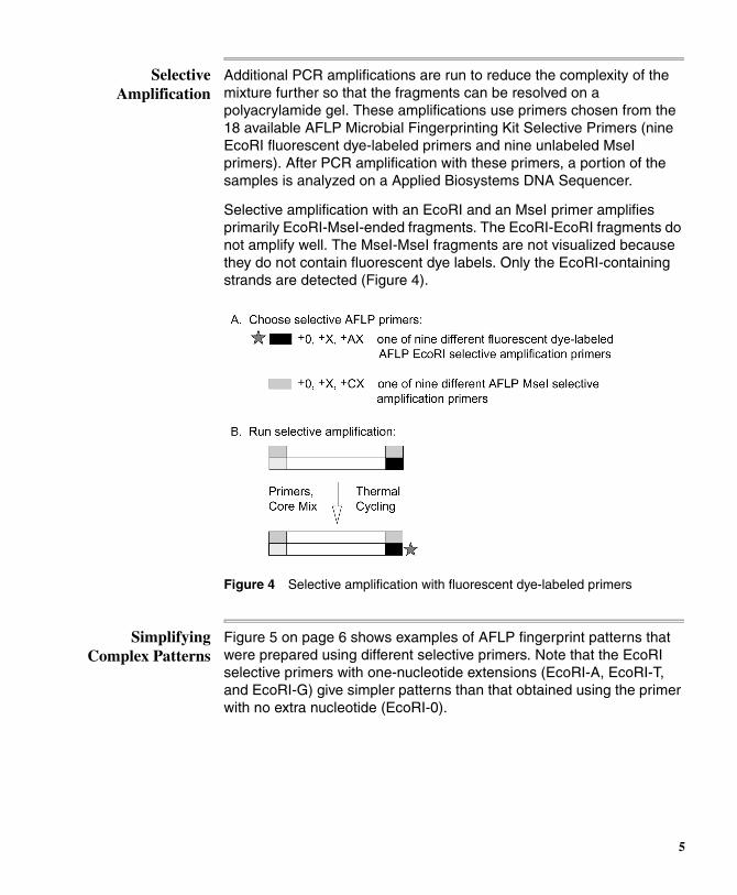
Selective Amplification
Additional PCR amplifications are run to reduce the complexity of the mixture further so that the fragments can be resolved on a polyacrylamide gel. These amplifications use primers chosen from the 18 available AFLP Microbial Fingerprinting Kit Selective Primers (nine EcoRI fluorescent dye-labeled primers and nine unlabeled MseI primers). After PCR amplification with these primers, a portion of the samples is analyzed on a Applied Biosystems DNA Sequencer.
Selective amplification with an EcoRI and an MseI primer amplifies primarily EcoRI-MseI-ended fragments. The EcoRI-EcoRI fragments do not amplify well. The MseI-MseI fragments are not visualized because they do not contain fluorescent dye labels. Only the EcoRI-containing strands are detected (Figure 4).
Figure 4 Selective amplification with fluorescent dye-labeled primers
Simplifying Complex Patterns
Figure 5 on page 6 shows examples of AFLP fingerprint patterns that were prepared using different selective primers. Note that the EcoRI selective primers with one-nucleotide extensions (EcoRI-A, EcoRI-T, and EcoRI-G) give simpler patterns than that obtained using the primer with no extra nucleotide (EcoRI-0).
5

Figure 5 AFLP fingerprints of E. coli W3110 Reference DNA. The MseI-CA and fluorescent dye-labeled EcoRI-0, EcoRI-A, EcoRI-T, and EcoRI-G selective primers (shown here top to bottom, respectively) were used.
If the complexity of the AFLP pattern is still too high at the +2/+2 level, we recommend reamplifying the preselective amplification sample with the preselective primers from the AFLP Ligation and Preselective Amplification Modules of the AFLP Regular and Small Plant Genome Mapping Kits (P/N 402004 and 402273, respectively).
Testing New Genomes
When testing novel genomes, you must be sure that the DNA restriction digest with EcoRI and MseI generates enough fragments for comparison of samples. There is a large variability in the number of restriction sites within microbial genomes. No assurances of kit performance are made for organisms not listed.
Empirical guidelines suggest that if the G-C content of the genome is >65%, MseI will not give a significant number of fragments. Optimal results are obtained with MseI when the G-C content is <50%. EcoRI also tends to produce more fragments in G-C-poor genomes. In cases where an organism’s G-C content is unknown, the effectiveness of the restriction enzymes must be determined empirically.
continued on next page
6
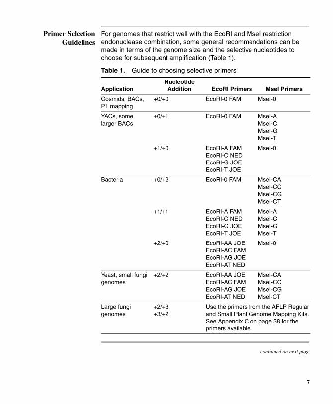
Primer Selection Guidelines
For genomes that restrict well with the EcoRI and MseI restriction endonuclease combination, some general recommendations can be made in terms of the genome size and the selective nucleotides to choose for subsequent amplification (Table 1).
continued on next page
Table 1. Guide to choosing selective primers
ApplicationNucleotide Addition EcoRI Primers MseI Primers
Cosmids, BACs, P1 mapping
+0/+0 EcoRI-0 FAM MseI-0
YACs, some larger BACs
+0/+1 EcoRI-0 FAM MseI-A MseI-C MseI-G MseI-T
+1/+0 EcoRI-A FAM EcoRI-C NED EcoRI-G JOE EcoRI-T JOE
MseI-0
Bacteria +0/+2 EcoRI-0 FAM MseI-CA MseI-CC MseI-CG MseI-CT
+1/+1 EcoRI-A FAM EcoRI-C NED EcoRI-G JOE EcoRI-T JOE
MseI-A MseI-C MseI-G MseI-T
+2/+0 EcoRI-AA JOE EcoRI-AC FAM EcoRI-AG JOE EcoRI-AT NED
MseI-0
Yeast, small fungi genomes
+2/+2 EcoRI-AA JOE EcoRI-AC FAM EcoRI-AG JOE EcoRI-AT NED
MseI-CA MseI-CC MseI-CG Msel-CT
Large fungi genomes
+2/+3 +3/+2
Use the primers from the AFLP Regular and Small Plant Genome Mapping Kits. See Appendix C on page 38 for the primers available.
7
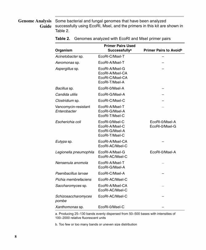
Genome Analysis Guide
Some bacterial and fungal genomes that have been analyzed successfully using EcoRI, MseI, and the primers in this kit are shown in Table 2.
Table 2. Genomes analyzed with EcoRI and MseI primer pairs
OrganismPrimer Pairs Used
Successfullya
a. Producing 25–130 bands evenly dispersed from 50–500 bases with intensities of 100–2000 relative fluorescent units
Primer Pairs to Avoidb
b. Too few or too many bands or uneven size distribution
Acinetobacter sp. EcoRI-C/MseI-T –
Aeromonas sp. EcoRI-A/MseI-T –
Aspergillus sp. EcoRI-A/MseI-G EcoRI-A/MseI-CA EcoRI-C/MseI-CA EcoRI-T/MseI-A
–
Bacillus sp. EcoRI-0/MseI-A –
Candida utilis EcoRI-G/MseI-A –
Clostridium sp. EcoRI-C/MseI-C –
Vancomycin-resistant Enterobacter
EcoRI-A/MseI-T EcoRI-G/MseI-A EcoRI-T/MseI-C
–
Escherichia coli EcoRI-0/MseI-C EcoRI-A/MseI-C EcoRI-G/MseI-A EcoRI-T/MseI-C
EcoRI-0/MseI-AEcoRI-0/MseI-G
Eutypa sp. EcoRI-A/MseI-CA EcoRI-AC/MseI-C
–
Legionella pneumophila EcoRI-A/MseI-G EcoRI-AC/MseI-C
EcoRI-0/MseI-A
Nensenula anomola EcoRI-A/MseI-T EcoRI-G/MseI-A
–
Paenibacillus larvae EcoRI-C/MseI-A –
Pichia membrefaciens EcoRI-AC/MseI-C
Saccharomyces sp. EcoRI-A/MseI-CA EcoRI-AC/MseI-C
–
Schizosaccharomyces pombe
EcoRI-AC/MseI-C –
Xanthomonas sp. EcoRI-0/MseI-C –
8
Note The list in Table 2 on page 8 is not exhaustive. Refer to the publications listed in Appendix B on page 35 for in-depth discussion of primer choices.
Fluorescent Dye-labeling and
Marker Detection
Applied Biosystems has adapted the AFLP technique for use with its ABI PRISM™ fluorescent dye-labeling and detection technology. PCR products are dye-labeled during amplification using a 5´ dye-labeled primer.
For high throughput, you can co-load up to three different reactions labeled with different colored dyes in a single lane on the ABI 373 or ABI PRISM 377 DNA Sequencer or in a single injection on the ABI PRISM
310 Genetic Analyzer. Load an internal lane size standard with a fourth color in every lane to size all amplification fragments accurately.
You can automate the scoring of the large numbers of markers that are typically generated by analyzing your results with GeneScan® Analysis and Genotyper® software.
9
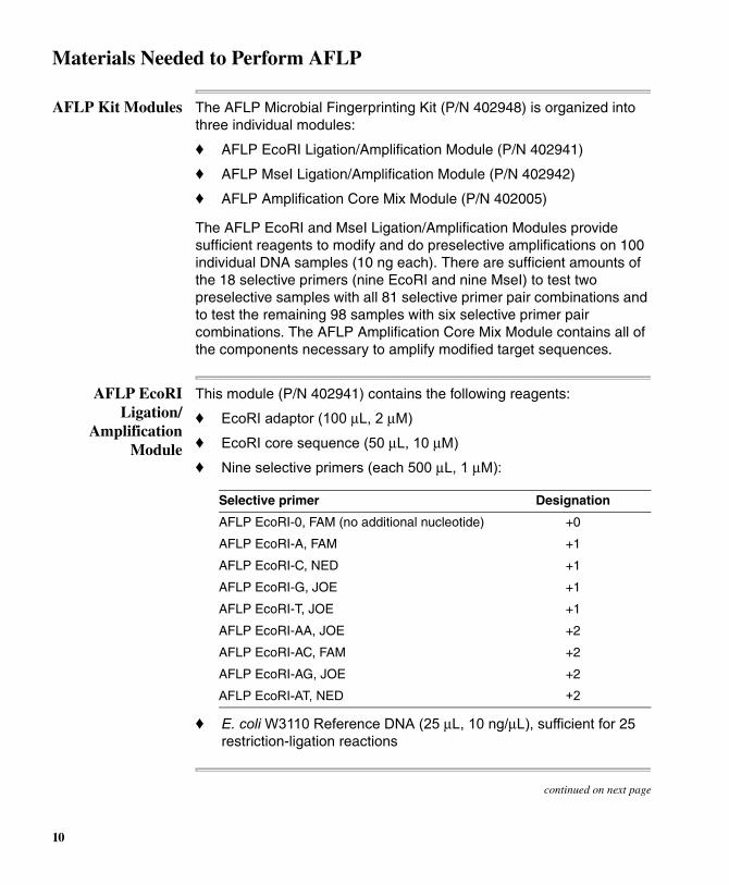
Materials Needed to Perform AFLP
AFLP Kit Modules The AFLP Microbial Fingerprinting Kit (P/N 402948) is organized into three individual modules:
♦ AFLP EcoRI Ligation/Amplification Module (P/N 402941)
♦ AFLP MseI Ligation/Amplification Module (P/N 402942)
♦ AFLP Amplification Core Mix Module (P/N 402005)
The AFLP EcoRI and MseI Ligation/Amplification Modules provide sufficient reagents to modify and do preselective amplifications on 100 individual DNA samples (10 ng each). There are sufficient amounts of the 18 selective primers (nine EcoRI and nine MseI) to test two preselective samples with all 81 selective primer pair combinations and to test the remaining 98 samples with six selective primer pair combinations. The AFLP Amplification Core Mix Module contains all of the components necessary to amplify modified target sequences.
AFLP EcoRI Ligation/
Amplification Module
This module (P/N 402941) contains the following reagents:
♦ EcoRI adaptor (100 µL, 2 µM)
♦ EcoRI core sequence (50 µL, 10 µM)
♦ Nine selective primers (each 500 µL, 1 µM):
♦ E. coli W3110 Reference DNA (25 µL, 10 ng/µL), sufficient for 25 restriction-ligation reactions
continued on next page
Selective primer Designation
AFLP EcoRI-0, FAM (no additional nucleotide) +0
AFLP EcoRI-A, FAM +1
AFLP EcoRI-C, NED +1
AFLP EcoRI-G, JOE +1
AFLP EcoRI-T, JOE +1
AFLP EcoRI-AA, JOE +2
AFLP EcoRI-AC, FAM +2
AFLP EcoRI-AG, JOE +2
AFLP EcoRI-AT, NED +2
10
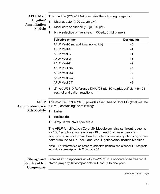
AFLP MseI Ligation/
Amplification Module
This module (P/N 402942) contains the following reagents:
♦ MseI adaptor (100 µL, 20 µM)
♦ MseI core sequence (50 µL, 10 µM)
♦ Nine selective primers (each 500 µL, 5 µM primer):
♦ E. coli W3110 Reference DNA (25 µL, 10 ng/µL), sufficient for 25 restriction-ligation reactions
AFLP Amplification Core
Mix Module
This module (P/N 402005) provides five tubes of Core Mix (total volume 7.5 mL) containing the following:
♦ buffer
♦ nucleotides
♦ AmpliTaq® DNA Polymerase
The AFLP Amplification Core Mix Module contains sufficient reagents for 1000 amplification reactions (10 µL each) of target genomic sequences. You determine how the selection occurs by choosing primer pairs from the AFLP EcoRI and MseI Ligation/Amplification Modules.
Note For information on ordering selective primers and other AFLP reagents individually, see Appendix C on page 38.
Storage and Stability of Kit
Components
Store all kit components at –15 to –25 °C in a non-frost-free freezer. If stored properly, kit components will last up to one year.
continued on next page
Selective primer Designation
AFLP MseI-0 (no additional nucleotide) +0
AFLP MseI-A +1
AFLP MseI-C +1
AFLP MseI-G +1
AFLP MseI-T +1
AFLP MseI-CA +2
AFLP MseI-CC +2
AFLP MseI-CG +2
AFLP MseI-CT +2
11
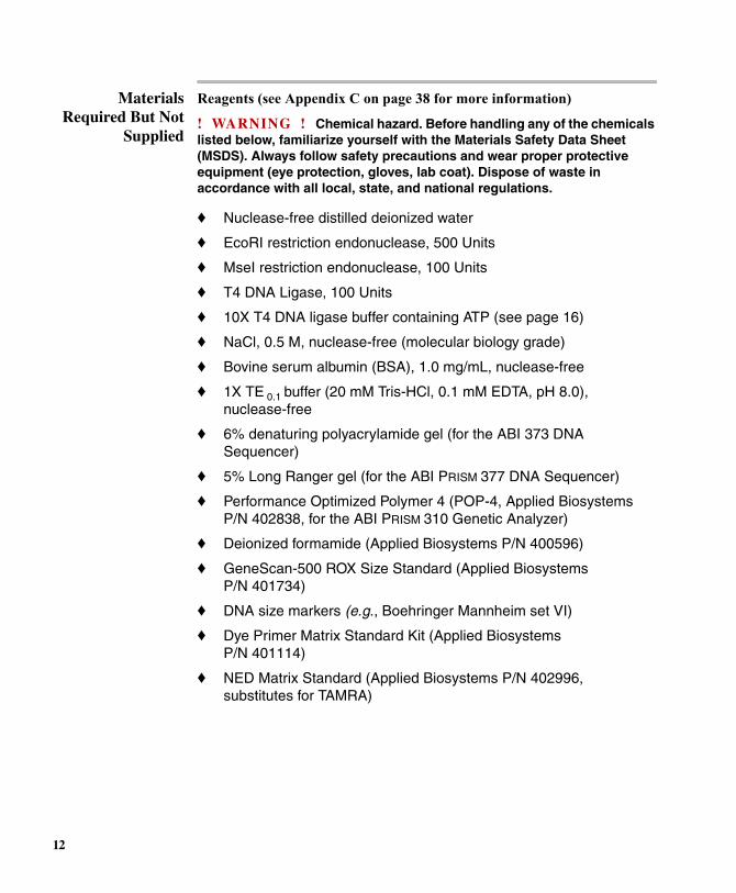
Materials Required But Not
Supplied
Reagents (see Appendix C on page 38 for more information)
! WARNING ! Chemical hazard. Before handling any of the chemicals listed below, familiarize yourself with the Materials Safety Data Sheet (MSDS). Always follow safety precautions and wear proper protective equipment (eye protection, gloves, lab coat). Dispose of waste in accordance with all local, state, and national regulations.
♦ Nuclease-free distilled deionized water
♦ EcoRI restriction endonuclease, 500 Units
♦ MseI restriction endonuclease, 100 Units
♦ T4 DNA Ligase, 100 Units
♦ 10X T4 DNA ligase buffer containing ATP (see page 16)
♦ NaCl, 0.5 M, nuclease-free (molecular biology grade)
♦ Bovine serum albumin (BSA), 1.0 mg/mL, nuclease-free
♦ 1X TE 0.1 buffer (20 mM Tris-HCl, 0.1 mM EDTA, pH 8.0), nuclease-free
♦ 6% denaturing polyacrylamide gel (for the ABI 373 DNA Sequencer)
♦ 5% Long Ranger gel (for the ABI PRISM 377 DNA Sequencer)
♦ Performance Optimized Polymer 4 (POP-4, Applied Biosystems P/N 402838, for the ABI PRISM 310 Genetic Analyzer)
♦ Deionized formamide (Applied Biosystems P/N 400596)
♦ GeneScan-500 ROX Size Standard (Applied Biosystems P/N 401734)
♦ DNA size markers (e.g., Boehringer Mannheim set VI)
♦ Dye Primer Matrix Standard Kit (Applied Biosystems P/N 401114)
♦ NED Matrix Standard (Applied Biosystems P/N 402996, substitutes for TAMRA)
12

Equipment
♦ Microcentrifuge
♦ Pipettors, 2 µL, 20 µL and 200 µL, with sterile pipette tips
♦ Gel-loading pipette tips, 0.17-mm flat (ABI PRISM 377)
♦ Sterile 0.5-ml microcentrifuge tubes
♦ Thermal cycler (Applied Biosystems)
♦ Sterile 0.2-mL MicroAmp® Thin-Walled Reaction Tubes and caps (GeneAmp® PCR Instrument Systems 2400 and 9600)
♦ Sterile GeneAmp Thin-Walled 0.5-mL Reaction Tubes (DNA Thermal Cycler 480)
13

Sample Preparation
Before Starting an AFLP Experiment
Before setting up an AFLP experiment, determine whether or not your genomic DNA restricts properly with EcoRI and MseI.
Preparing Samples for PCR
Amplification
To prepare samples for the AFLP preselective and selective amplification reactions, you must:
♦ anneal the adaptor pairs
♦ prepare a restriction-ligation enzyme master mix
♦ prepare the restriction-ligation reactions
♦ dilute the restriction-ligation reactions
continued on next page
Step Action
1 Digest 1–3 µg of DNA with the enzymes MseI and EcoRI separately, then with both together, according to the manufacturer’s instructions.
2 Load the digestion products in one lane on a 1.5% mini-agarose gel with size markers.
3 Stain with ethidium bromide.
! WARNING ! Ethidium bromide is a powerful mutagen and is moderately toxic. Wear gloves, a lab coat, and safety glasses when using this dye. After use, decontaminate ethidium bromide solutions before disposal.
4 View on a UV transilluminator.
For an example of what a successful digest looks like, see Figure 6 on page 18 (left half).
14
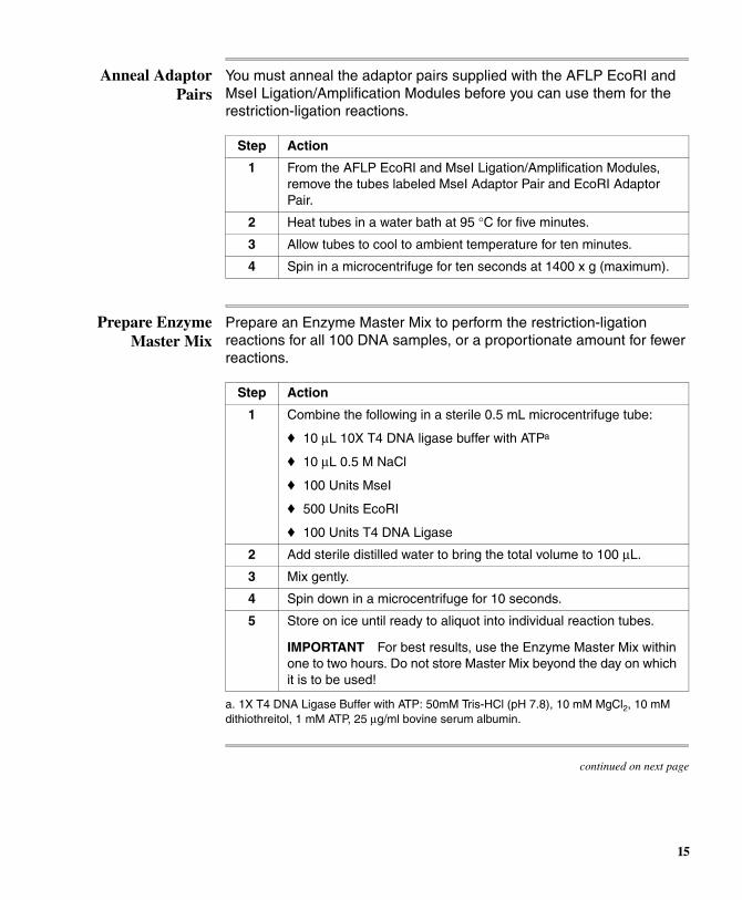
Anneal Adaptor Pairs
You must anneal the adaptor pairs supplied with the AFLP EcoRI and MseI Ligation/Amplification Modules before you can use them for the restriction-ligation reactions.
Prepare Enzyme Master Mix
Prepare an Enzyme Master Mix to perform the restriction-ligation reactions for all 100 DNA samples, or a proportionate amount for fewer reactions.
Step Action
1 From the AFLP EcoRI and MseI Ligation/Amplification Modules, remove the tubes labeled MseI Adaptor Pair and EcoRI Adaptor Pair.
2 Heat tubes in a water bath at 95 °C for five minutes.
3 Allow tubes to cool to ambient temperature for ten minutes.
4 Spin in a microcentrifuge for ten seconds at 1400 x g (maximum).
Step Action
1 Combine the following in a sterile 0.5 mL microcentrifuge tube:
♦ 10 µL 10X T4 DNA ligase buffer with ATPa
♦ 10 µL 0.5 M NaCl
♦ 100 Units MseI
♦ 500 Units EcoRI
♦ 100 Units T4 DNA Ligase
a. 1X T4 DNA Ligase Buffer with ATP: 50mM Tris-HCl (pH 7.8), 10 mM MgCl2, 10 mM dithiothreitol, 1 mM ATP, 25 µg/ml bovine serum albumin.
continued on next page
2 Add sterile distilled water to bring the total volume to 100 µL.
3 Mix gently.
4 Spin down in a microcentrifuge for 10 seconds.
5 Store on ice until ready to aliquot into individual reaction tubes.
IMPORTANT For best results, use the Enzyme Master Mix within one to two hours. Do not store Master Mix beyond the day on which it is to be used!
15
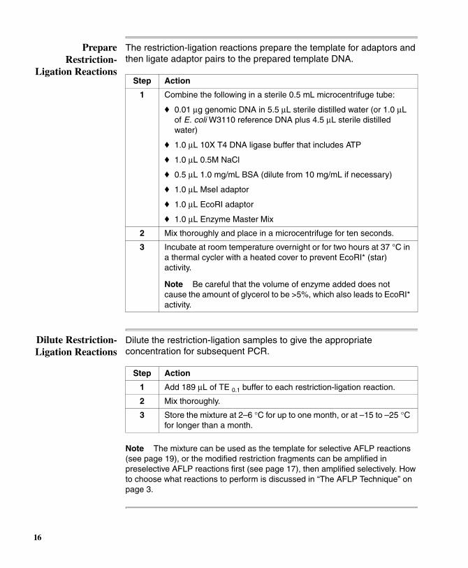
Prepare Restriction-
Ligation Reactions
The restriction-ligation reactions prepare the template for adaptors and then ligate adaptor pairs to the prepared template DNA.
Dilute Restriction-Ligation Reactions
Dilute the restriction-ligation samples to give the appropriate concentration for subsequent PCR.
Note The mixture can be used as the template for selective AFLP reactions (see page 19), or the modified restriction fragments can be amplified in preselective AFLP reactions first (see page 17), then amplified selectively. How to choose what reactions to perform is discussed in “The AFLP Technique” on page 3.
Step Action
1 Combine the following in a sterile 0.5 mL microcentrifuge tube:
♦ 0.01 µg genomic DNA in 5.5 µL sterile distilled water (or 1.0 µL of E. coli W3110 reference DNA plus 4.5 µL sterile distilled water)
♦ 1.0 µL 10X T4 DNA ligase buffer that includes ATP
♦ 1.0 µL 0.5M NaCl
♦ 0.5 µL 1.0 mg/mL BSA (dilute from 10 mg/mL if necessary)
♦ 1.0 µL MseI adaptor
♦ 1.0 µL EcoRI adaptor
♦ 1.0 µL Enzyme Master Mix
2 Mix thoroughly and place in a microcentrifuge for ten seconds.
3 Incubate at room temperature overnight or for two hours at 37 °C in a thermal cycler with a heated cover to prevent EcoRI* (star) activity.
Note Be careful that the volume of enzyme added does not cause the amount of glycerol to be >5%, which also leads to EcoRI* activity.
Step Action
1 Add 189 µL of TE 0.1 buffer to each restriction-ligation reaction.
2 Mix thoroughly.
3 Store the mixture at 2–6 °C for up to one month, or at –15 to –25 °C for longer than a month.
16

Amplification of Target Sequences
Overview This protocol has been optimized for the GeneAmp® PCR Systems 9600 and 2400 and the DNA Thermal Cycler 480. If you use a different thermal cycler, you may need to optimize the conditions. The temperature ramp times included in this protocol ensure identical products from any Applied Biosystems thermal cycler. Ramp time is crucial. If the temperature is increased too quickly, results may be inconsistent. See Appendix A on page 32 for troubleshooting tips.
Preselective Amplification
Sequences with adaptors ligated to both ends amplify exponentially and predominate in the final product.
Note Keep all reagents and tubes on ice until loaded into the thermal cycler.
continued on next page
Step Action
1 Combine the following in a PCR reaction tube (0.2 mL for the GeneAmp PCR System 9600 or 2400, 0.5 mL for the DNA Thermal Cycler 480):
♦ 4.0 µL diluted DNA prepared by restriction-ligation
♦ 0.5 µL AFLP EcoRI preselective primer
♦ 0.5 µL AFLP MseI preselective primer
♦ 15.0 µL AFLP Amplification Core Mix
Note If using the DNA Thermal Cycler 480, overlay your samples with 20 µL of light mineral oil.
2 Place the samples in a thermal cycler at ambient temperature.
3 Run the PCR method shown in Table 3, entering all ramp times as 0.01 (one second) on the GeneAmp PCR System 9600 and DNA Thermal Cycler 480 or 90% on the GeneAmp PCR System 2400.
4 Store at 2–6 °C.
Table 3. Thermal cycler parameters for preselective amplification
HOLD
CYCLE
HOLDEach of 20 Cycles
72 °C 2 min.
94 °C 20 sec.
56 °C 30 sec.
72 °C 2 min.
4 °C (forever)
17

Verify Successful Amplification
Run an agarose yield gel to check that amplification has occurred.
continued on next page
Step Action
1 Run 10 µL of each reaction on a 1.5% agarose gel in 1X TBE buffer at 4V/cm for 3–4 hours.
2 Stain the gel with ethidium bromide.
! WARNING ! Ethidium bromide is a powerful mutagen and is moderately toxic. Wear gloves, a lab coat, and safety glasses when using this dye.
3 View the gel on a UV transilluminator. A smear of product from 100–1500 bp should be clearly visible (Figure 6, right half).
Figure 6 Gel results after restriction digestion of 1–3 µg of DNA (left) and after preselective amplification (right)
18

Prepare Template Prepare the preselective amplification products for selective amplification.
Selective Amplification
Amplify the EcoRI- and MseI-modified fragments.
Step Action
1 Combine the following in a sterile 0.5 mL microcentrifuge tube:
♦ 10.0 µL preselective amplification product
♦ 190.0 µL TE 0.1 buffer
2 Mix thoroughly, then spin down in a microcentrifuge for ten seconds.
3 Store the diluted preselective amplification product at 2–6 °C if not used immediately.
Step Action
1 Combine the following in a PCR tube (0.2 mL for the GeneAmp PCR System 9600 or 2400, 0.5 mL for the DNA Thermal Cycler 480):
♦ 1.5 µL diluted preselective amplification product
♦ 0.5 µL MseI primer at 5 µM
♦ 0.5 µL dye-labeled EcoRI primer at 1 µM
♦ 7.5 µL AFLP Core Amplification Mix
Note If using the DNA Thermal Cycler 480, add 20 µL of light mineral oil to the tube.
2 Run PCR using the thermal cycler parameters shown in Table 4 on page 20.
Note For the GeneAmp PCR System 9600 and DNA Thermal Cycler 480, enter all ramp times as 0.01 (one second). For the GeneAmp PCR System 2400 enter all ramp times as 90%.
3 Store at 2–6 °C.
19

Table 4. Thermal cycler parameters for selective amplification
HOLD CYCLENumber of
Cycles
94 °C2 min.
94 °C20 sec.
66 °C30 sec.
72 °C2 min.
1
– 94 °C20 sec.
65 °C30 sec.
72 °C2 min.
1
– 94 °C20 sec.
64 °C30 sec.
72 °C2 min.
1
– 94 °C20 sec.
63 °C30 sec.
72 °C2 min.
1
– 94 °C20 sec.
62 °C30 sec.
72 °C2 min.
1
– 94 °C20 sec.
61 °C30 sec.
72 °C2 min.
1
– 94 °C20 sec.
60 °C30 sec.
72 °C2 min.
1
– 94 °C20 sec.
59 °C30 sec.
72 °C2 min
1
– 94 °C20 sec.
58 °C30 sec.
72 °C2 min.
1
– 94 °C20 sec.
57 °C30 sec.
72 °C2 min.
1
– 94 °C20 sec.
56 °C30 sec.
72 °C2 min.
20
60 °C30 min.
– 1
4 °Cforever
– 1
20
Evaluating Results
Overview You can evaluate the results of the AFLP reactions by using GeneScan software to analyze data from samples loaded and run on the ABI 373 or ABI PRISM 377 DNA Sequencer or on the ABI PRISM 310 Genetic Analyzer.
The following instructions describe step-by-step procedures for loading samples and performing electrophoresis on these instruments.
Preparing the Loading Buffer for
the ABI 373 and ABI PRISM 377
Prepare a loading buffer mix of the following reagents in the proportions shown in sufficient quantity for each sample:
♦ 1.25 µL deionized formamide
♦ 0.25 µL blue dextran/50 mM EDTA loading solution (supplied with the size standard)
♦ 0.5 µL of GeneScan-500 [ROX] size standard
! WARNING ! Chemical hazard: formamide is a teratogen and is harmful by inhalation, skin contact, and ingestion. Use in a well-ventilated area. Use chemical-resistant gloves and safety glasses when handling.
Note You can store any remaining loading buffer at 2–6 °C for a week.
continued on next page
21
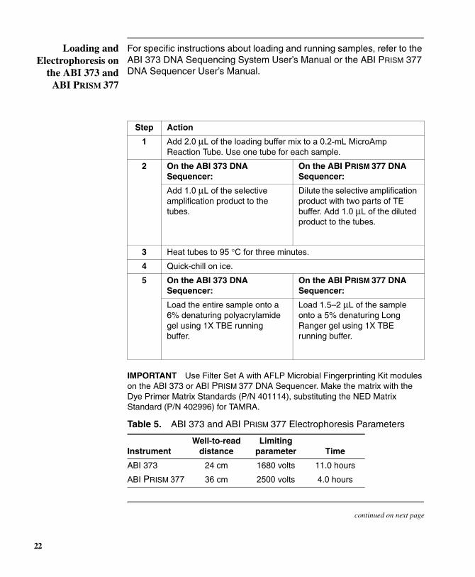
Loading and Electrophoresis on
the ABI 373 and ABI PRISM 377
For specific instructions about loading and running samples, refer to the ABI 373 DNA Sequencing System User’s Manual or the ABI PRISM 377 DNA Sequencer User’s Manual.
IMPORTANT Use Filter Set A with AFLP Microbial Fingerprinting Kit modules on the ABI 373 or ABI PRISM 377 DNA Sequencer. Make the matrix with the Dye Primer Matrix Standards (P/N 401114), substituting the NED Matrix Standard (P/N 402996) for TAMRA.
continued on next page
Step Action
1 Add 2.0 µL of the loading buffer mix to a 0.2-mL MicroAmp Reaction Tube. Use one tube for each sample.
2 On the ABI 373 DNA Sequencer:
On the ABI PRISM 377 DNA Sequencer:
Add 1.0 µL of the selective amplification product to the tubes.
Dilute the selective amplification product with two parts of TE buffer. Add 1.0 µL of the diluted product to the tubes.
3 Heat tubes to 95 °C for three minutes.
4 Quick-chill on ice.
5 On the ABI 373 DNA Sequencer:
On the ABI PRISM 377 DNA Sequencer:
Load the entire sample onto a 6% denaturing polyacrylamide gel using 1X TBE running buffer.
Load 1.5–2 µL of the sample onto a 5% denaturing Long Ranger gel using 1X TBE running buffer.
Table 5. ABI 373 and ABI PRISM 377 Electrophoresis Parameters
InstrumentWell-to-read
distanceLimiting
parameter Time
ABI 373 24 cm 1680 volts 11.0 hours
ABI PRISM 377 36 cm 2500 volts 4.0 hours
22
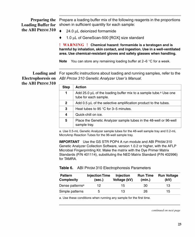
Preparing the Loading Buffer for the ABI PRISM 310
Prepare a loading buffer mix of the following reagents in the proportions shown in sufficient quantity for each sample:
♦ 24.0 µL deionized formamide
♦ 1.0 µL of GeneScan-500 [ROX] size standard
! WARNING ! Chemical hazard: formamide is a teratogen and is harmful by inhalation, skin contact, and ingestion. Use in a well-ventilated area. Use chemical-resistant gloves and safety glasses when handling.
Note You can store any remaining loading buffer at 2–6 °C for a week.
Loading and Electrophoresis on the ABI PRISM 310
For specific instructions about loading and running samples, refer to the ABI PRISM 310 Genetic Analyzer User’s Manual.
Step Action
1 Add 25.0 µL of the loading buffer mix to a sample tube.a Use one tube for each sample.
a. Use 0.5-mL Genetic Analyzer sample tubes for the 48-well sample tray and 0.2-mL MicroAmp Reaction Tubes for the 96-well sample tray.
IMPORTANT Use the GS STR POP4 A run module and ABI PRISM 310 Genetic Analyzer Collection Software, version 1.0.2 or higher, with the AFLP Microbial Fingerprinting Kit. Make the matrix with the Dye Primer Matrix Standards (P/N 401114), substituting the NED Matrix Standard (P/N 402996) for TAMRA.
2 Add 0.5 µL of the selective amplification product to the tubes.
3 Heat tubes to 95 °C for 3–5 minutes.
4 Quick-chill on ice.
5 Place the Genetic Analyzer sample tubes in the 48-well or 96-well sample tray.
Table 6. ABI PRISM 310 Electrophoresis Parameters
Pattern Complexity
Injection Time (sec.)
Injection Voltage (kV)
Run Time (min.)
Run Voltage (kV)
Dense patternsa
a. Use these conditions when running any sample for the first time.
continued on next page
12 15 30 13
Simple patterns 5 13 26 15
23
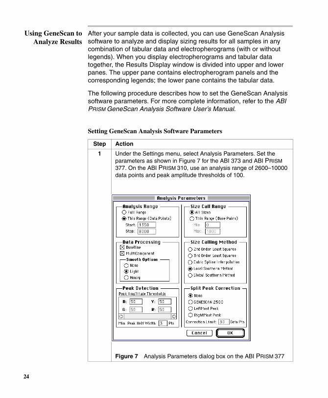
Using GeneScan to Analyze Results
After your sample data is collected, you can use GeneScan Analysis software to analyze and display sizing results for all samples in any combination of tabular data and electropherograms (with or without legends). When you display electropherograms and tabular data together, the Results Display window is divided into upper and lower panes. The upper pane contains electropherogram panels and the corresponding legends; the lower pane contains the tabular data.
The following procedure describes how to set the GeneScan Analysis software parameters. For more complete information, refer to the ABI PRISM GeneScan Analysis Software User’s Manual.
Setting GeneScan Analysis Software Parameters
Step Action
1 Under the Settings menu, select Analysis Parameters. Set the parameters as shown in Figure 7 for the ABI 373 and ABI PRISM
377. On the ABI PRISM 310, use an analysis range of 2600–10000 data points and peak amplitude thresholds of 100.
Figure 7 Analysis Parameters dialog box on the ABI PRISM 377
24

GeneScan-500 Size Standard
The GeneScan-500 standard is made of double-stranded DNA fragments, but only one of the strands is labeled with an ABI PRISM dye. Consequently, under denaturing conditions, even if the two strands migrate at different rates, only the one labeled strand is detected. Because of this, you can avoid split peaks, which result when two strands move through a denaturing gel at different rates. Under denaturing conditions, you can achieve a linear range of separation for fragment sizes of up to 500 bases (Figure 8 on page 26).
2 Click OK.
3 In the Analysis Control Window, define a size standard as follows:
a. Indicate the dye color of the Size Standard.
b. Choose Define New... from the pop-up window, and select a Sample File (data for one lane).
The size standard peaks within the defined Analysis Range appear.
c. Assign a size value to each peak.
d. Close the window and enter a standard name when a prompt appears.
4 Highlight the sample(s) to be analyzed and click on the Analyze button.
5 After a successful analysis, view your results in the Results Display window, and then save the project.
6 Select Save As from the File menu to save the data to a file.
Setting GeneScan Analysis Software Parameters (continued)
Step Action
25

Figure 8 Electropherogram of GeneScan-500 run under denaturing conditions
Using the Standard Sizing Curve
The Standard Sizing Curve is a measure of how well the standard definition matches the GeneScan size standard, and whether or not it is linear.
To align the data by size, GeneScan calculates a best-fit least squares curve for all samples. This is a third-order curve when you use the Third Order Least Squares size calling method. For all other size calling methods it is a second-order curve.
26
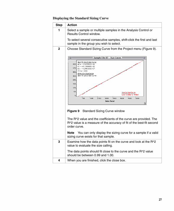
Displaying the Standard Sizing Curve
Step Action
1 Select a sample or multiple samples in the Analysis Control or Results Control window.
To select several consecutive samples, shift-click the first and last sample in the group you wish to select.
2 Choose Standard Sizing Curve from the Project menu (Figure 9).
Figure 9 Standard Sizing Curve window
The R^2 value and the coefficients of the curve are provided. The R^2 value is a measure of the accuracy of fit of the best-fit second order curve.
Note You can only display the sizing curve for a sample if a valid sizing curve exists for that sample.
3 Examine how the data points fit on the curve and look at the R^2 value to evaluate the size calling.
The data points should fit close to the curve and the R^2 value should be between 0.99 and 1.00.
4 When you are finished, click the close box.
27
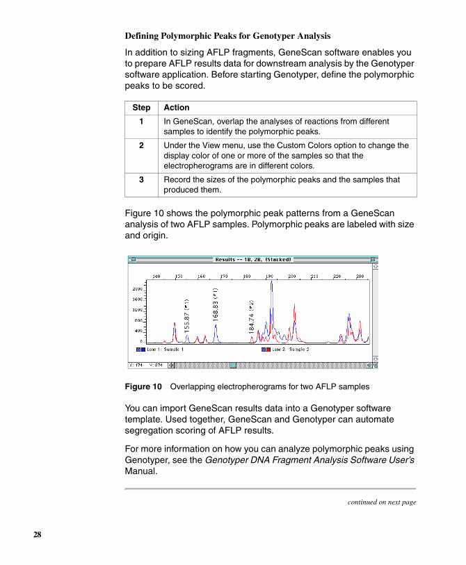
Defining Polymorphic Peaks for Genotyper Analysis
In addition to sizing AFLP fragments, GeneScan software enables you to prepare AFLP results data for downstream analysis by the Genotyper software application. Before starting Genotyper, define the polymorphic peaks to be scored.
Figure 10 shows the polymorphic peak patterns from a GeneScan analysis of two AFLP samples. Polymorphic peaks are labeled with size and origin.
Figure 10 Overlapping electropherograms for two AFLP samples
You can import GeneScan results data into a Genotyper software template. Used together, GeneScan and Genotyper can automate segregation scoring of AFLP results.
For more information on how you can analyze polymorphic peaks using Genotyper, see the Genotyper DNA Fragment Analysis Software User’s Manual.
continued on next page
Step Action
1 In GeneScan, overlap the analyses of reactions from different samples to identify the polymorphic peaks.
2 Under the View menu, use the Custom Colors option to change the display color of one or more of the samples so that the electropherograms are in different colors.
3 Record the sizes of the polymorphic peaks and the samples that produced them.
28

Evaluating ABI 373 DNA
Sequencer Results
If you run samples under the recommended electrophoresis conditions, and analyze them with GeneScan, resulting electropherogram data from the ABI 373 DNA Sequencer should look similar to data from samples run on the ABI PRISM 377 DNA Sequencer.
Figure 11 shows a representative electropherogram of fluorescent dye-labeled AFLP products run on an ABI 373 DNA Sequencer and analyzed using GeneScan analysis software. The analyzed products are DNA fragments modified with MseI and JOE dye-labeled EcoRI selective amplification primers. The JOE-labeled EcoRI fragments are displayed as peaks in the electropherogram.
Figure 11 Electropherogram of AFLP samples run on an ABI 373
continued on next page
29
Evaluating ABI PRISM 377
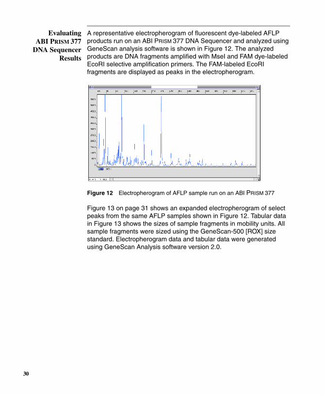
DNA Sequencer Results
A representative electropherogram of fluorescent dye-labeled AFLP products run on an ABI PRISM 377 DNA Sequencer and analyzed using GeneScan analysis software is shown in Figure 12. The analyzed products are DNA fragments amplified with MseI and FAM dye-labeled EcoRI selective amplification primers. The FAM-labeled EcoRI fragments are displayed as peaks in the electropherogram.
Figure 12 Electropherogram of AFLP sample run on an ABI PRISM 377
Figure 13 on page 31 shows an expanded electropherogram of select peaks from the same AFLP samples shown in Figure 12. Tabular data in Figure 13 shows the sizes of sample fragments in mobility units. All sample fragments were sized using the GeneScan-500 [ROX] size standard. Electropherogram data and tabular data were generated using GeneScan Analysis software version 2.0.
30
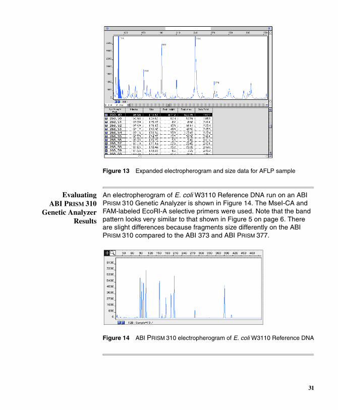
Figure 13 Expanded electropherogram and size data for AFLP sample
Evaluating ABI PRISM 310
Genetic Analyzer Results
An electropherogram of E. coli W3110 Reference DNA run on an ABI PRISM 310 Genetic Analyzer is shown in Figure 14. The MseI-CA and FAM-labeled EcoRI-A selective primers were used. Note that the band pattern looks very similar to that shown in Figure 5 on page 6. There are slight differences because fragments size differently on the ABI PRISM 310 compared to the ABI 373 and ABI PRISM 377.
Figure 14 ABI PRISM 310 electropherogram of E. coli W3110 Reference DNA
31
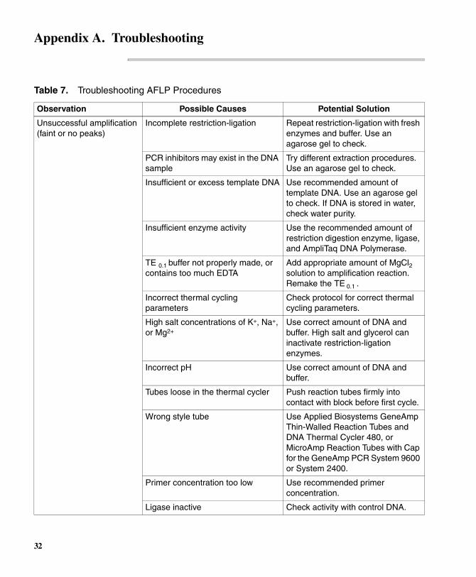
Appendix A. Troubleshooting
Table 7. Troubleshooting AFLP Procedures
Observation Possible Causes Potential Solution
Unsuccessful amplification (faint or no peaks)
Incomplete restriction-ligation Repeat restriction-ligation with fresh enzymes and buffer. Use an agarose gel to check.
PCR inhibitors may exist in the DNA sample
Try different extraction procedures. Use an agarose gel to check.
Insufficient or excess template DNA Use recommended amount of template DNA. Use an agarose gel to check. If DNA is stored in water, check water purity.
Insufficient enzyme activity Use the recommended amount of restriction digestion enzyme, ligase, and AmpliTaq DNA Polymerase.
TE 0.1 buffer not properly made, or contains too much EDTA
Add appropriate amount of MgCl2
solution to amplification reaction. Remake the TE 0.1 .
Incorrect thermal cycling parameters
Check protocol for correct thermal cycling parameters.
High salt concentrations of K+, Na+, or Mg2+
Use correct amount of DNA and buffer. High salt and glycerol can inactivate restriction-ligation enzymes.
Incorrect pH Use correct amount of DNA and buffer.
Tubes loose in the thermal cycler Push reaction tubes firmly into contact with block before first cycle.
Wrong style tube Use Applied Biosystems GeneAmp Thin-Walled Reaction Tubes and DNA Thermal Cycler 480, or MicroAmp Reaction Tubes with Cap for the GeneAmp PCR System 9600 or System 2400.
Primer concentration too low Use recommended primer concentration.
Ligase inactive Check activity with control DNA.
32

Inconsistent results with control DNA
Restriction incomplete Repeat the restriction-ligation.
Incorrect PCR thermal profile program
Choose correct temperature control parameters (refer to the GeneAmp PCR System 9600 User’s Manual).
GeneAmp PCR System 9600 misaligned lid
Align 9600 lid white stripes after twisting the top portion clockwise.
For DNA Thermal Cycler 480, improper tube placement in block
Refer to the DNA Thermal Cycler 480 User’s Manual.
Pipetting errors Calibrate pipettes, attach tips firmly, and check technique.
Combined reagents not spun to bottom of tube
Place all reagents in apex of tube. Spin briefly after combining.
Combined reagents left at room temperature or on ice for extended periods of time
Put tubes in block immediately after reagents are combined.
Extra peaks visible when sample is known to contain DNA from a single source
Contamination with exogenous DNA
Use appropriate techniques to avoid introducing foreign DNA during laboratory handling.
Incomplete restriction or ligation Extract the DNA again and repeat the restriction-ligation.
Samples not denatured before loading in the autosampler
Make sure the samples are heated at 95 °C for three minutes prior to loading.
Renaturation of denatured samples Load sample immediately following denaturation, or store on ice until ready.
Too much DNA in reaction, so that insufficient adaptor present
Use recommended amount of template DNA.
Too much DNA amplified and/or loaded resulting in crossover between color channels
Re-run PCR using less DNA or load less sample during electrophoresis.
Table 7. Troubleshooting AFLP Procedures (continued)
Observation Possible Causes Potential Solution
33

Signal continually gets weaker
Outdated or mishandled reagents Check expiration dates on reagents. Store and use according to manufacturers instructions. Compare with fresh reagents.
Degraded primers Store unused primers at –15 to –25 °C. Do not expose fluorescent dye-labeled primers to light for long periods of time.
Inconsistent sizing of known DNA sample
Inadvertent change in analysis parameters
Check settings for GeneScan analysis parameters.
Change in size-calling method Use same size-calling method.
Incorrect internal standard Use correct GeneScan size standard.
Change in electrophoresis temperature
Check the Log for the record of the electrophoresis temperature.
Data was not automatically analyzed
Sample Sheet not completed Complete Sample Sheet correctly.
Samples run faster than usual with decreased resolution
Incorrect buffer concentration Check if buffer concentration matches protocol requirements.
Incorrect run temperature Check the Log for the record of the electrophoresis temperature.
Table 7. Troubleshooting AFLP Procedures (continued)
Observation Possible Causes Potential Solution
34
Appendix B. References
Ausubel, F. M., Brent, R., Kingstin, R. E., Moore, D. D., Seidman, J. G., Smith, J. A., and Struhl, K., eds., 1987. Current Protocols in Molecular Biology, Greene Publishing Associates and Wiley-Interscience, John Wiley and Sons, New York.
Bachem, C. W. B., van der Hoeven, R. S., de Bruijn, S. M., Vreugdenhil, D., Zabeau, M., and Visser, R. G. F., 1996. “Visualization of differential gene expression using a novel method of RNA fingerprinting based on AFLP: analysis of gene expression during potato tuber development.” The Plant Journal 9: 745–753.
Ballvora, A., Hesselbach, J., Niewohner, J., Leister, D., Salamini, F., and Gebhardt, C., 1995. “Marker enrichment and high-resolution map of the segment of potato chromosome VII harbouring the nematode resistance gene Gro1.” Molecular and General Genetics 249: 82–90.
Bates, S. R. E., Knorr, D. A., Weller, J. N., and Ziegle, J. S., 1996. “Instrumentation for automated molecular marker acquisition and data analysis,” in Sobral, B. W. S., ed. The Impact of Plant Molecular Genetics, Birkhaüser, Boston, MA, pp. 239–255.
Becker, J., Vos, P., Kuiper, M., Salamini, F., and Heun, M., 1995. “Combined mapping of RFLP and AFLP markers in barley.” Molecular and General Genetics 249: 65–73.
Dijkshoorn, L., Aucken, H., Gerner-Smidt, P., Janssen, P., Kaufmann, M. E., Garaizar, J., Ursing, J., and Pitt, T. L., 1996. “Comparison of outbreak and nonoutbreak Acinetobacter baumanii strains by genotypic and phenotypic methods.” Journal of Clinical Microbiology 34: 1519–1525.
Folkertsma, R. T., Rouppe van der Voort, J. N. A., de Groot, K. E., van Zandvoort, P. M., Schots, A., Gommers, F. J., Helder, J., and Bakker, J., 1996. “Gene pool similarities of potato cyst nematode populations assessed by AFLP analysis.” Molecular Plant-Microbe Interactions 9: 47–54.
Heyndrickx, M., Vandemeulebroecke, K., Hoste, B., Janssen, P., Kersters, K., Vos, P., Logan, N. A., Ali, N., and Berkeley, R. C. W., 1996. “Reclassification of Paenibacillus (formerly Bacillus) pulvifaciens, a later subjective synonym of Paenibacillus (formerly Bacillus) larvae, as a subspecies of P. larvae, with emended descriptions of P. larvae as
35

P. larvae subsp. larvae and P. larvae subsp. pulvifaciens.” International Journal of Systematic Bacteriology 46: 270–279.
Huys, G., Coopman, R., Janssen, P., and Kersters, K., 1996. “High-resolution genotypic analysis of the genus Aeromonas by AFLP fingerprinting.” International Journal of Systematic Bacteriology 46: 572–580.
Janssen, P., Coopman, R., Huys, G., Swings, J., Bleeker, M., Vos, P., Zabeau, M., and Kersters, K., 1996. “Evaluation of the DNA fingerprinting method AFLP as a new tool in bacterial taxonomy.” Microbiology 142: 1881–1893.
Lin, J.-J., Kuo, J., Saunders, J. A., Beard, H. S., MacDonald, M. H., Kenworthy, W., Ude, G. N., and Matthews, B. F., 1996. “Identification of molecular markers in soybean comparing RFLP, RAPD and AFLP DNA mapping techniques.” Plant Molecular Biology Reporter 14: 156–169.
Meksem, K., Leister, D., Peleman, J., Zabeau, M., Salamini, F., and Gebhardt, C., 1995. “A high-resolution map of the R1 locus on chromosome V of potato based on RFLP and AFLP markers.” Molecular and General Genetics 249: 74–81.
Money, T., Reader, S., Qu, L. J., Dunford, R. P., and Moore, G., 1996. “AFLP-based mRNA fingerprinting.” Nucleic Acids Research 24: 2616–2617.
Sambrook, J., Fritsch, E. F., and Maniatis, T., 1989. Molecular Cloning: A Laboratory Manual, Cold Spring Harbor Press, NY.
Thomas, C. M., Vos, P., Zabeau, M., Jones, D. A., Norcott, K. A., Chadwick, B., and Jones, J. D. G., 1995. “Identification of amplified restriction fragment polymorphism (AFLP) markers tightly linked to the tomato Cf-9 gene for resistance to Cladosporum fulvum.” The Plant Journal 8: 785–794.
Valsangiacomo, C., Baggi, F., Gaia, V., Balmelli, T., Peduzzi, R., and Piffaretti, J. C., 1995. “Use of amplified fragment length polymorphism in molecular typing of Legionella pneumophila and application to epidemiological studies.” Journal of Clinical Microbiology 33: 1716–1719.
Van Eck, H. J., Rouppe van der Voort, J., Draaistra, J., van Zandwoort, P., van Enckevort, E., Segers, B., Peleman, J., Jacobsen, E., Helder, J., and Bakker, J., 1995. “The inheritance and chromosomal location of
36
AFLP markers in a non-inbred potato offspring.” Molecular Breeding 1: 397–410.
Vos, P., Hogers, R., Bleeker, M., Reijans, M., van de Lee, T., Hornes, M., Fritjers, A., Pot, J., Peleman, J., Kuiper, M. and Zabeau, M., 1995. “AFLP: a new concept for DNA fingerprinting.” Nucleic Acids Research 23: 4407–4414.
Zabeau, M. and Vos, P., 1993. “Selective Restriction Fragment Amplification: A general method for DNA Fingerprinting.” European Patent Application, EP 0534858.
37
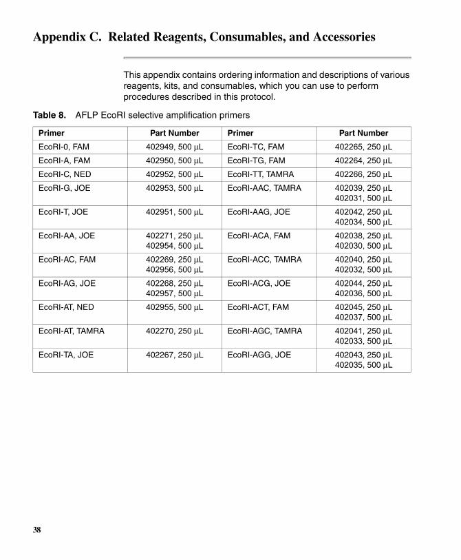
Appendix C. Related Reagents, Consumables, and Accessories
This appendix contains ordering information and descriptions of various reagents, kits, and consumables, which you can use to perform procedures described in this protocol.
Table 8. AFLP EcoRI selective amplification primers
Primer Part Number Primer Part Number
EcoRI-0, FAM 402949, 500 µL EcoRI-TC, FAM 402265, 250 µL
EcoRI-A, FAM 402950, 500 µL EcoRI-TG, FAM 402264, 250 µL
EcoRI-C, NED 402952, 500 µL EcoRI-TT, TAMRA 402266, 250 µL
EcoRI-G, JOE 402953, 500 µL EcoRI-AAC, TAMRA 402039, 250 µL402031, 500 µL
EcoRI-T, JOE 402951, 500 µL EcoRI-AAG, JOE 402042, 250 µL402034, 500 µL
EcoRI-AA, JOE 402271, 250 µL402954, 500 µL
EcoRI-ACA, FAM 402038, 250 µL402030, 500 µL
EcoRI-AC, FAM 402269, 250 µL402956, 500 µL
EcoRI-ACC, TAMRA 402040, 250 µL402032, 500 µL
EcoRI-AG, JOE 402268, 250 µL402957, 500 µL
EcoRI-ACG, JOE 402044, 250 µL402036, 500 µL
EcoRI-AT, NED 402955, 500 µL EcoRI-ACT, FAM 402045, 250 µL402037, 500 µL
EcoRI-AT, TAMRA 402270, 250 µL EcoRI-AGC, TAMRA 402041, 250 µL402033, 500 µL
EcoRI-TA, JOE 402267, 250 µL EcoRI-AGG, JOE 402043, 250 µL402035, 500 µL
38
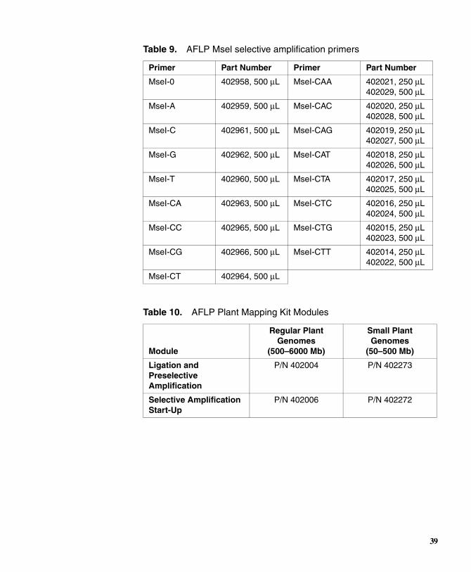
Table 9. AFLP Msel selective amplification primers
Primer Part Number Primer Part Number
MseI-0 402958, 500 µL MseI-CAA 402021, 250 µL 402029, 500 µL
MseI-A 402959, 500 µL MseI-CAC 402020, 250 µL 402028, 500 µL
MseI-C 402961, 500 µL MseI-CAG 402019, 250 µL 402027, 500 µL
MseI-G 402962, 500 µL MseI-CAT 402018, 250 µL 402026, 500 µL
MseI-T 402960, 500 µL MseI-CTA 402017, 250 µL 402025, 500 µL
MseI-CA 402963, 500 µL MseI-CTC 402016, 250 µL 402024, 500 µL
MseI-CC 402965, 500 µL MseI-CTG 402015, 250 µL 402023, 500 µL
MseI-CG 402966, 500 µL MseI-CTT 402014, 250 µL 402022, 500 µL
MseI-CT 402964, 500 µL
Table 10. AFLP Plant Mapping Kit Modules
Module
Regular Plant Genomes
(500–6000 Mb)
Small Plant Genomes
(50–500 Mb)
Ligation and Preselective Amplification
P/N 402004 P/N 402273
Selective Amplification Start-Up
P/N 402006 P/N 402272
39

Table 11. Related consumables and accessories
Name Description Vendor
AFLP Protocol Reagents and Equipment
AFLP Microbial Adaptor/Core Sequences
Consists of AFLP EcoRI and MseI adaptor pairs and core sequences
Applied Biosystems P/N 402943
E. coli W3110 DNA Reference DNA Applied Biosystems P/N 402990
T4 DNA ligase New England Biolabs
T4 DNA ligase buffer New England Biolabs
EcoRI restriction enzymes
Use higher concentration formulations of vendor- supplied enzymes
New England Biolabs
MseI restriction enzymes
Use higher concentration formulations of vendor- supplied enzymes
New England Biolabs
Bovine serum albumin (BSA)
Nuclease-free. Dilute 10 mg/mL solution supplied by vendor to 1.0 mg/mL
New England Biolabs
6% Pre-mixed polyacrylamide with 7.5 M urea in TBE buffer
Gel matrices for the ABI 373 DNA Sequencer
Amresco
LongRanger gel solutions
AT Biochem formulations. Used for the ABI PRISM
377 DNA Sequencer at 5% or 6% in TBE buffer
JT Baker
P/N 4730-02 for 250 mL
Performance Optimized Polymer 4 (POP-4)
Polymer solution used with the ABI PRISM 310
Applied Biosystems P/N 402838
10X TBE buffer stock Gibco
Deionized formamide Applied Biosystems P/N 400596
Gel-loading pipette tips, 0.17 mm flat, for the ABI PRISM 377
Rainin P/N GT-1514
40
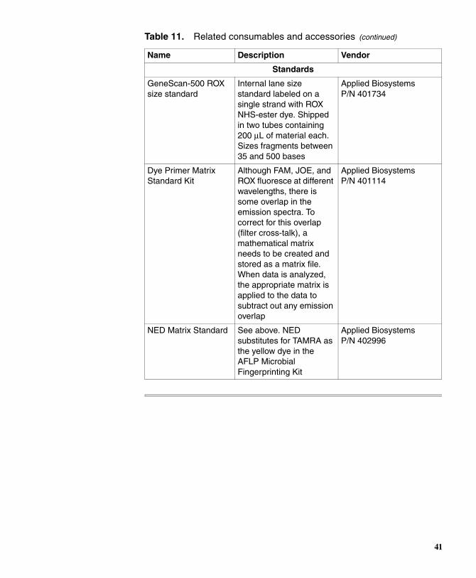
Standards
GeneScan-500 ROX size standard
Internal lane size standard labeled on a single strand with ROX NHS-ester dye. Shipped in two tubes containing 200 µL of material each. Sizes fragments between 35 and 500 bases
Applied Biosystems P/N 401734
Dye Primer Matrix Standard Kit
Although FAM, JOE, and ROX fluoresce at different wavelengths, there is some overlap in the emission spectra. To correct for this overlap (filter cross-talk), a mathematical matrix needs to be created and stored as a matrix file. When data is analyzed, the appropriate matrix is applied to the data to subtract out any emission overlap
Applied Biosystems P/N 401114
NED Matrix Standard See above. NED substitutes for TAMRA as the yellow dye in the AFLP Microbial Fingerprinting Kit
Applied Biosystems P/N 402996
Table 11. Related consumables and accessories (continued)
Name Description Vendor
41
Appendix D. Technical Support
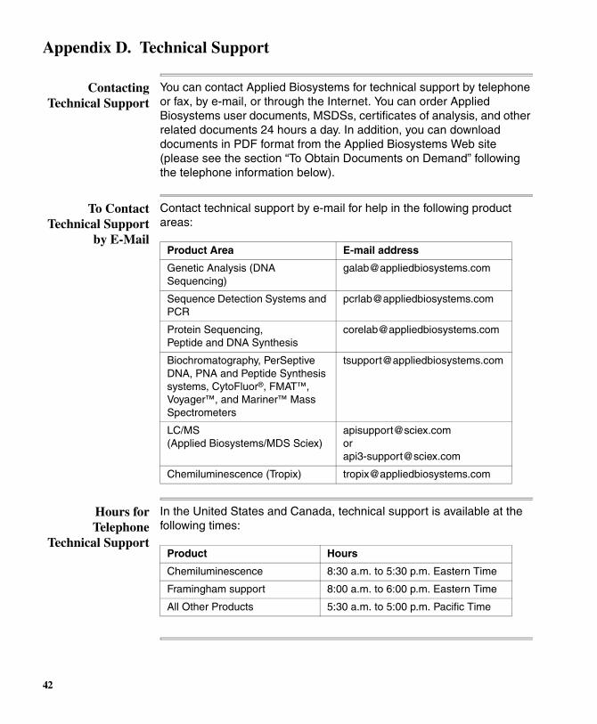
Contacting Technical Support
You can contact Applied Biosystems for technical support by telephone or fax, by e-mail, or through the Internet. You can order Applied Biosystems user documents, MSDSs, certificates of analysis, and other related documents 24 hours a day. In addition, you can download documents in PDF format from the Applied Biosystems Web site (please see the section “To Obtain Documents on Demand” following the telephone information below).
To Contact Technical Support
by E-Mail
Contact technical support by e-mail for help in the following product areas:
Hours for Telephone
Technical Support
In the United States and Canada, technical support is available at the following times:
Product Area E-mail address
Genetic Analysis (DNA Sequencing)
Sequence Detection Systems and PCR
Protein Sequencing, Peptide and DNA Synthesis
Biochromatography, PerSeptive DNA, PNA and Peptide Synthesis systems, CytoFluor®, FMAT™, Voyager™, and Mariner™ Mass Spectrometers
LC/MS (Applied Biosystems/MDS Sciex)
[email protected] or [email protected]
Chemiluminescence (Tropix) [email protected]
Product Hours
Chemiluminescence 8:30 a.m. to 5:30 p.m. Eastern Time
Framingham support 8:00 a.m. to 6:00 p.m. Eastern Time
All Other Products 5:30 a.m. to 5:00 p.m. Pacific Time
42

To Contact Technical Support
by Telephone or Fax
In North America
To contact Applied Biosystems Technical Support, use the telephone or fax numbers given below. (To open a service call for other support needs, or in case of an emergency, dial 1-800-831-6844 and press 1.)
Product or Product Area
Telephone Dial...
Fax Dial...
ABI PRISM® 3700 DNA Analyzer
1-800-831-6844, then press 8
1-650-638-5981
DNA Synthesis 1-800-831-6844, then press 21
1-650-638-5981
Fluorescent DNA Sequencing
1-800-831-6844, then press 22
1-650-638-5981
Fluorescent Fragment Analysis (includes GeneScan® applications)
1-800-831-6844, then press 23
1-650-638-5981
Integrated Thermal Cyclers (ABI PRISM® 877 and Catalyst 800 instruments)
1-800-831-6844, then press 24
1-650-638-5981
ABI PRISM ® 3100 Genetic Analyzer
1-800-831-6844, then press 26
1-650-638-5981
BioInformatics (includes BioLIMS®, BioMerge™, and SQL GT™ applications)
1-800-831-6844, then press 25
1-505-982-7690
Peptide Synthesis (433 and 43X Systems)
1-800-831-6844, then press 31
1-650-638-5981
Protein Sequencing (Procise® Protein Sequencing Systems)
1-800-831-6844, then press 32
1-650-638-5981
PCR and Sequence Detection
1-800-762-4001, then press 1 for PCR, 2 for the 7700 or 5700, 6 for the 6700 or dial 1-800-831-6844, then press 5
1-240-453-4613
43
Outside North America
Voyager™ MALDI-TOF Biospectrometry and Mariner™ ESI-TOF Mass Spectrometry Workstations
1-800-899-5858, then press 13
1-508-383-7855
Biochromatography (BioCAD® Workstations and Poros® Perfusion Chromatography Products)
1-800-899-5858, then press 14
1-508-383-7855
Expedite™ Nucleic acid Synthesis Systems
1-800-899-5858, then press 15
1-508-383-7855
Peptide Synthesis (Pioneer™ and 9050 Plus Peptide Synthesizers)
1-800-899-5858, then press 15
1-508-383-7855
PNA Custom and Synthesis
1-800-899-5858, then press 15
1-508-383-7855
FMAT™ 8100 HTS System and Cytofluor® 4000 Fluorescence Plate Reader
1-800-899-5858, then press 16
1-508-383-7855
Chemiluminescence (Tropix)
1-800-542-2369 (U.S. only), or 1-781-271-0045
1-781-275-8581
Applied Biosystems/MDS Sciex
1-800-952-4716 1-650-638-6223
RegionTelephone Dial...
Fax Dial...
Africa and the Middle East
Africa (English Speaking) and West Asia (Fairlands, South Africa)
27 11 478 0411 27 11 478 0349
South Africa (Johannesburg)
27 11 478 0411 27 11 478 0349
Middle Eastern Countries and North Africa (Monza, Italia)
39 (0)39 8389 481 39 (0)39 8389 493
Product or Product Area
Telephone Dial...
Fax Dial...
44

Eastern Asia, China, Oceania
Australia (Scoresby, Victoria)
61 3 9730 8600 61 3 9730 8799
China (Beijing) 86 10 64106608 86 10 64106617
Hong Kong 852 2756 6928 852 2756 6968
Korea (Seoul) 82 2 593 6470/6471 82 2 593 6472
Malaysia (Petaling Jaya) 60 3 758 8268 60 3 754 9043
Singapore 65 896 2168 65 896 2147
Taiwan (Taipei Hsien) 886 2 2358 2838 886 2 2358 2839
Thailand (Bangkok) 66 2 719 6405 66 2 319 9788
Europe
Austria (Wien) 43 (0)1 867 35 75 0 43 (0)1 867 35 75 11
Belgium 32 (0)2 712 5555 32 (0)2 712 5516
Czech Republic and Slovakia (Praha)
420 2 61 222 164 420 2 61 222 168
Denmark (Naerum) 45 45 58 60 00 45 45 58 60 01
Finland (Espoo) 358 (0)9 251 24 250 358 (0)9 251 24 243
France (Paris) 33 (0)1 69 59 85 85 33 (0)1 69 59 85 00
Germany (Weiterstadt) 49 (0) 6150 101 0 49 (0) 6150 101 101
Hungary (Budapest) 36 (0)1 270 8398 36 (0)1 270 8288
Italy (Milano) 39 (0)39 83891 39 (0)39 838 9492
Norway (Oslo) 47 23 12 06 05 47 23 12 05 75
Poland, Lithuania, Latvia, and Estonia (Warszawa)
48 (22) 866 40 10 48 (22) 866 40 20
Portugal (Lisboa) 351 (0)22 605 33 14 351 (0)22 605 33 15
Russia (Moskva) 7 095 935 8888 7 095 564 8787
South East Europe (Zagreb, Croatia)
385 1 34 91 927 385 1 34 91 840
Spain (Tres Cantos) 34 (0)91 806 1210 34 (0)91 806 1206
Sweden (Stockholm) 46 (0)8 619 4400 46 (0)8 619 4401
Switzerland (Rotkreuz) 41 (0)41 799 7777 41 (0)41 790 0676
The Netherlands (Nieuwerkerk a/d IJssel)
31 (0)180 331400 31 (0)180 331409
RegionTelephone Dial...
Fax Dial...
45
To Reach Technical Support
Through the Internet
We strongly encourage you to visit our Web site for answers to frequently asked questions and for more information about our products. You can also order technical documents or an index of available documents and have them faxed or e-mailed to you through our site. The Applied Biosystems Web site address is
http://www.appliedbiosystems.com/techsupp
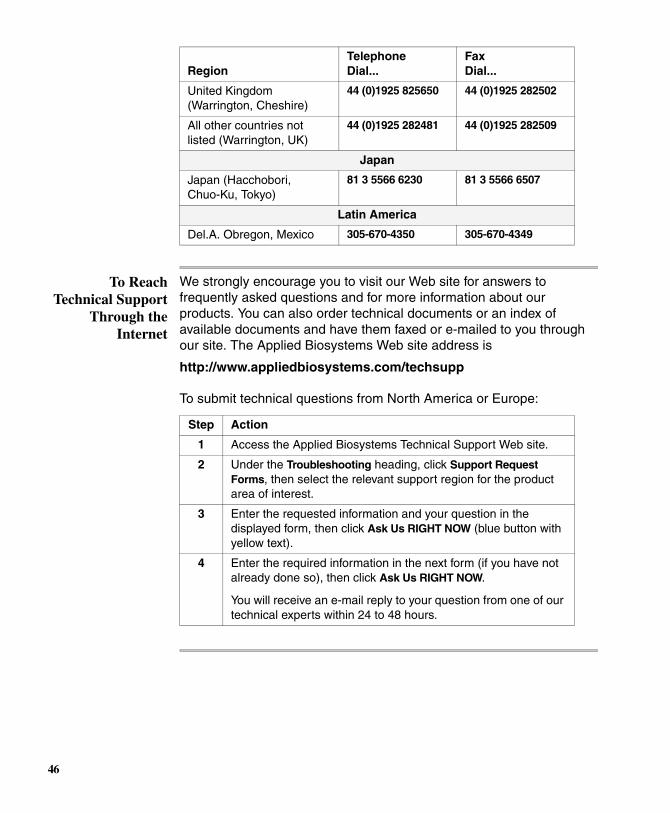
United Kingdom (Warrington, Cheshire)
44 (0)1925 825650 44 (0)1925 282502
All other countries not listed (Warrington, UK)
44 (0)1925 282481 44 (0)1925 282509
Japan
Japan (Hacchobori, Chuo-Ku, Tokyo)
81 3 5566 6230 81 3 5566 6507
Latin America
Del.A. Obregon, Mexico 305-670-4350 305-670-4349
RegionTelephone Dial...
Fax Dial...
To submit technical questions from North America or Europe:
Step Action
1 Access the Applied Biosystems Technical Support Web site.
2 Under the Troubleshooting heading, click Support Request Forms, then select the relevant support region for the product area of interest.
3 Enter the requested information and your question in the displayed form, then click Ask Us RIGHT NOW (blue button with yellow text).
4 Enter the required information in the next form (if you have not already done so), then click Ask Us RIGHT NOW.
You will receive an e-mail reply to your question from one of our technical experts within 24 to 48 hours.
46

To Obtain Documents on
Demand
Free, 24-hour access to Applied Biosystems technical documents, including MSDSs, is available by fax or e-mail or by download from our Web site.
To order documents... Then...
by index number
a. Access the Applied Biosystems Technical Support Web site at http://www.appliedbiosystems.com/techsupp
b. Click the Index link for the document type you want, then find the document you want and record the index number.
c. Use the index number when requesting documents following the procedures below.
by phone for fax delivery
a. From the U.S. or Canada, call 1-800-487-6809, or from outside the U.S. and Canada, call 1-858-712-0317.
b. Follow the voice instructions to order the documents you want.
Note There is a limit of five documents per request.
through the Internet for fax or e-mail delivery
a. Access the Applied Biosystems Technical Support Web site at http://www.appliedbiosystems.com/techsupp
b. Under Resource Libraries, click the type of document you want.
c. Enter or select the requested information in the displayed form, then click Search.
d. In the displayed search results, select a check box for the method of delivery for each document that matches your criteria, then click Deliver Selected Documents Now (or click the PDF icon for the document to download it immediately).
e. Fill in the information form (if you have not previously done so), then click Deliver Selected Documents Now to submit your order.
Note There is a limit of five documents per request for fax delivery but no limit on the number of documents you can order for e-mail delivery.
47

48

Headquarters850 Lincoln Centre DriveFoster City, CA 94404 USAPhone: +1 650.638.5800Toll Free: +1 800.345.5224Fax: +1 650.638.5884
Worldwide Sales OfficesApplied Biosystems vast distribution andservice network, composed of highly trainedsupport and applications personnel, reachesinto 150 countries on six continents. Forinternational office locations, please call ourlocal office or refer to our web site atwww.appliedbiosystems.com.
www.appliedbiosystems.com
Applied Biosystems is committed to providing theworld’s leading technology and information for lifescientists.
Printed in the USA, 06/2010Part Number 402977 Rev. F