Embed Size (px)
Citation preview

Nruroscienrv Vol. 7. No. 5, pp. 1265 to 1288, 1982 0~-~522,‘82~0S1265-23K)3.00/0
Printed in Great Britain Pergamon Press Ltd
0 1982 IBRO
AFFERENT CONNECTIONS OF DORSAL AND VENTRAL AGRANULAR INSULAR CORTEX IN THE HAMSTER
MESOCRICETUS AURATUS
R. L. REEP and S. S. WINANS*
Department of Anatomy, The University of Michigan Medical School, Ann Arbor, MI 48109, U.S.A.
Abstract-The agranular insular cortex is transitional in location and structure between the ventrally adjacent olfactory allocortex primutivus and dorsally adjacent sensory-motor isocortex. Its ventral anterior division receives major aflerent projections from olfactory areas of the limbic system (posterior primary olfactory cortex, posterolaterat cortical amygdaloid nucleus and lateral entorhinal cortex) while its dorsal anterior division does so from non-olfactory limbic areas (lateral and basolateral amygdaloid nuclei).
The medial segment of the mediodorsal thaiamic nucleus projects to both the ventral and dorsal divisions of the agranular insular cortex, to the former from its anterior portion and to the latter from its posterior portion. Other thalamic inputs to the two divisions arise from the gelatinosus, central medial, rhomboid and parafascicular nuclei. The dorsal division, but not the ventral division, receives input from neurons in the lateral hypothalamus and posterior hypothalamus.
The medial frontal cortex projects topographically and bilaterally upon both ventral and dorsal anterior insular cortex, to the former from the ventrally located medial orbital and infralimbic areas, to the latter from the dorsally-located anterior cingulate and medial precentral areas, and to both from the intermediately located prelimbic area. Similarly, the ipsilateral posterior agranular insular cortex and perirhinal cortex project in a topographic manner upon the two divisions of the agranular insular cortex.
Commissural input to both divisions originates from pyramidal neurons in the respective contralateral homotopical cortical area. In each case, pyramidal neurons in layer V contribute 90% of this projection and 10% arises from layer III pyramidals.
In the brainstem, the dorsal raphe nucleus projects to the ventral and dorsal divisions of the agranular insular cortex and the parabrachial nucleus projects to the dorsal division.
Based on their cytoarchitecture, pattern of aflerent connections and known functional properties, we consider the ventral and dorsal divisions of the agranular insular cortex to be, respectively, periallocorti- cal and proisocortical portions of the limbic cortex
The existence of an insular cortex in rodents and other non-primate mammals was first made clear by Maximilian Rose in cytoarchitectural and develop- mental studies on several orders of mammals.5’*52 He described agranular and granular divisions of the lis- sencephalic rodent cortex which correspond to corti- cal areas of similar location and structure in the oper- cularized insulae of primates including humans. Through his examination of normal embryological material, Rose found that in mice, rabbits and humans the insular cortex and underlying claustrum both appear to develop from cortical and striatal ele- ments that migrate ventrally to form a common cell plate (his cell strip X). He concluded that the insular cortex and claustrum are unique entities, distinct in origin and structure from both isocortex and stria- turn.
* Author to whom requests for reprints should be sent. Abbreviations: AI, agranular insular cortex; AId/AIp/
AIv, dorsal/posterior/ventral agranular insular cortex; HRP, horseradish peroxidase; MD, mediodorsal thalamic nucleus,
In more recent comparative cytoarchitectonic and embryological studies, Sanides54 and others1.Z*‘6*61.6* have emphasized the central position of agranular insular cortex in a structural transition from the rela- tively few layers of ventrally adjacent allocortex pri- mitivus (olfactory cortex) to the distinct six layers of dorsally adjacent isocortex. Furthermore, agranular insular cortex is but one portion of a belt of agranular cortex that also includes orbital frontal and medial frontal transition cortical areas.3s This belt may rep- resent a phylogenetic growth ring of the cortex, as suggested by Sanides.54
Several recent anatomical studies have demon- strated the striking parallels in afferent and efferent connections which exist among the insular, orbital and medial frontal transition cortical areas. A prime example is the presence, in several orders, of conver- gent input from the medi~or~l nucleus of the thala- mus, basofateral nucleus of the amygdala and dopaminergic cells of ventral tegmental area, in both the agranular insular and medial frontal cortical areaS 3.12.35.36
Comparative functional investigations on rodents,
1265

1266 R. L. Reep and S. S. Winans
carnivores and primates have revealed that all regions of the frontal agranular transition cortex influence
motivated and social behaviors 15,31,32~33.56~5g as well
as visceromotor and somatomotor responses usually associated with emotion and arousal (for a review. see Kaada2’). Such functions are often discussed coilec- tively within the conceptual framework of the term
‘limbic system’. Historically. the use of this term (see Whiteh5) in reference to cortical areas has emphasized medial zones, particularly the cingulate gyrus, to such an extent that the anatomical and functional conti- nuity among the medial, orbital and insular areas is
not commonly appreciated. The rodent agranular insular cortex consists of
several cytoarchitectural divisions, the exact number depending on the species.52 These divisions are
known to differ with respect to certain of their affer- ent connections, as was found by Krettek & Price in more recent autoradiographic studies of the cortical projections of the thalamus and amygdala in the rat,.35,3h Furthermore, these divisions are likely to have differing functional influences which should become clearer as more specific iesion/%ehavior studies are done. For these reasons, we began studies of the aff’erent and efferent connections of the dorsal and ventral agranular insular areas (Ald and Alv). which are the subdivisions of anterior agranular insu-
lar cortex in hamsters and rats. The efferent projec- tions are treated in a forthcoming report.4”
In a recent horseradish peroxidasc (HRP) study in the rat. Gerfen & Clavier ” reported differences in the thalamic inputs to AId and Aiv, but did not find
differences between Ald and Alv with respect to other inputs. Some of the known inputs to Aid and AIv were not described at all. The account below, which gives the results of our HRP study of AId and AIv, reveals many previously undescribed differences in the pattern of inputs to these two cortical areas and presents data concerning the cell types of origin, laminar distribution and topographic organization of particular projections.
EXPERIMENTAL PROCEDIJRES
In thirty male golden hamsters (M~socricetus uurut[cc)
ranging in weight from 95 145 grams. iontophoretic depo-
sits of horseradish peroxidase (Miles Laboratories, Inc..
Elkhart. Indiana), were made in the agranular insular cor-
tex or surrounding areas. HRP was prepared at a concen-
tration of 5OO~g;~l in 0.05 M fris buffer (ph 8.6). The hamster to be used was anesthetized with sodium pento-
barbital (75 mg/kg) and placed in a Kopf stereotaxic holder. A circular flap of skull bone was removed and the
dura beneath incised. A glass monofilament micropipette
(A-M Systems. Inc., Everett, WA) having a tip diameter of 12.-24 grn, and containing the HRP solution, was lowered
to the chosen coordinate position. A negative constant DC holding current of 0.1.-0.3 microamps was applied during
the descent, from a Grass S44 stimulator. This was to mini-
mize leakage of HRP from the pipette tip. For depositing the HRP. positive DC pulses of 0.5.2,Omicroamps (one per
second, 500 ms duration) were applied. After 5 I5 min the
current was turned off and the pipette left in place for up to
10min. During withdrawal of the pipette the ncgativc
holding current was agarn used.
Thirty to fifty hours after surgery. the animal was reanes-
thettzed and perfused through the heart with 2SOml of
warm (37 C) cacodylate-bufTcred saline solution (with
0.25 g sodium nitrite added as a vasodilator) followed by
250 ml of cacodylate-buffered 3”,, glutaraldehyde fixative.
The brain was removed from the skull and soaked over-
night in cold (4 C) cacodylate-buffered saline with 25”” su- crose added. The brain WS then cut corona@ at 40 50 itrn thickness on a freezing microtome and the sections col-
lectod and stored m a cold (on ice) solution consisting of
25”,, sucrose in ethylene glycol. From a few hours to a few
days after cutting. every fourth scctlon was reacted for
HRP using tetramethylbenzidine as the chromagen and the
procedures of de Olmos. Hardy & Heimer,” The reacted
sections were mounted from cold (4 Cl water onto glass
slides and after having air-dried for several haul-s. were
quickly dipped in alcohol and xqlene then overlald with
covcrslips. The distribution of labelled cells was examined
using dark-field illumination, and plotted on tracings of
adjacent Cresyl Violet stained sections which had been
made using a Bausch and Lomb microprojector. and upon
whtch relevant cytoarchitectu~d~ boundaries had been deh-
neated.
RESULTS
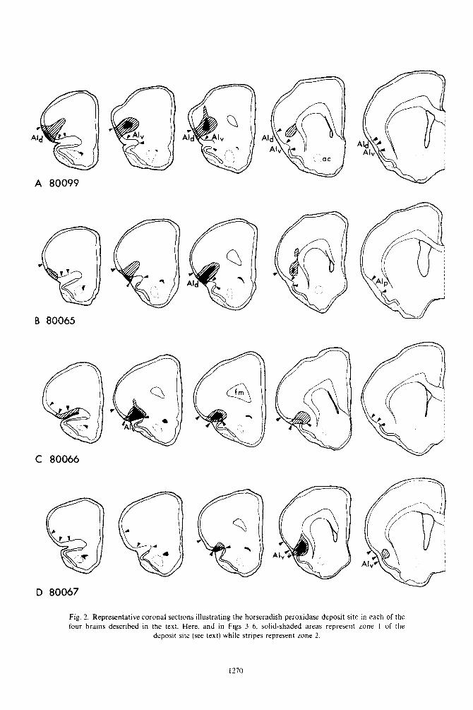
As noted by Newman & Winans?” an HRP depo- sit site can be parcelled into three distinct zones. When viewed with dark-field optics, these appear as: (1) a central area in which cells and the extracellular space are densely filled with HRP reaction product; (2) a middle zone in which cefls are densely filled but the extracellular space only moderately so; (3) an outer halo of lightly-labelled cell bodies and no extra- cellular label. As discussed by these authors, retro- grade transport apparently occurs only from zones 1 and 2. Thus in the present report all deposit site figures represent zones 1 and 2, and all references to the ‘deposit site’ refer to zones 1 and 2.
Labelled profiles were considered to be neurons only if there was a clearly demarcated cell body con- taining HRP reaction product. Often, dendritic branches containing label could be seen emanating from the cell body as well. Terminal fields labelled by anterograde transport of HRP were visible in much of our material but will be considered in a forthcoming report on the efferent connections of Aid and AIv.~~
C’ytoarchitecture cfagranular insular cortex
As shown in Fig. lA, the agranular insular cortex (AI) ties just dorsal to the rhinal fissure and extends from near the frontal pole anteriorly to perirhinaf cor- tex posteriorly. Throughout its length, AI is POS-
itioned between the ventrally adjacent primary olfac-
tory cortex (terminology of Heimer2*) and dorsally adjacent sensory-motor isocortex. Congruent with the
Afferents of agranular insular cortex 1267
early cytoarchitectural study of Rose5’ on several
orders of mammals, including the mouse and squirrel as representative rodents, and the more recent cytoar-
chitectural and experimental studies of Krettek & Price35,36 in the rat, the hamster AI can be divided
into anterior and posterior regions, and the anterior region subdivided into dorsal and ventral zones (AId and AIv), on both cytoarchitectural and connectionis- tic grounds. Area AIv curves around the buried
fundus of the rhinal fissure and is thus not wholly visible in a lateral surface view such as that shown in Fig. 1A. AId is about 2 mm in length and extends slightly farther anteriorly than does AIv. Posteriorly,
AId and AIv are replaced by AIp. In horizontal section (Fig. 1B) the lamina dissecans
is recognizable as a cell sparse zone between layers II/III and V in AIv and AId. Due to curvature of the
brain surface in the region AId and AIv, coronal sections such as those of Figs IC and 1D are oriented obliquely rather than perpendicularly to the pial sur- face, and thus exaggerate the width of layers V and VI. As illustrated in Figs 1C and lD, layers II and III of AIv are not distinct from one another and a promi- nent lamina dissecans separates layer II/III from layer V throughout AIv. In dorsally adjacent AId, layers II and III become clearly separated from each other and the lamina dissecans is gradually replaced by a granu- lar layer IV which is prominent in the lateral precen- tral area dorsal to AId. These latter two features dis- tinguish AId from AIv and, according to Sanides,54 are definitive characteristics of proisocortex versus periallocortex. Thus, AIv is a periallocortical region
by virtue of its position adjacent to the allocortex primitivus of primary olfactory cortex, its poorly de- veloped outer cell stratum (layers II/III) and the pres- ence of a lamina dissecans. AId is a proisocortical region due to its position adjacent to lateral precen- tral isocortex, its relatively well-developed outer cell stratum (distinct layers II and III) and its incipient granular layer IV within the lamina dissecans. AId corresponds to area ail of Rose” and AIv corre- sponds to his area ai2, as was previously noted by Krettek & Price.35
The claustrum lies between layer VI and the exter- nal capsule throughout AId and AIv (see Fig. 1D) and becomes noticeably thinner in AIp. The endopiriform nucleus is a ventral portion of the claustrum which lies deep to the primary olfactory cortex.36
Experimental results: dorsal agranular insular cortex
Brains 80099 and 80065 represent HRP deposits into dorsal and ventral portions of AId, respectively.
Brain 80099. The deposit site in brain 80099 (Fig. 2A) is centered in the middle of AId and all layers are affected. There is some involvement of the dorsally adjacent lateral precentral isocortex.
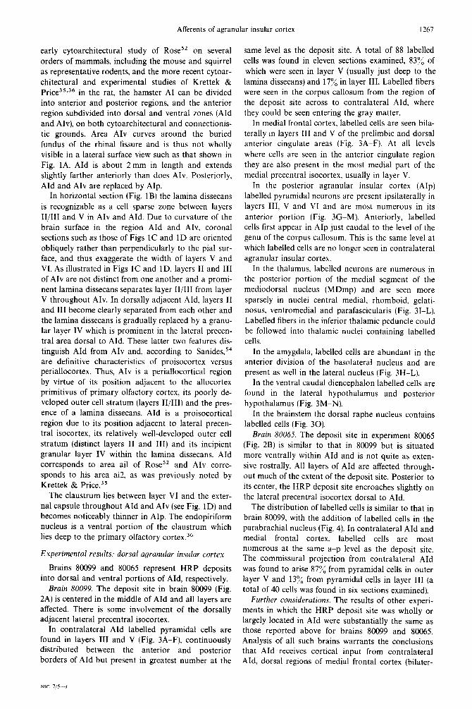
In contralateral AId labelled pyramidal cells are found in layers III and V (Fig. 3A-F), continuously distributed between the anterior and posterior borders of AId but present in greatest number at the
same level as the deposit site. A total of 88 labelled cells was found in eleven sections examined, 83% of
which were seen in layer V (usually just deep to the lamina dissecans) and 17% in layer III. Labelled fibers were seen in the corpus callosum from the region of the deposit site across to contralateral AId, where
they could be seen entering the gray matter. In medial frontal cortex, labelled cells are seen bila-
terally in layers III and V of the prelimbic and dorsal anterior cingulate areas (Fig. 3A-F). At all levels
where cells are seen in the anterior cingulate region they are also present in the most medial part of the medial precentral isocortex, usually in layer V.
In the posterior agranular insular cortex (AIp)
labelled pyramidal neurons are present ipsilaterally in layers III, V and VI and are most numerous in its
anterior portion (Fig. 3GM). Anteriorly, labelled cells first appear in AIp just caudal to the level of the genu of the corpus callosum. This is the same level at which labelled cells are no longer seen in contralateral
agranular insular cortex. In the thalamus, labelled neurons are numerous in
the posterior portion of the medial segment of the mediodorsal nucleus (MDmp) and are seen more
sparsely in nuclei central medial, rhomboid, gelati- nosus, ventromedial and parafascicularis (Fig. 31-L). Labelled fibers in the inferior thalamic peduncle could be followed into thalamic nuclei containing labelled cells.
In the amygdala, labelled cells are abundant in the anterior division of the basolateral nucleus and are present as well in the lateral nucleus (Fig. 3H-L).
In the ventral caudal diencephalon labelled cells are
found in the lateral hypothalamus and posterior hypothalamus (Fig. 3M-N).
In the brainstem the dorsal raphe nucleus contains
labelled cells (Fig. 30). Brain 80065. The deposit site in experiment 80065
(Fig. 2B) is similar to that in 80099 but is situated more ventrally within AId and is not quite as exten- sive rostrally. All layers of AId are affected through-
out much of the extent of the deposit site. Posterior to its center, the HRP deposit site encroaches slightly on the lateral precentral isocortex dorsal to AId.
The distribution of labelled cells is similar to that in
brain 80099, with the addition of labelled cells in the parabrachial nucleus (Fig. 4). In contralateral AId and medial frontal cortex, labelled cells are most numerous at the same a-p level as the deposit site. The commissural projection from contralateral AId was found to arise 87% from pyramidal cells in outer layer V and 13% from pyramidal cells in layer III (a total of 40 cells was found in six sections examined).
Further considerations. The results of other experi- ments in which the HRP deposit site was wholly or largely located in AId were substantially the same as those reported above for brains 80099 and 80065. Analysis of all such brains warrants the conclusions that AId receives cortical input from contralateral AId, dorsal regions of medial frontal cortex (bilater-

1268 R. L. Reep and S. S. Winans
ally and topographically) and ipsilateral AIp. The it, layers I-V are involved. There is no encroachement major thalamic input is from MDmp. with sparse to upon primary olfactory cortex but the ventral portion moderate projections arising in the other nuclei men- of AIv which curves around the fundus of the rhinal tioned above. Substantial amygdaloid projections fissure is affected. originate in the lateral and anterior basolateral nuclei. In contralateral AIv labelled pyramidal neurons are The lateral and posterior hypothalamic areas dorsal seen in layers III and V. most abundantly at the same raphe nucleus, parabrachial nucleus and locus coeru- lev,el as the deposit site (Fig. 5&D). In five sections leus were sometimes labelled in cases of HRP deposits examined, a total of 37 cells was found, 817, in layer into AId. V (usually just deep to the lamina dissecans) and 19%
in layer III (the deep portion of layer II/III). Labelled
Exprrimmtd result.s: wntrol cqrrrnuktr insulur c0rte.Y fibers were seen in the corpus callosum. from the deposit site across to contralateral AIv.
Brains 80066 and 80067 represent deposits into the In medial frontal cortex labelled cells are present most anterior and posterior portions of AIv, respect- bilaterally in the medial orbital and infralimbic ively. areas. usually in layers II and III. and in layers III
Brain KO066. The deposit site in experiment 80066 and V of the prelimbic area (Fig. 5,A-ME). Cells in the (Fig. 2C) is restricted to AIv except for slight medial orbital area are most numerous anterior to the encroachment upon the orbital cortex and extends deposit site. from its anterior boundary to near its posterior limit. Labelled pyramidal neurons are seen in layers III, Anteriorly, layers I--III are most extensively involved V and VI throughout the length of AIp (Fig. 5,F-L), while at the center of the deposit site and posterior to evenly distributed from a level just posterior to the
ac
ACd
ACv
AD
AHA
AId
AIP AIv
AM
AV
bc
BLa
BLP BM CE CeL CL CM
CP cst Cl
c2 C3 DP DR E ERC f fm
G
HL IL L
Fig. I. Location and structure of agranular insular cortex in the hamster. Part A is a lateral surface view
of the right side of the brain, indicating cytoarchitectonic boundaries as well as the planes of sections
shown in B. C and D.
LH
M
MDma
MDmp
anterior commissure
dorsal portion. anterior cingulate cortex
ventral portion. anterior cingulate cortex
anterodorsal thalamic nucleus
amygdalo-hippocampal area
dorsal agranular insular cortex
posterior agranular insular cortex
ventral agranular insular cortex
anteromedial thalamic nucleus
anteroventral thalamic nucleus
brachium conjunctivum
anterior portion. basolateral amygdaloid nucleus
posterior portion. basolateral amygdaloid nucleus
basotredial amygdaloid nucleus
central amygdaloid nucleus
central lateral thalamic nucleus
claustrum
central medial thalamic nucleus
cerebral peduncle
corticospinal tract
anterior cortical amygdaloid nucleus
posterolateral cortical amygdaloid nucleus
posteromedial cortical amygdaloid nucleus
dorsal peduncular cortex
dorsal raphe nucleus
endopiriform nucleus
entorhinal cortex
fornix forceps minor of corpus callosum gelatinosus (or submedial) thalamic nucleus
lateral hypothalamus
infralimbic cortex lateral amygdaloid nucleus
MH
ml
MO
mt
PaC
PBl
PBm
PC
PF
PL
POCp
PR
PT
PVa
PVP
RE
RH
sd
sm
TR
TT
VCD
VM 28L
28L’
lateral habenular nucleus
medial amygdaloid nucleus
anterior portion. medial segment of mediodorsal
thalamic nucleus
posterior portion. medial segment of mediodorsal
thalamic nucleus
medial habenular nucleus
medial lemniscus
medial orbital cortex
mammillothalamic tract
paracentral thalamic nucleus
lateral portion, parabrachial nucleus
medial portion, parabrachial nucleus
medial precentral cortex
parafascicular thalamic nucleus
prelimbic cortex
posterior primary olfactory cortex
perirhinal cortex
parataenial thalamic nucleus
anterior portion. paraventricular thalamic nucleus
posterior portion, paraventricular thalamic nu-
cleus
reuniens thalamic nucleus
rhomboid thalamic nucleus
supramammillary decussation
stria medullaris entorhinal cortical area TR
taenia tecta ventral caudal diencephalon
ventromedial thalamic nucleus lateral entorhinal cortical area 28L
lateral entorhinal cortical area 28L’

-6
6
Fig. 1.
1269
A
A 80099
B 80065
D 80067
Fig. 2. Representative coronal sections illustrating the horseradish peroxidase deposit site in each of the four brains described in the text. Here. and in Figs 3-6, solid-shaded areas represent zone I of the
deposit site (see text) while stripes represent zone 2.
t 270

Afferents of agranular insular cortex 1271
germ of the corpus callosum to the perirhinal cortex. tex, endopiriform nucleus, lateral entorhinal cortex
A few cells were found in the underlying claustrum as and posterolateral cortical amygdaloid nucleus. All
well. Pyramidal neurons are seen in layers III and V brains had labelled cells in the dorsal raphe nucleus,
of perirhinal cortex (Fig. 5M-0). and some had cells in locus coeruleus.
Tertiary olfactory areas (those receiving direct pro- jections from the olfactory bulb) containing labelled neurons include the posterior primary olfactory cor- tex (terminology of Heime?) (Fig. 5F-K), area 28L (terminology of Haug2’) of lateral entorhinal cortex (Fig. 5M-O), and the posterolateral cortical amygda- loid nucleus, C2 (terminology of Scalia & Winanss7) (Fig. 5J-K). In posterior primary olfactory cortex, 55 cells were found in 14 sections, 71% in layer III, 29”/;, in layer IIb. The endopiriform nucleus deep to pos- terior primary olfactory cortex contains a few labelled cells (Fig. 5,H-I).
Controi grains
Bruin 80067. In experiment 80067, the deposit site is centered in the posterior portion of AIv and involves layers I-VI (Fig. 2D). There is no encroachment upon the anterior primary olfactory cortex ventrally.
The distribution of labelled cells was similar to that in brain 80066 with the following qualifications.
In our experiments an angled dorsal approach was most often used to direct the HRP-filled micropipette into either AId or AIv. Often there was some labelling along the pipette track in areas dorsal to AId. In order to assess the effect of such track labelling on our results, deposits were made into these dorsally- located areas by lowering the pipette along the same trajectory as usual but not as far ventrally. In two such cases (not illustrated), the deposit site was situ- ated just dorsal to the posterior portion of AId at a level midway between the head of the caudate nucleus and the genu of the corpus callosum. Extrapolating from studies in rats, this corresponds to the granular insular taste area and sensory-motor isocortex. In brain 80021 deep cortical layers were affected and in brain 80100 more superficial layers were involved.
In contralateral AIv, labelled pyramidal cells are most numerous at about the same level as the deposit site and are not seen as far rostrally as in experiment 80066 (Fig. 6C-D). A total of 18 cells was found in six sections examined, 100% of which were in layer V.
In medial frontal cortex, layers II and III of the medial orbital area contain cells bilaterally (Fig. 6A-C) and these are located more posteriorly than those of experiment 80066.
Among the tertiary olfactory areas, posterior primary olfactory cortex contains labelled cells in layers IIb and III from the level of the genu of the corpus callosum posteriorly to its border with ento- rhinal cortex (Fig. 6E-K). This is in contrast to ex- periment 80066, in which labelled cells are very sparse in caudal portions of posterior primary olfactory cor- tex. In brain 80067 a total of 59 labelled cells was found in ten sections examined, 66% in layer III and 34% in layer IIb. The lateral entorhinal cortex con- tains labelled cells in areas 28L and 28L’ (Fig. 6L-M). The posterolateral cortical nucleus of the amygdala, C2, and the endopiriform nucleus also contain labelled cells (Fig. 6F-K).
In the cortex, labelled cells were seen in ipsilateral dorsal cortical regions adjacent to the deposit site and contralaterally in the same region as the deposit site. In the thalamus, labelled neurons were most numerous in the basal (taste) portion of the ventrome- dial nucleus, and in the paracentral, ventral lateral and ventrobasal nuclei. No other labelled cells were seen in any other areas in either of these brains.
HRP deposits in anterior portions of Alp produced no labelling in the mediodorsal thalamic nucleus, con- firming Krettek & Price’s3’ autoradiographic findings in the rat.
DISCUSSION
The present study has reported previously undocu- mented inputs to AId from the anterior cingulate and medial precentral areas bilaterally, the posterior agra- nular insular area ipsilaterally, the lateral nucleus of the amygdala, posterior hypothalamus, and medial and lateral parabrachial nuclei; to Aiv from the medial orbital and infralimibic areas bilaterally, and lateral entorhinal cortex ipsilaterally.
Further considerations. Other experiments in which the HRP deposit was wholly or largely confined to AIv gave results which were similar to those reported above for brains 80066 and 80067. Analysis of all such brains warrants the conciusions that AIv receives cor- tical input from contralateral AIv, ventral portions of medial frontal cortex (bilaterally and topographically organized; deposits involving the most ventral por- tion of AIv, which curves around the fundus of the rhinal fssure, resulted in the greatest number of labelled cells in infralimbic cortex), ipsilateral AIp and perirhinal cortex. The major thalamic input is from MDma, with other sparser projections originat- ing in the nuclei mentioned above, Tertiary olfactory inputs arise from the posterior primary olfactory cor-
The most striking findings of this study are that AId and AIv each receive a distinct combination of inputs, and that inputs to AId and AIv from a com- mon source usually arise from completely or partially separated regions of that source.
Cytoarchitectonics
Brodmann and Vogt introduced the distinction between isogenetic (or homogenetic) and allogenetic (or heterogenetic) cortical areas. As Stephan & Andy” point out, “the word ‘allocortex’ (the other cortex) functioned from the beginning as a collective term specifying the entire region of the ‘nonisocor- tex’... therefore, the allocortex has become more and more subdivided.” In contemporary usage the term

Fig. 3

Afferents of agranular insular cortex 1273
Fig. 3. Brain 80099. A series of representative coronal sections illustrating the distribution of horse-
radish peroxidase labelled neurons.

I’74 R. L. Reep and S. S. Winans
A F
A
Fig. 4.
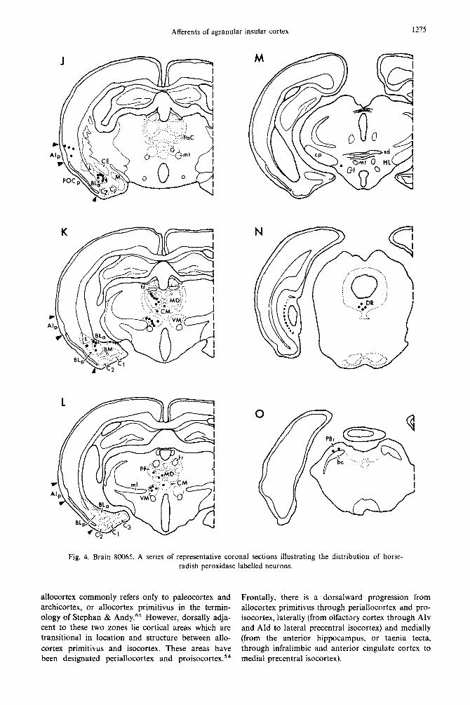
Afferents of agranular insular cortex 1275
Fig. 4. Brain 80065. A series of representative coronal sections illustrating the distribution of horse- radish peroxidase labelled neurons.
allocortex commonly refers onIy to paleocortex and Frontally, there is a dorsalward progression from archicortex, or allocortex primitivus in the termin- allocortex primitivus through ~riallocortex and pro- ology of Stephan & Andy.6’ However, dorsally adja- isocortex, laterally (from olfactory cortex through AIv cent to these two zones lie cortical areas which are and AId to lateral precentral isocortex) and medially transitional in location and structure between allo- (from the anterior hippocampus, or taenia tecta, cortex primitivus and isocortex. These areas have through infralimbic and anterior cingulate cortex to been designated periallocortex and proisocortex.54 medial precentral isocortex).

1276 R. L. Reep and S. S. Wmans
B
Fig. 5.

Afferents of agranular insular cortex 1277
Fig. 5. Brain 80066. A series of representative coronal sections illustrating the distribution of horse- radish peroxidase labelled neurons.

1278 R. L. Reep and S. S. Winans
The anterior agranular insular cortex (Ald and AIv) is one portion of a continuous belt of agranular cor- tex that includes other periallocortical and proisocor- tical transition regions, as well as precentral motor isocortex.‘* The transition areas are AIv and Ald laterally. orbital cortex frontally, and infralimbic. pre- limbic and anterior cingulate cortex medially. The precentral motor isocortex occupies the dorsolateral surface, extending from the anterior cingulate area to
AId and thereby completing the belt.
A
Because they receive direct input from the medio- dorsal thalamic nucleus, AId and AIv are usually con- sidered to be part of the ‘prefrontal’ cortex, which was defined by Rose & Woolseys3 as the total cortical projection area of MD. Although there is evidence that the rodent agranular insular cortex has func- tional properties similar to those of primate prefron- tal cortex.4’ the latter is characterized anatomically by its pronounced granularization whereas rodents have no such granular prefrontal cortex.33 Based on
Fig. 6

Afferents of agranular insular cortex 1219
K I I
I
f
CZ====
:’ ~‘--“~.~~\ / ;
~
6 i :
0 ;c: ::’
bib / u I l ..* ::..... ‘i
:.
’ . 0 0 ,..‘....,.,,
c’,;:..:: . . . . . . .,,, !‘. ? :” ,.
,&pc,
Fig. 6. Brain 80067. A series of representative coronal sections illustrating the distribution of horse- radish peroxidase labelled neurons.
comparative cytoarchitectonic studies, Sanides’4 has suggested that granular prefrontal cortex originated from agranular prefrontal cortex. Therefore, it may be that in rodents definitive funtional properties of pri- mate granular prefrontal cortex are present but are localized in presumptive granular zones within the agranular belt composed of insular, orbital and
medial frontal cortex.
Cortico-cortical connections Commissural input. Two previous reports exist con-
cerning the commissural connections between ipsi- lateral and contralateral AId and AIv. The HRP study of Gerfen & Clavier ” showed that AId and AIv
each receive input from contralateral agranular insu- lar cortex but did not discuss the precise location (i.e. AId versus AIv), laminar distribution, or cell types of

1280 R. L. Reep and S. S. Winans
origin for either of these projections. Similarly, the input from their contralateral homotopical counter- autoradiographic study of Beckstead demonstrated part. with little or no overlap. Thus, for example, Afd commissural connections between ipsilateral and receives input from contralateral AId but not from contralateral agranular insular cortex but the amino contralateral AIv. Furthermore, we found that the acid deposit sites (see his Figs 6 and 7) apparently cells of origin for these projections are pyramidal included both AId and AIv. neurons whose perikarya are located predominantly
Our results indicate that AId and AIv each receive in outer layer V (9~~~) and rarely in layer III (IO?(,). In
C MDmp MDmct
AId - L,BL,
\ I. ERC
VCD
Fig. 7. Schematic summary of the major afferent connections of dorsal and ventral agranular insular cortex. In A and B are illustrated, respectively, the topographic organization of inputs from lateral and medial frontal transition cortical areas to AId and AIv. In C the other major inputs to AId and AIv are
diagrammed.

Aflerents of agranular insular cortex 1281
contrast, Wise & Jones66 found that the commissural
projection of somatic sensory cortex in the rat orig- inates from comparable numbers of layer III and layer V pyramidal cells, or possibly to a greater extent from layer III cells (see their Fig. 17). Differences in migration rate which are known to exist between iso- cortex and agranular insular cortex during neuro- genesisz4 could conceivably result in different laminar
distributions of pyramidal neurons in these cortical areas, leading in turn to different laminar origins for
the commissural projections of isocortex versus agra- nular insular cortex.
Medialfrontal cortex. Projections from medial fron- tal cortical areas to AId and AIv have not been sys- tematically investigated previously, but were partially treated in the autoradiographic study of Beckstead. He found a projection from the prelimbic area to layers I, III and VI of agranular insular cortex bilater- ally, apparently to both AId and AIv (see his Fig.
2A-C). We have confirmed Beckstead’s finding that AId and AIv each receive input from the prelimbic area bilaterally, and have further demonstrated bila- teral inputs to AId from the anterior cingulate and medial precental areas dorsal to the prelimbic area, and to AIv from the infralimbic and medial orbital areas ventral to the prelimbic area. Thus, dorsally- located areas of medial frontal cortex project to the dorsal agranular insular area while ventrally-located areas of medial frontal cortex project to the ventral agranular insular area, as shown schematically in Fig. 7B.
Labelled neurons were most numerous in infralim- bit cortex when the deposit site involved layers II and III of the most ventral portion of AIv, which curves around the fundus of the rhinal fissure and is adjacent to anterior primary olfactory cortex. In single sections, HRP-filled axons could be followed medially from superficial layers of AIv directly to the infralim- bit area. Since some of our HRP deposits into AIv also encroached upon the anterior primary olfactory cortex to varying degrees, it is reasonable to question whether this area also receives input from infralimbic cortex. In our material, labelled fibers were never traced from anterior primary olfactory cortex to infra- limbic cortex, but were instead directed to the ventral taenia tecta and dorsal peduncular cortex (termin- ology of Haberly & Price18) just ventral to infralimbic cortex. This is in accord with the findings of Haberly & Price,lg in which injections of HRP into anterior primary olfactory cortex resulted in labelled cells in these two areas but not in the infralimbic cortex. It
must be noted, however, that their injections (see, for example, their Fig. 19) may not have significantly involved the most dorsal portion of anterior primary olfactory cortex, which is adjacent to AIv. Therefore, we cannot exclude a projection from infralimbic cor- tex to this area of primary olfactory cortex in addition to AIv.
Interestingly, the interconnected ventral areas AIv and infralimbic cortex both receive input from ter-
tiary olfactory areas: primary olfactory cortex, ento- rhinal cortex and posterolateral cortical amygdala (our results; Haberly & Price;” Kevetter & Winans;30 Krettek & Price36). No readily apparent
parallel can be drawn regarding common inputs to the dorsal areas AId and anterior cingulate/medial
precentral cortex. Posterior agranular insular cortex and perirhinal
cortex. Input to AIv from posterior agranular insular
cortex (AIp) and perirhinal cortex (PR) in the rat has
been reported by Haberly & Price,“though no topo- graphic organization was discussed. AS shown sche- matically in Fig. 7A, we found that while pyramidal
neurons throughout the entire length of AIp and PR project to AIv, only those in the anterior two-thirds of
AIp project to AId. The known inputs to AIp and PR in rodents are not topographically arranged; the anterior cortical, lateral, and anterior basolateral
amygdaloid nuclei all project to the full extent of AIp and PR.30*36
In the coronal plane, the level of the genu of the corpus callosum can be regarded as the point at which AIp replaces AIv and AId, since this is the same level at which labelled cells are no longer seen in
contralateral agranular insular cortex (commissural projection) and begin appearing in ipsilateral agranu- lar insular cortex (AIp). This boundary is indistinct cytoarchitectonically but is also defined by the pat- tern of other connections of AIP.~‘.~’
Thalamocortical connections
Our results confirm previous findings that AId and AIv each receive their major thalamic input from MD.12,13*17,35*3s Krettek & Price35 found that fibers
from MD terminate in layer III and outer layer I of the cortex. The terminology of Krettek & Price35 is used below to denote subdivisions of MD and it takes account of the differing terminology of Leonard3s and Heimer.”
Whereas our experiments have demonstrated that the anterior portion of the medial segment of MD (MDma) projects to AIv (see Fig. 8), the results of anterograde and retrograde tracing studies in the rat indicate that AIv receives its major input from the central segment of MD, MDc”*~~~~’ We refer this discrepancy to the fact that in the hamster there is no well-defined central segment of MD, either in Nissl- stained or myelin-stained sections. In the rat a central
segment is difficult to define in Nissl-stained material but appears as a fiber-rich zone in myelin-stained material.35 Since it has been reported that the rat MDc receives olfactory input from primary olfactory cortex, endopiriform nucleus and olfactory tuber- cle2’~36~48~58~60 and projects to AIv, it is natural to wonder if in the hamster, MDma, which projects to AIv, is the recipient of olfactory input. If so, it may be that a presumptive central segment of MD is con- tained within MDma in the hamster. According to Krettek 8~ Price,35 the only cortical projection of

11x:! K. L. Reep and S. S. Winans
MDma in the rat is to the prelimbic area of medial frontal cortex.
There is general agreement that in both the rat and hamster. AId receives its major thalamic input from
the posterior portion of the medial segment of MD,
MDmp (our results, Krettek & Price”’ Leonard.3Rf In their HRP study in the rat. Gerfen & Clavier” found that the boundary area between MDmp and the para- fascicular nucleus contained the greatest number of labelled cells following deposits into Aid. This is con- sistent with our observation that labelled cells in MDmp are contiguous with those in the parafascicu- lar nucleus. often within the same section, Other workers have also reported a projection from the para- fascicular nucleus to AId in the rat (experiment R4 of
Jones & Leavittzh, Krettek & Price’s). Our results indicate that Alv also receives input from the parafas- cicular nucleus in the hamster. Based on cytoarchitec- tural criteria and on its widespread connections with various cortical areas. the parafascicular nucleus has been treated as the posterior portion of the intra- laminar group of thalamic nuclei. with the central medial, paracentral and central lateral nuclei compris- ing the anterior portion.” The central medial nu- cleus, like the parafascicular, projects to both AId and
AIv in the hamster (our results) and the rat.“.” Our finding that the gelatinosus nucleus projects to
both Ald and AIv, but more heavily to the latter,
agrees with the report of Gerfen & Clavier” and with a conclusion drawn by ~lerkenham*~ in his study of the connections of the ventromedial thaiamic nucleus.
Several areas known to receive direct input from the main olfactory bulb, namely posterior primary olfactory cortex. lateral entorhinal cortex and the posterolateral cortical nucleus of the amygdala, C2.10~57 were found in the present study to project to Alv. The projection from C2 to AIv has been con- firmed autoradiographically in the hamster by Kevet- ter & Winans.3” Other workers have reported similar
olfactory projections to Atv in the rat, from posterior primary olfactory cortexl’~‘*~“h and from C2.3h
Fibers from these olfactory areas terminate in layer I of AIv,““.%
Ours is the first report of a projection from lateral entorhinal cortex to AIv. and this is reciprocated by a projection from AIv to lateral entorhinal cortex.J9 Some of our HRP deposits in ,AIv involved the border zone between Aiv and anterior primary olfactory cor- tex, It is therefore important to question whether encroachment on the anterior primary olfactory cor- tex resulted in labelled ceils in lateral entorhinal cor- tex, which like the former is a tertiary olfactory area. First, deposits in AIv which did not involve this border zone did produce labelled cells in lateral ento- rhinal cortex. Second, Haberly & Price” reported that in the rat, lateral entorhinal cortex projects only to medial areas of the olfactory forebrain and did not find labelled cells in lateral entorhinal cortex follow-
ing HRP injections into anterior primary olfactory cortex. Therefore, it appears that lateral entorhinal cortex projects to AIv but not to anterior primary olfactory cortex, in both hamsters and rats.
Our finding that the projection from posterior primary olfactory cortex to AIv originates mostly (65”~;~) from layer III cells and less so (357,)) from layer IIb cells is in accord with the results of Haberty & Price. ’ a
Considering the demonstrated anatomical separ- ation between the main and accessory (vomeronasal) olfactory systems’0~s7 and the probable differences in
function of these systems (see, for example, Johns’“, Wysocki”), it is notable that none of the tertiary ac- cessory olfactory areas (medial and posteromedial cortical nuclei of the amygdala. bed nucleus of the stria terminalis) were found to project to AIv. either in our study or in the autoradiographic study of Kevetter & Winans, ” also in the hamster.
Amygdalu prqjectians to AId
Previous reports are consistent with our finding of a substantial projection to Aid from the anterior por- tion of the basolaterai nucleus.“~3h Fibers from BLa terminate in two bands within AId, a heavy one in
layer V and a lighter one in layers I and II.“’ In the rat, reciprocal connections have been
reported to exist between BLa and MDmp,35.3h both of which project to AId in hamsters (our results) and rats.35.3” In a recent HRP study, Otterson & Ben- Ar?’ found that thalamic projections to BLa orig- inate in the interanteromedial, paraventricular and parataenial thalamic nuclei but not from MD. Simi-
larly. the autoradiographic study of Turner & Her- kenham” indicates that thalamic input to BLa is from the interanteromedial. central medial and para- taenial nuclei, As Otterson & Ben-Ari4’ suggest, it is likely that Krettek & Price’s injections into MD
encroached upon these nearby nuclei, thereby produc- ing terminal field labelling in BLa.
The subcortical projections of BLa are directed to the ventral striatum. ventral pallidurn and hypothala- muS,37.43.44 which are major output pathways of the
limbic system. The presence of labelled cells in the lateral nucleus
of the amygdala after AId deposits is consistent with Krettek & Price’s”’ finding that the cortical projec- tion field of the lateral nucleus appears to overlap with that of BL in the rat. Similarly, the results of Otterson & Ben-A@ and 0tterson4’ indicate a high degree of overlap in the sources of input to the lateral and basolateral nuclei. In cats there is a more distinct separation between the efferent cortical projections of these two nuclei,36 indicating either a greater separ- ation of the cortical fields or of the lateral and baso- lateral nuclei.
T/U> ventral caudul diencephulon and dopaminergic input to Ald
Several reports 5~12~17~40~47 have indicated the exist-

B

Afferents of agranular insular cortex 1285
ence of dopaminergic input to AId in the rat, originat- ing from cells in the anterior portion of the ventral
tegmental area, or area A10 in the terminology of Dahlstrom & Fuxe.g In the hamster, we have found projections to AId from the lateral and posterior hypothalamus. Most labelled cells in this area are
found in a region which appears to correspond to area All, a zone shown by Bjorklund & Nobin to contain catecholaminergic neurons in rats. The topo- graphic difference between rats and hamsters regard- ing the source of input to AId from the ventral caudal
diencephalon is paralleled by the topographic pattern of input from dopaminergic areas to nucleus accum-
bens. This nucleus receives input from areas A8, A9 and A10 in rats, but only from A10 in hamsters.43 As
suggested by Newman and Winans, there may be “dif- ferences in the topographic organization of the dopaminergic ‘nucleus’ in these two species.”
In the absence of histofluorescence studies in the hamster, our results are merely suggestive of
dopaminergic input to AId from the ventral caudal diencephalon.
Brainstem projections to Ald and AIv
The dorsal raphe nucleus projects to AId and AIv in both hamsters (our results) and rats.” This nucleus is the source of widespread serotonergic projections in
rats64 and may be in hamsters also. Noradrenergic input to AId and AIv originates
from locus coeruleus in rats.17*3g We have rarely seen
labelled cells in locus coeruleus in the hamster. Per- haps because of the profuse branching which occurs in noradrenergic neurons projecting to the cerebral
cortex 3g HRP is largely spread throughout the axo- nal branches rather than being transported poster- iorly to the cells of origin. Alternatively, noradrener- gic axon terminals may not uptake HRP as efficiently as non-noradrenergic terminals.
Our results indicate that the medial and lateral parabrachial nuclei project to AId in the hamster. Likewise, we have found a reciprocal projection from
AId to both divisions of the parabrachial nucleus4’ In their study of the efferent projections of the para- brachial nucleus in the rat, Saper & Loewy55 found
projections from the medial parabrachial nucleus to infralimbic cortex medially and granular insular (taste) cortex laterally (to layers V and VI in the lat- ter). According to their Fig. 4A, terminals are present in the anterior portion of AIp as well. It may be that in the hamster, input to AI from the parabrachial nucleus is directed to an area that includes posterior AId and anterior AIp.
Conclusions and possible functional implications
The use of the term ‘limbic lobe’ from the time of Broca onwards has referred to brain regions which surround the medial border of the hemisphere, adja- cent to the foramen of Monro, and to several cortical areas which form an outer ring around these central zones and constitute the limbic cortex.65 Although
many earlier anatomists (e.g. Elliot Smith) and some
later ones (e.g. Sanides, 54 White,65 Yakovle@“) have
understood the ring-like nature of limbic cortex and the fact that it includes orbital and insular, as well as
medial areas, the term ‘limbic cortex’ has commonly been applied only to the medially-located cingulate, retrosplenial and presubicular cortical areas, probably because of their connections with the anterior thala- mic nuclei (see, for example, Robertson & Kaitz’“) and the inclusion of the latter in the well known Papez circuit of the limbic system. The studies of Leo-
nard,38 Domesick,14 Beckstead and Krettek & Price35 have shown that projections from the anterior and mediodorsal thalamic nuclei overlap in the anter-
ior cingulate cortex, causing the realization that MD projects to limbic cortex as it is commonly defined. The ring of cortex understood by earlier anatomists
to constitute limbic cortex includes other areas which are known to receive MD input, namely medial fron- tal, orbital frontal and agranular insular areas. While the cortical projection field of MD is usually thought of as prefrontal cortex, we think it is more accurate to view it as limbic cortex in animals such as rodents, which have no granular prefrontal area. Furthermore, the projection from archicortex (pre- and parasubicu- lum) to the anterior thalamic nuclei,62 and the projec-
tion of the latter to medial limbic cortex3.i4 is paral- leled by a projection from paleocortex (primary olfac- tory cortex) to the mediodorsal thalamic nucleusand the projection of the latter to agranular insular cortex
(see Thalamocortical connections above). Thus, both these circuits involve projections from allocortex pri- mitivus to the thalamus, and from there to periallo- cortical and proisocortical areas. The MD cortical projection fields can also be viewed as periallocortical
and proisocortical portions of an agranular cortical belt which includes precentral isocortex as well. This agranular’ belt was first recognized on cytoarchitec-
tural grounds by von Economo and was later shown by Kaada” to possess functional continuity as well.
Behavioral studies in hamsters and rats have shown that lesions of agranular insular cortex, which typi- cally involve both AId and AIv, produce dramatic
changes in several species-typical behaviors, most notably those cued by olfactory stimuli. Eichenbaum and his co-workers15J6 found that male hamsters with lesions of MD or AI have normal odor detection thresholds but show reduced attraction to conspecific female or male odors. The hamsters’ odor discrimi- nation ability was also reduced. Animals with MD or AI lesions continued to mate successfully but spent more time (relative to normal, sham-operated, or
medial frontal-cortex-lesioned animals) sniffing non- genital body parts and often mounted the female in an inappropriate fashion. Similar effects on odor dis- crimination were seen in rats following lesions of MD or AI. These workers postulate that olfactory input to AI (via MD) influences odor discriminative aspects of odor preference and sexual performance but does not affect odor detection or the priming of sexual behav-

1286 R. L. Reep and S. S. Winans
ior by odor cues. Kolb and his co-workers have found that AI lesions in hamsters and rats result in weight loss due to transient aphagia, increased aggression. increased locomotor activity and changes in mating behavior.3’.32.33.59 Increased activity has also been seen in guinea-pigs following lesions of agranular insular cortex.41
In rats, electrodes placed in AI can mediate intra- cranial self-stimulation,3453” a response usually as- sociated with motivation and reward. Injection of
6-hydroxydopamine into the mesocortical dopamine fibers (from AlO) abolishes this response,’ and it is
attenuated by systemic injection of apomorphine, a
dopamine receptor agonist4* Similarly, electrolytic lesions of AI abolish intracranial self-stimulation
obtained from electrodes in AlO.’ These findings strongly imply that the dopaminergic pathway from Al0 to Ald plays a major role in the ability of Al to
support intracranial self-stimulation. By implication
this suggests that AId is a site where a rewarding or motivational bias is conferred either upon inputs to AId or outputs from it.
We suspect that by virtue of its extensive olfactory connections, Afv serves as an interface between olfac-
tory discrimination and related behaviors, while AId. due to its connections with the basolateral amygdala and dopaminergic centers. mediates the influence of affective biasing on non-olfactory behaviors. Behav-
ioral studies utilizing discrete lesions restricted to AId and AIv could determine whether this is the cast.
Acknowledgemetu~We thank L. L. Reep for her artistic
contributions and B. Johnson for typing the manuscript.
This research was supported by NIH postdoctoral grant
NS-06060 to Dr Reep and by NINCDS grant NS-14071 to
Dr Winans.
REFERENCES
1. Abbie A. A. (1940) Cortical lamination in the Monotremata. J. camp. Neural. 72, 42X-467.
2. Abbie A. A. (1942) Cortical lamination in a polyprotodont marsupial, Parumrles nusutu. J. camp. Neural. 76, 509 -536.
3. Beckstead R. M. (1976) Convergent thalamic and mesencephalic projections to the anterior medial cortex in the rat. J. comp. Nrurol. 166, 403-416.
4. Beckstead R. M. (1979) An autoradiographic examination of corticocortical and subcortical projections of the medio-
dorsal-projection (prefrontal) cortex in the rat. J. camp. Neural. 184, 43 62.
5. Berger B.. Thierry A. M.. Tassin J. P. & Moyne M. A. (1976) Dopaminergic innervation of the rat prefrontal cortex: a
flourescence histochemical study. Brain Res. 106, 133- 145.
6. Bjorklund A. & Nobin A. (1973) Flourescence histochemical and microspectro-flourometric mapping of dopamine
and noradrenaline cell groups in the rat diencephalon. Bruin Res. 51, 1933205.
7. Clavier R. M. & Corcoran M. E. (1976) Attenuation of self-stimulation from substantia nigra but not dorsal tegmental
noradrenergic bundle by lesions of sulcal prefrontal cortex. Brain Res. 113, 59969.
8. Clavier R. M. & Gerfen C. R. (1979) Self-stimulation of the sulcal prefrontal cortex in the rat: direct evtdence for
ascending dopamine mediation. Neuroscience Letters 12, 183- 187.
9. Dahlstrijm A. & Fuxe K. (1964) Evidence for the existence of monoamine-containing neurons in the central nervous
system-I. Demonstration of monoamines in the cell bodies of brain stem neurons. Acfa physiol. stand. 62, Suppl. 232.
l-55.
10. Davis B. J., Macrides F., Youngs W. M., Schneider S. P. & Rosene D. L. (1978) Efferents and centrifugal aRerents of
the main and accessory olfactory bulbs in the hamster. Brain Rrs. Bull. 3, 59-72.
11. de Olmos J.. Hardy H. & Heimer L. (1978) The afferent connections of the main and the accessory olfactory bulb
formations in the rat: an experimental HRP-study. J. camp. Neural. 181, 213-244.
12. Divac I., Bjorklund A., Lindvall 0. & Passingham R. E. (1978) Converging projections from the mediodorsal thalamic
nucleus and mesencephalic dopaminergic neurons to the neocortex in three species. J. camp. Neural. 180, 59-72.
13. Divac I., Kosmal A., Bjiirklund A. & Lindvall 0. (1978) Subcortical projections to the prefrontal cortex in the rat as
revealed by the horseradish peroxidase technique. Neuroscience 3, 785-796.
14. Domesick V. B. (1972) Thalamic relationships of medial cortex in the rat. Brain Behao. Eool. 6, 457-483.
15. Eichenbaum H., Shedlack K. J. & Eckmann K. W. (1980) Thalamocortical mechanisms in odor-guided behaviors-I.
Effects of lesions of the mediodorsal thalamic nucleus and frontal cortex on olfactory discrimination in the rat. Brain Brhao. Ecol. 17, 2555275.
16. Filiminoff I. N. (1947) A rational subdivision of the cerebral cortex. Arch. Neural. Psychiut. 58, 296311.
17. Gerfen C. R. & Clavier R. M. (1979) Neural inputs to the prefrontal agranular insular cortex in the rat: Horseradish
peroxidase study. Brain Res. Bull. 4, 347-353. 18. Haberly L. B. & Price J. L. (1978) Association and commissural fiber systems of the olfactory cortex in the rat I.
Systems originating in the piriform cortex and adjacent areas. J. camp. Neural. 178, 7 I l- 740.
19. Haberly L. B. & Price J. L. (1978) Association and commissural fiber systems of the olfactory cortex in the rat. II.
Systems originating in the olfactory peduncle. J. camp. Neural. 181, 781-808. 20. Haug F.-M. S. (1976) Sulphide silver pattern and cytoarchitectonics of parahippocampal areas in the rat. Ado. Anut.
Embryo/. Cell Biol. 52, l--73. 21. Heimer L. (1972) The olfactory connections of the diencephalon in the rat. Brain Behap. Evol. 6, 484523.
22. Heimer L. (1978) The olfactory cortex and the ventral striatum. In Limbic Mechanism.\: The Continuing .kolMion 01‘ [he Limbic System Concept (eds Livingston K. E. & Hornykiewicz 0.) pp. 95-187. Plenum Press, New York.

Afferents of agranular insular cortex 1287
23. Herkenham M. (1979) The afferent and efferent connections of the ventromedial thalamic nucleus in the rat. J. camp. Neural. 183, 487-518.
24. Hicks S. P. & D’Amato C. J. (1968) Cell migrations to the isocortex in the rat. Anat. Rec. 160, 619-634.
25. Johns M. A. (1980) The role of the vomeronasal system in mammalian reproductive physiology. In Chemical Signals..
Vertebrates and Aquatic Invertebrates (eds Muller-Schwarze D. & Silverstein R. M.) pp. 341-364. Plenum Press. New
York.
26. Jones E. G. & Leavit R. Y. (1974) Retrograde axonal transport and the demonstration of non-specific projections to the cerebral cortex and striatum from thalamic intralaminar nuclei in the rat, cat and monkey. J. camp: Neural. 154,
349-378.
27.
28.
29.
30.
31.
32.
33.
34.
35.
36.
37.
38.
39.
40.
41.
42.
43.
44.
4.5.
46.
47.
48.
49.
50.
51.
52.
53.
Kaada B. R. (1951) Somato-motor, autonomic and electrocorticographic responses to electrical stimulation of ‘rhinen-
cephalic’ and other structures in primates. cat and dog. Acta physiol. stand. 24, Suppl. 83, 1-285.
Kaada B. R. (1960) Cingulate, posterior orbital, anterior insular and temporal pole cortex. In ffandboo~ of’ Physiology, Section 1: ,~europ~~sjo~og~ (eds Field J.. Magoun H. W. & Hall V. E.) Vol. 2. pp. 1345-1372. American Physiological
Society, Washington, D. C.
Kevetter G. A. & Winans S. S. (1981) Connections of the corticomedial amygdala in the golden hamster-l. Efferents
of the ‘vomeronasal amygdala.’ J. camp. Neural. 197, 81-98.
Kevetter G. A. & Winans S. S. (1981) Connections of the corticomedial amydala in the golden hamster--II. Efferents
of the ‘olfactory amygdala.’ J. camp. Neural. 197, 99-l 12.
Kolb B. (1974a) Dissociation of the effects of lesions of the orbital or medial aspect of the prefrontal cortex of the rat
with respect to activity. B~~uz!. Bioi. 10, 329343.
Kolb B. (1974h) Social behavior of rats with chronic prefrontal lesions. J. camp. Physiol. Psych. 87, 46&474. Kolb B., Whishaw I. Q. & Schallert T. (1977) Aphagia, behavior sequencing and body weight set point following
orbital frontal lesions in rats. Physiol. Behav. 19, 93-103.
Koolhaus J. M., Mora F. & Phillips A. G. (1977) Ellects of food and water deprivation on self-stimulation of the
medial and sulcal prefrontal cortex and caudate putamen in the rat. Physiol. Behac. 18, 329331.
Krettek J. E. & Price J. L. (1977) The cortical projections of the mediodorsal nucleus and adjacent thalamic nuclei in
the rat. f. camp. Nruroi. 171, 357-192.
Krettek J. E. & Price J. L. (1977) Projections from the amygdaloid complex to the cerebral cortex and thalamus in the
rat and cat. J. camp. Neural. 172, 6877722.
Krettek J. E. & Price J. L. (1978) Amygdaloid projections to subcortical structures within the basal forebrain and
brainstem in the rat and cat. J. camp. Neural. 178, 225-254.
Leonard C. M. (1969) The prefrontal cortex of the rat-i. Cortical projection of the mediodorsal nucleus-II. Efferent
connections. Brain Res. 12, 321-343.
Levitt P. & Moore R. Y. (1978) Noradrenaline innervation of the neocortex in the rat. Brain Res. 139, 219-231.
Lindvall O., Bjorklund A. 81 Divac I. (1978) Organization of catecholamine neurons projecting to the frontal cortex in
the rat. Brain Res. 142, l-24.
Markowitsch H. J., Guldin W., Kessler J. & Riess R. (1980) Activity changes following sulcal, but not medial, ablation
of the prefrontal cortex of the guinea-pig. Physiol. Psych. 8, 32&324. Mora F., Phillips A. G., Koolhaus J. M. & Rolls E. T. (1976) Prefrontal cortex and neostriatum self-stimulation in the
rat: differential effects produced by a~morphine. Bruin Res. &ii. 1, 421-424. Newman R. & Winans S. (1980) An experimental study of the ventral striatum of the golden hamster-I. Neuronal
connections of the nucleus accumbens. J. camp. Neural. 191, 167-192.
Newman R. & Winans S. (1980) An experimental study of the ventral striatum of the golden hamster-II. Neuronal
connections of the olfactory tuhercle. J. camp. Neurol. 191, 1933212.
Otterson 0. P. (1980) Afferent connections to the amygdaloid complex of the rat and cat----II. Afferents from the
hypothalamus and basal telencephalon. J. camp. Nertrol. 194, 267-289.
Otterson 0. P. & Ben-Ari Y. (1979) ABerent connections to the amygdaloid complex of the rat and cat.-I. Projec-
tions from the thalamus. J. camp. Neurol. 187, 401-424.
Phillipson 0. T. (1979) Afferent projections to the ventral tegmental area of Tsai and interfascicular nucleus: a
horseradish peroxidase study in the rat. J. camp. Neural. 187, 117-144.
Powell T. P. S., Cowan W. M. & Raisman G. (1965) The central olfactory connexions. J. Anat. 99, 791-813.
Reep. R. I.. & Winans S. S. (1982) Efferent connections of dorsal and ventral agranular insular cortex in the hamster, Mesocricetus uurafus. Neuroscience 7 (in press).
Robertson R. T. & Kaitz S. S. (1981) Thalamic connections with limbic cortex-I. Thalamocortical connections. .I. camp. Neurol. 195, 501-525.
Rose M. (1928) Die Qntogenie der Inselrinde. Zugleich ein Beitrag zur histogenetischen Rindeneinteilung. J. Psychol. Neural. 36, 182-209.
Rose M. (1928) Die Inselrinde des Menschen und der Tieren. J. Psychol. Neural. 37, 467-624. Rose J. E. & Woolsey C. N. (1948) The orbitofrontal cortex and its connections with the mediodorsal nucleus in rabbit, sheep and cat. Assoc. Res. Nerv. Ment. L?is. 27, 21&232.
53a Routtenberg A. & Sloan M. (1972) Self stimulation in the frontal cortex of Rattus ~orvegjcus. Behao. Biol. 7,567-572. 54. Sanides F. (1970) Functional architecture of motor and sensory cortices in primates in light of a new concept of
neocortex evolution. In The Primate Bruin (eds Noback C. R. & Montagna W.) pp. 137-203. Appleton-Century- Crofts, New York.
55. Saper C. B. & Loewy A. D. (1980) Efferent connections of the parabrachial nucleus in the rat. Brain Res. 197,291-317.
56. Sapolsky R. M. & Eichenbaum H. (1980) Thalamocortical mechanisms in odor-guided behavior-II. Effects of lesions

1288 R. L. Reep and S. S. Winans
of the mediodorsal thalamic nucleus and frontal cortex on odor preference and sexual behavior in the hamster. bruin Behav. Evol. 17, 276-290.
57. Scalia F. & Winans S. S. (1975) The differential projections of the olfactory and accessory olfactory bulb in mammals. J. camp. Neuro/. 161, 31. 56.
58. Scott J. W. & Leonard C. M. (1971) The olfactory connections of the lateral hypothalamus in the rat, mouse and hamster. J. camp. Neuroi. 141, 331 -344.
59. Shipley J. E. & Kolb B. (f977) Neural correlates of species typical behavior in the Syrian golden hamster. J. romp. Physiol. Psych. 91, 1056-1073.
60. Siegel A., Fukushima T., Meibach R., Burke L., Edinger H. & Weiner S. (1977) The origin of the afferent supply to the mediodorsal thalamic nucleus: enhancement of HRP transport by selective lesions. Bruin Res. 135, I l-23.
61. Stephan H. & Andy 0. J. (1970) The allocortex in primates. In Thr Primate Brain (eds Noback C. R. & Montagna W.) pp. 109135. Appleton-century-Crofts, New York.
62. Swanson L. W. & Cowan W. M. (1977) An autoradiographic study of the organization of the efferent connections of the hippocampal formation in the rat. J. camp. Nrurol. 172, 49-84.
63. Turner 8. & Herkenham M. (1981) An autoradiographic study of thalamo-amygdaloid connections in the rat. Anat.
Rec. 199, 260A. 64. Ungerstedt U. (1971) Stereotaxic mapping of the monoamine pathways in the rat brain. Arta physiol. scund. 82, Suppl.
367, I-48. 65. White L. E. (1965) A morphological concept of the limbic lobe. fnt. Rev. N~arab~o~. 8, 1-34. 66. Wise S. P. & Jones E. G. (1976) The organization and postnatal development of the commissural projection of the rat
somatic sensory cortex. J. camp. Neural. 168, 313-344. 67. Wysocki C. J. (1979) Neuro-behavioral evidence for the involvement of the vomeronasal system in mammalian
reproduction, Neurosci. Biobehur. Rec. 3, 301-341. 68. Yakovlev P. 1. (1948) Motility. behavior and the brain. J. Nrrt:. Ment. Dis. 107, 313-335.
(Accepted 4 No~~n~ber 1981)