Embed Size (px)
Citation preview

Recent advancement to target Breast Cancer and Cancer Stem Cells
Prem Prakash Kushwaha and Shashank Kumar*
Center for Biochemistry and Microbial Sciences, Central University of Punjab, Bathinda-151001,
India
Phone: +91 933 564 7413; Email: [email protected]
Chapter 5
Advances in Biochemistry & Applications in Medicine
1
1. Introduction
Breast cancer is the second most prominent reason of cancer death in women after lung cancer, and it accounts for 25.2% of all cancer in women [1]. In the US, approximately about one in eight women (12%) develop invasive breast cancer [2]. In 2015, approximately 40,290 US women were expected to die from breast cancer. There was an estimation of 231,840 new cases of invasive breast cancer among US women in 2015 [3]. The chance of breast cancer causes woman’s death is about 3%. Various kinds of therapies such as hormonal, immuno-therapeutic agents, surgery and cytotoxic currently are being used to target the breast cancer. The response rate from these treatments comprises 60% to 80% for primary breast cancers and about 50% for metastases [4,5]. However, 20%-70% of patients showed reversion of cancer within five year of time [6]. Recurrence development allied with resistance to therapy and aug-mented death risk. In patients with primary breast cancer, combining cytotoxic and radiation therapy with anastrozole attained four-year survival rate of 91.6 % [7]. Gene mutations and dysregulation has been identified in breast cancers like the enhanced expression of the heparan sulfate interacting protein, p53 mutations (connecting with high histological grade) and mito-chondrial D-loop mutation (allied with lymph node-positive breast carcinoma) [7].
2. Botanicals for Breast Cancer Prevention
In the 20th century, people throughout the world now are focusing on natural products to cure different diseases. Eventually it was recognized that some natural products could in some instances be made more effective through the use of chemistry to produce semi-synthetic compounds (i.e. aspirin and later the penicillins) [8,9]. Vernonia amygdalina is an eatable Af-rican mountain plant from the Asteraceae family. A study reported that V. amygdalina extract

2
ww
w.openaccessebooks.com
Advances in Biochemistry & Applications in MedicineK
umar
S
has efficacy to inhibit the proliferative properties of MCF-7 and MKL-F cells [10,11]. Higher concentration of V. amygdalina, extracted by chloroform, hexane, butanol and ethyl acetate repressed the DNA synthesis by 76-98%. Flavonoids are the large class of pigments from the plant having a structure based on or alike to that of flavone. Most widely examined flavonoids are the catechins, flavopiridol, genistein, and quercetin. These flavonoids are known to inhibit the cancer proliferation and holds anti-tumour activities. Flavopiridol is a semisynthetic fla-vone derivative of the natural anti-inflammatory and immuno-modulatory alkaloid rohitukine [12]. For the first time, rohitukine was isolated from Amoora rohituka [13,14] and later from Schumanniophyton magnificum [15] and Dysoxylum binectariferum [16]. The study reported that rohitukine showed modest cytotoxicity against human HL-60 promyelocytic leukemia and HCT-116 colon cancer cells [17]. Green tea is a category of tea that is formed from Camellia sinensis leaves that have not undergone the same crushing and oxidation process used to make oolong and black tea. Green tea extracts exposed synergetic action with an anti-estrogens drug, tamoxifen. Tea extracts tested on estrogen receptor-positive MCF-7, ZR75, T47D human breast cancer cells in combination with tamoxifen were found that drug showed much more ef-ficient by suppressing the cell proliferation than alone [18]. Another study reported that green tea inhibits angiogenesis. Crude green tea extracts and epigallocatechin gallate (main green tea polyphenol) reduced the transcription of vascular endothelial growth factor (VEGF) in a dose-dependent manner. Withania somnifera is a widespread shrub used in Asian traditional medi-cine. The organic extracts from W. somnifera were tested among two dozen selected plants [19,20]. Extract treated cell culture (murine fibrosarcoma cell line L929sA) shown sensitiv-ity to TNF. The active ingredient in W. somnifera leaves and roots comprises C-28-steroidal lactone triterpenoids, with anolides were identified [21]. With aferin A inhibited the growth of breast cancer cell lines more efficiently than the standard. Resveratrol is a botanical poly-phenol that provides numerous health benefits including cancer prevention. Different studies revealed the potential of resveratrol on cancer cell signaling pathways [22]. It has been estab-lished that the outcome of resveratrol administration in in vitro and in vivo experimental mod-els depends upon the route of administration and dosage given [22]. In scientific community curcumin is a well-known phytochemical known for its anticancer potential. It is a phenolic compound derived from Curcumin longa (commonly known as turmeric). This phytochemical have tremendous ability to inhibit cancer cell cycle, induce apoptosis in cancer cells and check the metastasis ability of the cancer cells of different tissue origin [23]. Curcumin is known to target growth factors, transcription factors, cytokines and protein kinases in cancer cells. Literature depicts the diverse molecular targets modulated by curcumin in cancer cells and thereby providing prevention against the devastating disease [23].

3
Advances in Biochemistry & Applications in Medicine
3. Multi-targeted Chemotherapy in Breast Cancer
Targeted therapy comprises the compounds which have potential to target the molecule responsible for cancer origin. However, conventional chemotherapy acts on the all dividing cells which have potential to generate toxic effect and normal tissues damage; targeted drugs allow hitting the target in a precise manner and cells subpopulations which have directly im-plicated in tumor progression. For the first time, targeted therapy starts with the trastuzumab (monoclonal antibody), acts against HER2 for the treatment of metastatic breast cancer and Imatinib, a small tyrosine kinase inhibitor targeting BCR-Abl, in chronic myeloid leukemia [24]. Multi-targeted therapy accommodates development of a multi-target drug which is likely to harvest a drug that has efficacy to cooperate with lower affinity than a single-target drug because smaller drug-like molecule will bind to an assortment of different targets with corre-spondingly high affinity.
Human epidermal growth factor receptor 2 (HER2) is also known as receptor tyrosine-protein kinase erbB-2, CD340 and neu. It comprises as a member of the human epidermal growth factor receptor family (EGFR/ERBB/HER). Over expression of the HER2 gene has been shown complicated in the development of the aggressive types of breast tumor. HER2 has become as an important biomarker in recent years. It is the target of therapy for around thirty percent of the breast tumor patients. Recently it has been reported that two HER2 tar-geted therapy showed more beneficial rather than one [25]. Comparable to women having the higher risk for recurrence, this therapy is not advantageous for women having the lower risk for recurrence.
Figure 1: Structure of active natural phytochemicals

4
Advances in Biochemistry & Applications in Medicine
Recently a study reported that benefit was modest in addition to second HER2 targeted medicine, pertuzumab (Perjeta) compared to standard of care trastuzumab (Herceptin) [25]. Women patient getting trastuzumab alone shown 93.2% of women were not able to develop invasive breast cancer while receiving trastuzumab and pertuzumab, 94.1% of women had not developed invasive breast cancer. Mechanism of action of trastuzumab targets only HER2 while pertuzumab targets both HER2 and HER3. Obstruction of cancer cell growth signals by antibodies against HER2 and HER3 enhances the lower chance of treatment resistance. Ap-proximately 8% of all type of breast cancer patients have early HER2 positive breast cancer and may take advantage of these kinds of adjuvant therapy [25]. The study strengthens the concept of multi targeted drug therapy.
4. Identification of Novel miRNAs: New Paradigm in Breast Cancer Chemotherapy
In the multicellular organism, stem cells are the undifferentiated cell able to develop the same type of more cells and from several other kinds of cell arise by differentiation. From several decades, stem cells are using immuno-modulation or reconstitution, one of the meth-ods employed for cancer treatment. Stem cells have self-renewal capability with extremely replicative potential in multi-lineage differentiation capacity [27]. Cancer stem cells are the cells present in the tumor having potential to initiate the tumor [28]. Healthy cells pertains the three principal characters:
1. Self-renewal potential
2. Strict controls on stem cell numbers
3. Capability of differentiation, divide and to produce all functional element of the particular
Figure 2: Current therapies for HER2-expressing breast cells. Constitutively active HER2 receptors dimerise with other HER receptors on the surface of HER2-expressing breast cancer cells, activating downstream signaling pathways that mediate tumorigenic cell proliferation, survival and invasion. Lapatinib binds to HER2 and HER1 and inhibits tumorigenic receptor signaling, promotes apoptosis and cell cycle arrest. Trastuzumab prevents constitutive activation of HER2, induces internalisation and degradation of the protein and stimulates the immune system to recognise HER2-overexpressing cells. Adopted and modified from [26].

5
Advances in Biochemistry & Applications in Medicine
tissue. In comparison to healthy cells, cancer stem cells lost their control toward division and cell number. The cancer stem cells present in very minute amount in solid tumor growth. They are responsible for the development of tumor cells. Different markers have been identified and validated to isolate the breast cancer stem cells (Table 1).
MicroRNAs are the small non-coding RNAs which have potential to control an exten-sive range of biological progressions comprising carcinogenesis. Various studies have been found that miRNAs are profoundly dysregulated in cancer cells. Under certain circumstances, miRNA may act as either tumor suppressor or oncogenic. miRNAs comprises 20 to 25 nucle-otides in length binds to the 3’ untranslated regions (UTR) of mRNAs leads to the repression of target proteins via mRNA degradation or translational inhibition.
Transcription of the miRNA gene results in pri-miRNA formation in the nucleus which is cleaved by Drosha and DiGeorge Syndrome Critical Region Gene (DGCR8) proteins result-ing in the generation of pre-miRNA transcript. The exportin-5 then exports the pre-miRNA from the nucleus to the cytoplasm where it gets processed by Dicer, and other enzymes such as trans-activator RNA (tar) - binding protein (TRBP) leading to the formation of short RNA du-plexes. It then forms a miRISC/Ago complex with the help of some other proteins and factors. This complex then guides and performs some crucial functions in the cellular systems [35].
Table 1: Surface markers used to isolate the breast cancer stem cells
Figure 3: Classes of miRNAs in breast cancer. Adopted and modified from [34]
Marker Reference
CD44+CD49fhi CD133/2hi
ALDH1
CD44+/CD24- CD133
CD49f
[29]
[30]
[31]
[32]
CD61 [33]

Advances in Biochemistry & Applications in Medicine
6
Recently a study reported that a small RNA molecule, miR199a, supports to sustain the activity of stem cells in both cancerous and healthy breast cells [36]. miR199a regulates vari-ous factors that assist noncancerous mammary gland stem cells (MaSCs) resist differentiation and retain their capacity to self-renew in human as well as mouse breast cancer stem cells. It supports MaSCs to hold their stem cell activity by repressing the production of a protein called ligand-dependent corepressor (LCOR), which binds DNA to control the gene expression. Up-regulated miR199a expression in mouse MaSCs reduced the LCOR proteins and improved healthy stem cell function. Conversely, increased LCOR expression diminishes mammary gland stem cell activity. Tumor-expressed higher expression of miR199a correlates with poor survival rates, while the tumor with enhanced levels of LCOR shown better prognosis [36].
LCOR sensitizes the cells against the effects of signaling molecules such as interferon released from immune (macrophages) and epithelial cells in the mammary gland. These cells secrete interferon alpha (IFN-α) to encourage the cell differentiation and cell division inhibi-tion in the normal mammary gland development. The microRNA plays a crucial role during
Figure 4: Biogenesis of miRNA and mRNA degradation. Adopted and modified from [35]

Advances in Biochemistry & Applications in Medicine
7
tumorigenesis [37]. miR199a also protects MaSCs from IFN signaling and allowing them to remain undifferentiated and accomplished of self-renewal. Interferons have been extensively worked for the tumor treatment. These kinds of treatments become much more efficient if the cancer stem cells (interferon resistant) can be rendered sensitive by targeting the LCOR-mi-R199a pathway. These sorts of examinations expose a new property of breast cancer stem cells that give them compensations in their communications with the immune system. Therefore it characterizes an outstanding opportunity to adventure for refining cancer immunotherapy [36].
4. Combinatorial Immunotherapy: A New Hope in Early Stage Triple Negative Breast Cancer
Immunotherapy or biologic therapy is a technique for the treatment of cancer that boosts the body’s natural defences to fight against cancer. This kind of therapy uses substances which are made by the body or in the laboratory to improve or to restore the immune system function. In the phase I trial, combining the immune checkpoint inhibitor (durvalumab/MEDI4736) with chemotherapy as a pre-operative treatment for early stage triple negative breast cancer (TNBC) revealed a 71% pathologic complete response to the combination treatment [38]. Chemotherapies solely generate a complete pathologic response at the rate of around 35-40% for women with TNBC. Combinational therapy is important because women with breast can-cer who achieve complete response have excellent long-term survival. There is an essential requirement for new employment options to increase cure rates for women with TNBC. Phase I clinical trial sought to restrict the safety and tolerability of durvalumab in combination with weekly nabpaclitaxel and dose dense doxorubicin/cyclophosphamide (ddAC) [38]. There were no dose-limiting toxicities with the combination; Therefore the full dose of neo-adjuvant chemotherapy can be administered concurrently with the full dose of durvalumab. Among the seven patients in phase I clinical trial, five achieved the complete pathologic response, one had partial response with residual disease, and one had widespread residual cancer [38].

Advances in Biochemistry & Applications in Medicine
8
5. Novel PARP Inhibitor Against Advanced Breast Cancer
Enzyme Poly (ADP-ribose) polymerases (PARPs) have the capability to handover the ADP-ribose to the target proteins (poly ADPribosylation) [40,41]. Approximately 18 members of the PARP family have been reported yet, and several different genes encode these fam-ily proteins. PARP family shares homology in their conserved catalytic domain [40]. Several isoforms such as PARP1 and PARP2 are best known for their contribution to the DNA repair processes. Through the various studies, it is now clear that PARPs family have a necessary role in numerous cellular processes like proliferation and cell death [40].
In the majority, PARP substrates include nuclear proteins involved in nucleic acid me-tabolism, chromatin structure modulation, DNA synthesis and repair [41]. In the presence of DNA strand breaks, PARP also has potential to regulate and modify itself. It is the main ac-ceptors of poly ADP-ribose in vivo. Best characterized PARP family member is the PARP1. PARP2 is narrowly associated to PARP1 with 69% resemblance in their catalytic domain and recognized by the perseverance of PARP activity in PARP1-deficient cells [40,42].
Figure 5: Targeting immune targets 1 and 2 by different inhibitor molecules, alone or in combination. Inhibition might results in differential levels of responses in terms of antitumor activity and toxicity. Blockage of target 1 by lipillimumab might results into anti-tumor activity and toxicity both in tumor cells. Blockage of target 2 by lambrolizumab might results into more anti-tumor activity and comparatively less toxicity in tumor cells. By using the concept of combinato-rial therapy, blockage of target 1 and 2 by respective inhibitors might results into comparatively enhanced anti-tumor activity and toxicity in tumor cells. Adopted and modified from [39]
Figure 6 : (A) Structure of olaparib (PubChem CID: 23725625) (B) Cartoon structure of poly (ADP-ribose) polymerase (Cyan colour) (PDB ID: 3L3M) complexed with olaparib (Red colour). The 3D structure of the target receptor (PARP) was downloaded from RCSB-protein data bank in .pdb format. The olaparib structure was retrieved from NCBI Pub-Chem compounds database in .sdf format. Offline docking tool such as Auto Dock Tools 1.5.6 (ADT) was used for to study the drug-protein interaction. PyMOL molecular visualization tool was used for visualization of the interaction pattern in the drug-protein complex.(C) Schematic representation of hydrogen bonds and hydrophobic interactions be-tween olaparib and Poly (ADP-ribose) polymerase. Green colour represents hydrogen bonding (developed using Ligplot +v.1.4.5) ( Ligand bond, Non-ligand residues involved in hydrophobic contact, Non-ligand bond, Corresponding atoms involved in hydrophobic contact, Hydrogen bond and its length)

Advances in Biochemistry & Applications in Medicine
9
Lynparza (olaparib) significantly reduce the progression of breast cancer that is induced partially due to mutations in a gene called breast cancer (BRCA). BRCA gene mutations are responsible for around 3% of all breast cancers. A new study (phase III clinical trial) included 302 women with breast cancer that has spread to other organs. They were positive for BRCA gene mutation. Investigation of their breast cancer cells shown that cells have one of the hor-mone-sensing molecules either estrogen or progesterone or have none of these two hormone receptors along with HER2. These type of cancers termed as “triple negative breast cancers” [43].
Lynparza diminished the risk of cancer development by as much as 42% compared to additional conventional chemotherapy. Lynparza also showed much fewer side effects that conventional chemotherapy. In 60% patients receiving Lynparza, the tumor showed evident shrinkage. A similar reduction was noted in 29% women on standard chemotherapy. Serious side effects were seen in 37% women on Lynparza compared to 50% in women with conven-tional chemotherapy [43]. Lynparza (olaparib) inhibits the enzyme called poly ADP-ribose polymerase (PARP). Poly (ADP-ribose) polymerase (PARP) family proteins included some cellular processes such as DNA repair, genomic stability, and programmed cell death. BRCA gene works by stopping the repair of the damage caused by cancer to the DNA. PARP further prevents the damage repair by the cells. Lynparza can work only on cancers where the BRCA gene mutation is responsible for those diseases. Lynparza is already in the market for ovarian cancer that is caused by BRCA. Two other similar drugs for BRCA mutation induced ovarian cancer, include Zejula and Rubraca manufactured and made by Tesaro and Clovis Oncology respectively [43].
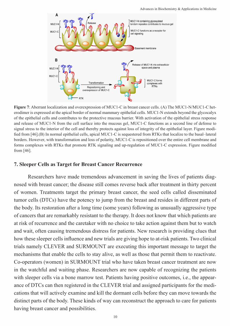
6. Targeting MUC1 Protein: A New Step Towards Targeted Drug Therapy in Breast Cancer
One of the O-linked glycosylated proteins called mucins plays an important defence function by forming a protective mucous barrier onto the epithelial cell surface. Mucins also take part and perform the crucial function in intracellular signaling [44]. It exhibited onto the apical surface of epithelial cells that line the mucosal cell surfaces of multiple tissues such as pancreas, stomach, lung, and breast providing lubrication and protection. When cells become malignant, the MUC1 protein changes and signals tumor cells to multiply. In a study, as cancer attacked formerly healthy mice, their immune systems provide antibodies to fight against the cancer cells [45]. Cancer cells illuminated green fluorescence, purporting the binding of the antibody to the abnormal MUC1 proteins and neglecting the healthy cells. Scientist signifies these kinds of new therapies as a “smart bombs” that can combat cancer cells by killing them without hitting any of the neighbouring healthy cells. That compares with traditional chemo-therapy that kills whatever it touches, including healthy tissue [45].
Advances in Biochemistry & Applications in Medicine
10
7. Sleeper Cells as Target for Breast Cancer Recurrence
Researchers have made tremendous advancement in saving the lives of patients diag-nosed with breast cancer; the disease still comes reverse back after treatment in thirty percent of women. Treatments target the primary breast cancer, the seed cells called disseminated tumor cells (DTCs) have the potency to jump from the breast and resides in different parts of the body. Its restoration after a long time (some years) following as unusually aggressive type of cancers that are remarkably resistant to the therapy. It does not know that which patients are at risk of recurrence and the caretaker with no choice to take action against them but to watch and wait, often causing tremendous distress for patients. New research is providing clues that how these sleeper cells influence and new trials are giving hope to at-risk patients. Two clinical trials namely CLEVER and SURMOUNT are executing this important message to target the mechanisms that enable the cells to stay alive, as well as those that permit them to reactivate. Co-operators (women) in SURMOUNT trial who have taken breast cancer treatment are now in the watchful and waiting phase. Researchers are now capable of recognizing the patients with sleeper cells via a bone marrow test. Patients having positive outcomes, i.e., the appear-ance of DTCs can then registered in the CLEVER trial and assigned participants for the medi-cations that will actively examine and kill the dormant cells before they can move towards the distinct parts of the body. These kinds of way can reconstruct the approach to care for patients having breast cancer and possibilities.
Figure 7: Aberrant localization and overexpression of MUC1-C in breast cancer cells. (A) The MUC1-N/MUC1-C het-erodimer is expressed at the apical border of normal mammary epithelial cells. MUC1-N extends beyond the glycocalyx of the epithelial cells and contributes to the protective mucous barrier. With activation of the epithelial stress response and release of MUC1-N from the cell surface into the mucous gel, MUC1-C functions as a second line of defense to signal stress to the interior of the cell and thereby protects against loss of integrity of the epithelial layer. Figure modi-fied from [46].(B) In normal epithelial cells, apical MUC1-C is sequestered from RTKs that localize to the basal–lateral borders. However, with transformation and loss of polarity, MUC1-C is repositioned over the entire cell membrane and forms complexes with RTKs that promote RTK signaling and up-regulation of MUC1-C expression. Figure modified from [46].

Advances in Biochemistry & Applications in Medicine
11
8. Biological Production of Anticancer Secondary Metabolites
Ontario grown onions (Ruby Ring onion variety) contain one of the highest concen-trations of quercetin, a type of flavonoid. The study revealed that the red onion has not only high levels of quercetin but also large amounts of anthocyanin which enriches the scavenging properties of quercetin molecules [47]. Anthocyanins are the water-soluble vacuolar pigments, provides colours to several vegetables and fruits. Depending on their pH, it produces different colours such as red, purple or blue. Anthocyanins pass sense that the red onions (darkest in colour) would have the most cancer-fighting power. Onions activate pathways that encourage cancer cells to undergo cell death. They develop an abnormal environmental condition for cancer cells, and they obstruct interaction between cancer cells which hinders growth. A new extraction procedure needs to be explored which will reduce the use of chemicals making the quercetin found in onions more suitable for consumption. Another extraction technique uti-lizes solvents that can transmit a toxic residue which is then ingested in food. Proper extrac-tion strategy is must which can reduce the chemical residues is crucial to explore the onion’s cancer-fighting properties in nutraceuticals as well as in pill form [47].
Figure 8: (A) Conventional approach to target the cancer remedies.Observation of the cancerous cells demonstrated that a special type of cell exists with the characteristics associated with stem cells called cancer stem cells. Cancer stem cells are tumor establishing and more prominently have the potency to give rise to all other cancer cell types found in a particular cancer. These stem cells of cancer can exist as a distinct population among other cells and cause metastasis and give rise to new tumors. Unfortunately, these cells can be very elusive and most of the cancer drugs have no or little effect on them. (B) In a novel approach, targeting stem cells can repress the chance of recurrence of cancer.
(Adopted and modified from https://ninithi.com/2015/07/26/smart-nanoparticles-to-target-and-destroy-cancer-recur-ring-stem-cells/)

Advances in Biochemistry & Applications in Medicine
12
9. Novel Biomarker for Identification of Early Stage Breast Cancer
The definition of the biomarker conferring to the National Cancer Institute is “a biologi-cal molecule found in body fluids, blood and other tissues that are a symbol of a normal or abnormal progression or disease such as cancer. Biomarkers characteristically distinguish an affected or diseased person from people without any disease. Variations can be due to some factors such as transcriptional alterations, somatic or germ line mutations and post-transla-tional modifications. In the cellular systems, there is the astonishing variety of biomarkers is present such as proteins (enzymes or receptors), nucleic acids (microRNA or other non-coding RNA), antibodies and peptides [48]. A biomarker is a collection of various alterations of gene expression, metabolomic as well as proteomic signatures. It can be immediately identified in the circulatory system (plasma, serum or whole blood) or excretory secretion system (urine, stool, sputum or nipple discharge), and thus it can be easily evaluated [48].
Conventional treatment is inadequate for many early stage tumors that have grown past the in situ stage and failed to stop their spread to separate sites in the body. Recently research-ers have identified SMARCE1 (SWI/SNF-related matrix-associated actin-dependent regulator of chromatin subfamily E member 1), a gene overexpressed in the subset of early stage cancers that are likely to become aggressively invasive making it conceivable to distinguish poorly in-vasive tumors from those that will likely expand and metastasize [49]. The researchers found that fifty percent of the early stage cancers with high SMARCE1 expression will metastasize at some point in the ten to fifteen years after their first diagnosis. SMARCE1 drives invasion by regulating the expression of secreted proteases that degrade basement membrane, an ECM bar-rier surrounding all epithelial tissues. In functional studies, SMARCE1 promotes invasion of in situ cancers growing within primary human mammary tissues and required for metastasis in vivo [49]. Mechanistically, SMARCE1 drives invasion by forming an SWI/SNF-independent complex with the transcription factor ILF3. In patients diagnosed with early-stage cancers, the
Figure 9: Quercitin and anthocyanins present in onion

Advances in Biochemistry & Applications in Medicine
13
SMARCE1 expression is a strong predictor of eventual relapse and metastasis. Collectively, these findings establish SMARCE1 as a key driver of invasive progression in early-stage tu-mors [49].
10. RIOKinase: A Novel Target for Breast Cancer
RIOK1 or RIO kinase 1 is one of the novel cytoplasmic component of the pre-40S pre-ri-bosomal particle(s) in mammalian systems, required for the final stages of cytoplasmic pre-40S maturation [51]. RioK1 is a new interactor protein of arginine methyltransferase 5 (PRMT5) which competes with pICln for interaction and modulates PRMT5 complex formation and substrate specificity [51]. RIOK1 may reveal new insights into defects underlying breast and related cancers and may reveal new therapeutic possibilities for these cancers. RIOK1 knock-down (not RIOK2 or RIOK3) in colon, breast and lung cancer cell lines exceedingly reduces cell proliferation and invasiveness [52]. These consequences mainly remarked in RAS mutant cancer cells. In contrast, the growth of RAS wild-type Caco-2 and Bcr-Abl-driven K562 cells is not influenced by the RIOK1 knockdown, recommending a precise obligation for RIOK1 in the context of oncogenic RAS signaling. Furthermore, RIOK1 activates NF-κB signaling and
Figure 10: Schematic view of normal, in situ, invasive, and metastatic carcinoma progression. Normally, breast ducts consists basement membrane and luminal epithelial and myoepithelial cells. Stromaportion of the cells compriseseveral leukocytes, fibroblasts, and endothelial cells. Enhanced expression of SMARCE1 induces the protease expression which degrades the basement membrane. In in situ carcinomas the myoepithelial cells are altered by both phenotypically and epigenetically and their number decreases due to the basement membrane degradation. The number of stromal fibro-blasts, lymphocytes, and endothelial cells increases with the same time. Myoepithelial cells and basement membrane loss results in invasive carcinomas, in which tumor cells can invade surrounding tissues andmight migrate to distant organs, ultimately leading to metastases. Adopted and modified from [50].

Advances in Biochemistry & Applications in Medicine
14
promotes cell cycle progression. A study revealed that pro-invasive proteins metadherin and Stathmin1 to be regulated by RIOK1. In conclusion, RIOK1 could be a possible therapeutic target, particularly in RAS-driven carcinomas [52].
11. Summary
Over the past few decades, considerable progress has been made in breast cancer treat-ment. Beside that survival rates for patient of breast cancer remain poor which underlies the need for further advances in therapy. Effort of researchers and scientist worldwide towards the better efficacy of breast cancer therapy includes the recent advances in breast cancer treatment such as multi-targeted chemotherapy; identification of novel anticancer miRNAs; combina-torial immunotherapy; novel PARP inhibitors; identification of novel biomarkers for early breast cancer detection, biological production of anticancer secondary metabolites; and target-ing O-linked glycoproteins, sleeper cells and RIOK1. Finally the advancement in breast cancer therapy and or drug discovery needs to be study in much more depth for further improvements in outcomes for patients with the disease.
12. Acknowledgment
PPK acknowledges financial support from University Grants Commission, India in the form of CSIR-UGC Junior Research fellowship. SK acknowledges Department of Sci-ence and Technology, India, for providing financial support in the form of DST-SERB Grant [EEQ/2016/000350]. SK also acknowledges Central University of Punjab, Bathinda for pro-viding necessary infrastructure facility.
13. References
1. Report, W. C. (2014). International Agency for Research on Cancer. World Health Organization.
2. Report, W. C. (2008). International Agency for Research on Cancer. Retrieved 26 February, 2011.
3. American Cancer Society. Breast Cancer Facts & Figures 2015-2016. Atlanta: American Cancer Society, Inc. 2015.
4. Bartsch, R., Wenzel, C., & Steger, G. G. Trastuzumab in the management of early and advanced stage breast cancer. Biologics: targets & therapy, 2007; 1(1): 19.
5. Vici, P., Colucci, G., Gebbia, V., Amodio, A., Giotta, F., Belli, F., ...& Brandi, M. First-line treatment with epirubicin and vinorelbine in metastatic breast cancer. Journal of clinical oncology, 2002; 20(11): 2689-2694.
6. Goldhirsch, A., Gelber, R. D., & Castiglione, M. Relapse of breast cancer after adjuvant treatment in premenopausal and perimenopausal women: patterns and prognoses. Journal of Clinical Oncology, 1988; 6(1): 89-97.
7. Van Pham, P. Breast Cancer Stem Cells & Therapy Resistance. 2015, Springer.
8. Newman, D. J., Cragg, G. M., & Snader, K. M. The influence of natural products upon drug discovery. Natural Prod-uct Reports, 2000; 17(3): 215-234.
9. Butler, M. S. The role of natural product chemistry in drug discovery. Journal of natural products, 2004; 67(12):

Advances in Biochemistry & Applications in Medicine
15
2141-2153.
10. Yedjou, C., Izevbigie, E., & Tchounwou, P. B. Preclinical assessment of Vernoniaamygdalina leaf extracts as DNA damaging anti-cancer agent in the management of breast cancer. Journal of Environmental Research and Public Health, 2008; 5(5): 337-341.
11. Oyugi, D. A., Luo, X., Lee, K. S., Hill, B., & Izevbigie, E. B. Activity markers of the anti-breast carcinoma cell growth fractions of Vernoniaamygdalina extracts. Experimental biology and medicine, 2009; 234(4), 410-417.
12. Cragg, G. M., & Newman, D. J. Plants as a source of anti-cancer agents. Journal of Ethnopharmacology, 2005; 100(1): 72-79.
13. Harmon, A. D., Weiss, U., & Silverton, J. V. The structure of rohitukine, the main alkaloid of Amoorarohituka (Syn. Aphanamixispolystachya)(meliaceae). Tetrahedron Letters, 1979; 20(8): 721-724.
14. Chang, X., Firestone, G. L., & Bjeldanes, L. F. Inhibition of growth factor-induced Ras signaling in vascular en-dothelial cells and angiogenesis by 3, 3′-diindolylmethane. Carcinogenesis, 2005; 27(3): 541-550.
15. Houghton, P. J., & Hairong, Y. Further chromone alkaloids from Schumanniophytonmagnificum. Plantamedica, 1987; 53(03): 262-264.
16. Lakdawala, A. D., Shirole, M. V., Mandrekar, S. S., & Dohadwalla, A. N. Immunopharmacological potential of rohitukine-a novel compound isolated from the plant Dysoxylum binectariferum. Asia Pacific Journal of Pharmacol-ogy, 1988; 3(2): 91-98.
17. Ismail, I. S., Nagakura, Y., Hirasawa, Y., Hosoya, T., Lazim, M. I. M., Lajis, N. H., ...& Morita, H. Chrotacumines A− D, Chromone Alkaloids from Dysoxylumacutangulum. Journal of Natural Products, 2009; 72(10): 1879-1883.
18. Sartippour, M. R., Pietras, R., Marquez-Garban, D. C., Chen, H. W., Heber, D., Henning, S. M., ...& Rao, J. Y. The combination of green tea and tamoxifen is effective against breast cancer. Carcinogenesis, 2006; 27(12): 2424-2433.
19. Ali-Shtayeh, M. S., Yaniv, Z., & Mahajna, J. Ethnobotanical survey in the Palestinian area: a classification of the healing potential of medicinal plants. Journal of Ethnopharmacology, 2000; 73(1): 221-232.
20. Kaileh, M., Berghe, W. V., Boone, E., Essawi, T., & Haegeman, G. Screening of indigenous Palestinian medicinal plants for potential anti-inflammatory and cytotoxic activity. Journal of Ethnopharmacology, 2007; 113(3): 510-516.
21. Jayaprakasam, B., Zhang, Y., Seeram, N. P., & Nair, M. G. Growth inhibition of human tumor cell lines by withano-lides from Withaniasomnifera leaves. Life Sciences, 2003; 74(1): 125-132.
22. Carter, L. G., D’Orazio, J. A., & Pearson, K. J. Resveratrol and cancer: focus on in vivo evidence. Endocrine-Related Cancer, 2014; 21(3): R209-R225.
23. Shanmugam, M. K., Rane, G., Kanchi, M. M., Arfuso, F., Chinnathambi, A., Zayed, M. E., ...& Sethi, G. The multi-faceted role of curcumin in cancer prevention and treatment. Molecules, 2015; 20(2): 2728-2769.
24. Rosland, G. V., & Engelsen, A. S. T. Novel points of attack for targeted cancer therapy. Basic & Clinical Pharmacol-ogy & Toxicology, 2015; 116(1): 9-18.
25. vonMinckwitz, G., Procter, M., de Azambuja, E., Zardavas, D., Benyunes, M., Viale, G., ... & Knott, A. Adju-vant Pertuzumab and Trastuzumab in Early HER2-Positive Breast Cancer. New England Journal of Medicine. 2017; 377:122-131.
26. Jones, K. L., & Buzdar, A. U. Evolving novel anti-HER2 strategies. The lancet oncology, 2009; 10(12): 1179-1187.
27. Reya, T., Morrison, S. J., Clarke, M. F., & Weissman, I. L. Stem cells, cancer, and cancer stem cells. Nature, 2001; 414(6859): 105-111.

Advances in Biochemistry & Applications in Medicine
16
28. Sagar, J., Chaib, B., Sales, K., Winslet, M., & Seifalian, A. Role of stem cells in cancer therapy and cancer stem cells: a review. Cancer Cell International, 2007; 7(1): 9.
29. Meyer, M. J., Fleming, J. M., Lin, A. F., Hussnain, S. A., Ginsburg, E., & Vonderhaar, B. K. CD44posCD49fhiCD133/2hi defines xenograft-initiating cells in estrogen receptor–negative breast cancer. Cancer Research, 2010; 70(11): 4624-4633.
30. Ginestier, C., Hur, M. H., Charafe-Jauffret, E., Monville, F., Dutcher, J., Brown, M., ...& Schott, A. ALDH1 is a marker of normal and malignant human mammary stem cells and a predictor of poor clinical outcome. Cell Stem Cell, 2007; 1(5): 555-567.
31. Wright, M. H., Calcagno, A. M., Salcido, C. D., Carlson, M. D., Ambudkar, S. V., & Varticovski, L. Brca1 breast tumors conain distinct CD44+/CD24-and CD133+ cells with cancer stem cell characteristics. Breast Cancer Research, 2008; 10(1): R10.
32. Cariati, M., Naderi, A., Brown, J. P., Smalley, M. J., Pinder, S. E., Caldas, C., & Purushotham, A. D. Alpha-6 in-tegrin is necessary for the tumourigenicity of a stem cell like subpopulation within the MCF7 breast cancer cell line. International Journal of Cancer, 2008; 122(2): 298-304.
33. Vaillant, F., Asselin-Labat, M. L., Shackleton, M., Forrest, N. C., Lindeman, G. J., & Visvader, J. E. The mammary progenitor marker CD61/β3 integrin identifies cancer stem cells in mouse models of mammary tumorigenesis. Cancer Research, 2008; 68(19): 7711-7717.
34. Gurses, H. E., Hatipoğlu, O. F., Gunduz, M., & Gunduz, E. MicroRNAs as therapeutic targets in human breast can-cer. In A Concise Review of Molecular Pathology of Breast Cancer. 2015. InTech.
35. Piva, R., Spandidos, D. A., & Gambari, R. From microRNA functions to microRNA therapeutics: Novel targets and novel drugs in breast cancer research and treatment (Review). International Journal of Oncology, 2013; 43(4): 985-994.
36. Celia-Terrassa, T., Liu, D. D., Choudhury, A., Hang, X., Wei, Y., Zamalloa, J., ...& Smith, H. A. Normal and cancer-ous mammary stem cells evade interferon-induced constraint through the miR-199a-LCOR axis. Nature Cell Biology. 2017; 19:711-723.
37. Skaftnesmo, K. O., Prestegarden, L., Micklem, D. R., & Lorens, J. B. MicroRNAs in tumorigenesis. Current phar-maceutical biotechnology, 2007; 8(6): 320-325.
38. Szekely, B., Silber, A. L., & Pusztai, L. New Therapeutic Strategies for Triple-Negative Breast Cancer. Oncology (Williston Park, NY), 2017; 31(2).
39. Dranoff, G. Balancing tumor immunity and inflammatory pathology. 2013; 19(9): 1100-1101
40. Ame, J. C., Spenlehauer, C., & de Murcia, G. The PARP superfamily. Bioessays, 2004; 26(8): 882-893.
41. d’Amours, D., Desnoyers, S., d’SILVA, I., & Poirier, G. G. Poly (ADP-ribosyl) ation reactions in the regulation of nuclear functions. Biochemical Journal, 1999; 342(2): 249-268.
42. Ame, J. C., Rolli, V., Schreiber, V., Niedergang, C., Apiou, F., Decker, P., ...& de Murcia, G. PARP-2, A novel mammalian DNA damage-dependent poly (ADP-ribose) polymerase. Journal of Biological Chemistry, 1999; 274(25): 17860-17868.
43. Rafii, S., Gourley, C., Kumar, R., Geuna, E., Ang, J. E., Rye, T., ...& De Greve, J. Baseline clinical predictors of antitumor response to the PARP inhibitor olaparib in germline BRCA1/2 mutated patients with advanced ovarian cancer. 2017; 8:47154-47160.
44. Cullen, P. J. Signaling Mucins: The New Kids on the MARK Block. Critical Reviews™ in Eukaryotic Gene Expres-sion, 2007; 17(3):241-257.

Advances in Biochemistry & Applications in Medicine
17
45. Dreau, D., Moore, L. J., Alvarez-Berrios, M. P., Tarannum, M., Mukherjee, P., & Vivero-Escoto, J. L. Mucin-1-Antibody-Conjugated Mesoporous Silica Nanoparticles for Selective Breast Cancer Detection in a Mucin-1 Transgenic Murine Mouse Model. Journal of Biomedical Nanotechnology, 2016; 2(12): 2172-2184.
46. Kufe, D. W. MUC1-C oncoprotein as a target in breast cancer: activation of signaling pathways and therapeutic ap-proaches. Oncogene, 2013; 32(9): 1073-1081.
47. Murayyan, A. I., Manohar, C. M., Hayward, G., & Neethirajan, S. Antiproliferative activity of Ontario grown onions against colorectal adenocarcinoma cells. Food Research International, 2017; 96: 12-18.
48. Henry, N. L., & Hayes, D. F. Cancer biomarkers. Molecular oncology, 2012; 6(2): 140-146.
49. Sokol, E. S., Feng, Y. X., Jin, D. X., Tizabi, M. D., Miller, D. H., Cohen, M. A., ... &Jaenisch, R. SMARCE1 is required for the invasive progression of in situ cancers. Proceedings of the National Academy of Sciences, 2017; 114: 4153-4158
50. Polyak, K. Breast cancer: origins and evolution. The Journal of Clinical Investigation, 2007; 117(11): 3155-3163.
51. Guderian, G., Peter, C., Wiesner, J., Sickmann, A., Schulze-Osthoff, K., Fischer, U., & Grimmler, M. RioK1, a new interactor of protein arginine methyltransferase 5 (PRMT5), competes with pICln for binding and modulates PRMT5 complex composition and substrate specificity. Journal of Biological Chemistry, 2011; 286(3): 1976-1986.
52. Weinberg, F., Reischmann, N., Fauth, L., Taromi, S., Mastroianni, J., Köhler, M., ...& Uhl, F. M. The Atypical Kinase RIOK1 Promotes Tumor Growth and Invasive Behavior. EBioMedicine. 2017; 20:79-97.


![Advances in Biochemistry & Applications in Medicine...Advances in Biochemistry & Applications in Medicine 3 other methods like BMI cut-offs [11]. 2. Contributing factors There are](https://img.pdfslide.us/doc/110x75/5e9e0154d939184fdc75428e/advances-in-biochemistry-applications-in-medicine-advances-in-biochemistry.jpg)