Embed Size (px)
Citation preview
1
Adult Fabry Disease Standard Operating Procedures These standard operating procedures (SOPs) have been prepared in 2012 (to
assist commissioning of services for Adult Fabry Disease in England) by a group
of prescribing physicians working in designated treatment centres at the invitation
of the National Specialist Commissioning team. The SOP is designed to regulate
practice in England and is not a clinical guideline for use elsewhere. Physicians
and commissioners have examined the clinical evidence in the context of the cost
of treatment as it pertains to the healthcare system in England.
Document controller: Dr Derralynn Hughes [email protected]
Last update: January 2013

2
Membership of the LSD Expert Advisory Group (January 2013)
Mr Dan Brown
Dr Anupam Chakrapani
Dr Maureen Cleary
Ms Tanya Collin-Histed
Prof Tim Cox
Dr Charlotte Dawson
Dr Patrick Deegan
Dr Chris Hendriksz
Dr Tarek Hiwot
Dr Derralynn Hughes
Dr Simon Jones
Dr Ana Jovanovic
Dr Edmund Jessop
Dr Robin Lachmann
Mrs Christine Lavery
Ms Toni Matheson
Prof Atul Mehta
Mr Allan Muir
Dr Elaine Murphy
Dr Uma Ramaswami
Dr Reena Sharma
Ms Sheela Upadhyaya
Dr Ashok Vellodi
Dr Suresh Vijay
Prof Ed Wraith
3
ABSTRACT Anderson-Fabry disease (AFD) is a rare, X-linked lysosomal storage
disorder that leads to accumulation of globotriasylceramide throughout the
body. The disease usually presents in childhood, is progressive and
results in increasing disability and premature death. Males and females can be
affected although the disease in females is usually milder and of later onset.
Treatment used to be entirely symptomatic, but the advent of enzyme
replacement therapy has made it necessary to
have explicit guidelines for the diagnosis, assessment, treatment and
follow up of patients and families.
4
Contents
1.0 Anderson-Fabry Disease: an overview 2.0 Baseline Assessment at Diagnosis, Presentation or commencement of Therapy 3.0Treatment of Anderson-Fabry Disease 4.0 Follow-up APPENDIX 1 Fabry Disease: Paediatric Patients
5
2.0 1.0 Anderson-Fabry Disease: an overview
Anderson-Fabry disease (AFD)—also known as Fabry disease is a
rare, X-linked lysosomal storage disorder (LSD), caused by an inborn deficiency
of α-galactosidase A (α-Gal A). The resulting inability to catabolise
glycosphingolipids causes progressive accumulation of globotriaosylceramide
(CTH) in endothelial cells, vascular smooth muscle, erector pilori muscles in the
skin, myocardium, corneal epithelial cells and in organs such as the kidney,
pancreas, bowel and lung (Peters et al 2001). The resulting symptoms usually
appear during childhood and adolescence (Ries et al 2003), affect many organ
systems and may lead to progressive disease and premature death.
AFD is the second commonest of the 40 LSDs (after Gaucher disease), with an
incidence of one in 117,000 in Australia (Meikle et al 1999) and one in 476,000 in
the Netherlands (Poorthuis et al 1999) and occurs in all racial groups. More
recently, however, a screening study of new born males in Italy has suggested
that the incidence may be as high as 1:3100 (Spada et al). Milder,
atypical AFD with symptoms confined to one organ may be more common (see
below).
The gene for α galactosidase A is on Xq22 and more than 350 mutations have
been identified (Desnick et al. 2001; Schafer et al 2004). Most are small
deletions or insertions and numerous single based substitutions leading to
missense or nonsense mutations. The mutations are usually ‘private’ (restricted
to a single or few families) and usually lead to complete lack of detectable
enzyme in males (Garmon & Garboczi, 2004). The diagnosis is often missed
(Morgan &d’A Crawford, 1988;; Mehta, Lewis & Lavery, 2002);; in UK males it
takes a mean
of 8.18 years from onset of neuropathic pain and a mean of 10.70 years from the
onset of angiokeratoma.
The inheritance of AFD follows an X-linked pattern. Hemizygous males carry a
defective X-chromosome and develop classical AFD. Heterozygous females
have one normal and one abnormal X chromosome; they usually have
6
disease which has later onset than hemizygous males. However, a number of
studies have demonstrated a significant burden of disease in females
(;MacDermott et al 2001; Whybra et al 2001, Deegan et al 2006 ). Indeed, all the
manifestations described in males may also occur in females, though typically
the disease has a later onset, slower progression and less severe clinico-
pathological changes.
1.1 Clinical Features Although clinically heterogenous, classical AFD is usually a slowly progressive
disease in which signs and symptoms change as the patient ages
(Mehta et al, 2004). The main causes of death are renal failure, heart disease or
stroke around the age of 50 years for hemizygous men (MacDermott et al 2001
[a]) and 70 years for obligate carrier women (MacDermott et al 2001 [b]).
Disease progression in AFD Childhood and adolescence ( ≤16- years) • Acroparaesthesiae Pain and AFD crises
• Angiokeratomas
• Opthalmological abnormalities, especially cornea verticillata
• Hearing impairment
• Dyshydrosis (hypohydrosis and hyperhydrosis)
• Non specific bowel disturbances
• Lethargy and tiredness
Early adulthood (17 – 30 years) • More extensive angiokeratomas
• Proteinuria, lipiduria, haematuria
• Oedema
• Fever
• Hypo- or anhidrosis
• Heat sensitivity
7
• Diarrhoea, abdominal pain
Later adulthood (age > 30 years) • Cardiac involvement (see later)
• Impaired renal function
• Stroke or TIA
1.2 Diagnosis It is common for Anderson-Fabry Disease to be misdiagnosed as the differential
diagnosis of many of the associated clinical features is wide. It is
important that diagnosis is confirmed in all newly presenting patients including
those presenting in the context of family screening. In males the diagnosis may
be confirmed by enzymatic analysis of leucocyte or plasma alpha galactosidase
A and/or DNA analysis of the alpha galactosidase A gene. In women genotyping
is essential as enzymatic levels of the female heterozygote may lie within the
normal range. In cases where DNA analysis is non-conclusive examination of
urine for CTH and cornea for verticillata may assist in the diagnosis.
A diagnosis of Fabry disease is only confirmed where a mutation previously
documented as causing relevant pathology is identified. If a new mutation/
sequence variant is identified this should be accompanied by biochemical
evidence of decreased enzyme activity in males (decreased in expression
systems in females) and evidence of substrate accumulation in urine or on
biopsy.
Family Screening Pedigree analysis should be made of each patient presenting with Anderson-
Fabry disease. This should be done by physicians and nurses with training and
experience in genetic counselling. Access to regional genetic services should be
available. Written informed consent should be obtained prior to any genetic test
and support and counselling should be available at each stage. Confidentiality
8
between family members should be maintained at all times and permission of the
GP should be sought prior to testing/investigating any individual.
1.3 Treatment Two enzyme formulations are licensed in Europe for the treatment of AFD:
agalsidase alfa (Replagal™, Shire HGT) at a dose of 0.2mg/kg
intravenously every two weeks and agalsidase beta (Fabrazyme™, Genzyme
Corporation). at a dose of 1mg/kg intravenously every two weeks. Agalsidase
beta was approved by the FDA in the USA in 2003. Agalsidase alfa is produced
using a genetically engineered human fibroblast cell line. Agalsidase beta is
produced using a Chinese hamster ovary cell line. The product licences are
based on the National Institute of Health (NIH) study using agalsidase alfa
(Schiffman et al 2001) and the trial using agalsidase beta conducted by the
Mount Sinai School of Medicine study group (MSSG) (Eng et al 2001). Both
studies were randomised, double-blind and placebo-controlled, but differed in
details of their entry criteria and outcome measures. The NIH study recruited 28
men aged over 18 years with neuropathic pain; the MSSG study included 56 men
and two women aged over 16 years with serum creatinine ≤ 2.2 mg/dl (194.5
μmol/L). The actively treated group in the NIH study (14 men, mean age 34.0)
were older and had more clinically measurable disease than in
the MSSG study (27 men and 2 women, mean age 32.0). The randomised
phases of studies were approximately equal —24 weeks (NIH study) and 20
weeks (MSSG study) —followed in both studies by 24 weeks open-label
treatment and an extension phase. These trials have shown both preparations to
be broadly equivalent in the doses used as measured by laboratory assessment
of treated versus placebo groups
(eg. statistically significant reductions in urine and plasma CTH content, renal
histology).
The NIH group has subsequently reported that algalsidase alfa treatment has
resulted in significant reduction in abnormal cerebral perfusion and the resolution
9
of abnormally increased cerebrovascular blood flow (Moore
et al 2001 [a]; Moore et al 2001 [b]; Moore et al 2002). Treatment with
algalsidase alfa has also been shown to stabilise renal function, cardiac
abnormalities and pain (Beck et al, 2004); to improve quality of life (Hoffman et al
2004) and to improve hearing (Hajioff et al, 2002). Treatment with agalsidase
beta has been shown to improve or stabilise or improve cardiac function (Waldek
et al) and renal function (Germain et al 2007). A recently completed phase IV
randomised
double blind trial of agalsidase beta shows a reduction in clinical progression of
the disease in treated patients with respect to renal, cardiac and CNS events
(Banikazemi et al 2007).
The safety of enzyme replacement therapy in young children has been recently
demonstrated in two clinical trials. A clinical trial of Replagal in children aged 2 to
18 years was performed. Safety and efficacy of enzyme replacement therapy
with agalsidase alfa, 0.2 mg/kg infused over 40 minutes every 2 weeks for 23
weeks, were studied in a multicentre open-label trial in nine boys and four girls.
Median age at the start of the study was 11.0 years (range, 3.5–18 years).: Fifty-
four adverse events were reported in 11 patients. No serious adverse events
related to enzyme replacement therapy were reported. Twelve of the 54 adverse
events were considered possibly or probably related to enzyme replacement
therapy. Infusion reactions (8 mild, 3 moderate) occurred in four boys, in seven
infusions. One boy developed IgG antibodies, although he continued to make
good clinical progress. At the end of the study, two of the four boys and the one
girl on regular pain medication at baseline had stopped taking analgesics. Brief
Pain Inventory (BPI) scores decreased in most patients by week 12 and were
sustained until the end of the study. This change was greater in the boys, who
had higher (worse) BPI scores at baseline. Pain-related quality of life scores also
10
decreased during the study. Plasma globotriaosylceramide concentrations and
urinary globotriaosylceramide:sphingomyelin ratios decreased after 12 and 23
weeks of therapy, particularly in the boys. Increases in sweat volume were
recorded in three out of five of the boys and in one of two girls tested after 23
weeks of treatment. Enzyme replacement therapy with agalsidase alfa in children
with Fabry disease is well tolerated and, in the short term, appears to decreases
pain and to improve pain-related quality of life (Ramaswami et al. 2007).
In the second international, open-label study, children with AFD were treated with
1.0 mg/kg Fabrazyme biweekly. A total of 16 symptomatic children (14 males, 2
females) with a mean age of 12.1 years (range 8 to 16 years) were enrolled. The
patients were first observed for a period of 12 weeks, and then treated for a
period of 48 weeks. Treatment was generally well tolerated, and the adverse
events reported consisted mainly of headache, pain, abdominal pain, fever,
rhinitis, rigors and nausea. In addition, one serious adverse event related to study
treatment was reported; the patient recovered quickly but treatment was
discontinued. Histological evaluation of the capillary endothelium (vasculature) in
the skin, for CTH accumulation, was conducted using light microscopy at
Baseline and Week 24. All samples were scored by a group of 3 independent
pathologists in a blinded manner, according to a none-mild-moderate-severe
scale (0-1-2-3). At Baseline, 11 of 15 patients (including the 2 youngest)
presented a moderate GL-3 accumulation (i.e., a score of 2). None of the 2
females presented a detectable accumulation at Baseline. For one male patient
the result is pending. After 24 weeks of treatment, all 16 patients showed a score
of 0 in the skin. Plasma CTH levels, measured by mass spectrometry, were
abnormal at Baseline for all males (but not for the 2 females) and normalised
(i.e., < 7.03 μg/mL) after 24 weeks of treatment. (Wraith et al 2008).
ERT seems to be well tolerated by patients with AFD. Antibody function has
11
been reported with both preparations but there is no clear evidence of any impact
on clinical efficacy of treatment (Linthorst et al, 2004). For the purposes of these
guidelines it is assumed that both preparations will be available for prescription
and patients will be offered a choice of products.
Evidence for treatment effect on clinical manifestation of AFD Level of evidence for improvement is presented in bold.
Cardiac LVH- 1b/A, (Macdermott 2007)
Conduction abnormalities- 1b/A (,Waldek, 2003)
Cardiac Endothelium CTH 1b/A (Eng 2001)
Systolic/diastolic dysfunction IIa/B (Weidemann 2003)
Renal Urine CTH-1b/A (Eng 2001 Schiffman 2001)
Renal CTH-1b/A (Eng 2001,Schiffman 2001)
Proteinuria
Reduced GFR/CrCl-1b/A (Schiffman 2001)
Neurovascular Dermal CTH-1b/A (Eng 2001)
Hearing impairment- 1b/A (Hajioff 2003)
Pain-1b/A (Schiffman 2001)
Neuropathy-IIa/B (Hiltz, 2004)
Gastrointestinal GI symptoms-IIb/B (Dehout 2004)
Quality of Life Reduced EQ5D/ SF36-1b/A (Eng 2001, Schiffman 2001)
1,4 Principles of treatment and assessment of response
Appropriate setting for the management of Anderson Fabry Disease
An experienced consultant, who is part of an approved NCG designated
centre in accordance with UK NHS strategy, should supervise the treatment of
12
patients with Anderson-Fabry Disease. Effective and high-quality care requires a
multi-speciality and multi-disciplinary team familiar with the range of clinical
problems likely to be encountered. The following represent the core range of
essential accessible expertise and services, which may be available at the
treatment centre or in a neighbouring hospital. There should be clear policies and
protocols for access to these services.
• Nurse specialists
• Clinical pathology
• Clinical Genetics
• Paediatrician with expertise in Fabry Disease
• Radiology
• Cardiology
• Neurology
• Dermatology
• Opthalmology
• Audiology
• Pharmacy facilities and expertise
• Renal service, including rapid access to haemodialysis. (Patients with renal
failure should be managed jointly with a renal physician)
• Primary care liaison
• Palliative care physicians/nurses
• Physiotherapy/rehabilitation
• Administrative support for case registration, audit and clinical trials
• Social services and financial advice
• Patient support group (possibly through national organizations)
2.0 Baseline Assessment at Diagnosis, Presentation or commencement of therapy: Once a diagnosis is confirmed the aim of further investigations is to provide a
precise assessment of the severity of the clinical manifestations of the
disease in the patient. This will further allow the requirement for
13
specific and adjunctive therapies to be considered and will provide the
baseline against which the effectiveness of such therapies will be assessed.
Recommended Investigations for patients with Anderson-Fabry disease General: 1. Medical history and family pedigree
2. Clinical examination
3. Vital signs
4. Pain score (BPI)
5. Age appropriate Quality of Life score (SF-36 or EQ5D), Fabry Specific
paediatric health related questionnaire (this is only in FOS at present)
6. Severity Score Index for adults– MSSI (Whybra et al 2004) Cardiac: 1. ECG
2. 24 hour ECG
3. Echocardiogram
4. Symptom limiting exercise testing / vmAX to be considered in selected adults
eg those with exercise-induced arrhythmia Renal: 1. Glomerular Filtration Rate: Cr51 EDTA OR estimated GFR (mdrd). Conahan-
Barratt method for patients less than 16 years of age.
2. If (1) is not available the 24 hour urine Cr Clearance may be performed
3. 24 Hour urine protein (in children over 10 years if appropriate)
4. Spot urine Alb/Creatinine ratio or protein/Creatinine ratio (this is what we do
routinely). For children aim to perform three consecutive early morning urine
samples
5. Renal biopsy- at the discretion of the renal physician
6. Renal USS
14
Neurology 1. T2 weighted MRI brain examination (CT if MRI precluded by pacemaker etc).
In children if clinically indicated or does not require a general anaesthetic.
Opthalmology: 1. Slit-lamp examination (cornea verticillata)
2. Retroillumination (AFD cataract)
3. Retinal examination (vascular abnormalities)
Audiology: 1. Pure tone audiogram or age appropriate hearing assessments
2. Vestibular examination
Laboratory Investigations: 1. Full blood count
2. Urea & electrolytes and creatinine
3. Liver function tests
4. Fasting lipid profile (not in children)
5. Plasma CTH where available
Urine 1. Urine CTH (10 mls of urine in a universal container sent either
immediately or frozen as for blood above)
15
3.0 Treatment of Anderson-Fabry Disease
Therapy of Anderson-Fabry Disease comprises both specific replacement of the
deficient alpha-galactosidase A (enzyme replacement therapy) and supportive or
adjunctive therapy of complications of the condition. Adjunctive therapies include
treatment of pain, hypertension and angiokeratoma and should be available to all
patients who are symptomatic. There are no randomised controlled trials of these
therapies in Anderson Fabry Disease and the evidence for their effectiveness is
largely derived from experience in other conditions and is therefore Grade C
evidence level IV.
3.1 Adjunctive treatment Pain Chronic pain: anticonvulsants (eg carbamazepine, gabapentin)
AFD crises or when necessary: strong analgesia including opiates,
Minimisation of activities that trigger painful crises eg physical exertion,
temperature changes, emotional stress
Angiokeratoma Removal (if desired by the patient) with argon laser therapy Renal disease
Early stages of impairment:
ACE inhibitors or Angiotensin receptor 2 blockers when proteinuria exceeds
300mg/24 hour (in patients without renal artery stenosis)
Renal failure: dialysis or transplantation
Cardiovascular disease Chest pain: anti-anginals (calcium antagonists, nitrates)
16
Heart failure: Diuretics, ACE inhibitors, digoxin,
Atrial ventricular tachycarrhythmia: antiarrhythmics, anticoagulants, ICDs
(http://www.nice.org.uk/Guidance/TA11)
Symptomatic bradycardia: pacemaker
Hypertension Rigorous control eg ACE ingibitors. Avoid beta blockers where
sinus bradycardia
Hyperlipidaemia is common in patients with Anderson-Fabry disease: it should be
treated according to local/national guidelines
Gastrointestinal symptoms Low-fat diet, small and frequent meals, motility agents
Neurovascular Disease Aspirin, clopidogrel
3.2 Enzyme replacement therapy A number of randomised controlled trials have investigated the therapeutic effect
of recombinant alpha galactosidase A on the clinical manifestations of Anderson
Fabry Disease providing evidence at level 1b and recommendation grade A
(Eng, 2001, Schiffman 2001). No trial has yet addressed the appropriate starting
time of treatment or the group of patients most likely to benefit from therapy.
However this is a chronic, progressive disorder. The aim of treatment is to
prevent progression and where disease is already manifest to try and reverse or
stabilise the disease. It is anticipated that treatment will be most successful when
started early in the course of the disease. Conversely treatment late in the course
of the disease may have limited efficacy
The manifestations responsive to ERT have been used to devise criteria for
initiating therapy.
17
Starting Criteria In males with ‘classical mutations (leucocyte enzyme activity <1%) enzyme
replacement therapy should commence at diagnosis. In females and those males
with ‘later onset’ mutations with higher levels of leucoyte enzyme activity enzyme
replacement therapy should commence when one of the following criteria are
fulfilled. (An appendic of mutations is in preparation)
1. General symptoms of Anderson-Fabry disease, specifically
-Uncontrolled pain leading to a need to alter lifestyle or pain that interferes
with quality of life * Pain is often a first manifestation of the disease and
therapy started at this stage is also intended to arrest progression to
involvement of other organ systems.
2. Evidence of renal disease a.Clinically significant reduction in Glomerular Filtration Rate (< 80 ml/min
adjusted according to age)
b.In males Proteinuria >300 mgs/24 hours.
c. In males Microaluminuria where a renal biopsy showed endothelial deposits,
vascular or interstitial changes
d. In children: persistent microalbiminuria ( three consecutive early morning urine
samples or 3 early morning urine samples over a period of one month).
3. Evidence of cardiac disease A. ECG
a.presence of left ventricular hypertrophy (Romhilt-Estes or Cornell
criteria)
b, Isolated repolarisation abnormalities (in absence of other causes
such as hypertension, aortic stenosis)
c. Conduction abnormalities: (Short PR interval, 1, 2 or 3 degree heart
18
block, bundle branch block)
B. Echocardiogram
a .Increased left ventricular mass (in patients with concentric remodelling or
hypertrophy) Criteria (Devereux et al 1977,1986)
Normal LVMI defined as < 134 gm/m2 for men and < 110 gm/m2 in females.
Relative wall thickness (RWT) calculated as ((IVS + PW)/LVed) at the
mitral valve level.
LV remodelling or LVH defined as a RWT > 0.4514.
LV geometry defined as normal (normal LV mass and normal RWT), concentric
remodelling (normal LV mass and increased RWT), eccentric LVH (increased LV
mass and normal RWT), and concentric LVH (increased LV mass and increased
RWT).
b. Increased left ventricular wall thickness (13 mm in any segment).
c. Left atrial enlargement
d. Valvular thickening/insufficiency
e. Systolic impairment (regional wall motion abnormality or reduction in
left ventricular ejection fraction (< 50%)
f. Diastolic dysfunction (using age corrected Doppler assessment )
C. Arrhythmia
a.24 hour ECG (or other documented ECG evidence) showing bradyarrhythmia,
atrial arrhythmia, ventricular tachycardia.
D. Ischaemic heart disease: positive exercise test, PET scan in
the ABSENCE of angiographically significant epicardial coronary artery
disease.
4. Evidence of Neurovascular disease
-Previous stroke or TIA in the absence of other risk factors
-Progression of abnormal cerebral MRI scans
5. Gastrointestinal symptoms such as pain, vomiting or altered bowel habit
which are significantly reducing quality of life and not attributable to other
19
pathology.
Please see Appendix 1 for paediatric baseline assessments
Exclusion Criteria for Enzyme Replacement Therapy:
1. The presence of another life-threatening illness or disease where the
prognosis is unlikely to be improved by enzyme replacement therapy.
2. Patients with Fabry disease who are deemed too severely affected to benefit
from enzyme replacement therapy (Eg Severely incapacitated following
stroke/ Dementia ).
3. End stage renal failure requiring dialysis in the absence of other starting
criteria
4.Severe cardiac fibrosis/ ICD/PM in the absence of other starting criteria
Delivery of Enzyme Replacement Therapy: Patients will be offered either Replagal or Fabrazyme.
1. Replagal 0.2 mg/kg in 100 mls of saline over 40 minutes, or
2. Fabrazyme 1.0 mg/kg in 500 mls of saline over 4 hours, reducing to 90
minutes as tolerated.
Fabrazyme There has been discussion about the prescription of Fabrazyme at doses lower than 1.0mg/kg. This has been termed “the licensed dose”. The strict requirements of the Medicines Act and the ABPI Code of Practice for the Pharmaceutical Industry are that promotion of products must be in accordance with the summary of product characteristics (SPC) which forms the wording of the marketing license for a product granted by the competent authority. The SPC for Fabrazyme describes dosing at both 1.0 mg/kg and at 0.3 mg/kg. Prescribing at these doses is therefore in accordance with the SPC for Fabrazyme and both doses are therefore “licensed” and may be prescribed
20
at the professional discretion of licensed medical practitioners in accordance with the SPC as appropriate to the clinical requirements of individual patients.
Pre-medication with paracetamol, chlorpheniramine, hydroxyzine or
hydrocortisone will be given at the discretion of the prescribing clinician. The first
1-3 infusions idose of enzyme replacement should be given in hospital with full
monitoring and
resuscitation facilities available. If an infusion reaction occurs then further doses
should be given in hospital with pre-medication as above. When the clinician is
confident that infusions will proceed without serious or life-threatening reaction
then patients may be offered home infusion therapy. This will be initiated by an
accredited home care nursing service but ultimately, after appropriate training,
enzyme may be administered by the patient himself.
Persistent reaction to enzyme infusion should be assessed by clinician and the
existence of antibodies to alpha-galactosidase A investigated. In the case of
anaphylactic-type reactions this should be treated as a medical emergency,
infusions suspended and the existence of IgE antibodies immediately
investigated.
4.0 Follow-up: For patients receiving enzyme replacement therapy:
If patient are receiving shared care with a local centre then the responsibility for
result interpretation, treatment decisions and dose adjustment will rest with the
NCG-designated centre. Protocols for management of shared care patients
should available.
At each infusion (unless patients self-administering enzyme at home): 1. Vital signs
2. Adverse events
3. Concomitant medications
21
Every 6 months: 1. Medical history and concomitant medications
2. Clinical examination
3. Vital signs
4. Pain score (BPI)
5. Quality of Life score (SF-36 or EQ5D)
6. Full blood count, urea & electrolytes, liver function, fasting lipid profile (12
monthly suffice for children unless they are on ERT)
8. Urine tests (Albumin/creatinine ratio, urine protein)
9. ECG (12 monthly unless they are on ERT)/
Every 12 months: As at six months with the addition of:
1. GFR
2. 24 hour urinary protein: creatinine ratio or albumin: creatinine ratio.
( spot urine albumin/creatinine and protein/creatinine ratios in children as 3
consec early morning samples).
3. Echocardiogram
4. 24 hour ECG
5. MRI scan if abnormal at baseline (2 years if normal at baseline)
6. Audiology
7. Quality of Life score
8. Assay for alpha-galactosidase A antibodies.
9. Plasma and Urine CTH
NB: In children – GFR, 24 hour ECG, 24 hour urinary protein, and MRI brain will
be performed depending on age of patient and, if co-operative. Please see
Appendix 1 of baseline assessments for further details.
5.0 Efficacy end-points:
22
An improvement in or a prevention of deterioration in:
1. Renal function (defined by GFR or 24 hour urine creatine clearance or
proteinuria)
2. Pain scores
3. Age appropriate Quality of Life measurement
4. Cardiac structure and function
5. Neurological status
6. Growth and development in Children
7. Composite endpoint using a severity score index if available
Actions to be considered if there has been no change in symptoms after 12 months therapy: 1. Increase the dose of enzyme as part of a clinical trial
2. Change to the alternative enzyme product
3. Stop treatment
4. Continue on same dose
Safety end-points: Safety should be monitored by:
1. Clinical examination
2. Vital signs
3. Routine bloods
Adverse events: Divide into infusion and non-infusion related and scored as mild, moderate or
severe.
Indications for cessation of specific treatment Specific treatment may be withdrawn under the following circumstances:
GENERAL:
23
1. Intolerable and unavoidable adverse effects.
2. Intercurrent illness, where either long-term quality of life or expected
survival is such that the patient will gain no significant benefit from specific
treatment for Fabry disease.
3. At the request of the patient, or properly allocated guardian acting in the
patient’s best interests, if the patient is properly deemed not competent.
4. If the circumstances of the patient’s lifestyle are such that sufficient
compliance with treatment is not possible. Such cases might include
intravenous drug abuse associated with a peripatetic lifestyle.
5. If the health and wellbeing of medical and/or nursing staff are placed
under significant threat as a result of the actions or lifestyle of the patient.
6. Emigration of the patient outside the jurisdiction of the UK, when
administration and funding of the treatment becomes the responsibility of
Health Services in the new country of residence / domicile.
SPECIFIC: To be considered annually from the first anniversary of start of ERT
Objective evidence of progression in measured clinical criteria which are not
(1) Attributable to a secondary pathology
(2) Commensurate with natural age-related decline
(3) Remediable by increasing dose, changing product or institution of other
simple therapeutic measure.
(4) Within the normal measured variation of that laboratory parameter.
(5) Out weighed in clinical significance by stabilisation or improvement in one of
the other criteria.
On the basis of current major criteria these might include:
a. Worsening of pain beyond baseline
b. Deterioration of GFR or proteinuria (?20% decline)
c. Progressive impairment of systolic or diastolic dysfunction resulting in
worsening heart failure symptoms
d. New presentation of clinically significant neurovascular disease
24
Shared Care Protocol The service supports the use of shared-care arrangements with physicians
based closer to the patient’s home. The principles and responsibilities of the
shared care arrangements between Trusts should be outlined in Shared Care
policy
documents
Interaction between centres All centres will undertake to give the patient the opportunity of a second medical
opinion if requested. All centres will inform patients of their right to seek advice
from another centre and from the MPS society.
Adult treatment centres will undertake to refer patients in the paediatric age
group to a paediatric centre of the patient’s and parents’ choice. Paediatric
centres will undertake to refer patients to the adult centre of their choice on
reaching the age of 16-18. Adult and paediatric centres undertake to ensure as
much as possible a seamless transfer of care.
Audit: Each treatment centre should audit their service. This will include audit of service
provision including patient satisfaction surveys and audit of treatment efficacy
using the end points outlined above. Other audit activity will be national and
based on input into the national registry when developed.
25
References Banikazemi M, Bultas J, Waldek S, Wilcox WR, Whitley CB, McDonald M, Finkel
R, Packman S, Bichet DG, Warnock DG, Desnick RJ; Fabry Disease Clinical
Trial Study Group. Agalsidase-beta therapy for advanced Fabry disease: a
randomized trial.Ann Intern Med. 2007 Jan 16;146(2):77-86.
Beck M, Ricci R, Widmer U, Dehout F, Garciade Lorenzo A, Kampmann C,
Linhart A, Sunder-Plassman G, Houge G. Ramaswani U, Gal A & Mehta A. Fabry
disease: overall effects of agalsidase alfa treatment.European Journal of Clinical
Investigation 2004, 34, 838-844
Cable W J, Kolodny E H, Adams R D. Fabry disease, impaired autonomic
function. Neurology 1982; 32: 498-502
Deegan PB, Baehner AF, Barba Romero MA, Hughes DA, Kampmann C, Beck
M; European FOS Investigators.Natural history of Fabry disease in females in the
Fabry Outcome Survey.J Med Genet. 2006 Apr;43(4):347-52.
Dehout, F., Roland, D., Treille de Granseigne., Guillaume, B., and Van
Maldergem., L Relief of gastrointestinal symptoms under enzyme replacement
therapy (corrected) in patients with Fabry disease J Inherit Metab Dis.
2004;27(4):499-505. Erratum in: J Inherit Metab Dis. 2004;27(5):620.
Desnick RJ, Ioannou YA, Eng CM: α-Galactosidase A deficiency: Fabry disease.
In: The Metabolic and Molecular Basis of Inherited disease. 8th Edition, Vol. 3.
Scriver CR, Beaudet Al, Sly WS, Valk D. (Eds) McGraw Hill, New York, (2001):
3733-3774.
Devereux RB, Alonso DR, Lutas EM, Gottlieb GJ, Campo E, Sachs I, Reichek N.
Echocardiographic assessment of left ventricular hypertrophy: comparison to
necropsy findings. American Journal of Cardiology. 1986;57(6):450-458.
Devereux RB, Reichek N. Echocardiographic determination of left ventricular
mass in man. Anatomic validation of the method. Circulation. 1977;55(4):613-
618.
Eng C M, Guffon N, Wilcox W R, et al. Safety and efficacy of recombinant human
α-galactosidase A replacement in Fabry disease. N Engl J Med 2001; 345: 9-16
Garman, S.C, and Garbocz.i D.N. The molecular defect leading to Fabry disease:
26
structure of human alpha-galactosidase. J Mol Biol 2004; 337: 319-
Germain DP, Waldek S, Banikazemi M, Bushinsky DA, Charrow J, Desnick RJ,
Lee P, Loew T, Vedder AC, Abichandani R, Wilcox WR, Guffon N.Sustained,
long-term renal stabilization after 54 months of agalsidase beta therapy in
patients with Fabry disease. J Am Soc Nephrol. 2007 May;18(5):1547-57.
Grabowski GA, Leslie N, Wenstrup R. Enzyme therapy for Gaucher’s disease:
the first 5 years. Blood Reviews, (1998), 12, 115-133.
Grewal R P, Barton N W. Fabry’s disease presenting with stroke. Clin Neuro
Neurosurg 1992; 94 (2): 77-9
Hajioff D, Enever Y, Quiney Y, Zuckerman J, MacDermott K & Mehta A. Hearing
loss in Fabry disease: the effect of agalsidase alfa replacement therapy. Journal
of
Inherited and Metabolic Diseases, 2003., 26, 8, 787-794.
Hiltz, M.J., Brys, M., Marthol, H., Stemper, B.,. Dutsch, M., Enzyme replacement
therapy improves the function of C-, Aδ and Aβ nerve fibre in Fabry Neuropathy.
Neurology, 62, 1066-1072, 2004
Lee J-K, Kim G-H, Kim J-S, et al. Identification of four novel mutations in five
unrelated Korean families with Fabry disease. Clin Genet 2000; 58 (3): 228-33
Mehta A, Ricci R, Widmer U, Dehout F, Garcia de Lorenzo A, Kampmann C,
Linhart A, Sunder-Plassmann G, Ries M & Beck M. On behalf of the FOS
investigators. Fabry disease defined: baseline clinical manifestations of 366
patients in FOS – the Fabry Outcome Survey. European Journal of Clinical
Investigation, 2004, 34, 236-42.
Linthorst G E, Hollak C E M, Donker-Koopman W E, Strijland A , Aerts J M F G.
Enzyme therapy for Fabry disease: Neutralizing antibodies toward agalsidase
alpha and beta. Kidney International, 2004, 66, 1589-1595.
Mehta A, Lewis S and Lavery C. Treatment of lysosomal storage disorders.
(Editorial) British Medical Journal, 2003, 327, 462-463.
MacDermot K D, Holmes A, Miners A H. Anderson-Fabry disease: clinical
manifestations and impact of disease in a cohort of 98 hemizygous males. J Med
Genet 2001(a); 38: 750-60
27
(MacDermot K D, Holmes A, Miners A H. Anderson-Fabry disease: clinical
manifestations and impact of disease in a cohort of 60 obligate carrier females. J
Med Genet 2001(b); 38: 769-75
Meikle P J, Hopwood J J, Clague A E, Carey W F. Prevalence of lysosomal
storage disorders. JAMA 1999; 281: 249-54
Mitsias P, Levine S R. Cerebrovascular complications of Fabry's disease. Ann
Neurol 1996; 40 (1): 8-17
(Moore D F, Herscovitch P, Schiffmann R. Selective arterial distribution of
cerebral hyperperfusion in Fabry disese. J Neuroimaging 2001(a); 11 (3): 303-7
(Moore D F, Scott L T, Gladwin M T, et al. Regional cerebral hyperperfusion and
nitric oxide pathway deregulation in Fabry disease: reversal by enzyme
replacement therapy. Circulation 2001(b); 104: 1506-12
Moore D F, Altarescu A, Ling G S F, et al. Cerebrovascular hyperdynamicity in
Fabry disease with reversal following enzyme replacement therapy. Stroke 2002;
33 (2): 525-31
Morgan SH, Crawford MA. Anderson-Fabry disease. Brit Med J. 1988, 297,
872-873.
Nakao S, Takenaka T, Maeda M, et al. An atypical varient of Fabry’s disease in
men with left ventricular hypertrophy. N Engl J Med 1995; 333: 288-93
Ohshima T, Murray GJ, Swaim WD et al. Alpha-galactosidase A deficient mice:
a model of Fabry disease. Proc. Natl. Acad. Sci. USA. (1997) 94 (6), 2540 –
2544.
Pastores G M, Thadhani R. Enzyme replacement therapy for Anderson-Fabry
disease. Lancet 2001; 358: 601-603
Pastores G M, Thadhani R. Advances in the management of Anderson-Fabry
disease: enzyme replacement therapy. Expert Opin Biol Ther 2002; 2 (3): 1-9
Peters F P J, Vermeulen A, Kho T L. Anderson-Fabry’s disease: α-galactosidase
deficiency. Lancet 2001; 357: 138-40
Poorthuis B J, Wevers R A, Kleijer W J, et al. The frequency of lysosomal
storage diseases in the Netherlands. Hum Genet 1999; 105: 151-6
28
Ramaswami U, S Wendt, G Pintos-Morell, R Parini, C Whybra, JA Leon Leal, F Santus
and M Beck. Enzyme Replacement Therapy with Agalsidase Alfa in children with
Fabry Disease. Acta Paediatrica 2007; 96: 122-7
Ries, M., Ramaswami, U et al. The early clinical phenotype of Fabry
disease: A study of 35 European children. Eur J Pediatr 2003; 162:
767-772.
Rosenberg D M, Ferrans V J, Fulmer J D, et al. Chronic airflow obstruction in
Fabry’s disease. Am J Med 1980;; 68: 898-905
Sachdev B, Takenaka T, Teraguchi H, et al. Prevalence of Anderson-Fabry
disease in male patients with late onset hypertrophic cardiomyopathy. Circulation
2002; 105; 1407-11
Schaeffer E, Gal A & Mehta A. Genotype phenotype correlations in Fabry
Disease Acta Paediactrica, 2004.
Schiffman R, Kopp J B, Austin A H 3rd, et al. Enzyme replacement therapy in
Fabry disease: a randomised controlled trial. JAMA 2001; 285: 2743-9
Shelley E D, Shelley W B, Kurczynski T W. Painful fingers, heat intolerance,
telangiectases of the ear: easily ignored childhood signs of Fabry disease.
Pediatr Dermatol 1995; 12: 215-19
Sher N A, Letson R D, Desnick R J. The ocular manifestations of Fabry’s
disease. Arch Ophthalmol 1979; 97: 671-6
Spinelli L, Pisani A, Sabbatini M, Petretta M, Andreucci MV, Procaccini D, Lo
Surdo N, Federico S, Cianciaruso B. Enzyme replacement therapy with
agalsidase b improves cardiac involvement in Fabry’s disease.
Clin Genet 2004 1-8
Tsakiris D, Simpson H K L, Jones E H P, et al. Rare diseases in renal
replacement therapy in the ERA-EDTA Registry. Nephrol Dial Transplant 1996;
11: 4-20
Waldek, S., PR interval and the response to enzyme replacement therapy for
Fabry’s disease. New England Journal of Medicine 348: 1186-1187 2003
29
Frank Weidemann, MD*; Frank Breunig, MD*; Meinrad Beer, MD; Joern
Sandstede, MD; Oliver Turschner, MD; Wolfram Voelker, MD; Georg Ertl, MD;
Anita Knoll, MD; Christoph Wanner, MD; Jörg M. Strotmann, MD Improvement of
Cardiac Function During Enzyme Replacement Therapy in Patients With Fabry
Disease A Prospective Strain Rate Imaging Study Circulation. 2003;108:1299-
1301.)
Whybra C, Wendrich K, Ries M, Gal A, Beck M. Clinical manifestations in female
Fabry disease patients, Schieppati A, Daina E, Sessa A, Remuzzi G (Eds.).
Rare kidney diseases. Contrib Nephrol. Basel, Karger, 2001, 136, 245 – 250.
Whybra C, Kampmann C, Krummenauer F, Ries M, Mengel E, Miebach E,
Baehner F, Kim K, Bajbouj M, Schwarting A, Gal A, Beck M The Mainz Severity
Score Index: a new instrument for quantifying the Anderson–Fabry disease
phenotype, and the response of patients to enzyme replacement therapy
Clin Genet 2004 1-9 Wraith JE, Tylki-Szymanska A, Guffon N, Lien YH, Tsimaratos M, Vellodi A,
Germain DP. Safety and efficacy of enzyme replacement therapy with agalsidase
beta: an international, open-label study in pediatric patients with Fabry disease.
J Pediatr. 2008 Apr;152(4):563-70, 570.e1.
30
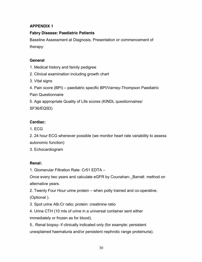
APPENDIX 1 Fabry Disease: Paediatric Patients Baseline Assessment at Diagnosis, Presentation or commencement of
therapy:
General 1. Medical history and family pedigree
2. Clinical examination including growth chart
3. Vital signs
4. Pain score (BPI) – paediatric specific BPI/Varney-Thompson Paediatric
Pain Questionnaire
5. Age appropriate Quality of Life scores (KINDL questionnaires/
SF36/EQ5D)
Cardiac: 1. ECG
2. 24 hour ECG whenever possible (we monitor heart rate variability to assess
autonomic function)
3. Echocardiogram
Renal: 1. Glomerular Filtration Rate: Cr51 EDTA –
Once every two years and calculate eGFR by Counahan-_Barratt method on
alternative years.
2. Twenty Four Hour urine protein – when potty trained and co-operative.
(Optional ).
3. Spot urine Alb:Cr ratio; protein: creatinine ratio
4. Urine CTH (10 mls of urine in a universal container sent either
immediately or frozen as for blood).
5.. Renal biopsy- if clinically indicated only (for example: persistent
unexplained haematuria and/or persistent nephrotic range proteinuria).
31
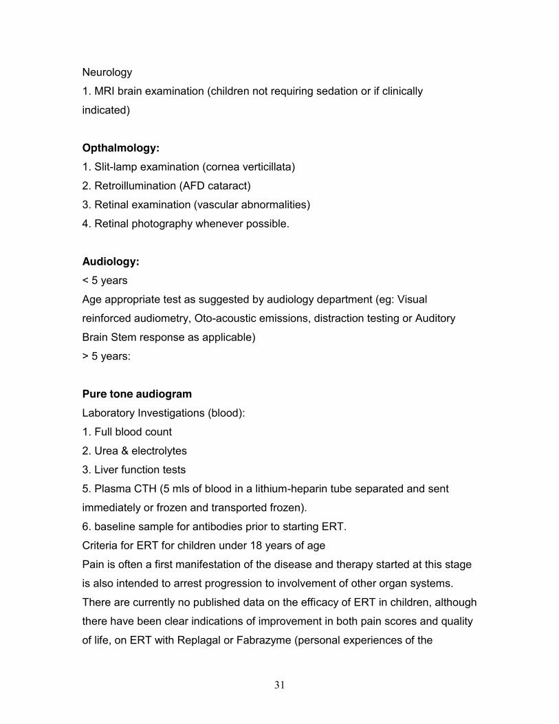
Neurology
1. MRI brain examination (children not requiring sedation or if clinically
indicated)
Opthalmology: 1. Slit-lamp examination (cornea verticillata)
2. Retroillumination (AFD cataract)
3. Retinal examination (vascular abnormalities)
4. Retinal photography whenever possible.
Audiology: < 5 years
Age appropriate test as suggested by audiology department (eg: Visual
reinforced audiometry, Oto-acoustic emissions, distraction testing or Auditory
Brain Stem response as applicable)
> 5 years:
Pure tone audiogram Laboratory Investigations (blood):
1. Full blood count
2. Urea & electrolytes
3. Liver function tests
5. Plasma CTH (5 mls of blood in a lithium-heparin tube separated and sent
immediately or frozen and transported frozen).
6. baseline sample for antibodies prior to starting ERT.
Criteria for ERT for children under 18 years of age
Pain is often a first manifestation of the disease and therapy started at this stage
is also intended to arrest progression to involvement of other organ systems.
There are currently no published data on the efficacy of ERT in children, although
there have been clear indications of improvement in both pain scores and quality
of life, on ERT with Replagal or Fabrazyme (personal experiences of the
32
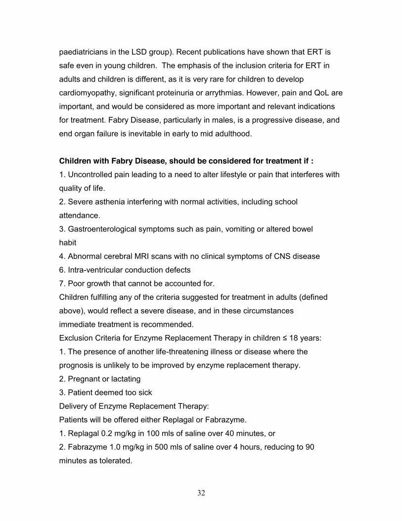
paediatricians in the LSD group). Recent publications have shown that ERT is
safe even in young children. The emphasis of the inclusion criteria for ERT in
adults and children is different, as it is very rare for children to develop
cardiomyopathy, significant proteinuria or arrythmias. However, pain and QoL are
important, and would be considered as more important and relevant indications
for treatment. Fabry Disease, particularly in males, is a progressive disease, and
end organ failure is inevitable in early to mid adulthood.
Children with Fabry Disease, should be considered for treatment if : 1. Uncontrolled pain leading to a need to alter lifestyle or pain that interferes with
quality of life.
2. Severe asthenia interfering with normal activities, including school
attendance.
3. Gastroenterological symptoms such as pain, vomiting or altered bowel
habit
4. Abnormal cerebral MRI scans with no clinical symptoms of CNS disease
6. Intra-ventricular conduction defects
7. Poor growth that cannot be accounted for.
Children fulfilling any of the criteria suggested for treatment in adults (defined
above), would reflect a severe disease, and in these circumstances
immediate treatment is recommended.
Exclusion Criteria for Enzyme Replacement Therapy in children ≤ 18 years:
1. The presence of another life-threatening illness or disease where the
prognosis is unlikely to be improved by enzyme replacement therapy.
2. Pregnant or lactating
3. Patient deemed too sick
Delivery of Enzyme Replacement Therapy:
Patients will be offered either Replagal or Fabrazyme.
1. Replagal 0.2 mg/kg in 100 mls of saline over 40 minutes, or
2. Fabrazyme 1.0 mg/kg in 500 mls of saline over 4 hours, reducing to 90
minutes as tolerated.
33
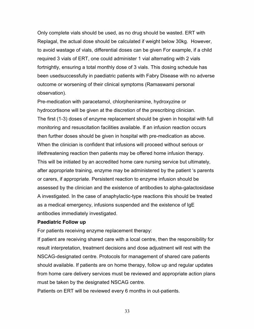
Only complete vials should be used, as no drug should be wasted. ERT with
Replagal, the actual dose should be calculated if weight below 30kg. However,
to avoid wastage of vials, differential doses can be given For example, if a child
required 3 vials of ERT, one could administer 1 vial alternating with 2 vials
fortnightly, ensuring a total monthly dose of 3 vials. This dosing schedule has
been usedsuccessfully in paediatric patients with Fabry Disease with no adverse
outcome or worsening of their clinical symptoms (Ramaswami personal
observation).
Pre-medication with paracetamol, chlorpheniramine, hydroxyzine or
hydrocortisone will be given at the discretion of the prescribing clinician.
The first (1-3) doses of enzyme replacement should be given in hospital with full
monitoring and resuscitation facilities available. If an infusion reaction occurs
then further doses should be given in hospital with pre-medication as above.
When the clinician is confident that infusions will proceed without serious or
lifethreatening reaction then patients may be offered home infusion therapy.
This will be initiated by an accredited home care nursing service but ultimately,
after appropriate training, enzyme may be administered by the patient ‘s parents
or carers, if appropriate. Persistent reaction to enzyme infusion should be
assessed by the clinician and the existence of antibodies to alpha-galactosidase
A investigated. In the case of anaphylactic-type reactions this should be treated
as a medical emergency, infusions suspended and the existence of IgE
antibodies immediately investigated.
Paediatric Follow up For patients receiving enzyme replacement therapy:
If patient are receiving shared care with a local centre, then the responsibility for
result interpretation, treatment decisions and dose adjustment will rest with the
NSCAG-designated centre. Protocols for management of shared care patients
should available. If patients are on home therapy, follow up and regular updates
from home care delivery services must be reviewed and appropriate action plans
must be taken by the designated NSCAG centre.
Patients on ERT will be reviewed every 6 months in out-patients.
34
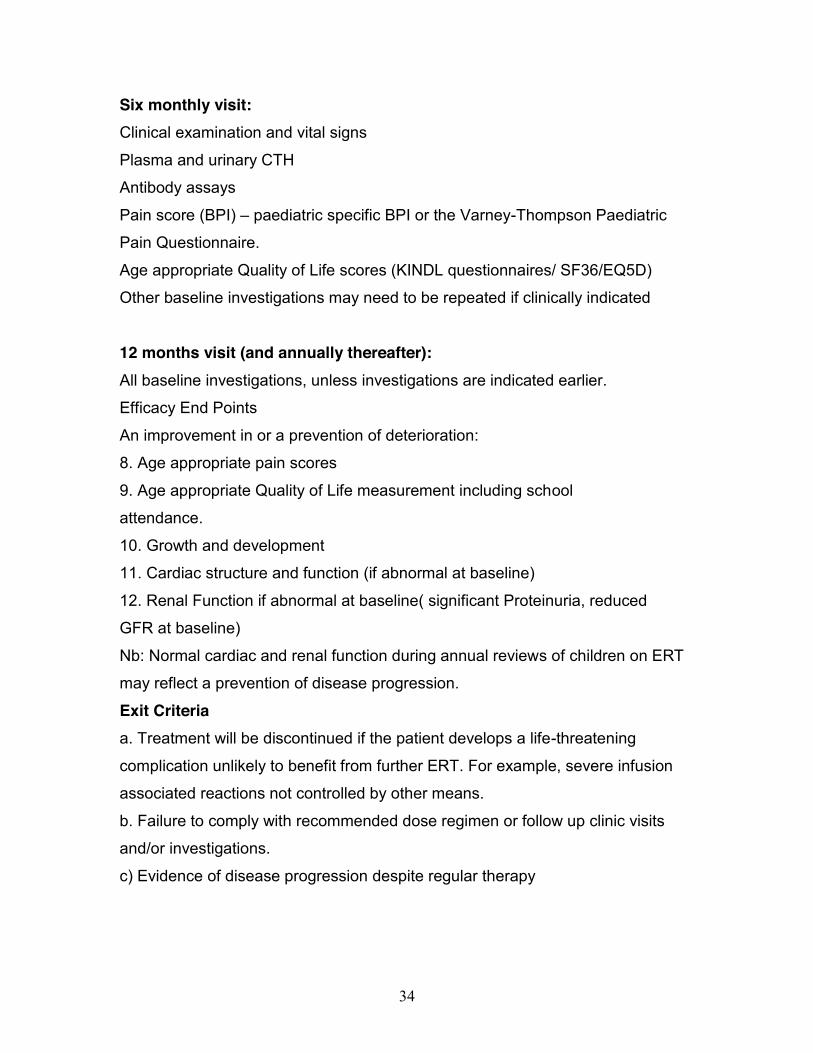
Six monthly visit: Clinical examination and vital signs
Plasma and urinary CTH
Antibody assays
Pain score (BPI) – paediatric specific BPI or the Varney-Thompson Paediatric
Pain Questionnaire.
Age appropriate Quality of Life scores (KINDL questionnaires/ SF36/EQ5D)
Other baseline investigations may need to be repeated if clinically indicated
12 months visit (and annually thereafter): All baseline investigations, unless investigations are indicated earlier.
Efficacy End Points
An improvement in or a prevention of deterioration:
8. Age appropriate pain scores
9. Age appropriate Quality of Life measurement including school
attendance.
10. Growth and development
11. Cardiac structure and function (if abnormal at baseline)
12. Renal Function if abnormal at baseline( significant Proteinuria, reduced
GFR at baseline)
Nb: Normal cardiac and renal function during annual reviews of children on ERT
may reflect a prevention of disease progression.
Exit Criteria a. Treatment will be discontinued if the patient develops a life-threatening
complication unlikely to benefit from further ERT. For example, severe infusion
associated reactions not controlled by other means.
b. Failure to comply with recommended dose regimen or follow up clinic visits
and/or investigations.
c) Evidence of disease progression despite regular therapy

![Operating Procedures 1 G2 - OPERATING PROCEDURES [6 Exam Questions - 6 Groups] G2APhone operating procedures; USB/LSB utilization conventions; procedural](https://img.pdfslide.us/doc/110x75/56649e4d5503460f94b4351a/operating-procedures-1-g2-operating-procedures-6-exam-questions-6-groups.jpg)