Embed Size (px)
Citation preview
e50 • CID 2010:50 (15 April) • BRIEF REPORT
B R I E F R E P O R T
Acute Necrotizing EncephalopathyProgressing to Brain Deathin a Pediatric Patient with NovelInfluenza A (H1N1) Infection
Alexandra Martin1 and Erin Parrish Reade2
1Pediatric Residency Program and 2Division of Critical Care Medicine,Department of Pediatrics, University of Tennessee Collegeof Medicine–Chattanooga
We report a case of encephalopathy progressing to brain
death in a pediatric patient with confirmed infection with
novel influenza H1N1. Although neurologic dysfunction as-
sociated with H1N1 has been described, we believe this to
be the first published report of brain death associated with
H1N1 infection.
Neurologic complications of influenza have been well described
in the literature and date back to the diagnosis of encephalitis
lethargica during the 1918 influenza pandemic [1]. Neurologic
manifestations of influenza are now known to include enceph-
alitis, acute disseminated encephalomyelitis, Guillain-Barre syn-
drome, transverse myelitis, and acute necrotizing encephalop-
athy (ANE). Reports of ANE began surfacing from Japan during
the influenza epidemics of the mid- and late 1990s [2–6]. In-
fluenza-associated central nervous system (CNS) dysfunction
has also been reported, although infrequently, in Europe and
the United States [7–10]. Other infections associated with ANE
include human herpesvirus–6 infection, measles, parainfluenza
infection, and Mycoplasma infection [11].
According to studies from Japan, in the most severe cases
of influenza-associated ANE, patients develop altered mental
status with or without seizures and then rapidly progress to a
comatose state within a mean of 24–72 hours from the onset
of fever and upper respiratory symptoms [2, 3, 6]. Seizures are
often resistant to antiepileptic medications [4]. Death, which
occurs in roughly 30% of cases, results largely from cardiores-
Received 23 November 2009; accepted 12 January 2010; electronically published 10 March2010.
Reprints or correspondence: Dr Erin Parrish Reade, University of Tennessee College ofMedicine–Chattanooga, Dept of Pediatrics, Div of Critical Care Medicine, T. C. ThompsonChildren’s Hospital, 910 Blackford St, Chattanooga, TN 37403 ([email protected]).
Clinical Infectious Diseases 2010; 50:e50–e52� 2010 by the Infectious Diseases Society of America. All rights reserved.1058-4838/2010/5008-00E1$15.00DOI: 10.1086/651501
piratory compromise or complications from mechanical ven-
tilation [2].
To our knowledge, encephalopathy associated with the novel
H1N1 influenza strain was first reported in a case series of 4
pediatric patients in the United States in May 2009 [12]. All 4
patients had mild seizures and/or altered mental status, and all
recovered fully without any neurological sequelae at discharge.
Here, we report what we believe to be the first published case
of H1N1 encephalopathy progressing to brain death in a pe-
diatric patient in the United States.
Case presentation. A 7-year-old, previously healthy, pre-
sumably immunocompetent female of Mandarin Chinese de-
scent presented to a community emergency department with
a 1-day history of fever (temperature, 39.4�C) and malaise but
no upper respiratory symptoms. She had not been vaccinated
for seasonal influenza, and she had not received the H1N1
vaccine. She was diagnosed with influenza A by rapid screen
on nasal wash specimen and then discharged home with in-
structions for supportive care with oral fluids and ibuprofen.
Approximately 4 hours after discharge from the first emergency
department visit, she became confused, lethargic, unable to
walk, and incontinent of bladder and bowel function. Ap-
proximately 7 hours after onset of these new symptoms at
home, she presented again to the local emergency department.
She was febrile (temperature, 40.8�C), she was obtunded, and
she required emergent intubation for airway protection. A non-
contrast head computed tomography scan revealed diffuse ce-
rebral edema, infarcts of the basal ganglia bilaterally, and ef-
facement of the fourth ventricle (Figure 1a). At this point, she
was transferred to the pediatric intensive care unit at T. C.
Thompson Children’s Hospital in Chattanooga, Tennessee, for
further care.
Upon arrival at the pediatric intensive care unit, she had a
Glasgow Coma Score of 6T and an exam consistent with se-
vere neurologic compromise. In the absence of neuromuscular
blockade, she displayed no spontaneous movement and had
minimal withdrawal to pain. Cough, gag, and corneal reflexes
were all absent. Her pupils were 5 mm and nonreactive to light.
She also demonstrated myoclonus. The family denied any toxic
ingestion and denied use of salicylates. The only medication
given at home was ibuprofen.
Initial laboratory abnormalities included aspartate amino-
transferase level of 103, alanine aminotransferase level of 61,
prothrombin time of 17.2, and international normalized ratio
of 1.34. Her initial complete blood count and serum electrolytes
were all normal.
BRIEF REPORT • CID 2010:50 (15 April) • e51
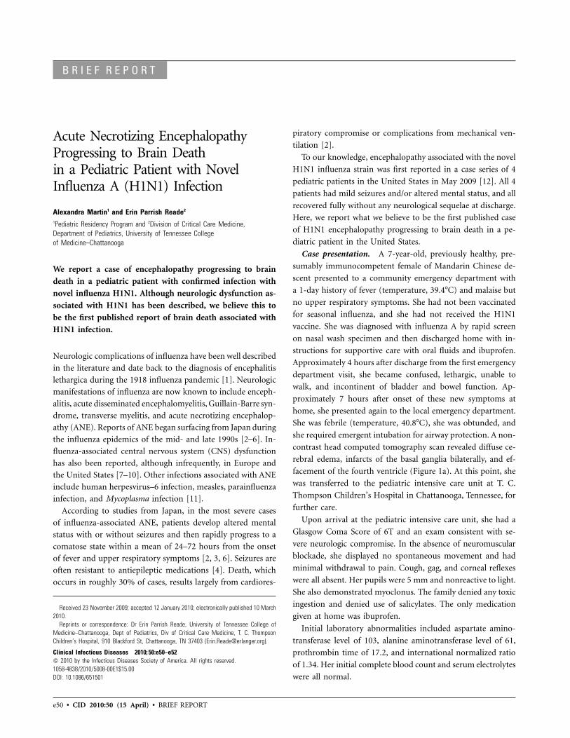
Figure 1. a, Noncontrast head computed tomography scan reveals extensive hypodensity in the white matter as well as the basal ganglia andthalami consistent with global insult. In addition, there is generalized cerebral edema. b, Magnetic resonance imaging of the brain (with fluid attenuationinversion recovery) demonstrates extensive and diffuse white matter and subcortical white matter signal abnormality that includes the bilateral thalamias well as increasing cerebral edema.
Initial management consisted of temperature control with a
cooling blanket and rectal acetaminophen, elevation of the head
of bed to 40�, mild hyperventilation, hypertonic saline, and
mannitol because of signs of increased intracranial pressure.
She was also empirically treated for bacterial meningitis with
vancomycin and ceftriaxone as well as oseltamavir for influ-
enza. Approximately 4 hours after admission, she demon-
strated bradycardia and hypertension that were treated with
boluses of mannitol and hypertonic saline. These signs of her-
niation syndrome were followed by severe hypotension that
necessitated the use of fluid boluses and multiple vasopressive
agents to maintain blood pressure. Neurosurgical consultation
was obtained to determine if intracranial pressure monitoring
and/or decompressive craniectomy were treatment options in
this patient. The patient’s parents refused consent for lumbar
puncture.
Because of concern for stroke, magnetic resonance imaging
(MRI) and magnetic resonance angiography of the brain were
performed (Figure 1b). It was determined that she was not a
candidate for intracranial pressure monitoring or decompres-
sive craniectomy because of brainstem herniation. She quickly
developed diabetes insipidus that required vasopressin infu-
sion. On hospital day 3, she was pronounced brain dead fol-
lowing 2 brain death examinations performed by different
physicians 24 hours apart, a nuclear medicine cerebral blood
flow study that showed no cerebral perfusion, and an electro-
encephalograph consistent with electrical silence. Autopsy was
refused by the family. By the time of death, we received notice
that a polymerase chain reaction (PCR) assay performed by the
Tennessee state laboratory was positive for H1N1 influenza
infection.
Discussion. To our knowledge, this is the first reported case
of novel influenza A (H1N1)-associated ANE resulting in brain
death in the United States. First described in Japan in 1979,
ANE was formally characterized in 1995 and is defined as acute
encephalopathy following a viral febrile illness with rapid de-
terioration of level of consciousness; multifocal, symmetric le-
sions seen on computed tomography or MRI in the thalamus,
cerebral and cerebellar medullae, and brainstem; no cerebro-
spinal fluid pleocytosis; elevation of serum aminotransferases
of variable degrees; and exclusion of resembling diseases [11].
The clinical course of ANE is rapidly progressive; patients pre-
sent with fever and nonspecific symptoms, such as cough, em-
esis, and/or diarrhea, and quickly develop neurologic dysfunc-
tion. Approximately 18% of cases of ANE in Japan have been
associated with influenza A infection [3]. The strain most fre-
quently associated with ANE is influenza A, H3N2 subtype,
although cases associated with H1N1 and influenza B have also
been described [2, 7, 13–15]. The disease is associated with
significant morbidity and mortality, and survivors usually ex-
hibit at least short-term neurologic sequelae [6]. In addition
to antiviral therapies such as oseltamavir, corticosteroids and
intravenous immune globulin have been used to treat selected
cases of ANE in Japan, with varying degrees of patient im-

e52 • CID 2010:50 (15 April) • BRIEF REPORT
provement [16]. Currently, there is no definitive treatment for
ANE, and management of these patients centers upon sup-
portive care for neurologic failure and treatment of increased
intracranial pressure if present.
At present, it is unknown whether influenza virus physically
enters the CNS or whether neurological dysfunction is secondary
to other effects. On postmortem pathological examination, some
authors have found evidence of petechial hemorrhages, conges-
tion of intraparenchymal thalamic vessels, microthrombi, and
vasogenic edema, suggesting that CNS dysfunction may result
from vascular damage without evidence of direct penetration
across the blood brain barrier [3]. Other authors describe high
plasma concentrations of interleukin 6 and tumor necrosis fac-
tor–a, suggesting that proinflammatory cytokines may be me-
diators of damage [1], although not all reports show elevation
in inflammatory markers [4]. Very rarely do authors report direct
evidence of influenza virus in the CNS [17]. In a survey of 94
Japanese hospitals over 9 influenza seasons, Togashi et al [2]
reported that only 10% of cases had PCR detection of influenza
in the cerebrospinal fluid. Fujimoto et al [18] showed PCR de-
tection in 5 of 10 ANE patients.
It is unclear whether there are anatomical or biochemical
differences that allow the virus entry into the CNS or that lead
to remote effects on the brain or whether there are mutations
in certain strains of the influenza virus that confer more neu-
rotropic properties. Because the symptoms have such rapid onset
and viral antigens have been found so rarely in brain tissue and
cerebrospinal fluid, most authors have suggested that a cytokine-
mediated process, rather than direct invasion of the CNS, is the
pathophysiologic mechanism in this disease.
It appears that CNS sequelae of influenza occur with dis-
proportionate frequency in patients of Asian descent, despite
the fact that influenza itself is a very common winter febrile
illness in all developed countries. Some authors suggest that
the increased use of certain drugs, namely diclofenac sodium,
mephenamate, and ephedrine, in Japan may play a role [4].
Per history, the patient described here had no exposure to
diclofenac sodium, mephenamate, or ephedrine, although she
had taken ibuprofen, another nonsteroidal anti-inflammatory
drug.
During the time of this patient’s presentation, the H1N1
pandemic was nearing its peak in the southeastern United
States. Our institution treated a large number of children with
confirmed H1N1 infection during this period, most of whom
recovered fully. The patient we focus on in this report, although
presumably exposed to the same strain of virus circulating in
the community, was the only one with very rapid and fatal
CNS dysfunction and happened to be of Asian descent. There-
fore, it is unclear whether her Asian heritage was related to her
illness.
In summary, we describe a pediatric patient with docu-
mented novel influenza A (H1N1) infection who presented with
the well-described features of ANE and progressed to brain
death. Although influenza is a relatively benign illness in the
majority of healthy children, physicians who care for children
in the United States should be aware of ANE and should main-
tain a high degree of clinical suspicion in any child presenting
with acute mental status changes in the setting of influenza
infection.
Acknowledgments
Potential conflicts of interest. A.M. and E.P.R.: no conflicts.
References
1. Vilensky JA, Foley P, Gilman S. Children and encephalitis lethargica:a historical review. Pediatr Neurol 2007; 37:79–84.
2. Togashi T, Matsuzono Y, Narita M, Morishima T. Influenza-associatedacute encephalopathy in Japanese children in 1994–2002. Virus Res2004; 103:75–78.
3. Sugaya N. Influenza-associated encephalopathy in Japan. Semin PediatrInfect Dis 2002; 13(2):79–84.
4. Shinjoh M, Bamba M, Jozaki K, Takahashi E, Koinuma G, Sugaya N.Influenza A–associated encephalopathy with bilateral thalamic necrosisin Japan. Clin Infect Dis 2000; 31:611–613.
5. Fujimoto Y, Motohiro S, Tsuyuki M, Okada M, Tsuzuki K. InfluenzaA virus encephalopathy with symmetric thalamic lesions. Eur J Pediatr2000; 159:319–321.
6. Mizuguchi M, Abe J, Mikkaichi K, et al. Acute necrotizing encepha-lopathy of childhood: a new syndrome presenting with multifocal, sym-metric brain lesions. J Neurol Neurosurg Psychiatry 1995; 58:555–561.
7. Soldo I, Duvnjak M, Lisnjic D, et al. Encephalitis or encephalopathyduring an influenza-A epidemic. Coll Antropol 2003; 27(1):19–22.
8. Smidt MH, Stroink H, Bruinenberg JF, Peeters M. Encephalopathyassociated with influenza A. Eur J Paediatr Neurol 2004; 8:257–260.
9. Steininger C, Popow-Kraupp T, Laferl H, et al. Acute encephalopathyassociated with influenza A virus infection. Clin Infect Dis 2003; 36:567–574.
10. Weitkamp JH, Spring MD, Brogan T, Moses H, Block KC, Wright PF.Influenza A virus–associated acute necrotizing encephalopathy in theUnited States. Pediatr Infect Dis J 2004; 23:259–263.
11. Mizuguchi M. Acute necrotizing encephalopathy of childhood: a novelform of acute encephalopathy prevalent in Japan and Taiwan. BrainDev 1997; 19:81–92.
12. Centers for Disease Control and Prevention. Neurologic complicationsassociated with novel influenza A (H1N1) virus infection in children—Dallas, Texas, May 2009. MMWR Morb Mortal Wkly Rep 2009; 58:773–778.
13. Long SS. Emerging infections in children: influenza and acute nec-rotizing encephalopathy. Adv Exp Med Biol 2005; 568:1–9.
14. Grose C. The puzzling picture of acute necrotizing encephalopathyafter influenza A and B virus infection in young children. Pediatr InfectDis J 2004; 23:253–254.
15. Maricich SM, Neul JL, Lotze TE, et al. Neurologic complications as-sociated with influenza A in children during the 2003–2004 influenzaseason in Houston, Texas. Pediatrics 2004; 114(5):e626–e633.
16. Okumura A, Mizuguchi M, Kidokoro H, et al. Outcome of acutenecrotizing encephalopathy in relation to treatment with corticoste-roids and gammaglobulin. Brain Dev 2009; 31: 221–227.
17. Toovey S. Influenza-associated central nervous system dysfunction: aliterature review. Travel Med Infect Dis 2008; 6:114–124.
18. Fujimoto S, Kobayashi M, Uemura O, et al. PCR on cerebrospinalfluid to show influenza-associated acute encephalopathy or encepha-litis. Lancet 1998; 352:873–875.