1
ACTIVATION OF MATRIPTASE-2 IN HEK CELLS IS A TRANS-MECHANISM Eva Maurer, Marit Stirnberg and Michael Gütschow University of Bonn, Pharmaceutical Institute, Pharmaceutical Chemistry I, An der Immenburg 4, 53121 Bonn [email protected] 1. Introduction 2. Overexpression of matriptase-2 in HEK cells Figure 2: Subcellular localization of matriptase-2. Immunofluorescence detection of matriptase-2 in transfected HEK cells using monoclonal mouse anti-c-Myc antibody (red). Nuclei were stained with DAPI (blue; 10 ng per ml). The white scale bar represents 10 μm. HEK cells were stably transfected with cDNA encoding human matriptase-2 (HEK-MT2 WT ) and the inactive variant of matriptase-2 with the mutated catalytic triad Ser 753 (HEK-MT2 S753A ). Both constructs contain a C-terminal c-Myc and His 6 tag. Immunofluorescence analysis of permeabilized and non- permeabilized cells expressing MT2 S753A revealed the intracellular (Fig. 2A) and membrane (Fig. 2B) localization. A HEK-MT2 S753A , permeabilized C HEK-mock, permeabilized B HEK-MT2 S753A , non-permeabilized D HEK-mock, non-permeabilized 3. Identification of matriptase-2 forms in membranes and conditioned medium Figure 3: Immunoblot analysis of matriptase-2 in transfected HEK-MT2 WT cells. A, B. 10 μg total protein of conditioned medium (CM), membrane fractions (MF) and cytosolic fractions (CF) of HEK- MT2 WT and HEK-mock cells were separated under reducing (A) and non-reducing conditions (B). Matriptase-2 protein was visualized using monoclonal mouse anti-c-Myc antibody. M, molecular mass marker. Matriptase-2 was visible in membranes of HEK cells expressing MT2 WT under reducing (Fig. 3A) and non-reducing conditions (Fig. 3B) as a single 120 kDa band. An additional >170 kDa signal under non-reducing conditions indicated that matriptase-2 forms homo- or hetero-meric complexes, tethered by disulfide bonds. In the conditioned medium of HEK-MT2 WT cells the catalytic domain of 30 kDa was detected under reducing, but a 55 kDa band under non-reducing conditions. Thus, matriptase-2 is shed as an active two-chain protein in which the catalytic domain is disulfide-linked to a part of the stem region. Matriptase-2 is a recently identified member of the Type II Transmembrane Serine Proteases (TTSPs) [1]. Activation occurs via a proteolytic cleavage of the single-chain zymogen to the two-chain, disulfide-connected form. Mutations are correlated to iron-refractory iron deficiency anemia (IRIDA) [2, 3] and a lack of matriptase-2 has been linked to the inability to suppress the expression of hepcidin, the key regulator of iron homeostasis [4]. Matriptase-2 inhibition appears to be a novel therapeutic approach for the treatment of hemochromatosis [5]. In this study, we characterized the activation of matriptase-2 [6]. Figure 1: Role of matriptase-2 in iron homeostasis. Matriptase-2 suppresses hepcidin expression through proteolytic processing of hemojuvelin (m-HJV). This process results in the loss of m-HJV to act as a coreceptor for bone morphogenetic proteins (BMPs). BMPs signal through SMAD proteins and this leads to the expression of the hepcidin gene HAMP. 6. Future prospects Future studies are intended to characterize the proteolytic processing of matriptase-2 in hepatocytes. Such experiments should include the identification of possible interaction partners in hetero-meric membrane complexes of matriptase- 2. In additional investigations, the physiological role of matriptase-2 in iron homeostasis will be further explored, in particular the transcriptional and posttranscriptional regulation of matriptase-2 in hepatocytes. 5. Activation occurs via a trans-mechanism 4. Autocatalytic activation of matriptase-2 References and Acknowledgement 1. Velasco, G., Cal, S., Quesada, V., Sánchez, L. M. and López-Otín, C. (2002) Matriptase-2, a membrane-bound mosaic serine proteinase predominantly expressed in human liver and showing degrading activity against extracellular matrix proteins. J. Biol. Chem. 277, 37637-37646 2. Finberg, K. E., Heeney, M. M., Campagna, D. R, Aydinok, Y., Pearson, H. A., Hartman, K. R., Mayo, M. M., Samuel S. M., Strouse, J. J., Markianos, K., Andrews, N. C. and Fleming, M. D. (2008) Mutations in TMPRSS6 cause iron-refractory iron deficiency anemia (IRIDA). Nat. Genet. 40, 569- 571 3. Du, X., She, E., Gelbart, T., Truksa, J., Lee, P., Xia, Y., Khovananth, K., Mudd, S., Mann, N., Moresco, E. M., Beutler, E and Beutler, B. (2008) The serine protease TMPRSS6 is required to sense iron deficiency. Science 320, 1088-1 4. Silvestri, L., Pagani, A., Nai, A., De Domenico, I., Kaplan, J. and Camaschella, C. (2008) The serine protease matriptase-2 (TMPRSS6) inhibits hepcidin activation by cleaving membrane hemojuvelin. Cell Metabol. 8, 502–511 5. Sisay, M.T., Steinmetzer, T., Stirnberg, M., Maurer, E., Hammami, M., Bajorath, J., Gütschow, M. (2010) Identification of the first low-molecular-weight inhibitors of matriptase-2. J. Med. Chem. 53,5523-35 6. Stirnberg, M. Maurer, E., Horstmeyer, A., Kolb, S., Frank, S., Bald, T., Arenz, K., Janzer, A., Prager, K., Wunderlich, P., Walter, J., Gütschow, M. (2010) The proteolytic processing of the serine protease matriptase-2: identification of the cleavage sites required for its autocatalytic release from the cell surface. Biochem. J. 430, 87–95 This work was supported by Deutsche Forschungsgemeinschaft [grant number SFB645]; and by the North Rhine-Westphalia International Graduate Research School Biotech-Pharma.SFB, GRK 804, Support by Prof. Jochen Walter and Katharina Arenz, University of Bonn, is acknowledged. E.M. gratefully acknowledges receipt of a fellowship from Bayer Schering Pharma AG. A B HEK-mock HEK-MT2 WT HEK-MT2 WT HEK-mock M 130 -- 95 -- 72 -- 55 -- 34 -- 26 -- kDa A CM MF CF HEK-mock HEK-MT2 WT CM MF CF CM MF CF HEK-MT2 S753A 130 -- 95 -- 72 -- 55 -- 34 -- 26 -- kDa B M CM MF CF CM MF CF CM MF CF HEK-mock HEK-MT2 WT HEK-MT2 S753A Figure 4: Immunoblot analysis of catalytically inactive matriptase-2 in transfected HEK-MT2 S753A cells. A, B. 10 μg total protein of conditioned medium (CM), membrane fractions (MF) and cytosolic fractions (CF) of HEK-mock, HEK-MT2 WT and HEK-MT2 S753A cells were separated under reducing (A) and non-reducing conditions (B). Matriptase-2 protein was visualized using monoclonal mouse anti-c-Myc antibody. M, molecular mass marker. In order to investigate the requirement of matriptase-2 activity for activation cleavage, HEK-MT2 S753A cells were examined. Immunoblot analysis revealed a 120 kDa fragment in membrane fractions under reducing (Fig. 4A) and non- reducing conditions (Fig. 4B) as observed for HEK-MT2 WT cells. No derivatives of the mutated S753A variant were visible in the conditioned medium indicating that matriptase-2 undergoes an autocatalytic activation. Figure 5: Immunoblot analysis of co-transfected MT2 WT -His/MT2 S753A - Myc cells. A, B. 10 μg total protein of conditioned medium (CM) and membrane fractions (MF) of MT2 WT -Myc-His, MT2 WT -His, MT2 S753A -Myc and co-transfected MT2 WT -His/MT2 S753A -Myc cells were separated under reducing conditions. Matriptase-2 protein was visualized using monoclonal mouse anti-c-Myc antibody (A) or anti-His antibody (B). M, molecular mass marker. To analyze whether the autoactivation process is catalyzed by the same (cis-) or by another (trans-mechanism) matriptase-2 molecule, HEK cells were co-transfected with two vectors (Fig. 5A). No fragments were detectable in the conditioned medium of single transfected MT2 S753A -Myc cells. However, a weak signal was visualized in the conditioned medium of co-transfected MT2 WT -His/MT2 S753A -Myc cells using the anti-c-Myc antibody. Thus, activation cleavage of the inactive S753A mutant form was catalyzed by wild type matriptase-2 suggesting the trans-mechanism. BMP receptor matriptase-2 m-HJV BMP SMADs HAMP iron plasma level from Silvestri et al., Cell Metab. (2008), modified

ACTIVATION OF MATRIPTASE-2 IN HEK CELLS IS A TRANS …

  • Upload
    others

  • View
    1

  • Download
    0

Embed Size (px)

Citation preview

ACTIVATION OF MATRIPTASE-2 IN HEK CELLS

IS A TRANS-MECHANISM

Eva Maurer, Marit Stirnberg and Michael GütschowUniversity of Bonn, Pharmaceutical Institute, Pharmaceutical Chemistry I, An der Immenburg 4, 53121 Bonn

[email protected]

1. Introduction

2. Overexpression of matriptase-2 in HEK cells

Figure 2: Subcellular localization of matriptase-2.

Immunofluorescence detection of matriptase-2 in transfected HEK cells using monoclonal mouse anti-c-Myc antibody (red). Nuclei were stained with DAPI (blue; 10 ng per ml). The white scale bar represents 10 µm.

HEK cells were stably transfected with cDNA encoding human matriptase-2 (HEK-MT2WT) and the inactive variant of matriptase-2 with the mutated catalytic triad Ser753

(HEK-MT2S753A). Both constructs contain a C-terminal c-Myc and His6

tag. Immunofluorescence analysis of permeabilized and non-permeabilized cells expressing MT2S753A revealed the intracellular (Fig. 2A) and membrane (Fig. 2B) localization.

A HEK-MT2S753A, permeabilized

C HEK-mock, permeabilized

B HEK-MT2S753A, non-permeabilized

D HEK-mock, non-permeabilized

3. Identification of matriptase-2 forms in

membranes and conditioned medium

Figure 3: Immunoblot

analysis of matriptase-2 in

transfected HEK-MT2WT cells.

A, B. 10 µg total protein of conditioned medium (CM),

membrane fractions (MF) and cytosolic fractions (CF) of HEK-MT2WT and HEK-mock cells were separated under reducing (A) and non-reducing

conditions (B). Matriptase-2 protein was visualized using monoclonal mouse anti-c-Mycantibody. M, molecular mass marker.

Matriptase-2 was visible in membranes of HEK cells expressing MT2WT under reducing (Fig. 3A) and non-reducing conditions (Fig. 3B) as a single 120 kDa band. An additional >170 kDa signal under non-reducing conditions indicated that matriptase-2 forms homo- or hetero-meric complexes, tethered by disulfide bonds. In the conditioned medium of HEK-MT2WT cells the catalytic domain of 30 kDa was detected under reducing, but a 55 kDa band under non-reducing conditions. Thus, matriptase-2 is shed as an active two-chain protein in which the catalytic domain is disulfide-linked to a part of the stem region.

Matriptase-2 is a recently identified member of the Type II Transmembrane Serine Proteases (TTSPs) [1]. Activation occurs via a proteolytic cleavage of the single-chain zymogen to the two-chain, disulfide-connected form. Mutations are correlated to iron-refractory iron deficiency anemia (IRIDA) [2, 3] and a lack of matriptase-2 has been linked to the inability to suppress the expression of hepcidin, the key regulator of iron homeostasis [4]. Matriptase-2 inhibition appears to be a novel therapeutic approach for the treatment of hemochromatosis [5]. In thisstudy, we characterized the activation of matriptase-2 [6].

Figure 1: Role of matriptase-2 in iron homeostasis. Matriptase-2 suppresses hepcidin expression through proteolytic processing of hemojuvelin (m-HJV). This process results in the loss of m-HJV to act as a coreceptor for bone morphogenetic proteins (BMPs). BMPs signal through SMAD proteins and this leads to the expression of the hepcidin gene HAMP.

6. Future prospects

Future studies are intended to characterize the proteolytic processing of matriptase-2 in hepatocytes. Such experiments should include the identification of possible interaction partners in hetero-meric membrane complexes of matriptase-2. In additional investigations, the physiological role of matriptase-2 in iron homeostasis will be further explored, in particular the transcriptional and posttranscriptional regulation of matriptase-2 in hepatocytes.

5. Activation occurs via a trans-mechanism

4. Autocatalytic activation of matriptase-2

References and Acknowledgement

1. Velasco, G., Cal, S., Quesada, V., Sánchez, L. M. and López-Otín, C. (2002) Matriptase-2, a membrane-bound mosaic serine proteinase predominantly expressed in human liver and showing degrading activity against extracellular matrix proteins. J. Biol. Chem. 277, 37637-376462. Finberg, K. E., Heeney, M. M., Campagna, D. R, Aydinok, Y., Pearson, H. A., Hartman, K. R., Mayo, M. M., Samuel S. M., Strouse, J. J., Markianos, K., Andrews, N. C. and Fleming, M. D. (2008) Mutations in TMPRSS6 cause iron-refractory iron deficiency anemia (IRIDA). Nat. Genet. 40, 569-

5713. Du, X., She, E., Gelbart, T., Truksa, J., Lee, P., Xia, Y., Khovananth, K., Mudd, S., Mann, N., Moresco, E. M., Beutler, E and Beutler, B. (2008) The serine protease TMPRSS6 is required to sense iron deficiency. Science 320, 1088-14. Silvestri, L., Pagani, A., Nai, A., De Domenico, I., Kaplan, J. and Camaschella, C. (2008) The serine protease matriptase-2 (TMPRSS6) inhibits hepcidin activation by cleaving membrane hemojuvelin. Cell Metabol. 8, 502–5115. Sisay, M.T., Steinmetzer, T., Stirnberg, M., Maurer, E., Hammami, M., Bajorath, J., Gütschow, M. (2010) Identification of the first low-molecular-weight inhibitors of matriptase-2. J. Med. Chem. 53,5523-356. Stirnberg, M. Maurer, E., Horstmeyer, A., Kolb, S., Frank, S., Bald, T., Arenz, K., Janzer, A., Prager, K., Wunderlich, P., Walter, J., Gütschow, M. (2010) The proteolytic processing of the serine protease matriptase-2: identification of the cleavage sites required for its autocatalytic release from the

cell surface. Biochem. J. 430, 87–95

This work was supported by Deutsche Forschungsgemeinschaft [grant number SFB645]; and by the North Rhine-Westphalia International Graduate Research School Biotech-Pharma.SFB, GRK 804, Support by Prof. Jochen Walter and Katharina Arenz, University of Bonn, is acknowledged.

E.M. gratefully acknowledges receipt of a fellowship from Bayer Schering Pharma AG.

A BHEK-mock HEK-MT2WT HEK-MT2WTHEK-mock

M

130 --

95 --

72 --

55 --

34 --

26 --

kDa

A

CM MF CF

HEK-mock HEK-MT2WT

CM MF CF CM MF CF

HEK-MT2S753A

130 --

95 --

72 --

55 --

34 --

26 --

kDa

B

M CM MF CF CM MF CF CM MF CF

HEK-mock HEK-MT2WT HEK-MT2S753A

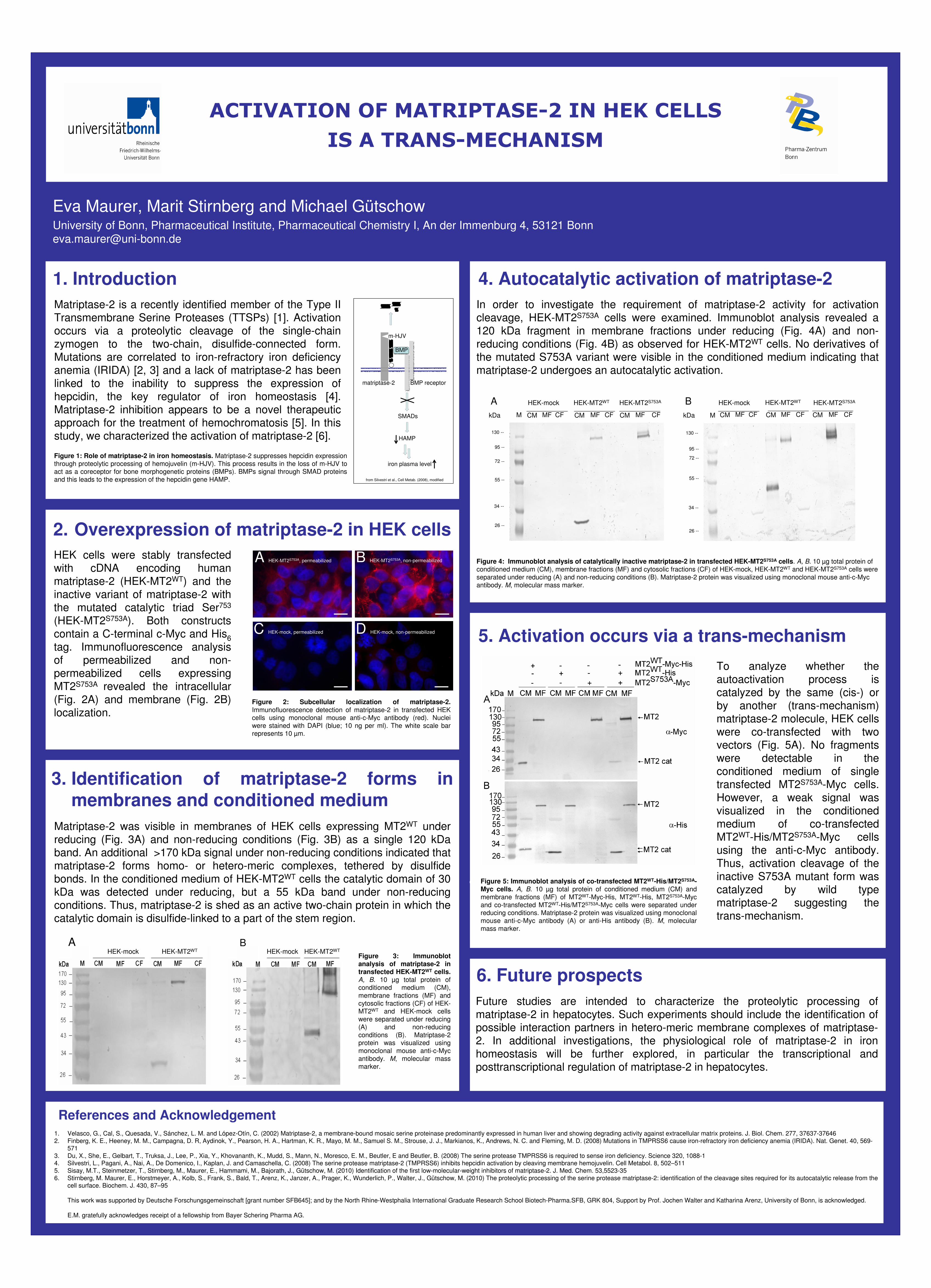
Figure 4: Immunoblot analysis of catalytically inactive matriptase-2 in transfected HEK-MT2S753A cells. A, B. 10 µg total protein of conditioned medium (CM), membrane fractions (MF) and cytosolic fractions (CF) of HEK-mock, HEK-MT2WT and HEK-MT2S753A cells were

separated under reducing (A) and non-reducing conditions (B). Matriptase-2 protein was visualized using monoclonal mouse anti-c-Mycantibody. M, molecular mass marker.

In order to investigate the requirement of matriptase-2 activity for activation cleavage, HEK-MT2S753A cells were examined. Immunoblot analysis revealed a 120 kDa fragment in membrane fractions under reducing (Fig. 4A) and non-reducing conditions (Fig. 4B) as observed for HEK-MT2WT cells. No derivatives of the mutated S753A variant were visible in the conditioned medium indicating thatmatriptase-2 undergoes an autocatalytic activation.

Figure 5: Immunoblot analysis of co-transfected MT2WT-His/MT2S753A-

Myc cells. A, B. 10 µg total protein of conditioned medium (CM) and

membrane fractions (MF) of MT2WT-Myc-His, MT2WT-His, MT2S753A-Myc and co-transfected MT2WT-His/MT2S753A-Myc cells were separated under reducing conditions. Matriptase-2 protein was visualized using monoclonal mouse anti-c-Myc antibody (A) or anti-His antibody (B). M, molecularmass marker.

To analyze whether the autoactivation process is catalyzed by the same (cis-) or by another (trans-mechanism) matriptase-2 molecule, HEK cells were co-transfected with two vectors (Fig. 5A). No fragments were detectable in the conditioned medium of single transfected MT2S753A-Myc cells. However, a weak signal was visualized in the conditioned medium of co-transfected MT2WT-His/MT2S753A-Myc cells using the anti-c-Myc antibody. Thus, activation cleavage of the inactive S753A mutant form was catalyzed by wild type matriptase-2 suggesting thetrans-mechanism.

BMP receptormatriptase-2

m-HJV

BMP

SMADs

HAMP

iron plasma level

from Silvestri et al., Cell Metab. (2008), modified