Embed Size (px)
Citation preview

Submitted 3 November 2017Accepted 15 February 2018Published 9 March 2018
Corresponding authorBorros M. Arneth,[email protected]
Academic editorGiuseppe Nocentini
Additional Information andDeclarations can be found onpage 18
DOI 10.7717/peerj.4462
Copyright2018 Arneth
Distributed underCreative Commons CC-BY 4.0
OPEN ACCESS
Activation of CD4 and CD8 T cellreceptors and regulatory T cells inresponse to human proteinsBorros M. ArnethInstitute of Laboratory Medicine and Pathobiochemistry, Molecular Diagnostics, University Hospital of theUniversities of Giessen and Marburg UKGM, Justus Liebig University Giessen, Giessen, Hessen, Germany
ABSTRACTThis study assessed in detail the influence of four different human proteins on theactivation of CD4+ and CD8+ T lymphocytes and on the formation of regulatory Tcells. Human whole-blood samples were incubated with four different human proteins.The effects of these proteins on the downstream immune-system response, on theexpression of extracellular activation markers on and intracellular cytokines in Tlymphocytes, and on the number of regulatory T cells (T-reg cells) were investigated viaflow cytometry. Incubation with β-actin or glyceraldehyde 3-phosphate dehydrogenase(GAPDH), which are cytoplasmic proteins, increased the expression of both extra-cellular activation markers (CD69 and HLA-DR) and intracellular cytokines but didnot significantly affect the number of T-reg cells. In contrast, incubation with humanalbumin or insulin, which are serum proteins, reduced both extracellular activationmarkers and intracellular cytokine expression and subsequently increased the numberof T-reg cells. These findings may help to explain the etiological basis of autoimmunediseases.
Subjects Allergy and Clinical Immunology, Immunology, Internal Medicine, Pathology,PediatricsKeywords Regulatory T cells, CD4+ T helper cells, Lymphocytes, CD8+ cytotoxic T cells,Interferon-gamma
INTRODUCTIONThe stability and plasticity of various T lymphocyte subgroups parallel their variousphysiological functions and suggest selection mechanisms within the context of T cellmaturation (Shah & Zúñiga-Pflücker, 2014). The various T helper subsets (i.e., Th1, Th2,and Th17) and regulatory T cells (T-reg cells) are interconvertible. Over time, T helper cellscan differentiate into other cell types of this group via the activation of specific transcriptionfactors. Indeed, the T cell receptor (TCR) spectrum of various T helper subsets is plastic,and such plasticity in terms of TCR spectra and of the antigens recognized results in varioussubgroups of T helper cells. Notably, the expression of CD4 and/or CD8 co-receptors—andthus differentiation into one of these two cell types—is fixed once it has occurred (Shah &Zúñiga-Pflücker, 2014).
The respective TCR spectra of T cells are fixed into a CD4+ TCR spectrum and/or aCD8+ TCR spectrum through stable differentiation into CD4+ and/or CD8+ T cells. Thisdifferentiation occurs at an extremely early stage during development through positive
How to cite this article Arneth (2018), Activation of CD4 and CD8 T cell receptors and regulatory T cells in response to human proteins.PeerJ 6:e4462; DOI 10.7717/peerj.4462
and negative selection of the respective T cells during thymic T cell development (Shah &Zúñiga-Pflücker, 2014). Although the plasticity of T helper cell subgroups allows increasedvariability in T cell populations later in life, fixation of CD4 and CD8 TCR spectra (Shah &Zúñiga-Pflücker, 2014) leads to permanent changes throughout the lifetime of an organism.
Several authors have described immunological memory as nothing more than ‘‘leftover’’cells from former immunizations (Zinkernagel et al., 1996; Zinkernagel, 2003; Zinkernagel& Hengartner, 2006). Therefore, improved knowledge of CD4+ and CD8+T cell activationcould lead to a detailed understanding of both immunological memory as a ‘‘leftovers’’ ofspecific activated CD4+ and CD8+T cells and the immune reactions that occur later in life.
Core idea/core hypothesisThe present study hypothesized that self-molecules can be recognized as foreign when atthe wrong location. To confirm or disprove this hypothesis, whole blood from healthysubjects was incubatedwith various human proteins, and the response regarding CD4+ andCD8+ T lymphocyte activation was examined.
To investigate the detailed effects of humanproteins onCD4+ andCD8+T lymphocytesand T-reg cells, whole-blood samples from healthy volunteers (N = 20) were incubatedwith four different human proteins (GAPDH, β-actin, insulin, and albumin).
These proteins were selected because they are prominent intracellular (actin andGAPDH) and extracellular (albumin, insulin), which were also purely available.
Then, the degree of the resulting T cell activation was quantified via flow-cytometricmeasurements of surface markers: CD69, which is an early T cell activation marker;HLA-DR, which is a late T cell activation marker, CD4, which is a T helper cell marker(CD4+ T cells); CD8, which is a cytotoxic T cell marker (CD8+ T cells); and CD3, whichis predominantly located on T cells. Intracellular cytokine expression levels were alsoexamined.
The development of autoimmune diseases and the role ofregulatory T cellsThe hypotheses accepted to date assumes that autoimmune diseases are acquired throughinnate susceptibility, including genetic predisposition, in combination with externalinfluences. Genetic predisposition is primarily because different individuals expressdifferent MHC molecule variants on the surfaces of their cells. Some MHC variantspresent more components that are similar to the body’s own structures, triggering anautoreactive immune response. Such genetic factors, in conjunction with stress-causingenvironmental factors such as viruses, other infections, and pregnancy, can result in anautoimmune disease. Another theory is the hygiene hypothesis, which holds that thedevelopment of immune diseases may be promoted by too little confrontation of theimmune system with bacteria in the environment. Overall, women are more often affectedby autoimmune diseases.
Furthermore, T cell selection during maturation in the thymus is considered important.Autoreactive T cells may dominate if they are not eliminated in the appropriate manner. Inaddition, cross-reactivity of bacterial and/or viral protein fragments and self-protein
Arneth (2018), PeerJ, DOI 10.7717/peerj.4462 2/22
fragments might be important in the development of autoimmune diseases. Lastly,regulatory T cells (T-reg cells) are important in controlling effector T cells and preventingthe development of autoimmune diseases. The mechanisms by which T-reg cells controleffector T cells are complex.
METHODSEthics approval and consent to participateThe institutional review board of the University of Mainz (Mainz, Germany) approvedthis research (ukm-2005-2742834). All participants gave written informed consent toparticipate in the study, and the consent procedure was approved by the institutionalreview board of the University of Mainz. All research was carried out in compliance withthe Helsinki Declaration. All methods were performed in accordance with the relevantguidelines and regulations.
SubjectsWhole-blood samples treated with ethylenediaminetetraacetic acid (EDTA) were collectedfrom healthy volunteers (N = 20). The samples were incubated with and without varioushuman proteins and analyzed by flow cytometry.
Flow cytometryThe samples were analyzed using a FACSCalibur (Becton Dickinson, San Diego, CA, USA)flow cytometer. An antibody mixture containing antibodies against CD4, CD8, CD3, andCD69 and/or antibodies against CD4, CD8, CD3, and HLA-DR (Becton Dickinson; SanDiego, CA, USA) was added to 50 µl of each sample. The anti-CD69 antibody and/or theanti-HLA-DR antibody were conjugated with R-phycoerythrin (R-PE), the most sensitivefluorochrome among those available for staining. The antibodies specific to CD8, CD3,and CD4 were conjugated with fluorescein isothiocyanate (FITC), peridinin chlorophyllprotein (PerCP), and allophycocyanin (APC), respectively.
FITC, PerCP, APC, and PE fluorescence levels were measured and graphed. The cellswere initially gated based on the CD3 PerCP signal and side scatter. The CD3+ populationwas considered the T lymphocyte population. In total, 30,000 events in the T lymphocytegate were recorded per sample.
Lymphocyte activationThe whole-blood sample from each individual was divided into 7 1-ml aliquots. Thesamples were either treated or untreated, as follows: Tube 1, concanavalin A as positivecontrol (Sigma-Aldrich, Bremen, Germany); Tube 2, untreated sample as negative control;Tube 3, human albumin (Fresenius, Bad Homburg, Germany); Tube 4, human β-actin(Abcam; Cambridge, UK); Tube 5, human glyceraldehyde 3-phosphate dehydrogenase(GAPDH; Sigma-Aldrich, Bremen, Germany); Tube 6, human insulin (Eli Lilly; BadHomburg, Germany); and Tube 7, untreated sample as negative control. All proteinswere used at the same two concentrations (low: 2 µg/ml and high: 2 mg/ml), which wereconfirmed using the Bradford assay. For incubations at the 2-mg/ml concentration, theproteins were pre-incubated with a BioTrek transfection reagent (Stratagene, La Jolla, CA,
Arneth (2018), PeerJ, DOI 10.7717/peerj.4462 3/22
USA). For intracellular cytokine assays (IL-2, IFg) and for HLA-DR activation, anti-CD28and CD49d were used to co-stimulate T cells.
Incubation periodFor the CD69 assay whole-blood samples with or without added proteins were placedin an incubator at 37 ◦C (98.6 ◦F) for 14 h (overnight). The samples were gently mixedapproximately once every 2 h. Flow-cytometric analyses were performed the following day.
For the HLA-DR assay lymphocyte cultures were incubated at 37 ◦C (98.6 ◦F) for 72 h.
Protein purityAll experiments were performed using proteins isolated from human plasma, except forinsulin, which was a recombinant protein identical to human insulin. All proteins werepurified using the Protein Purification Kit from Norgen (BioCat, Thorold, ON, Canada)based on a spin column chromatography. Limulus amebocyte lysate (LAL) was used toensure that no additives and/or pyrogens were included in the protein solutions (<0.1IU/mg pyrogen by LAL).
Erythrocyte lysisErythrocyte lysis was performed prior to culturing for the HLA-DR, the intracellularinterferon gamma activation, the interleukin-2 experiments as well as for the Treg cellanalysis (pleases sees below). On the other hand, for the lymphocyte activation experimentsby overnight incubation and CD69 measurement (overnight incubation with antigens,please see above) whole blood samples were used. In this cases the erythrocyte lyses wasdone after overnight incubation (after 14 h). Erythrocytes were lysed for 20 min with 450 µlof BD lysis buffer (1×) according to the manufacturer’s instructions (Becton Dickinson,San Diego, CA, USA). For CD69 measurements the samples were then analyzed by flowcytometry.
For culturing lymphocytes cells were isolated and cleaned by using anti- human CD3MACS microbeads (MACS; Miltenyi Biotec, Bergisch Gladbach, Germany) prior to takinglymphocytes into culture. Culturemediumwas Dulbeccomodified eagle medium (DMEM,Gibco, USA).
Live/dead-cell assaysFor all experiments, samples (tubes) were examined for live and dead cells using a Live/DeadViability Cytotoxicity Kit (Invitrogen MP 03224; Invitrogen, Carlsbad, CA, USA) formammalian cells from Molecular Probes Invitrogen. Only experiments with 95% or morecells alive have been included into the evaluation.
Analysis of the intracellular activation markers interferon-gamma (IFN-γ)and interleukin-2 (IL-2)Flow-cytometric analysis of the intracellular activation marker IFN-γ and IL-2 wereconducted using BD FastImmune CD4 and/or CD8 Intracellular Cytokine Staining Kits.1. The BD FastImmune CD4 Intracellular Cytokine Three-Color Kit contained the
following:Anti-Hu- IFγ Kit (BD Cat. No. 340970) and/or Anti-Hu-IL-2 FITC
Arneth (2018), PeerJ, DOI 10.7717/peerj.4462 4/22
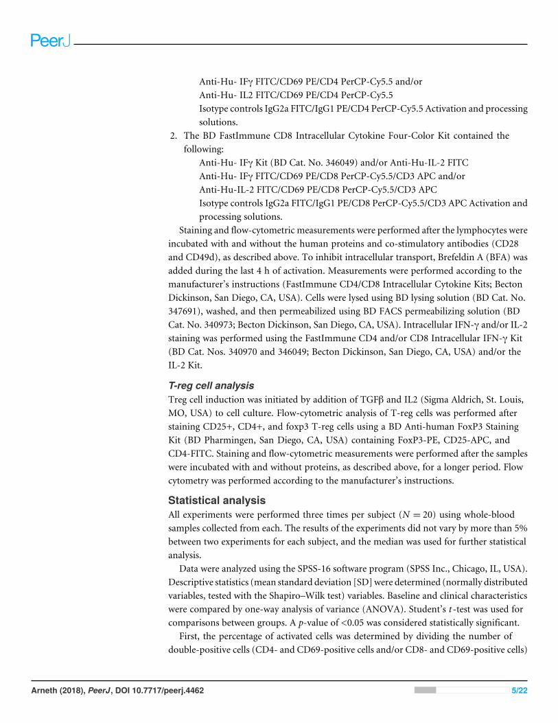
Anti-Hu- IFγ FITC/CD69 PE/CD4 PerCP-Cy5.5 and/orAnti-Hu- IL2 FITC/CD69 PE/CD4 PerCP-Cy5.5Isotype controls IgG2a FITC/IgG1 PE/CD4 PerCP-Cy5.5 Activation and processingsolutions.
2. The BD FastImmune CD8 Intracellular Cytokine Four-Color Kit contained thefollowing:
Anti-Hu- IFγ Kit (BD Cat. No. 346049) and/or Anti-Hu-IL-2 FITCAnti-Hu- IFγ FITC/CD69 PE/CD8 PerCP-Cy5.5/CD3 APC and/orAnti-Hu-IL-2 FITC/CD69 PE/CD8 PerCP-Cy5.5/CD3 APCIsotype controls IgG2a FITC/IgG1 PE/CD8 PerCP-Cy5.5/CD3 APC Activation andprocessing solutions.
Staining and flow-cytometric measurements were performed after the lymphocytes wereincubated with and without the human proteins and co-stimulatory antibodies (CD28and CD49d), as described above. To inhibit intracellular transport, Brefeldin A (BFA) wasadded during the last 4 h of activation. Measurements were performed according to themanufacturer’s instructions (FastImmune CD4/CD8 Intracellular Cytokine Kits; BectonDickinson, San Diego, CA, USA). Cells were lysed using BD lysing solution (BD Cat. No.347691), washed, and then permeabilized using BD FACS permeabilizing solution (BDCat. No. 340973; Becton Dickinson, San Diego, CA, USA). Intracellular IFN-γ and/or IL-2staining was performed using the FastImmune CD4 and/or CD8 Intracellular IFN-γ Kit(BD Cat. Nos. 340970 and 346049; Becton Dickinson, San Diego, CA, USA) and/or theIL-2 Kit.
T-reg cell analysisTreg cell induction was initiated by addition of TGFβ and IL2 (Sigma Aldrich, St. Louis,MO, USA) to cell culture. Flow-cytometric analysis of T-reg cells was performed afterstaining CD25+, CD4+, and foxp3 T-reg cells using a BD Anti-human FoxP3 StainingKit (BD Pharmingen, San Diego, CA, USA) containing FoxP3-PE, CD25-APC, andCD4-FITC. Staining and flow-cytometric measurements were performed after the sampleswere incubated with and without proteins, as described above, for a longer period. Flowcytometry was performed according to the manufacturer’s instructions.
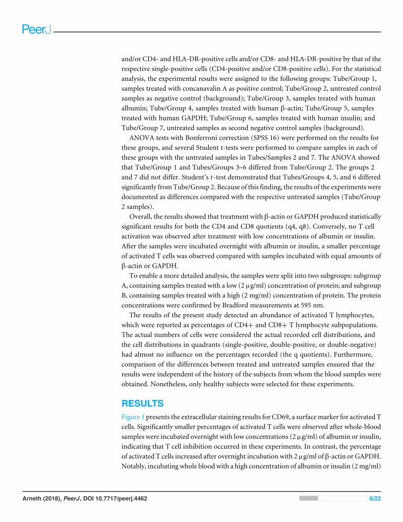
Statistical analysisAll experiments were performed three times per subject (N = 20) using whole-bloodsamples collected from each. The results of the experiments did not vary by more than 5%between two experiments for each subject, and the median was used for further statisticalanalysis.
Data were analyzed using the SPSS-16 software program (SPSS Inc., Chicago, IL, USA).Descriptive statistics (mean standard deviation [SD]were determined (normally distributedvariables, tested with the Shapiro–Wilk test) variables. Baseline and clinical characteristicswere compared by one-way analysis of variance (ANOVA). Student’s t -test was used forcomparisons between groups. A p-value of <0.05 was considered statistically significant.
First, the percentage of activated cells was determined by dividing the number ofdouble-positive cells (CD4- and CD69-positive cells and/or CD8- and CD69-positive cells)
Arneth (2018), PeerJ, DOI 10.7717/peerj.4462 5/22
and/or CD4- and HLA-DR-positive cells and/or CD8- and HLA-DR-positive by that of therespective single-positive cells (CD4-positive and/or CD8-positive cells). For the statisticalanalysis, the experimental results were assigned to the following groups: Tube/Group 1,samples treated with concanavalin A as positive control; Tube/Group 2, untreated controlsamples as negative control (background); Tube/Group 3, samples treated with humanalbumin; Tube/Group 4, samples treated with human β-actin; Tube/Group 5, samplestreated with human GAPDH; Tube/Group 6, samples treated with human insulin; andTube/Group 7, untreated samples as second negative control samples (background).
ANOVA tests with Bonferroni correction (SPSS 16) were performed on the results forthese groups, and several Student t-tests were performed to compare samples in each ofthese groups with the untreated samples in Tubes/Samples 2 and 7. The ANOVA showedthat Tube/Group 1 and Tubes/Groups 3–6 differed from Tube/Group 2. The groups 2and 7 did not differ. Student’s t -test demonstrated that Tubes/Groups 4, 5, and 6 differedsignificantly fromTube/Group 2. Because of this finding, the results of the experiments weredocumented as differences compared with the respective untreated samples (Tube/Group2 samples).
Overall, the results showed that treatment with β-actin or GAPDH produced statisticallysignificant results for both the CD4 and CD8 quotients (q4, q8). Conversely, no T cellactivation was observed after treatment with low concentrations of albumin or insulin.After the samples were incubated overnight with albumin or insulin, a smaller percentageof activated T cells was observed compared with samples incubated with equal amounts ofβ-actin or GAPDH.
To enable a more detailed analysis, the samples were split into two subgroups: subgroupA, containing samples treated with a low (2 µg/ml) concentration of protein; and subgroupB, containing samples treated with a high (2 mg/ml) concentration of protein. The proteinconcentrations were confirmed by Bradford measurements at 595 nm.
The results of the present study detected an abundance of activated T lymphocytes,which were reported as percentages of CD4+ and CD8+ T lymphocyte subpopulations.The actual numbers of cells were considered the actual recorded cell distributions, andthe cell distributions in quadrants (single-positive, double-positive, or double-negative)had almost no influence on the percentages recorded (the q quotients). Furthermore,comparison of the differences between treated and untreated samples ensured that theresults were independent of the history of the subjects from whom the blood samples wereobtained. Nonetheless, only healthy subjects were selected for these experiments.
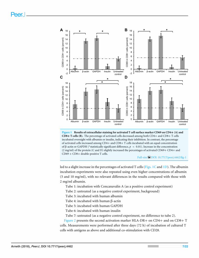
RESULTSFigure 1 presents the extracellular staining results for CD69, a surfacemarker for activated Tcells. Significantly smaller percentages of activated T cells were observed after whole-bloodsamples were incubated overnight with low concentrations (2µg/ml) of albumin or insulin,indicating that T cell inhibition occurred in these experiments. In contrast, the percentageof activated T cells increased after overnight incubation with 2 µg/ml of β-actin or GAPDH.Notably, incubating whole bloodwith a high concentration of albumin or insulin (2mg/ml)
Arneth (2018), PeerJ, DOI 10.7717/peerj.4462 6/22
-2
0
2
4
6
8
10
12
14
16
18
Albumin β-actin GAPDH Insulin Untreatedcontrol
Albumin β-actin GAPDH Insulin Untreatedcontrol
Albumin β-actin GAPDH Insulin Untreatedcontrol
Albumin β-actin GAPDH Insulin Untreatedcontrol
CD
69 in
CD
4+ c
ells
(per
cent
)
CD
69 in
CD
8+ c
ells
(per
cent
)
CD
69 in
CD
4+ c
ells
(per
cent
)
CD
69 in
CD
8+ c
ells
(per
cent
)
-2
0
2
4
6
8
10
12
14
16
18
0
2
4
6
8
10
12
14
16
18
0
2
4
6
8
10
12
14
16
18
A B
C D
Figure 1 Results of extracellular staining for activated T cell surface marker CD69 on CD4+ (A) andCD8+ T cells (B). The percentage of activated cells decreased among both CD4+ and CD8+ T cellsincubated overnight with albumin or insulin, indicating their inhibition. In contrast, the percentageof activated cells increased among CD4+ and CD8+ T cells incubated with an equal concentrationof β-actin or GAPDH (*statistically significant difference, p < 0.01). Increase in the concentration(2 mg/ml) of the protein (C and D) slightly increased the percentages of activated CD69+ CD4+ andCD69+ CD8+ double-positive T cells.
Full-size DOI: 10.7717/peerj.4462/fig-1
led to a slight increase in the percentages of activated T cells (Figs. 1C and 1D). The albuminincubation experiments were also repeated using even higher concentrations of albumin(5 and 10 mg/ml), with no relevant differences in the results compared with those with2 mg/ml albumin.
Tube 1: incubation with Concanavalin A (as a positive control experiment)Tube 2: untreated (as a negative control experiment, background)Tube 3: incubated with human albuminTube 4: incubated with human β-actinTube 5: incubated with human GAPDHTube 6: incubated with human insulinTube 7: untreated (as a negative control experiment, no difference to tube 2).Figure 2 presents the second activation marker HLA-DR+ on CD4+ and on CD8+ T
cells. Measurements were performed after three days (72 h) of incubation of cultured Tcells with antigens as above and additional co-stimulation with CD28.
Arneth (2018), PeerJ, DOI 10.7717/peerj.4462 7/22

0
1
2
3
4
5
6
7
8
Albumin β-actin GAPDH Insulin Untreatedcontrol
0
1
2
3
4
5
6
7
8
Albumin β-actin GAPDH Insulin Untreatedcontrol
0
1
2
3
4
5
6
7
8
Albumin β-actin GAPDH Insulin Untreatedcontrol
0
1
2
3
4
5
6
7
8
Albumin β-actin GAPDH Insulin Untreatedcontrol
A B
C D
HLA
-DR
in C
D4+
cel
ls (p
erce
nt)
HLA
-DR
in C
D4+
cel
ls (p
erce
nt)
HLA
-DR
in C
D8+
cel
ls (p
erce
nt)
HLA
-DR
in C
D8+
cel
ls (p
erce
nt)
Figure 2 Results of extracellular staining for activated T cell surface marker HLA-DR on CD4+ (A)and CD8+ T cells (B). The percentage of activated cells decreased among both CD4+ and CD8+ T cellsincubated overnight with albumin or insulin, indicating their inhibition. In contrast, the percentage ofactivated cells increased among both CD4+ and CD8+ T cells incubated with an equal concentrationof β-actin or GAPDH (*statistically significant difference, p < 0.01). Increase in the concentration(2 mg/ml) of the protein (C and D) increased the percentages of activated HLA-DR+ CD4+ andHLA-DR+ CD8+ double-positive T cells.
Full-size DOI: 10.7717/peerj.4462/fig-2
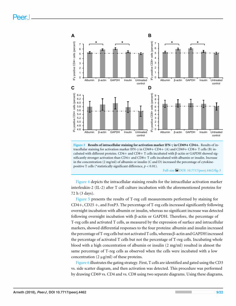
Figure 3 depicts the intracellular staining results for the activation marker IFγ afterwhole-blood samples were incubated with the aforementioned proteins. Similar to thetrend for surface markers, the levels of intracellular IFγ indicated that incubation withβ-actin or GAPDH led to significantly stronger T cell activation than incubation withalbumin or insulin. Again, however, incubating the samples with a high concentrationof albumin or insulin (2 mg/ml) increased the percentage of cytokine-positive T cells(Figs. 3C and 3D).
Arneth (2018), PeerJ, DOI 10.7717/peerj.4462 8/22
Albumin β-actin GAPDH Insulin Untreatedcontrol
Albumin β-actin GAPDH Insulin Untreatedcontrol
Albumin β-actin GAPDH Insulin Untreatedcontrol
Albumin β-actin GAPDH Insulin Untreatedcontrol
0
1
2
3
4
5
6
7
0
1
2
3
4
5
6
7
4.6 4.8 5.0 5.2 5.4 5.6 5.8 6.0 6.2 6.4
0 1 2 3 4 5 6 7 8 9
IFγ
posi
tive
CD
4+ c
ells
(per
cent
)
IFγ
posi
tive
CD
8+ c
ells
(per
cent
)
IFγ
posi
tive
CD
4+ c
ells
(per
cent
)
IFγ
posi
tive
CD
8+ c
ells
(per
cent
)
A B
C D
Figure 3 Results of intracellular staining for activation marker IFN-γ in CD69+ CD4+. Results of in-tracellular staining for activation marker IFN-γ in CD69+ CD4+ (A) and CD69+ CD8+ T cells (B) in-cubated with different proteins. CD4+ and CD8+ T cells incubated with β-actin or GAPDH showed sig-nificantly stronger activation than CD4+ and CD8+ T cells incubated with albumin or insulin. Increasein the concentration (2 mg/ml) of albumin or insulin (C and D) increased the percentage of cytokine-positive T cells (*statistically significant difference, p< 0.01).
Full-size DOI: 10.7717/peerj.4462/fig-3
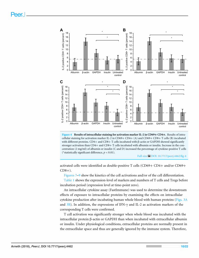
Figure 4 depicts the intracellular staining results for the intracellular activation markerinterleukin-2 (IL-2) after T cell culture incubation with the aforementioned proteins for72 h (3 days).
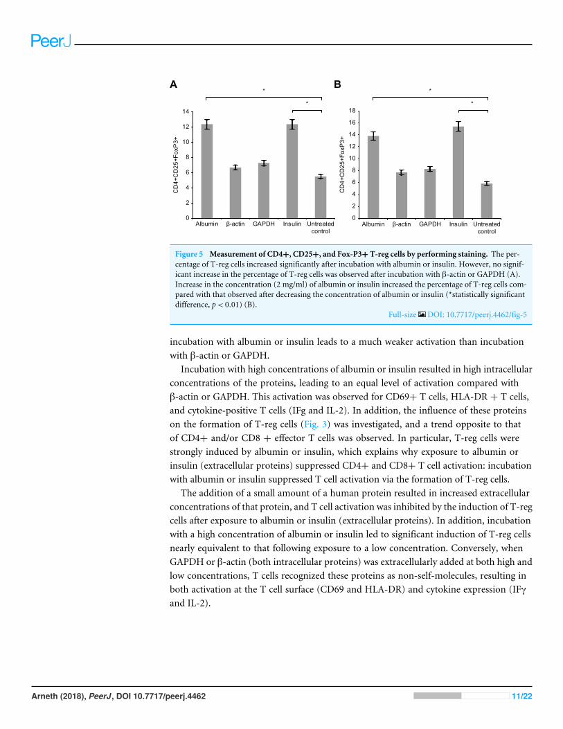
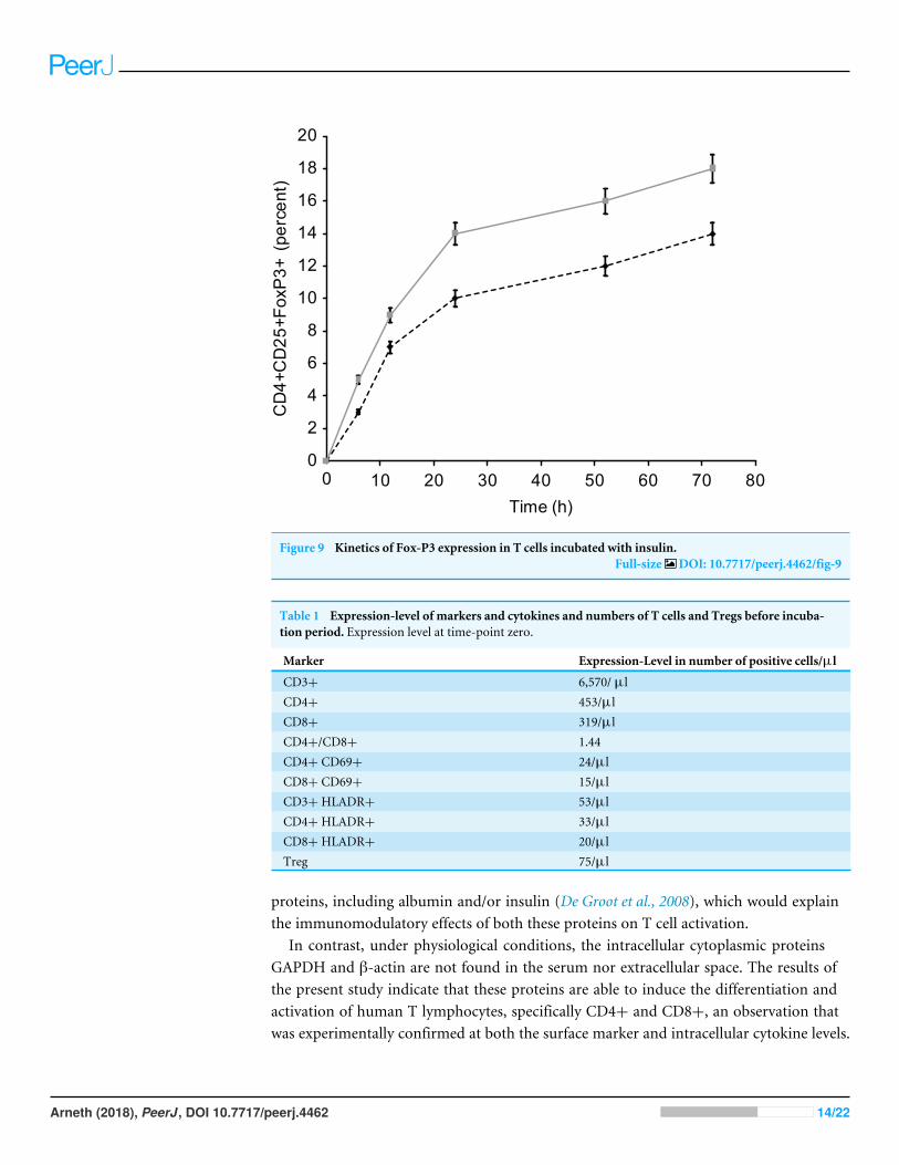
Figure 5 presents the results of T-reg cell measurements performed by staining forCD4+, CD25+, and FoxP3. The percentage of T-reg cells increased significantly followingovernight incubation with albumin or insulin, whereas no significant increase was detectedfollowing overnight incubation with β-actin or GAPDH. Therefore, the percentage ofT-reg cells and activated T cells, as measured by the expression of surface and intracellularmarkers, showed differential responses to the four proteins: albumin and insulin increasedthe percentage of T-reg cells but not activatedT cells, whereasβ-actin andGAPDH increasedthe percentage of activated T cells but not the percentage of T-reg cells. Incubating wholeblood with a high concentration of albumin or insulin (2 mg/ml) resulted in almost thesame percentage of T-reg cells as observed when the cells were incubated with a lowconcentration (2 µg/ml) of these proteins.
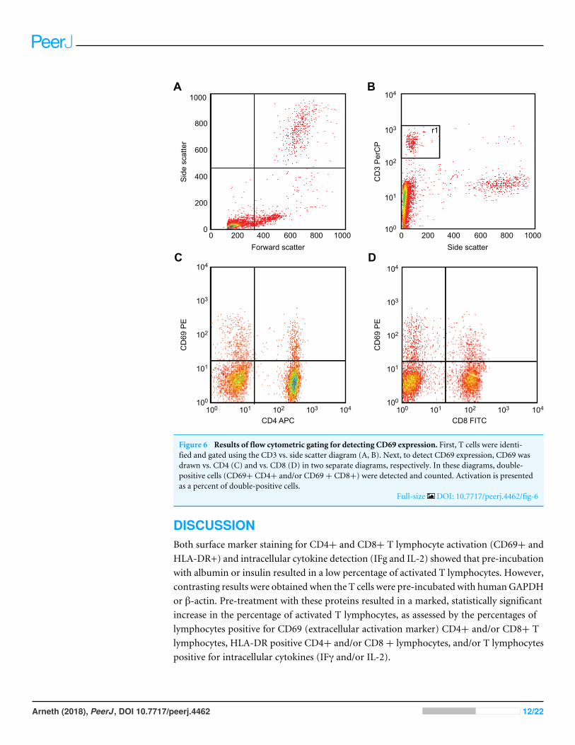
Figure 6 illustrates the gating strategy. First, T cells are identified and gated using the CD3vs. side scatter diagram, and then activation was detected. This procedure was performedby drawing CD69 vs. CD4 and vs. CD8 using two separate diagrams. Using these diagrams,
Arneth (2018), PeerJ, DOI 10.7717/peerj.4462 9/22
0
2
4
6
8
10
12
14
16
18
Albumin β-actin GAPDH Insulin Untreatedcontrol
*
*
0
2
4
6
8
10
12
14
16
18
Albumin β-actin GAPDH Insulin
*
*
02468
1012141618
Albumin β-actin GAPDH Insulin
*
*
*
02468
1012141618
Albumin β-actin GAPDH Insulin
*
*
*
IL-2
pos
itive
CD
4+ T
cel
ls (p
erce
nt)
IL-2
pos
itive
CD
8+ T
cel
ls (p
erce
nt)
IL-2
pos
itive
CD
4+ T
cel
ls (p
erce
nt)
IL-2
pos
itive
CD
8+ T
cel
ls (p
erce
nt)
Untreatedcontrol
Untreatedcontrol
Untreatedcontrol
A B
C D
Figure 4 Results of intracellular staining for activation marker IL-2 in CD69+ CD4+. Results of intra-cellular staining for activation marker IL-2 in CD69+ CD4+ (A) and CD69+ CD8+ T cells (B) incubatedwith different proteins. CD4+ and CD8+ T cells incubated with β-actin or GAPDH showed significantlystronger activation than CD4+ and CD8+ T cells incubated with albumin or insulin. Increase in the con-centration (2 mg/ml) of albumin or insulin (C and D) increased the percentage of cytokine-positive T cells(*statistically significant difference, p< 0.01).
Full-size DOI: 10.7717/peerj.4462/fig-4
activated cells were identified as double-positive T cells (CD69+ CD4+ and/or CD69+CD8+).
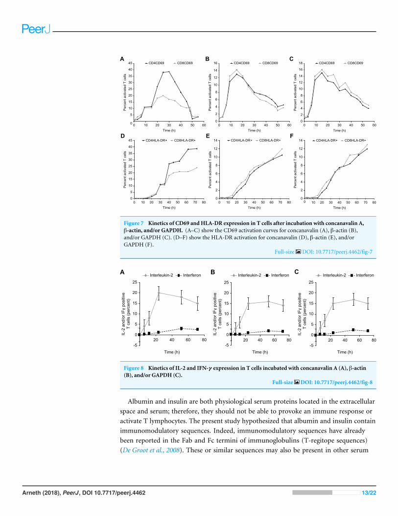
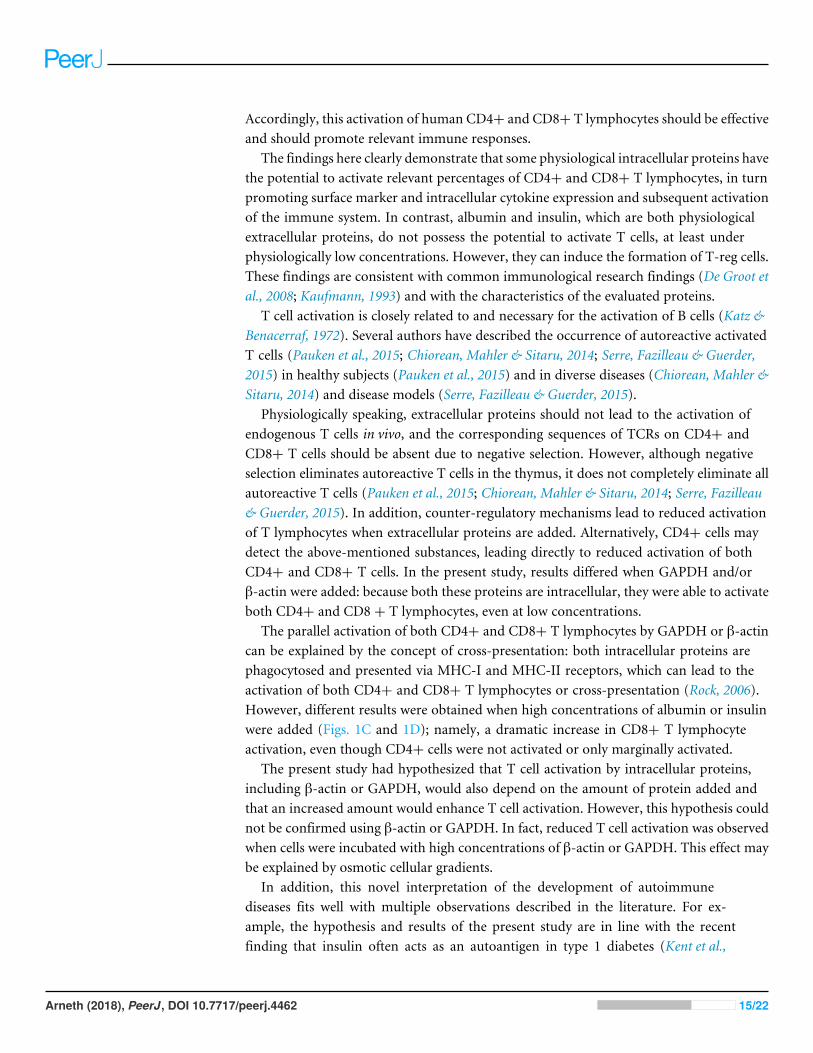
Figures 7–9 show the kinetics of the cell activations and/or of the cell differentiation.Table 1 shows the expression-level of markers and numbers of T cells and Tregs before
incubation period (expression level at time-point zero).An intracellular cytokine assay (FastImmune) was used to determine the downstream
effects of exposure to intracellular proteins by examining the effects on intracellularcytokine production after incubating human whole blood with human proteins (Figs. 3Aand 3B). In addition, the expressions of IFN-γ and IL-2 as activation markers of thecorresponding T cells were confirmed.
T cell activation was significantly stronger when whole blood was incubated with theintracellular protein β-actin or GAPDH than when incubated with extracellular albuminor insulin. Under physiological conditions, extracellular proteins are normally present inthe extracellular space and thus are generally ignored by the immune system. Therefore,
Arneth (2018), PeerJ, DOI 10.7717/peerj.4462 10/22
0
2
4
6
8
10
12
14
Albumin β-actin GAPDH Insulin Untreatedcontrol
CD4+CD25+FoxP3+
0
2
4
6
8
10
12
14
16
18
Albumin β-actin GAPDH Insulin Untreatedcontrol
CD4+CD25+FoxP3+
*
* *
*
A B
Figure 5 Measurement of CD4+, CD25+, and Fox-P3+ T-reg cells by performing staining. The per-centage of T-reg cells increased significantly after incubation with albumin or insulin. However, no signif-icant increase in the percentage of T-reg cells was observed after incubation with β-actin or GAPDH (A).Increase in the concentration (2 mg/ml) of albumin or insulin increased the percentage of T-reg cells com-pared with that observed after decreasing the concentration of albumin or insulin (*statistically significantdifference, p< 0.01) (B).
Full-size DOI: 10.7717/peerj.4462/fig-5
incubation with albumin or insulin leads to a much weaker activation than incubationwith β-actin or GAPDH.
Incubation with high concentrations of albumin or insulin resulted in high intracellularconcentrations of the proteins, leading to an equal level of activation compared withβ-actin or GAPDH. This activation was observed for CD69+ T cells, HLA-DR + T cells,and cytokine-positive T cells (IFg and IL-2). In addition, the influence of these proteinson the formation of T-reg cells (Fig. 3) was investigated, and a trend opposite to thatof CD4+ and/or CD8 + effector T cells was observed. In particular, T-reg cells werestrongly induced by albumin or insulin, which explains why exposure to albumin orinsulin (extracellular proteins) suppressed CD4+ and CD8+ T cell activation: incubationwith albumin or insulin suppressed T cell activation via the formation of T-reg cells.
The addition of a small amount of a human protein resulted in increased extracellularconcentrations of that protein, and T cell activation was inhibited by the induction of T-regcells after exposure to albumin or insulin (extracellular proteins). In addition, incubationwith a high concentration of albumin or insulin led to significant induction of T-reg cellsnearly equivalent to that following exposure to a low concentration. Conversely, whenGAPDH or β-actin (both intracellular proteins) was extracellularly added at both high andlow concentrations, T cells recognized these proteins as non-self-molecules, resulting inboth activation at the T cell surface (CD69 and HLA-DR) and cytokine expression (IFγand IL-2).
Arneth (2018), PeerJ, DOI 10.7717/peerj.4462 11/22
800
800
1000
1000
600
600
400
400
200
2000
0 800 10006004002000100
104
103
102
101
100
100
104
104
103
103
102
102
101
101100
100
104
104
103
103
102
102
101
101
Sid
e sc
atte
r
Side scatterForward scatter
CD
3 P
erC
P
CD
69 P
E
CD
69 P
E
CD4 APC CD8 FITC
r1
A B
C D
Figure 6 Results of flow cytometric gating for detecting CD69 expression. First, T cells were identi-fied and gated using the CD3 vs. side scatter diagram (A, B). Next, to detect CD69 expression, CD69 wasdrawn vs. CD4 (C) and vs. CD8 (D) in two separate diagrams, respectively. In these diagrams, double-positive cells (CD69+ CD4+ and/or CD69+ CD8+) were detected and counted. Activation is presentedas a percent of double-positive cells.
Full-size DOI: 10.7717/peerj.4462/fig-6
DISCUSSIONBoth surface marker staining for CD4+ and CD8+ T lymphocyte activation (CD69+ andHLA-DR+) and intracellular cytokine detection (IFg and IL-2) showed that pre-incubationwith albumin or insulin resulted in a low percentage of activated T lymphocytes. However,contrasting results were obtained when the T cells were pre-incubated with humanGAPDHor β-actin. Pre-treatment with these proteins resulted in a marked, statistically significantincrease in the percentage of activated T lymphocytes, as assessed by the percentages oflymphocytes positive for CD69 (extracellular activation marker) CD4+ and/or CD8+ Tlymphocytes, HLA-DR positive CD4+ and/or CD8 + lymphocytes, and/or T lymphocytespositive for intracellular cytokines (IFγ and/or IL-2).
Arneth (2018), PeerJ, DOI 10.7717/peerj.4462 12/22
0
5
10
15
20
25
30
35
40
45
0 10 20 30 40 50 60Time (h)
Per
cent
act
ivat
ed T
cel
ls
CD4CD69 CD8CD69
Time (h)
0
2
4
6
8
10
12
14
16
18
0 10 20 30 40 50 60
Per
cent
act
ivat
ed T
cel
ls
CD4CD69 CD8CD69
0
5
10
15
20
25
30
35
40
45
0 10 20 30 40 50 60 70 80Time (h)
Per
cent
act
ivat
ed T
cel
ls
CD4HLA-DR+ CD8HLA-DR+
0
2
4
6
8
10
12
14
0 10 20 30 40 50 60 70 80Time (h)
Per
cent
act
ivat
ed T
cel
ls
CD4HLA-DR+ CD8HLA-DR+
0
2
4
6
8
10
12
14
0 10 20 30 40 50 60 70 80Time (h)
Per
cent
act
ivat
ed T
cel
ls
CD4HLA-DR+ CD8HLA-DR+
0
2
4
6
8
10
12
14
16
0 10 20 30 40 50 60Time (h)
Per
cent
act
ivat
ed T
cel
ls
CD4CD69 CD8CD69A B C
D E F
Figure 7 Kinetics of CD69 and HLA-DR expression in T cells after incubation with concanavalin A,β-actin, and/or GAPDH. (A–C) show the CD69 activation curves for concanavalin (A), β-actin (B),and/or GAPDH (C). (D–F) show the HLA-DR activation for concanavalin (D), β-actin (E), and/orGAPDH (F).
Full-size DOI: 10.7717/peerj.4462/fig-7
A B C
-5
0
5
10
15
20
25
20 40 60 80
IL-2
and
/or I
Fγ p
ositi
veT
cells
(per
cent
)
Time (h)
Interleukin-2 Interferon
-5
0
5
10
15
20
25
20 40 60 80
IL-2
and
/or I
Fγ p
ositi
veT
cells
(per
cent
)
Time (h)
Interleukin-2 Interferon
-5
0
5
10
15
20
25
20 40 60 80
IL-2
and
/or I
Fγ p
ositi
veT
cells
(per
cent
)
Time (h)
Interleukin-2 Interferon
Figure 8 Kinetics of IL-2 and IFN-γ expression in T cells incubated with concanavalin A (A), β-actin(B), and/or GAPDH (C).
Full-size DOI: 10.7717/peerj.4462/fig-8
Albumin and insulin are both physiological serum proteins located in the extracellularspace and serum; therefore, they should not be able to provoke an immune response oractivate T lymphocytes. The present study hypothesized that albumin and insulin containimmunomodulatory sequences. Indeed, immunomodulatory sequences have alreadybeen reported in the Fab and Fc termini of immunoglobulins (T-regitope sequences)(De Groot et al., 2008). These or similar sequences may also be present in other serum
Arneth (2018), PeerJ, DOI 10.7717/peerj.4462 13/22
0
2
4
6
8
10
12
14
16
18
20
0 10 20 30 40 50 60 70 80
CD
4+C
D25
+Fox
P3+
(pe
rcen
t)
Time (h)
Figure 9 Kinetics of Fox-P3 expression in T cells incubated with insulin.Full-size DOI: 10.7717/peerj.4462/fig-9
Table 1 Expression-level of markers and cytokines and numbers of T cells and Tregs before incuba-tion period. Expression level at time-point zero.
Marker Expression-Level in number of positive cells/µl
CD3+ 6,570/ µlCD4+ 453/µlCD8+ 319/µlCD4+/CD8+ 1.44CD4+ CD69+ 24/µlCD8+ CD69+ 15/µlCD3+HLADR+ 53/µlCD4+HLADR+ 33/µlCD8+HLADR+ 20/µlTreg 75/µl
proteins, including albumin and/or insulin (De Groot et al., 2008), which would explainthe immunomodulatory effects of both these proteins on T cell activation.
In contrast, under physiological conditions, the intracellular cytoplasmic proteinsGAPDH and β-actin are not found in the serum nor extracellular space. The results ofthe present study indicate that these proteins are able to induce the differentiation andactivation of human T lymphocytes, specifically CD4+ and CD8+, an observation thatwas experimentally confirmed at both the surface marker and intracellular cytokine levels.
Arneth (2018), PeerJ, DOI 10.7717/peerj.4462 14/22
Accordingly, this activation of human CD4+ and CD8+ T lymphocytes should be effectiveand should promote relevant immune responses.
The findings here clearly demonstrate that some physiological intracellular proteins havethe potential to activate relevant percentages of CD4+ and CD8+ T lymphocytes, in turnpromoting surface marker and intracellular cytokine expression and subsequent activationof the immune system. In contrast, albumin and insulin, which are both physiologicalextracellular proteins, do not possess the potential to activate T cells, at least underphysiologically low concentrations. However, they can induce the formation of T-reg cells.These findings are consistent with common immunological research findings (De Groot etal., 2008; Kaufmann, 1993) and with the characteristics of the evaluated proteins.
T cell activation is closely related to and necessary for the activation of B cells (Katz &Benacerraf, 1972). Several authors have described the occurrence of autoreactive activatedT cells (Pauken et al., 2015; Chiorean, Mahler & Sitaru, 2014; Serre, Fazilleau & Guerder,2015) in healthy subjects (Pauken et al., 2015) and in diverse diseases (Chiorean, Mahler &Sitaru, 2014) and disease models (Serre, Fazilleau & Guerder, 2015).
Physiologically speaking, extracellular proteins should not lead to the activation ofendogenous T cells in vivo, and the corresponding sequences of TCRs on CD4+ andCD8+ T cells should be absent due to negative selection. However, although negativeselection eliminates autoreactive T cells in the thymus, it does not completely eliminate allautoreactive T cells (Pauken et al., 2015; Chiorean, Mahler & Sitaru, 2014; Serre, Fazilleau& Guerder, 2015). In addition, counter-regulatory mechanisms lead to reduced activationof T lymphocytes when extracellular proteins are added. Alternatively, CD4+ cells maydetect the above-mentioned substances, leading directly to reduced activation of bothCD4+ and CD8+ T cells. In the present study, results differed when GAPDH and/orβ-actin were added: because both these proteins are intracellular, they were able to activateboth CD4+ and CD8 + T lymphocytes, even at low concentrations.
The parallel activation of both CD4+ and CD8+ T lymphocytes by GAPDH or β-actincan be explained by the concept of cross-presentation: both intracellular proteins arephagocytosed and presented via MHC-I and MHC-II receptors, which can lead to theactivation of both CD4+ and CD8+ T lymphocytes or cross-presentation (Rock, 2006).However, different results were obtained when high concentrations of albumin or insulinwere added (Figs. 1C and 1D); namely, a dramatic increase in CD8+ T lymphocyteactivation, even though CD4+ cells were not activated or only marginally activated.
The present study had hypothesized that T cell activation by intracellular proteins,including β-actin or GAPDH, would also depend on the amount of protein added andthat an increased amount would enhance T cell activation. However, this hypothesis couldnot be confirmed using β-actin or GAPDH. In fact, reduced T cell activation was observedwhen cells were incubated with high concentrations of β-actin or GAPDH. This effect maybe explained by osmotic cellular gradients.
In addition, this novel interpretation of the development of autoimmunediseases fits well with multiple observations described in the literature. For ex-ample, the hypothesis and results of the present study are in line with the recentfinding that insulin often acts as an autoantigen in type 1 diabetes (Kent et al.,
Arneth (2018), PeerJ, DOI 10.7717/peerj.4462 15/22
2005; Nakayama et al., 2005; Von Herrath, 2005; Wilson, 2005). In addition, thepresent study fits well with the observation that myocardial infarction can beaccompanied and complicated by autoimmune reactions (Liao & Cheng, 2006).
Currently, the mosaic of autoimmunity is only partially understood (Gershwin, 2008).However, the expression of autoantibodies, which are often associated with autoimmunedisease, increases in patients during early disease stages. Over the past 10 years, suchautoantibodies have increasingly been demonstrated to have high diagnostic value(Gershwin, 2008). Notably, many of these autoantibodies are directed against intracellularmolecules themselves, including ribonucleoproteins (RNPs) and double-stranded DNA (dsDNA) in systemic lupus patients (SLE-patients) and centromeres and type 1 topoisomerasein patients with scleroderma (Gershwin, 2008). Regardless, the mechanisms underlying thisprocess remain poorly understood. The hypothesis and results of the present study couldprovide a possible explanation for such nonspecific, bystander-autoimmune phenomena.
Recently, cellular ubiquitination and proteolysis systems have been proposed as beingclosely correlated with the balance between immunity and tolerance (Gomez-Martin,Diaz-Zamudio & Alcocer-Varela, 2008).
CD4+ T cells are known to play an important role in the development of autoimmunediseases (Slavin et al., 2002), with their inhibition leading to the suppression of diseasedevelopment (Slavin et al., 2002). Interestingly, transfected CD4+ T lymphocytes are usedas the preferred vector in experimental gene therapy to treat autoimmune diseases inanimals (Slavin et al., 2002).
Two well-accepted alternative hypotheses for the development of autoimmune diseasesare the virus hypothesis and the molecular mimicry hypothesis. The viral hypothesis statesthat new, unknown viruses are responsible for the development of autoimmune diseases;however, this hypothesis remains unconfirmed in light of current knowledge (Denman &Rager-Zisman, 2004). Accordingly, future studies that attempt to detect unknown virusesshould be performed, and the strong postulates of Koch may need to be changed (Denman& Rager-Zisman, 2004).
Cardiovascular disease has been shown to occur much more frequently in patients withrheumatoid arthritis and to more often lead to death in these patients than in healthyindividuals (Sarzi-Puttini et al., 2005).
The present study has shown that, in principle, physiological autoreactive T lymphocytesare present even in healthy individuals. Several studies have shown the presence of a largerepertoire of T lymphocytes oriented toward various organs and/or tissues in healthy mice(Sharma et al., 2007). If these autoimmune T lymphocytes expand, an autoimmune attackmay occur (Sharma et al., 2007). In addition, several authors have reported the spontaneousdevelopment of autoimmune diseases in mice; these diseases target the skin, lungs, liver,and tail when T-reg cells (CD4+, CD25+, and Foxp3+ cells) are absent (Foxp3 protein-deficient scurfy mice Sharma et al., 2007; Davies, 2008; Goodnow et al., 2005). These resultsshow that additional mechanisms in healthy individuals prevent autoimmune attacks,including important T-reg cells, which the present study investigated. However, theimmune system is regulated by other control systems; indeed, multiple control systems
Arneth (2018), PeerJ, DOI 10.7717/peerj.4462 16/22
appear to be necessary, particularly due to the possibility of normal, healthy individualsdeveloping autoimmune diseases.
These mechanisms are both important and necessary for the accurate control of theself/non-self recognition process.
Over the past decade, several studies have reported that all immune-system componentsappear to be involved in the development and modulation of atherosclerosis (Bassi et al.,2009). In particular, pentraxins and antibodies appear to play important roles. Pentraxinsare ancient multimer molecules that are also important to the recognition of self- andnon-self structures and are functionally complementary to the cellular recognition of self-and non-self molecules (Bottazzi et al., 2006). As described above, these observations matchthe hypothesis outlined by the present study.
Recently, a mechanism has been described in which intracellular endogenous proteinsare presented by MHC-II due to autophagy by antigen-presenting cells (Münz, 2012). Thiseffect might be dependent on the exact physiological context and on the concentrationof the respective intracellular protein in the plasma, as shown by the present study. Theoccurrence of this autophagy-driven presentation of intracellular peptides to MHC-IIduring infections and similar situations could explain the hygiene hypothesis. To date,autophagy has been described in connection with infection with the herpes simplex virusand influenza viruses (Münz, 2012). If no infection occurs in early childhood, then theMHC repertoires are different, and no cross-presentation occurs. However, if infectionsoccur in early childhood, the intracellular antigen spectra might better display via theMHC-II pathway, and resistance to autoimmune diseases might increase.
Herein a strong division of the antigenic spectrum into an extracellular part, consistingof CD4 T cell receptors, and an intracellular part, consisting of CD8 T cell receptors ispostulated.
With regard to the phenomenon of cross-presentation (presentation of exogenousproteins by the MHC-I receptor), the following must be said.
Scientific work in recent years has shown that cross-presentation is a limited, highlyspecialized, and precisely regulated process (Fehrens et al., 2014). The process is limitedonly to specific specialized cells (DC) (Fehrens et al., 2014) and specific organelles withinthese cells (Brode & Macary, 2004). In addition, many specialized enzymes are involved inprecisely regulating the process (Brode & Macary, 2004).
Furthermore, as has been described, only antigens already bound to specific receptors(immunoglobulins and Fc receptors) can be taken up by these cells and then processed bycross-presentation (Platzer, Stout & Fiebiger, 2014).
However, if this is the case, then the phenomenon of cross-presentation does not in anyway contradict the division of the antigen spectrum into an extracellular and intracellularspectrum, as described here, and does not preclude the representation of these divisions bythe various T cell receptors and the various lymphocyte populations (CD4+ and CD8+ Tlymphocytes).
On the contrary, if uptake and processing of an antigen is possible via cross-presentationonly after binding to specific receptors (immunoglobulins and Fc receptors Platzer, Stout& Fiebiger, 2014), then that antigen has already been recognized as foreign, ensuing that
Arneth (2018), PeerJ, DOI 10.7717/peerj.4462 17/22
the antigen is not a body-own-specific structure of the extracellular space. This stronglysupports the correctness of the relationship postulated here.
In order to establish these findings it is necessary to test a line of further extracellular andintracellular proteins for their immune activation potential in future studies. In addition,species difference studies should also be performed in future research.
CONCLUSIONSOverall, in addition to the location being important for immunization, as described herein,other mechanisms may be relevant to the development of autoimmune diseases, includingmolecular mimicry, viral infections, and redox reactions of antibodies, all of which maybe responsible for the development of autoantibodies. However, the results of the presentstudy suggest an additional mechanism; namely, the misguided formation of proteins thatbecome antigens.
Several aspects of the experiments conducted in the present study have been reported inpart by other researchers, although their research was conducted within different contexts.For instance, the existence of an autoreactive T cell repertoire, even in healthy individuals,has been reported by several authors (Kamate et al., 2007; Walker & Abbas, 2002; Munket al., 1989; Zou et al., 2008; Vella et al., 2009). Their findings are consistent with thoseof the present study, and a hypothetical explanation for this phenomenon is providedherein. In addition, an autoreactive T cell response toward insulin was reported by Ito andYang but only in patients with autoimmune diabetes (Ito et al., 1993; Yang et al., 2014).Lastly, induction of T-reg cells in response to a human protein—namely, intravenousimmunoglobulin—was reported byMaddur et al. (2001) and Cousens et al. (2013).
The present study confirmed and extended their experimental results, placed themwithin the context of disease development, and provided an explanation. The presentstudy is in line with the following novel proposal of autoimmune regulation: Whenself-molecules occur at the wrong place in an intact organism, they are recognized asnon-self-molecules, leading to the activation of immune cells. Therefore, structural proteinabnormalities, mutations, or errors, including those in signaling, are not necessary toinduce immunization against the organism itself (self-immunization); indeed, a meremislocalized protein is sufficient.
ACKNOWLEDGEMENTSThere are no acknowledgements.
ADDITIONAL INFORMATION AND DECLARATIONS
FundingThe author received no funding for this work.
Competing InterestsThe author declares there are no competing interests.
Arneth (2018), PeerJ, DOI 10.7717/peerj.4462 18/22
Author Contributions• Borros M. Arneth conceived and designed the experiments, performed the experiments,analyzed the data, contributed reagents/materials/analysis tools, prepared figures and/ortables, authored or reviewed drafts of the paper, approved the final draft.
Human EthicsThe following information was supplied relating to ethical approvals (i.e., approving bodyand any reference numbers):
The institutional review board of the University of Mainz (Mainz, Germany) approvedthis research.
Data AvailabilityThe following information was supplied regarding data availability:
The raw data are provided in a Supplemental File.
Supplemental InformationSupplemental information for this article can be found online at http://dx.doi.org/10.7717/peerj.4462#supplemental-information.
REFERENCESBassi N, Zampieri S, Ghirardello A, TononM, ZenM, Cozzi F, Doria A. 2009. Pen-
traxins, anti-pentraxin antibodies, and atherosclerosis. Clinical Reviews in Allergy &Immunology 37:36–43 DOI 10.1007/s12016-008-8098-6.
Bottazzi B, Garlanda C, Salvatori G, Jeannin P, Manfredi A, Mantovani A. 2006.Pentraxins as a key component of innate immunity. Current Opinion in Immunology18:10–15 DOI 10.1016/j.coi.2005.11.009.
Brode S, Macary P. 2004. Cross-presentation: dendritic cells and macrophages bite offmore than they can chew! Immunology 112:345–351DOI 10.1111/j.1365-2567.2004.01920.x.
Chiorean R, Mahler M, Sitaru C. 2014.Molecular diagnosis of autoimmune skindiseases. Romanian Journal of Morphology and Embryology 55(3 Suppl):1019–1033.
Cousens L, Najafian N, Mingozzi F, ElyamanW,Mazer B, Moise L, Messitt T, SuY, SayeghM, High K, Khoury S, Scott D, De Groot A. 2013. In vitro and in vivostudies of IgG-derived Treg epitopes (Tregitopes): a promising new tool for toleranceinduction and treatment of autoimmunity. Journal of Clinical Immunology 33(Suppl1):43–49 DOI 10.1007/s10875-012-9762-4.
Davies AJS. 2008. Immunological tolerance and the autoimmune response. Autoimmu-nity Reviews 7:538–543 DOI 10.1016/j.autrev.2008.04.007.
De Groot A, Moise L, McMurry J, Wambre E, Van Overtvelt L, Moingeon P, Scott DW,MartinW. 2008. Activation of natural regulatory T cells by IgG Fc–derived peptide‘‘T-regitopes’’. Blood 112:3303–3311 DOI 10.1182/blood-2008-02-138073.
Denman AM, Rager-Zisman B. 2004. Viruses and autoimmune diseases—adaptingKoch’s postulates. Autoimmun Rev 3:355–361 DOI 10.1016/j.autrev.2004.03.001.
Arneth (2018), PeerJ, DOI 10.7717/peerj.4462 19/22
Fehrens C, UngerW, Garcia-Vallejo J, Van Kooyk Y. 2014. Understanding the biologyof antigen cross-presentation for the design of vaccines against cancer. Frontiers inImmunology 5:Article 149 DOI 10.3389/fimmu.2014.00149.
GershwinME. 2008. Editorial. The mosaic of autoimmunity. Autoimmun Rev 7:161–163DOI 10.1016/j.autrev.2007.11.021.
Gomez-Martin D, Diaz-ZamudioM, Alcocer-Varela J. 2008. Ubiquitination systemand autoimmunity: the bridge towards the modulation of the immune response.Autoimm Rev 7:284–290 DOI 10.1016/j.autrev.2007.11.026.
Goodnow CC, Sprent J, Fazekas de St Groth B, Vinuesa CG. 2005. Cellular andgenetic mechanisms of self-tolerance and autoimmunity. Nature 435:590–597DOI 10.1038/nature03724.
Ito Y, NiedaM, Uchigata Y, NishimuraM, Tokunaga K, Kuwata S, Obata F, TadokoroK, Hirata Y, Omori Y. 1993. Recognition of human insulin in the context of HLA-DRB1*0406 products by T cells of insulin autoimmune syndrome patients andhealthy donors. Journal of Immunology 151(10):5770–5776.
Kamate C, Lenting P, Van den Berg H, Mutis T. 2007. Depletion of CD4+/CD25 highregulatory T cells may enhance or uncover factor VIII-specific T-cell responses inhealthy individuals. Journal of Thrombosis and Haemostasis 5:611–651DOI 10.1111/j.1538-7836.2007.02336.x.
Katz DH, Benacerraf B. 1972. The regulatory influence of activated T cells on B cellresponses to antigen. Advances in Immunology 15:1–94DOI 10.1016/S0065-2776(08)60683-5.
Kaufmann S. 1993. Immunity to intracellular bacteria. Annual Review of Immunology11:129–163 DOI 10.1146/annurev.iy.11.040193.001021.
Kent SC, Chen Y, Bregoli L, Clemmings SM, Kenyon NS, Ricordi C, Hering BJ, HaflerDA. 2005. Expanded T-cells from pancreatic lymph nodes of type 1 diabetic subjectsrecognize an insulin epitope. Nature 435(7039):224–228.
Liao YH, Cheng X. 2006. Autoimmunity in myocardial infarction. International Journalof Cardiology 112:21–26 DOI 10.1016/j.ijcard.2006.05.009.
MaddurMS, Vani J, Hegde P, Lacroix-Desmazes S, Kaveri SV, Bayry J. 2001. Inhibitionof differentiation, amplification, and function of human TH17 cells by intravenousimmunoglobulin. Journal of Allergy and Clinical Immunology 127(3):823–830DOI 10.1016/j.jaci.2010.12.1102.
MunkM, Schoel B, Modrow S, Karr R, Young R, Kaufmann S. 1989. T lymphocytesfrom healthy individuals with specificity to self-epitopes shared by the mycobacterialand human 65-kilodalton heat shock protein. Journal of Immunology 143:2844–2849.
Münz. 2012. Antigen processing for MHC class II presentation via autophagy. Frontiersin Immunology 3:Article 9 DOI 10.3389/fimmu.2012.00009.
NakayamaM, Abiru N, Moriyama H, Babaya N, Liu E, Miao D, Yu L,Wegmann DR,Hutton JC, Elliott JF, Eisenbarth GS. 2005. Prime role for an insulin epitope in thedevelopment of type 1 diabetes in NOD mice. Nature 12:220–223.
Pauken K, Nelson C, Martinov T, Spanier J, Heffernan J, Sahli N, QuarnstromC, OsumK, Schenkel J, Jenkins M, Blazar B, Vezys V, Fife B. 2015. Cutting
Arneth (2018), PeerJ, DOI 10.7717/peerj.4462 20/22
edge: identification of autoreactive CD4+ and CD8+ T cell subsets resis-tant to PD-1 pathway blockade. Journal of Immunology 194(8):3551–3555DOI 10.4049/jimmunol.1402262.
Platzer B, Stout M, Fiebiger E. 2014. Antigen cross-presentation of immune complexes.Frontiers in Immunology 5:Article 140.
Rock LK. 2006. Exiting the outside world for cross-presentation. Immunity 25:523–524DOI 10.1016/j.immuni.2006.09.003.
Sarzi-Puttini P, Atzeni F, Shoenfeld Y, Ferraccioli G. 2005. TNFa, rheumatoid arthri-tis, and heart failure: a rheumatological dilemma. Autoimmun Rev 4:153–161DOI 10.1016/j.autrev.2004.09.004.
Serre L, Fazilleau N, Guerder S. 2015. Central tolerance spares the private high-avidityCD4+ T-cell repertoire specific for an islet antigen in NOD mice. European Journalof Immunology Epub ahead of print DOI 10.1002/eji.201445290.
Shah DK, Zúñiga-Pflücker JC. 2014. An overview of theintrathymic intricacies of T celldevelopment. Journal of Immunology 192(9):4017–4023DOI 10.4049/jimmunol.1302259.
Sharma R, JarjourWN, Zheng L, Gaskin F, Fu SM, Ju ST. 2007. Large functionalrepertoire of regulatory T-cell suppressible autoimmune T cells in scurfy mice.Journal of Autoimmunity 29:10–19 DOI 10.1016/j.jaut.2007.04.001.
Slavin AJ, Tarner IH, Nakajima A, Urbanek-Ruiz I, McBride J, Contag CH, FathmanCG. 2002. Adoptive cellular gene therapy of autoimmune disease. Autoimmun Rev1:213–219 DOI 10.1016/S1568-9972(02)00051-4.
Vella L, YuM, Fuhrmann S, El-AmineM, Epperson D, Finn O. 2009.Healthy individ-uals have T-cell and antibody responses to the tumor antigen cyclin B1 that whenelicited in mice protect from cancer. Proceedings of the National Academy of Sciencesof the United States of America 106(33):14010–14015 DOI 10.1073/pnas.0903225106.
VonHerrathM. 2005. Immunology: insulin trigger for diabetes. Nature 12:151–152.Walker L, Abbas A. 2002. The enemy within: keeping self-reactive T cells at bay in the
periphery. Nature Reviews Immunology 2:11–19 DOI 10.1038/nri701.Wilson DB. 2005. Immunology: insulin auto-antigenicity in type 1 diabetes. Nature
24:5–6.Yang J, Chow I-T, Sosinowski T, Torres-Chinn N, GreenbaumC, James E, Kappler
J, Davidson H, KwokaW. 2014. Autoreactive T cells specific for insulin B:11-23 recognize a low-affinity peptide register in human subjects with autoimmunediabetes. Proceedings of the National Academy of Sciences of the United States ofAmerica 111(41):14840–14845 DOI 10.1073/pnas.1416864111.
Zinkernagel RM. 2003. On natural and artificial vaccinations. Annual Review of Im-munology 21:515–546 DOI 10.1146/annurev.immunol.21.120601.141045.
Zinkernagel RM, BachmannMF, Kuendig TM, Oehen S, Pircher HP, HengartnerH. 1996. On immunological memory. Annual Review of Immunology 14:333–367DOI 10.1146/annurev.immunol.14.1.333.
Arneth (2018), PeerJ, DOI 10.7717/peerj.4462 21/22

Zinkernagel RM, Hengartner H. 2006. Protective immunity by pre-existent neutralizingantibody titers and preactivated T cells but not by so-called immunological memory.Immunological Reviews 211:310–319 DOI 10.1111/j.0105-2896.2006.00402.x.
Zou J, Hannier S, Cairns L, Barker R, Rees A, Turner A, Phelps R. 2008.Healthyindividuals have good pasture autoantigen-reactive T cells. Journal of the AmericanSociety of Nephrology 19(2):396–404 DOI 10.1681/ASN.2007050546.
Arneth (2018), PeerJ, DOI 10.7717/peerj.4462 22/22