Embed Size (px)
Citation preview

Acidosis Drives Damage-associated Molecular Pattern(DAMP)-induced Interleukin-1 Secretion via aCaspase-1-independent Pathway*
Received for publication, April 19, 2013, and in revised form, September 3, 2013 Published, JBC Papers in Press, September 10, 2013, DOI 10.1074/jbc.M113.478941
Michelle E. Edye, Gloria Lopez-Castejon, Stuart M. Allan1, and David BroughFrom the Faculty of Life Sciences, University of Manchester, Manchester M13 9PT, United Kingdom
Background: IL-1� is a “master” proinflammatory cytokine central to host responses to injury and infection. Dangermolecules (DAMPs) activate IL-1� via activation of a protease called caspase-1.Results: Acidosis promotes alternative DAMP-induced processing of IL-1� independent of caspase-1.Conclusion: Acidosis is a regulator of inflammatory pathways.Significance:Multiple pathways may contribute to the activation of IL-1 during disease.
The proinflammatory cytokine IL-1� is a key mediator ofinflammatory responses that contribute to and exacerbate braininjury. IL-1� is synthesized by microglia in the brain as an inac-tive precursor (pro-IL-1�). Cleavage of pro-IL-1� to a matureform is stimulated by damage-associated molecular patterns(DAMPs). These DAMPs are sensed by a pattern recognitionreceptor called NLRP3, which forms an inflammasome, result-ing in the activation of caspase-1 and cleavage of pro-IL-1�. Todate, regulation of the inflammasome in culture has been stud-ied under normal culture conditions, and it is not known howDAMPs signal under disease relevant conditions such as acido-sis. Given the presence of acidosis in pathological states, ourobjective was to test the hypothesis that acidic conditions mod-ify DAMP-induced IL-1� release from cultured primary mouseglial cells. When LPS-primed glial cells were stimulated withDAMPs under acidic conditions (pH 6.2), the predominantIL-1� form secreted was the 20-kDa rather than the 17-kDacaspase-1-dependent species. Lactic acidosis, induced by theaddition of 25mM lactic acid, also induced the release of 20-kDaIL-1�. This 20-kDa product was produced independently ofNLRP3 and caspase-1 but was inhibited by the cathepsin Dinhibitor pepstatin A. These data suggest that under disease rel-evant acidosis, DAMPs and lactic acid induce the secretion ofIL-1� independently of the inflammasome. Therapeutic strate-gies directed to the inhibition of IL-1� processing should there-fore consider alternative processing of IL-1� in addition tocaspase-1-dependent processing.
Non-communicable diseases (cardiovascular disease, diabe-tes, stroke, and cancer) kill more people than all other causescombined and are thus recognized as a global healthcare prior-ity (1, 2). Inflammation has been strongly implicated in non-
communicable diseases (3) and, in this context (the absence ofinfection), is considered “sterile” and represents a major drugtarget (4). IL-1� is a primary mediator of sterile inflammatoryresponses, and given its established contribution to disease (5),understanding the mechanisms of its processing and releasecould lead to the identification of new therapeutic targets.IL-1� is an established contributor to acute brain injury, such ascaused by stroke (6).In the brain, IL-1� is produced primarily by microglia in
response to an inflammatory stimulus as an inactive precursor(pro-IL-1�). Pro-IL-1� requires proteolytic cleavage to anactive mature form that signals at IL-1 receptor I on responsivecells (7). Activation of IL-1� is regulated by inflammasomes,which aremultimolecular complexes defined by the presence ofa cytosolic pattern recognition receptor. Of particular rele-vance to sterile inflammation is the pattern recognition recep-tor NLRP3 (NLR family, pyrin domain-containing 3) (8). Afteractivation,NLRP3 interactswith the adaptor proteinASC (apo-ptosis-associated speck-like protein containing a CARD) andprocaspase-1 to form an inflammasome, resulting in activationof caspase-1 and its processing of pro-IL-1� to a mature activeform (9). NLRP3 senses molecules released from dead anddying cells, so-called damage-associated molecular patterns(DAMPs)2 (10). Treatment of activated macrophages withnecrotic cells also drives NLRP3 inflammasome-dependentIL-1� secretion (11). How diverse DAMPs activate NLRP3 isnot known, but several host signals are thought to integratethese stimuli (9). To date, studies investigating regulation of theinflammasome in vitro have been conducted under physiolog-ical or “normal culture” conditions. However, during disease,there are profound changes in the intercellularmilieu (such as adrop in pH), and how DAMPs signal under these particularconditions is not known.Lactic acidosis commonly occurs during disease conditions
and results from an increase in anaerobic glycolysis due to tis-sue hypoxia. In the brain following a stroke, parenchymal pH
* This work was supported by the Wellcome Trust (to D. B. and G. L.-C.), theMedical Research Council (to S. M. A.), and Neusentis UK and the Biotech-nology and Biological Sciences Research Council (BBSRC; to M. E. E.).Author’s Choice—Final version full access.
1 To whom correspondence should be addressed: Faculty of Life Sciences,University of Manchester, AV Hill Bldg., Oxford Rd., Manchester M13 9PT,United Kingdom. Tel.: 44-161-275-5255; Fax: 44-161-275-5948; E-mail:[email protected].
2 The abbreviations used are: DAMP, damage-associated molecular pattern;Ac-YVAD-CHO, acetyl-YVAD-aldehyde; MSU, monosodium urate; CPPD,calcium pyrophosphate dihydrate; KO, knock-out; LDH, lactate dehydro-genase; ANOVA, analysis of variance.
THE JOURNAL OF BIOLOGICAL CHEMISTRY VOL. 288, NO. 42, pp. 30485–30494, October 18, 2013Author’s Choice © 2013 by The American Society for Biochemistry and Molecular Biology, Inc. Published in the U.S.A.
OCTOBER 18, 2013 • VOLUME 288 • NUMBER 42 JOURNAL OF BIOLOGICAL CHEMISTRY 30485
by guest on January 19, 2020http://w
ww
.jbc.org/D
ownloaded from

can drop as low as 6.2 (12). It was reported recently that P2X7receptor-dependent release of IL-1� from culturedmicroglia isaffected by acidosis (13). Stimulation of microglia with theP2X7 receptor agonist ATP at pH 6.2 results in the secretion ofa 20-kDa species of IL-1� as opposed to the more convention-ally reported 17-kDa form. The 20-kDa cleavage product doesnot require caspase-1 but rather depends upon the proteasecathepsin D (13). Several other non-caspase proteases alsocleave pro-IL-1� (14, 15), and proteinase-3 has been implicatedin acute arthritis (16). Acidosis was also recently described as adanger signal that could activate the NLRP3 inflammasome(17). Thus, during disease, pro-IL-1� can be cleaved by a varietyof proteases that may act to shape the inflammatory response.Here, we sought to determine whether acidic conditions
modify DAMP-induced IL-1� release from cultured primaryglial cells. We report that acidosis induced a shift from the17-kDa to the 20-kDa IL-1� product in response to DAMPstimulation and that this was independent of the NLRP3inflammasome. We also report that the addition of lactic aciddirectly to the culture induced the secretion of 20-kDa IL-1�,raising the possibility that lactic acidosis could drive IL-1-de-pendent inflammatory responses during disease.
EXPERIMENTAL PROCEDURES
Materials—RPMI 1640 medium, DMEM, penicillin/strepto-mycin solution, LPS (Escherichia coli O26:B6), ATP, L-lacticacid, cathepsin D inhibitor pepstatin A, and cathepsin B inhib-itor CA-074 methyl ester were purchased from Sigma. Thecaspase-1 inhibitor acetyl-YVAD-aldehyde (Ac-YVAD-CHO)and calpain inhibitor III were purchased from Calbiochem.Monosodium urate (MSU) and calcium pyrophosphate dihy-drate (CPPD) were purchased from InvivoGen. FBS was pur-chased from PAA Laboratories. HEPES-buffered salt solutioncomprised 145 mM NaCl, 2.5 mM KCl, 1 mM MgCl2, 1.8 mM
CaCl2, 20 mM HEPES, 10 mM glucose, and 0.01% BSA asdescribed previously (13). The pH of the HEPES-buffered saltsolution was adjusted using 1 M NaOH. Antibodies againsthuman and mouse IL-1� and mouse IL-1� were purchasedfrom R&D Systems. HRP-conjugated secondary antibodieswere from DAKO.Cell Culture—Initial studies used the human monocytic cell
line THP-1, which provides a well characterized secretionmodel of IL-1�. THP-1 cells were cultured in RPMI 1640medium supplemented with 10% FBS, 100 units/ml penicillin,and 100�g/ml streptomycin. Cells were plated in 24-well platesat a density of 5 � 105 cells/well and treated with phorbol12-myristate 13-acetate (0.5 �M). After 3 h, the medium wasremoved, fresh medium was added, and cells were incubatedovernight (37 °C, 5% CO2). Cells were then LPS-primed (1�g/ml, 4 h) to induce pro-IL-1� and NLRP3 expression. Theculture medium then replaced with HEPES-buffered salt solu-tion for subsequent treatments.DAMP-dependent inflammatory responses in cultures of
mixed glia reflect inflammatory responses produced by anischemic brain (18), so we considered these cultures to repre-sent an appropriate system for examining the effects of pH onDAMP-induced inflammation in the brain. Primary mixed gliafrom 1–4-day-old mice (C57BL/6J or NLRP3 knock-out (KO))
were cultured as described previously (18). Briefly,meningeswereremoved, and cells were dissociated by trituration. Cells wereseeded at one brain/60 cm2 and maintained in growth medium(DMEMsupplementedwith 10%FBS, 100units/ml penicillin, and100�g/ml streptomycin).Cellswere incubated at 37 °C in5%CO2until confluent (�14 days in vitro). Cultures were then incubatedwith LPS (1 �g/ml, 18–24 h). The growth medium was replacedwith serum-freemediumorHEPES-buffered salt solution prior totreatment. All procedureswere performed in accordancewith theAnimal (Scientific Procedures) Act 1986.Treatments—Prior to treatment with DAMPs (ATP, MSU,
and CPPD), the culture medium was replaced with HEPES-buffered salt solutionwith the pH adjusted to 7.4 or 6.2. Prior tolactic acid treatment, the culture medium was replaced withserum-free medium. Primed human THP-1 or mouse mixedglial cultures were treated with ATP (5.5 mM, 1 h), MSU (250�g/ml, 1 h), CPPD (250 �g/ml, 1 h), or lactic acid (25 mM, 8 h).In subsequent studies, cellswere pretreatedwith caspase-1 (Ac-YVAD-CHO, 100 �M), cathepsin B (CA-074 methyl ester; 50�M), and cathepsin D (pepstatin A, 50 �M) inhibitors or theirrespective vehicles (1% Me2SO or MeOH) for 15 min prior toDAMP/lactic acid treatment. Supernatants were collected andstored at �20 °C until required.WesternBlotting—Supernatantswere collected and, if required,
concentrated using 10-kDa cutoff filters (Millipore). Samples ofthis supernatant were resolved on 12% polyacrylamide gels fordetection of IL-1� or IL-1�. Proteins were transferred to a nitro-cellulosemembrane, and specific proteinswere detected byWest-ern blotting with anti-human IL-1�, anti-mouse IL-1�, or anti-mouse IL-1�, followed by a secondary HRP-conjugated antibody,and subsequently detected using enhanced chemiluminescencereagents (ECL, Amersham Biosciences).ELISA—Supernatants were analyzed for IL-1� and IL-1�
using specific ELISA kits for human or mouse IL-1 from R&DSystems according to the manufacturer’s instructions.Cell Death Assay—Lactate dehydrogenase (LDH) assay was
used as an indicator of cell death. LDH release from cells wasdetected using the CytoTox 96� non-radioactive cytotoxicityassay (Promega) according to the manufacturer’s instructions.Statistics—Statistical analysis was performed using GraphPad
Prism v5. For comparisons between two groups, Student’s t testwas used. For comparisons involving three or more experimentalgroups, a one-way analysis of variance (ANOVA) with a Bonfer-roni multiple-comparisons post hoc test was used. Data areexpressed as means � S.E. for the number of repeats indicated inthe figure legends. p � 0.05 was considered significant.
RESULTS
DAMPs Induce Alternative IL-1� Processing at Acidic pH inTHP-1 Cells—In LPS-primed THP-1 cells incubated at pH 6.2,mature IL-1� release was not observed in the absence ofDAMP, in contrast to a previous report (17). However, at pH6.2, both 20- and 17-kDa forms were present after treatmentwith the NLRP3 inflammasome-activating DAMPs CPPD andMSU (Fig. 1A). At pH 7.4, only the 17-kDa form was present inthe culture supernatants after DAMP treatment (Fig. 1A). Inthese THP-1 cells, CPPD proved to be a powerful inducer ofIL-1� release, with levels readily detected by ELISA (Fig. 1B).
Acidosis-regulated IL-1 Release
30486 JOURNAL OF BIOLOGICAL CHEMISTRY VOLUME 288 • NUMBER 42 • OCTOBER 18, 2013
by guest on January 19, 2020http://w
ww
.jbc.org/D
ownloaded from

There was no difference in DAMP-induced toxicity betweencells maintained at pH 7.4 and 6.2 (Fig. 1C), suggesting that theeffect cannot be explained by toxicity of the pH change. Thus,these data indicate that at acidic pH (pH 6.2), DAMPs inducethe release of 20-kDa IL-1� from THP-1 cells.
We subsequently showed that neither the caspase-1 inhibitorAc-YVAD-CHO(Fig. 2A) nor the cathepsinB inhibitorCA-074methyl ester (Fig. 2B) blocked the production of 20-kDa IL-1�fromLPS-primedTHP-1cells inducedbyCPPDatpH6.2, thereby
suggesting alternative processing of IL-1� independent ofcaspase-1 in THP-1 cells at acidic pH. Consistent with the previ-ously reported effect of acidic conditions on P2X7 receptor-de-pendent IL-1� release (13), the cathepsin D inhibitor pepstatin Ainhibited CPPD-induced release of 20-kDa IL-1� from LPS-primed THP-1 cells, whereas the release of 17-kDa IL-1� wasunaffected (Fig. 2C).Overall, thesedata suggest that inTHP-1cellsat pH 6.2, CPPD induces the release of a cathepsin D-dependentbut not caspase-1-dependent IL-1� that is 20 kDa in size.
FIGURE 1. Effect of extracellular pH on IL-1� processing in THP-1 cells. LPS-primed THP-1 cells were treated with vehicle (Veh), CPPD (250 �g/ml), or MSU(250 �g/ml) for 1 h at pH 6.2 or 7.4. Processing of IL-1� was observed by Western blotting of the supernatants (A), with quantification of levels released by ELISA(B). The effect of pH and DAMPs on cell death was measured by the release of LDH (C). ELISA and LDH data are means � S.E. of three separate experiments.Western blots are representative of three separate experiments.
FIGURE 2. DAMP-induced release of 20-kDa IL-1� is caspase-1-independent. LPS-primed THP-1 cells were incubated at pH 6.2 and treated with thecaspase-1 inhibitor Ac-YVAD-CHO (YVAD; 100 �M; A), the cathepsin B inhibitor CA-074 methyl ester (Ca074Me; 50 �M; B), or the cathepsin D inhibitor pepstatinA (PepA; 50 �M; C) for 15 min prior to CPPD treatment (250 �g/ml, 1 h). Processing of IL-1� was observed by Western blotting of the supernatants (blots), withquantification of levels released by ELISA (graphs). ELISA data are means � S.E. of three separate experiments. Western blots are representative of threeseparate experiments. Statistical analysis was performed using a one-way ANOVA with a Bonferroni post hoc test. ns, not significant; *, p � 0.05; ***, p � 0.001.
Acidosis-regulated IL-1 Release
OCTOBER 18, 2013 • VOLUME 288 • NUMBER 42 JOURNAL OF BIOLOGICAL CHEMISTRY 30487
by guest on January 19, 2020http://w
ww
.jbc.org/D
ownloaded from
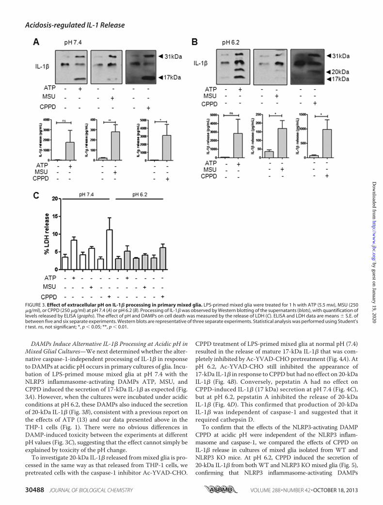
DAMPs Induce Alternative IL-1� Processing at Acidic pH inMixed Glial Cultures—We next determined whether the alter-native caspase-1-independent processing of IL-1� in responseto DAMPs at acidic pH occurs in primary cultures of glia. Incu-bation of LPS-primed mouse mixed glia at pH 7.4 with theNLRP3 inflammasome-activating DAMPs ATP, MSU, andCPPD induced the secretion of 17-kDa IL-1� as expected (Fig.3A). However, when the cultures were incubated under acidicconditions at pH 6.2, these DAMPs also induced the secretionof 20-kDa IL-1� (Fig. 3B), consistent with a previous report onthe effects of ATP (13) and our data presented above in theTHP-1 cells (Fig. 1). There were no obvious differences inDAMP-induced toxicity between the experiments at differentpH values (Fig. 3C), suggesting that the effect cannot simply beexplained by toxicity of the pH change.To investigate 20-kDa IL-1� released frommixed glia is pro-
cessed in the same way as that released from THP-1 cells, wepretreated cells with the caspase-1 inhibitor Ac-YVAD-CHO.
CPPD treatment of LPS-primed mixed glia at normal pH (7.4)resulted in the release of mature 17-kDa IL-1� that was com-pletely inhibited by Ac-YVAD-CHOpretreatment (Fig. 4A). AtpH 6.2, Ac-YVAD-CHO still inhibited the appearance of17-kDa IL-1� in response to CPPD but had no effect on 20-kDaIL-1� (Fig. 4B). Conversely, pepstatin A had no effect onCPPD-induced IL-1� (17 kDa) secretion at pH 7.4 (Fig. 4C),but at pH 6.2, pepstatin A inhibited the release of 20-kDaIL-1� (Fig. 4D). This confirmed that production of 20-kDaIL-1� was independent of caspase-1 and suggested that itrequired cathepsin D.To confirm that the effects of the NLRP3-activating DAMP
CPPD at acidic pH were independent of the NLRP3 inflam-masome and caspase-1, we compared the effects of CPPD onIL-1� release in cultures of mixed glia isolated from WT andNLRP3 KO mice. At pH 6.2, CPPD induced the secretion of20-kDa IL-1� from bothWT and NLRP3 KOmixed glia (Fig. 5),confirming that NLRP3 inflammasome-activating DAMPs
FIGURE 3. Effect of extracellular pH on IL-1� processing in primary mixed glia. LPS-primed mixed glia were treated for 1 h with ATP (5.5 mM), MSU (250�g/ml), or CPPD (250 �g/ml) at pH 7.4 (A) or pH 6.2 (B). Processing of IL-1� was observed by Western blotting of the supernatants (blots), with quantification oflevels released by ELISA (graphs). The effect of pH and DAMPs on cell death was measured by the release of LDH (C). ELISA and LDH data are means � S.E. ofbetween five and six separate experiments. Western blots are representative of three separate experiments. Statistical analysis was performed using Student’st test. ns, not significant; *, p � 0.05; **, p � 0.01.
Acidosis-regulated IL-1 Release
30488 JOURNAL OF BIOLOGICAL CHEMISTRY VOLUME 288 • NUMBER 42 • OCTOBER 18, 2013
by guest on January 19, 2020http://w
ww
.jbc.org/D
ownloaded from

induced the secretion of IL-1� from LPS-primed cells indepen-dently of the NLRP3 inflammasome under acidic conditions.Lactic Acidosis Induces IL-1� Release from Mixed Glial
Cultures—During cerebral ischemia, there is a marked eleva-tion in lactic acid that contributes to the drop in tissue pH (12,19). Thus, we hypothesized that lactic acid could stimulate
the release of IL-1� in an ischemic brain. Treatment of LPS-primed mixed glia with lactic acid induced the release of20-kDa IL-1� and a minor amount of 17-kDa IL-1� (Fig. 6A).This effect required LPS priming because lactic acid failed toinduce expression or release of IL-1� in its absence (Fig. 6, Band C). Again, there was no obvious change in toxicity with
FIGURE 4. DAMP-induced release of 20-kDa IL-1� is caspase-1-independent in primary mixed glia. LPS-primed mixed glia were pretreated with thecaspase-1 inhibitor Ac-YVAD-CHO (YVAD; 100 �M) or the cathepsin D inhibitor pepstatin A (PepA; 50 �M) for 15 min prior to CPPD treatment (250 �g/ml, 1 h) atpH 7.4 (A and C) or pH 6.2 (B and D). Processing of IL-1� was observed by Western blotting of the supernatants (blots), with quantification of levels released byELISA (graphs). ELISA data are means � S.E. of between five and six separate experiments. Western blots are representative of three separate experiments.Statistical analysis was performed using a one-way ANOVA with a Bonferroni post hoc test. ns, not significant; **, p � 0.01; ***, p � 0.001.
Acidosis-regulated IL-1 Release
OCTOBER 18, 2013 • VOLUME 288 • NUMBER 42 JOURNAL OF BIOLOGICAL CHEMISTRY 30489
by guest on January 19, 2020http://w
ww
.jbc.org/D
ownloaded from

lactic acid treatment (Fig. 6D), suggesting that the IL-1�release observed with lactic acid cannot simply be explainedby toxicity.Lactic acid-induced release of 17-kDa IL-1�was inhibited by
Ac-YVAD-CHO, whereas release of 20-kDa IL-1� was unaf-fected (Fig. 7A). Lactic acid also induced the release of 20-kDaIL-1� from NLRP3 KO mixed glia (Fig. 7B), confirming theNLRP3 inflammasome- and caspase-1-independent release.Consistent with data presented above, lactic acid-induced20-kDa IL-1� was inhibited by the cathepsin D inhibitor pep-statin A (Fig. 7C).Lactic Acid Induces IL-1� Processing and Release fromMixed
Glial Cultures—The effects of lactic acid on the IL-1 systemwere not limited to IL-1� because treatment of LPS-primedmixed glia with lactic acid also induced the release of processedIL-1� (Fig. 8A), which we have reported previously to be a keyearly mediator of inflammation following acute brain injury(20, 21). Lactic acid-induced IL-1� release was also indepen-dent of the NLRP3 inflammasome (Fig. 8B) but required thecalcium-dependent protease calpain because calpain inhibitorIII inhibited lactic acid-induced IL-1� processing and release(Fig. 8C). Thus, in line with the NLRP3-independent actions oflactic acid on IL-1� release described above, these data suggestthat lactic acid also induces the release of NLRP3-independentbut calpain-dependent IL-1� release from glia.
DISCUSSION
Most studies on IL-1� release in in vitro systems are con-ducted under physiological conditions and in peripheral
immune cells such as macrophages. The objective of this studywas to investigate IL-1� secretion under disease relevant con-ditions, with particular reference to ischemic stroke, for which
FIGURE 5. CPPD-induced 20-kDa IL-1� is NLRP3-independent in primarymixed glia. Mixed glial cultures prepared from WT and NLRP3 KO mice weretreated with CPPD (250 �g/ml, 1 h) at pH 6.2. Processing of IL-1� wasobserved by Western blotting of the supernatants (blot), with quantificationof levels released by ELISA (graph). ELISA data are means � S.E. of three sep-arate experiments. Western blots are representative of three separate exper-iments. Statistical analysis was performed using a one-way ANOVA with aBonferroni post hoc test. ns, not significant; **, p � 0.01.
FIGURE 6. Lactic acid induces the release of 20-kDa IL-1� from primarymixed glia. LPS-primed mixed glia were treated with lactic acid (25 mM, 8 h)or ATP (5.5 mM, 1 h). Processing of IL-1� was observed by Western blotting ofthe supernatants (A), with quantification of levels of IL-1� in cell lysates (B)and released into the supernatants (C) measured by ELISA. The effect of lacticacid and ATP on cell death was measured by the release of LDH (D). ELISA dataare means � S.E. of six separate experiments. Western blots are representa-tive of three separate experiments. LDH data are means � S.E. of three sepa-rate experiments. Statistical analysis was performed using a one-way ANOVAwith a Bonferroni post hoc test. ns, not significant; ***, p � 0.001.
Acidosis-regulated IL-1 Release
30490 JOURNAL OF BIOLOGICAL CHEMISTRY VOLUME 288 • NUMBER 42 • OCTOBER 18, 2013
by guest on January 19, 2020http://w
ww
.jbc.org/D
ownloaded from

lactic acidosis is relevant (19). Under acidic conditions, “classi-cal” NLRP3 inflammasome-activating DAMPs such as ATP(22) and MSU and CPPD crystals (23) induced the release of aprocessed IL-1� that was completely independent of theNLRP3 inflammasome and caspase-1, and this occurred in bothperipheral macrophage-like cells and also glial cells from thecentral nervous system. Although we and others have reportedthat MSU and CPPD stimulate IL-1-independent inflammatoryresponses (18, 24, 25), when used to stimulate IL-1 production,they are associated with the NLRP3 inflammasome- andcaspase-1-dependent processing (23).We also showed that lac-tic acid induced the release of processed IL-1� that was againindependent of the NLRP3 inflammasome and caspase-1.Caspase-1-independent processing of IL-1� has been reported
previously and may impact certain diseases even more signifi-cantly than caspase-1-processed IL-1� (14–16).In our experiments, priming to induce the synthesis of pro-
IL-1� was required prior to lactic acid (or DAMP)-inducedIL-1� release. The TLR4 agonist LPS is a tool for priming cellsin culture, but little is known about priming in vivo in an injuredbrain. Brain injury often results in disruption of the blood-brainbarrier (6), which would allow circulating peripheral molecules toenter the brain and act on glia. The acute phase protein serumamyloidA is elevated inplasmaduring inflammation andhasbeenshown to prime glial cells to release IL-1 following subsequentDAMP stimulation in vitro in a way similar to LPS (18).
The contribution of IL-1 to ischemic brain injury is wellestablished (6), with the utility of the IL-1 receptor antagonist as
FIGURE 7. Lactic acid-induced release of 20-kDa IL-1� occurs independently of caspase-1 in primary mixed glia. LPS-primed mixed glia were pretreatedwith 100 �M Ac-YVAD-CHO (YVAD; A) or 50 �M pepstatin A (PepA; C) for 15 min prior to lactic acid treatment (25 mM, 8 h). Mixed glial cultures prepared from WTand NLRP3 KO mice were treated with 25 mM lactic acid (8 h; B). Processing of IL-1� was observed by Western blotting of the supernatants (blots), withquantification of levels released by ELISA (graphs). ELISA data are means � S.E. of between three and six separate experiments. Western blots are representativeof three separate experiments. Statistical analysis was performed using a one-way ANOVA with a Bonferroni post hoc test. ns, not significant; **, p � 0.01; ***,p � 0.001.
Acidosis-regulated IL-1 Release
OCTOBER 18, 2013 • VOLUME 288 • NUMBER 42 JOURNAL OF BIOLOGICAL CHEMISTRY 30491
by guest on January 19, 2020http://w
ww
.jbc.org/D
ownloaded from

a therapeutic under clinical investigation (26). Brain acidosisafter ischemia contributes to neuronal injury, and the damagingeffects are mediated, at least in part, by acid-sensing ion chan-nels (27, 28). Our data provide the rationale that acidosis andneuronal injury could also be linked via the IL-1 system. Levelsof lactic acid increase in an ischemic brain due to a lack ofoxygen and the up-regulation of anaerobic glycolysis. Stimula-tion of LPS-primed cultures of glial cells with lactic acid or withNLRP3 inflammasome-activating DAMPs under acidic condi-tions induced cathepsin D-dependent release of 20-kDa IL-1�,consistentwith a previous report showingATP-induced releaseof 20-kDa IL-1� from microglia under acidic conditions (13).This cathepsinD cleavage of IL-1� adds to a growing number ofproteases now known to cleave pro-IL-1�. These proteases
cleave IL-1� at different sites to produce different size frag-ments corresponding to differing activities (29). The putativecathepsin D-dependent product at 20 kDa is suggested to be�5-fold less active at IL-1 receptor I than the caspase-1-gener-ated 17-kDa product (29). Thus, if generated in vivo, it could actas an agonist or maybe a partial agonist of IL-1 receptor I andcontribute to inflammatory responses during ischemia; there-fore, further experiments to understand the exact nature of itsrole in vivo are required to help direct any future targeting ofthis pathway. Acidosis was also recently described as aninducer of the NLRP3 inflammasome (17). Although we didobserve 17-kDa IL-1� following lactic acid treatment (e.g.Fig. 7A), it was the minor species compared with the 20-kDaform. Together, these data strongly suggest that acidosis is
FIGURE 8. Lactic acid induces release of IL-1� from primary mixed glia. LPS-primed WT mixed glia were treated with 25 mM lactic acid for 8 h, and IL-1�processing and release was measured (A). LPS-primed mixed glia from WT and NLRP3 KO mice were treated with lactic acid (25 mM; 8 h; B). LPS-primed WTmixed glia were pretreated with calpain inhibitor III (Cal; 50 �M) for 15 min prior to lactic acid treatment (25 mM, 8 h; C). Processing of IL-1� was observed byWestern blotting of the supernatants (blots), with quantification of levels released by ELISA (graphs). ELISA data are means � S.E. of between three and sixseparate experiments. Western blots are representative of three separate experiments. Statistical analysis was performed using Student’s t test (A) or one-wayANOVA with a Bonferroni post hoc test (B and C). ns, not significant; *, p � 0.05; **, p � 0.01; ***, p � 0.001.
Acidosis-regulated IL-1 Release
30492 JOURNAL OF BIOLOGICAL CHEMISTRY VOLUME 288 • NUMBER 42 • OCTOBER 18, 2013
by guest on January 19, 2020http://w
ww
.jbc.org/D
ownloaded from

an important disease relevant regulator of IL-1�-processingpathways.Until recently, the precursor of IL-1� (pro-IL-1�) was
thought to be as biologically active as the mature 17-kDa format IL-1 receptor I. However, recent research has shown thatprocessed IL-1� is significantly more biologically active thanthe pro-form (30, 31), which highlights IL-1� processing as abiologically important mechanism. In addition to its effects onIL-1�, lactic acid also induced the release of processed IL-1�,which was 17 kDa and was inhibited by a calpain inhibitor,consistent with the involvement of a calpain protease in itsprocessing (7). DAMPs are known to induce the secretion ofprocessed IL-1� (32), and DAMP-induced processing andsecretion of IL-1� can be dependent, partially dependent, orindependent of the NLRP3 inflammasome and caspase-1,depending upon the DAMP (32). Our data suggest that lacticacid-induced IL-1� secretion occurs independently of theinflammasome. In addition to the effects of IL-1� in acute braininjury, IL-1� is also known to play an important role in braininjury (20, 21, 33). IL-1� is up-regulated before IL-1� and asearly as 4 h post-ischemic brain injury in areas of subsequentneuronal loss, suggesting that it may be the key driver of thedamaging early inflammatory response (20). Thus, the lacticacid-induced IL-1� release we have described may contributeto damage post-ischemic injury.In summary, these data provide new insights into the mech-
anisms of IL-1 production during disease relevant conditions.We have shown that under acidic conditions, both proinflam-matory forms of the IL-1 family (IL-1� and IL-1�) are regulatedindependently of the NLRP3 inflammasome. Thus, consider-ation of this observation should be made when investigatinginhibitors of the NLRP3 inflammasome to target IL-1 in dis-ease, particularly where changes in the local environment maydictate the mechanism of IL-1� processing.
Acknowledgments—We are grateful to Dr. Vishva Dixit (Genentech)for providing the NLRP3 KO mice and to Prof. Nancy Rothwell forcritically evaluating the manuscript.
REFERENCES1. Reardon, S. (2011) A world of chronic disease. Science 333, 558–5592. Angell, S. Y., Danel, I., and DeCock, K. M. (2012) Global health. Global indi-
cators and targets for noncommunicable diseases. Science 337, 1456–14573. Rock, K. L., Latz, E., Ontiveros, F., and Kono, H. (2010) The sterile inflam-
matory response. Annu. Rev. Immunol. 28, 321–3424. Dinarello, C. A. (2010) Anti-inflammatory agents: present and future.Cell
140, 935–9505. Dinarello, C. A. (2011) Blocking interleukin-1� in acute and chronic au-
toinflammatory diseases. J. Int. Med. 269, 16–286. Allan, S. M., Tyrrell, P. J., and Rothwell, N. J. (2005) Interleukin-1 and
neuronal injury. Nat. Rev. Immunol. 5, 629–6407. Luheshi, N.M., Rothwell, N. J., and Brough,D. (2009)Dual functionality of
interleukin-1 family cytokines: implications for anti-interleukin-1 ther-apy. Br. J. Pharmacol. 157, 1318–1329
8. Strowig, T., Henao-Mejia, J., Elinav, E., and Flavell, R. (2012) Inflam-masomes in health and disease. Nature 481, 278–286
9. Schroder, K., and Tschopp, J. (2010) The inflammasomes. Cell 140,821–832
10. Chen, G. Y., and Nuñez, G. (2010) Sterile inflammation: sensing and re-acting to damage. Nat. Rev. Immunol. 10, 826–837
11. Iyer, S. S., Pulskens, W. P., Sadler, J. J., Butter, L. M., Teske, G. J., Ulland,T. K., Eisenbarth, S. C., Florquin, S., Flavell, R. A., Leemans, J. C., andSutterwala, F. S. (2009) Necrotic cells trigger a sterile inflammatory re-sponse through the Nlrp3 inflammasome. Proc. Natl. Acad. Sci. U.S.A.106, 20388–20393
12. Nemoto, E. M., and Frinak, S. (1981) Brain tissue pH after global brainischemia and barbiturate loading in rats. Stroke 12, 77–82
13. Takenouchi, T., Iwamaru, Y., Sugama, S., Tsukimoto, M., Fujita, M., Seki-gawa, A., Sekiyama, K., Sato,M., Kojima, S., Conti, B., Hashimoto,M., andKitani, H. (2011) The activation of P2X7 receptor induces cathepsinD-de-pendent production of a 20-kDa form of IL-1� under acidic extracellularpH in LPS-primed microglial cells. J. Neurochem. 117, 712–723
14. Fantuzzi, G., Ku,G., Harding,M.W., Livingston, D. J., Sipe, J. D., Kuida, K.,Flavell, R. A., andDinarello, C.A. (1997) Response to local inflammation ofIL-1�-converting enzyme- deficient mice. J. Immunol. 158, 1818–1824
15. Stehlik, C. (2009) Multiple interleukin-1�a-converting enzymes contrib-ute to inflammatory arthritis. Arthritis Rheum. 60, 3524–3530
16. Joosten, L. A., Netea, M. G., Fantuzzi, G., Koenders, M. I., Helsen, M. M.,Sparrer, H., Pham, C. T., van derMeer, J.W., Dinarello, C. A., and van denBerg,W. B. (2009) Inflammatory arthritis in caspase 1 gene-deficientmice:contribution of proteinase 3 to caspase 1-independent production of bio-active interleukin-1�. Arthritis Rheum 60, 3651–3662
17. Rajamäki, K., Nordström, T., Nurmi, K., Åkerman, K. E., Kovanen, P. T.,Öörni, K., and Eklund, K. K. (2013) Extracellular acidosis is a novel dangersignal alerting innate immunity via the NLRP3 inflammasome. J. Biol.Chem. 288, 13410–13419
18. Savage, C. D., Lopez-Castejon, G., Denes, A., and Brough, D. (2012)NLRP3-inflammasome activating DAMPs stimulate an inflammatory re-sponse in glia in the absence of primingwhich contributes to brain inflam-mation after injury. Front. Immunol. 3, 288
19. Sun, P. Z., Cheung, J. S., Wang, E., and Lo, E. H. (2011) Association be-tween pH-weighted endogenous amide proton chemical exchange satu-ration transferMRI and tissue lactic acidosis during acute ischemic stroke.J. Cereb. Blood Flow Metab. 31, 1743–1750
20. Luheshi, N. M., Kovács, K. J., Lopez-Castejon, G., Brough, D., and Denes,A. (2011) Interleukin-1� expression precedes IL-1� after ischemic braininjury and is localised to areas of focal neuronal loss and penumbral tis-sues. J. Neuroinflammation 8, 186
21. Greenhalgh, A. D., Brough, D., Robinson, E. M., Girard, S., Rothwell, N. J.,andAllan, S.M. (2012) Interleukin-1 receptor antagonist is beneficial aftersubarachnoid haemorrhage in rat by blocking haem-driven inflammatorypathology. Dis. Model. Mech. 5, 823–833
22. Mariathasan, S., Weiss, D. S., Newton, K., McBride, J., O’Rourke, K.,Roose-Girma, M., Lee, W. P., Weinrauch, Y., Monack, D. M., and Dixit,V. M. (2006) Cryopyrin activates the inflammasome in response to toxinsand ATP. Nature 440, 228–232
23. Martinon, F., Pétrilli, V., Mayor, A., Tardivel, A., and Tschopp, J. (2006)Gout-associated uric acid crystals activate the NALP3 inflammasome.Nature 440, 237–241
24. Guerne, P. A., Terkeltaub, R., Zuraw, B., and Lotz,M. (1989) Inflammatorymicrocrystals stimulate interleukin-6 production and secretion by humanmonocytes and synoviocytes. Arthritis Rheum. 32, 1443–1452
25. Bouchard, L., de Médicis, R., Lussier, A., Naccache, P. H., and Poubelle,P. E. (2002) Inflammatory microcrystals alter the functional phenotype ofhuman osteoblast-like cells in vitro: synergism with IL-1 to overexpresscyclooxygenase-2. J. Immunol. 168, 5310–5317
26. Emsley,H.C., Smith, C. J., Georgiou, R. F., Vail, A.,Hopkins, S. J., Rothwell,N. J., and Tyrrell, P. J. (2005) A randomised phase II study of interleukin-1receptor antagonist in acute stroke patients. J. Neurol. Neurosurg. Psychi-atry 76, 1366–1372
27. Xiong, Z. G., Zhu, X. M., Chu, X. P., Minami, M., Hey, J., Wei, W. L.,MacDonald, J. F., Wemmie, J. A., Price, M. P., Welsh, M. J., and Simon,R. P. (2004) Neuroprotection in ischemia: blocking calcium-permeableacid-sensing ion channels. Cell 118, 687–698
28. Gao, J., Duan, B., Wang, D. G., Deng, X. H., Zhang, G. Y., Xu, L., and Xu,T. L. (2005) Coupling between NMDA receptor and acid-sensing ionchannel contributes to ischemic neuronal death. Neuron 48, 635–646
29. Hazuda, D. J., Strickler, J., Simon, P., and Young, P. R. (1991) Structure-
Acidosis-regulated IL-1 Release
OCTOBER 18, 2013 • VOLUME 288 • NUMBER 42 JOURNAL OF BIOLOGICAL CHEMISTRY 30493
by guest on January 19, 2020http://w
ww
.jbc.org/D
ownloaded from
function mapping of interleukin 1 precursors. Cleavage leads to a confor-mational change in the mature protein. J. Biol. Chem. 266, 7081–7086
30. Afonina, I. S., Tynan,G. A., Logue, S. E., Cullen, S. P., Bots,M., Lüthi, A. U.,Reeves, E. P., McElvaney, N. G., Medema, J. P., Lavelle, E. C., and Martin,S. J. (2011) Granzyme B-dependent proteolysis acts as a switch to enhancethe proinflammatory activity of IL-1�.Molecular cell 44, 265–278
31. Zheng, Y., Humphry, M., Maguire, J. J., Bennett, M. R., and Clarke, M. C.(2013) Intracellular interleukin-1 receptor 2 binding prevents cleavageand activity of interleukin-1�, controlling necrosis-induced sterile inflam-
mation. Immunity 38, 285–29532. Gross, O., Yazdi, A. S., Thomas, C. J., Masin, M., Heinz, L. X., Guarda, G.,
Quadroni, M., Drexler, S. K., and Tschopp, J. (2012) Inflammasome acti-vators induce interleukin-1� secretion via distinct pathways with differ-ential requirement for the protease function of caspase-1. Immunity 36,388–400
33. Boutin, H., LeFeuvre, R. A., Horai, R., Asano, M., Iwakura, Y., and Roth-well, N. J. (2001) Role of IL-1� and IL-1� in ischemic brain damage. J. Neu-rosci. 21, 5528–5534
Acidosis-regulated IL-1 Release
30494 JOURNAL OF BIOLOGICAL CHEMISTRY VOLUME 288 • NUMBER 42 • OCTOBER 18, 2013
by guest on January 19, 2020http://w
ww
.jbc.org/D
ownloaded from

Michelle E. Edye, Gloria Lopez-Castejon, Stuart M. Allan and David BroughInterleukin-1 Secretion via a Caspase-1-independent Pathway
Acidosis Drives Damage-associated Molecular Pattern (DAMP)-induced
doi: 10.1074/jbc.M113.478941 originally published online September 10, 20132013, 288:30485-30494.J. Biol. Chem.
10.1074/jbc.M113.478941Access the most updated version of this article at doi:
Alerts:
When a correction for this article is posted•
When this article is cited•
to choose from all of JBC's e-mail alertsClick here
http://www.jbc.org/content/288/42/30485.full.html#ref-list-1
This article cites 33 references, 11 of which can be accessed free at
by guest on January 19, 2020http://w
ww
.jbc.org/D
ownloaded from