Embed Size (px)
Citation preview
Dr. Mohamed Alshekhani
Background:
• Endoscopic imaging technologies provide detailed contrast enhancement of the mucosal surface & blood vessels offer an alternative to dye-based chromoendoscopy.
• Include narrow-band imaging (NBI) (Olympus), flexible spectral imaging color enhancement (FICE) (Fujinon)& i-SCAN (PENTAX).
• Aachieved by the observation of light transmission at selected wavelengths because the interaction of particular tissue structures with light is wavelength dependent.
• Selective light transmittance is accomplished by optical filtering of white light in NBI, whereas FICE & i-SCAN both accomplish this through software-driven post-image processing.
NBI:
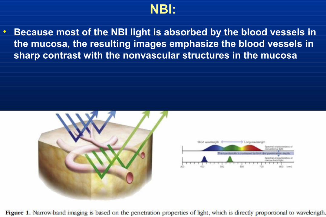
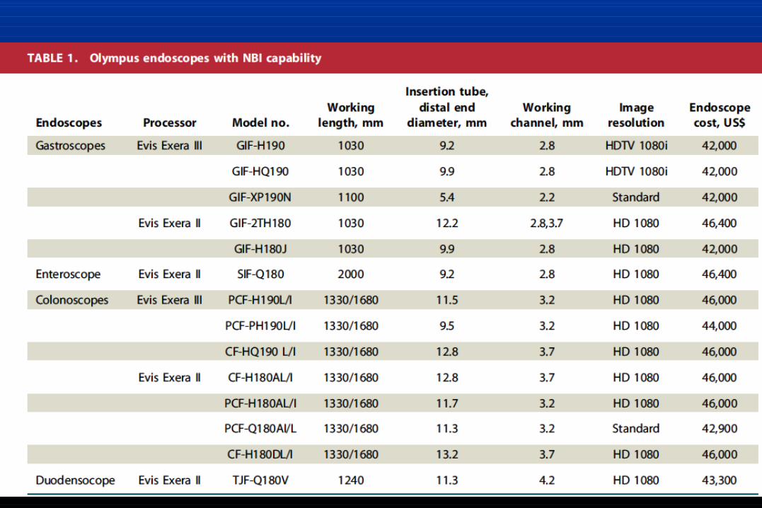
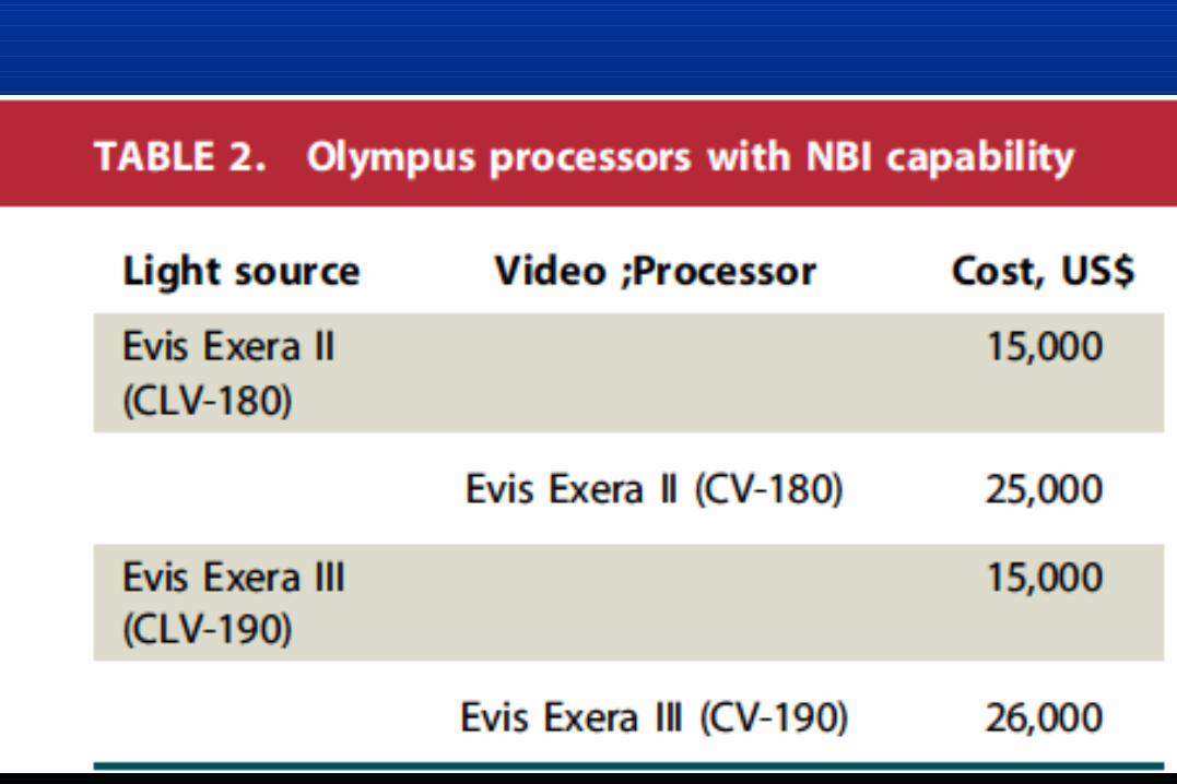
• NBI is an endoscopic optical image enhancement, from Olympus. • NBI is based on the penetration properties of light, which is
directly proportional to wavelength.• Short wavelengths penetrate only superficially into the mucosa,
whereas longer wavelengths are capable of penetrating more deeply into tissue .
• The placement of a NBI filter directly in front of the xenon arc lamp produces 2 narrow bands of light centered at the specific wavelengths of 415&540 nm,corresponding to the primary& secondary light absorption peaks of hemoglobin,respectively.
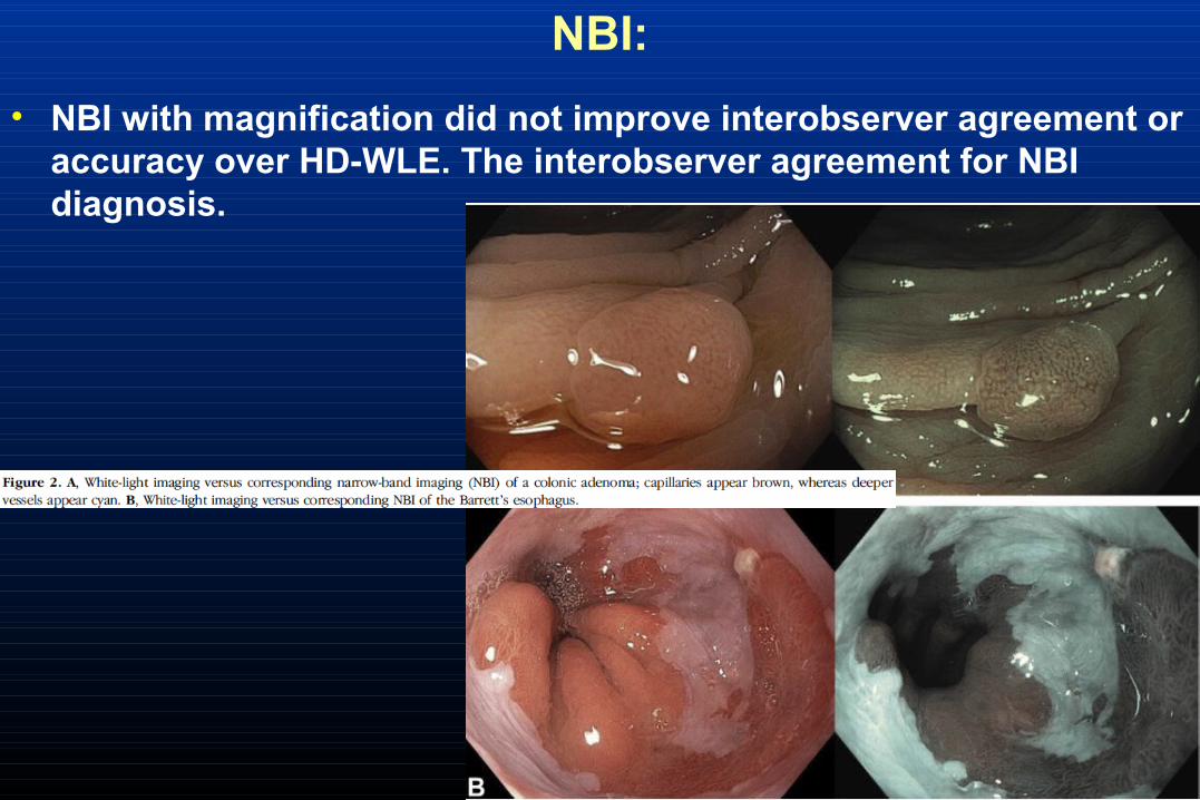
• Capillaries in the superficial mucosa are highlighted by the 415-nm wavelength &appear brown.
• The longer 540-nm wavelength penetrates slightly more deeply into the mucosa &submucosa and makes the deeper veins appear blue-green (cyan).
NBI:
• Because most of the NBI light is absorbed by the blood vessels in the mucosa, the resulting images emphasize the blood vessels in sharp contrast with the nonvascular structures in the mucosa
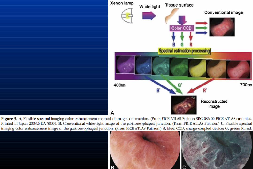
FICE:Flexible spectral image color enhancement
• Digital imaging post-processing system of Fujinon.• It takes white-light endimages from the video processor &
mathematically processes it by certain wavelengths ranges.• 3 single-wavelength images selected & assigned to red, green &
blue monitor inputs, to display a composite color-enhanced image • 10 factory-determined presets available for diff color displays
• Each preset can be button-activated from a computer keyboard.
• The factory-preset wavelengths can also be manually altered.
• There are 60 possible permutations of the available wavelengths ( 400- 695 nm) that can be manipulated in 5-nm increments.
• The endoscope push-button controller can be programmed to enable switching between the CWL image & up to 3 FICE presets.
• The switch to FICE from WLE occurs almost instantaneously.
• Optimal preset(s) for tissue diag or differen not been established
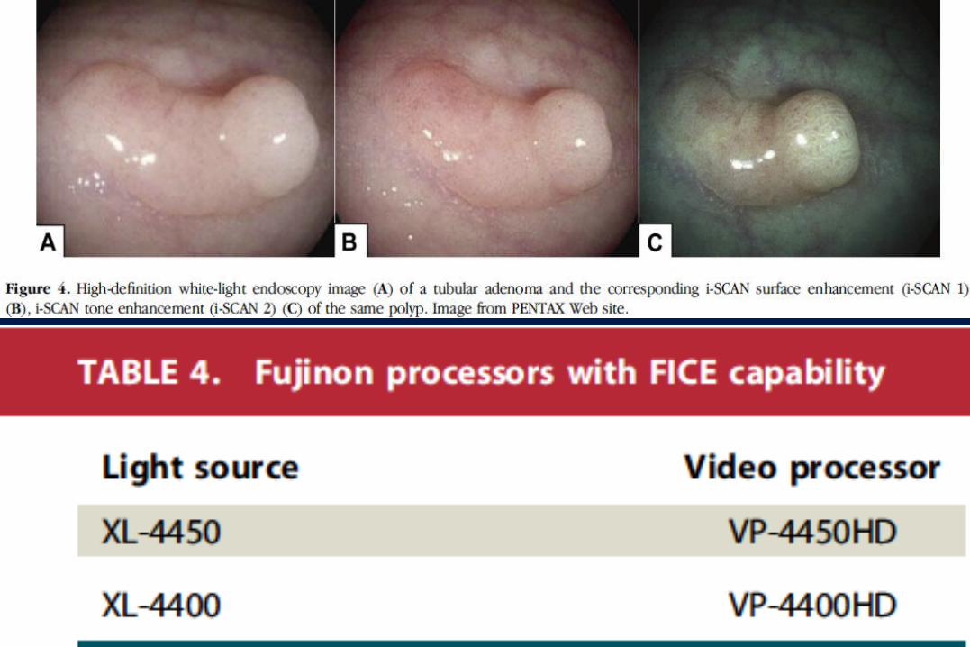
i-SCAN:
• A software-based digital, postprocessing(like FICE IE technology from PENTAX provides digital contrast to endoimages.
• 3 i-SCAN modes: 1, 2, 3,by touching a button on the endoscope. • The switch from WLE to i-SCAN occurs almost instantaneously.• i-SCAN 1 is a surface-enhancement (SE)& contrast-enhancement (CE) mode
enhances contrast &mucosal surface detail including mucosal surface texture & sharpened views of surface vessels.
• The image remains as bright as conventional WLE.• i-SCAN 2 comprises SE, CE,&tone enhancement (TE) mode&increases the
contrast between the mucosa & blood vessels, improving the visibility of blood vessels.
• i-SCAN 3 comprises SE, CE, TE mode g. provides mucosal SE &improved visualization of blood vs including dimly lit far-fields.
• i-SCAN 3 differs from i-SCAN 2 primarily in its ability to illuminate more distant regions better.
• Unlike NBI, red remains the predominant blood vessel color in all i-SCAN modes.

NBI:EFFICACY STUDIES
• Most studied. • Barrett’s esophagus, high-grade dysplasia, adenocarcinoma• NBI sharpens visualization of the squamo-columnar boundary,can
potentially detect BE)& associated dysplasia. • HD-NBI without magnification was superior to SD-WLE in detecting
dysplasia& higher grades of dysplasia were found
• A randomized, crossover study showed no difference in the detection of high-grade dysplasia (HGD) between HD-WLE & HD-NBI without magnification
• A recent multicenter, randomized, crossover trial: use of NBI allowed fewer biopsy specimens per patient &detected a higher proportion of areas with HGD.
• These trials suggest that both NBI / HD may individually contribute to increased detection of Barrett’ s&associated dysplasia.
NBI:
• NBI with magnification did not improve interobserver agreement or accuracy over HD-WLE. The interobserver agreement for NBI diagnosis.
NBI:
• GERD• NBI as a diagnostic tool for GERD requires further validation with
randomized, prospective studies.• GERD patients had a significantly increased number of
capillaries,or capillary dilation &tortuosity of intrapapillary capillary, microerosions and increased vascularity at the squamocolumnar junction.
NBI:• Gastric neoplasia:• NBI without magnification increased diagnostic yield for detection
of gastric lesions, including dysplasia&intestinal metaplasia compared with routine WLE.
• Magnifying endoscopy with NBI found to be useful in diagnosing gastric neoplasia,significantly better than HD-WLE &M-NBI finding of a light blue crest on the epithelial surface of gastric mucosa correlated with histological evidence of intestinal metaplasia.
• Experienced endoscopists recommended M-NBI over NBI alone for the detection of gastric cancer.
• The vessel plus surface architecture classification system by M-NBI to describe the microvascular /microsurface changes in the stomach in gastric cancer diagnosed high-grade adenomas or early carcinomas at a higher sensitivity than HDWLE.
NBI:
• M-NBI to detect gastric small depressive lesions 10 mm or smaller & diagnostic accuracy was signifi cantly higher for NBI than for HD-WLE but the difference was not statistically significant.
• The accuracy& specificity of M-NBI were greater than those of HD-WLE,but not SS.
• NBI appears to be a useful adjunct in the diagnosis of gastric neoplasia, but cannot replace biopsy at this time.
NBI:
• Colon polyps• There was no statistically significant difference between WLE (SD
& HD) & NBI for the detection of patients with colorectal polyps.• The Cochrane review also compared NBI with SD-WLE, found that
polyp and adenoma detection might be superior with NBI compared with SD-WLE. NBI was superior to SD-WLE in patients with at least 1 colorectal polyp or adenoma.
• NBI with or without magnification is precise in identifying adenomas based on visualization alone.
NBI:
• Ulcerative colitis• No difference between the NBI and HD-WLE groups in the
proportion of patients with at least 1 area of dysplasia• NBI may be an adjunct in vivo tool for the assessment of the grade
of inflammation in patients with quiescent UC.
FICE & I-SCAN:
• Esophagus• Detection of HGD in BE, FICE & chromoendoscopy both had a
Sensitivity of directed biopsies alone for the detection of lesions which was not statistically significant.
• A study examined the ability to more clearly visualize palisade vessels &distinguish the demarcation between BE mucosa & gastric mucosa by using FICE images & WLE; FICE images was significantly higher than that of WLE images
• The diagnostic yield of reflux esophagitis was significantly higher in the i-SCAN group compared with the HD-WLE group.
FICE & I-SCAN:
• Gastric neoplasia• Depressed-type early gastric cancer: Greater median color
differences between malignant lesions&the surrounding mucosa were present in FICE images compared with conventional images, resulting in images with better contrast
• Although magnified i-SCAN with TE& SE slightly increased the diagnostic yield in gastric neoplasia , there was no significant difference compared with magnified HDWLE.
62
FICE & I-SCAN:
• Polyps• No difference between the FICE & HD-WLE groups in adenoma
detection rate,total number of adenomas, or detection of subgroups of adenomas.
• The number of hyperplastic polyps was also the same in both groups&results were the same for both the screening & the diagnostic colonoscopy subgroups.
• Withdrawal time was similar in both groups
• The overall accuracy achieved by FICE with magnification in the diagnosis of adenomas (87.0%) wassignifi cantly greater than FICE without magnification
• HD i-SCAN detected significantly more patients with colorectal neoplasia (38%) compared with SD-WLE.
• The prediction of neoplastic& non-neoplastic colorectal lesions was more precise in the i-SCAN 2 group compared with HD-WLE .
FICE & I-SCAN:
• Polyps• There were no significant differences between HD-WLE& i-SCAN in
the characterization of polyps of smaller than 10 • Histology was correctly predicted with a sensitivity, specificity,&
accuracy of 95%, 82%& 92%, respectively.
COMPARATIVE STUDIES:
• NBI versus i-SCAN• NBI&i-SCAN had a significantly higher sensitivity & improved
accuracy compared with HDWLE, for the prediction of adenomas • There were no significant differences between the NBI&i-SCAN
with good intra- / interobserver agreement between.
NBI versus FICE
• Several studies have compared NBI and FICE for polyp detection, all of which have shown no difference.
• Neither NBI nor FICE increased the mean number of adenomas detected per patient compared with HD-WLE
• The percentage of missed adenomas also did not differ between the 3 groups.
SAFETY
• There have been no reported complications attributed to the use of NBI, FICE, or i-SCAN.
FINANCIAL CONSIDERATIONS
• Electronic chromoendoscopy has the potential to avoid costs associated with tissue sampling; however, this is currently not the standard of care.
• There are no unique Current Procedural Terminology codes for NBI, FICE, or i-SCAN.