Embed Size (px)
Citation preview
Accurate mass spectrometry elucidates a misleading metabonate formed from amine-�containing drugs in reactive metabolite screening assays
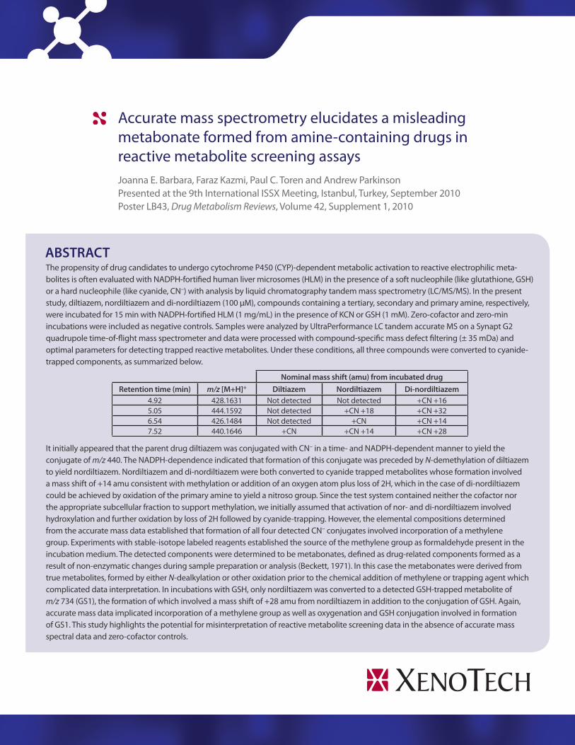
The propensity of drug candidates to undergo cytochrome P450 (CYP)-�dependent metabolic activation to reactive electrophilic meta-�bolites is often evaluated with NADPH-�fortified human liver microsomes (HLM) in the presence of a soft nucleophile (like glutathione, GSH) or a hard nucleophile (like cyanide, CN–) with analysis by liquid chromatography tandem mass spectrometry (LC/MS/MS). In the present study, diltiazem, nordiltiazem and di-�nordiltiazem (100 µM), compounds containing a tertiary, secondary and primary amine, respectively, were incubated for 15 min with NADPH-�fortified HLM (1 mg/mL) in the presence of KCN or GSH (1 mM). Zero-�cofactor and zero-�min incubations were included as negative controls. Samples were analyzed by UltraPerformance LC tandem accurate MS on a Synapt G2 quadrupole time-�of-�flight mass spectrometer and data were processed with compound-�specific mass defect filtering (± 35 mDa) and optimal parameters for detecting trapped reactive metabolites. Under these conditions, all three compounds were converted to cyanide-�trapped components, as summarized below.
Nominal mass shift (amu) from incubated drugRetention time (min) m/z [M+H]+ Diltiazem Nordiltiazem Di-nordiltiazem
4.92 428.1631 Not detected Not detected +CN +16 5.05 444.1592 Not detected +CN +18 +CN +32 6.54 426.1484 Not detected +CN +CN +14 7.52 440.1646 +CN +CN +14 +CN +28
It initially appeared that the parent drug diltiazem was conjugated with CN– in a time-� and NADPH-�dependent manner to yield the conjugate of m/z 440. The NADPH-�dependence indicated that formation of this conjugate was preceded by N-�demethylation of diltiazem to yield nordiltiazem. Nordiltiazem and di-�nordiltiazem were both converted to cyanide trapped metabolites whose formation involved a mass shift of +14 amu consistent with methylation or addition of an oxygen atom plus loss of 2H, which in the case of di-�nordiltiazem could be achieved by oxidation of the primary amine to yield a nitroso group. Since the test system contained neither the cofactor nor the appropriate subcellular fraction to support methylation, we initially assumed that activation of nor-� and di-�nordiltiazem involved hydroxylation and further oxidation by loss of 2H followed by cyanide-�trapping. However, the elemental compositions determined from the accurate mass data established that formation of all four detected CN– conjugates involved incorporation of a methylene group. Experiments with stable-�isotope labeled reagents established the source of the methylene group as formaldehyde present in the incubation medium. The detected components were determined to be metabonates, defined as drug-�related components formed as a result of non-�enzymatic changes during sample preparation or analysis (Beckett, 1971). In this case the metabonates were derived from true metabolites, formed by either N-�dealkylation or other oxidation prior to the chemical addition of methylene or trapping agent which complicated data interpretation. In incubations with GSH, only nordiltiazem was converted to a detected GSH-�trapped metabolite of m/z 734 (GS1), the formation of which involved a mass shift of +28 amu from nordiltiazem in addition to the conjugation of GSH. Again, accurate mass data implicated incorporation of a methylene group as well as oxygenation and GSH conjugation involved in formation of GS1. This study highlights the potential for misinterpretation of reactive metabolite screening data in the absence of accurate mass spectral data and zero-�cofactor controls.
ABSTRACT
Joanna E. Barbara, Faraz Kazmi, Paul C. Toren and Andrew Parkinson Presented at the 9th International ISSX Meeting, Istanbul, Turkey, September 2010 Poster LB43, Drug Metabolism Reviews, Volume 42, Supplement 1, 2010
In vitro screening assays involving trapping of electrophiles by hard (e.g., cyanide ion, CN–) and soft (e.g., glutathione, GSH) nucleophiles are commonly employed to determine the propensity of new drug candidates to be converted by cytochrome P450 (CYP) (and occasionally other drug metabolizing enzymes) to reactive electrophilic metabolites. The assay typically involves incubating the drug with appropriate subcellular fractions and cofactors to support bioactivation, in the presence of a trapping agent, followed by profiling for trapped metabolites, commonly by liquid chromatography/mass spectrometry. The calcium channel blocker diltiazem (Figure 1) is a quasi-�irreversible, metabolism-�dependent inhibitor of cytochrome P450 (CYP) 3A4. Inhibition involves formation of a metabolite intermediate complex (MIC) which is proposed to comprise a metabolite formed by oxidative biotransformation of the alkylamine group that coordinately binds to the ferrous heme iron of CYP3A4 (Hanson et al., 2010). The metabolite has not been identified but is commonly proposed as the nitroso metabolite of di-�nordiltiazem, which has never been isolated. Like diltiazem, nordiltiazem forms an MIC with CYP3A4, but di-�nordiltiazem does not form a spectrophotometrically similar complex. In fact, Hanson et al. (2010) recently proposed that di-�nordiltiazem may inhibit MIC formation from diltiazem and nordiltiazem. In the present study, diltiazem, nordiltiazem and di-�nordiltiazem (Figure 1), which contain a tertiary, secondary and primary amine, respectively, were incubated with NADPH-�fortified human liver microsomes (HLM) in the presence of CN– or GSH. Samples were profiled by high-�resolution accurate LC/MS/MS for trapped metabolites potentially associated with the inhibition of CYP by diltiazem.
INTRODUCTION
RESULTS
Table 1.
Nominal mass shift (amu) from incubated drug
Name Retention time (min)
m/z [M+H]+ Diltiazem Nordiltiazem Di-nordiltiazem
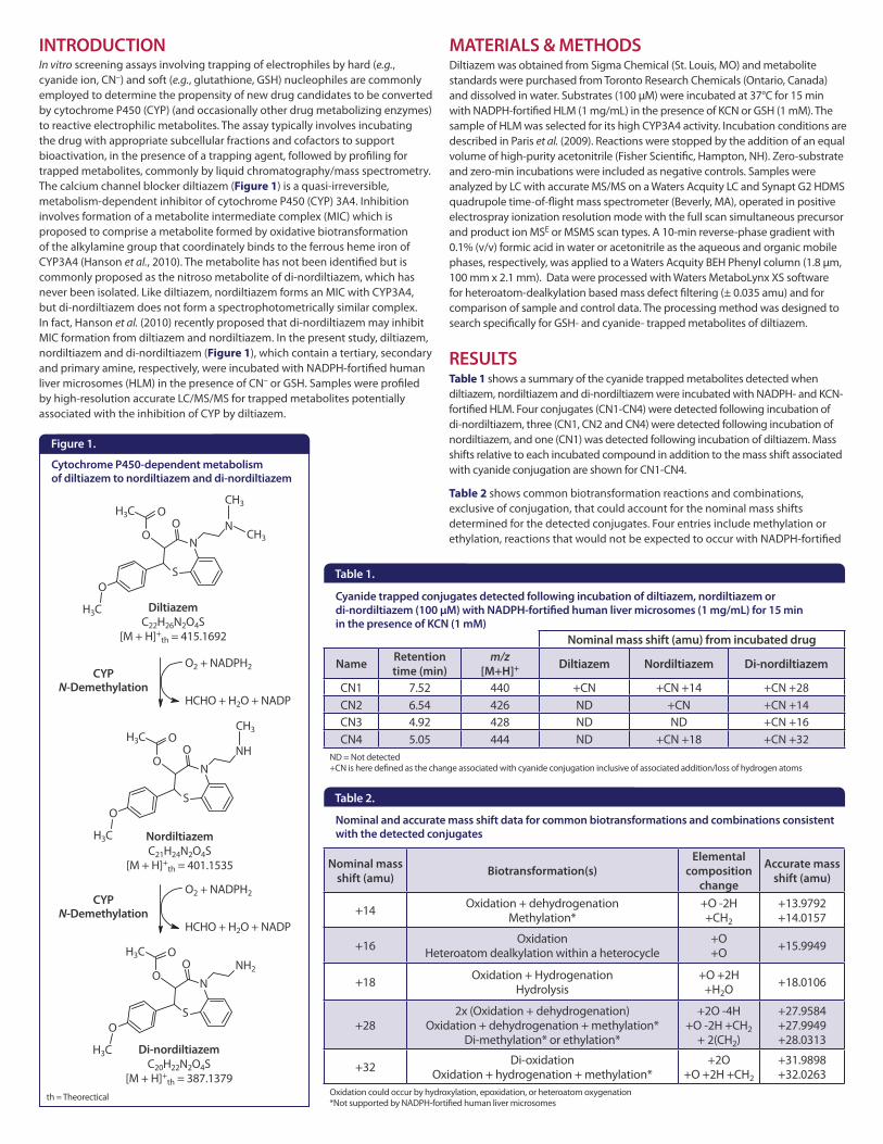
CN1 7.52 440 +CN +CN +14 +CN +28CN2 6.54 426 ND +CN +CN +14CN3 4.92 428 ND ND +CN +16CN4 5.05 444 ND +CN +18 +CN +32
Cyanide trapped conjugates detected following incubation of diltiazem, nordiltiazem or di-nordiltiazem (100 µM) with NADPH-fortified human liver microsomes (1 mg/mL) for 15 min in the presence of KCN (1 mM)
Table 1 shows a summary of the cyanide trapped metabolites detected when diltiazem, nordiltiazem and di-�nordiltiazem were incubated with NADPH-� and KCN-�fortified HLM. Four conjugates (CN1-�CN4) were detected following incubation of di-�nordiltiazem, three (CN1, CN2 and CN4) were detected following incubation of nordiltiazem, and one (CN1) was detected following incubation of diltiazem. Mass shifts relative to each incubated compound in addition to the mass shift associated with cyanide conjugation are shown for CN1-�CN4.
Table 2 shows common biotransformation reactions and combinations, exclusive of conjugation, that could account for the nominal mass shifts determined for the detected conjugates. Four entries include methylation or ethylation, reactions that would not be expected to occur with NADPH-�fortified
Diltiazem was obtained from Sigma Chemical (St. Louis, MO) and metabolite standards were purchased from Toronto Research Chemicals (Ontario, Canada) and dissolved in water. Substrates (100 µM) were incubated at 37°C for 15 min with NADPH-�fortified HLM (1 mg/mL) in the presence of KCN or GSH (1 mM). The sample of HLM was selected for its high CYP3A4 activity. Incubation conditions are described in Paris et al. (2009). Reactions were stopped by the addition of an equal volume of high-�purity acetonitrile (Fisher Scientific, Hampton, NH). Zero-�substrate and zero-�min incubations were included as negative controls. Samples were analyzed by LC with accurate MS/MS on a Waters Acquity LC and Synapt G2 HDMS quadrupole time-�of-�flight mass spectrometer (Beverly, MA), operated in positive electrospray ionization resolution mode with the full scan simultaneous precursor and product ion MSE or MSMS scan types. A 10-�min reverse-�phase gradient with 0.1% (v/v) formic acid in water or acetonitrile as the aqueous and organic mobile phases, respectively, was applied to a Waters Acquity BEH Phenyl column (1.8 µm, 100 mm x 2.1 mm). Data were processed with Waters MetaboLynx XS software for heteroatom-�dealkylation based mass defect filtering (± 0.035 amu) and for comparison of sample and control data. The processing method was designed to search specifically for GSH-� and cyanide-� trapped metabolites of diltiazem.
MATERIALS & METHODS
ND = Not detected +CN is here defined as the change associated with cyanide conjugation inclusive of associated addition/loss of hydrogen atoms
Table 2.
Nominal mass shift (amu) Biotransformation(s)
Elemental composition
change
Accurate mass shift (amu)
+14 Oxidation + dehydrogenation Methylation*
+O -�2H +CH2
+13.9792 +14.0157
+16 Oxidation Heteroatom dealkylation within a heterocycle
+O +O +15.9949
+18 Oxidation + Hydrogenation Hydrolysis
+O +2H +H2O +18.0106
+282x (Oxidation + dehydrogenation)
Oxidation + dehydrogenation + methylation* Di-�methylation* or ethylation*
+2O -�4H +O -�2H +CH2
+ 2(CH2)
+27.9584 +27.9949 +28.0313
+32 Di-�oxidation Oxidation + hydrogenation + methylation*
+2O +O +2H +CH2
+31.9898 +32.0263
Nominal and accurate mass shift data for common biotransformations and combinations consistent with the detected conjugates
Oxidation could occur by hydroxylation, epoxidation, or heteroatom oxygenation *Not supported by NADPH-�fortified human liver microsomes
DiltiazemC22H26N2O4S
[M + H]+th = 415.1692
NordiltiazemC21H24N2O4S
[M + H]+th = 401.1535
O2 + NADPH2
HCHO + H2O + NADP
CYP N-Demethylation
O2 + NADPH2
HCHO + H2O + NADP
CYP N-Demethylation
Di-nordiltiazemC20H22N2O4S
[M + H]+th = 387.1379
Figure 1.
Cytochrome P450-dependent metabolism of diltiazem to nordiltiazem and di-nordiltiazem
th = Theorectical
HLM. The accurate mass shift associated with each entry is shown to illustrate that some entries with the same nominal mass are easily distinguished with accurate mass data.
Table 3 (next page) shows accurate mass and associated elemental composition data for the cyanide-�trapped conjugates. Formation of CN1 through CN4 from incubated di-�nordiltiazem involved the incorporation of at least one meth-�ylene group in addition to cyanide conjugation. A methylene group was also incorporated in formation of CN1 from nordiltiazem.
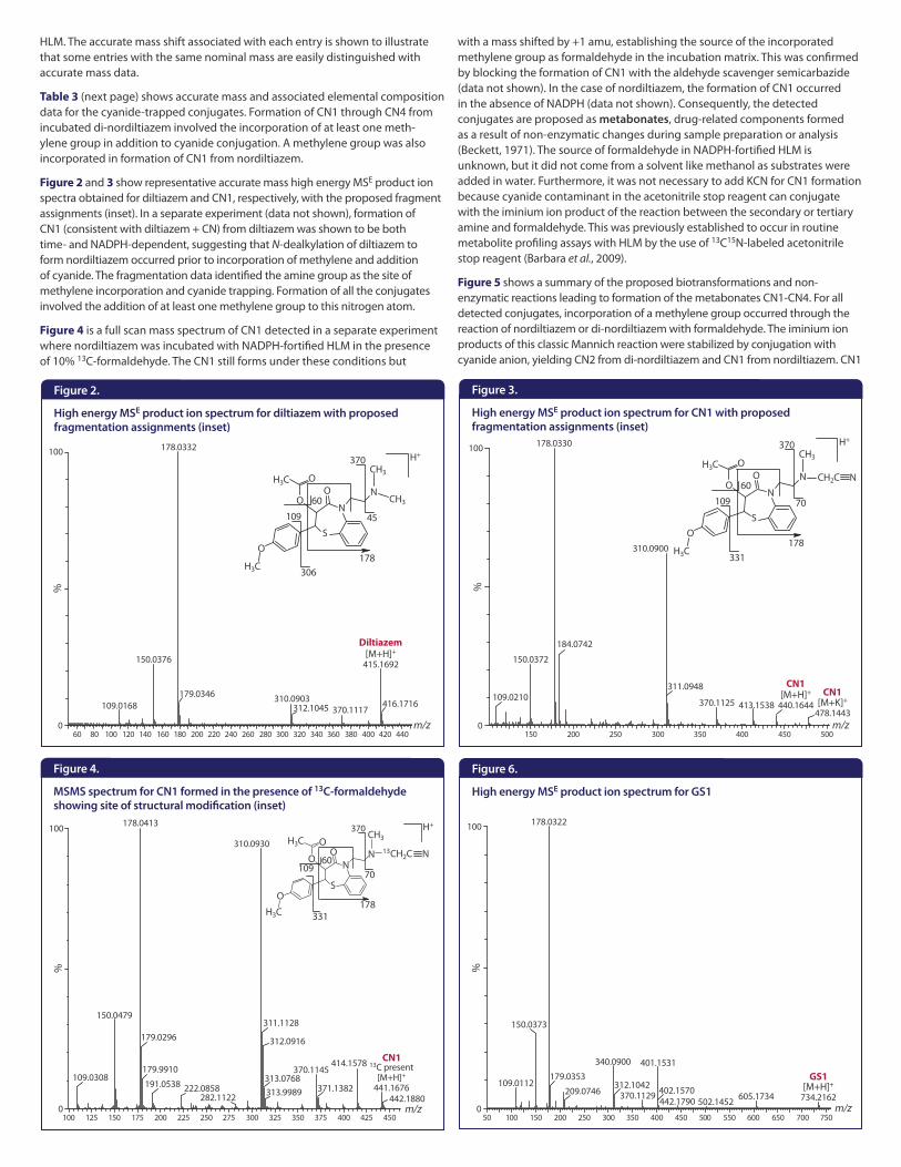
Figure 2 and 3 show representative accurate mass high energy MSE product ion spectra obtained for diltiazem and CN1, respectively, with the proposed fragment assignments (inset). In a separate experiment (data not shown), formation of CN1 (consistent with diltiazem + CN) from diltiazem was shown to be both time-� and NADPH-�dependent, suggesting that N-�dealkylation of diltiazem to form nordiltiazem occurred prior to incorporation of methylene and addition of cyanide. The fragmentation data identified the amine group as the site of methylene incorporation and cyanide trapping. Formation of all the conjugates involved the addition of at least one methylene group to this nitrogen atom.
Figure 4 is a full scan mass spectrum of CN1 detected in a separate experiment where nordiltiazem was incubated with NADPH-�fortified HLM in the presence of 10% 13C-�formaldehyde. The CN1 still forms under these conditions but
with a mass shifted by +1 amu, establishing the source of the incorporated methylene group as formaldehyde in the incubation matrix. This was confirmed by blocking the formation of CN1 with the aldehyde scavenger semicarbazide (data not shown). In the case of nordiltiazem, the formation of CN1 occurred in the absence of NADPH (data not shown). Consequently, the detected conjugates are proposed as metabonates, drug-�related components formed as a result of non-�enzymatic changes during sample preparation or analysis (Beckett, 1971). The source of formaldehyde in NADPH-�fortified HLM is unknown, but it did not come from a solvent like methanol as substrates were added in water. Furthermore, it was not necessary to add KCN for CN1 formation because cyanide contaminant in the acetonitrile stop reagent can conjugate with the iminium ion product of the reaction between the secondary or tertiary amine and formaldehyde. This was previously established to occur in routine metabolite profiling assays with HLM by the use of 13C15N-�labeled acetonitrile stop reagent (Barbara et al., 2009).
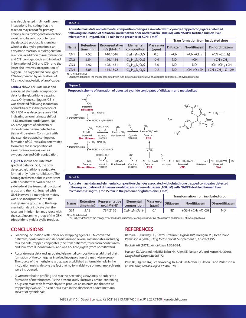
Figure 5 shows a summary of the proposed biotransformations and non-�enzymatic reactions leading to formation of the metabonates CN1-�CN4. For all detected conjugates, incorporation of a methylene group occurred through the reaction of nordiltiazem or di-�nordiltiazem with formaldehyde. The iminium ion products of this classic Mannich reaction were stabilized by conjugation with cyanide anion, yielding CN2 from di-�nordiltiazem and CN1 from nordiltiazem. CN1
High energy MSE product ion spectrum for diltiazem with proposed fragmentation assignments (inset)
High energy MSE product ion spectrum for CN1 with proposed fragmentation assignments (inset)
Figure 6.
High energy MSE product ion spectrum for GS1
Figure 4.
MSMS spectrum for CN1 formed in the presence of 13C-formaldehyde showing site of structural modification (inset)
Diltiazem
CN1CN1
CN1
GS1
Figure 3.Figure 2.
REFERENCESBarbara JE, Buckley DB, Kazmi F, Yerino P, Ogilvie BW, Horrigan MJ, Toren P and Parkinson A (2009). Drug Metab Rev 41:Supplement 3, Abstract 195.
Beckett AH (1971). Xenobiotica 1:365-384.
Hanson KL, VandenBrink BM, Babu KN, Allen KE, Nelson WL and Kunze KL (2010). Drug Metab Dispos 38:963-72.
Paris BL, Ogilvie BW, Scheinkoenig JA, Ndikum-(2009). Drug Metab Dispos 37:2045-205.
16825 W 116th Street | Lenexa, KS 66219 | 913.438.7450 | fax 913.227.7100 | xenotechllc.com
• Following incubation with CN– or GSH trapping agents, HLM converted diltiazem, nordiltiazem and di-nordiltiazem to several metabonates, including four cyanide-trapped conjugates (one from diltiazem, three from nordiltiazem and four from di-nordiltiazem) and one GSH conjugate (from nordiltiazem).
• Accurate mass data and associated elemental compositions established that formation of the conjugates involved incorporation of a methylene group. The source of the methylene group was established as formaldehyde in the incubation matrix, despite the fact that no formaldehyde or methanol solvents were introduced.
• In vitroformation of metabonates. As the present study illustrates, amine-containing drugs can react with formaldehyde to produce an iminium ion that can be trapped by cyanide. This can occur even in the absence of added methanol solvent or cyanide salt.
CONCLUSIONS
Proposed scheme of formation of detected cyanide conjugates of diltiazem and metabolites
CYP N-Demethylation
DetectedDiltiazem
CYP N-Demethylation
DetectedNordiltiazem
Not detected DetectedCN1
DetectedDi-nordiltiazem
Not detected DetectedCN2
DetectedCN3
DetectedCN4
Figure 5.
Table 3.
Accurate mass data and elemental composition changes associated with cyanide-trapped conjugates detected
microsomes (1 mg/mL) for 15 min in the presence of KCN (1 mM)Transformation from incubated drug
Name Retention time (min)
Representative m/z [M+H]+
Elemental composition
Mass error (ppm) Diltiazem Nordiltiazem Di-nordiltiazem
CN1 7.52 440.1646 C23H25N3O4S 0.5 +CN +CN +CH2 +CN +2(CH2)
CN2 6.54 426.1484 C22H23N3O4S -0.9 ND +CN +CN +CH2
CN3 4.92 428.1631 C22H25N3O4S -3.0 ND ND +CN +CH2 +2HCN4 5.05 444.1592 C22H25N3O5S -0.2 ND +CN +O +2H +CN +CH2 +O +2H
Table 4.
Accurate mass data and elemental composition changes associated with glutathione trapped conjugates detected
microsomes (1mg/mL) for 15 min in the presence of glutathione (1 mM)
Transformation from incubated drug
Name Retention time (min)
Representative m/z [M+H]+
Elemental composition
Mass error (ppm) Diltiazem Nordiltiazem Di-nordiltiazem
GS1 5.13 734.2166 C32H39N5O11S2 0.1 ND +GSH +CH2 +O -2H ND
ND = Not detected
ND = Not detected
was also detected in di-nordiltiazem incubations, indicating that the reaction may repeat for primary amines, but a hydrogenation reaction would also have to occur to form the detected product. It is unclear whether this hydrogenation is an enzymatic reaction. A hydrogenation reaction, in addition to methylenation and CN– conjugation, is also involved in formation of CN3 and CN4, and the latter also involves incorporation of oxygen. The oxygenated conjugate CN4 fragmented by neutral loss of 16 amu, characteristic of an N-oxide.
Table 4 shows accurate mass and associated elemental composition data for the glutathione trapping assay. Only one conjugate (GS1) was detected following incubation of nordiltiazem in the presence of GSH. GS1 was detected at m/z 734, indicating a nominal mass shift of +333 amu from nordiltiazem. No GSH conjugates of diltiazem or di-nordiltiazem were detected in this in vitro system. Consistent with the cyanide-trapped conjugates, formation of GS1 was also determined to involve the incorporation of a methylene group as well as oxygenation and GSH conjugation.
Figure 6 shows accurate mass spectral data for GS1, the only detected glutathione conjugate, formed only from nordiltiazem. The conjugated metabolite is consistent with nordiltiazem oxidized to an aldehyde at the N-methyl functional group and then conjugated with GSH. However, a methylene moiety was also incorporated into the methylamine group and the frag-mentation data indicate that the resultant iminium ion may react with the cysteine amine group of the GSH tripeptide to yield a cyclic product.