Embed Size (px)
Citation preview

Speckle-Tracking Echocardiography Elucidates the Effectof Pacing Site on Left Ventricular Synchronization in theNormal and Infarcted Rat MyocardiumMichal Mor1., Wesam Mulla1., Sigal Elyagon1, Hovav Gabay1,2, Shani Dror1, Yoram Etzion1,3"*,
Noah Liel-Cohen4"
1 Cardiac Arrhythmia Research Laboratory, Department of Physiology and Cell Biology, Faculty of Health Sciences, Ben-Gurion University of the Negev, Beer-Sheva, Israel,
2 Department of Emergency Medicine, Recanati School for Community Health Professions, Faculty of Health Sciences and PREPARED Center for Emergency Response
Research, Ben-Gurion University of the Negev, Beer-Sheva, Israel, 3 Division of Internal Medicine, Soroka University Medical Center, Beer-Sheva, Israel, 4 Cardiology
Department, Soroka University Medical Center, Beer-Sheva, Israel
Abstract
Background: Right ventricular (RV) pacing generates regional disparities in electrical activation and mechanical function(ventricular dyssynchrony). In contrast, left ventricular (LV) or biventricular (BIV) pacing can improve cardiac efficiency in thesetting of ventricular dyssynchrony, constituting the rationale for cardiac resynchronization therapy (CRT). Animal models ofventricular dyssynchrony and CRT currently relay on large mammals which are expensive and not readily available to mostresearchers. We developed a methodology for double-site epicardial pacing in conscious rats. Here, following post-operative recovery, we compared the effects of various pacing modes on LV dyssynchrony in normal rats and in rats withischemic cardiomyopathy.
Methods: Two bipolar electrodes were implanted in rats as follows: Group A (n = 6) right atrial (RA) and RV sites; Group B(n = 7) RV and LV sites; Group C (n = 8) as in group B in combination with left coronary artery ligation. Electrodes wereexteriorized through the back. Following post-operative recovery, two-dimensional transthoracic echocardiography wasperformed during pacing through the different electrodes. Segmental systolic circumferential strain (Ecc) was used toevaluate LV dyssynchrony.
Results: In normal rats, RV pacing induced marked LV dyssynchrony compared to RA pacing or sinus rhythm, as measuredby the standard deviation (SD) of segmental time to peak Ecc, SD of peak Ecc, and the average delay between opposingventricular segments. LV pacing and, to a greater extend BIV pacing diminished the LV dyssynchrony compared to RVpacing. In rats with extensive MI, the effects of LV and BIV pacing were markedly attenuated, and the response of individualanimals was variable.
Conclusions: Rodent cardiac pacing mimics important features seen in humans. This model may be developed as a simplenew tool to study the pathophysiology of ventricular dyssynchrony and CRT.
Citation: Mor M, Mulla W, Elyagon S, Gabay H, Dror S, et al. (2014) Speckle-Tracking Echocardiography Elucidates the Effect of Pacing Site on Left VentricularSynchronization in the Normal and Infarcted Rat Myocardium. PLoS ONE 9(6): e99191. doi:10.1371/journal.pone.0099191
Editor: Alena Talkachova, University of Minnesota, United States of America
Received February 7, 2014; Accepted May 12, 2014; Published June 10, 2014
Copyright: � 2014 Mor et al. This is an open-access article distributed under the terms of the Creative Commons Attribution License, which permits unrestricteduse, distribution, and reproduction in any medium, provided the original author and source are credited.
Funding: This work was supported by a research grant from the Clalit Research Institute, Clalit health services, Israel (Yoram Etzion and Noah Liel-Cohen). Thefunders had no role in study design, data collection and analysis, decision to publish, or preparation of the manuscript.
Competing Interests: The authors have declared that no competing interests exist.
* E-mail: [email protected]
. These authors contributed equally to this work.
" These authors also contributed equally to this work.
Introduction
Dyssynchronous contraction of the left ventricle (LV) can
markedly worsen the outcome of heart failure (HF) independently
of traditional risk factors [1,2]. This pathophysiology is often
derived from or exacerbated by native conduction delay, such as
left bundle branch block (LBBB) or artificial pacing of the right
ventricle (RV) [3–5]. Based on QRS widening, it is estimated that
at least 25–30% of patients with HF exhibit dyssynchronous LV
contraction [6,7]. Clinical studies employing biventricular (BIV)
pacing (cardiac resynchronization therapy; CRT) to treat HF
patients with dyssynchronous contraction have revealed significant
benefits in terms of contractile performance, improved quality of
life, and long-term survival [8].
Although dyssynchronous heart failure (DHF) has been
extensively studied in mechanical terms and although the clinical
effectiveness of CRT is remarkable, there are multiple issues that
remain ill-defined at present. It is, for example, estimated that
around 30% of patients do not benefit from CRT, and the clinical
criteria to identify CRT non-responders remain obscure and
PLOS ONE | www.plosone.org 1 June 2014 | Volume 9 | Issue 6 | e99191

controversial. In addition, there is variability in individual patient
vulnerability to DHF progression in response to conventional RV
pacing, which is difficult to predict at present [4,9]. Only recently
has the impact of dyssynchrony and BIV pacing on molecular
signaling and the ionic properties of the myocardium been realized
[9,10]. Animal studies have shown that DHF is associated with
regional molecular changes (collectively termed ‘molecular polar-
ization’), including downregulation of Ca2+ handling proteins and
connexin 43, activation of stress response kinases and increased
expression of tumor necrosis factor in the lateral LV wall. Most of
these changes can be reversed by CRT [11]. Additional studies
identifying regional changes in ionic channels and multiple gene
transcripts [12,13] have revealed a highly complicated picture in
which multiple players are involved. Therefore, comprehensive
understanding of the effects of various pacing modes at the
molecular level will require extensive additional scientific efforts.
One of the problems hampering progress in this field of research
is the lack of suitable animal models that can be utilized for routine
experimentation. Current studies almost exclusively utilize large
animals (mainly dogs) and are based on the implantation of human
devices. Such an experimental setting is highly demanding. In
addition, the large animal models can not be manipulated
genetically, an issue that is becoming increasingly critical when
it is necessary to reveal molecular mechanisms. While rat and
mouse models are extensively used in research on HF [14], these
animal models are largely missing in research on DHF and CRT,
mainly due to technical difficulties in electrode implantation. Two
independent studies using M-mode echocardiography have
managed to demonstrate dyssynchrony between the septum and
the LV lateral wall in the murine heart [13,15]. In addition, in the
pioneering study of Bilchick et al. 2006 mice were implanted with
a single pacing electrode on the RV free wall and were paced for
seven days using an implanted miniature pacemaker. Using
microarray analysis, this study demonstrated indications for
molecular polarization as a result of prolonged RV pacing
[13,15]. Thus, it appears that rodents may mimic important
features that are seen in large mammals in regard to the
mechanical effect of ventricular pacing. However, to the best of
our knowledge, the mechanical effect of RV pacing as compared
with LV and BIV pacing has not yet been defined in rodents.
In the present study, using an implanted device that was
developed for pacing and recordings in conscious rodents [16], we
acutely evaluated the effect of pacing site on LV synchronization
in normal and infarcted rats following post operative recovery. We
hypothesized that the mechanical effects of LV pacing and BIV
pacing should be favorable compared to RV pacing. Elucidation
of this issue seems critical in order to clarify whether rodent models
may be further developed as simple experimental tools to study the
molecular biology of DHF and CRT. Utilizing speckle-tracking 2-
dimensional echocardiography, a method that has been repeatedly
validated in rats for segmental strain analysis over the past decade
[17–20], we confirmed the existence of mechanical dyssynchrony
when RV pacing was applied. In addition, we elucidated the
electrical and mechanical effects of LV pacing and BIV pacing.
Overall, our results support the notion that the effects of pacing in
rodents can recapitulate important features observed in large
animal models and humans.
Methods
Ethics statementAll animal studies reported in this article were approved by the
institutional ethics committee, Faculty of Health Sciences, Ben-
Gurion University of the Negev, Israel. At the end of all
experiments animals were killed using IV KCl injection under
deep isoflurane anesthesia.
Implantable rat pacing deviceThe implantable device that was used to pace the heart at
different locations was originally designed for long-term atrial
pacing and recording in freely moving rats [16]. In this device, the
electrical connections to the epicardial surface are achieved using
miniature-bipolar hook electrodes, which have been described in
detail previously [16]. The specific design of the miniature-bipolar
hook electrode allows it to be positioned on a desired location on
the epicardial surface of the atria or ventricles through a small
lateral thoracotomy without the need of additional suturing [16].
The implantable device consists of an 8-pin ‘female’ connector
that is attached by flexible insulated electrical wires to a set of two
miniature bipolar hook electrodes (Figure 1a). Peripheral elec-
trodes that are implanted on the back for pseudo ECG
measurements also exist (Figure 1a,c). Before implantation, the
8-pin ‘female’ connector was covered with a latex sheath to
prevent direct contact with the subcutaneous tissue during the
operation. Electron beam radiation was applied for sterilization of
the device before use.
Surgical proceduresAdult male Sprague-Dawley rats (250–300 g) were anesthetized
(ketamine/xylazine 75/5 mg/kg, IP) and mechanically ventilated.
The animals were placed supine on a warmed heating and under
sterile conditions a skin incision in the left thorax was performed
and a subcutaneous tunnel was extended posteriorly to the upper
back of the animal. The 8-pin ‘female’ connector (covered with a
latex sheath) was inserted through the subcutaneous tunnel.
Thereafter, lateral thoracotomy was performed and the two
miniature -bipolar hook electrodes were positioned as follows:
Group A; on the right atrium (RA) and RV apex (RV). Group B;
on the RV and on the posterobasal aspect of the left ventricle (LV).
Group C; similar electrode implantation as in group B, as well as
proximal left anterior descending coronary artery ligation to
induce myocardial infarction (MI). In order to insert electrodes on
the right side (for positioning on the RA and RV) a small
subcutaneous tunnel was done in the chest wall through which the
electrodes were moved to the right side of the thorax. The
coronary artery was ligated by 6/0 surgical suture and MI was
confirmed by the presence of regional cyanosis in the myocardial
surface. Following chest closure, each animal was placed in a
prone position and the back connector was exteriorized through
the skin of the back (Figure 1B). Postoperative recovery and
analgesia were done as described previously [16].
Cardiac pacing and echocardiograpyNormal rats were evaluated 6–8 days following the surgical
procedure. MI rats were evaluated 14 days following the surgical
procedure. For the echocardiograpic analysis, each animal was
sedated (isoflurane 1%) and placed on a heating pad. ECG
recordings (Nihon Kodhen, RMC1100) were monitored online
throughout the experiment and were also digitized and stored for
offline analysis in each pacing mode (PCI-6024E, National
Instruments, Austin, TX, USA). Electrical stimulation (2 ms
square pulses) was applied through isolation units (Iso-Flex,
AMPI, Israel) at a double diastolic threshold. The cycle length
that was used to induce override pacing in the normal rats was set
to 150 ms (400 bpm) which invariably induced override pacing in
these animals. In the MI rats, baseline heart rate was sometimes
slightly higher than 400 bpm. Therefore, the override pacing cycle
length was set to 140 ms (428 bpm), which managed to override
Strain Analysis in Paced Rats
PLOS ONE | www.plosone.org 2 June 2014 | Volume 9 | Issue 6 | e99191
the spontaneous heart rate of these animals. During BIV pacing,
two independent isolation units were used for simultaneous but
independent RV apical and LV pacing. A program developed by
YE [16] was used to control data acquisition, electrical stimulation
and off-line analysis. QT interval measurements were manually
done off-line on the digital signals under high time and voltage
magnification scales. These measurements were performed using
dedicated cursors which marked the isoelectric line and stressed
out the time of return of the ECG signal to this line. The measured
QT intervals of five independent complexes were averaged in each
condition to reduce sampling error. Two-dimensional transtho-
racic echocardiography was performed with a Vivid 7 echocar-
diography system (GE Healthcare, Milwaukee WI, USA) by short-
axis mid-ventricular scans at the level of the papillary muscle tips.
Images were obtained with a 10 S transducer (5.4,11.8 MHz)
using an image depth of 2.5 cm and a frame rate of 225 frames/s
as was repeatedly validated in the literature in rat studies [17-20].
The acquisition protocol was as follows: Following optimal
positioning of the transducer on the chest wall, it was fixed in
place by the operator, while 2D clips (20 s each) were sequentially
recorded in sinus rhythm followed by override pacing at the
different pacing sites. Of note, we took special care that the
transducer will stay in the exact same position throughout the
experiment to allow for accurate comparison between the different
pacing modes in each animal. The procedure was always done by
two experimentalist, one acquiring there echocardiography clips
and the other controlling the pacing and confirming continuous
electrical capture in each pacing mode. At the end of each
experiment, the animal was sacrificed and correct positioning of
the epicardial electrodes was confirmed by post mortem operation.
Offline analysis of LV fractional area shortening (LV-FAS) was
done by calculating the LV areas during maximal systole (LVAs)
and during maximal diastole (LVAd) and then expressing LV-FAS
as: (LVAd-LVAs)/LVAd6100.
Circumferential 2-dimensional strain analysisBased on the existing literature in rodents, we restricted our
current analysis to circumferential strain (Ecc) which is more
reliable than radial strain in the small heart of rats [18,19]. Ecc
was evaluated as described in the literature [17–19]. Briefly,
analysis of mid-level short-axis images was done with dedicated
software (‘‘2D-strain’’, EchoPAC, GE healthcare, Vingmed,
Norway). Endocardial border at end-systole and the region of
interest from the endocardium to the epicardial edge were
determined by the user (Figure 1d). Thereafter, an automatic
speckle tracking analysis was performed for the short-axis images
to track the motion of speckles contained within the region of
interest on a frame-to-frame basis [21]. Ecc curves, expressed in
percentage values as a function of time were exported to a
customized program (MATLAB software, MathWorks) to calcu-
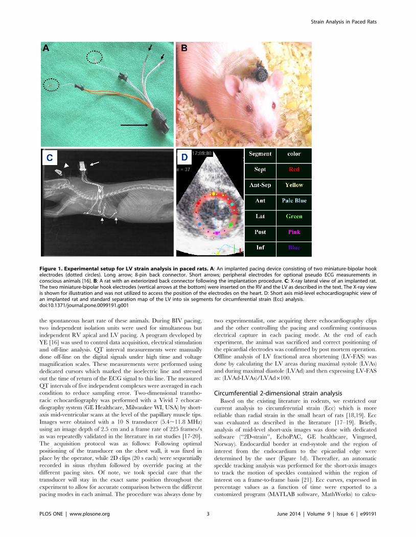
Figure 1. Experimental setup for LV strain analysis in paced rats. A: An implanted pacing device consisting of two miniature-bipolar hookelectrodes (dotted circles). Long arrow; 8-pin back connector. Short arrows; peripheral electrodes for optional pseudo ECG measurements inconscious animals [16]. B: A rat with an exteriorized back connector following the implantation procedure. C: X-ray lateral view of an implanted rat.The two miniature-bipolar hook electrodes (vertical arrows at the bottom) were inserted on the RV and the LV as described in the text. The X-ray viewis shown for illustration and was not utilized to access the position of the electrodes on the heart. D: Short axis mid-level echocardiographic view ofan implanted rat and standard separation map of the LV into six segments for circumferential strain (Ecc) analysis.doi:10.1371/journal.pone.0099191.g001
Strain Analysis in Paced Rats
PLOS ONE | www.plosone.org 3 June 2014 | Volume 9 | Issue 6 | e99191

late the time from onset of QRS to peak Ecc (time to peak Ecc)
and the value of maximal Ecc (across the six mid-level segments
(Figure 1d, Figure 2a). The values of maximal Ecc are shown as %
of segmental length at onset QRS complex. At least five
consecutive beats were analyzed under each pacing condition in
each animal. Initial determination of the region of interest from
the endocardium to the epicardial edge was performed under no
pacing conditions and was kept as constant as possible during the
different pacing conditions. Analysis of all rats in each group was
done by a single observer (MM - normal rats, WM - MI rats). An
independent observer who is highly experienced in echocardio-
graphic methodologies including 2D strain analysis (NLC)
confirmed the consistency of the analysis performed by the two
observers.
Statistical analysisValues are expressed as means 6SE. Due to the relatively small
sample size in each group, analysis of the difference between
pacing conditions was done using Kruskal-Wallis one way analysis
of variance on ranks with Student-Neuman-Keuls post hoc test. All
statistical tests were done using SigmaStat 3.1 (Systat Software,
Inc, Point Richmond, CA). The criterion for significance was set at
P,0.05.
Results
RV pacing versus RA pacing or sinus rhythm in normalrats
As a first step we aimed to compare the effect of RV pacing to
both no pacing and RA pacing modes. This analysis was done in
order to confirm that the override pacing itself does not induce
rate-dependent LV dyssynchrony compared to the no pacing
mode (sinus rhythm). For that purpose, group A (n = 6) was
instrumented with a bipolar RA pacing electrode and a bipolar
RV pacing electrode, and override pacing in these two locations
(150 ms CL) was compared to the no pacing state. The obtained
Ecc traces of the different LV segments typically demonstrated no
major differences between RA pacing and sinus rhythm. In
contrast, RV pacing invariably induced marked changes in the
segmental strain pattern. The RV pacing typically produced an
early systolic contraction in the inferior or posterior segments,
which was associated, in some cases, with a clear stretching effect
in opposing segments (Figure 2A). Summary of the segmental time
to peak Ecc and the segmental peak Ecc patterns under the
different pacing modes is presented figure 2B. Analysis of
important global parameters derived from the segmental Ecc
analysis under the different pacing modes is shown in Figure 3.
The global time to peak Ecc was found to be slightly shorter
during RA pacing compared to no pacing (85.664.1%, p,0.05)
an was also shorter compared to RV pacing (Figure 3A). Global
peak Ecc (Figure 3B) was not different between RA pacing and no
pacing but was markedly attenuated during RV pacing
(61.063.7% compared to no pacing, p,0.01 compared to both
RA pacing and no pacing). While the Ecc analysis identified a
marked effect of RV pacing on strain parameters, concurrent
analysis of LV-FAS did not identify a significant effect of the
different pacing modes on this parameter (Figure 3C, see
discussion). Specific analysis of LV dyssynchrony variables
indicated marked deleterious effects of RV pacing on all the
calculated parameters, including standard deviation of time to
peak Ecc (Figure 3D), standard deviation of peak Ecc (Figure 3E)
and the average time difference between the peak Ecc of opposing
segments (Delta TP opposing segments, Figure 3F). There was no
statistical difference in these parameters between RA pacing and
sinus rhythm.
Effects of different ventricular pacing modes in normalrats
In the next stage, we evaluated group B (n = 7) rats, which had
bipolar pacing leads in the RV and LV. The effect of override
pacing in these two locations alone or simultaneously (BIV pacing)
was compared to the no pacing mode. In terms of electrical
activity, the ECG recording indicated QRS prolongation during
RV pacing as compared to LV and BIV pacing (Figure 4A).
However, the unclear separation between the QRS and the T-
wave in the rodent heart [22,23], did not allow quantitative
analysis of QRS width during the various pacing modes.
Nevertheless, an analysis of QT duration indicated marked QT
prolongation during RV pacing (138.365.3%, p,0.01) as
compared to the no pacing mode. This marked QT prolongation
was reduced by LV pacing (124.962.9%) and even further
reduced by BIV pacing (109.163.6%), as shown with detailed
statistical analysis in Figure 4B. In addition, recordings of
epicardial electrograms from the RV electrode when pacing the
LV and vice versa, was performed in two animals from group B.
These recordings supported a longer sequence of activation during
RV pacing compared to LV pacing (Figure 4C).
We next evaluated the mechanical dyssynchrony during the
various pacing modes. Summary of the segmental time to peak
Ecc and the segmental peak Ecc under the different pacing modes
in group B are presented figure 5. Analysis of important global
parameters derived from the segmental Ecc analysis under the
different pacing modes is shown in Figure 6. RV pacing
significantly reduced both the global time to peak Ecc and the
global peak Ecc as compared to no pacing. These parameters
recovered significantly during LV and BIV pacing (Figure 6A, B).
In addition, while a prominent increase in all the parameters of
LV dyssynchrony was noted during RV pacing, LV pacing and
particularly BIV pacing markedly improved all three parameters
of LV dyssynchrony (Figure 6 D–E). Of note, the segmental
pattern of the time to peak Ecc during RV pacing (Figure 5) was
somewhat different from that observed in group A (Figure 2),
possibly due to variability in the positioning of the RV electrode on
the epicardial surface of the RV (see limitations section in our
discussion). As in group A (Figure 3), we could not detect
significant changes in LV-FAS during the different pacing modes
(Figure 6C).
Effects of different ventricular pacing modes in rats withischemic-induced heart failure
To elucidate the influence of ischemic HF on the electrome-
chanical results of our model, group C (n = 8) was subjected to a
combined procedure of electrode implantation and left coronary
artery ligation. We analyzed the effect of pacing two weeks
following surgery, at a time when prominent scar tissue and
myocardial remodeling were present (Figure 7A, B). Out of the
eight animals with confirmed MI, one had electrical capture only
in the LV and was excluded from analysis. Two additional animals
had electrical capture only in the RV and were therefore analyzed
only in the no pacing and RV pacing modes. Final statistical
analysis was done only on the five MI rats that exhibited electrical
capture in both RV and LV sites. In electrophysiological terms,
the MI rats demonstrated baseline prolongation of the QT interval
compared to the normal rats (94.064.4 ms vs. 70.564.4 ms, p,
0.001). However, the effect of pacing on the QT interval followed
a similar trend to that in the normal rats. Indeed, RV pacing
Strain Analysis in Paced Rats
PLOS ONE | www.plosone.org 4 June 2014 | Volume 9 | Issue 6 | e99191

statistically increased the QT interval as compared with no pacing
mode (118.663.7%, p,0.05), while the QT during LV and BIV
pacing did not differ from sinus rhythm (113.964.7% and
106.263.9%, respectively, see details in Figure 7C).
Evaluation of segmental Ecc in group C revealed typical
akinesia or dyskinesia in the anteroseptal, anterior and lateral
segments at baseline (Figure 7D). No apparent effect of pacing on
the magnitude and temporal pattern of the peak Ecc was observed
in these rats (Figure 7E, Figure 8 A,B). In addition, while
dyssynchrony parameters were much higher in the MI animals at
baseline, there were no apparent effects of the different pacing
modes on these parameters, excluding a non-significant tendency
Figure 2. Segmental Ecc analysis during RA and RV pacing. A: Representative strain waveforms in an instrumented rat subjected to no pacing(upper), RA pacing (middle) and RV (lower). The colors of the different waveforms match those in the segmental map in Figure 1D. White dotted linesrepresent the global (average) strain of all six segments. B: Average segmental time to peak Ecc (left) and peak Ecc (right) in seven animals subjectedno pacing, RA pacing, and RV pacing.doi:10.1371/journal.pone.0099191.g002
Strain Analysis in Paced Rats
PLOS ONE | www.plosone.org 5 June 2014 | Volume 9 | Issue 6 | e99191

to increase in the ‘delta TP opposing segments’ parameter during
RV pacing (Figure 8 D–F). Looking more closely at the effect of
pacing on the ‘delta TP of opposing segments’ in individual rats
(Figure 9 A), we observed a consistent tendency for this parameter
to increase during RV pacing in 6 out of the 7 evaluated animals.
Compared with RV pacing the effects of LV pacing and BIV
pacing on the delta TP of opposing segments were variable. While
these pacing modes seemed to have a beneficial effect in some
cases (e.g., animals 2 and 3), there was a worsening of this
parameter in animal 5 when LV and BIV pacing were applied
(Figure 9 A). An additional analysis specifically focusing on the two
opposing segments that had maximal temporal dyssynchrony
during RV pacing detected a rather similar pattern (Figure 9 B, C).
Discussion
The present study introduces a new model of ventricular pacing
in an instrumented rat, which allowed us for the first time to
systematically evaluate the effects of pacing site on LV synchro-
nization in the rodent heart. We used speckle-tracking echocar-
diography analysis of Ecc, which has been reported to reliably and
sensitively detect changes in LV dyssynchrony under various
clinically relevant conditions in the rat heart [17–20]. The speckle-
tracking echocardiographic methodology has previously been
applied extensively in large animal models and in people and
has been shown to be much more sensitive to changes in LV
dyssynchrony than global echocardiographic measures, such as
fractional shortening [24]. Therefore, in the present study, it is not
Figure 3. RV pacing induces marked LV dyssynchrony compared to RA pacing. Summary of echocardiographic parameters in group Asubjected to no pacing, RA pacing, and RV pacing. A: Average time to peak Ecc of all six segments (global). B: Average peak Ecc of all six segments(global). C: LV fractional area shortening. D: Standard deviation of the time to peak Ecc of different segments. E: Standard deviation of the peak Ecc ofdifferent segments. F: Analysis of ‘delta TP opposing segments’, i.e., the average time difference between the peak Ecc of opposing segments. * P,0.05, ** P,0.01. Horizontal lines above\below the bar graphs indicate significant differences between pacing modes in the post-hoc analysis. Notethat in D the Kruskal-Wallis test was significant. However, the post-hoc analysis could not reveal the source of difference between the groups.doi:10.1371/journal.pone.0099191.g003
Strain Analysis in Paced Rats
PLOS ONE | www.plosone.org 6 June 2014 | Volume 9 | Issue 6 | e99191

Figure 4. RV pacing induces electrical dyssynchrony compared to LV and BIV pacing. A: Representative ECG recordings from aninstrumented rat during sinus rhythm (no pacing) and during RV, LV and BIV pacing. Arrows indicate the termination of the T wave in each trace. B:Summary of QT interval analysis in six instrumented rats. ** P,0.01. Horizontal lines above the bar graph indicate significant differences betweenpacing modes in the post-hoc analysis. C: Example of epicardial electrograms during RV pacing (left) and LV pacing (right). In each recording uppertrace is a peripheral ECG signal (Lead I), middle trace is the epicardial electrogram and lower trace shows the time of cardiac pacing. Note that theepicardial activation in the RV electrode correlates with a late negative vector in the ECG signal. In contrast, the epicardial activation during RV pacingis correlated with a late positive vector in the ECG signal. Measurements of the time from end of stimulus artifact to the peaks of these late vectors inthe ECG (dash vertical lines) indicate shorter activation time during LV pacing. Analysis of epicardial electrograms in an additional animal from groupB demonstrated similar findings (not shown).doi:10.1371/journal.pone.0099191.g004
Figure 5. Segmental Ecc analysis during RV, LV and BIV pacing. Average segmental time to peak Ecc (A) and peak Ecc (B) in seven animalsfrom group B subjected to no pacing, RV pacing, LV pacing and BIV pacing.doi:10.1371/journal.pone.0099191.g005
Strain Analysis in Paced Rats
PLOS ONE | www.plosone.org 7 June 2014 | Volume 9 | Issue 6 | e99191

surprising that there were no significant effects of pacing on LV-
FAS, while the effects of pacing site on segmental and global Ecc
pattern were consistent and significant.
The main findings of our Ecc analysis indicate that while the
rodent heart has some clear differences compared to the human
heart in aspects such as action potential repolarization and
calcium handling [22,23,25], its mechanical response to the effect
of various pacing modes seems to have important similarities to the
large mammalian heart. While previous studies in the mouse heart
have already demonstrated negative effects of acute RV pacing on
Figure 6. RV pacing induces mechanical dyssynchrony compared to LV and BIV pacing. Summary of echocardiographic parameters in sixanimals subjected to no pacing, RV, LV and BIV pacing. A: Average time to peak Ecc of all six segments (global). B: Average peak Ecc of all sixsegments (global) C: LV fractional area shortening D: Standard deviation of the time to peak Ecc of different segments. E: Standard deviation of thepeak Ecc of different segments. F: Average time difference between the peak Ecc of opposing segments (‘delta TP opposing segments’, see Figure 3for details). * P,0.05, ** P,0.01. Horizontal lines above\below the bar graphs indicate significant differences between pacing modes in the post-hocanalysis.doi:10.1371/journal.pone.0099191.g006
Strain Analysis in Paced Rats
PLOS ONE | www.plosone.org 8 June 2014 | Volume 9 | Issue 6 | e99191

LV synchrony using M-mode echocardiography, [13,15] our study
is the first to utilize speckled tracking analysis under various pacing
modes in order to view the full segmental pattern during RA, RV,
LV and BIV pacing. The results show clear beneficial effects of LV
and to a greater extend BIV pacing on LV synchrony compared to
RV pacing. In terms of electrical synchrony, we found it difficult to
reliably measure the QRS duration during the different pacing
modes due to the absence of apparent differentiation between the
QRS complex and the T wave in the rat ECG. However, the
effects of different pacing modes on the QT duration, which were
consistent and significant (Figure 4 A,B), seem to reflect, at least
partially, changes in QRS duration that are embedded in the QT
interval. In addition, analysis of epicardial electrograms also
supports longer activation time during RV pacing compared to
LV pacing (Figure 4 C). Thus, LV and BIV pacing appear to
induce beneficial effects on electrical synchrony compared with
RV pacing in the rat heart. In terms of mechanical synchrony our
results demonstrate that LV-only pacing has favorable outcome
which is almost similar to the effect of BIV pacing. While many
studies support the notion that LV-only pacing may have
beneficial effects in HF patients [26], recent data in children with
normal cardiac function and complete AV block demonstrated
striking similarity to the results of our rat model in regard to LV-
only pacing [27]. Moreover, LV-only pacing appears to be
associated with good clinical outcome in this and other studies in
the pediatric population [28,29]. It should be mentioned that the
segmental findings of our study as well as the pediatric studies
stated above appear to contrast the findings of [30], which
demonstrated reduced Ecc near the LV pacing site compare to
remote sites near the RV in the dog model of acute pacing.
However, the segmental Ecc analysis in the above study was done
by MRI using a much higher spatial resolution dividing the mid
LV into 24 segments and 8 longitudinal segments. Thus the
analysis focused on segments which are not in the same transverse
plane and are much more refined to the actual vicinity of the
pacing electrodes.
Figure 7. Combined model of pacing and ischemic heart failure. A: Anterior image of a heart subjected to electrodes implantation incombination with left coronary ligation. RV electrode and LV electrode are marked by short and long black arrows, respectively. White arrow;coronary artery ligation site. B: Transverse sections of the heart shown in A. Apical to basal sections are ordered from left to right. C: Summary of QTinterval analysis in five instrumented rats with MI. * P,0.01. Horizontal line above the bar graph indicate significant difference in the post-hocanalysis. D: Representative strain waveforms of two instrumented rats with MI under control conditions. E: Average segmental time to peak Ecc (left)and peak Ecc (right) in five rats with ischemic heart failure subjected to no pacing, RV, LV and BIV pacing.doi:10.1371/journal.pone.0099191.g007
Strain Analysis in Paced Rats
PLOS ONE | www.plosone.org 9 June 2014 | Volume 9 | Issue 6 | e99191

The addition of MI-induced HF to the double site ventricular
pacing model was performed as a proof of concept for the
usefulness of our rat model in simulating a scenario that is
clinically relevant. Our results indicate decreased beneficial effect
of LV or BIV pacing in animals with extensive MI. These results
are consistent with various clinical results indicating that the
beneficial effect of CRT is highly dependent on the scar burden
and is lost when the scar burden passes a certain limit [31,32]. A
recent study in a dog model indicated, however, a clear beneficial
effect of CRT in the setting of ,20% scar burden [33]. In this
context, a limitation of our current analysis was the lack of a
quantitative measurement of scar burden. However, based on the
echocardiographic analysis of akinetic/dyskinetic segments, LV-
FAS analysis (Figure 8C), and the macroscopic view of the MI in
Figure 8. Attenuated effects of pacing in rats subjected to extensive MI. Summary of echocardiographic parameters in five rats withischemic heart failure subjected to no pacing, RV pacing, LV pacing, and BIV pacing. A: Average time to peak Ecc of all six segments (global). B:Average peak Ecc of all six segments (global) C: LV fractional area shortening D: Standard deviation of the time to peak Ecc of different segments. E:Standard deviation of the peak Ecc of different segments. F: Average time difference between the peak Ecc of opposing segments (‘delta TPopposing segments’, see Figure 3 for details). Kruskal-Wallis test did not reveal a significant difference between the different pacing modes for any ofthe presented parameters.doi:10.1371/journal.pone.0099191.g008
Strain Analysis in Paced Rats
PLOS ONE | www.plosone.org 10 June 2014 | Volume 9 | Issue 6 | e99191

the hearts (Figure 7A, B), it appears that the MIs of the present
study were rather extensive in nature and were far beyond the
20% scar burden that was calculated in the dog model of
Rademakers et al. [33]. Importantly, our initial analysis did not
detect a marked deleterious effect of RV pacing in the MI animals
when LV dyssynchrony parameters were compared to the no
pacing mode (Figure 8). This phenomenon may be partially
attributed to the nature the dyssynchrony parameters that were
selected in our study, which may lose their sensitivity when high
values are already present at the baseline. Nevertheless, it may also
be possible that in the presence of an extensive MI, the effect of
RV pacing on LV dyssynchrony is indeed small due to the high
LV dyssynchrony that is already present at baseline. Looking
specifically at the temporal synchrony of opposing segments, which
appears to be specifically useful both clinically and experimentally
[19,34], the picture that we obtained was somewhat different.
Indeed, using this measure, we could identify a tendency of
increased dyssynchrony during RV pacing in the majority of rats
(Figure 9), a finding suggesting that this pacing still imposes a
deleterious effect in the extensive MI setting but is harder to detect
and analyze in this context. An examination of opposing segments
also revealed marked variability in the response of individual rats
with ischemic HF to the effect of LV and BIV pacing. While, in
some animals, these modes of pacing seemed to be favorable
compared to RV pacing, an opposite tendency was clearly noted
in one out of the five fully analyzed rats. Clearly, more extensive
work is required to further define the parameters that are
responsible for the observed variability in the setting of MI.
Quantitative analysis of scar burden and precise post mortem
analysis of the location of LV pacing site in relation to the scar
Figure 9. Individual variability in the effect of pacing in rats subjected to extensive MI. A: Average time difference between the peak Eccof opposing segments in seven individual rats with MI (MI1 to MI7). Note the tendency of increased delta TP in all rats during RV pacing. B: Analysis ofdelta TP in each animal focused solely on the two opposing segments presenting maximal dyssynchrony during RV pacing. C: Delta TP focused onthe two opposing segment presenting maximal dyssynchrony in seven individual rats with MI (as in Figure 9B).doi:10.1371/journal.pone.0099191.g009
Strain Analysis in Paced Rats
PLOS ONE | www.plosone.org 11 June 2014 | Volume 9 | Issue 6 | e99191

tissue, as well as invasive hemodynamic measurements during the
different pacing modes, would all make a valuable contribution to
further analysis in this complex setting.
The current rat model opens a window of opportunity for
various studies regarding DHF and CRT using a rather simple
and economically efficient model. The study might also pave the
way to the application of double site pacing in genetically
engineered mice models. New technical advances in the develop-
ment of wireless pacing for mice, [35] imply that such studies may
indeed become more feasible in the near future.
LimitationsThere are several limitations to the present model. The absence
of atrial sensing electrode and dependence on override pacing
prevents atrioventricular synchrony. However, since the main
comparisons of different ventricular pacing modes all rely on
override pacing, this confounder should be equal in the different
ventricular pacing modes. In addition, the pseudo LBBB pattern
induced by RV pacing may not necessarily represent all the
features of true LBBB. In this context, the epicardial location of
our RV electrodes is also important to note, since this setting
differs from the most common clinical RV pacing situation, where
the RV lead is endocardial. Finally, the patterns of LV
dyssynchrony observed in different animals are not completely
identical, as can be appreciated by comparison of the segmental
time to peak patterns in Figure 2 (group A) and Figure 5 (group B).
The source of this variability between the groups was not clear to
us. One of the things we suspect is that although the position of
electrode implantation seems identical to the operator, the small
size of the rat heart did not allow for detection of subtle differences
in the position of the implantation, specifically in the anterior-
posterior dimension. Such difference may affect the electrical
activation sequence. Thus, if an electrode is positioned in a more
anterior position close to the apex, the first LV segment that is
activated is the inferior one as seen in the summary of animals in
group A (Figure 2C). On the other hand, if the electrode is
positioned in a more posterior location then the first LV segment
that is activated is the posterior one as was dominantly observed in
the animals of group B (Figure 5). This possibility implies that
more attention to the exact location of the RV electrode should be
used in future studies. Nevertheless, our current analysis also
indicates that regardless of the exact segmental pattern of the time
to peak strain, the variability of this parameter is remarkably larger
during RV pacing compared to LV pacing and BIV pacing.
Conclusions
The present study shows the feasibility of an electrodes
implantation procedure for double site epicardial pacing in rats,
and the ability to combine this technique with conventional MI
surgery. By the use of ECG recordings and speckle-tracking
echocardiography we found that rodent pacing mimics important
electromechanical features seen in large mammals and in people.
Thus, this model may become a simple new tool to study the
pathophysiology of ventricular dyssynchrony and CRT.
Supporting Information
Checklist S1 NC3Rs ARRIVE Guidelines Checklist.
(DOC)
Acknowledgments
The authors thank Prof. Amos Katz for important scientific insights. We
also thank Yossi Tsadok and Dr. Noa Bachner-Hinenzon for technical
assistance with the 2DS analysis.
Author Contributions
Conceived and designed the experiments: MM WM YE NLC. Performed
the experiments: MM WM SE HG SD. Analyzed the data: MM WM SE
SD. Contributed reagents/materials/analysis tools: MM WM HG. Wrote
the paper: MM WM YE NLC.
References
1. Bader H, Garrigue S, Lafitte S, Reuter S, Jais P, et al. (2004) Intra-left
ventricular electromechanical asynchrony. A new independent predictor of
severe cardiac events in heart failure patients. J Am Coll Cardiol 43: 248–256.
2. Cho GY, Song JK, Park WJ, Han SW, Choi SH, et al. (2005) Mechanical
dyssynchrony assessed by tissue Doppler imaging is a powerful predictor of
mortality in congestive heart failure with normal QRS duration. J Am Coll
Cardiol 46: 2237–2243.
3. Ludwig DR, Tanaka H, Friehling M, Gorcsan J, 3rd, Schwartzman D (2013)
Further deterioration of LV ejection fraction and mechanical synchrony during
RV apical pacing in patients with heart failure and LBBB. J Cardiovasc Transl
Res 6: 425–429.
4. Rickard J, Cheng A, Spragg D, Cantillon D, Chung MK, et al. (2013) QRS
narrowing is associated with reverse remodeling in patients with chronic right
ventricular pacing upgraded to cardiac resynchronization therapy. Heart
Rhythm 10: 55–60.
5. Tops LF, Schalij MJ, Bax JJ (2009) The Effects of Right Ventricular Apical
Pacing on Ventricular Function and Dyssynchrony: Implications for Therapy.
J Am Coll Cardiol 54: 764–776.
6. Bleeker GB, Bax JJ, Steendijk P, Schalij MJ, van der Wall EE (2006) Left
ventricular dyssynchrony in patients with heart failure: pathophysiology,
diagnosis and treatment. Nat Clin Pract Cardiovasc Med 3: 213–219.
7. Kashani A, Barold SS (2005) Significance of QRS complex duration in patients
with heart failure. J Am Coll Cardiol 46: 2183–2192.
8. Leclercq C, Singh JP (2011) Cardiac resynchronization therapy: from treatment
to prevention. Eur Heart J 32: 1580–1582.
9. Cho H, Barth AS, Tomaselli GF (2012) Basic science of cardiac resynchroniza-
tion therapy: molecular and electrophysiological mechanisms. Circ Arrhythm
Electrophysiol 5: 594–603.
10. Saba S, Mehdi H, Mathier MA, Islam MZ, Salama G, et al. (2010) Effect of
Right Ventricular Versus Biventricular Pacing on Electrical Remodeling in the
Normal Heart. Circ Arrhythm Electrophysiol 3: 79–87.
11. Chakir K, Daya SK, Tunin RS, Helm RH, Byrne MJ, et al. (2008) Reversal of global
apoptosis and regional stress kinase activation by cardiac resynchronization.
Circulation 117: 1369–1377. doi: 1310.1161/CIRCULATIONAHA.1107.706291.
Epub 702008 Mar 706293.
12. Barth AS, Aiba T, Halperin V, DiSilvestre D, Chakir K, et al. (2009) Cardiac
Resynchronization Therapy Corrects Dyssynchrony-Induced Regional Gene
Expression Changes on a Genomic Level. Circ Cardiovasc Genet 2: 371–378.
13. Bilchick KC, Saha SK, Mikolajczyk E, Cope L, Ferguson WJ, et al. (2006)
Differential regional gene expression from cardiac dyssynchrony induced by
chronic right ventricular free wall pacing in the mouse. Physiol Genomics 26:
109–115.
14. Houser SR, Margulies KB, Murphy AM, Spinale FG, Francis GS, et al. (2012)
Animal models of heart failure: a scientific statement from the American Heart
Association. Circ Res 111: 131–150.
15. Kontogeorgis A, Kaba RA, Kang E, Feig JE, Gupta PP, et al. (2008) Short-term
pacing in the mouse alters cardiac expression of connexin43. BMC Physiol 8:8:
10.1186/1472-6793-1188-1188.
16. Etzion Y, Mor M, Shalev A, Dror S, Etzion O, et al. (2008) New insights into the
atrial electrophysiology of rodents using a novel modality: the miniature-bipolar
hook electrode. Am J Physiol Heart Circ Physiol 295: H1460–1469 Epub 2008
Jul 1425.
17. Popovic ZB, Benejam C, Bian J, Mal N, Drinko J, et al. (2007) Speckle-tracking
echocardiography correctly identifies segmental left ventricular dysfunction
induced by scarring in a rat model of myocardial infarction. Am J Physiol Heart
Circ Physiol 292: H2809–2816.
18. Bachner-Hinenzon N, Ertracht O, Leitman M, Vered Z, Shimoni S, et al. (2010)
Layer-specific strain analysis by speckle tracking echocardiography reveals
differences in left ventricular function between rats and humans. Am J Physiol
Heart Circ Physiol 299: H664–672.
19. Bonios M, Chang CY, Pinheiro A, Dimaano VL, Higuchi T, et al. (2011)
Cardiac resynchronization by cardiosphere-derived stem cell transplantation in
an experimental model of myocardial infarction. J Am Soc Echocardiogr 24:
808–814.
20. Bachner-Hinenzon N, Ertracht O, Malka A, Leitman M, Vered Z, et al. (2012)
Layer-specific strain analysis: investigation of regional deformations in a rat
Strain Analysis in Paced Rats
PLOS ONE | www.plosone.org 12 June 2014 | Volume 9 | Issue 6 | e99191

model of acute versus chronic myocardial infarction. Am J Physiol Heart Circ
Physiol 303: H549–558.
21. Rappaport D, Adam D, Lysyansky P, Riesner S (2006) Assessment of myocardial
regional strain and strain rate by tissue tracking in B-mode echocardiograms.
Ultrasound Med Biol 32: 1181–1192.
22. Salama G, London B (2007) Mouse models of long QT syndrome. J Physiol 578:
43–53.
23. Nerbonne JM (2004) Studying cardiac arrhythmias in the mouse–a reasonable
model for probing mechanisms? Trends Cardiovasc Med 14: 83–93.
24. Dandel M, Hetzer R (2009) Echocardiographic strain and strain rate imaging–
clinical applications. Int J Cardiol 132: 11–24.
25. Kaese S, Verheule S (2012) Cardiac electrophysiology in mice: a matter of size.
Front Physiol 3: 345.
26. Strik M, van Deursen CJ, van Middendorp LB, van Hunnik A, Kuiper M, et al.
(2013) Transseptal conduction as an important determinant for cardiac
resynchronization therapy, as revealed by extensive electrical mapping in the
dyssynchronous canine heart. Circ Arrhythm Electrophysiol 6: 682–689.
27. Tomaske M, Breithardt OA, Bauersfeld U (2009) Preserved cardiac synchrony
and function with single-site left ventricular epicardial pacing during mid-term
follow-up in paediatric patients. Europace 11: 1168–1176.
28. Tomaske M, Breithardt OA, Balmer C, Bauersfeld U (2009) Successful cardiac
resynchronization with single-site left ventricular pacing in children. Int J Cardiol
136: 136–143.
29. Janousek J, van Geldorp IE, Krupickova S, Rosenthal E, Nugent K, et al. (2013)
Permanent cardiac pacing in children: choosing the optimal pacing site: amulticenter study. Circulation 127: 613–623.
30. Prinzen FW, Hunter WC, Wyman BT, McVeigh ER (1999) Mapping of
regional myocardial strain and work during ventricular pacing: experimentalstudy using magnetic resonance imaging tagging. J Am Coll Cardiol 33: 1735–
1742.31. Adelstein EC, Tanaka H, Soman P, Miske G, Haberman SC, et al. (2011)
Impact of scar burden by single-photon emission computed tomography
myocardial perfusion imaging on patient outcomes following cardiac resynchro-nization therapy. Eur Heart J 32: 93–103.
32. Ypenburg C, Roes SD, Bleeker GB, Kaandorp TA, de Roos A, et al. (2007)Effect of total scar burden on contrast-enhanced magnetic resonance imaging on
response to cardiac resynchronization therapy. Am J Cardiol 99: 657–660. Epub2007 Jan 2004.
33. Rademakers LM, van Kerckhoven R, van Deursen CJ, Strik M, van Hunnik A,
et al. (2010) Myocardial infarction does not preclude electrical and hemody-namic benefits of cardiac resynchronization therapy in dyssynchronous canine
hearts. Circ Arrhythm Electrophysiol 3: 361–368.34. Prinzen FW, Vernooy K, De Boeck BW, Delhaas T (2011) Mechano-energetics
of the asynchronous and resynchronized heart. Heart Fail Rev 16: 215–224.
35. Laughner JI, Marrus SB, Zellmer ER, Weinheimer CJ, Macewan MR, et al.(2013) A fully implantable pacemaker for the mouse: from battery to wireless
power. PLoS One 8: e76291.
Strain Analysis in Paced Rats
PLOS ONE | www.plosone.org 13 June 2014 | Volume 9 | Issue 6 | e99191