Embed Size (px)
Citation preview
Accurate Detection in Volumetric Images usingElastic Registration based Validation
Dominic Mai1,2, Jasmin Durr3, Klaus Palme2,3, and Olaf Ronneberger1,2
1Computer Science Department, University of Freiburg2BIOSS Centre of Biological Signalling Studies, University of Freiburg
3Institute for Biologie II, University of [email protected]
Abstract. In this paper, we propose a method for accurate detectionand segmentation of cells in dense plant tissue of Arabidopsis Thaliana.We build upon a system that uses a top down approach to yield thecell segmentations: A discriminative detection is followed by an elasticalignment of a cell template. While this works well for cells with a dis-tinct appearance, it fails once the detection step cannot produce reliableinitializations for the alignment. We propose a validation method for thealigned cell templates and show that we can thereby increase the averageprecision substantially.
1 Introduction
Multi class segmentation is an important task in biomedical image analysis.It enables statistically meaningful analysis of signals by relating them to theunderlying structures. There are basically two approaches to this problem: 1. Inthe bottom up approach, one generates a set of region hypotheses that are laterclassified and merged to obtain the class label, e.g. [9,3]. 2. In the top downapproach one uses a detector to obtain coarse object localizations that are refinedby a finer grained alignment of a model to the data, e.g. [10,2].
In [10] we presented a paper that deals with detection and alignment of plantcells in volumetric data in a top down approach. The goal of this paper was todetect single cells of a certain layer from an Arabidopsis root and to reconstructthis cell layer. Arabidopsis thaliana is a model organism widely used in plant bi-ology [7,11]. We use a rigid cell detector based on 3D HOG features to coarselylocalize the cells, similar to Dalal and Trigg’s approach for 2D human detec-tion [5]. Then we align a template image (sharp mean image) for the respectivecell type to the data using elastic registration. Finally we reconstruct the root ina greedy fashion by assembling the aligned cell templates iteratively, beginningwith the aligned detection whose associated detection filter received the highestscore.
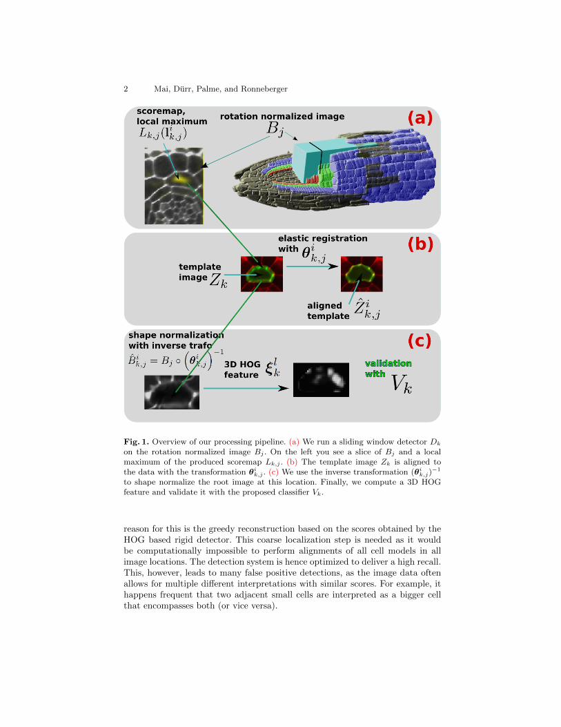
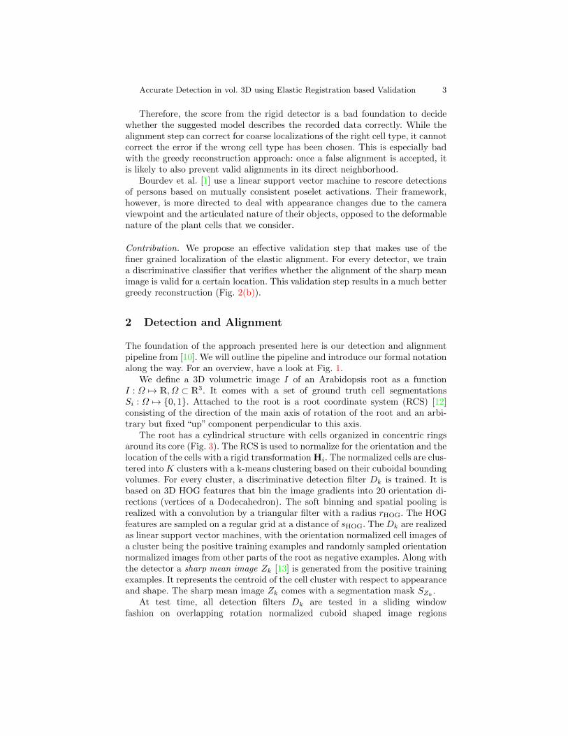
This approach works well for the cell layer 3 (Fig. 3), as the rigid detectionfilter produces reliable hypotheses for the alignment. Unfortunately it fails toproduce satisfactory results for cell layer 4 as is illustrated in Fig. 2(a). The
2 Mai, Durr, Palme, and Ronneberger
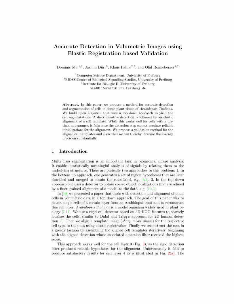
Fig. 1. Overview of our processing pipeline. (a) We run a sliding window detector Dk
on the rotation normalized image Bj . On the left you see a slice of Bj and a localmaximum of the produced scoremap Lk,j . (b) The template image Zk is aligned tothe data with the transformation θi
k,j . (c) We use the inverse transformation (θik,j)
−1
to shape normalize the root image at this location. Finally, we compute a 3D HOGfeature and validate it with the proposed classifier Vk.
reason for this is the greedy reconstruction based on the scores obtained by theHOG based rigid detector. This coarse localization step is needed as it wouldbe computationally impossible to perform alignments of all cell models in allimage locations. The detection system is hence optimized to deliver a high recall.This, however, leads to many false positive detections, as the image data oftenallows for multiple different interpretations with similar scores. For example, ithappens frequent that two adjacent small cells are interpreted as a bigger cellthat encompasses both (or vice versa).
Accurate Detection in vol. 3D using Elastic Registration based Validation 3
Therefore, the score from the rigid detector is a bad foundation to decidewhether the suggested model describes the recorded data correctly. While thealignment step can correct for coarse localizations of the right cell type, it cannotcorrect the error if the wrong cell type has been chosen. This is especially badwith the greedy reconstruction approach: once a false alignment is accepted, itis likely to also prevent valid alignments in its direct neighborhood.
Bourdev et al. [1] use a linear support vector machine to rescore detectionsof persons based on mutually consistent poselet activations. Their framework,however, is more directed to deal with appearance changes due to the cameraviewpoint and the articulated nature of their objects, opposed to the deformablenature of the plant cells that we consider.
Contribution. We propose an effective validation step that makes use of thefiner grained localization of the elastic alignment. For every detector, we traina discriminative classifier that verifies whether the alignment of the sharp meanimage is valid for a certain location. This validation step results in a much bettergreedy reconstruction (Fig. 2(b)).
2 Detection and Alignment
The foundation of the approach presented here is our detection and alignmentpipeline from [10]. We will outline the pipeline and introduce our formal notationalong the way. For an overview, have a look at Fig. 1.
We define a 3D volumetric image I of an Arabidopsis root as a functionI : Ω 7→ R, Ω ⊂ R3. It comes with a set of ground truth cell segmentationsSi : Ω 7→ 0, 1. Attached to the root is a root coordinate system (RCS) [12]consisting of the direction of the main axis of rotation of the root and an arbi-trary but fixed “up” component perpendicular to this axis.
The root has a cylindrical structure with cells organized in concentric ringsaround its core (Fig. 3). The RCS is used to normalize for the orientation and thelocation of the cells with a rigid transformation Hi. The normalized cells are clus-tered into K clusters with a k-means clustering based on their cuboidal boundingvolumes. For every cluster, a discriminative detection filter Dk is trained. It isbased on 3D HOG features that bin the image gradients into 20 orientation di-rections (vertices of a Dodecahedron). The soft binning and spatial pooling isrealized with a convolution by a triangular filter with a radius rHOG. The HOGfeatures are sampled on a regular grid at a distance of sHOG. The Dk are realizedas linear support vector machines, with the orientation normalized cell images ofa cluster being the positive training examples and randomly sampled orientationnormalized images from other parts of the root as negative examples. Along withthe detector a sharp mean image Zk [13] is generated from the positive trainingexamples. It represents the centroid of the cell cluster with respect to appearanceand shape. The sharp mean image Zk comes with a segmentation mask SZk
.At test time, all detection filters Dk are tested in a sliding window
fashion on overlapping rotation normalized cuboid shaped image regions
4 Mai, Durr, Palme, and Ronneberger
(a) (b)
Fig. 2. Greedy reconstructions of layer 4 of the root r06. (green) cells are correctlyaligned detections of cells with an IOU ≥ 0.5, (red) cells are falsely aligned detectionswith an IOU < 0.5. (a) Reconstruction based on the scores from the rigid detector.Many locations of the root are occupied by false detections (average precision = 0.64).(b) The proposed validation step produces much better scores and thus leads to abetter reconstruction (average precision = 0.88).
Bj , j ∈ 1, . . . , NB of the root, that are sampled in 10 steps. The rotationnormalization is based on the RCS. The sliding window is realized as a convo-lution operation that is efficiently computed in the Fourier domain. This resultsin score maps Lk,j : R3 7→ R. The detection locations lik,j ∈ R3, i ∈ 1, . . . , Nk,j(ith detection for the detection filter Dk on the rotation normalized image Bj)are the local maxima of these maps. All local maxima need to be > 0. Within thevolume of the segmentation mask SZk
, all local maxima except the best scoringlocal maxima are suppressed.
The corresponding sharp mean image Zk : R3 7→ R is put at the locationlik,j and is subsequently aligned to the rotation normalized image region Bjwith an elastic registration based on the combinatorial optimization from [8].The elastic registration yields a transformation θik,j : R3 7→ R3 that is used for
obtaining the aligned sharp mean image Zik,j = Zk θik,j and the corresponding
aligned segmentation mask SiZk,j= SZk
θik,j . Note that the Zk and SZkare only
dependent on the k, but the aligned images Zik,j and the aligned segmentation
masks SiZk,jare also dependent on the respective rotation normalized image
region Bj and the implied transformation θik,j .As last step, the aligned sharp mean images are transformed back into the
coordinate system of the original root to obtain a reconstruction. This is done inan iterative greedy fashion, beginning with the aligned sharp mean image Zik,jcorresponding to the highest scoring detection location lik,j . The indices k, j, iare formally given by
k, j, i = arg maxk∈1,...,Kj∈1,...,NBi∈1,...,Nk,j
Lk,j(lik,j) . (1)
The aligned sharp mean image Zik,j is transformed back to the original rootimage, then it is removed from the pool of available detection hypotheses andthe detection hypotheses with the next best score is processed. Note that oncea location in the original root image is occupied, it is not possible to put otheraligned images at this location. This gives a crucial importance to the ordering

Accurate Detection in vol. 3D using Elastic Registration based Validation 5
of the aligned candidates implied by Eqn. 1: Due to the continuous nature of thecells wrt. deformation, we usually have multiple competing aligned candidatesper ground truth location. It is crucial to pick the well aligned candidates firstduring this greedy iterative reconstruction, as a badly aligned candidate can notbe corrected and will probably also prevent subsequent well aligned candidatesin its direct neighborhood.
If the scores delivered by the rigid detector fail to provide a good sorting ofthe aligned candidates prior to the greedy reconstruction, the results for layer 4are not satisfactory (Fig. 2(a)). We propose a validation of the aligned sharpmean images that makes use of the finer grained information that is availabledue to the alignment. This results in much better reconstruction results as wewill show in the experiments section (Fig. 2(b)).
3 Training of the Alignment Classifiers
In order to validate a candidate alignment of the sharp mean template we pro-pose to use the metric induced by a discriminative classifier. To this end wewill use a support vector machine, as it gives a normalized score around zero.Values > 1 indicate a confident decision for the positive class (well aligned can-didate), values < −1 indicate a confident decision for the negative class (badlyaligned candidate). As SVMs have good generalization properties, decision valuesin the interval [−1, 1] mark a gradual change between the classes.
In our case the data used for training and testing is the 3D HOG represen-tation of the root image within the support of the aligned cell template. Thesupport vector machine, however, needs input data of a fixed size. This meansthat we cannot use the image data “below” the aligned template directly, as itsvolume is variable due to the elastic alignment. Therefore we will use the inversetransformation θ−1 to warp the image data onto the cell template. This assuresthat all training and test data for a cluster k will have the same number offeatures.
We need to mine positive (+) and negative (−) training examples from atraining and validation root to train the validation classifier Vk. We compare thealigned segmentation masks SiZk,j
with the ground truth segmentations. TheIntersection over Union is the measure MIOU used in the PASCAL VOC [6]challenge to assess the quality of a detection:
MIOU(S1, S2) =
∫ΩS1(x) · S2(x) dx∫
Ωmax
(S1(x), S2(x)
)dx
. (2)
This area based measure is well suited to evaluate the degree of alignment ina detection setting, especially when it is based on 3D segmentation masks. Weassign the class +,− based on the rule that is also used for the evaluationof the complete pipeline. An aligned candidate is accepted, iff the intersectionover union of the corresponding aligned segmentation mask with ground truth

6 Mai, Durr, Palme, and Ronneberger
segmentation Sl is greater than 0.5:
c(SiZk,j) =
+, MIOU(SiZk,j
, Sl) ≥ 0.5
−, MIOU(SiZk,j, Sl) < 0.5
. (3)
We shape normalize the corresponding root image by transforming it with theinverse transformation:
Bik,j = Bj (θik,j
)−1. (4)
After the this transformation, we extract the 3D HOG feature that will be usedin the training for the validation classifiers Vk.
ξlk = f(Bik,j), f : (Ω 7→ R) 7→ RNfk (5)
The function f transforms the image Bik,j into a vectorial feature representation,i.e. the 3D HOG feature, and crops it along the support of the sharp mean im-age Zk. For simplicity of notation we replace the indices j, k with l ∈ 1, . . . , Nk,as its no longer important, from which Bj the ξlk originates. After the classifi-cation of the training examples into (+) and (−) with Eqn. 3 we end up with aset S+k = l+1 , . . . , l
+N+ for positive examples and a set S−k = l−1 , . . . , l
−N− for
the negative training examples for every cluster k.As wish to investigate the effect of the model complexity of the classifier,
train a linear support vector machine V link a RBF kernel support vector machine
V RBFk . We use 5-fold cross validation to estimate suitable parameters for the
outlier penalty c and the radius γ of the radial basis function for V RBFk . The
training is done with libsvm [4].
3.1 Validating Aligned Templates
The setting at test time is identical to the mining of the training examples forthe Vk, except that we run the greedy iterative reconstruction of the root imageafter the detection and alignment phase. For each detection location lik,j , weperform the elastic registration of the corresponding sharp mean image Zk tothe data and yield the transformation θik,j . Then we shape normalize the root
image at this location by warping it with the inverse transformation (θik,j)−1
and compute the 3D HOG features ξlk (Eqn. 5). Thus we end up with a setof aligned template image candidates and the corresponding 3D HOG featurerepresentations of the locally shape normalized root image:(
Zlk, ξlk
)with k ∈ 1, . . . ,K and l ∈ 1, . . . , Nk . (6)
We perform the iterative greedy reconstruction, but replace the sorting inducedby the rigid detector scores (Eqn. 1) with a sorting based on the proposed vali-dation classifier. We begin with the best scoring candidate image
Zlk with k, l = arg maxk∈1,...,Kl∈1,...,Nk
Vlin,RBFk (ξlk) . (7)
Accurate Detection in vol. 3D using Elastic Registration based Validation 7
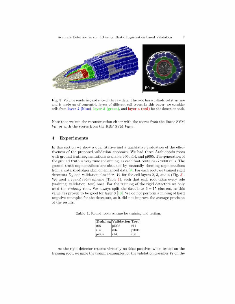
Fig. 3. Volume rendering and slice of the raw data. The root has a cylindrical structureand is made up of concentric layers of different cell types. In this paper, we considercells from layer 2 (blue), layer 3 (green), and layer 4 (red) for the detection task.
Note that we run the reconstruction either with the scores from the linear SVMVlin or with the scores from the RBF SVM VRBF.
4 Experiments
In this section we show a quantitative and a qualitative evaluation of the effec-tiveness of the proposed validation approach. We had three Arabidopsis rootswith ground truth segmentations available: r06, r14, and pi005. The generation ofthe ground truth is very time consuming, as each root contains ∼ 2500 cells. Theground truth segmentations are obtained by manually checking segmentationsfrom a watershed algorithm on enhanced data [9]. For each root, we trained rigiddetectors Dk and validation classifiers Vk for the cell layers 2, 3, and 4 (Fig. 3).We used a round robin scheme (Table 1), such that each root takes every role(training, validation, test) once. For the training of the rigid detectors we onlyused the training root. We always split the data into k = 15 clusters, as thisvalue has proven to be good for layer 3 [10]. We do not perform a mining of hardnegative examples for the detectors, as it did not improve the average precisionof the results.
Table 1. Round robin scheme for training and testing.
Training Validation Test
r06 pi005 r14r14 r06 pi005pi005 r14 r06
As the rigid detector returns virtually no false positives when tested on thetraining root, we mine the training examples for the validation classifier Vk on the

8 Mai, Durr, Palme, and Ronneberger
training and the validation root. We train a linear support vector machine V link
and a RBF support vector machine V RBFk using libsvm [4].
Our test setup is a detection setting. To assess the quality of the detections,we use the same method as in the PASCAL VOC challenge [6]. We accept adetection as valid, iff the intersection over union of the predicted segmentationmask with the ground truth segmentation is ≥ 0.5. All subsequent detectionsof the same ground truth cell that are not suppressed during the reconstructioncount as false positives. We investigate all combinations
r06, r14, pi005 × layer 2, layer 3, layer 4 × Dk, Vlink , V RBF
k
and thus end up with 27 experiments. The sliding window detection is performedon the rotation normalized root images Bj and takes ∼ 50s on a six core work-station. We compute the necessary convolutions in the Fourier domain, thereforethe runtime is not dependent on the size of the detection filter Dk. The align-ment of a cell template to the image data is computed with the combinatorialregistration from our previous work [10], using a gradient orientation based dataterm. The computation of one alignment takes ∼ 1.5s. The scoring of the alignedcell templates with the validation classifier takes < 0.1s. These steps are nearlyperfectly parallelizable. When executed on a computing cluster with 5×32 coresthe detection for a whole root takes ∼ 5min, the alignment of the cell templatesin average ∼ 20min, depending on the number of cell hypotheses. The limitingfactor with our setup was the hard disk IO.
The iterative greedy reconstruction is more difficult to parallelize and takesin average ∼ 30min, also dependent on the number of aligned cell candidates.For some more statistics of the roots, have a look at Table 2.
Table 2. Statistics for the roots (“GT” = ground truth).
r06 r14 pi005
size (voxels) 1030 × 433 × 384 944 × 413 × 360 855 × 458 × 329
layer 2 #GT cells 542 487 554layer 3 #GT cells 216 188 208layer 4 #GT cells 266 211 222
Bj arrangement 3 × 36 3 × 36 2 × 36Bj size (voxels) 301 × 101 × 131 301 × 101 × 131 301 × 101 × 131
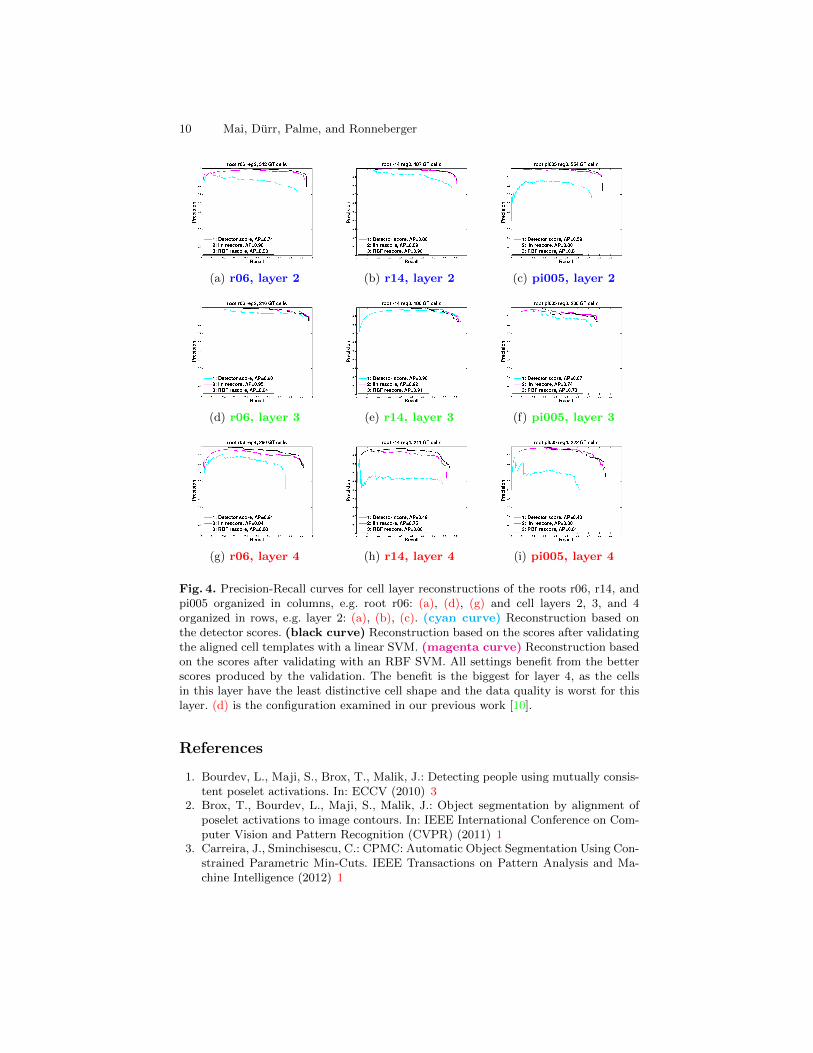
Our findings are summarized in Fig. 4 as precision-recall graphs and in Ta-ble 3 as the mean average precision (AP) per cell layer. The average precisionis computed as the area under the precision-recall curve. Our original process-ing pipeline (cyan curve) works reasonably well for layer 2 and layer 3 withAP = 0.71 and AP = 0.82 respectively. It fails for layer 4 with AP = 0.52.The reason for this can be found in the less distinctive cell shapes of this layer andin its location within the root. For the volumetric recording of layer 4, the lighthas to pass layers 1, 2, and 3 during the recording with a confocal microscope,which results in a more distorted signal.

Accurate Detection in vol. 3D using Elastic Registration based Validation 9
When performing the greedy reconstruction based on the scores of the pro-posed validation approach, we yield substantially better results for the difficultlayer 4. For an illustration see Fig. 2. For every other root and layer combinationwe also achieve better results through the validation scores. The linear SVMbased scores (black curve) achieve the best reconstructions on layer 3. Withthe RBF SVM based scores (red curve), we achieve the best reconstructions forlayer 2 and layer 4. The performance of the linear scoring and the RBF scor-ing is very similar, maybe with a slight edge for the RBF based rescoring. Thetraining times of the SVMs are practically identical. When training directly onthe kernel matrix with libsvm [4], the training takes under a minute including across validation based grid search for the SVM parameters γ and C.
For the validation with the RBF SVM, one needs to compute between 50×and 200× longer compared to the validation with the linear SVM. However, thetime needed to compute a validation is dominated by the disk IO, which leadsto a similar overall computation time.
Table 3. Mean average precision (AP) for scoring strategy and cell layer.
layer 2 layer 3 layer 4
raw detector score 0.71 0.82 0.52linear SVM 0.86 0.87 0.80RBF SVM 0.88 0.86 0.83
5 Conclusions
In this paper we presented a validation strategy for detections in volumetric im-ages that leverages the fine grained localization provided by the elastic alignmentof a cell template image to the underlying image data. We use a metric based ontrained discriminative classifiers to decide whether this alignment was successfulor not. This validation step comes at practically no extra cost given the aligneddetections. However, it achieves to boost the detection accuracy substantially,especially in regions of lower data quality, where the scores of the rigid detectorare no longer reliable. We believe that this validation strategy should also workfor other object classes in 2D and 3D images when the intra class appearancevariation mainly stems from an elastic deformation of the objects.
Acknowledgements
This work was supported by the Excellence Initiative of the German Federaland State Governments: BIOSS Centre for Biological Signalling Studies (EXC294) and the Bundesministerium fur Bildung und Forschung (SYSTEC, 0101-31P5914).
10 Mai, Durr, Palme, and Ronneberger
(a) r06, layer 2 (b) r14, layer 2 (c) pi005, layer 2
(d) r06, layer 3 (e) r14, layer 3 (f) pi005, layer 3
(g) r06, layer 4 (h) r14, layer 4 (i) pi005, layer 4
Fig. 4. Precision-Recall curves for cell layer reconstructions of the roots r06, r14, andpi005 organized in columns, e.g. root r06: (a), (d), (g) and cell layers 2, 3, and 4organized in rows, e.g. layer 2: (a), (b), (c). (cyan curve) Reconstruction based onthe detector scores. (black curve) Reconstruction based on the scores after validatingthe aligned cell templates with a linear SVM. (magenta curve) Reconstruction basedon the scores after validating with an RBF SVM. All settings benefit from the betterscores produced by the validation. The benefit is the biggest for layer 4, as the cellsin this layer have the least distinctive cell shape and the data quality is worst for thislayer. (d) is the configuration examined in our previous work [10].
References
1. Bourdev, L., Maji, S., Brox, T., Malik, J.: Detecting people using mutually consis-tent poselet activations. In: ECCV (2010) 3
2. Brox, T., Bourdev, L., Maji, S., Malik, J.: Object segmentation by alignment ofposelet activations to image contours. In: IEEE International Conference on Com-puter Vision and Pattern Recognition (CVPR) (2011) 1
3. Carreira, J., Sminchisescu, C.: CPMC: Automatic Object Segmentation Using Con-strained Parametric Min-Cuts. IEEE Transactions on Pattern Analysis and Ma-chine Intelligence (2012) 1
Accurate Detection in vol. 3D using Elastic Registration based Validation 11
4. Chang, C.C., Lin, C.J.: LIBSVM: A library for support vector machines. ACMTransactions on Intelligent Systems and Technology 2, 27:1–27:27 (2011) 6, 8, 9
5. Dalal, N., Triggs, B.: Histograms of oriented gradients for human detection. In:Schmid, C., Soatto, S., Tomasi, C. (eds.) International Conference on ComputerVision & Pattern Recognition (2005) 1
6. Everingham, M., Van Gool, L., Williams, C.K.I., Winn, J., Zisserman, A.: Thepascal visual object classes (voc) challenge. International Journal of ComputerVision 88(2), 303–338 (Jun 2010) 5, 8
7. Fernandez, R., Das, P., Mirabet, V., Moscardi, E., Traas, J., Verdeil, J., Malandain,G., Godin, C.: Imaging plant growth in 4d: robust tissue reconstruction and lin-eaging at cell resolution. Nature methods 7(7), 547–553 (2010) 1
8. Komodakis, N., Tziritas, G., Paragios, N.: Performance vs computational efficiencyfor optimizing single and dynamic mrfs: Setting the state of the art with primal-dual strategies. Computer Vision and Image Understanding 112(1), 14–29 (2008)4
9. Liu, K., Schmidt, T., T.Blein, Durr, J., Palme, K., Ronneberger, O.: Joint 3d cellsegmentation and classification in the arabidopsis root using energy minimizationand shape priors. In: IEEE International Symposium on Biomedical Imaging (ISBI)(2013) 1, 7
10. Mai, D., Fischer, P., Blein, T., Durr, J., Palme, K., Brox, T., Ronneberger, O.:Discriminative detection and alignment in volumetric data. In: Weickert, J., Hein,M., Schiele, B. (eds.) GCPR. Lecture Notes in Computer Science, vol. 8142, pp.205–214. Springer (2013) 1, 3, 7, 8, 10
11. Marcuzzo, M., Quelhas, P., Campilho, A., Maria Mendonca, A., Campilho, A.:Automated arabidopsis plant root cell segmentation based on svm classificationand region merging. Comput. Biol. Med. 39(9), 785–793 (2009) 1
12. Schmidt, T., Pasternak, T., Liu, K., Blein, T., Aubry-Hivet, D., Dovzhenko, A.,Durr, J., Teale, W., Ditengou, F.A., Burkhardt, H., Ronneberger, O., Palme, K.:The irocs toolbox – 3d analysis of the plant root apical meristem at cellular res-olution. The Plant Journal 77(5), 806–814 (Mar 2014), http://lmb.informatik.uni-freiburg.de//Publications/2014/SLBR14 3
13. Wu, G., Jia, H., Wang, Q., Shen, D.: Sharpmean: Groupwise registration guidedby sharp mean image and tree-based registration. NeuroImage 56(4) (2011) 3


![[Archived] Infusion systems guidance · Powered volumetric infusion pumps, together with an appropriate administration set, are intended to provide an accurate flow of fluids over](https://img.pdfslide.us/doc/110x75/5e146239bca18d7f377b5e39/archived-infusion-systems-guidance-powered-volumetric-infusion-pumps-together.jpg)