Embed Size (px)
Citation preview
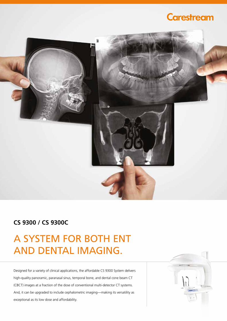
CS 9300 / CS 9300C
Designed for a variety of clinical applications, the affordable CS 9300 System delivers
high-quality panoramic, paranasal sinus, temporal bone, and dental cone beam CT
(CBCT) images at a fraction of the dose of conventional multi-detector CT systems.
And, it can be upgraded to include cephalometric imaging—making its versatility as
exceptional as its low dose and affordability.
A SYSTEM FOR BOTH ENT AND DENTAL IMAGING.
CS 9300 / CS 9300C SYSTEM
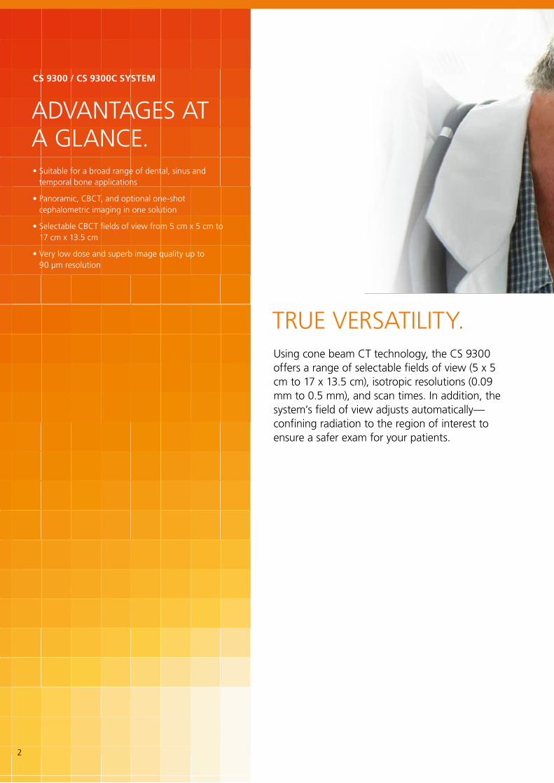
TRUE VERSATILITY.
ADVANTAGES AT A GLANCE.
Using cone beam CT technology, the CS 9300 offers a range of selectable fields of view (5 x 5 cm to 17 x 13.5 cm), isotropic resolutions (0.09 mm to 0.5 mm), and scan times. In addition, the system’s field of view adjusts automatically—confining radiation to the region of interest to ensure a safer exam for your patients.
• Suitable for a broad range of dental, sinus and temporal bone applications
• Panoramic, CBCT, and optional one-shot cephalometric imaging in one solution
• Selectable CBCT fields of view from 5 cm x 5 cm to 17 cm x 13.5 cm
• Very low dose and superb image quality up to 90 μm resolution
2

SINUS AND TEMPORAL BONE FIELDS OF VIEW
FIELDS OF VIEW SCANNING TIME
RESOLUTION
REGION OF INTEREST
Sinus17 x 13.5 cm
28 sec.
0.3 – 0.5 mm
• Paranasal sinus evaluation
• Chronic sinusitis with and without polyps
• Image guided sinus surgery
• Craniofacial evaluation
• Chronic sinusitis with and without polyps
• Treatment follow up (17x11 fast)
• Pediatric sinus evaluation (17x11 fast)
• Cochlear implant evaluation and follow up
• Otosclerosis and cholesteatoma
• Superior semicircular canal dehiscence
• Image guided temporal surgery
• Chronic otitis media
• Cholesteatoma
• Superior semicircular canal dehiscence
• Cochlear implant evaluation and follow up
• Otosclerosis and cholesteatoma
• Superior semicircular canal dehiscence
Sinus17 x 11 cm
12 or 20 sec. 0.25 - 0.5 mm
Temporal bone17 x 6 cm
20 sec.
0.20 - 0.5 mm
Temporal bone8 x 8 cm
20 sec.
0.20 - 0.3 mm
Temporal bone5 x 5 cm
12 or 20 sec. 0.09 - 0.2 mm
SAMPLE IMAGES RECOMMENDED APPLICATIONS
3
FIELDS OF VIEW SCANNING TIME
RESOLUTION
REGION OF INTEREST
SAMPLE IMAGES RECOMMENDED APPLICATIONS
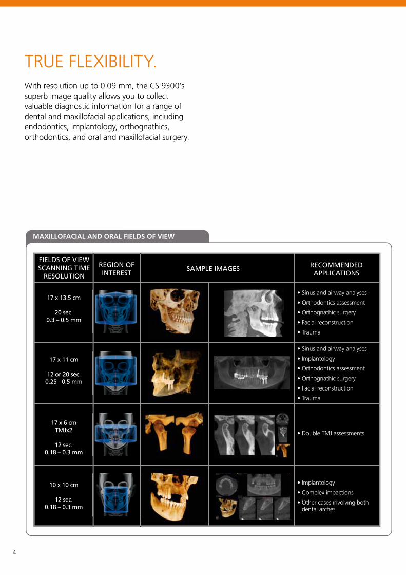
MAXILLOFACIAL AND ORAL FIELDS OF VIEW
17 x 13.5 cm
20 sec. 0.3 – 0.5 mm
17 x 11 cm
12 or 20 sec.0.25 - 0.5 mm
17 x 6 cm TMJx2
12 sec.
0.18 – 0.3 mm
10 x 10 cm
12 sec. 0.18 – 0.3 mm
• Sinus and airway analyses
• Orthodontics assessment
• Orthognathic surgery
• Facial reconstruction
• Trauma
• Sinus and airway analyses
• Implantology
• Orthodontics assessment
• Orthognathic surgery
• Facial reconstruction
• Trauma
• Double TMJ assessments
• Implantology
• Complex impactions
• Other cases involving both dental arches
TRUE FLEXIBILITY.With resolution up to 0.09 mm, the CS 9300’s superb image quality allows you to collect valuable diagnostic information for a range of dental and maxillofacial applications, including endodontics, implantology, orthognathics, orthodontics, and oral and maxillofacial surgery.
4
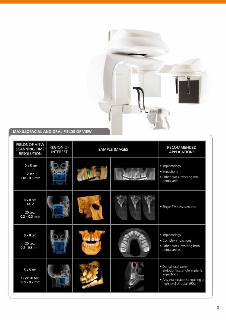
FIELDS OF VIEW SCANNING TIME
RESOLUTION
REGION OF INTEREST
SAMPLE IMAGES RECOMMENDED APPLICATIONS
MAXILLOFACIAL AND ORAL FIELDS OF VIEW
10 x 5 cm
12 sec. 0.18 - 0.5 mm
8 x 8 cm TMJx1
20 sec. 0.2 – 0.3 mm
8 x 8 cm
20 sec. 0.2 - 0.3 mm
5 x 5 cm
12 or 20 sec.0.09 - 0.2 mm
• Implantology
• Impactions
• Other cases involving one dental arch
• Single TMJ assessments
• Implantology
• Complex impactions
• Other cases involving both dental arches
• Dental local cases: Endodontics, single implants, impactions
• Any examinations requiring a high level of detail (90μm)
5
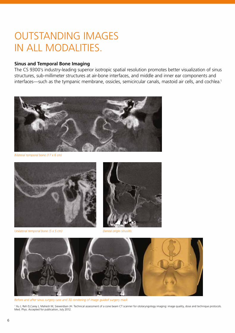
OUTSTANDING IMAGES IN ALL MODALITIES. Sinus and Temporal Bone ImagingThe CS 9300’s industry-leading superior isotropic spatial resolution promotes better visualization of sinus structures, sub-millimeter structures at air-bone interfaces, and middle and inner ear components and interfaces—such as the tympanic membrane, ossicles, semicircular canals, mastoid air cells, and cochlea.1
Bilateral temporal bone (17 x 6 cm)
Unilateral temporal bone (5 x 5 cm) Dental origin sinusitis
Before and after sinus surgery case and 3D rendering of image guided surgery mask
1 Xu J, Reh D,Carey J, Mahesh M, Siewerdsen JH. Technical assessment of a cone beam CT scanner for otolaryngology imaging: image quality, dose and technique protocols. Med. Phys. Accepted for publication, July 2012.
6
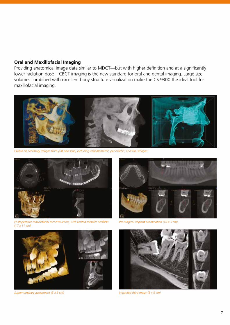
Oral and Maxillofacial ImagingProviding anatomical image data similar to MDCT—but with higher definition and at a significantly lower radiation dose—CBCT imaging is the new standard for oral and dental imaging. Large size volumes combined with excellent bony structure visualization make the CS 9300 the ideal tool for maxillofacial imaging.
Create all necessary images from just one scan, including cephalometric, panoramic, and TMJ images
Postoperative maxillofacial reconstruction, with limited metallic artifacts (17 x 11 cm)
Supernumerary assessment (5 x 5 cm)
Pre-surgical implant examination (10 x 5 cm)
Impacted third molar (5 x 5 cm)
7
30 x 30 cm (12 x 12 in.)24 x 30 cm (10 x 12 in.)
24 x 24 cm (10 x 10 in.)18 x 24 cm (8 x 10 in.)
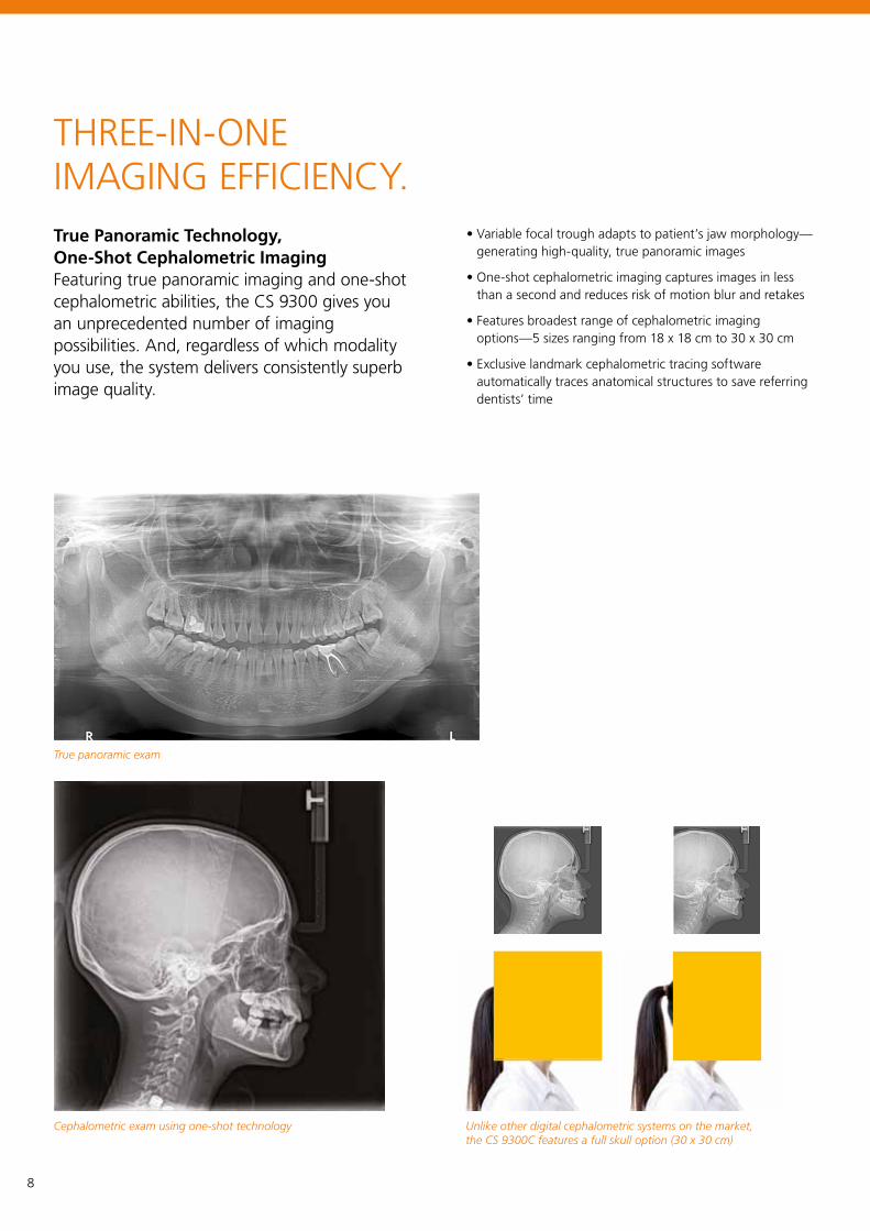
THREE-IN-ONE IMAGING EFFICIENCY.True Panoramic Technology, One-Shot Cephalometric ImagingFeaturing true panoramic imaging and one-shot cephalometric abilities, the CS 9300 gives you an unprecedented number of imaging possibilities. And, regardless of which modality you use, the system delivers consistently superb image quality.
• Variable focal trough adapts to patient’s jaw morphology—generating high-quality, true panoramic images
• One-shot cephalometric imaging captures images in less than a second and reduces risk of motion blur and retakes
• Features broadest range of cephalometric imaging options—5 sizes ranging from 18 x 18 cm to 30 x 30 cm
• Exclusive landmark cephalometric tracing software automatically traces anatomical structures to save referring dentists’ time
True panoramic exam
Unlike other digital cephalometric systems on the market, the CS 9300C features a full skull option (30 x 30 cm)
Cephalometric exam using one-shot technology
8
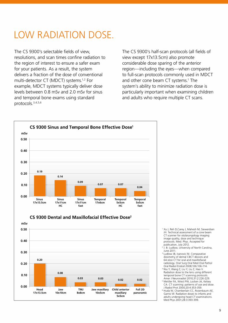
The CS 9300’s selectable fields of view, resolutions, and scan times confine radiation to the region of interest to ensure a safer exam for your patients. As a result, the system delivers a fraction of the dose of conventional multi-detector CT (MDCT) systems.1,2 For example, MDCT systems typically deliver dose levels between 0.8 mSv and 2.0 mSv for sinus and temporal bone exams using standard protocols.3,4,5,6
LOW RADIATION DOSE.
The CS 9300’s half-scan protocols (all fields of view except 17x13.5cm) also promote considerable dose sparing of the anterior region—including the eyes—when compared to full-scan protocols commonly used in MDCT and other cone beam CT systems.1 The system’s ability to minimize radiation dose is particularly important when examining children and adults who require multiple CT scans.
CS 9300 Sinus and Temporal Bone Effective Dose1
CS 9300 Dental and Maxillofacial Effective Dose2
mSv
mSv
0.00
0.00
0.10
0.10
0.20
0.20
0.30
0.30
0.40
0.40
0.50
0.50 1 Xu J, Reh D,Carey J, Mahesh M, Siewerdsen JH. Technical assessment of a cone beam CT scanner for otolaryngology imaging: image quality, dose and technique protocols. Med. Phys. Accepted for publication, July 2012.
2 J. B. Ludlow, University of North Carolina, June 2011.
3 Ludlow JB, Ivanovic M. Comparative dosimetry of dental CBCT devices and 64-slice CT for oral and maxillofacial radiology. Oral Surg Oral Med Oral Pathol Oral Radiol Endod 2008;106:106-114.
4 Niu Y, Wang Z, Liu Y, Liu Z, Hao V. Radiation dose to the lens using different temporal bone CT scanning protocols. Amer J Neuroradiol 2010;31:2:226-229.
5 Mettler FA, Wiest PW, Locken JA, Kelsey CA. CT scanning: patterns of use and dose. J Radiol Prot 2000;20:4:353-359.
6 Huda W, Chamberlain CC, Rosenbaum AE, Garrisi W. Radiation doses to infants and adults undergoing head CT examinations. Med Phys 2001;28:3:393-399.
9
TMJ8x8cm
Jaw maxillary 10x5cm
Child anterior maxillary
5x5cm
Full 2Dpanoramic
Head17x13.5cm
Jaw10x10cm
Sinus17x11cm
fast
Temporal 17x6cm
Temporal 5x5cm
HC
Temporal 5x5cm
fast
Sinus17x13.5cm
0.19
0.14
0.090.07 0.07
0.04
Sinus17x11cm
HC
0.020.020.030.03
0.08
0.20
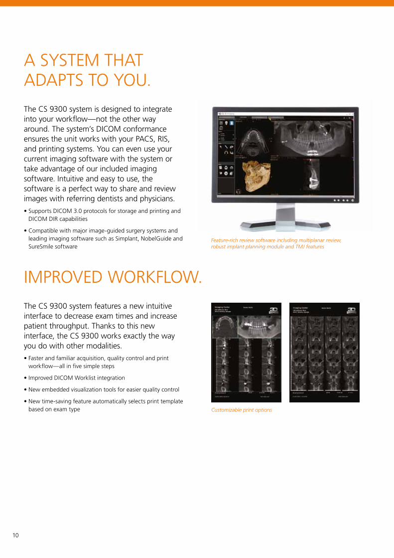
A SYSTEM THAT ADAPTS TO YOU.
The CS 9300 system is designed to integrate into your workflow—not the other way around. The system’s DICOM conformance ensures the unit works with your PACS, RIS, and printing systems. You can even use your current imaging software with the system or take advantage of our included imaging software. Intuitive and easy to use, the software is a perfect way to share and review images with referring dentists and physicians.
• Supports DICOM 3.0 protocols for storage and printing and DICOM DIR capabilities
• Compatible with major image-guided surgery systems and leading imaging software such as Simplant, NobelGuide and SureSmile software
Customizable print options
Feature-rich review software including multiplanar review, robust implant planning module and TMJ features
The CS 9300 system features a new intuitive interface to decrease exam times and increase patient throughput. Thanks to this new interface, the CS 9300 works exactly the way you do with other modalities.
• Faster and familiar acquisition, quality control and print workflow—all in five simple steps
• Improved DICOM Worklist integration
• New embedded visualization tools for easier quality control
• New time-saving feature automatically selects print template based on exam type
IMPROVED WORKFLOW.
10
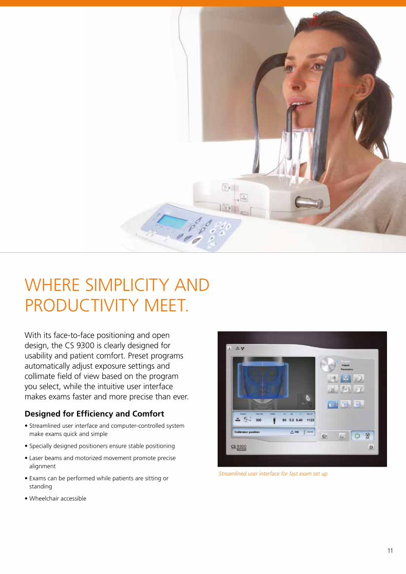
WHERE SIMPLICITY AND PRODUCTIVITY MEET.
With its face-to-face positioning and open design, the CS 9300 is clearly designed for usability and patient comfort. Preset programs automatically adjust exposure settings and collimate field of view based on the program you select, while the intuitive user interface makes exams faster and more precise than ever.
Designed for Efficiency and Comfort• Streamlined user interface and computer-controlled system
make exams quick and simple
• Specially designed positioners ensure stable positioning
• Laser beams and motorized movement promote precise alignment
• Exams can be performed while patients are sitting or standing
• Wheelchair accessible
Streamlined user interface for fast exam set up
11
A TRUSTED COMPANY, A PROVEN PRODUCT.With over a century of experience, no one knows medical and dental imaging like Carestream. From installation and training to technical support and integration services, we provide the expert services you need to keep your equipment operating at peak performance—and the CS 9300 is no exception: with more than 4,000 units installed, the system is built on a proven CBCT platform and backed by our comprehensive service and support packages.
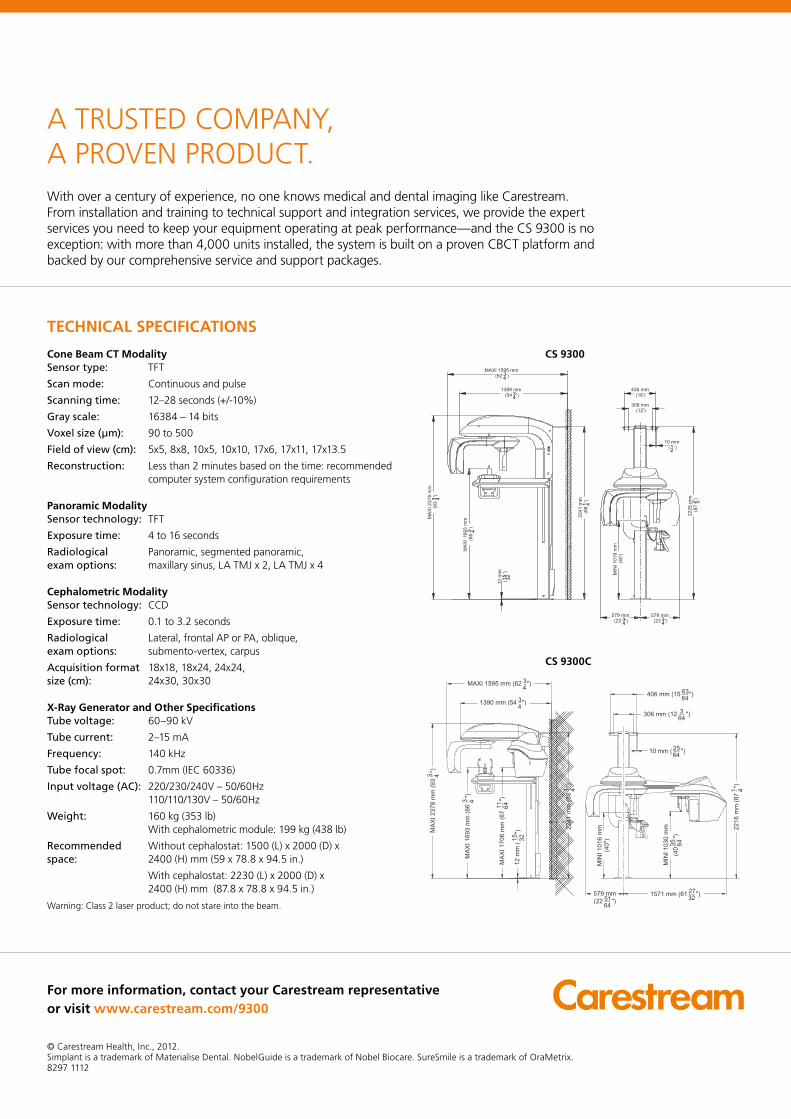
Cone Beam CT ModalitySensor type: TFT
Scan mode: Continuous and pulse
Scanning time: 12–28 seconds (+/-10%)
Gray scale: 16384 – 14 bits
Voxel size (µm): 90 to 500
Field of view (cm): 5x5, 8x8, 10x5, 10x10, 17x6, 17x11, 17x13.5
Reconstruction: Less than 2 minutes based on the time: recommended computer system configuration requirements
Panoramic ModalitySensor technology: TFT
Exposure time: 4 to 16 seconds
Radiological Panoramic, segmented panoramic, exam options: maxillary sinus, LA TMJ x 2, LA TMJ x 4
Cephalometric ModalitySensor technology: CCD
Exposure time: 0.1 to 3.2 seconds
Radiological Lateral, frontal AP or PA, oblique, exam options: submento-vertex, carpus
Acquisition format 18x18, 18x24, 24x24, size (cm): 24x30, 30x30
X-Ray Generator and Other SpecificationsTube voltage: 60–90 kV
Tube current: 2–15 mA
Frequency: 140 kHz
Tube focal spot: 0.7mm (IEC 60336)
Input voltage (AC): 220/230/240V – 50/60Hz 110/110/130V – 50/60Hz
Weight: 160 kg (353 lb) With cephalometric module: 199 kg (438 lb)
Recommended Without cephalostat: 1500 (L) x 2000 (D) x space: 2400 (H) mm (59 x 78.8 x 94.5 in.)
With cephalostat: 2230 (L) x 2000 (D) x 2400 (H) mm (87.8 x 78.8 x 94.5 in.)
Warning: Class 2 laser product; do not stare into the beam.
For more information, contact your Carestream representative or visit www.carestream.com/9300
© Carestream Health, Inc., 2012.Simplant is a trademark of Materialise Dental. NobelGuide is a trademark of Nobel Biocare. SureSmile is a trademark of OraMetrix.8297 1112
TECHNICAL SPECIFICATIONS
CS 9300
CS 9300C