Embed Size (px)
Citation preview
Journal of Pharmacy and Pharmacology 6 (2018) 175-187 doi: 10.17265/2328-2150/2018.02.011
A Simple Procedure to Fabricate Paper Biosensor and Its Applicability—NADH/NAD+ Redox System
Isabel Ribau and Elvira Fortunato
Departamento de Ciências dos Materiais, Faculdade de Ciências e Tecnologia, Universidade Nova de Lisboa, Campus de Caparica,
Caparica 2829-516, Portugal
Abstract: A simple device which incorporates three electrodes (working electrode, counter electrode and reference electrode) was constructed to be used currently in laboratories without elevated cost. It does not need more than 2 µL of electrolyte, sample or working solution, his support material is paper, and the working electrode which is based on carbon ink can incorporate enzymes and cofactors. To test this concept we started this investigation using the NADH/NAD+ redox couple which is an omnipresent coenzyme in living systems but is also a challenge to electrochemistry. The paper sensor fabrication was simple, rapid and cheaper. NADH was incorporated in the carbon ink by mixing both and, this mixture was used to print the working electrode. The direct electrochemical system NADH/NAD+ signal obtained, using this device, appeared at low potentials. A quasi-reversible diffusional redox process was achieved and regeneration of the NADH after oxidation was reached. This small paper device was not only used to study the redox process of NAD+/NADH, but also its behavior in the presence of electroactive (ascorbic acid) and non-eletroactive species (glucose). Key words: NADH/NAD+, screen-printing biosensor, paper device.
1. Introduction
NADH molecule can be oxidized through the
The redox system NADH/NAD+ has been very important in nature, not only because it is a ubiquitous coenzyme, but also because it participates in many cellular reactions. This system is important in pharmaceutical and food industry as well as in biotechnology industry since it is present in drugs, biosensors, but also in biofuel cells and bioreactors [1-7], nevertheless is expensive and the redox process is usually irreversible [3, 6].
In the NAD+ reduction, the reversible redox process occurs in nicotinamide ring, which accepts two electrons from a substrate in the presence of an appropriated enzyme, forming NADH: SH2 + NAD+ ⇄ S + NADH+ H+ [3]. This reaction is particularly important since it allows the identification of non-eletroactive substrates that interact with NAD+ and take part in its reduction [8].
Corresponding author: Isabel Ribau, Ph.D., researcher,
research fields: biosensors and bioeletrochemistry.
nicotinamide or adenosine groups [8]. The NADH is able to oxidize at potential higher than +0.4 V (Ag/AgCl) on carbon paste electrodes [3, 8]. Adenosine free in solution is oxidized at +1.2 V (vs. SCE, pH 7.0) in carbon electrodes. Nicotinamide presents an oxidation peak at +0.45 V (vs. SCE, pH 7.0) in carbon electrodes. In the same conditions the peak at +1.05 V was attributed to the formation of an adduct between phosphate anions and NADH through adenine and nicotinamide groups. If in phosphate medium, using a glassy carbon electrode, a potential higher than +0.9 V is applied to nicotinamide, it promotes the surface blocking with adducts [8].
The oxidation of NADH in bare electrodes occurs via radical cation intermediates, which may lead to the fouling process and surface poisoning with the formation of inactive NAD2 dimers (adsorbed on the electrode surface), adducts or the formation NADH (active form). As a result of these reactions, low sensitivity, selectivity and stability are achieved [3, 5, 8, 9].
Some research has been done to study the direct
D DAVID PUBLISHING
A Simple Procedure to Fabricate Paper Biosensor and Its Applicability—NADH/NAD+ Redox System
176
electrochemistry of NAD+/NADH and the possible causes of the reduction irreversibility, with the formation of inactive form NAD2 reported in some studies [6, 8, 10]. In one of these researches, it was possible to regenerate NAD+ to the active form, 1,4-NADH, applying a regeneration potential [6]. The reduction reaction of NAD+ on glassy carbon was irreversible and under diffusion control, at a formal potential, E0’ (NAD+/NADH) = -0.885 V (pH 5.8). In another article the formal redox potential of NAD+/NADH was -0.560 V (vs. SCE), or -0.315 V (vs. NHE, pH 7) and the E0’ variation with pH was -30.3 mV/pH [5]. For a reversible process, in unmodified carbon surfaces, the formal potential of the redox coupled NAD+/ NADH was -0.32 V (NHE, pH 7) but the heterogeneous kinetic was slower and interferences could occur [3, 6]. Using different electrochemical techniques, the apparent formal heterogeneous electron-transfer rate constant was estimated as (6.1 ± 2) × 10-14 cms-1 and (2.5 ± 1) × 10-14 cms-1 [6]. These low values indicate very slow kinetics of the NAD+ reduction reaction on a glassy carbon electrode and reflect the over potential necessary for the redox reaction which is related to both redox kinetics and mass transport.
One strategy employed to overcome these difficulties (fouling and overvoltage and side reactions) was the use of mediator-modified electrodes, where the mediators are used to shuttle electrons from NADH to the electrode surface and allow electron transport between them [5, 9-16]. Some mediators (electrocatalysts) were immobilized on the electrode surface by covalent attachment, electrochemical polymerization, incorporation in carbon paste, adsorption, self-assembly and via entrapment in polymeric matrices [4, 17-19]. Another strategy is modification of the electrode surface with a polymeric substance, using electrodes modified with carbon nanotubes, nanofibers or using enzymatic methods that follow bioelectrocatalytic reaction [2, 4, 13, 15, 16,
20-23]. Investigation in this area usually explores the mediator use or the surface modification to improve the electrochemical detection [15, 21, 24-28]. Some literature has been published related to the study of the redox couple NADH/NAD+ in screen printing electrodes [1, 4, 21, 24-27, 29-34]. The sensor built using this technique can incorporate the three electrodes (working electrode, reference and counter electrode), can be easily produced and miniaturized, work with a minimum volume (2 µL), it needs low reagent consumption, it can be fabricated in many supports (paper, glass) and sensors are disposable devices that can be used in many science fields [26, 35]. In a screen-printing electrode prototype, the oxidation of NADH (0.4 mM) occurred in a potential range from +0.18 V to +0.44 V but the signals were not well-defined [34]. At a screen-printing electrodes modified with MWCNTs (multiwalled carbon nanotubes), or AuNPs (gold nanoparticles) or with PNRs (polyneutral red films), it was possible to verify that the best response of the redox system NAD+/NADH was obtained with the modification using MWCNTs, which was used as an amperometric NADH detector [27].
The goal of this report is to present a sensor that can be easily fabricated, which is cheaper and environmentally friend and allows the electrochemical study of the redox couple NADH/NAD+. With this aim, we present an NADH/ NAD+ biosensor, for sensing subtracts that can interact with the cofactor.
There are significant advantages of using this biosensor, the first is that it is possible to obtain the oxidation and reduction peak of the redox couple NADH/NAD+ in lower potentials, second is the possibility to sense either electroactive as non-electroactive species, the third is that the fabricated device is not time-consuming (is quick and simple), the fourth is the low cost of the device, the fifth is the ecological approach as this device is made with paper.
A Simple Procedure to Fabricate Paper Biosensor and Its Applicability—NADH/NAD+ Redox System
177
2. Material and Methods
2.1 Materials
All reagents used were of analytical grade β-nicotinamide adenine dinucleotide, NADH (reduced disodium salt hydrate), β-D-glucose, ascorbic acid, potassium chloride and potassium ferrocyanide were acquired from Sigma-Aldrich. All solutions were prepared with buffer. All buffers used in this work were commercial and purchased from ROTH (Germany). The electrolyte was a buffer solution with potassium chloride (0.1 M).
2.2 Fabrication of the Biosensors
The fabrication procedure that will be described below, is not time consuming (it takes 15 minutes to do 5 biosensors), is low cost (the price is lower than 0.2 Euros per electrode) and is ecological (the biosensors is constructed with paper, and the silver and carbon ink can be removed and recovered).
The carbon ink and Ag/AgCl ink were purchased from Conductive Compounds. The working electrode was of carbon ink which was added to the solid β-nicotinamide adenine dinucleotide (see Section 2.3 Working Electrode Preparation).
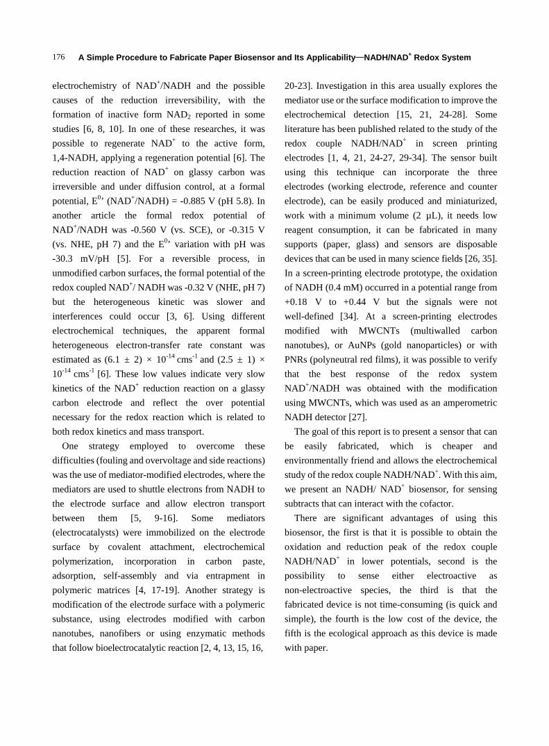
A Xerox Color Qube 8570 printer was used to print the hydrophobic region of the devices. The paper used was Whatman n.0 1 chromatographic paper, and the wax was obtained from Xerox. After the wax printing, the wax was heat treated during 10 s in a hot plate (150 oC). After that, the paper, cooled at room temperature, was ready to perform the screen printing technique. The configuration system design was a three-electrode system with an Ag/AgCl as the reference electrode, a carbon counter electrode and a working electrode based on carbon ink, Figs. 1A and 1B.
The counter electrode was printed with conductive carbon ink, which was deposited above the hydrophobic matrix (wax). Then the mesh was removed and the device was allowed to heat at hot plate
Fig. 1 Biosensor with a three-electrode configuration system design with an Ag/AgCl reference electrode, a carbon counter electrode and a working electrode based in carbon ink (A). SEM (scanning electron microscopy) images of the working electrode surface (B). Upper image scale is 1 µm, and down image scale is 100 µm. Observations were carried out using a Carl Zeiss AURIGA Cross Beam (FIB-SEM) workstation coupled with energy dispersive X-ray spectroscopy (EDS) from Oxford Instruments. The materials have been previously coated with an Ir conductive film for avoiding charge effects.
A Simple Procedure to Fabricate Paper Biosensor and Its Applicability—NADH/NAD+ Redox System
178
(60 oC) during 5 minutes. Once the construction of the counter was concluded, the working electrode was printed and dried the same way as described before. The reference electrode, fabricated with Ag/AgCl ink had the same screen printing treatment.
2.3 Working Electrode Preparation
A mixture with the cofactor (NADH) enclosed in the carbon ink was prepared and used to make the working electrode.
2.4 Electrochemical Detection
During the electrochemical measurements, a drop of the interest solutions (2 µL) was spotted in the hydrophobic channel between the wax-limited zones (working area) and it was dispersed through the paper matrix in a few seconds, being in contact with the three electrodes. The electrochemical behavior of each biosensor was experimentally characterized through cyclic voltammetry.
All electrochemical acquisitions and measurements were performed in a Gamry ESA419 data acquisition system, using PHE 200 physical electrochemical and PV 220 physical electrochemical software coupled with a Gamry instruments (reference 600) potentiostat/galvanostat (ZRA) and the data analysis was processed by Gamry software package. All the experimental procedures were performed in normal atmosphere in the presence of oxygen.
3. Results and Discussion
The electrochemical behavior of the redox couple NAD+/NADH has been analyzed in the last years due to its role as a cofactor, and as an electron carrier in living organisms [5, 6, 14].
In this research work the cofactor electrochemical behavior was studied using the PBS buffer (pH 7) with KCl (0.1 M) as the electrolyte. This pH was chosen because NADH is instable in highly alkaline and acidic solutions due to its rapid degradation [27].
3.1 Electrode Surface Area
The electrochemical characterization of the electrodes was made using the standard heterogeneous rate constant (k0) and the effective electroactive area.
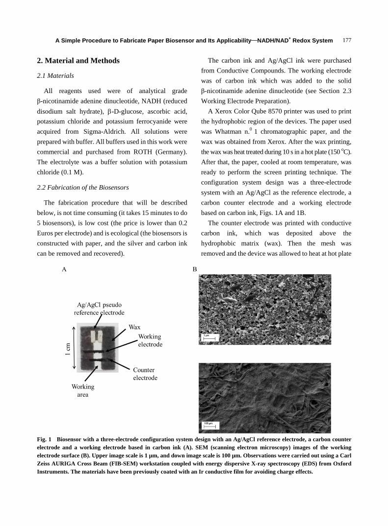
To obtain the electrode real surface area, a redox model par, potassium ferri(III)cyanide/ ferro(II)cyanide, K3/K4Fe(CN)6, was used. The cyclic voltammograms were recorded in the sweep between 20 mV·s-1 and 150 mV·s-1 in PBS buffer (pH 7) with 100 mM KCl as the supporting electrolyte. From these recordings, the peak currents were measured, in Fig. 2.
The peak potential average of the reduction and oxidation, E1/2, is related to the formal potential E0’ by Eq. 2, E1/2 = E0’+ (RT/nF)1 ln(Dr/Do). As the diffusion coefficient is related to the peak current vs. scan rate square root slope, Dr/Do is approximately one and E1/2 = E0’. The formal reduction potential can be estimated from the average of the reduction and oxidation peak potentials and a value of E0’ = (Ep
c+Epa)/2.
The results presented are the medium values obtained with two screen-printed electrodes: (Ep
a = (+269 ± 1) mV vs. Ag/AgCl; Epc = (+163 ± 6)
mV vs. Ag/AgCl), a midpoint, E0’, of (+216 ± 3) mV vs. Ag/AgCl, and peak separation of (+107 ± 7) mV vs.
Ag/AgCl. Peak currents vary linearly with the square root of the scan rate, in the studied rate range, thus denoting a diffusion controlled process.
The peak currents ratio obtained in these experiments (|ip
a/ipc| = +0.72 ± 0.05), and the peak to
peak separation enable us to conclude that in these conditions the potassium ferri(III) cyanide /ferro (II) cyanide do not present a reversible behavior as it was expected. It is possible to verify that the ratio ip /v1/2 is independent of the scan rate (in the range between 150 mV·s-1 and 20 mV. s-1), ip
a/v1/2 (+29.1 ± 1.2) µA and ip
c /v1/2 (-20.8 ± 1.3) µA, but Ep varies with the scan
rate and △Ep also increases with the scan rate indicating that in these conditions the redox par behaves like a quasi-reversible system.
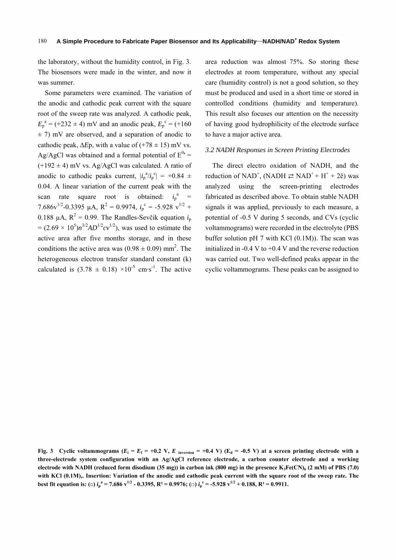
A Sim
Fig. 2 Cycliwith a three-eelectrode withwith KCl (0.1
better fit equa
The elect
using the r
standard con
low values,
if it is high
In these an
electron tra
developed
coefficient v
experimenta
independent
allowed the
parameters.
between E
k/(aDo)
= 0.5), whic
(diffusion co
10-6 cm2·s-1
ferrocyanide
transfer coef
the heterog
mple Procedu
ic voltammogrelectrode systeh NADH (redu1 M). Insertion
ation is: ( ) ipa
trochemical r
rate of hete
nstant (k) [3
the electron t
it indicates f
nalyses, to d
ansfer const
by Nicholso
value, , can
al curves with
t of in th
determinatio
In these inte
Ep and the kin
), where = (
ch is used to
oefficient for 1, and for D
e) the value
fficient value
eneous elect
ure to Fabrica
rams (Ei = Ef =em configuratiuced form disodn: Variation ofa = 29.974 v1/2 -
reversibility c
erogeneous e
6, 37]. Whe
transfer kinet
fast electron
determine th
tant, we us
on [36, 37
n be accesse
h the theoret
he range 0.3
n of k, witho
ervals of , th
netic paramet
(Do/Dr)1/2, a =
calculate the
ferri cyanide
Dr (diffusion
e 6.5 × 10⁻⁶
e of 0.5, it wa
tron transfer
ate Paper Bios
= +0.6 V, E inv
ion with an Agdium (35 mg)) f the anodic an
- 0.1463R² = 0.9
can be evalu
electron tran
en the k pres
tics are slow,
transfer kine
he heterogene
sed the met
]. The tran
ed by compa
tical ones. E
3 to 0.7, w
ut knowing th
here is a rela
ter (Eq. 1,
= nFv/RT whe
e k. Using fo
e) the value 7
n coefficient 6 cm2·s-1 an
as possible to
constant, k
sensor and It
version = -0.2 V)g/AgCl referenin carbon ink nd cathodic pe
9976; (□) ipc =
uated
nsfer
sents
, but
etics.
eous
thod
nsfer
aring
Ep is
which
hese
ation
=
en
r Do
7.6 ×
for
nd a
find
k, of
(2.6
the
elec
are
T
equ
150
was
3.5
(2.6
elec
is th
is th
is th
in c
surf
Ran
obta
syst
A
dete
ts Applicabili
(Ed = -0.5 V) nce electrode, a(800 mg) in th
eak current wi
-18.419 v1/2 - 0
63 ± 0.46) ×
value found
ctrodes (5.2 ×
in the presen
To estimate
uation was us
0 mV·s-1. Fro
s possible to
± 0.7 mm2
69 × 105)n3/2
ctrons exchan
he electroche
he concentrat
he scan rate (
cm2/s), was u
face area (3
ndles-Sevčik
ained for pota
tem [38].
After five
ermined again
ity—NADH/NA
at a screen ata carbon coun
he presence K3Fith the square
0.4838R² = 0.99
10-2 cm·s-1.
d in other stu
× 10-6 cm·s-1)
nce of a quasi
surface ar
sed, in a rang
om the slope
calculate th2. The Randl2AD1/2cv1/2, (w
nge in the red
emically activ
ion of the elec
(V/s) and D i
used to estima
.5 mm2) an
equation,
assium ferroc
months the
n, using bios
AD+ Redox S
t a screen prin
nter electrode aFe(CN)6 (1 mM
e root of the sw
911.
This value is
udies with sc
) [1] and indi
-reversible sy
rea the Ra
ge between 20
of the ip vs
he medium ac
les-Sevčik eq
where n is th
dox process, h
ve electrode a
ctric species i
s the diffusio
ate the effect
nd it was ob
through the
cyanide/ferric
e electrode
ensors that w
System 179
nting electrodeand a working
M) of PBS (7.0)weep rate. The
s bigger than
reen-printing
cates that we
ystem.
andles-Sevčik
0 mV·s-1 and
. v1/2 plots it
ctive area as
quation, ip =
he number of
here n = 1; A
area in cm2; c
in mol/cm3; v
on coefficient
tive electrode
btained from
parameters
cyanide redox
area was
were stored in
9
e g ) e
n
g
e
k
d
t
s
=
f
A
c
v
t
e
m
s
x
s
n
A Sim
180
the laborator
The biosens
was summer
Some par
the anodic a
root of the s
Epa = (+232
± 7) mV are
cathodic pea
Ag/AgCl wa
(+192 ± 4) m
anodic to c
0.04. A line
scan rate
7.686v1/2-0.3
0.188 µA, R
= (2.69 × 10
active area
conditions th
heterogeneo
calculated i
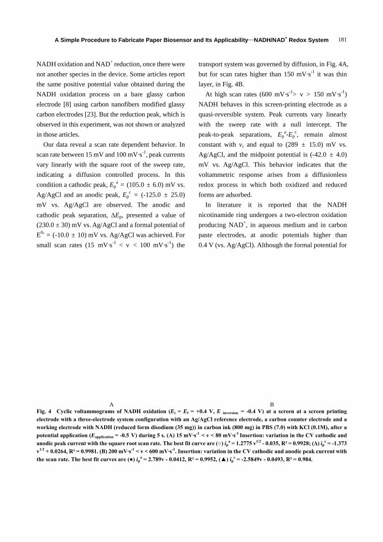
Fig. 3 Cyclithree-electrodelectrode withwith KCl (0.1
best fit equati
mple Procedu
ry, without th
sors were ma
r.
rameters were
and cathodic
sweep rate wa
± 4) mV and
e observed, a
ak, Ep, with
as obtained a
mV vs. Ag/Ag
athodic peak
ear variation
square ro
3395 µA, R2
R2 = 0.99. Th
05)n3/2AD1/2cv
after five m
he active area
ous electron t
s (3.78 ± 0.
ic voltammogrde system conh NADH (redu1M),. Insertion
ion is: ( ) ipa =
ure to Fabrica
he humidity c
ade in the wi
e examined.
peak current
as analyzed.
d an anodic p
and a separat
h a value of (+
and a formal p
gCl was calc
ks current, |ip
of the curre
oot is ob
= 0.9974, ip
he Randles-S
v1/2), was use
months storag
a was (0.98 ±
transfer stand
18) ×10-5 cm
rams (Ei = Ef nfiguration wiuced form disodn: Variation of
= 7.686 v1/2 - 0.3
ate Paper Bios
control, in Fig
inter, and no
The variatio
t with the sq
A cathodic p
peak, Epc = (+
tion of anodi
+78 ± 15) mV
potential of E
ulated. A rati
pa/ip
c| = +0.8
nt peak with
btained: ipa
pc = -5.928 v
evčik equatio
ed to estimate
ge, and in th
± 0.09) mm2.
dard constant
m·s-1. The ac
= +0.2 V, E i
ith an Ag/AgCdium (35 mg)) f the anodic an
3395, R² = 0.99
sensor and It
g. 3.
ow it
on of
quare
peak,
+160
ic to
V vs.
E0' =
io of
84 ±
h the
= 1/2 +
on ip
e the
hese
The
t (k)
ctive
area
elec
care
mus
con
Thi
of h
to h
3.2
T
redu
ana
fabr
sign
pote
volt
buf
init
was
cyc
inversion = +0.4 Cl reference ein carbon ink nd cathodic pe
976; (□) ipc = -5
ts Applicabili
a reduction
ctrodes at ro
e (humidity c
st be produce
ntrolled cond
is result also
having good
have a major
NADH Respo
The direct el
uction of NA
alyzed using
ricated as des
nals it was ap
ential of -0.5
tammograms
ffer solution p
ialized in -0.4
s carried out.
lic voltammo
V) (Ed = -0.5electrode, a c(800 mg) in th
eak current wi
5.928 v1/2 + 0.18
ity—NADH/NA
was almost
oom temperat
control) is no
ed and used in
ditions (hum
focuses our
hydrophilicit
active area.
onses in Scre
lectro oxidat
AD+, (NADH
g the scr
scribed above
pplied, previ
V during 5
) were record
pH 7 with KC
4 V to +0.4 V
Two well-de
ograms. These
V) at a screecarbon countehe presence K3Fith the square
88, R² = 0.9911
AD+ Redox S
75%. So s
ture, without
ot a good solu
n a short time
midity and t
attention on
ty of the elec
een Printing E
tion of NAD
NAD+ + H
reen-printing
e. To obtain s
iously to each
seconds, and
ded in the elec
Cl (0.1M)). T
V and the reve
efined peaks
e peaks can b
en printing eler electrode anFe(CN)6 (2 mM root of the sw
1.
System
storing these
t any special
ution, so they
e or stored in
temperature).
the necessity
trode surface
Electrodes
DH, and the
H+ + 2ē) was
electrodes
stable NADH
h measure, a
d CVs (cyclic
ctrolyte (PBS
The scan was
erse reduction
appear in the
be assigned to
ectrode with and a working
M) of PBS (7.0)weep rate. The
e
l
y
n
.
y
e
e
s
s
H
a
c
S
s
n
e
o
a g ) e
A Sim
NADH oxidnot another the same poNADH oxidelectrode [8]carbon electrobserved in tin those artic
Our data scan rate betvary linearlyindicating acondition a cAg/AgCl anmV vs. Agcathodic pea(230.0 ± 30)E0' = (-10.0 small scan
Fig. 4 Cyclielectrode withworking electpotential appanodic peak cv1/2 + 0.0264, Rthe scan rate.
mple Procedu
dation and NAspecies in the
ositive potentdation proce] using carborodes [23]. Buthis experimecles. reveal a scan
tween 15 mV y with the sqa diffusion cathodic peak
nd an anodic g/AgCl are ak separation) mV vs. Ag/A
± 10) mV vsrates (15 mV
ic voltammogrh a three-electtrode with NADlication (Eapplic
current with thR² = 0.9981. (B. The best fit cu
ure to Fabrica
AD+ reductione device. Somtial value obtess on a baron nanofibersut the reducti
ent, was not sh
n rate dependand 100 mV·
quare root ofcontrolled p
k, Epa = (105
peak, Epc =
observed. Tn, Ep, preseAgCl and a fos. Ag/AgCl wV·s-1 < v <
A rams of NADHtrode system coDH (reduced f
cation = -0.5 V) he square root sB) 200 mV·s-1 <urves are (●) ip
ate Paper Bios
n, once there wme articles retained duringre glassy cars modified glon peak, whichown or analy
dent behavior·s-1, peak currf the sweep process. In
5.0 ± 6.0) mV(-125.0 ± 2
The anodic ented a valuormal potentiawas achieved.
100 mV·s-1)
H oxidation (Eonfiguration wform disodiumduring 5 s. (A)scan rate. The
< v < 600 mV·s-
pa = 2.789v - 0.
sensor and It
were eport g the rbon lassy ch is yzed
r. In rents rate, this
V vs.
25.0) and
ue of al of . For ) the
tranbut laye
ANAquawithpeaconAg/mVvoltredoform
Innicopropast0.4
Ei = Ef = +0.4 with an Ag/AgC
m (35 mg)) in ca) 15 mV·s-1 < vbest fit curve a1. Insertion: va0412, R² = 0.99
ts Applicabili
nsport systemfor scan rate
er, in Fig. 4BAt high scan
ADH behavesasi-reversible h the sweep
ak-to-peak snstant with v
/AgCl, and thV vs. Ag/AgC
tammetric reox process ims are adsorbn literatureotinamide rinducing NADte electrodesV (vs. Ag/Ag
V, E inversion =
Cl reference elarbon ink (800v < 80 mV·s-1 Inare (○) ip
a = 1.2ariation in the 952, (▲) ip
c = -
ity—NADH/NA
m was governees higher tha. rates (600 m in this screesystem. Pea
p rate witheparations, , and equal he midpoint Cl. This behesponse arisin which botbed.
it is repng undergoes
D+, in aqueous, at anodicgCl). Althoug
B = -0.4 V) at a lectrode, a car0 mg) in PBS (7nsertion: varia
2775 v1/2 - 0.035CV cathodic a-2.5849v - 0.04
AD+ Redox S
ed by diffusioan 150 mV·s-
mV·s-1> v > en-printing eak currents vh a null intEp
a-Epc, rem
to (289 ± 1potential is (havior indicaes from a th oxidized
orted that a two-electr
us medium anc potentials gh the formal
screen at a scrbon counter el7.0) with KCl (ation in the CV5, R² = 0.9928; and anodic pea493, R² = 0.984.
System 181
on, in Fig. 4A-1 it was thin
150 mV·s-1)lectrode as avary linearlytercept. The
main almost5.0) mV vs.
(-42.0 ± 4.0)ates that thediffusionlessand reduced
the NADHron oxidationnd in carbonhigher than
l potential for
creen printinglectrode and a(0.1M), after aV cathodic and(Δ) ip
c = -1.373k current with.
A, n
) a y e t .
) e s d
H n n n r
g a a d 3 h
A Sim
182
NAD+/NAD
0.56 V (SCE
for high p
appropriated
electrode it
near +0.250
surface treat
For a rev
Ep = 59/n (
case, Ep is
oxidation in
related with
printing elec
the surface)
holes, fissure
but also d
environment
with the liter
to the data r
printing ele
electrodes a
redox coup
reversible s
result, based
transfer occu
ink. So this i
be used in am
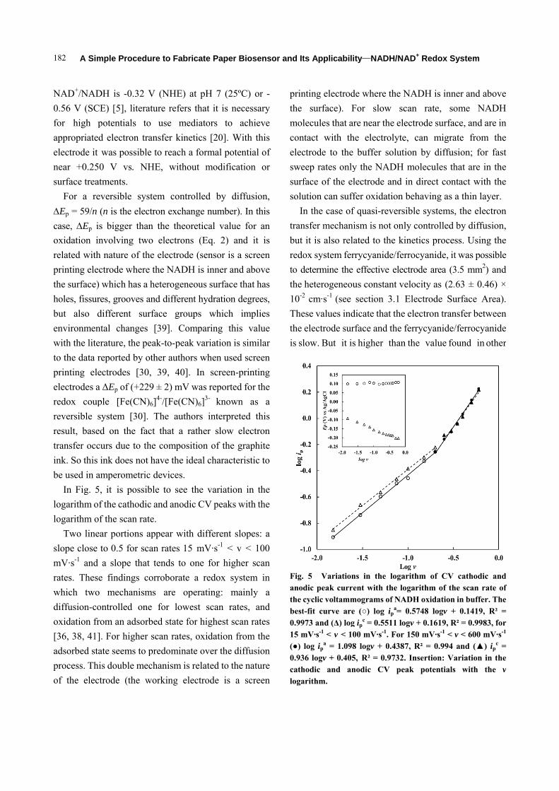
In Fig. 5,
logarithm of
logarithm of
Two linea
slope close t
mV·s-1 and
rates. These
which two
diffusion-co
oxidation fro
[36, 38, 41]
adsorbed sta
process. Thi
of the elect
mple Procedu
DH is -0.32 V
E) [5], literat
potentials to
d electron tra
was possible
0 V vs. NH
tments.
versible syst
(n is the elect
s bigger than
nvolving two
nature of the
ctrode where
which has a h
es, grooves an
different surf
tal changes
rature, the pe
eported by ot
ectrodes [30,
ΔEp of (+229
ple [Fe(CN)6
ystem [30].
d on the fac
urs due to th
ink does not h
mperometric
, it is possib
f the cathodic
f the scan rate
ar portions ap
to 0.5 for sca
a slope that
e findings co
mechanism
ontrolled one
om an adsorb
. For higher s
ate seems to p
is double mec
trode (the w
ure to Fabrica
V (NHE) at p
ture refers th
use media
ansfer kinetic
to reach a fo
HE, without
tem controlle
tron exchange
n the theoreti
o electrons (
e electrode (s
the NADH is
heterogeneou
nd different h
face groups
[39]. Comp
ak-to-peak va
ther authors w
39, 40]. In
9 ± 2) mV wa
6]4-/[Fe(CN)6]
The authors
ct that a rath
e compositio
have the idea
devices.
ble to see the
and anodic C
e.
ppear with d
an rates 15 m
tends to one
orroborate a
ms are opera
e for lowest
bed state for h
scan rates, ox
predominate o
chanism is rel
working electr
ate Paper Bios
pH 7 (25ºC)
hat it is neces
ators to ach
cs [20]. With
ormal potentia
modification
ed by diffus
e number). In
ical value fo
Eq. 2) and
sensor is a sc
s inner and ab
us surface that
hydration degr
which imp
aring this v
ariation is sim
when used sc
n screen-prin
as reported for
]3- known a
s interpreted
her slow elec
on of the grap
al characterist
e variation in
CV peaks with
different slope
mV·s-1 < v <
e for higher
redox system
ating: mainl
scan rates,
highest scan r
xidation from
over the diffu
lated to the na
rode is a sc
sensor and It
or -
ssary
hieve
this
al of
n or
sion,
n this
or an
it is
reen
bove
t has
rees,
plies
value
milar
reen
nting
r the
as a
this
ctron
phite
tic to
n the
h the
es: a
100
scan
m in
ly a
and
rates
m the
usion
ature
reen
prin
the
mol
con
elec
swe
surf
solu
In
tran
but
redo
to d
the
10-2
The
the
is sl
Fig.anothe best0.9915 m(●) 0.93cathloga
ts Applicabili
nting electrod
surface). F
lecules that ar
ntact with th
ctrode to the
eep rates only
face of the el
ution can suff
n the case of
nsfer mechani
it is also rela
ox system fer
determine the
heterogeneou2 cm·s-1 (see
ese values ind
electrode sur
low. But it is
. 5 Variationdic peak currecyclic voltammt-fit curve are973 and (Δ) logmV·s-1 < v < 10log ip
a = 1.09836 logv + 0.405hodic and anarithm.
ity—NADH/NA
de where the N
For slow sc
re near the ele
he electrolyte
buffer solut
y the NADH
lectrode and
fer oxidation
f quasi-revers
ism is not onl
ated to the ki
rrycyanide/fer
effective elec
us constant v
section 3.1
dicate that the
rface and the f
s higher than
ns in the logaent with the lo
mograms of NAe (○) log ip
a= g ip
c = 0.5511 lo00 mV·s-1. For 8 logv + 0.4385, R² = 0.9732.nodic CV pe
AD+ Redox S
NADH is inn
can rate, so
ectrode surfa
e, can migra
tion by diffus
molecules th
in direct con
behaving as
sible systems,
ly controlled
inetics proce
rrocyanide, it
ctrode area (3
elocity as (2.
Electrode Su
e electron tran
ferrycyanide/
n the value fo
arithm of CV ogarithm of thADH oxidation
0.5748 logv +ogv + 0.1619, R 150 mV·s-1 < 7, R² = 0.994 . Insertion: Va
eak potentials
System
ner and above
ome NADH
ce, and are in
ate from the
sion; for fast
hat are in the
ntact with the
a thin layer.
, the electron
by diffusion,
ss. Using the
t was possible
3.5 mm2) and
63 ± 0.46) ×
urface Area).
nsfer between
/ferrocyanide
ound in other
cathodic andhe scan rate of in buffer. The
+ 0.1419, R² =R² = 0.9983, forv < 600 mV·s-1
and (▲) ipc =
ariation in thes with the v
e
H
n
e
t
e
e
n
,
e
e
d
×
.
n
e
r
d f e = r 1 = e v
A Simple Procedure to Fabricate Paper Biosensor and Its Applicability—NADH/NAD+ Redox System
183
studies with screen-printing electrodes (5.2 × 10⁻⁶ cm·s-1) [29].
In both amperometric biosensors and biofuel cells an efficient regeneration of NADH/NAD+ is required. In this context we developed a system that allowed the regeneration of both species. It is necessary to use another carbon ink to increase electron transfer velocity between the electrode surface and the redox system.
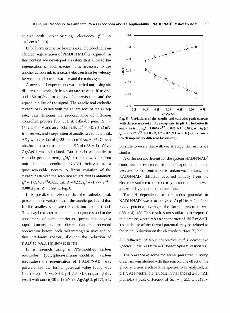
A new set of experiments was carried out, using six different electrodes, in low scan rate between 10 mV·s-1 and 150 mV·s-1, to analyze the persistence and the reproducibility of the signal. The anodic and cathodic current peak varies with the square root of the sweep rate, thus denoting the predominance of diffusion controlled process [36, 38]. A cathodic peak, Ep
a = (+82 ± 4) mV and an anodic peak, Ep
c = (-159 ± 2) mV is observed, and a separation of anodic to cathodic peak, ∆Ep, with a value of (+221 ± 2) mV vs. Ag/AgCl was obtained and a formal potential, E0', of (-38 ± 1) mV vs.
Ag/AgCl was calculated. But a ratio of anodic to cathodic peaks current, |ip
a/ipc| estimated was far from
unit. In this condition NADH behaves as a quasi-reversible system. A linear variation of the current peak with the scan rate square root is obtained: ip
a = 1.0946 v1/2-0.035 µA, R = 0.99, ipc = -1.777 v1/2 +
0.0803 µA, R = 0.99, in Fig. 6. It is possible to observe that the cathodic peak
presents more variation than the anodic peak, and that for the smallest scan rate the variation is almost null. This may be related to the reduction process and to the appearance of some interferent species that have a rapid kinetics as the dimer. But the potential application before each voltammogram may reduce this interferent species, allowing the reduction of NAD+ to NADH in slow scan rate.
In a research using a PPS-modified carbon electrodes (poly(phenosafranin)-modified carbon electrodes) the regeneration of NADH/NAD+ was possible and the formal potential value found was (-365 ± 2) mV vs. SHE, pH 7.0 [9]. Comparing this result with ours ((-38 ± 1) mV vs. Ag/AgCl, pH 7), it is
Fig. 6 Variations of the anodic and cathodic peak current with the square root of the sweep rate, in pH 7. The better fit equation is: (◌) ip
a = 1.0946 v1/2 - 0.035, R² = 0.988, n = 6; (□) ip
c = -1.777 v1/2 + 0.0803, R² = 0.9893, n = 6 (six measures which implied six different biosensors).
possible to verify that with our strategy, the results are similar.
A diffusion coefficient for the system NADH/NAD+ could not be estimated from the experimental data, because its concentration is unknown. In fact, the NADH/NAD+ diffusion occurred initially from the electrode surface to the electrolyte solution, and it was governed by gradient concentration.
The pH dependence of the redox potential of NADH/NAD+ was also analyzed. At pH from 3 to 9 the redox potential average, the formal potential was (-33 ± 4) mV. This result is not similar to the reported in literature, which refer a dependence of -30.3 mV/pH. The stability of the formal potential may be related to the initial reduction on the electrode surface [5, 32].
3.3 Influence of Nonelectroactive and Electroactive Species in the NADH/NAD+ Redox System Responses
The presence of some molecules presented in living organism was studied with this sensor. The effect of the glucose, a non electroactive species, was analyzed, in pH 7. At a neutral pH, glucose in the range of 2-15 mM, promotes a peak difference of ΔEp = (+235 ± 12) mV
A Simple Procedure to Fabricate Paper Biosensor and Its Applicability—NADH/NAD+ Redox System
184
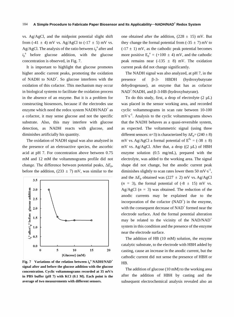
vs. Ag/AgCl, and the midpoint potential slight shift from (-41 ± 4) mV vs. Ag/AgCl to (-17 ± 5) mV vs. Ag/AgCl. The analysis of the ratio between ip
a after and ip
a before glucose addition, with the glucose concentration is observed, in Fig. 7.
It is important to highlight that glucose promotes higher anodic current peaks, promoting the oxidation of NADH to NAD+. So glucose interferes with the oxidation of this cofactor. This mechanism may occur in biological systems to facilitate the oxidation process in the absence of an enzyme. But it is a problem for constructing biosensors, because if the electrodes use enzyme which need the redox system NADH/NAD+ as a cofactor, it may sense glucose and not the specific substrate. Also, this may interfere with glucose detection, as NADH reacts with glucose, and diminishes artificially his quantity.
The oxidation of NADH signal was also analyzed in the presence of an eletroactive species, the ascorbic acid at pH 7. For concentration above between 0.75 mM and 12 mM the voltammograms profile did not change. The difference between potential peaks, ΔEp, before the addition, (233 ± 7) mV, was similar to the
Fig. 7 Variations of the relation between ip
a NADH/NAD+
signal after and before the glucose addition with the glucose concentration. Cyclic voltammograms recorded at 35 mV/s in PBS buffer (pH 7) with KCl (0.1 M). Each point is the average of two measurements with different sensors.
one obtained after the addition, (228 ± 15) mV. But they change the formal potential from (-35 ± 7) mV to (-17 ± 1) mV, as the cathodic peak potential becomes more positive Ep
a = (+100 ± 4) mV, and the cathodic peak remains near (-135 ± 8) mV. The oxidation current peak did not change significantly.
The NADH signal was also analyzed, at pH 7, in the presence of β-3- HBDH (hydroxybutyrate dehydrogenase), an enzyme that has as cofactor NAD+/NADH, and β-3-HB (hydroxybutyrate).
To do this study, first, a drop of electrolyte (2 µL) was placed in the sensor working area, and recorded cyclic voltammograms in scan rate between 10-100 mV·s-1. Analysis to the cyclic voltammograms shows that the NADH behaves as a quasi-reversible system, as expected. The voltammetric signal (using three different sensors: n=3) is characterized by ∆Ep= (240 ± 8) mV vs. Ag/AgCl a formal potential of E0' = (-38 ± 8) mV vs. Ag/AgCl. After that, a drop ((2 µL) of HBH enzyme solution (0.5 mg/mL), prepared with the electrolyte, was added to the working area. The signal shape did not change, but the anodic current peak diminishes slightly to scan rates lower them 50 mV·s-1, and the ∆Ep obtained was (227 ± 2) mV vs. Ag/AgCl (n = 3), the formal potential of (-8 ± 15) mV vs.
Ag/AgCl (n = 3) was obtained. The reduction of the anodic currents may be explained due to the incorporation of the cofactor (NAD+) in the enzyme, with the consequent decrease of NAD+ formed near the electrode surface. And the formal potential alteration may be related to the vicinity of the NAD/NAD+ system in this condition and the presence of the enzyme near the electrode surface.
The addition of HB (10 mM) solution, the enzyme catalytic substrate, to the electrode with HBH added by casting, cause an increase in the anodic current, but the cathodic current did not sense the presence of HBH or HB.
The addition of glucose (10 mM) to the working area after the addition of HBH by casting and the subsequent electrochemical analysis revealed also an
A Simple Procedure to Fabricate Paper Biosensor and Its Applicability—NADH/NAD+ Redox System
185
increase in the anodic current, but also a change in the E0' to positive values (+6 ± 10) mV vs. Ag/AgCl (n = 3)) and a typical ∆Ep = (+229.0 ± 14) mV vs.
Ag/AgCl (n = 3).
4. Conclusions
In this work, we used a simple procedure to fabricate a paper device, using wax to limit the hydrophobic and hydrophilic areas of the biosensor. We immobilized the cofactor in the matrix of the working electrode, which was printed on paper. This procedure is easy and quick, and does not need any drastic treatment.
In this work, the electrode electrochemical reaction is a complex process, and has many challenges. One of them is the increase of the rate of electron transfer between the electrode and NAD/NADH system. This velocity depends not only on the electron transfer, but also on mass transport velocity.
By comparison of the mass transport velocity and charge transference it is possible to identify a quasi-reversible reaction where the electrode process is governed by kinetics and diffusion. In all situations analyzed in this work the system behaved as a quasi-reversible. Some characteristics observed here were attributed to the nature of the electrode (screen-printing) that conditioned kinetics and reduced the mass transport velocity, which can be perceived in the peak-to-peak separation. One strategy used by some researcher is the utilization a solution of 3-APDES (aminopropyldimethly ethoxysilane) prepared with water to improve the hydrophilicity of the electrode surface (graphite) and of the paper channel and enhance the electron transfer velocity [42].
It is important to highlight that even with these obstacles, it was possible to obtain the oxidation and reduction well defined NAD+/NADH signal that was not reported in the literature. It was also possible to test this sensor with non-electroactive molecules such as glucose, and with eletroactive species as ascorbic acid, which shows its versatility. The first one interacts with the electrode and promotes higher oxidation currents,
but the ascorbic acid did not interfere with the electrochemical signal.
In future researches it will be interesting to use this approach, and incorporate enzymes relevant to health and to the environment, in the carbon ink, to detect there subtracts. It will be also important using the screen-printing technique and the capability of NAD+/NADH interaction with glucose to develop cheaper biofuel cell.
References [1] Sekretaryova, A., Eriksson, M., and Turner, A. 2016.
“Bioelectrocatalytic Systems for Health Applications.” Biotechnology Advances 34: 177-97.
[2] Wooten, M., and Gorski, W. 2010. “Facilitation of NADH Electro-Oxidation at Treated Carbon Nanotubes.” Anal. Chem. 82: 1299. doi: 10.1021/ac902301b
[3] Álvarez-González, I., Saidman, S., Lobo-Castañón, M. J., Miranda-Ordieres, A. Tuñón-Blanco, P. 2000. “Electrocatalytic Detection of NADH and Glycerol by NAD(+)-Modified Carbon Electrodes.” Anal. Chem. 72: 520-7.
[4] Govindhan, M., Amiri, M., and Chen, A. 2015. “Au Nanoparticle/Graphene Nanocomposite as a Platform for the Sensitive Detection of NADH in Human Urine” Biosensors and Bioelectronics 66: 474-80.
[5] Radoi, A., and Compagnone, D. 2009. “Recent Advances in NADH Electrochemical Sensing Design.” Bioeletrochemistry 76: 126-34.
[6] Ali, I., and Omanovic, S. 2013. “Kinetics of Electrochemical Reduction of NAD+ on a Glassy Carbon Electrode.” Int. J. Electrochem. Sci. 8: 4283-304.
[7] Hughes, G., Pemberton, R., Fielden, P., and Hart, J. 2015. “Development of a Novel Reagentless, Screen-Printed Amperometric Biosensor Based on Glutamate Dehydrogenase and NAD+, Integrated with Multi-walled Carbon Nanotubes for the Determination of Glutamate in Food and Clinical Applications.” Sensors and Actuators B 216: 614-21.
[8] Katekawa, E., Maximiano, F., Rodrigues, L., Delbem, M., and Serrano, S. 1999. “Electrochemical Oxidation of NADH at a Bare Glassy Carbon Electrode in Different Supporting Electrolytes.” Analytica Chimica Acta 385: 345-52.
[9] Saleh, F., Rahman, M., Okajima, T., Mao, L., and Ohsaka, T. 2011. “Determination of Formal Potential of NADH/NAD+ Redox Couple and Catalytic Oxidation of NADH Using Poly (Phenosafranin)-Modified Carbon Electrodes.” Bioeletrochemistry 80: 121-7.

A Simple Procedure to Fabricate Paper Biosensor and Its Applicability—NADH/NAD+ Redox System
186
[10] Gorton, L., and Domınguez, E. 2002. “Electrocatalytic Oxidation of NAD (P)/H at Mediator Modified Electrodes.” Reviews in Molecular Biotechnology 82: 371-92.
[11] Popescu, I., Domínguez, E., Narváez, A., Pavlov, V., and Katakis, I. 1999. “Electrocatalytic Oxidation of NADH at Graphite Electrodes Modified with Osmium Phenanthrolinedione.” Journal of Electroanalytical Chemistry 464: 208-14.
[12] Prieto-Simón, B., and Fàbregas, E. 2004. “Comparative Study of Electron Mediators Used in the Electrochemical Oxidation of NADH.” Biosensors and Bioelectronics 19: 1131-8.
[13] Zhou, j. L., Nie, P. P., Zheng, H, T., and Zhang. J. M. 2009. “Progress of Electrochemical Biosensors Based on Nicotinamide Adenine Dinucleotide (Phosphate)- Dependent Dehyfrogenases.” Chinese Journal of Analytical Chemistry 37: 617-23.
[14] Zanardi, C., Ferrari, E., Pigani, F., and Seeber, R. 2015. “Development of an Electrochemical Sensor for NADH Determination Based on a Caffeic Acid Redox Mediator Supported on Carbon Black.” Chemosensors 3: 118-28.
[15] Eguílaz, M., Gutierrez, F., González-Domínguez, J., Matínez, M., and Rivas, G. 2016. “Single-Walled Carbon Nanotubes Covalently Functionalized with Polytyrosine: A New Material for the Development of NADH-Based Biosensors.” Biosensors and Bioeletronics 86: 308-14.
[16] Friedl, J., and Stimming, U. 2017. “Determining Electron Transfer Kinetics at Porous Electrodes.” Electrochimica Acta 227: 235-45.
[17] Balamurugan, A., Ho, K-C., Chen, S-M., and Huang, T-Y. 2010. “Electrochemical Sensing of NADH Based on Meldola Blue Immobilized Silver Nanoparticle-Conducting Polymer Electrode.” Colloids and Surfaces A: Physicochem. Eng. Aspects 362: 1-7.
[18] Lates, V., Gligor, D., Muresan, L., and Popescu, I. 2011. “Comparative Investigation of NADH Electrooxidation at Graphite Electrcdes Modified with Two New Phenothiezine Derivatives.” J. Electroanal. Chem. 661: 192-7.
[19] Tan, B., Hickey, D., Milton, R., Giroud, F., and Minteer, S. 2015. “Regeneration of the NADH Cofactor by a Rhodium Complex Immobilized on Multi-walled Carbon Nanotubes.” Journal of the Electrochemical Society 162: H102-7.
[20] Kumar, S., and Cheng, S-M. 2008. “Electroanalysis of NADH Using Conducting and Redox Active Polymer/Carbon Nanotubes Modified Electrodes—A Review.” Sensors 8: 739-66.
[21] Radoi, A., Compagnone, D., Devic, E., and Palleschi, G. 2007. “Low Potential Detection of NADH with Prussian
Blue Bulk Modified Screen-Printed Electrodes and Recombinant NADH Oxidase from Thermus Thermophilus.” Sensors and Actuators B 121: 501-6.
[22] Rivas, G., Rubianes, M., Rodríguez, M., Ferreyra, N., Luque, G., Pedano, M., Miscoria, S., and Parrado, C. 2007. “Carbon Nanotubes for Electrochemical Biosensing.” Talanta 74: 291-307.
[23] Arvinte, A., Valentini, F., Radoi, A., Arduiini, F., Tamburri, E., Rotariu, L., Palleshi, G., and Bala, C. 2007. “The NADH Electrochemical Detection Performed at Carbon. Nanofibers Modified Glassy Carbon Electrode. Electroanalysis.” Electroanalysis 19: 1455-9.
[24] Vasilescu. A., Andressscu, S., Bala, C., Litescu, S., Noguer, T., and Marty, J-L. 2003. “Screen-Printed Electrodes with Electropolymerized Meldola Blue as Versatile Detectors in Biosensors.” Biosensors and Bioelectronics 18: 781-90.
[25] Doumèche, B., and Blum, L. 2010. “NADH oxidAtion on Screen-Printed Electrode Modified with a New Phenothiazine Diazonium Salt.” Electrochemistry Communications 12: 1398-402.
[26] Taleat, Z., Khoshroo, A., and Mazloum-Ardakani, M. 2014. “Screen-Printed Electrodes for Biosensing: A Review (2008-2013).” Microchim Acta 181: 865-91.
[27] Sahin, M., and Ayranci, E. 2015. “Electrooxidation of NADH on Modified Screen-Printed Electrodes: Effects of Conducting Polymer and Nanomaterials.” Electrochimica Acta 166: 261-70.
[28] Ensafi, A., Alinajafi, H., Jafari.Asl, M., Rezaei, B., and Ghazaei, F. 2016. “Cobalt Ferrite Nanoparticles Decorated on Exfoliated Graphene Oxide, Application for Amperometric Determination of NADH and H2O2.” Materials Science and Engineering C 60: 276-84.
[29] Gurban, A., Noguer, T., Bala, C., and Rotariu, L. 2008. “Improvement of NADH Detection Using Prussian Blue Modified Screen-Printed Electrodes and Different Strategies of Immobilisation.” Sensors and Actuators B 128: 536-44.
[30] Avramescu, A., Andreescu, S., Noguer, T., Bala, C., Andreescu, D., and Marty, J-L. 2002. “Biosensors Designed for Environmental and Food Quality Control Based on Screen-Printed Graphite Electrodes with Different Configurations.” Anal. Bioanal. Chem. 374: 25-32.
[31] Blanco, E., Foster, C., Cumba, L., Cramo, D., and Banks, C. 2016. “Can Solvent Induced Surface Modifications Applied to Screen-Printed Platforms Enhance Their Electroanalytical Performance?” Analyst 141: 2783-90.
[32] Radoi, A., Compagnone, D., Valcarcel, M., Placidi, P., Materazzi, S., Moscone, D., and Palleschi. G. 2008. “Detection of NADH via Electrocatalytic Oxidation at Single-Walled Carbon Nanotubes Modified with
A Simple Procedure to Fabricate Paper Biosensor and Its Applicability—NADH/NAD+ Redox System
187
Variamine Blue.” Electrochimica Acta 53: 2161-9. [33] Prieto-Simón, B., Macanás, J., Muñoz, M., and Fàbregas,
E. 2007. “Evaluation of Different Mediator-modified Screen-printed Electrodes used in a Flow System as Amperometric Sensors for NADH.” Talanta 71: 2102-7.
[34] Fang, L., Wang, S-H., and Liu, C-C. 2008. “An Electrochemical Biosensor of the ketone. 3-[beta]-hydroxybutyrate for Potential Diabetic Patient Management.” Sensors and Actuators B 129: 818-825.
[35] Mohamed, H. 2016. “Data Analysis Strategies for Targeted and Untargeted LC-MS Metabolomic Studies: Overview and Workflow.” Trends in Analytical Chemistry 82: 1-11.
[36] Nicholson, R. 1965. “Theory and Application of Cyclic Voltammetry for Measurement of Electrode Reaction Kinetics.” Analitical Chemistry 37 (11): 1351-5.
[37] Santhiago, M., and Kubota, L. 2013. “A New Approach for Paper-Based Analytical Devices with Electrochemical Detection Based on Graphite Pencil Electrodes.” Sensors and Actuators B 177: 224-30.
[38] Bard, A., and Faulkner, L. 2001. Electrochemical Methods, Fundamentals and Applications, 2nd edition. New York: Wiley.
[39] Määtänen, A., Vanamo, U., Ihalainen, P., Pulkkinen, P., Tenhu, H., Bobacka, J., and Peltonen, J. 2013. “A Low-Cost Paper-Based Inkjet-Printed Platform for Electrochemical Analyses.” Sensors and Actuators B: Chemical 177: 153-62.
[40] Wang, J., Tian, B., Nascimento, V., and Angnes, L. 1998. “Performance of Screen-Printed Carbon Electrodes Fabricated from Different Carbon Inks.” Electrochim Acta A 43: 3459-65.
[41] Laviron, E. 1974. “Adsorption, Autoinhibition and Autocatalysis in Polarography and in Linear Potential Sweep Voltammetry.” Electroanalytical Chemistry and Interfacial Electrochemistry 52: 355.
[42] Nie, Z., Deiss, F., Liu, X., Akbulut, O., and Whitesides, G. 2010. “Integration of Paper-Based Microfluidic Devices with Commercial Electrochemical Readers.” Lab. Chip. 10: 3163-9.