Embed Size (px)
Citation preview

En
do
crin
e-R
ela
ted
Can
cer
ResearchE von Dobschuetz et al. A registry-based study of thyroid
paraganglioma22 :2 191–204
A registry-based study of thyroidparaganglioma: histological andgenetic characteristics
Ernst von Dobschuetz, Helena Leijon1, Camilla Schalin-Jantti2, Francesca Schiavi3,
Michael Brauckhoff4,†, Mariola Peczkowska5, Giovanna Spiazzi6, Serena Dematte7,
Maria Enrica Cecchini7, Paola Sartorato8, Jolanta Krajewska9, Kornelia Hasse-Lazar9,
Katarzyna Roszkowska-Purska10, Elisa Taschin3, Angelica Malinoc11, Lars A Akslen12,
Johanna Arola1, Dariusz Lange13, Ambrogio Fassina14, Gianmaria Pennelli14,
Mattia Barbareschi15, Jutta Luettges16, Aleksander Prejbisz5, Andrzej Januszewicz5,
Tim Strate, Birke Bausch17, Frederic Castinetti18, Barbara Jarzab9, GiuseppeOpocher3,
Charis Eng19 and Hartmut P H Neumann11
Section of Endocrine Surgery, Department of General, Visceral and Thoracic Surgery, Krankenhaus Reinbek,
St. Adolf Stift, Academic Teaching Hospital University of Hamburg, Reinbek, Germany1Department of Pathology, Helsinki University Central Hospital and University of Helsinki, Helsinki, Finland2Division of Endocrinology, Department of Medicine, Helsinki University Central Hospital and University of Helsinki,
Helsinki, Finland3Familial Cancer Clinic and Onco-Endocrinology, Veneto Institute of Oncology, IRCCS, Padova, Italy4Department of Endocrine Surgery, University of Bergen, Bergen, Norway5Department of Hypertension, Institute of Cardiology, Warsaw, Poland6Endocrinology, Diabetes and Metabolism, University of Verona and Azienda Ospedaliera Universitaria
Integrata Verona, Verona, Italy7Units of Internal Medicine, Santa Chiara General Hospital, Trento, Italy8Department of Internal Medicine, General Hospital, Montebelluna, Treviso, Italy9Department of Nuclear Medicine and Endocrine Oncology, Maria Sklodowska-Curie Memorial Cancer Center
and Institute of Oncology, Gliwice Branch, Gliwice, Poland10Department of Pathology and Laboratory Diagnostics, Skłodowska-Curie Memorial Institute of Oncology,
Warsaw, Poland11Section for Preventive Medicine, Department of Nephrology and General Medicine, Albert-Ludwigs-University
of Freiburg, Hugstetter Straße 55, 79106 Freiburg, Germany12Section for Pathology, Department of Clinical Medicine, Centre for Cancer Biomarkers CCBIO,
University of Bergen, Bergen, Norway13Department of Tumor Pathology, M.Sklodowska-Curie Memorial Cancer Center and Institute of Oncology,
Gliwice Branch, Gliwice, Poland14Surgical Pathology and Cytopathology Unit, Department of Medicine, DIMED, University of Padova, Padova, Italy15Department of Pathology, Santa Chiara Regional Hospital, Trento, Italy16Department of Pathology, Marienkrankenhaus, Hamburg, Germany17Department of Gastroenterology, Albert-Ludwigs-University of Freiburg, Freiburg, Germany18Department of Endocrinology, La Timone Hospital, Hopitaux de Marseille and Centre de Recherche en
Neurobiologie et Neurophysiologie de Marseille, Aix-Marseille University, Marseille, France19Genomic Medicine Institute, Lerner Research Institute and Taussig Cancer Institute, Cleveland Clinic,
Cleveland, Ohio, USA†M Brauckhoff died on September 10, 2014. We dedicate this manuscript to his memory.
http://erc.endocrinology-journals.org q 2015 Society for EndocrinologyDOI: 10.1530/ERC-14-0558 Printed in Great Britain
Published by Bioscientifica Ltd.
Correspondence
should be addressed
to H P H Neumann
hartmut.neumann@
uniklinik-freiburg.de
Abstract
The precise diagnosis of thyroid neoplasias will guide surgical management. Primary thyroid
paraganglioma has been rarely reported. Data on prevalence, immunohistochemistry (IHC),
and molecular genetics in a systematic series of such patients are pending. We performed a
multinational population-based study on thyroid paraganglioma and analyzed prevalence,
Key Words
" neuroendocrinology
" thyroid
" molecular genetics

En
do
crin
e-R
ela
ted
Can
cer
Research E von Dobschuetz et al. A registry-based study of thyroidparaganglioma
22 :2 192
IHC, and molecular genetics. Patients with thyroid paraganglioma were recruited from the
European-American-Head-and-Neck-Paraganglioma-Registry. Demographic and clinical data
were registered. Histopathology and IHC were re-investigated. All patients with thyroid
paraganglioma underwent molecular genetic analyses of the SDHA, SDHB, SDHC, SDHD,
SDHAF2, VHL, RET, TMEM127, and MAX genes. Analyses included Sanger sequencing
and multiplex ligation-dependent probe amplification (MLPA) for detection of large
rearrangements. Of 947 registrants, eight candidates were initially identified. After
immunohistochemical analyses of these eight subjects, 5 (0.5%) were confirmed to have
thyroid paraganglioma. IHC was positive for chromogranin, synaptophysin, and S-100 and
negative for calcitonin in all five thyroid paragangliomas, whereas the three excluded
candidate tumors stained positive for pan-cytokeratin, a marker excluding endocrine tumors.
Germline variants, probably representing mutations, were found in four of the five confirmed
thyroid paraganglioma cases, two each in SDHA and SDHB, whereas the excluded cases had no
mutations in the tested genes. Thyroid paraganglioma is a finite entity, which must be
differentiated from medullary thyroid carcinoma, because medical, surgical, and genetic
management for each is different. Notably, approximately 80% of thyroid paragangliomas
are associated with germline variants, with implications for additional tumors and a potential
risk for the family. As opposed to sporadic tumors, surgical management and extent of
resection are different for heritable tumors, each guided by the precise gene involved.
http://erc.endocrinology-journals.org q 2015 Society for EndocrinologyDOI: 10.1530/ERC-14-0558 Printed in Great Britain
Published by Bioscientifica Ltd.
Endocrine-Related Cancer
(2015) 22, 191–204
Introduction
Neuroendocrine tumors of the thyroid gland are rare, and
virtually all are medullary thyroid carcinoma (Ferri et al.
2009). Paragangliomas of the head and neck are neuro-
endocrine neoplasms developing from parasympathetic
paraganglia, which occur at skull base and cervical sites,
mostly the carotid body and the tympanic, vagal, and
jugular paraganglia. The inferior laryngeal paraganglia can
be located within the thyroid capsule explaining why
this tumor might occur in the thyroid gland.
Head and neck paragangliomas can occur as sporadic
tumors or as a manifestation of familial paraganglioma
syndromes type 1–5 (PGL 1 to PGL 5). PGL 1 to PGL 5 are
associated with germline mutations of the genes enco-
ding succinate dehydrogenase subunit D, SDHD (PGL 1),
succinate dehydrogenase complex assembly factor 2,
SDHAF2 (PGL 2), succinate dehydrogenase subunit C,
SDHC (PGL 3), succinate dehydrogenase subunit B, SDHB
(PGL 4), and succinate dehydrogenase subunit A, SDHA
(PGL 5), the SDHx genes (Zantour et al. 2004, Boedeker
et al. 2009). Heritable non-SDHx paraganglioma can also
occur in tumor syndromes mainly associated with
pheochromocytomas such as von Hippel–Lindau disease
(associated with mutations of the VHL gene), multiple
endocrine neoplasia type 2 (RET gene), and neurofibro-
matosis type 1 (NF1 gene) (Boedeker et al. 2009, Neumann
et al. 2009, Burnichon et al. 2010, Castro-Vega et al. 2014,
Yang et al. 2015). In patients carrying germline mutations
of the new susceptibility genes FH, PHD1 (EGLN2), and
PHD2 (EGLN1), head and neck paragangliomas have not
been reported (Castro-Vega et al. 2014, Yang et al. 2015).
Somatic mutations of the HIF2A (EPAS1) gene have been
reported in abdominal paraganglial tumors but not in head
and neck paragangliomas (Toledo et al. 2013). We therefore
sought to determine the frequency and characteristics
of thyroid paragangliomas in our population-based
European-American-Head-and-Neck-Paraganglioma-
Registry based in Freiburg, Germany.
Methods
Patients and data collection
We utilized the population-based European-American-
Head-and-Neck-Paraganglioma-Registry (European-
American-HNPGL-Registry), which is based in Freiburg/
Germany and currently has 947 registrants. Registration
policy has been described in detail previously (Neumann
et al. 2004, 2009, Schiavi et al. 2005). Of these 947
registrants with head and neck paragangliomas, only
eight carried the initial (working) diagnosis of thyroid
paraganglioma. For these eight, we re-reviewed registered
rst
Dia
gn
osi
s
pa
rag
an
gli
om
a
ma
de
tio
nA
fter
1st
op
era
tio
n
tio
nA
fter
1st
op
era
tio
n
tio
nA
fter
3ye
ars
eA
fter
12
years
tio
nA
fter
1st
op
era
tio
n
tio
nA
fter
1st
op
era
tio
n
tio
nA
fter
1st
op
era
tio
ne
Aft
er
som
ew
eeks
ter
som
ew
eeks
to12
years
En
do
crin
e-R
ela
ted
Can
cer
Research E von Dobschuetz et al. A registry-based study of thyroidparaganglioma
22 :2 193
data for their putative thyroid paraganglioma including
demographic information such as age, sex, symptoms,
location and size of the tumor, radiological imaging,
and results from fine-needle aspiration cytology and
histopathology of the surgical specimens. Tumors were
diagnosed as malignant, only if patients showed metastases
(Lloyd et al. 2004).
nts
wh
oen
tere
dth
ere
gis
try
wit
hth
ed
iag
no
sis
of
thyr
oid
para
gan
gli
om
a
en
ign
/
ali
gn
an
tS
ym
pto
ms
Hy
pe
r-
ten
sio
n
Pre
op
era
tiv
e
ult
raso
no
-
gra
ph
y
Fin
en
ee
dle
bio
psy
be
fore
firs
to
pe
rati
on
Init
ial
dia
gn
osi
s
Tre
atm
en
t
ba
sed
on
fi
dia
gn
osi
s
en
Rela
tive
test
ed
as
SDH
B-p
osi
tive
No
No
No
td
on
ePG
LH
orm
on
esu
bst
itu
en
No
de
inth
en
eck
No
Yes
Ad
en
om
aPG
LH
orm
on
esu
bst
itu
en
No
de
inth
en
eck
No
No
No
td
on
eH
em
an
gio
-p
eri
cyto
ma
Ho
rmo
ne
sub
stit
uen
Lum
pin
the
neck
No
Yes
Foll
icu
lar
neo
pla
sia
Foll
icu
lar
thyr
oid
can
cer
Rad
ioact
ivio
din
een
Swall
ow
ing
dif
ficu
ltie
sY
es
Yes
Foll
icu
lar
neo
pla
sia
PG
LH
orm
on
esu
bst
itu
al
No
No
Yes
No
td
on
ePG
LH
orm
on
esu
bst
itu
en
Inci
den
tal
fin
din
gN
oY
es
Foll
icu
lar
neo
pla
sia
PG
LH
orm
on
esu
bst
itu
en
Swall
ow
ing
dif
ficu
ltie
sN
oY
es
Susp
icio
us
for
atu
mo
rN
eu
roen
do
crin
etu
mo
r,PG
LR
ad
ioact
ivio
din
e
din
five
pati
en
ts(1
,2,3,4,an
d5)
an
dexc
lud
ed
inth
ree
pati
en
ts(A
,B
,an
dC
)b
yth
isst
ud
y.‘I
nit
ial
dia
gn
osi
s’w
as
revi
sed
af
reg
istr
y.
Histology and immunohistochemistry
Tumor materials from eight patients who had a working
(initial) diagnosis of thyroid paraganglioma were collected
and new slides from the paraffin blocks were centrally
sectioned and re-analyzed by our internal reference center
in the Department of Pathology of the University of
Helsinki Central Hospital using conventional H&E his-
tology and immunohistochemistry (IHC). The panel of
IHC included staining against chromogranin A, synapto-
physin, S-100, pan-cytokeratin (CK-PAN), calcitonin,
thyroid transcription factor 1 (TTF1), MIB1, and p53.
Slides were processed through deparaffinization in xylene
followed by rehydration with graded alcohol series.
Endogenous peroxidase was blocked with 0.3% Dako
REAL Peroxidase-Blocking Solution. Detection was per-
formed using a Dako REAL EnVision/HRP detection
system in Autostainer (Labvision Autostainer 480S). Slides
were counterstained with Meyer’s hematoxylin and
mounted in mounting medium (Fluka, Eukitt Quick-
hardening mounting medium). In each immunohisto-
chemical staining run, a positive and a negative control
were used. Each of the different IHC procedures was
performed on the same day for all eight cases and blindly
scored by J A and H L with 100% concordance.
These results were compared with those primarily
obtained in the participating centers. The diagnosis
paraganglioma was newly established according to the
criteria of the WHO (DeLellis 2004). All patients provided
written informed consent.
Tab
le1
Cli
nic
al
fin
din
gs
ineig
ht
pati
e
Ca
seA
ge
Se
x
Pa
rag
an
gli
om
a
con
firm
ed
B m
127
Male
Yes
B
232
Male
Yes
B
336
Fem
ale
Yes
B
437
Fem
ale
Yes
B
571
Fem
ale
Yes
B
A42
Fem
ale
No
M
B67
Fem
ale
No
B
C69
Male
No
B
Th
ed
iag
no
sis
of
para
gan
gli
om
aw
as
con
firm
e(l
ast
colu
mn
),b
ut
alw
ays
befo
reen
try
into
the
Genetic mutation analyses
All patients were offered molecular testing for germline
mutations of the genes, which have been reported to
cause head and neck paragangliomas: SDHA, SDHAF2,
SDHB, SDHC, SDHD, VHL, RET, MAX, and TMEM127
(Neumann et al. 2004, Schiavi et al. 2005, Burnichon et al.
2010). In these genes we looked for mutations in all exons
except for RET, for which exons 8, 10, 11, and 13–16 only
were analyzed. Mutation screening was not performed for
the new candidate genes FH, EGLN2, EGLN1, and EPAS1
http://erc.endocrinology-journals.org q 2015 Society for EndocrinologyDOI: 10.1530/ERC-14-0558 Printed in Great Britain
Published by Bioscientifica Ltd.

Figure 1
Immunohistochemistry of case 1: H&E, chromogranin, synaptophysin,
S-100, CK-PAN, calcitonin, TTF1, and internal control for CK-PAN.
Note that there is positive staining of the tumor for chromogranin and
synaptophysin, and of sustentacular cells for S-100. The internal control
for CK-PAN shows positive staining of epithelial cells.
En
do
crin
e-R
ela
ted
Can
cer
Research E von Dobschuetz et al. A registry-based study of thyroidparaganglioma
22 :2 194
(Toledo et al. 2013, Castro-Vega et al. 2014, Yang et al. 2015),
because such patients seem to have only extremely rare head
and neck paragangliomas: in addition, EPAS1 mutations
are typically associated with polycythemia, which was not
seen in any of our registrants. Genomic DNA was obtained
from EDTA-anticoagulated whole blood. We performed
bidirectional Sanger sequencing of the coding regions
and splice sites of all genes. In order to find a deletion or
duplication of the listed genes, we performed multiplex
ligation-dependent probe amplification (MLPA) analyses
for VHL, SDHB, SDHC, SDHD, SDHAF2, MAX, and SDHA
and semi-quantitative multiplex PCR for TMEM127. We did
Figure 2
Immunohistochemistry of case 2: H&E, chromogranin, synaptophysin, S-100, CK
for chromogranin and synaptophysin, and of sustentacular cells for S-100. Neg
http://erc.endocrinology-journals.org q 2015 Society for EndocrinologyDOI: 10.1530/ERC-14-0558 Printed in Great Britain
not scan RET for large rearrangements. The sequences
of the primers used for these analyses and PCR conditions
are available upon request. To elucidate the extent of large
deletions, breakpoints were located through quantitative
real-time PCR gene-dosage determination, and then
characterized by long-range PCR and nucleotide sequen-
cing. The results were subjected to in silico analyses using
the programs SIFT, MutationTaster, and PolyPhen-2 in
order to predict pathogenicity of the DNA variants.
They are considered to be probably pathogenic if at least
two of the three software packages predicted them to be
damaging/pathogenic.
-PAN, calcitonin, and TTF1. Note that there is positive staining of the tumor
ative staining is shown for CK-PAN, calcitonin, and TTF1.
Published by Bioscientifica Ltd.

Figure 3
Immunohistochemistry of case 3: H&E, chromogranin, synaptophysin, S-100, CK-PAN, calcitonin, and TTF1. Note that there is positive staining of the tumor
for chromogranin and synaptophysin, and of sustentacular cells for S-100. Negative staining is shown for CK-PAN, calcitonin, and TTF1.
En
do
crin
e-R
ela
ted
Can
cer
Research E von Dobschuetz et al. A registry-based study of thyroidparaganglioma
22 :2 195
Results
Prevalence of thyroid paraganglioma
As of May 1, 2014, the European-American-HNPGL-Registry
based in Freiburg, Germany, comprises 947 registrants.
Among these 947 patients, 939 carried the diagnosis of head
and neck paragangliomas (HNPs): 55% had tympanojugular
HNPs, 45% had carotid body tumors, 5% had vagal HNPs,
and 2% had HNPs with other locations; bilateral HNPs
and HNPs at several locations were present. Of the 947
patients, eight carried the putative diagnosis of thyroid
Figure 4
Immunohistochemistry of case 4: H&E, chromogranin, synaptophysin, S-100, CK
for chromogranin and synaptophysin, and of sustentacular cells for S-100. Neg
http://erc.endocrinology-journals.org q 2015 Society for EndocrinologyDOI: 10.1530/ERC-14-0558 Printed in Great Britain
paraganglioma. After comprehensive re-analysis (described
below), three tumors did not meet the WHO criteria of
thyroid paraganglioma (see below); thus, in the European-
American-HNPGL-Registry, the prevalence of documented
thyroid paraganglioma was estimated to be 0.5% (5/944).
Patients
The eight patients with the initial working diagnosis of
thyroid paraganglioma comprised five females and three
males with ages at diagnosis of 27–71 (median 40) years.
-PAN, calcitonin, and TTF1. Note that there is positive staining of the tumor
ative staining is shown for CK-PAN, calcitonin, and TTF1.
Published by Bioscientifica Ltd.

Figure 5
Immunohistochemistry of case 5: H&E, chromogranin, synaptophysin, S-100, CK-PAN, calcitonin, and TTF1. Note that there is positive staining of the tumor
for chromogranin and synaptophysin, and of sustentacular cells for S-100. Negative staining is shown for CK-PAN, calcitonin, and TTF1.
En
do
crin
e-R
ela
ted
Can
cer
Research E von Dobschuetz et al. A registry-based study of thyroidparaganglioma
22 :2 196
All patients had focal enlargement of the thyroid gland.
One had dysphagia. Thyroid ultrasonography revealed
tumors with a largest diameter of 22–60 (median 44) mm.
None of the patients had either additional neuroendo-
crine tumors or metastases. Clinical findings are given in
Table 1. Family history for paraganglial tumors was
positive only in one patient (case 1).
Fine-needle aspiration cytology
Fine-needle aspiration cytology was performed in five of
the eight patients. Cytology revealed findings suggesting a
‘tumor’ in one, an adenoma in one, and a follicular
neoplasia in three patients (Table 1).
Table 2 Results of immunohistochemistry performed in this study
Case Chromogranin A Synaptophysin S-100 Calcit
1 Positive Positive Positive Nega
2 Positive Positive Positive Nega
3 Positive Positive Positive Nega
4 Positive Positive Positive Nega
5 Positive Positive Positive Nega
A Negative Negative Negative NegaB Positive Positive Negative A few
celC Positive Positive Negative (a few
positive cells)Nega
http://erc.endocrinology-journals.org q 2015 Society for EndocrinologyDOI: 10.1530/ERC-14-0558 Printed in Great Britain
Treatment
All patients underwent surgery for removal of the thyroid
tumor. This was performed in two patients by hemithyr-
oidectomy and in six patients by total thyroidectomy.
Histology and IHC
Histology was studied using hematoxylin and eosin (H&E)
stains and IHC was systematically performed and re-
investigated at one of the participating centers (Helsinki).
Histology together with IHC revealed that five of the eight
tumors had the characteristics of paragangliomas (Figs 1,
2, 3, 4, and 5). On the basis of conventional histology,
onin TTF1 CytokeratinPAN Diagnosis
tive Negative Negative Thyroidparaganglioma
tive Negative Negative Thyroidparaganglioma
tive Negative Negative Thyroidparaganglioma
tive Negative Negative Thyroidparaganglioma
tive Negative Negative Thyroidparaganglioma
tive Negative Positive No paragangliomapositive
lsPositive Positive No paraganglioma
tive Negative Positive No paraganglioma
Published by Bioscientifica Ltd.

Tab
le3
Resu
lts
of
mu
tati
on
scre
en
ing
.Fo
ur
pati
en
tsh
ad
blo
od
DN
Ava
rian
tsin
the
SDH
Ao
rSD
HB
gen
es
Ca
seG
en
eR
efS
eq
Nu
cle
oti
de
va
ria
tio
na
nd
pre
dic
ted
eff
ect
on
pro
tein
Pro
tein
do
ma
in
Pu
bli
cd
ata
ba
ses
Pa
tho
ge
nic
ity
clu
es
(in
silico
an
aly
sis)
db
SNP
ESP
HG
MD
Ala
mu
tso
ftw
are
Case
1SD
HB
NM
_003000.2
c.530G
OA
p.A
rg177H
is4Fe
–4S
ferr
e-
do
xin
-typ
ers
150437793
(MA
F:T
Z0.0
00/0
)
Eu
r.A
m.:
TZ
0.0
0%
CM
130559
path
og
en
icp
hen
oty
pe:
ph
eo
chro
-m
ocy
tom
a
Hig
hly
con
serv
ed
nu
cleo
tid
e(p
hyl
oP:
0.8
4(K
5.2
;1.1
))A
fr.
Am
.:T
Z0.0
2%
Hig
hly
con
serv
ed
am
ino
aci
d,
up
toB
aker’
sye
ast
(co
nsi
deri
ng
14
speci
es)
Small
ph
ysic
och
em
ical
dif
fere
nce
betw
een
Arg
an
dH
is(G
ran
tham
dis
t.:
29
(0–2
15))
Alig
nG
VG
D:
C25
(GV
:0.0
0–G
D:
28.8
2)
SIFT
:d
ele
teri
ou
s(s
core
:0,
med
ian
:4.0
7)
Mu
tati
on
Tast
er:
dis
ease
cau
sin
g(P
valu
e:
1)
Po
lyp
hen
-2H
um
Div
:p
oss
ibly
dam
ag
ing
–sc
ore
0.6
11
(sen
siti
vity
:0.8
7;
speci
fici
ty:
0.9
1)
Po
lyp
hen
-2H
um
Var:
ben
ign
–sc
ore
0.3
80
(sen
siti
vity
:0.8
5;
speci
fici
ty:
0.7
9)
Case
2SD
HB
NM
_003000.2
c.201-1
339_2
39d
eli
nsA
-lu
Yb
8p
.?N
ot
rep
ort
ed
inth
eli
tera
ture
Case
3SD
HA
NM
_004168.2
c.394T
OC
p.T
rp132A
rgFA
D-b
ind
ing
do
main
No
tre
po
rted
inth
eli
tera
ture
Hig
hly
con
serv
ed
nu
cleo
tid
e(p
hyl
oP:
0.9
6(K
5.2
;1.1
))H
igh
lyco
nse
rved
am
ino
aci
d,
up
toB
aker’
sye
ast
(co
nsi
deri
ng
13
speci
es)
Mo
dera
tep
hys
ico
chem
ical
dif
fere
nce
betw
een
Trp
an
dA
rg(G
ran
tham
dis
t.:
101
(0–2
15))
Alig
nG
VG
D:
C65
(GV
:0.0
0–G
D:
101.2
9)
SIFT
:d
ele
teri
ou
s(s
core
:0,
med
ian
:3.7
4)
Mu
tati
on
Tast
er:
dis
ease
cau
sin
g(P
valu
e:
1)
Po
lyp
hen
-2H
um
Div
:p
rob
ab
lyd
am
ag
ing
–sc
ore
1.0
00
(sen
siti
vity
:0.0
0;
speci
fici
ty:
1.0
0)
Po
lyp
hen
-2H
um
Var:
pro
bab
lyd
am
ag
ing
–sc
ore
0.9
98
(sen
siti
vity
:0.1
8;
speci
fici
ty:
0.9
8)
En
do
crin
e-R
ela
ted
Can
cer
Research E von Dobschuetz et al. A registry-based study of thyroidparaganglioma
22 :2 197
http://erc.endocrinology-journals.org q 2015 Society for EndocrinologyDOI: 10.1530/ERC-14-0558 Printed in Great Britain
Published by Bioscientifica Ltd.
Tab
le3
Co
nti
nu
ed
Ca
seG
en
eR
efS
eq
Nu
cle
oti
de
va
ria
tio
na
nd
pre
dic
ted
eff
ect
on
pro
tein
Pro
tein
do
ma
in
Pu
bli
cd
ata
ba
ses
Pa
tho
ge
nic
ity
clu
es
(in
silico
an
aly
sis)
db
SNP
ESP
HG
MD
Ala
mu
tso
ftw
are
Case
4SD
HA
NM
_004168.2
c.1799G
OA
p.A
rg600G
lnFu
mara
tere
du
ctase
/su
ccin
ate
deh
ydro
ge-
nase
flavo
-p
rote
in-l
ike
C-t
erm
inal
rs112656
(MA
F:A
Z0.0
14/3
1)
Eu
r.A
m.:
TZ
0.0
0%
No
tre
po
rted
inth
eli
tera
ture
Hig
hly
con
serv
ed
nu
cleo
tid
e(p
hyl
oP:
0.9
1(K
5.2
;1.1
))A
fr.
Am
.:T
Z0.0
0%
Hig
hly
con
serv
ed
am
ino
aci
d,
up
toB
aker’
sye
ast
(co
nsi
deri
ng
13
speci
es)
Small
ph
ysic
och
em
ical
dif
fere
nce
betw
een
Arg
an
dG
ln(G
ran
tham
dis
t.:
43
(0–2
15))
Alig
nG
VG
D:
C35
(GV
:0.0
0–G
D:
42.8
1)
SIFT
:d
ele
teri
ou
s(s
core
:0,
med
ian
:3.7
4)
Mu
tati
on
Tast
er:
dis
ease
cau
sin
g(P
valu
e:
1)
Po
lyp
hen
-2H
um
Div
:p
rob
ab
lyd
am
a-
gin
g–
sco
re1.0
00
(sen
siti
vity
:0.0
0;
speci
fici
ty:
1.0
0)
Po
lyp
hen
-2H
um
Var:
pro
bab
lyd
am
a-
gin
g–
sco
re0.9
99
(sen
siti
vity
:0.0
9;
speci
fici
ty:
0.9
9)
NA
,n
ot
ap
pli
cab
le.
Furt
her
exp
lan
ati
on
so
fab
bre
viati
on
wil
lb
ep
rovi
ded
on
req
uest
.En
do
crin
e-R
ela
ted
Can
cer
Research E von Dobschuetz et al. A registry-based study of thyroidparaganglioma
22 :2 198
http://erc.endocrinology-journals.org q 2015 Society for EndocrinologyDOI: 10.1530/ERC-14-0558 Printed in Great Britain
there were cells with diffuse sheet-like patterns of growth
or nests (the classic Zellballen). These cells had mostly a
clear or basophilic abundant cytoplasm according to H&E
staining and some variations in the nuclei. These cells
represented chief cells. The mitotic rate was low. Around
the sheets or nests of chief cells, there were cells that
represented sustentacular cells. Between tumor cells, there
were highly vascularized fibrous septa. IHC of all these five
tumors revealed chief cells positive for the neuroendocrine
markers chromogranin A and synaptophysin. Sustenta-
cular cells were positive for S-100. CK-PAN, TTF1, and
calcitonin were also investigated and were negative in all
primary thyroid paragangliomas. Both the histology and
immunohistochemical profile were consistent with the
WHO classification of tumors (DeLellis 2004).
The levels of the proliferation marker MIB1 were low,
between 3 and 5% in all five thyroid paragangliomas, and
p53 was negative, which did not indicate a mutation in
this tumor-suppressor gene.
Three cases (cases A, B, and C in Tables 1, 2, and 3) that
were initially included in the study as thyroid paragan-
gliomas were excluded because of their immunohisto-
chemical profiles (Figs 6, 7, and 8). All these cases stained
positive with CK-PAN, a marker which would exclude
paraganglioma. The tumor of patient A is only positive
for CK-PAN, which resulted in an immunohistochemical
diagnosis of an epithelial neoplasia. The tumor of
patient B is a medullary thyroid carcinoma or a metastasis
of a neuroendocrine tumor. The tumor of patient C is
a metastasis of a neuroendocrine tumor or a calcitonin-
negative medullary thyroid carcinoma. A summary of the
IHC results is given in Table 2.
Molecular genetics and family history
All eight patients were analyzed for nine known para-
ganglioma predisposition genes as part of this study. The
three cases that were excluded as thyroid paragangliomas
were found not to carry any germline mutations in the
nine genes. Of the five patients with confirmed thyroid
paragangliomas, four were found to have germline DNA
variants, which probably represent mutations (Table 3).
In the first confirmed thyroid paraganglioma case, the
youngest of our series who was 27 years old at the time
of diagnosis, the mutation was known from a relative
carrying the germline DNA variant SDHB c.664GOA
p.Arg177His. This family consisted of ten members and
one unrelated wife (1st generation). Of the ten relatives,
there were eight mutation carriers and two members in
whom the mutation is not present (Fig. 9). Six mutation
Published by Bioscientifica Ltd.

Figure 6
Immunohistochemistry of case A: H&E, chromogranin, synaptophysin, S-100, CK-PAN, calcitonin, and TTF1. Note that there is positive staining for CK-PAN
and negative staining for chromogranin, synaptophysin, S-100, calcitonin, and TTF1.
En
do
crin
e-R
ela
ted
Can
cer
Research E von Dobschuetz et al. A registry-based study of thyroidparaganglioma
22 :2 199
carriers had paragangliomas or pheochromocytomas.
Among these six patients is the 27-year-old patient with
thyroid paraganglioma (III.1), the first symptomatic
patient of this family (II.1) who presented with meta-
chronic bilateral pheochromocytoma. One 80-year-old
mutation carrier underwent computerized tomography
of the neck, chest, and abdomen with normal results.
One 14-year-old mutation carrier was not investigated
thus far. The two relatives without mutations had not
undergone surgery for any tumor. In silico analyses of the
variant associated with disease indicated this variant to be
deleterious according to the SIFT program, to be disease
Figure 7
Immunohistochemistry of case B: H&E, chromogranin, synaptophysin, S-100, CK
CK-PAN, but also for chromogranin, synaptophysin, and TTF1. Staining for S-10
http://erc.endocrinology-journals.org q 2015 Society for EndocrinologyDOI: 10.1530/ERC-14-0558 Printed in Great Britain
causing according to the MutationTaster program, and
to be possibly damaging according to the PolyPhen-2
program. Together, the results of the in silico analyses
and the segregation of mutation and phenotype in the
pedigree make the germline variant most probably
pathogenic. The phenotypic data are in accordance with
the well-known reduced penetrance of tumors in subjects
carrying mutations of the SDHB gene.
The second case (case 2) was a 32-year-old male who
was found to have a complex rearrangement, consisting of
a partial deletion of intron 2 and exon 3 of the SDHB gene
and an Alu insertion (Alu family Yb8) (Fig. 10). The latter
-PAN, calcitonin, and TTF1. Note that there is positive staining not only for
0 is negative, and staining for calcitonin shows only very few positive cells.
Published by Bioscientifica Ltd.
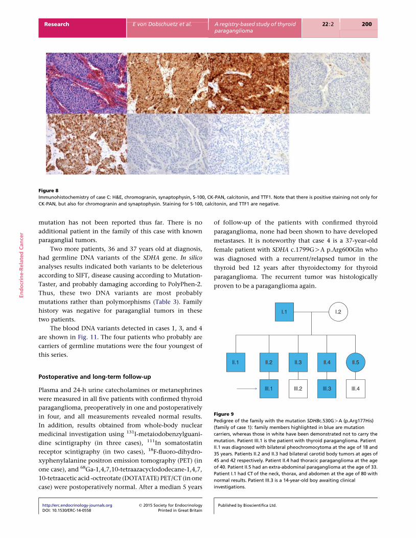
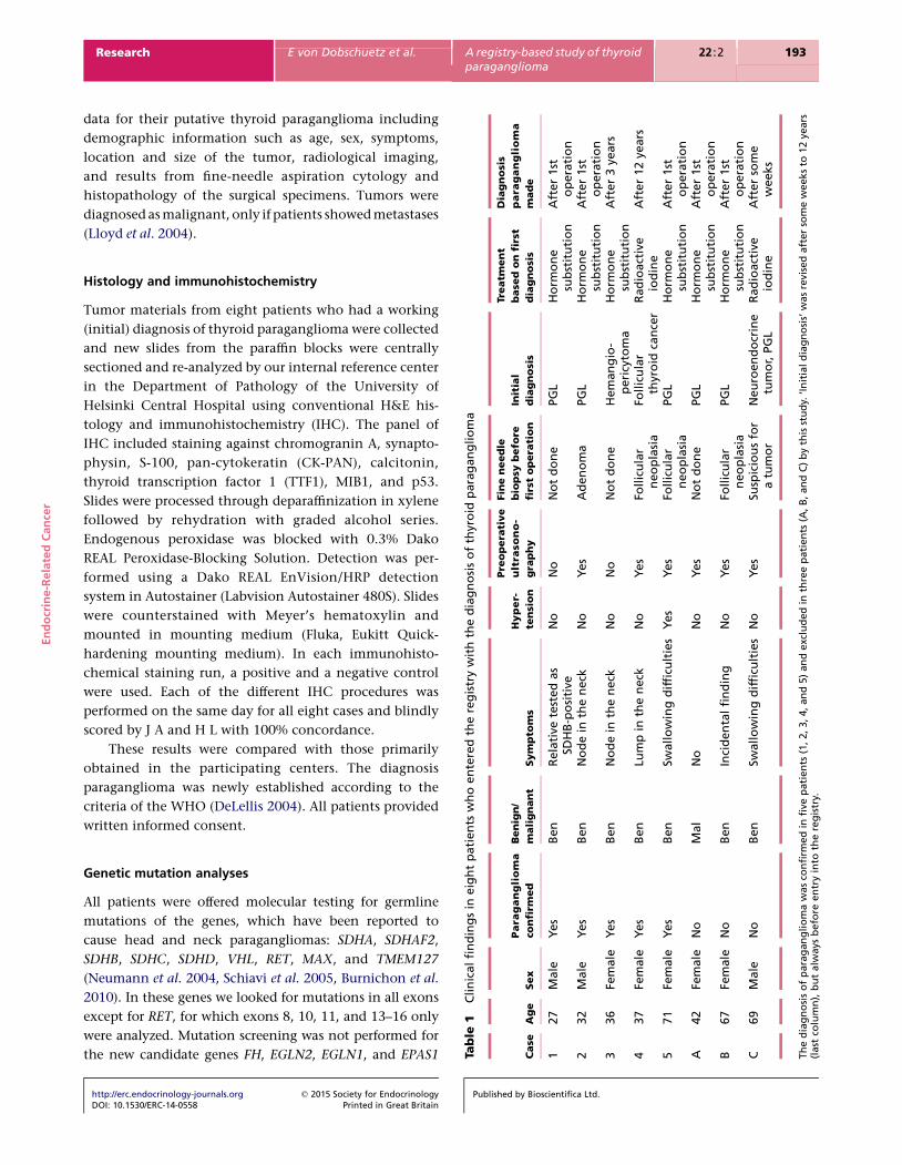
Figure 8
Immunohistochemistry of case C: H&E, chromogranin, synaptophysin, S-100, CK-PAN, calcitonin, and TTF1. Note that there is positive staining not only for
CK-PAN, but also for chromogranin and synaptophysin. Staining for S-100, calcitonin, and TTF1 are negative.
l.1 l.2
En
do
crin
e-R
ela
ted
Can
cer
Research E von Dobschuetz et al. A registry-based study of thyroidparaganglioma
22 :2 200
mutation has not been reported thus far. There is no
additional patient in the family of this case with known
paraganglial tumors.
Two more patients, 36 and 37 years old at diagnosis,
had germline DNA variants of the SDHA gene. In silico
analyses results indicated both variants to be deleterious
according to SIFT, disease causing according to Mutation-
Taster, and probably damaging according to PolyPhen-2.
Thus, these two DNA variants are most probably
mutations rather than polymorphisms (Table 3). Family
history was negative for paraganglial tumors in these
two patients.
The blood DNA variants detected in cases 1, 3, and 4
are shown in Fig. 11. The four patients who probably are
carriers of germline mutations were the four youngest of
this series.
ll.5ll.4ll.3ll.2ll.1lll.1 lll.2 lll.3 lll.4
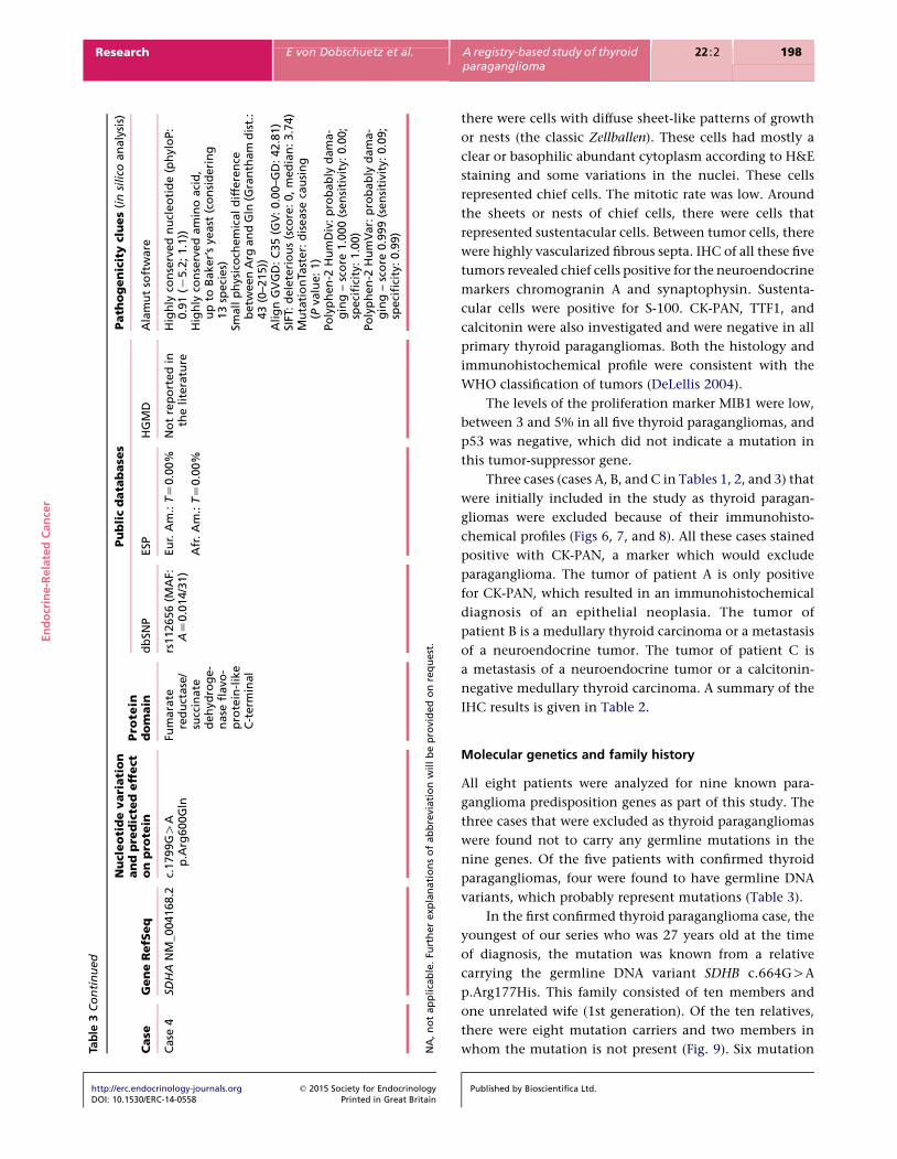
Figure 9
Pedigree of the family with the mutation SDHBc.530GOA (p.Arg177His)
(family of case 1): family members highlighted in blue are mutation
carriers, whereas those in white have been demonstrated not to carry the
mutation. Patient III.1 is the patient with thyroid paraganglioma. Patient
II.1 was diagnosed with bilateral pheochromocytoma at the age of 18 and
35 years. Patients II.2 and II.3 had bilateral carotid body tumors at ages of
45 and 42 respectively. Patient II.4 had thoracic paraganglioma at the age
of 40. Patient II.5 had an extra-abdominal paraganglioma at the age of 33.
Patient I.1 had CT of the neck, thorax, and abdomen at the age of 80 with
normal results. Patient III.3 is a 14-year-old boy awaiting clinical
investigations.
Postoperative and long-term follow-up
Plasma and 24-h urine catecholamines or metanephrines
were measured in all five patients with confirmed thyroid
paraganglioma, preoperatively in one and postoperatively
in four, and all measurements revealed normal results.
In addition, results obtained from whole-body nuclear
medicinal investigation using 131I-metaiodobenzylguani-
dine scintigraphy (in three cases), 111In somatostatin
receptor scintigraphy (in two cases), 18F-fluoro-dihydro-
xyphenylalanine positron emission tomography (PET) (in
one case), and 68Ga-1,4,7,10-tetraazacyclododecane-1,4,7,
10-tetraacetic acid -octreotate (DOTATATE) PET/CT (in one
case) were postoperatively normal. After a median 5 years
http://erc.endocrinology-journals.org q 2015 Society for EndocrinologyDOI: 10.1530/ERC-14-0558 Printed in Great Britain
of follow-up of the patients with confirmed thyroid
paraganglioma, none had been shown to have developed
metastases. It is noteworthy that case 4 is a 37-year-old
female patient with SDHA c.1799GOA p.Arg600Gln who
was diagnosed with a recurrent/relapsed tumor in the
thyroid bed 12 years after thyroidectomy for thyroid
paraganglioma. The recurrent tumor was histologically
proven to be a paraganglioma again.
Published by Bioscientifica Ltd.

Figure 10
Genomic sequence and electropherogram of the SDHB genomic
rearrangement of case 2: the genomic sequences of intron 2 and exon 3
(in bold) are shown in the figure, and the intronic repeat markers are
shown in italics (AluSz and AluJb). The deleted region is highlighted in red
and the inserted segment (AluYb8sequence) with its polyA tail in green.
Long-range PCR was used to amplify the deleted region and produced an
approximately 1.1 kb mutant product compared with the approximately
2.5 kb WT genomic sequence, predicting a deletion of approximately
1.4 kb; bidirectional sequencing of the mutant PCR product confirmed
the partial deletion of intron 2 and exon 3 and showed the insertion of
approximately 150 bp corresponding to an AluYb8 sequence (shown in
the figure).
En
do
crin
e-R
ela
ted
Can
cer
Research E von Dobschuetz et al. A registry-based study of thyroidparaganglioma
22 :2 201
http://erc.endocrinology-journals.org q 2015 Society for EndocrinologyDOI: 10.1530/ERC-14-0558 Printed in Great Britain
Published by Bioscientifica Ltd.
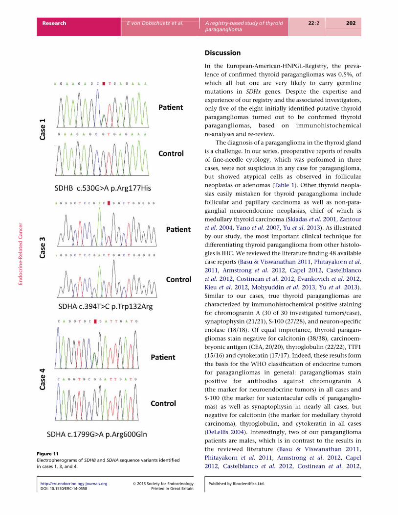
Figure 11
Electropherograms of SDHB and SDHA sequence variants identified
in cases 1, 3, and 4.
En
do
crin
e-R
ela
ted
Can
cer
Research E von Dobschuetz et al. A registry-based study of thyroidparaganglioma
22 :2 202
http://erc.endocrinology-journals.org q 2015 Society for EndocrinologyDOI: 10.1530/ERC-14-0558 Printed in Great Britain
Discussion
In the European-American-HNPGL-Registry, the preva-
lence of confirmed thyroid paragangliomas was 0.5%, of
which all but one are very likely to carry germline
mutations in SDHx genes. Despite the expertise and
experience of our registry and the associated investigators,
only five of the eight initially identified putative thyroid
paragangliomas turned out to be confirmed thyroid
paragangliomas, based on immunohistochemical
re-analyses and re-review.
The diagnosis of a paraganglioma in the thyroid gland
is a challenge. In our series, preoperative reports of results
of fine-needle cytology, which was performed in three
cases, were not suspicious in any case for paraganglioma,
but showed atypical cells as observed in follicular
neoplasias or adenomas (Table 1). Other thyroid neopla-
sias easily mistaken for thyroid paraganglioma include
follicular and papillary carcinoma as well as non-para-
ganglial neuroendocrine neoplasias, chief of which is
medullary thyroid carcinoma (Skiadas et al. 2001, Zantour
et al. 2004, Yano et al. 2007, Yu et al. 2013). As illustrated
by our study, the most important clinical technique for
differentiating thyroid paraganglioma from other histolo-
gies is IHC. We reviewed the literature finding 48 available
case reports (Basu & Viswanathan 2011, Phitayakorn et al.
2011, Armstrong et al. 2012, Capel 2012, Castelblanco
et al. 2012, Costinean et al. 2012, Evankovich et al. 2012,
Kieu et al. 2012, Mohyuddin et al. 2013, Yu et al. 2013).
Similar to our cases, true thyroid paragangliomas are
characterized by immunohistochemical positive staining
for chromogranin A (30 of 30 investigated tumors/case),
synaptophysin (21/21), S-100 (27/28), and neuron-specific
enolase (18/18). Of equal importance, thyroid paragan-
gliomas stain negative for calcitonin (38/38), carcinoem-
bryonic antigen (CEA, 20/20), thyroglobulin (22/22), TTF1
(15/16) and cytokeratin (17/17). Indeed, these results form
the basis for the WHO classification of endocrine tumors
for paragangliomas in general: paragangliomas stain
positive for antibodies against chromogranin A
(the marker for neuroendocrine tumors) in all cases and
S-100 (the marker for sustentacular cells of paraganglio-
mas) as well as synaptophysin in nearly all cases, but
negative for calcitonin (the marker for medullary thyroid
carcinoma), thyroglobulin, and cytokeratin in all cases
(DeLellis 2004). Interestingly, two of our paraganglioma
patients are males, which is in contrast to the results in
the reviewed literature (Basu & Viswanathan 2011,
Phitayakorn et al. 2011, Armstrong et al. 2012, Capel
2012, Castelblanco et al. 2012, Costinean et al. 2012,
Published by Bioscientifica Ltd.

En
do
crin
e-R
ela
ted
Can
cer
Research E von Dobschuetz et al. A registry-based study of thyroidparaganglioma
22 :2 203
Evankovich et al. 2012, Kieu et al. 2012, Mohyuddin et al.
2013, Yu et al. 2013).
Of our five confirmed thyroid paraganglioma patients,
four were found very likely to carry germline mutations
in SDHx. In contrast, none of the three patients who
eventually turned out to have non-paraganglioma thyroid
tumors (cases A, B, and C, Table 1, Figs 6, 7, and 8) were
found to have germline mutations in the nine known
pheochromocytoma–paraganglioma-related genes. There-
fore, in addition to IHC, germline genetic analysis could
be helpful in differentiating thyroid paraganglioma from
other types of thyroid neoplasms that mimic this histology.
Thyroid paraganglioma is a rare entity with approxi-
mately 60 cases that are reported, including our series
(LaGuette et al. 1997, Yano et al. 2007, Ferri et al. 2009,
Gonzalez Poggioli et al. 2009, Basu & Viswanathan 2011,
Phitayakorn et al. 2011, Varsavsky et al. 2011, Armstrong
et al. 2012, Capel 2012, Castelblanco et al. 2012, Costinean
et al. 2012, Evankovich et al. 2012, Kieu et al. 2012,
Mohyuddin et al. 2013, Yu et al. 2013). A major question
is whether the rare entity of thyroid paraganglioma is
of practical clinical relevance. Our series demonstrates that
this diagnosis dictates specific treatment and hence the
consequences regarding correct treatment are obvious: one
female patient was primarily diagnosed with a papillary
thyroid carcinoma. Consequently, the patient was sub-
jected to radioactive iodine radiation. Only the relapse
of her tumor 12 years later led to the identification of a
paraganglioma, resulting in the retrospective diagnosis of
a primary thyroid paraganglioma and not papillary thyroid
carcinoma. From an outcome point of view, it is important
to know that thyroid paraganglioma may show local
invasive growth but confirmed metastases are extremely
rare (Massaioli et al. 1979, Mohyuddin et al. 2013).
Finally, it has to be emphasized that we found in
most patients with thyroid paraganglioma germline
DNA variants, which most probably represent germline
mutations. This opens avenues for genetic family scree-
ning and preventive medicine. Patients with germline
mutations of the SDHB gene, as found in two of our
patients, may display adrenal, retroperitoneal, pelvic, or
thoracic paragangliomas or metachronous head and neck
paragangliomas such as carotid glomus tumors (Haegert
et al. 1974, Hughes et al. 1997), which may become
malignant and therefore have to undergo lifelong high-
risk clinical surveillance (Neumann et al. 2004). Similar
risk profiles will be identified once a statistically significant
number of patients with mutations of the SDHA gene
are available.
http://erc.endocrinology-journals.org q 2015 Society for EndocrinologyDOI: 10.1530/ERC-14-0558 Printed in Great Britain
In summary, our systematic, population-based series
of thyroid paragangliomas indicates that thyroid para-
ganglioma is an important entity to be differentiated from
other thyroid tumors, mainly medullary thyroid carci-
noma, despite its prevalence of approximately 0.5% in
HNPs, because clinical management is vastly different. The
fact that the genetic load of thyroid paraganglioma is
seemingly 80% is also significant, with implications for
both patient management and family members.
Declaration of interest
The authors declare that there is no conflict of interest that could be
perceived as prejudicing the impartiality of the research reported.
Funding
This work was supported by: grants from the Deutsche Krebshilfe (grant
number 107995 to H P H Neumann), the Sondra J and Stephen R Hardis
Endowment (to C Eng), and the Arthur Blank Foundation, Atlanta, GA, USA
(to C Eng). B Jarzab and J Krajewska were supported by Polish National
Science Center grant number NN401410639. Mariola Peczkowska, Alek-
sander Prejbisz and Andrzej Januszewicz were supported by a grant from
the Polish Ministry of Science and Higher Education (N N402 386038).
Author contribution statement
B Jarzab, G Opocher, C Eng and H P H Neumann shared senior authorship.
References
Armstrong MJ, Chiosea SI, Carty SE, Hodak SP & Yip L 2012 Thyroid
paragangliomas are locally aggressive. Thyroid 22 88–93. (doi:10.1089/
thy.2011.0110)
Basu S & Viswanathan S 2011 Primary paraganglioma of thyroid presenting
as solitary thyroid mass. Journal of Cancer Research and Therapeutics 7
385–387. (doi:10.4103/0973-1482.87028)
Boedeker CC, Erlic Z, Richard S, Kontny U, Gimenez-Roqueplo A-P, Cascon
A, Robledo M, de Campos JM, van Nederveen FH, de Krijger RR et al.
2009 Head and neck paragangliomas in von Hippel–Lindau disease and
multiple endocrine neoplasia type 2. Journal of Clinical Endocrinology
and Metabolism 94 1938–1944. (doi:10.1210/jc.2009-0354)
Burnichon N, Briere JJ, Libe R, Vescovo L, Riviere J, Tissier F, Jouanno E,
Jeunemaitre X, Benit P, Tzagoloff A et al. 2010 SDHA is a tumor
suppressor gene causing paraganglioma. Human Molecular Genetics 19
3011–3020. (doi:10.1093/hmg/ddq206)
Capel I, Gil MP, Marques G, Barcons S & Rigla M 2012 Paraganglioma
cervical simulando nodulo tiroideo con afectation del nervio
recurrente. Endocrinologıa y Nutricion 59 274–275. (doi:10.1016/
j.endonu.2011.09.011)
Castelblanco E, Gallel P, Ros S, Gatius S, Valls J, De-Cubas AA, Maliszewska A,
Yebra-Pimentel MT, Menarguez J, Gamallo C et al. 2012
Thyroid paraganglioma. Report of 3 cases and description of an
immunohistochemical profile useful in the differential diagnosis with
medullary thyroid carcinoma, based on complementary DNA array
results. Human Pathology 43 1103–1112. (doi:10.1016/j.humpath.2011.
08.022)
Published by Bioscientifica Ltd.

En
do
crin
e-R
ela
ted
Can
cer
Research E von Dobschuetz et al. A registry-based study of thyroidparaganglioma
22 :2 204
Castro-Vega LJ, Buffet A, De Cubas AA, Cascon A, Menara M, Khalifa E,
Amar L, Azriel S, Bourdeau I, Chabre O et al. 2014 Germline mutations
in FH confer predisposition to malignant pheochromocytomas and
paragangliomas. Human Molecular Genetics 23 2440–2446.
(doi:10.1093/hmg/ddt639)
Costinean S, Balatti V, Bottoni A, Old M, Croce C & Wakely PE Jr 2012
Primary intrathyroidal paraganglioma: histopathology and novel
molecular alterations. Human Pathology 43 2371–2375. (doi:10.1016/
j.humpath.2012.06.021)
DeLellis RA 2004 Paraganglioma. In Pathology and Genetics of Tumours of
the Endocrine Organs. World Health Organization Classification of
Tumours Pathology and Genetics of Tumours of Endocrine Organs, p 117.
Eds RA DeLellis, RV Lloyd, PU Heitz & C Eng. Lyon: IARC Press.
Evankovich J, Dedhia RC, Bastaki JM, Tublin M & Johnson JT 2012
Primary sclerosing paraganglioma of the thyroid gland: a case report.
Annals of Otology, Rhinology, and Laryngology 121 510–515.
Ferri E, Manconi R, Armato E & Ianniello F 2009 Primary paraganglioma of
thyroid gland: a clinicopathologic and immunohistochemical study
with review of the literature. Acta Otorhinolaryngologica Italica 29 97–102.
Gonzalez Poggioli N, Lopez Amado M & Yebra Pimentel M 2009
Paraganglioma of the thyroid gland: a rare entity. Endocrine Pathology
20 62–65. (doi:10.1007/s12022-009-9066-2)
Haegert DG, Wang NS, Farrer PA, Seemayer TA & Thelmo W 1974
Non-chromaffin paragangliomatosis manifesting as a cold thyroid
nodule. American Journal of Clinical Pathology 61 561–570.
Hughes JH, El-Mofty S, Sessions D & Liapis H 1997 Primary intrathyroidal
paraganglioma with metachronous carotid body tumor: report of a case
and review of the literature. Pathology, Research and Practice 193
791–796 discussion 797–799. (doi:10.1016/S0344-0338(97)80059-3)
Kieu V, Yuen A, Tassone P & Hobbs CG 2012 Cervical paraganglioma
presenting as thyroid neoplasia. Otolaryngology and Head and Neck
Surgery 146 516–518. (doi:10.1177/0194599811419229)
LaGuette J, Matias-Guiu X & Rosai J 1997 Thyroid paraganglioma: a
clinicopathologic and immunohistochemical study of three cases.
American Journal of Surgical Pathology 21 748–753. (doi:10.1097/
00000478-199707000-00002)
Lloyd RV, Tischler AS, Kimura N, McNicol AM & Young WF 2004 Adrenal
tumors. In Pathology and Genetics of Tumours of the Endocrine Organs
World Health Organization Classification of Tumours, pp 137–138.
Eds RA DeLellis, RV Lloyd, PU Heitz & C Eng. Lyon: IARC Press.
Massaioli N, Balbo G, Fausone G & Negro D 1979 Paraganglioma
branchiomerico endotiroideo (non cromaffine). Descrizione di un caso
clinico. Minerva Chirurgica 34 867–874.
Mohyuddin N, Ferrer K & Patel U 2013 Malignant paraganglioma of the
thyroid gland with synchronous bilateral carotid body tumors. Ear,
Nose, & Throat Journal 92 E20–E23.
Neumann HP, Pawlu C, Peczkowska M, Bausch B, McWhinney SR, Muresan
M, Buchta M, Franke G, Klisch J, Bley TA et al. 2004 Distinct clinical
http://erc.endocrinology-journals.org q 2015 Society for EndocrinologyDOI: 10.1530/ERC-14-0558 Printed in Great Britain
features of paraganglioma syndromes associated with SDHB and SDHD
gene mutations. Journal of the American Medical Association 292
943–951. (doi:10.1001/jama.292.8.943)
Neumann HP, Erlic Z, Boedeker CC, Rybicki LA, Robledo M, Hermsen M,
Schiavi F, Falcioni M, Kwok P, Bauters C et al. 2009 Clinical
predictors for germline mutations in head and neck paraganglioma
patients: cost reduction strategy in genetic diagnostic process as
fall-out. Cancer Research 69 3650–3656. (doi:10.1158/0008-5472.
CAN-08-4057)
Phitayakorn R, Faquin W, Wei N, Barbesino G & Stephen AE 2011
Thyroid-associated paragangliomas. Thyroid 21 725–733. (doi:10.1089/
thy.2010.0362)
Schiavi F, Boedeker CC, Bausch B, Peczkowska M, Gomez CF, Strassburg T,
Pawlu C, Buchta M, Salzmann M, Hoffmann MM et al. 2005 Predictors
and prevalence of paraganglioma syndrome associated with mutations
of the SDHC gene. Journal of the American Medical Association 294
2057–2063. (doi:10.1001/jama.294.16.2057)
Skiadas PK, Kakavoulis TN & Gikonti IJ 2001 Normalisation of blood
pressure and heart rate after excision of a thyroid paraganglioma.
European Journal of Surgery 167 392–394. (doi:10.1080/
110241501750215320)
ToledoRA, Qin Y, SrikantanS,MoralesNP, Li Q,Deng Y,Kim SW, PereiraMA,
Toledo SP, Su X et al. 2013 In vivo and in vitro oncogenic effects of
HIF2A mutations in pheochromocytomas and paragangliomas.
Endocrine-Related Cancer 20 349–359. (doi:10.1530/ERC-13-0101)
Varsavsky M, Cortes Berdonces M, Alonso G, Garcia Martin A & Munoz
Torres M 2011 Adenopatia metastasica de un microcarcinoma tiroideo,
diagnostico final de un supuesto paraganglioma. Endocrinologıa y
Nutricion 58 143–144. (doi:10.1016/j.endonu.2010.10.0090)
Yang C, Zhuang Z, Fliedner SM, Shankavaram U, Sun MG, Bullova P, Zhu R,
Elkahloun AG, Kourlas PJ, Merino M et al. 2015 Germ-line PHD1
and PHD2 mutations detected in patients with pheochromocytoma/
paraganglioma-polycythemia. Journal of Molecular Medicine 93 93–104.
(doi:10.1007/s00109-014-1205-7)
Yano Y, Nagahama M, Sugino K, Ito K & Kameyama K 2007 Paraganglioma
of the thyroid: report of a male case with ultrasonographic imagings,
cytologic, histologic, and immunohistochemical features. Thyroid 17
575–578. (doi:10.1089/thy.2006.0284)
Yu BH, Sheng WQ & Wang J 2013 Primary paraganglioma of thyroid gland:
a clinicopathologic and immunohistochemical analysis of three cases
with a review of the literature. Head and Neck Pathology 7 373–380.
(doi:10.1007/s12105-013-0467-7)
Zantour B, Guilhaume B, Tissier F, Louvel A, Jeunemaitre X,
Gimenez-Roqueplo AP & Bertagna X 2004 A thyroid nodule revealing
a paraganglioma in a patient with a new germline mutation in the
succinate dehydrogenase B gene. European Journal of Endocrinology 151
433–438. (doi:10.1530/eje.0.1510433)
Received in final form 7 January 2015Accepted 16 January 2015Made available online as an Accepted Preprint16 January 2015
Published by Bioscientifica Ltd.