Embed Size (px)
Citation preview
191 International Journal of Scientific Study | August 2015 | Vol 3 | Issue 5
Bilateral Carotid Body Paraganglioma: A Rare Case ReportDinesh Kulkarni1, Manoj Dongare2, Manik Deshpande3
1Consultant Histopathologist, Aurangabad, Maharashtra, India, 2Consultant Oncosurgeon, MGM MCRI and Manik Hospital & Research Centre, Aurangabad, Maharashtra, India, 3Consultant Anesthesiologist, Manik Hospital and Research Centre, Aurangabad, Maharashtra, India
clinical course, about 10% of carotid body paragangliomas turn malignant. However, evidence of metastasis is the only definitive criterion to label the tumor as malignant. Metastasis can occur to regional lymph nodes and lungs.4 Paragangliomas are characterized grossly by the brown color of the cut surface and microscopically by the presence of well-defined nests of uniform cuboidal cells (Zellballen) separated by highly vascularized fibrous septae.1-4 Individual cells have an abundant granular basophilic cytoplasm. Bizzare nuclei and vascular invasion are sometimes found, but these should not be taken as evidence of malignancy. There are no reliable morphologic criteria to separate benign from malignant, but high mitotic activity and decreased reactivity for neuropeptides suggest malignancy.4 Characteristic symptoms are headaches, hypertension, palpitations, diaphoresis, and sweating.
CASE REPORT
A 32-year-old male came with complaints of swelling in both sides of the neck for 3 years. The swellings were rounded, soft to firm, freely mobile, non-tender. He was non-hypertensive. General and systemic examinations were within normal limits.
INTRODUCTION
Paraganglioma is the generic term applied to tumors arising along the sympathetic and parasympathetic paraganglia regardless of its location, except arising from adrenal medulla. These are of neuroendocrine origin derived from the embryonic neural crest.1,2 They are a category of chromaffin cell tumors, secreting catecholamines in 50-60% of cases. The extra-adrenal location accounts for 10-30% of cases.3
Carotid body paragangliomas are the important group of extra-adrenal paraganglioma. The first carotid body paraganglioma was reported by Marchand in 1891. They are located at the bifurcation of common carotid artery.4 They are more frequently seen in people living at high altitude. Men are affected more frequently than women and in the third and fourth decade. Most of them follow a benign
Case Report
AbstractAccording to the World Health Organization classification of tumors 2004, paragangliomas are a type of neuroendocrine tumors derived from the embryonic neural crest. The adrenal gland and the extra-adrenal paraganglia of the autonomic nervous system are the most common places for its occurrence. When found in the adrenal gland they are called pheochromocytoma, but in the extra-adrenal paraganglia they are described as paraganglioma. The carotid body paraganglioma is relatively common and has a benign course, but bilateral existence is rare. We present here a case of bilateral carotid body paraganglioma in a 32 years male. Ultrasound of the neck was done, which detected the bilateral mass in carotid bifurcation region. Adrenaline and noradrenaline levels were within normal limits. In two sittings, tumors on both sides of the neck were excised, and histopathological study diagnosed them to be bilateral paraganglioma. Surgical excision is the treatment of choice. One of the possible causes of hypertension in young adults is paraganglioma.
Key words: Bilateral, Carotid body paraganglioma, Neuroendocrine, Surgery
Access this article online
www.ijss-sn.com
Month of Submission : 06-2015 Month of Peer Review : 07-2015 Month of Acceptance : 07-2015 Month of Publishing : 08-2015
Corresponding Author: Dr. Dinesh Kulkarni, 42/101, Manish Apartment, Sahakar Nagar, Aurangabad, Maharashtra, India. Phone: +91-9850018299. E-mail: [email protected]
DOI: 10.17354/ijss/2015/373
Kulkarni, et al.: Bilateral Carotid Body Paraganglioma
192International Journal of Scientific Study | August 2015 | Vol 3 | Issue 5
Ultrasonography (USG) neck showed masses in bilateral carotid spaces. Magnetic image resonance (MRI) neck revealed bilateral homogenous masses in carotid spaces at the carotid bifurcation, right 4 cm × 3 cm and left sided 2.5 cm × 2 cm. Magnetic resonance angiography showed splaying of internal and external carotid arteries at bifurcation on both sides. Plasma epinephrine and norepinephrine were within normal limits. His X-ray chest, two-dimensional echocardiogram, hemogram, electrolytes, liver and kidney function tests were within normal limits, and he was medically fit for surgery.
Under general anesthesia, bilateral tumor masses were excised completely in two sittings along with surrounding lymph nodes. The excised masses were sent for histopathology which revealed it to be bilateral paraganglioma and bilateral lymph nodes showed reactive hyperplasia and were free from metastasis.
Post-operative USG, MRI neck and color Doppler did not reveal residual tumor.
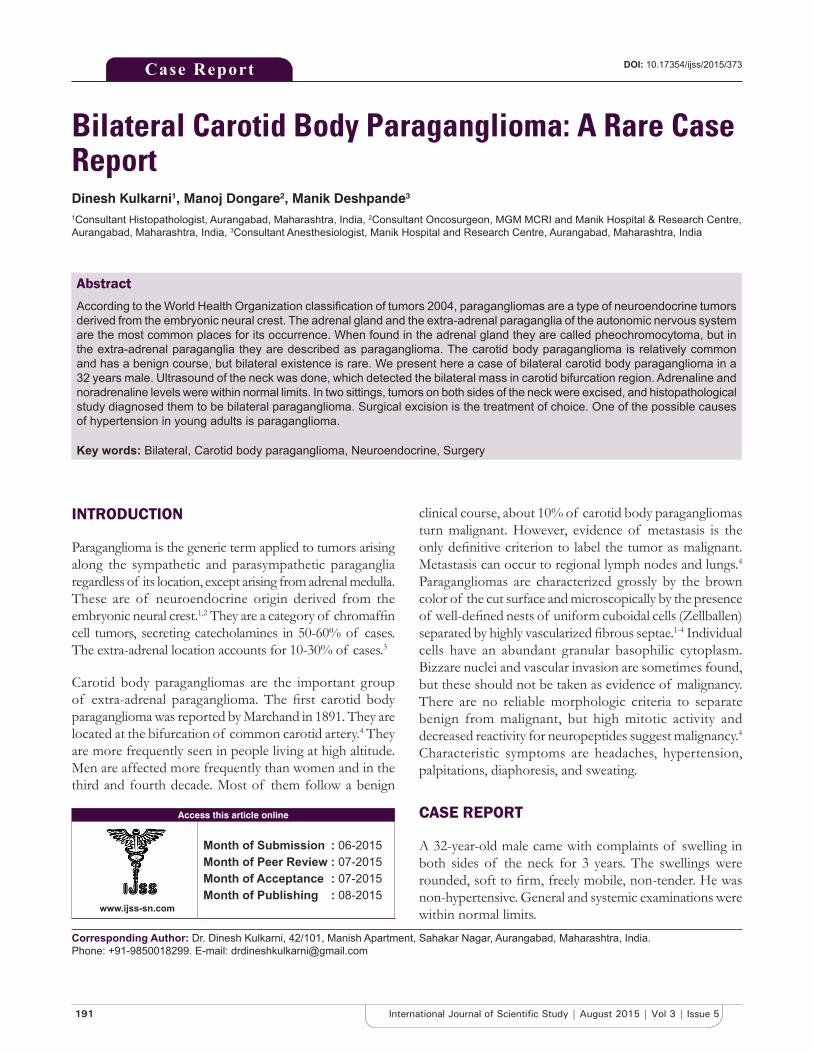
MorphologyReceived two specimen in a space of 15 days. On gross, right-sided mass was single rounded 4 cm × 2 cm × 1 cm grayish firm mass, cut surface brownish firm. Also received a single 1 cm × 1 cm grayish soft tissue, cut surface grayish. Also received another specimen after 15 days with two oval 2 cm × 1.5 cm × 1 cm grayish firm mass, cut surface brownish firm. Also received a single 1 cm × 1 cm grayish soft tissue, cut surface grayish. No necrosis or haemorrhages seen (Figure 1).
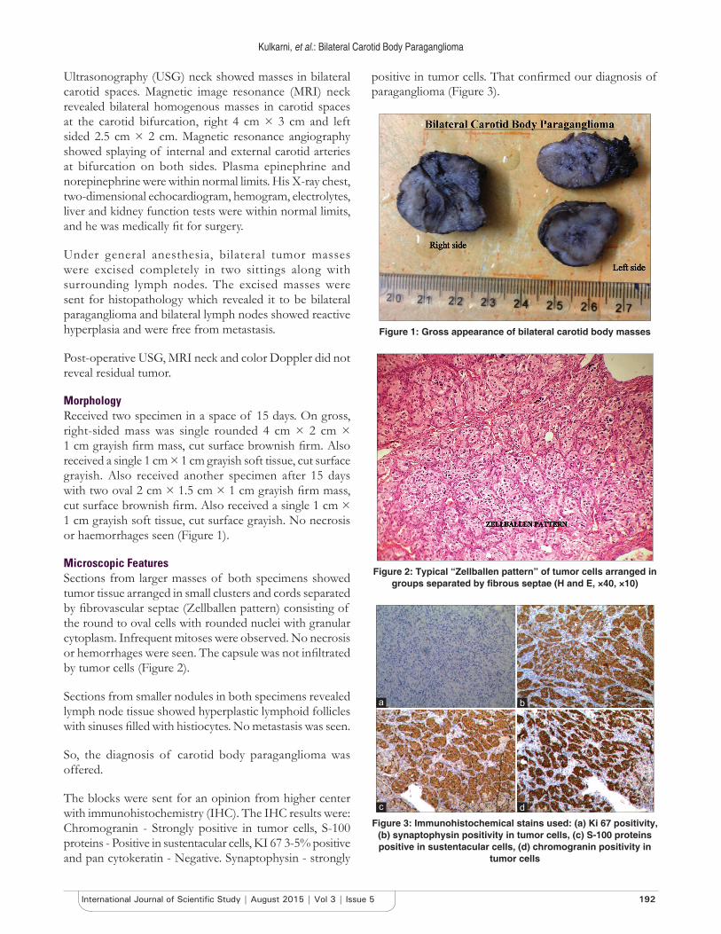
Microscopic FeaturesSections from larger masses of both specimens showed tumor tissue arranged in small clusters and cords separated by fibrovascular septae (Zellballen pattern) consisting of the round to oval cells with rounded nuclei with granular cytoplasm. Infrequent mitoses were observed. No necrosis or hemorrhages were seen. The capsule was not infiltrated by tumor cells (Figure 2).
Sections from smaller nodules in both specimens revealed lymph node tissue showed hyperplastic lymphoid follicles with sinuses filled with histiocytes. No metastasis was seen.
So, the diagnosis of carotid body paraganglioma was offered.
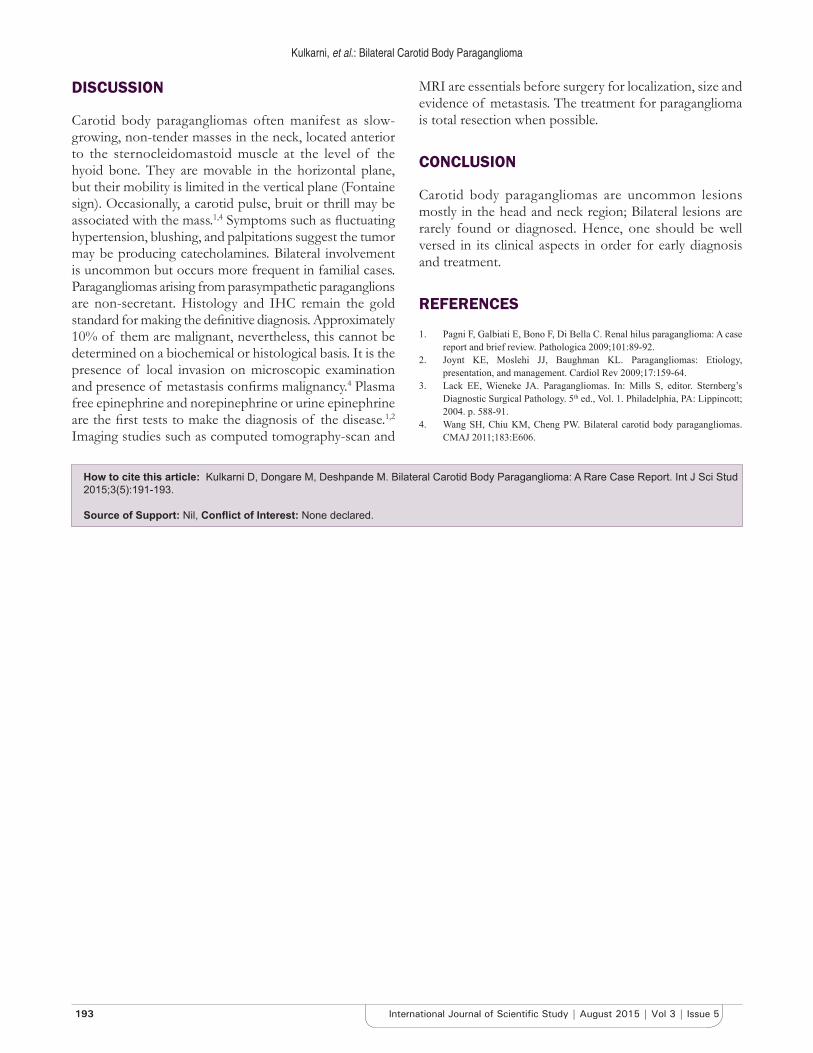
The blocks were sent for an opinion from higher center with immunohistochemistry (IHC). The IHC results were: Chromogranin - Strongly positive in tumor cells, S-100 proteins - Positive in sustentacular cells, KI 67 3-5% positive and pan cytokeratin - Negative. Synaptophysin - strongly
positive in tumor cells. That confirmed our diagnosis of paraganglioma (Figure 3).
Figure 1: Gross appearance of bilateral carotid body masses
Figure 2: Typical “Zellballen pattern” of tumor cells arranged in groups separated by fibrous septae (H and E, ×40, ×10)
Figure 3: Immunohistochemical stains used: (a) Ki 67 positivity, (b) synaptophysin positivity in tumor cells, (c) S-100 proteins positive in sustentacular cells, (d) chromogranin positivity in
tumor cells
dc
ba
Kulkarni, et al.: Bilateral Carotid Body Paraganglioma
193 International Journal of Scientific Study | August 2015 | Vol 3 | Issue 5
DISCUSSION
Carotid body paragangliomas often manifest as slow-growing, non-tender masses in the neck, located anterior to the sternocleidomastoid muscle at the level of the hyoid bone. They are movable in the horizontal plane, but their mobility is limited in the vertical plane (Fontaine sign). Occasionally, a carotid pulse, bruit or thrill may be associated with the mass.1,4 Symptoms such as fluctuating hypertension, blushing, and palpitations suggest the tumor may be producing catecholamines. Bilateral involvement is uncommon but occurs more frequent in familial cases. Paragangliomas arising from parasympathetic paraganglions are non-secretant. Histology and IHC remain the gold standard for making the definitive diagnosis. Approximately 10% of them are malignant, nevertheless, this cannot be determined on a biochemical or histological basis. It is the presence of local invasion on microscopic examination and presence of metastasis confirms malignancy.4 Plasma free epinephrine and norepinephrine or urine epinephrine are the first tests to make the diagnosis of the disease.1,2 Imaging studies such as computed tomography-scan and
MRI are essentials before surgery for localization, size and evidence of metastasis. The treatment for paraganglioma is total resection when possible.
CONCLUSION
Carotid body paragangliomas are uncommon lesions mostly in the head and neck region; Bilateral lesions are rarely found or diagnosed. Hence, one should be well versed in its clinical aspects in order for early diagnosis and treatment.
REFERENCES
1. Pagni F, Galbiati E, Bono F, Di Bella C. Renal hilus paraganglioma: A case report and brief review. Pathologica 2009;101:89-92.
2. Joynt KE, Moslehi JJ, Baughman KL. Paragangliomas: Etiology, presentation, and management. Cardiol Rev 2009;17:159-64.
3. Lack EE, Wieneke JA. Paragangliomas. In: Mills S, editor. Sternberg’s Diagnostic Surgical Pathology. 5th ed., Vol. 1. Philadelphia, PA: Lippincott; 2004. p. 588-91.
4. Wang SH, Chiu KM, Cheng PW. Bilateral carotid body paragangliomas. CMAJ 2011;183:E606.
How to cite this article: Kulkarni D, Dongare M, Deshpande M. Bilateral Carotid Body Paraganglioma: A Rare Case Report. Int J Sci Stud 2015;3(5):191-193.
Source of Support: Nil, Conflict of Interest: None declared.