Embed Size (px)
Citation preview
Research ArticleA Novel Thermostable GH3 𝛽-Glucosidasefrom Talaromyce leycettanus with BroadSubstrate Specificity and Significant SoybeanIsoflavone Glycosides-Hydrolyzing Capability
Xinxin Li, Wei Xia, Yingguo Bai, Rui Ma, Hong Yang, Huiying Luo, and Pengjun Shi
Key Laboratory for Feed Biotechnology of the Ministry of Agriculture, Feed Research Institute,Chinese Academy of Agricultural Sciences, Beijing 100081, China
Correspondence should be addressed to Pengjun Shi; [email protected]
Received 13 September 2017; Accepted 17 September 2018; Published 23 October 2018
Academic Editor: Jose D. Fontana
Copyright © 2018 Xinxin Li et al.This is an open access article distributed under the Creative Commons Attribution License, whichpermits unrestricted use, distribution, and reproduction in any medium, provided the original work is properly cited.
A novel 𝛽-glucosidase gene (Bgl3B) of glycoside hydrolase (GH) family 3 was cloned from the thermophilic fungus Talaromyceleycettanus JM12802 and successfully expressed in Pichia pastoris. The deduced Bgl3B contains 860 amino acid residues with acalculated molecular mass of 91.2 kDa. The purified recombinant Bgl3B exhibited maximum activities at pH 4.5 and 65∘C andremained stable at temperatures up to 60∘C and pH 3.0−9.0, respectively. The enzyme exhibited broad substrate specificities,showing 𝛽-glucosidase, glucanase, cellobiase, xylanase, and isoflavone glycoside hydrolase activities, and its activities werestimulated by short-chain alcohols. The catalytic efficiencies of Bgl3B were 693 and 104/mM/s towards pNPG and cellobiose,respectively. Moreover, Bgl3B was highly effective in converting isoflavone glycosides to aglycones at 37∘C within 10 min, withthe hydrolysis rates of 95.1%, 76.0%, and 75.3% for daidzin, genistin, and glycitin, respectively. These superior properties makeBgl3B potential for applications in the food, animal feed, and biofuel industries.
1. Introduction
Plant cell walls contain three major polymers, i.e., cellu-lose, hemicellulose, and lignin [1]. Cellulose is the largestcomponent and consists of glucose residues linked by a𝛽-1,4-glycosidic bond [2, 3]. It can be degraded into glu-cose through the cooperation of three types of enzymes,that is, endo-𝛽-1,4-glucanase (EC3.2.1.4), exoglucanase (alsoknown as cellobiohydrolase, EC3.2.1.91), and 𝛽-glucosidase(EC3.2.1.21) [4, 5].𝛽-Glucosidase, also referred to as 𝛽-d-glucoside glu-
cohydrolase, is a key rate-limiting enzyme that is capableof specifically catalyzing the hydrolysis of 𝛽-1,4-glycosidiclinkages that existed in oligosaccharides, alkyl- or aryl 𝛽-d-glucosides, and cyanogenic glycosides from the nonreducingends [6].The classification of 𝛽-glucosidase generally followsthe CAZY dataset (http://www.cazy.org), in which glycosidehydrolases (GH) are divided into 153 families. The enzymes
having 𝛽-glucosidase activities are confined in the familiesGH1, 3, 5, 9, 30, and 116 [7], and those of GH3 contain threedomains: the (𝛽/𝛼)
8bucket domain, the (𝛽/𝛼)
6sandwich
domain, and the Fn3-like region [8]. Glucosidases are widelyused inmany industries. In the flavor field, 𝛽-glucosidase cancatalyze the production of flavor compounds and remarkablyimprove food aroma [9, 10]. In the animal feed, 𝛽-glucosidaseis supplemented to convert soybean isoflavone glycosides[5, 11]. 𝛽-Glucosidase is also used in the detergent, cosmetic,and food products [12].
The thermophilic fungus Talaromyces is well-knownfor producing various extracellular enzymes [13, 14].Considering the great potential of thermophilic 𝛽-glucosidases for application in the food and feed field,a novel GH3 𝛽-glucosidase-encoding gene from thethermophilic Talaromyces leycettanus was overexpressed inPichia pastoris in the present study. The enzymatic propertieswere characterized comprehensively. The recombinant
HindawiBioMed Research InternationalVolume 2018, Article ID 4794690, 9 pageshttps://doi.org/10.1155/2018/4794690
2 BioMed Research International
𝛽-glucosidase was found to have broad substrate specificity,high catalytic efficiency of fiber oligosaccharides, and greatsoybean isoflavone glycosides-degrading capability, thusrepresenting a promising enzyme candidate for applicationin the feed field.
2. Experimental Section
2.1. Strains, Media, Vectors, and Chemicals. The filamen-tous fungus T. leycettanus JCM12802 was purchased fromJapan Collection of Microorganisms and cultured at 45∘Cin the wheat bran medium [7]. Escherichia coli Trans1-T1 (TransGen, Beijing, China) and Pichia pastoris GS115(Invitrogen, Carlsbad, CA) used for gene cloning andheterologous expression were cultured at 37∘C and 30∘C,respectively. Luria-Bertani (LB) medium supplemented with50 𝜇g/ml ampicillin and yeast peptone dextrose mediumwere used for the cultivation of prokaryotic and eukaryoticcells, respectively. The vectors pEASY-T3 (TransGen) andpPIC9 (Invitrogen) were applied for cloning and expres-sion, respectively. The DNA purification kit, LA Taq DNApolymerase, and restriction enzymes were purchased fromTaKaRa (Otsu, Japan). The FastPfu DNA polymerase andFungal DNA Mini kit were purchased from TIANGEN (Bei-jing, China) and the Omega Biotek (Doraville, GA), respec-tively. The total RNA isolation system kit and T4 DNA ligasewere purchased from Promega (Madison, WI). The restric-tion endonucleases and endo-𝛽-N-acetylglucosaminidase H(EndoH)were obtained fromNewEngland Biolabs (Ipswich,MA). Substrates 4-nitrophenyl 𝛽-glucopyranoside (pNPG),barley 𝛽-d-glucan, medium viscosity carboxymethylcellu-lose sodium (CMC-Na), lichenan, beechwood xylan AvicelPH-101, amygdalin, and six isoflavone standards (daidzin,genistin, glycitin, daidzein, genistein, and glycitein) weresupplied by Sigma-Aldrich (St. Louis, MO) [7]. Xyloglucan,laminarin, and oligosaccharides (cellobiose, cellotriose, cel-lotetraose, cellopentaose, and laminaritetraose) were sup-plied by Megazyme (Wicklow, Ireland). All other chemicalswere of analytic grade and commercially available.
2.2. Gene Cloning and Sequence Analysis. The mycelia ofstrain JCM12802 were collected after 4-day-growth in wheatbran medium. The genomic DNA and total RNA wereextracted and purified in accordance with the manufacturer’sinstructions of Fungal DNA Mini kit and the SV Total RNAIsolation System, respectively. TransScript� One-Step gDNARemoval and cDNA Synthesis SuperMix kit (TransGen) wereused for the production of cDNAs.
According to the conserved regions of fungal 𝛽-glucosidases of GH3, two conserved sequences SSNIDD andGLDMT (A) MPGD (S) were identified, and a degenerateprimer set DP-F and DP-R (Table S1) was designedaccordingly to amplify the core region of the objective gene.The purified PCR products with target size were linkedwith the cloning vector pEASY-T3 (TransGen), which wasthen transformed into E. coli Trans1-T1 for cloning andsequencing. According to the sequencing results, two specificprimer sets (Table S1) were designed to amplify the up- anddown-stream sequences. The PCR products were purified,
sequenced, and assembled with the core region to givethe full-length 𝛽-glucosidase gene Bgl3B. According to thesequence of Bgl3B, two expression primers (Table S1) weredesigned to amplify the cDNA fragment coding for themature Bgl3B without the signal peptide-coding sequence.An annealing temperature of 60∘C was set.
The nucleotide sequences of Bgl3Bwere assembled by theVectorNTI 10.0 software (Invitrogen). Sequence comparisonswith known GH3 𝛽-glucosidase sequences were accom-plished by using the BlastN, BlastX, and BlastP at NCBI(http://www.ncbi.nlm.nih.gov/BLAST) based on the homol-ogy search. Alignment of multiple protein sequences wasconducted using the ClustalX software (http://www.clustal.org/) and rendered by the ESPript 3.0 (http://espript.ibcp.fr/ESPript/cgi-bin/ESPript.cgi). The potential signal peptidewas predicted to use the SignalP 4.0 server (http://www.cbs.dtu.dk/services/SignalP/).Theonline programsNetNGlyc1.0 Server (http://www.cbs.dtu.dk/services/NetNGlyc/) andNetOGlyc 4.0 Server (http://www.cbs.dtu.dk/services/NetOGlyc/) were used to predict the different type glyco-sylation sites. The three-dimensional structures of Bgl3B andits complex with cellobiose were homology modeled withthe 𝛽-glucosidase, Af𝛽G, from Aspergillus fumigatus (PDB:5FJI A) by applying the Discovery Studio v2.5 (Accelrys, SanDiego, CA).
2.3. Expression and Purification of the Heterologous Protein.The cDNA fragment of Bgl3B and vector pPIC9 were bothdigested by EcoRI and NotI and then ligated to construct therecombinant plasmid pPIC9-Bgl3B. The sequence-verifiedrecombinant plasmid pPIC9-Bgl3B was then linearized byBglII and purified with DNA purification kit. The purifiedproducts were transformed into P. pastoris GS115 competentcells via electroporation using the Gene Pulser X cell Elec-troporation System (Bio-Rad, Hercules, CA). Positive trans-formants were screened on minimal dextrose (MD) platesand grown in corresponding medium of different stagesaccording the previous methods [7, 15]. The cell-free culturesupernatants were gathered by high-speed centrifugation(12,000 g) at 4∘C for 10min and concentrated using aVivaflowultrafiltration membrane (Vivascience, Hannova, Germany)that can trap protein with a molecular mass of more than3 kDa. The crude enzyme was further purified using aFPLC HiTrap Q Sepharose XL 6 ml column (GE Healthcare,Uppsala, Sweden) according to the previous methods ofprotein purification [7, 15]. Fractions showing 𝛽-glucosidaseactivities (as described below) were identified using sodiumdodecyl sulfate-polyacrylamide gel electrophoresis (SDS-PAGE) and pooled for further studies.
2.4. Enzyme Activity Assays
2.4.1. pNP Method. When using pNPG as the substrate, thestandard systems containing enzyme solution (250 𝜇l) and 1mM of pNPG in 100 mM citric acid-Na
2HPO4buffer (250
𝜇l, pH 4.5) were treated at 65∘C for 10 min and terminatedby the addition of 1 M of Na
2CO3(1.5 mL). After cooling to
room temperature, the amount of the liberated p-nitrophenol(pNP) was measured spectrophotometrically at 405 nm [7,

BioMed Research International 3
15]. One unit (U) of 𝛽-glucosidase activity was defined as theamount of enzyme which generated 1 𝜇mol of pNP per minunder the assay conditions [15, 16].
2.4.2. GOD-POD Method. To measure the Bgl3B activitiestowards cellobiose (4 mM), laminaritetraose (5 mM), cel-lotriose (1%), cellotetraose (1%), cellopentaose (1%), gentio-biose (1%), genistin (1%), daidzin (1%), or amygdalin (1%),the GOD-POD method was used to determine the amountof reducing glucose with a commercial kit (Biosino, Bei-jing, China) [15]. The standard reaction systems containingenzyme solution (70 𝜇l) and 2 mM cellobiose (70 𝜇l) in 100mM citric acid-Na
2HPO4buffer (pH 4.5) were incubated at
65∘C for 10min, terminated in a boiling water bath and addedwith moderate GOD-POD coloring solution (2.1 ml) [7].Theamount of released glucose was estimated by measuring theabsorbance at 520 nm. One unit of 𝛽-glucosidase activity wasdefined as the amount of enzyme which generated 1 mmol ofglucose per min under the assay conditions [17].
2.4.3. Dinitrosalicylic Acid (DNS) Method. DNS method wasused to measure the Bgl3B activity towards polysaccharides[18]. The reaction systems containing substrate solution (900𝜇l, 1% [w/v] of laminarin, barley 𝛽-d-glucan, Avicel, xylan,or CMC-Na or 0.5% [w/v] of lichenan in 100 mM citric acid-Na2HPO4, pH 4.5) and 100 𝜇l of enzyme were treated at 65∘C
for 10min.The reactionswere stopped by the addition ofDNSreagent (1.5 ml), followed by a boiling water bath for 5 minand cooling to room temperature. The absorbance at 540 nmwas measured. One unit (U) of enzyme activity was definedas the amount of enzyme which generated 1 𝜇mol of reducingsugar per min under the assay conditions [19]. A Bio-Radmicroplate absorbance reader was applied, and all assays weremeasured three times.
2.5. Biochemical Characterization
2.5.1. pH and Temperature Properties. The enzyme propertiesof purified recombinant Bgl3B were determined by using thepNPmethod.The activity-pHprofilewasmeasured at 60∘C in100mM citric acid-Na
2HPO4(pH 3.0–8.0) and 100mMTris-
HCl (pH 8.0–9.0) for 10 min [15]. The activity-temperatureprofile was measured at the optimal pH and different tem-peratures (30–90∘C) for 10 min.The pH stability and thermalstability were estimated by measuring the residual enzymeactivities under optimal conditions (pH 4.5, 65∘C, and 10min) after preincubation of the enzyme in different buffers(pH 2.0–10.0) at 37∘C for 1 h or at the optimal pH and 60∘C or70∘C for different periods (5, 10, 20, 30, and 60 min) withoutsubstrate.
2.5.2. Effect of Chemicals. To estimate the effects of variouschemical reagents on the activity of Bgl3B, the standardreaction system supplemented with 5 mM of Pb2+, Na+, Ni2+,K+, Cu2+, Li+, Fe3+, Ag+, Cr3+,Mn2+, Zn2+, Ca2+, Co2+,Mg2+,SDS, EDTA, or 𝛽-mercaptoethanol was subject to enzymeactivity assay and compared to the blank control without anychemical addition [7, 20].
2.5.3. Effect of Alcohols. Alcohols are strong nucleophilereagents of 𝛽-glucosidases [21]. The effects of methanol,ethanol, and propanol on Bgl3B activities against pNPGwerealso studied by measuring the residual enzyme activities inthe presence of different concentrations of alcohols underoptimal reaction conditions (pH4.5 and 65∘C for 10min).Thealcohol stability of Bgl3B was determined by measuring theresidual activities after pretreatment with ethanol of differentconcentration (up to 50%) at pH 4.5 and 30∘C for 4 h.
2.6. Substrate Specificity and Kinetic Parameters
2.6.1. Substrate Specificity. The substrate specificity of thepurified recombinant Bgl3B was determined under optimalreaction conditions using various pNP derivatives (1 mM),oligosaccharides (5 mM), polysaccharides (1% [w/v]), orsoybean isoflavone glycosides (1% [w/v]) as the substrate.
2.6.2. Kinetic Parameters. The kinetic parameters (Km, Vmax,and kcat) of Bgl3B were estimated at pH 4.5 and 65∘C for 5min, with different concentrations of cellobiose (1 to 10 mM)or pNPG (0.2 to 1.5 mM) as the substrate. The experimentswere repeated three times, and each experiment had threereplicates. The data were calculated and analyzed accordingto the Lineweaver-Burk method [7].
2.7. Assay of Glucose Tolerance. The inhibitory effect ofglucose on the purified recombinant Bgl3B was assayed byfitting the Dixon plot [22].The enzyme (500 𝜇l) was added toglucose solution of different concentrations (12 ml, 0.01–2.0M), followed by 1 h-incubation at room temperature. Thesystems contained pNPG (12.5 ml, 3 or 4 mM), 100 mM citricacid-Na
2HPO4buffer (250ml, pH 4.5), and the same amount
of glucose [15].The residual enzyme activities were measuredunder optimal conditions (pH 4.5 and 65∘C for 10 min). TheKi value was determined by drawing two linear functions ofreaction velocities and glucose concentrations in the presenceof 3 or 4 mM of pNPG.
2.8. Analysis of the Hydrolysis Products of Soybean IsoflavoneGlycosides. A commercial 𝛽-glucosidase from Sigma-Aldrich (G4511, from almonds) was used for comparisonof the soybean isoflavone glycosides-degrading ability withBgl3B.The systems containing soybean flour solution (50 𝜇l,10% [w/v]) and each enzyme solution (200𝜇l, 0.05U) in citricacid-Na
2HPO4buffer (100 mM, pH 4.5) were incubated at
37∘C for 10 min, and the reactions were terminated in an icewater bath. The hydrolysates were collected by high-speedcentrifugation (12,000 g, 4∘C and 10 min) and ultrafiltratedusing a Vivaflow ultrafiltration membrane (Vivascience) thatcan trap a molecular mass of more than 3 kDa and weresubject to the high-performance liquid chromatography(HPLC) analysis with the Waters HP1100 (Milford, MA)equipped with a C18 column (5 mm × 250 mm) [15]. Thechromatograms were detected at 254 nm. The calibrationcurves of six isoflavone standards were prepared to calculatethe isoflavone contents in samples. The reactions withoutany enzyme were set as blank controls. Each experiment hadthree replicates.

4 BioMed Research International
kDa
100
75
65
45
35
25
15
M 1 2 3
Endo H
Figure 1: SDS-PAGE analysis of the purified recombinant Bgl3B. Lane M, the standard protein molecular weight markers; lane 1, the crudeenzyme; lane 2, the purified enzyme; and lane 3, the purified enzyme after digestion with Endo H.
3. Results
3.1. Gene Cloning and Sequence Analysis. The cDNA of Bgl3B(GenBank accession number: MF445381) from T. leycettanusJCM12802 consists of 2,583 base pairs and encodes 860amino acid residues with the calculated isoelectric point andmolecular weight values of 4.83 and 91.2 kDa, respectively.SignalP analysis indicated that deduced Bgl3B contains ofa putative signal peptide of 19 amino acid residues atthe N-terminus. The enzyme contains a total of thirty-sixpotential glycosylation sites (12 N-glycosylation and 24 O-glycosylation sites, respectively). Sequence alignment showedthat deduced Bgl3B had the highest identity of 78% with athermostable 𝛽-glucosidase from Thermoascus aurantiacusIFO9748 (GenBank accession no. AAZ95587.1) and 75%identity with the structure-resolved 𝛽-glucosidase from A.fumigatus (PDB no: 5FJI). The results of BLAST analysisand multiple alignments indicated that Bgl3B belongs to thefamily GH3 (Figure S1). Homology modeling indicated thatdeduced Bgl3B contains three typical domains of GH3 𝛽-glucosidases (Figure S2): the N-terminal TIM-barrel domain,the C-terminal 𝛼/𝛽 sandwich domain, and the fibronectintype III (Fn3)-domain [8]. The putative catalytic residues areD261 and E490.
3.2. Expression and Purification of the Recombinant Bgl3B.The cDNA fragment of Bgl3B without the signal peptide-encoding sequence was overexpressed in P. pastoris GS115.After methanol induction in BMMY medium, the culturesupernatants showed significant 𝛽-glucosidase activities of1.5 U/ml. This result indicated that the recombinant Bgl3Bwas successfully expressed and secreted into the medium.The enzymewas purified into electrophoretic homogeneity asshown in SDS-PAGE (Figure 1).The apparent molecular massof the purified recombinant Bgl3B was close to 100.0 kDa,which is higher than the theoretically predicted molecular
mass (91.2 kDa). After 1 h-digestion with Endo H at 37∘C, theprotein showed a molecular mass of approximately 91.0 kDa.It indicated that N-glycosylation occurred in recombinantBgl3B during the heterologous expression in P. pastoris.
3.3. Biochemical Properties of the Purified Recombinant Bgl3B.The enzymatic properties of the purified recombinant Bgl3Bwere determined using pNPG as the substrate. When assayedat 60∘C, the pH optimum for Bgl3B activity was found tobe 4.5 (Figure 2(a)). At pH 4.5, the purified Bgl3B exhibitedoptimal activity at 65∘C, and remained>50%of themaximumactivity at 50–75∘C (Figure 2(b)). The enzyme showedstability over a wide pH range, retaining >80% of the originalactivity at pH from 3.0 to 9.0 (Figure 2(c)). And the enzymewas stable at 60∘C, but lost >80% of the activity at 70∘C after1 h (Figure 2(d)).
The effects of various chemicals on the activities ofpurifiedBgl3B are shown inTable 1.Themajority of chemicalstested had little or no effect on Bgl3B. But when Ag+ and Fe3+were added, the enzyme lost more than 90% of the activities.
The effects of short-chain alcohols on the Bgl3B activityare shown Figure 3. The presence of alcohols can enhancethe enzymatic activities distinctly; ethanol even enhancedthe activities up to 130%. The optimal concentrations ofmethanol, ethanol, and propanol were found to be 20%, 15%,and 5% (v/v), respectively.When supplementedwith differentconcentrations of ethanol, the Bgl3B retained stable in thepresence of 0–25% ethanol, retaining more than 60% of thehydrolytic activity after incubation at 30∘C for 4 h.
3.4. Substrate Specificity. The substrate specificities of thepurified recombinant Bgl3B towards various substrates areshown in Table 2. The enzyme had significant activities of𝛽-glucosidase, glucanase, cellobiase, xylanase, and isoflavoneglycoside hydrolase. Of the tested substrates, pNPG wasthe most favorable substrate for Bgl3B, with the specific

BioMed Research International 5
Table 1: Effects of metal ions and chemical reagents (5 mM) on the activity of purified recombinant Bgl3Ba.
Chemical Relative activity (%) Chemical Relative activity (%)Control 100.0 ± 1.5 Zn2+ 94.2 ± 1.5K+ 100.1 ± 0.6 Mn2+ 89.3 ± 1.9Mg2+ 98.3 ± 0.9 Cu2+ 83.1 ± 0.3Pb2+ 98.1 ± 0.4 Fe3+ 5.2 ± 0.3Ni2+ 97.8 ± 0.0 Ag+ NDNa+ 97.3 ± 0.3 𝛽-Mercaptoethanol 95.6 ± 0.1Ca2+ 96.8 ± 1.1 EDTA 89.8 ± 1.1Cr3+ 96.2 ± 0.6 SDS 83.9 ± 0.3Co2+ 94.9 ± 0.2aThe data are shown as the mean ± SD (n = 3); ND, not detected.
Rela
tive a
ctiv
ity (%
)
120
100
80
60
40
20
0
pH2 3 4 5 6 7 8 9 10
(a)
Rela
tive a
ctiv
ity (%
)
120
100
80
60
40
20
030 40 50 60 70 80 90
Temperature (∘C)
(b)
Rela
tive a
ctiv
ity (%
)
120
100
80
60
40
20
0
pH2 3 4 5 6 7 8 9 10
(c)
Rela
tive a
ctiv
ity (%
)
120
140
100
80
60
40
20
00 10 20 30 40 50 60
50∘C
60∘C
70∘C
Time (min)
(d)
Figure 2: Biochemical characterization of the purified recombinant Bgl3B. (a) Effect of pH on the Bgl3B activity; (b) effect of temperatureon the Bgl3B activity; (c) pH stability; (d) thermostability. Each value in the panel represents the mean ± SD (n = 3).
activities of 222.8 ± 6.7 U/mg. Besides the 𝛽-linked syntheticsubstrate pNPG, it was active towards natural substrategenistin and daidzin. Of the tested saccharides, cellobiose(𝛽-1,4-linked) was the most suitable substrate, followed bylaminaritetraose ([𝛽-d-Glc-1,3)]
3-d-linked), laminarin (𝛽-
1,3-linked), and lichenan (1,3:(1,4)2-𝛽-d-linked).
3.5. Kinetic Parameters and Glucose Tolerance. When usingpNPG and cellobiose as the substrates, the𝐾m,𝑉max, and 𝑘catvalues of Bgl3B were determined to be 1.03 and 7.63 mM,469 and 526 𝜇mol/min/mg, and 714 and 800 /s, respectively.Catalytic efficiencies (𝑘cat/𝐾m) of Bgl3B against pNPG andcellobiose were 693 and 105 /mM/s, respectively. With pNPG

6 BioMed Research International
Table 2: The substrate specificity of the purified recombinant Bgl3Ba.
Substrate Glycosyl linkage Specific activity (U/mg) Relative activity (%)a
Aryl-glycosidespNPG (2 mM) 𝛽-Glucose 222.8 ± 6.7 100.0Genistin (1%) 𝛽-Glucose 69.7 ± 0.2 31.3Daidzin (1%) 𝛽-Glucose 50.9 ± 0.2 22.8Amygdalin (1%) - 146.9 ± 0.1 65.9OligosaccharidesCellobiose (4 mM) 𝛽-1,4-Glucose 189.5 ± 1.8 100.0Cellotriose (1%) 𝛽-1,4-Glucose 185.0 ± 4.1 97.6Cellotetraose (1%) 𝛽-1,4-Glucose 94.1 ± 2.1 49.7Cellopentaose (1%) 𝛽-1,4-Glucose 85.8 ± 1.5 45.3Laminaritetraose (5 mM) [𝛽-d-Glc-1,3)]
3-d-Glc 173.0 ± 4.1 91.3
PolysaccharidesLaminarin (1%) 𝛽-1,3-Glucan 25.7 ± 0.9 100.0Lichenan (0.5%) 1,3:(1,4)
2-𝛽-d-Glucan 25.6 ± 0.6 99.8
Barley 𝛽-d-glucan (1%) 1,3:1,4-𝛽-d-Glucan 7.2 ± 0.2 28.1Avicel (1%) 𝛽-1,4-Glucose 6.6 ± 0.2 25.5Xylan (1%) 𝛽-1,4-Xylose 4.1 ± 0.5 16.1CMC-Na (1%) 𝛽-1,4-Glucose 5.9 ± 0.4 23.0aThe data are shown as mean ± SD (n = 3).The specific activities of Bgl3B towards pNPG, cellobiose, and laminarin are defined as 100% for the aryl-glycosides,oligosaccharides, and polysaccharides, respectively.
Rela
tive a
ctiv
ity (%
)
140
130
120
110
100
900 5 10 15 20 25 30 35 40
Alcohol concentration (%, v/v)methanolethanolpropanol
(a)
Rela
tive a
ctiv
ity (%
)
140
120
100
80
60
40
20
00 10 20 30 40 50 60
Ethanol concentration (%, v/v)
(b)
Figure 3: Effect of short-chain alcohols on the Bgl3B activity (a) and stability (b). Each value in the panel represents the mean ± SD (n = 3).
as the substrate, Bgl3B showed a low Ki value (glucoseinhibition constant), which was measured to be 7.1 mM.
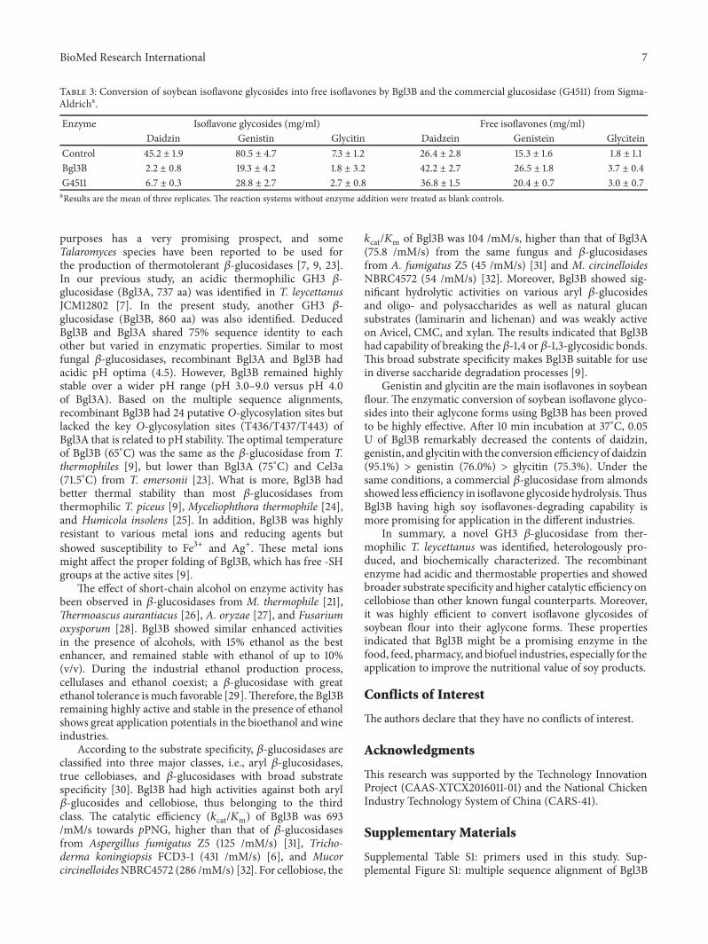
3.6. Hydrolysis of Soybean Isoflavone Glycosides. Some 𝛽-glucosidases have capability of degrading soybean isoflavoneglycosides [15]. HPLC analysis indicated that the soybeanflour extract contained 133.0 mg/ml of isoflavone glucosides(daidzin, genistin, and glycitin) and 43.5 mg/ml of isoflavoneaglycones (daidzein, genistein, and glycitein) (Table 3).Whentreating the soybean flour extract with Bgl3B at 37∘C for 10min, the amounts of the isoflavone glucosides were reduced
to 23.3 mg/ml, while the amounts of isoflavone aglyconeswere increased to 72.4 mg/ml. Under the same conditions,the commercial 𝛽-glucosidase G4511 converted the samesoybean isoflavones into 38.2 mg/ml isoflavone glycosidesand 60.2 mg/ml free aglycones. It indicated that Bgl3B hadgreater soybean isoflavone glycosides-degrading ability thanthe commercial 𝛽-glucosidase from almond.
4. Discussion
The genus Talaromyces that is widely used in the productionof lignocellulose-degrading enzymes with industrial
BioMed Research International 7
Table 3: Conversion of soybean isoflavone glycosides into free isoflavones by Bgl3B and the commercial glucosidase (G4511) from Sigma-Aldricha.
Enzyme Isoflavone glycosides (mg/ml) Free isoflavones (mg/ml)Daidzin Genistin Glycitin Daidzein Genistein Glycitein
Control 45.2 ± 1.9 80.5 ± 4.7 7.3 ± 1.2 26.4 ± 2.8 15.3 ± 1.6 1.8 ± 1.1Bgl3B 2.2 ± 0.8 19.3 ± 4.2 1.8 ± 3.2 42.2 ± 2.7 26.5 ± 1.8 3.7 ± 0.4G4511 6.7 ± 0.3 28.8 ± 2.7 2.7 ± 0.8 36.8 ± 1.5 20.4 ± 0.7 3.0 ± 0.7aResults are the mean of three replicates. The reaction systems without enzyme addition were treated as blank controls.
purposes has a very promising prospect, and someTalaromyces species have been reported to be used forthe production of thermotolerant 𝛽-glucosidases [7, 9, 23].In our previous study, an acidic thermophilic GH3 𝛽-glucosidase (Bgl3A, 737 aa) was identified in T. leycettanusJCM12802 [7]. In the present study, another GH3 𝛽-glucosidase (Bgl3B, 860 aa) was also identified. DeducedBgl3B and Bgl3A shared 75% sequence identity to eachother but varied in enzymatic properties. Similar to mostfungal 𝛽-glucosidases, recombinant Bgl3A and Bgl3B hadacidic pH optima (4.5). However, Bgl3B remained highlystable over a wider pH range (pH 3.0–9.0 versus pH 4.0of Bgl3A). Based on the multiple sequence alignments,recombinant Bgl3B had 24 putative O-glycosylation sites butlacked the key O-glycosylation sites (T436/T437/T443) ofBgl3A that is related to pH stability. The optimal temperatureof Bgl3B (65∘C) was the same as the 𝛽-glucosidase from T.thermophiles [9], but lower than Bgl3A (75∘C) and Cel3a(71.5∘C) from T. emersonii [23]. What is more, Bgl3B hadbetter thermal stability than most 𝛽-glucosidases fromthermophilic T. piceus [9], Myceliophthora thermophile [24],and Humicola insolens [25]. In addition, Bgl3B was highlyresistant to various metal ions and reducing agents butshowed susceptibility to Fe3+ and Ag+. These metal ionsmight affect the proper folding of Bgl3B, which has free -SHgroups at the active sites [9].
The effect of short-chain alcohol on enzyme activity hasbeen observed in 𝛽-glucosidases from M. thermophile [21],Thermoascus aurantiacus [26], A. oryzae [27], and Fusariumoxysporum [28]. Bgl3B showed similar enhanced activitiesin the presence of alcohols, with 15% ethanol as the bestenhancer, and remained stable with ethanol of up to 10%(v/v). During the industrial ethanol production process,cellulases and ethanol coexist; a 𝛽-glucosidase with greatethanol tolerance ismuch favorable [29].Therefore, the Bgl3Bremaining highly active and stable in the presence of ethanolshows great application potentials in the bioethanol and wineindustries.
According to the substrate specificity, 𝛽-glucosidases areclassified into three major classes, i.e., aryl 𝛽-glucosidases,true cellobiases, and 𝛽-glucosidases with broad substratespecificity [30]. Bgl3B had high activities against both aryl𝛽-glucosides and cellobiose, thus belonging to the thirdclass. The catalytic efficiency (𝑘cat/𝐾m) of Bgl3B was 693/mM/s towards pPNG, higher than that of 𝛽-glucosidasesfrom Aspergillus fumigatus Z5 (125 /mM/s) [31], Tricho-derma koningiopsis FCD3-1 (431 /mM/s) [6], and MucorcircinelloidesNBRC4572 (286 /mM/s) [32]. For cellobiose, the
𝑘cat/𝐾m of Bgl3B was 104 /mM/s, higher than that of Bgl3A(75.8 /mM/s) from the same fungus and 𝛽-glucosidasesfrom A. fumigatus Z5 (45 /mM/s) [31] and M. circinelloidesNBRC4572 (54 /mM/s) [32]. Moreover, Bgl3B showed sig-nificant hydrolytic activities on various aryl 𝛽-glucosidesand oligo- and polysaccharides as well as natural glucansubstrates (laminarin and lichenan) and was weakly activeon Avicel, CMC, and xylan. The results indicated that Bgl3Bhad capability of breaking the 𝛽-1,4 or 𝛽-1,3-glycosidic bonds.This broad substrate specificity makes Bgl3B suitable for usein diverse saccharide degradation processes [9].
Genistin and glycitin are the main isoflavones in soybeanflour. The enzymatic conversion of soybean isoflavone glyco-sides into their aglycone forms using Bgl3B has been provedto be highly effective. After 10 min incubation at 37∘C, 0.05U of Bgl3B remarkably decreased the contents of daidzin,genistin, and glycitinwith the conversion efficiency of daidzin(95.1%) > genistin (76.0%) > glycitin (75.3%). Under thesame conditions, a commercial 𝛽-glucosidase from almondsshowed less efficiency in isoflavone glycoside hydrolysis.ThusBgl3B having high soy isoflavones-degrading capability ismore promising for application in the different industries.
In summary, a novel GH3 𝛽-glucosidase from ther-mophilic T. leycettanus was identified, heterologously pro-duced, and biochemically characterized. The recombinantenzyme had acidic and thermostable properties and showedbroader substrate specificity and higher catalytic efficiency oncellobiose than other known fungal counterparts. Moreover,it was highly efficient to convert isoflavone glycosides ofsoybean flour into their aglycone forms. These propertiesindicated that Bgl3B might be a promising enzyme in thefood, feed, pharmacy, and biofuel industries, especially for theapplication to improve the nutritional value of soy products.
Conflicts of Interest
The authors declare that they have no conflicts of interest.
Acknowledgments
This research was supported by the Technology InnovationProject (CAAS-XTCX2016011-01) and the National ChickenIndustry Technology System of China (CARS-41).
Supplementary Materials
Supplemental Table S1: primers used in this study. Sup-plemental Figure S1: multiple sequence alignment of Bgl3B

8 BioMed Research International
with the GH3 𝛽-glucosidases from Thermoascus auran-tiacus (BGLI), A. fumigatus (5FJI), and T. leycettanusJCM12802 (Bgl3A). Identical and similar amino acids areindicated by black and gray boxes, respectively. The cat-alytic residues, nucleophile Asp261 and acid/base residueGlu490, are indicated. Supplemental Figure S2: the homologymodeled structure of Bgl3B with the crystal structure of 𝛽-glucosidase from A. fumigatus (5FJI) as the template. TheN-terminal catalytic domain (TIM-barrel), the C-terminalcatalytic domain (𝛼/𝛽 sandwich), and the fibronectin typeIII domain (Fn3) are indicated in green, yellow, and blue,respectively. Supplemental Figure S3: HPLC chromatogramsof the soybean isoflavones. The purple line indicates thesoybean flour extract; the red line indicates the soybean flourextract hydrolyzed by 0.05 U of Bgl3B at 37∘C for 10 min;the black line indicates the soybean flour extract hydrolyzedby 0.05 U of commercial 𝛽-glucosidase G4511 from Sigma-Aldrich at 37∘C for 10 min. (Supplementary Materials)
References
[1] K. Keegstra, K. W. Talmadge, W. D. Bauer, and P. Albersheim,“The Structure of Plant Cell Walls: III. A Model of the Walls ofSuspension-cultured Sycamore Cells Based on the Interconnec-tions of the Macromolecular Components,” Plant Physiology,vol. 51, no. 1, pp. 188–197, 1973.
[2] R. Gilad, L. Rabinovich, S. Yaron et al., “Cell, a noncellulosomalfamily 9 enzyme from Clostridium thermocellum, is a proces-sive endoglucanase that degrades crystalline cellulose,” Journalof Bacteriology, vol. 185, no. 2, pp. 391–398, 2003.
[3] S. I. Pathan, L. Zifcakova, M. T. Ceccherini, O. L. Pantani, T.Vetrovsky, and P. Baldrian, “Seasonal variation and distributionof total and active microbial community of 𝛽-glucosidaseencoding genes in coniferous forest soil,” Soil Biology & Bio-chemistry, vol. 105, pp. 71–80, 2017.
[4] S. J. Horn, G. Vaaje-Kolstad, B. Westereng, and V. G. H. Eijsink,“Novel enzymes for the degradation of cellulose,” Biotechnologyfor Biofuels, vol. 5, 2012.
[5] C. E. Nelson, A. Rogowski, C. Morland, J. A. Wilhide, H.J. Gilbert, and J. G. Gardner, “Systems analysis in Cellvibriojaponicus resolves predicted redundancy of 𝛽-glucosidasesand determines essential physiological functions,” MolecularMicrobiology, vol. 104, no. 2, pp. 294–305, 2017.
[6] Z. Zhang, J.-L. Liu, J.-Y. Lan, C.-J. Duan, Q.-S. Ma, and J.-X. Feng, “Predominance of Trichoderma and Penicillium incellulolytic aerobic filamentous fungi from subtropical andtropical forests inChina, and their use in finding highly efficient𝛽-glucosidase,” Biotechnology for Biofuels, vol. 7, no. 1, 2014.
[7] W. Xia, X. Xu, L. Qian et al., “Engineering a highly activethermophilic 𝛽-glucosidase to enhance its pH stability andsaccharification performance,” Biotechnology for Biofuels, vol. 9,no. 1, 2016.
[8] M. Gudmundsson, H. Hansson, S. Karkehabadi et al., “Struc-tural and functional studies of the glycoside hydrolase family3 𝛽-glucosidase Cel3A from the moderately thermophilic fun-gus Rasamsonia emersonii:,” Acta Crystallographica Section D:Structural Biology, vol. 72, no. 7, pp. 860–870, 2016.
[9] H. Mallek-Fakhfakh and H. Belghith, “Physicochemical prop-erties of thermotolerant extracellular 𝛽-glucosidase from
Talaromyces thermophilus and enzymatic synthesis of cello-oligosaccharides,” Carbohydrate Research, vol. 419, pp. 41–50,2016.
[10] S. Romo-Sanchez, M. Arevalo-Villena, E. Garcıa Romero, H.L. Ramirez, and A. Briones Perez, “Immobilization of 𝛽-Glucosidase and Its Application for Enhancement of AromaPrecursors in Muscat Wine,” Food and Bioprocess Technology,vol. 7, no. 5, pp. 1381–1392, 2014.
[11] K.D. R. Setchell, “Absorption andmetabolismof soy isoflavones- From food to dietary supplements and adults to infants,”Journal of Nutrition, vol. 130, no. 3, 2000.
[12] C. Panintrarux, S. Adachi, Y. Araki, Y. Kimura, and R. Matsuno,“Equilibrium yield of n-alkyl-𝛽-d-glucoside through conden-sation of glucose and n-alcohol by 𝛽-glucosidase in a biphasicsystem,” Enzyme andMicrobial Technology, vol. 17, no. 1, pp. 32–40, 1995.
[13] Y. Li, Y. Wang, T. Tu et al., “Two acidic, thermophilic GH28polygalacturonases from Talaromyces leycettanus JCM 12802with application potentials for grape juice clarification,” FoodChemistry, vol. 237, pp. 997–1003, 2017.
[14] S. You, T. Tu, L. Zhang et al., “Improvement of the thermosta-bility and catalytic efficiency of a highly active 𝛽-glucanasefromTalaromyces leycettanus JCM12802 by optimizing residualcharge-charge interactions,” Biotechnology for Biofuels, vol. 9,no. 1, 2016.
[15] X. Yang, R. Ma, P. Shi et al., “Molecular Characterization of aHighly-Active Thermophilic 𝛽-Glucosidase from Neosartoryafischeri P1 and Its Application in the Hydrolysis of SoybeanIsoflavoneGlycosides,” PLoS ONE, vol. 9, no. 9, p. e106785, 2014.
[16] R. M. Peralta, M. K. Kadowaki, H. F. Terenzi, and J. A. Jorge,“A highly thermostable 𝛽-glucosidase activity from the ther-mophilic fungus Humicola grisea var. thermoidea: Purificationand biochemical characterization,” FEMS Microbiology Letters,vol. 146, no. 2, pp. 291–295, 1997.
[17] R. Miksch and G. Wiedemann, “Blood sugar determinationwith the GOD-POD-ABTS method using uranylacetate fordeproteinization,”Zeitschrift fur medizinische Labortechnik, vol.14, no. 1, pp. 27–33, 1973.
[18] G. L. Miller, “Use of dinitrosalicylic acid reagent for determina-tion of reducing sugar,” Analytical Chemistry, vol. 31, no. 3, pp.426–428, 1959.
[19] K. Zhao, P. Xue, andG.Gu, “Study on determination of reducingsugar content using 3, 5-dinitrosalicylic acid method,” FoodScience, vol. 8, p. 128, 2008.
[20] S.-J. Ding, W. Ge, and J. A. Buswell, “Secretion, purificationand characterisation of a recombinant Volvariella volvaceaendoglucanase expressed in the yeast Pichia pastoris,” Enzymeand Microbial Technology, vol. 31, no. 5, pp. 621–626, 2002.
[21] A. Karnaouri, E. Topakas, T. Paschos, I. Taouki, and P.Christakopoulos, “Cloning, expression and characterization ofan ethanol tolerant GH3 𝛽-glucosidase from Myceliophthorathermophila,” PeerJ, vol. 2013, no. 1, 2013.
[22] C. A. Uchima, G. Tokuda, H. Watanabe, K. Kitamoto, andM. Arioka, “Heterologous expression in pichia pastoris andcharacterization of an endogenous thermostable and high-glucose-tolerant 𝛽-glucosidase from the termite Nasutitermestakasagoensis,” Applied and Environmental Microbiology, vol.78, no. 12, pp. 4288–4293, 2012.
[23] P. Murray, N. Aro, C. Collins et al., “Expression in Tricho-derma reesei and characterisation of a thermostable family3 𝛽-glucosidase from the moderately thermophilic fungus

BioMed Research International 9
Talaromyces emersonii,” Protein Expression and Purification,vol. 38, no. 2, pp. 248–257, 2004.
[24] J. Zhao, C. Guo, C. Tian, and Y. Ma, “Heterologous Expressionand Characterization of a GH3 𝛽-Glucosidase from Ther-mophilic Fungi Myceliophthora thermophila in Pichia pas-toris,” Applied Biochemistry and Biotechnology, vol. 177, no. 2,pp. 511–527, 2015.
[25] X. Xu, J. Li, P. Shi et al., “The use of T-DNA insertionalmutagen-esis to improve cellulase production by the thermophilic fungusHumicola insolens Y1,” Scientific Reports, vol. 6, 2016.
[26] N. J. Parry, D. E. Beever, E. Owen, I. Vandenberghe, J. VanBeeumen, and M. K. Bhat, “Biochemical characterization andmechanism of action of a thermostable 𝛽-glucosidase purifiedfrom Thermoascus aurantiacus,” Biochemical Journal, vol. 353,no. 1, pp. 117–127, 2001.
[27] C. Riou, J. Salmon, M. Vallier, Z. Gunata, and P. Barre,“Purification, characterization, and substrate specificity of anovel highly glucose-tolerant 𝛽-glucosidase from Aspergillusoryzae,” Applied and Environmental Microbiology, vol. 64, no.10, pp. 3607–3614, 1998.
[28] P. Christakopoulos, P. W. Goodenough, D. Kekos, B. J. Macris,M. Claeyssens, and M. K. Bhat, “Purification and character-isation of an extracellular 𝛽-glucosidase with transglycosyla-tion and exo-glucosidase activities from Fusarium oxysporum,”European Journal of Biochemistry, vol. 224, no. 2, pp. 379–385,1994.
[29] H. Jørgensen, J. Vibe-Pedersen, J. Larsen, and C. Felby, “Lique-faction of lignocellulose at high-solids concentrations,” Biotech-nology and Bioengineering, vol. 96, no. 5, pp. 862–870, 2007.
[30] P. Tiwari, B. N.Misra, andN. S. Sangwan, “𝛽 -glucosidases fromthe fungus Trichoderma: An efficient cellulase machinery inbiotechnological applications,” BioMed Research International,vol. 2013, 2013.
[31] D. Liu, R. Zhang, X. Yang et al., “Characterization of athermostable 𝛽-glucosidase from Aspergillus fumigatus Z5, andits functional expression in Pichia pastoris X33,”Microbial CellFactories, vol. 11, article 25, 2012.
[32] Y. Kato, T. Nomura, S. Ogita, M. Takano, and K. Hoshino, “Twonew 𝛽-glucosidases from ethanol-fermenting fungus Mucorcircinelloides NBRC 4572: Enzyme purification, functionalcharacterization, and molecular cloning of the gene,” AppliedMicrobiology andBiotechnology, vol. 97, no. 23, pp. 10045–10056,2013.

Hindawiwww.hindawi.com
International Journal of
Volume 2018
Zoology
Hindawiwww.hindawi.com Volume 2018
Anatomy Research International
PeptidesInternational Journal of
Hindawiwww.hindawi.com Volume 2018
Hindawiwww.hindawi.com Volume 2018
Journal of Parasitology Research
GenomicsInternational Journal of
Hindawiwww.hindawi.com Volume 2018
Hindawi Publishing Corporation http://www.hindawi.com Volume 2013Hindawiwww.hindawi.com
The Scientific World Journal
Volume 2018
Hindawiwww.hindawi.com Volume 2018
BioinformaticsAdvances in
Marine BiologyJournal of
Hindawiwww.hindawi.com Volume 2018
Hindawiwww.hindawi.com Volume 2018
Neuroscience Journal
Hindawiwww.hindawi.com Volume 2018
BioMed Research International
Cell BiologyInternational Journal of
Hindawiwww.hindawi.com Volume 2018
Hindawiwww.hindawi.com Volume 2018
Biochemistry Research International
ArchaeaHindawiwww.hindawi.com Volume 2018
Hindawiwww.hindawi.com Volume 2018
Genetics Research International
Hindawiwww.hindawi.com Volume 2018
Advances in
Virolog y Stem Cells International
Hindawiwww.hindawi.com Volume 2018
Hindawiwww.hindawi.com Volume 2018
Enzyme Research
Hindawiwww.hindawi.com Volume 2018
International Journal of
MicrobiologyHindawiwww.hindawi.com
Nucleic AcidsJournal of
Volume 2018
Submit your manuscripts atwww.hindawi.com