Embed Size (px)
Citation preview
GH3-mediated Auxin Homeostasis Links Growth Regulationwith Stress Adaptation Response in Arabidopsis*□S
Received for publication, November 13, 2006, and in revised form, January 8, 2007 Published, JBC Papers in Press, February 1, 2007, DOI 10.1074/jbc.M610524200
Jung-Eun Park‡, Ju-Young Park§, Youn-Sung Kim‡, Paul E. Staswick¶, Jin Jeon�, Ju Yun‡, Sun-Young Kim‡,Jungmook Kim�, Yong-Hwan Lee§, and Chung-Mo Park‡1
From the ‡Molecular Signaling Laboratory, Department of Chemistry, Seoul National University, Seoul 151-742, Korea, the §Schoolof Agricultural Biotechnology and Center for Agricultural Biomaterials, Seoul National University, Seoul 151-742, Korea, the¶Department of Agronomy and Horticulture, University of Nebraska, Lincoln, Nebraska 68583, and the �Department of PlantBiotechnology, Agricultural Plant Stress Research Center and Biotechnology Research Institute, Chonnam National University,Gwangju 500-757, Korea
Plants constantly monitor environmental fluctuations tooptimize their growth and metabolism. One example isadaptive growth occurring in response to biotic and abioticstresses. Here, we demonstrate that GH3-mediated auxinhomeostasis is an essential constituent of the complex net-work of auxin actions that regulates stress adaptationresponses in Arabidopsis. Endogenous auxin pool is regu-lated, at least in part, through negative feedback by a group ofauxin-inducible GH3 genes encoding auxin-conjugatingenzymes. An Arabidopsis mutant, wes1-D, in which a GH3gene WES1 is activated by nearby insertion of the 35Senhancer, exhibited auxin-deficient traits, including reducedgrowth and altered leaf shape. Interestingly, WES1 is alsoinduced by various stress conditions as well as by salicylicacid and abscisic acid. Accordingly, wes1-D was resistant toboth biotic and abiotic stresses, and stress-responsive genes,such as pathogenesis-related genes and CBF genes, were up-regulated in this mutant. In contrast, a T-DNA insertionalmutant showed reduced stress resistance. We therefore pro-pose that GH3-mediated growth suppression directs reallo-cation of metabolic resources to resistance establishment andrepresents the fitness costs of induced resistance.
Plants are frequently exposed to diverse biotic and abioticstresses throughout their life cycle. A number of stress signalinggenes have been identified through molecular genetic studiesand genome-wide screens, and a range of stress signaling path-ways have been elucidated. However, the underlying molecularand biochemical mechanisms remain poorly understood in
most cases, primarily due to the complexity of interactionsbetween multiple signaling pathways (1–3).Diverse growth hormones are involved in mediating stress
responses and signalings. Salicylic acid (SA)2 is a primarygrowthhormone thatmediates plant disease resistance and abi-otic stress responses. Several key components of SA signalinghave been functionally characterized, among which the NPR1(nonexpressor of PR1) and TGA transcription factors are thebest understood (4, 5). Furthermore, the SA signaling pathwaysare interconnected with other growth hormone signaling path-ways (1, 2, 6). The interplays of SA with jasmonic acid (JA) andethylene have been extensively studied (6, 7). Biotic stress aswell as abiotic stress also promotes abscisic acid (ABA) biosyn-thesis, further suggesting that growth hormones interact withone another in stress signaling and stress tolerance. Notably,recent reports have shown that light plays a crucial role in bothbiotic and abiotic stress responses. SA biosynthesis, PR-1(pathogenesis-related 1) induction by SA, and hypersensitiveresponse all require functional phytochromes (8, 9). Similarly,light is also essential for cold-induced CBF expression (10).
Commonly observed symptoms of infected or stressed plantsinclude growth retardation and reduced metabolism, whichmay be caused by the reallocation of metabolic resourcesbetween different physiological pathways in order to maximizeplant survival under stress conditions (11–13). Auxin has beenimplicated in such adaptive responses, a notion that is furthersupported by genome-wide analysis. It has been reported thatthe endogenous indole-3-acetic acid (IAA) level substantiallyincreases upon pathogen infections (14), and expression ofsome auxin-regulated genes is altered in infected plants (15). Inaddition, most pathovars of Pseudomonas syringae and otherbacterial pathogens produce a large amount of IAA (16). Fur-thermore, it has been demonstrated that enhanced antibacte-rial resistance is intimately related to repression of auxin signal-ing (17), strongly suggesting that auxin modulates plantresponses to pathogen infections.Onemechanism by which plants coordinate auxin-mediated
processes is to maintain the endogenous pool of auxins at anappropriate level. This can be achieved by regulating auxin
* This work was supported by the BK 21, Biogreen 21 (20050301034456)and National Research Laboratory programs, a grant from the PlantSignaling Network Research Center, Korea Science and EngineeringFoundation Grant R02-2003-000-10001-0, Korea Research FoundationGrant 2005-070-C00129, and Plant Diversity Research Center of 21stCentury Frontier Research Program Grant PF0330404-02 (to J. K.). Thecosts of publication of this article were defrayed in part by the paymentof page charges. This article must therefore be hereby marked “adver-tisement” in accordance with 18 U.S.C. Section 1734 solely to indicatethis fact.
□S The on-line version of this article (available at http://www.jbc.org) containssupplemental Figs. S1–S5.
1 To whom correspondence should be addressed. Tel.: 82-2-880-6640; Fax:82-2-889-1568; E-mail: [email protected].
2 The abbreviations used are: SA, salicylic acid; JA, jasmonic acid; ABA, abscisicacid; IAA, indole-3-acetic acid; RT, reverse transcription; SAG, SA glucoside;MS, Murashige and Skoog.
THE JOURNAL OF BIOLOGICAL CHEMISTRY VOL. 282, NO. 13, pp. 10036 –10046, March 30, 2007© 2007 by The American Society for Biochemistry and Molecular Biology, Inc. Printed in the U.S.A.
10036 JOURNAL OF BIOLOGICAL CHEMISTRY VOLUME 282 • NUMBER 13 • MARCH 30, 2007
by guest on June 2, 2019http://w
ww
.jbc.org/D
ownloaded from

biosynthesis and distribution among different organs and byconjugate formation with sugars, peptides, and amino acids(18–20). Although the physiological importance of conju-gates in auxin homeostasis is not yet fully understood, it isgenerally accepted that conjugate formation plays a criticalrole in auxin action.Auxin functions to an extent by regulating a group of primary
responsive genes: Aux/IAA genes, GH3 genes, and small aux-in-up RNAs (SAUR) (19).Members of theAux/IAA gene familyhave been studied in light regulation of auxin responses (18, 20).Although none of the SAUR genes are as yet functionally char-acterized, the SAUR proteins have been shown to bind to cal-cium/calmodulin (21), suggesting a role for the calcium ion inauxin signaling. Several GH3 genes have been studied usingmutants with altered gene expression. GH3-overexpressingmutants, such as dfl1-D (22), dfl2-D (23), and ydk1-D (24), dis-play reduced growth and altered light responses, suggesting arole for these GH3 proteins in light-auxin interactions.Recently, the GH3 proteins have been biochemically character-ized. JAR1 has been found to activate JA by conjugating it toisoleucine (25). Several other GH3 genes encode enzymes thatconjugate amino acids to IAA (26, 27). It is notable that theGH3.5 enzyme targets both IAA and SA, suggesting a role forthis enzyme in IAA-SA interactions (26). The rapid inductionof the GH3 genes by auxin may help to regulate auxin homeo-stasis by conjugating excess auxins to amino acids. However,their functional mechanisms may not be so simple, becauseonly a fraction of the GH3 genes are induced by auxin (19).Here, we demonstrate that both biotic and abiotic stress
adaptation responses are mediated, at least in part, by auxinhomeostasis governed through negative feedback regulation bya group of GH3 enzymes. Overproduction of an IAA-conjugat-ing GH3 enzyme causes growth reduction, which is intimatelylinked to enhanced stress resistance. Stress-induced turnoverof endogenous auxins by the GH3 enzymes is likely to be anadaptive strategy that coordinately modulates growth rate andstress resistance.
EXPERIMENTAL PROCEDURES
Plant Materials and Growth Conditions—All Arabidopsisthaliana lines used were in the Columbia ecotype (Col-0)unless otherwise specified. Plants were grown in a controlledculture room at 23 °C with a relative humidity of 60% underlong day conditions (16 h light and 8 h dark) with white lightillumination (110 �mol photons m�2 s�1) provided byfluorescent FLR40D/A tubes (Osram, Seoul, Korea).Isolation of wes1-D and wes1—Ecotype Col-0 was trans-
formed with the activation tagging vector pSKI015, as previ-ously described (28). To select herbicide-resistant transfor-mants, T0 seeds were collected, sown in soil, and sprayed twicea week with a 1:1,000 dilution (in water) of Finale (AgrEvo,Montvale, NJ) containing 5.78% Basta. The herbicide-resistantseeds were further selected through 2–3more generations, andhomozygotic seeds were obtained. A dwarfed mutant (wes1-D)was chosen for further analysis in this work. wes1 was isolatedfrom a pool of T-DNA insertion lines (SALK 151766.46.55X,Arabidopsis Biological Resource Center; Ohio State University).
The single T-DNA insertion event in wes1-D was verified bygenomic Southern blot hybridization using the 35S enhancersequence as a probe, followed by analysis of segregation ratios.The sequences flanking the insertion site were determined by athermal asymmetric interlaced PCR (29).Analysis of Transcript Levels—Transcript levels were ana-
lyzed either by Northern blot hybridization or by reverse tran-scription (RT)-PCR-based Southern blot hybridization or byquantitative real time RT-PCR. Total RNA was isolated fromaerial parts of 2-week-old plants, unless otherwise specified, orplant organs using the RNeasy Plant Mini Kit (Qiagen, Valen-cia, CA). For Northern blot hybridization, �25–30 �g of totalRNA samples were separated using 1.2% denaturing formalde-hyde-agarose gel electrophoresis and hybridized to probeslabeled with [32P]dCTP.
Semiquantitative RT-PCR was employed to measure thetranscript levels. Total RNA samples were pretreated exten-sively with RNase-free DNase I to remove any contaminatinggenomic DNAs. The first strand cDNA was synthesized from1–2 �g of total RNA in a 20-�l reaction volume using Super-script II reverse transcriptase (Invitrogen). RT-PCR was per-formed for 15–30 cycles, depending on the linear range of PCRamplification for each gene, using PfuTurbo polymerase (Strat-agene, La Jolla, CA). Each cycle was performed at 94 °C for 0.5min, 60 °C for 0.5 min, and 72 °C for 4 min with a final cycle at72 °C for 10min to finish polymerization.Whenever necessary,positive and negative control genes were included to assure thereaction conditions.For RT-PCR-based Southern blot hybridization, PCR prod-
ucts were electrophoresed on 1% agarose gel and transferred toHybond-N� nylon membrane (Amersham Biosciences). Themembrane was hybridized with gene-specific probes labeledwith [32P]dCTP using the Megaprime DNA labeling system(Amersham Biosciences).Real time RT-PCR was carried out in 96-well blocks with an
Applied Biosystems 7500 real time PCR system (Foster City,CA) using the SYBRGreen Imaster mix in a 25-�l volume. Theprimers were designed using the Primer Express Softwareinstalled into the system. The two-step thermal cycling profileused was 15 s at 94 °C and 1 min at 65 °C. A tubulin gene wasincluded as an internal control to normalize variations in cDNAamounts used. The reactions were performed in triplicate foreach run. The comparative ��CTmethod was used to evaluatethe relative quantities of each amplified product in the samples.The threshold cycle (CT) was automatically determined foreach reaction by the system set with default parameters.Growth Hormone Treatments—Plants were grown on
Murashige and Skoog (MS)-agar plates containing 0.5� MSsalts with vitamins, 0.5 g liter�1 4-morpholineethanesulfonicacid, and 0.8% Phytagar (Duchefa, Haarlem, The Netherlands)supplementedwith IAA, JA, ABA (10�Meach), SA (0.1 or 1mM
unless otherwise specified), 1-amino cyclopropane-1-carbox-ylic acid (ACC) (50 �M), or 1-N-naphthylphthalamic acid (20�M).Whole plant parts of 2-week-old plants were used for totalRNA extraction.To accurately assess the effects of IAA on primary root
growth and lateral root development,MS-agar plates were sup-
Auxin in Stress Adaptation
MARCH 30, 2007 • VOLUME 282 • NUMBER 13 JOURNAL OF BIOLOGICAL CHEMISTRY 10037
by guest on June 2, 2019http://w
ww
.jbc.org/D
ownloaded from

plemented with 0–10 �M IAA. Measurements from 30 rootswere averaged and analyzed statistically using Student’s t test.Pathogen Inoculations, Trypan Blue Staining, and Determi-
nation of Bacterial Growth—Cells of P. syringae pv. tomatostrain DC3000 were grown for 2 days at 28 °C in King’s Bmedium supplemented with 50 �g liter�1 rifampicin. Bacterialcells were harvested by centrifugation and resuspended at 107colony-forming units ml�1 in 10 mM MgCl2 and 250 ppmTween 80. Plants were inoculated by spraying with bacterialsuspensions until the leaf surface was fully covered with finedroplets. Inoculated plants were incubated for 16 h at 25 °C and100% relative humidity and then transferred to a growth cham-ber set at 25 °C and 80% relative humidity and grown underlong day conditions. In order to visualize microscopic lesions,leaves were stained with trypan blue as previously described(30).To determine bacterial growth in plants, whole leaves of
4-week-old plants were infiltrated with bacterial suspensions(105 colony-forming units/ml) of P. syringae pv. tomato strainDC3000 by applying a vacuum for 2 min followed by suddenrelease using a vacuumpump (modelDOA-P104-AA; SangwooS & T, Seoul, Korea). Leaf discs of 0.5 cm in diameter wereharvested, using a hole puncher, from the leaves at the indicatedtime intervals after infiltration, and bacterial growth was deter-mined as previously described (31). The assays were repeated inthree replicates, each consisting of five leaf discs from differentinoculated leaves. Statistical significance of the measurementswas determined using Student’s t test.Abiotic Stress Tolerance Assays—Drought stress tolerance
assays were carried out essentially as previously described (32)with minor modifications. Plants were germinated and grownfor 10 days in pots filled with themixture of perlite and vermic-ulite at a ratio of 1:1 under normal growth conditions (23 °C,60% relative humidity). Each pot (9 cm in diameter) contained�40 g of themixture in dryweight. Drought stress was imposedby withholding water for 2 weeks, and survival rates were cal-culated for each plant group 5 days after rewatering. Threeindependent measurements, each consisting of 50 plants, wereaveraged and statistically analyzed using Student’s t test.For high salinity tolerance assays, plants were germinated
and grown vertically on MS-agar plates for 10 days and thentransferred to MS liquid culture containing 200 mM NaCl andincubated under constant light for 3 days. To quantify damagefrom high salinity, leaf samples were soaked overnight in 95%EtOH at 65 °C, and the chlorophyll contents were measuredspectrophotometrically (chlorophyll a/b (�g/ml) � OD664.2 �5.24 � OD648.6 � 22.24). Eleven measurements were averaged,and statistical significance was determined using a student ttest.For freezing tolerance assays, �30 plants grown for 3 weeks
on MS-agar plates were incubated for 1.5 h at �7 °C andallowed to recover at 23 °C for 1 week. Three independentmeasurements of survival rates were averaged and statisticallyanalyzed using Student’s t test.
Fully expanded rosette leaves from 2-week-old plants grownin soil were used to measure freezing-induced electrolyte leak-age as previously described (33). One excised leaflet was placedin a 5-ml test tube containing 100�l of deionizedwater, and the
tube was placed in a circulator bath (PolyScience, Cheshire,UK) set at 0 °C. After equilibration for 1 h, ice chips were addedto each tube. The temperature of the bath was programmed todecrease to �9 °C with a 1 °C decrement every 30 min. Thetubes were removed from the bath when the designated tem-perature was reached and placed immediately on ice to allowgradual thawing overnight. The leaflets were then carefullytransferred to a 50-ml tube containing 20ml of deionized waterand shaken overnight at 40 rpm. The conductivity of the solu-tionwasmeasured. The tubes with the leaflets were autoclaved,and the conductivity of the solution was measured again. Thedegree of electrolyte leakage was calculated as the percentageconductivity before autoclaving over that following autoclav-ing. Ten replicates were performed for each temperature treat-ment and statistically analyzed using Student’s t test.Growth Hormone Measurements—Endogenous auxins were
extracted from aerial tissues of 4-week-old plants as previouslydescribed (27), except that the final purification step by highpressure liquid chromatography was eliminated. Internalstandards were [13C6]IAA (Cambridge Isotope Laboratories,Andover,MA); [13C6]IAA-Asp and [13C6]IAA-Glu (kindly pro-vided by J. Cohen); and [13C6]IAA-Ala and [13C6]IAA-Leu (syn-thesized as previously described (34)). Samples were derivat-ized using 2,3,4,5,6-pentafluorobenzyl bromide (35), purifiedby silica gel SPE chromatography, and dissolved in chloroform.Analyses were carried out on a Finnigan Trace gas chromato-graph (inlet 280 °C), which was coupled to a DSQ mass spec-trometer using negative chemical ionization. The reagent gaswas methane with a source temperature of 200 °C, and theinstrument was operated in the SIM mode. The ions collectedfor endogenous auxins werem/z 174, 245, 271, 285, and 287 forIAA, IAA-Ala, IAA-Asp, IAA-Glu, and IAA-Leu, respectively.Quantitative data were obtained from integrated peak areas aspreviously described (36).Extraction and quantification of endogenous SA and SA glu-
coside (SAG) were performed using the leaf tissues from2-week-old plants as previously described (37). ABA contentswere determined using the Phytodetek ABA Immunoassay Kitaccording to the procedure provided by themanufacturer (Ide-tek, Sunnyvale, CA). Three independent measurements wereaveraged for both SA and ABA. Statistical significance wasdetermined using Student’s t test.
RESULTS
WES1 Activation Causes Growth Retardation in wes1-D—From a screening of a pool of activation-tagged Arabidopsismutants, we isolated a severely dwarfedmutant,wes1-D, whichexhibited reduced growth and small plant organs. The geneticlocus was designated WES1 (for WESO 1, meaning a dwarfedstature in Korean. wes1-D additionally exhibited small curledleaves, reduced apical dominance, and slightly early flowering(Fig. 1A and supplemental Fig. 1, A and B).Notably, the phenotypic alterations observed in wes1-D are
quite similar to those of axr2-1, an auxin-resistant mutant witha mutation in domain II of IAA7 (supplemental Fig. 1A), and tothose of dfl1-D, dfl2-D, and ydk1-D, in which auxin-inducibleGH3 genes are overexpressed (22–24). In contrast, a T-DNAinsertional mutant, wes1, was phenotypically indistinguishable
Auxin in Stress Adaptation
10038 JOURNAL OF BIOLOGICAL CHEMISTRY VOLUME 282 • NUMBER 13 • MARCH 30, 2007
by guest on June 2, 2019http://w
ww
.jbc.org/D
ownloaded from
from wild type plants but with slightly larger leaves (�6–7%increase; Fig. 1A and supplemental Fig. 1B). It does not seemthat the absence of discernible phenotypic changes in wes1 issimply due to functional redundancy among the GH3 proteins.It may be also attributable to the stress inducibility of theWES1gene, since the wes1 mutant differentially responded to stressconditions (see below). These observations suggested that thewes1-D phenotype might be related to auxin action. However,unlike axr2-1, which exhibits short hypocotyls in both light anddarkness, the dwarfed phenotype of wes1-D occurred only in
the light (data not shown), suggesting a different molecularmechanism underlying the wes1-D phenotype.
Thermal asymmetric interlaced PCR was employed to mapthe T-DNA insertion site in the wes1-D genome (29). Togetherwith analysis of segregation ratios (3:1 � wes1-D/wild type �basta-resistant plants/basta-susceptible plants) and genomicSouthern blot hybridization (supplemental Fig. 2A), it revealedthat a single copy of the 35S enhancer was inserted into the
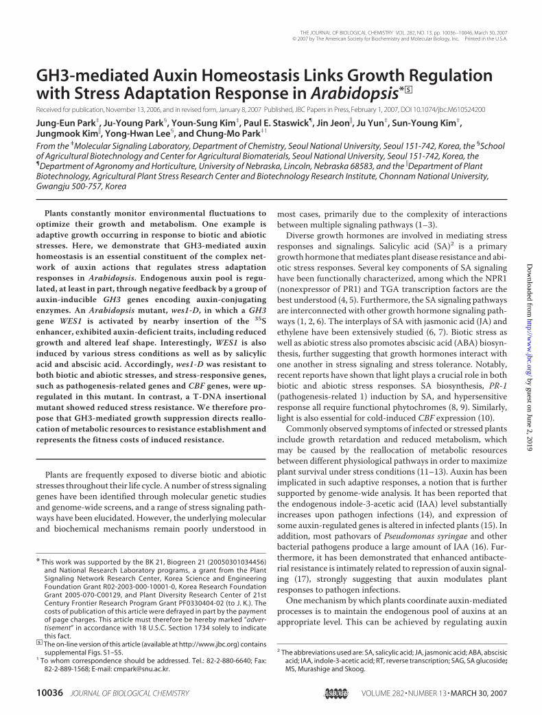
FIGURE 1. wes1-D phenotype and WES1 expression patterns. A, dwarfedgrowth in wes1-D. WES1 was overexpressed under the control of the CaMV 35Spromoter in Arabidopsis (35S::WES1) to confirm the wes1-D phenotype. AT-DNA insertional mutant, wes1, was also included for comparison. B, map-ping of T-DNA insertion site in the wes1-D genome. The T-DNA insertion site isindicated by an arrowhead. wes1 has a single T-DNA insertion in the secondexon of WES1 (At4g27260) (arrow). C, WES1 activation in wes1-D. D, effects ofIAA and ABA on WES1 expression. E, effects of other growth hormones onWES1 expression. F, organ-specific expression of WES1. Total RNA sampleswere extracted separately from the cauline leaves (CL), flowers (FL), shootapex (SA), rosette leaves (RL), roots (RT), and stems (ST). Three-week-old plantswere used for dissection of plant organs except for flowers. In C and F, WES1expression was examined by RT-PCR-based Southern blot hybridization, anda tubulin gene (Tub) was included as a loading control. In D and E, whole plantparts of 2-week-old plants were used for total RNA extraction, and quantita-tive real time RT-PCR was employed for measurements of WES1 expression.Triplicate runs were averaged, and statistical significance was determinedusing Student’s t test. Bars, S.E.
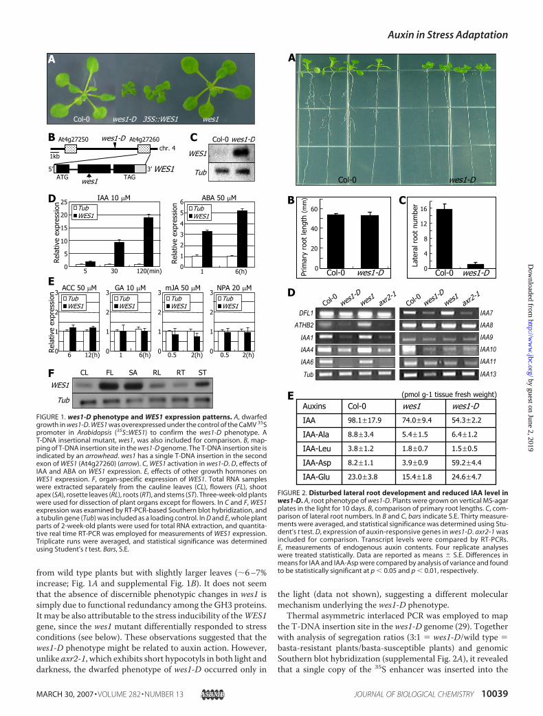
FIGURE 2. Disturbed lateral root development and reduced IAA level inwes1-D. A, root phenotype of wes1-D. Plants were grown on vertical MS-agarplates in the light for 10 days. B, comparison of primary root lengths. C, com-parison of lateral root numbers. In B and C, bars indicate S.E. Thirty measure-ments were averaged, and statistical significance was determined using Stu-dent’s t test. D, expression of auxin-responsive genes in wes1-D. axr2-1 wasincluded for comparison. Transcript levels were compared by RT-PCRs.E, measurements of endogenous auxin contents. Four replicate analyseswere treated statistically. Data are reported as means � S.E. Differences inmeans for IAA and IAA-Asp were compared by analysis of variance and foundto be statistically significant at p � 0.05 and p � 0.01, respectively.
Auxin in Stress Adaptation
MARCH 30, 2007 • VOLUME 282 • NUMBER 13 JOURNAL OF BIOLOGICAL CHEMISTRY 10039
by guest on June 2, 2019http://w
ww
.jbc.org/D
ownloaded from

region between At4g27250 and At4g27260. RT-PCR-basedSouthern blot hybridization indicated that, whereas the level ofAt4g27260 transcript is extremely low in wild type plants, it ispresent at a very high level inwes1-D (Fig. 1C and supplementalFig. 2B), suggesting that overexpression of the locus may be themolecular cause for the wes1-D phenotype. Sequence analysisrevealed that WES1 is identical to AtGH3/GH3.5, the expres-sion profile of which has been studied using a promoter trapline (38). In contrast, the level of At4g27250 transcript wasunaltered in wes1-D, and transgenic plants overexpressing thelocus did not exhibit any phenotypic alterations (data notshown).To confirm that overexpression of WES1 is associated with
the wes1-D phenotype,WES1 was expressed under the controlof the CaMV 35S promoter in Arabidopsis. Twenty-eight of the36 transgenic lines (35S::WES1) obtained displayed a phenotypeessentially identical to that of wes1-D (Fig. 1A). However,growth retardation and leaf curling were slightly less severe inthe transgenic lines compared withwes1-D. This may be due toa gene dosage effect, since the level of WES1 transcript waslower in the 35S::WES1 transgenic plants than in wes1-D (sup-plemental Fig. 2B). The phenotypic changes in the remainingeight transgenic lines were only marginal, and the WES1 tran-script level was only slightly induced (data not shown), furthersuggesting that overexpression of WES1 underlies the wes1-Dphenotype.WES1 encodes a polypeptide of 612 residues (supplemental
Fig. 2C) that exhibits a high sequence identity with soybeanGH3 (39) andArabidopsisGH3homologues (supplemental Fig.3). Three sequence motifs (I–III) that are found in all of theknown GH3 proteins (26) were also identified at the equivalentpositions in WES1 (supplemental Fig. 2C).WES1 Is Induced by Auxin and ABA—SeveralGH3 genes are
known to be induced by auxin (22, 38).We examined the effectsof auxin and other growth hormones onWES1 expression. IAArapidly induced WES1 within 5 min after treatment (Fig. 1D).Interestingly, ABA also activatedWES1 expression to a consid-erable level (Fig. 1D), suggesting a role forWES1 in ABA-regu-lated stress response. Since WES1 is auxin-inducible, an auxintransport inhibitor, 1-N-naphthylphthalamic acid, was alsoincluded in the assays. It did not confer any discernible effectson the WES1 expression (Fig. 1E). This may be because thebasal level of theWES1 expression is extremely low under nor-mal growth conditions.Other growth hormones, such as 1-amino cyclopropane-1-
carboxylic acid, GA, and methyl JA, did not confer any signifi-cant effects onWES1 expression (Fig. 1E), althoughWES1 wasslightly induced by 1-amino cyclopropane-1-carboxylic acidbut slightly repressed by methyl JA. The slight repression ofWES1 by methyl JA may be related to the antagonistic roles ofJA and SA in pathogen resistance (see below) (Fig. 4B).High levels of WES1 transcript were detected in the shoot
apex and flower tissues, and moderate levels were found in thestems (Fig. 1F). However, it was relatively lower in the leavesand roots. WES1 was expressed to a relatively high level inyoung seedlings and gradually decreased throughout the lifecycle (supplemental Fig. 2D), although the overall transcriptlevels were low and could be detected only by RT-PCR.
Endogenous IAALevel Is Reduced inwes1-D—Examination ofwes1-D roots grown on vertical MS-agar plates indicated thatprimary root growth was not discernibly affected (Fig. 2, A andB). However, the number of lateral roots was dramaticallyreduced inwes1-D (Fig. 2C), suggesting again a role forWES1 inauxin action. Consistent with this, a subset of Aux/IAA geneswas suppressed inwes1-D and axr2-1 but induced to detectablelevels in wes1 (Fig. 2D). ATHB2, which is involved in light reg-ulation of auxin responses (e.g. shade avoidance response) (40),was also affected in a similar manner. These observations sug-gested that WES1 might play a role in auxin-mediated growthregulation.It has been shown thatGH3 enzymes inactivate IAAby form-
ing conjugates with amino acids (27). Accordingly, the IAA-Asp level is elevated in dfl1-D (22, 27).We thus anticipated thatthe endogenous level of auxins would be altered in wes1-D andwes1. To examine this, we measured endogenous auxin levelsusing 4-week-old leaves. The IAA-Asp content was 7.2 timeshigher in wes1-D than in wild type plants, whereas it wasdecreased by about 50% inwes1 (Fig. 2E), suggesting thatWES1synthesizes IAA-Asp in planta. The amounts of free IAA were
FIGURE 3. Reduced auxin responses of wes1-D roots. Seeds were germi-nated and grown on auxin-free MS-agar plates for 3 days. The young seed-lings were then transferred to MS-agar plates supplemented with varyingconcentrations of IAA and further grown vertically for 7 days before takingphotographs. Two representative seedlings grown in the presence of differ-ent IAA concentrations were displayed.
Auxin in Stress Adaptation
10040 JOURNAL OF BIOLOGICAL CHEMISTRY VOLUME 282 • NUMBER 13 • MARCH 30, 2007
by guest on June 2, 2019http://w
ww
.jbc.org/D
ownloaded from

98.1 and 54.3 pmol g�1 tissue fresh weight in wild type plantsand wes1-D, respectively. In wes1-D, the 45% decrease in freeIAA was found to be statistically significant in four replicateanalyses (p � 0.05). In contrast, the difference in meansbetween wes1 and wild type plants was not statistically signifi-cant, which might be due to functional redundancy among theGH3 enzymes. These data confirm the correlation between thereduction of active auxins and the wes1-D phenotype. It is alsoenvisioned that the auxin-inducibleWES1 gene may representa negative feedback loop thatmaintains the endogenous level ofactive auxins at an appropriate level (see “Discussion”).Primary root growth is inhibited by excess amounts of exog-
enous auxins and is frequently used as an assay system for auxinactivity. We examined the sensitivity of wes1-D roots to exoge-nous auxin. Primary root growth in wes1-D was slightly butreproducibly resistant to exogenous IAA (supplemental Fig. 4),and this effect was most evident at an IAA concentration rangeof 10�8 to 10�6 M (p � 0.05). In contrast, wes1 was more sensi-tive to IAA (p � 0.05). This observation is consistent with theproposed biochemical role of WES1 in auxin metabolism andthe reduction of free IAA in wes1-D (Fig. 2E).Auxin feeding experiments strongly supported the above
notion.When seedlings were grown onMS-agar plates supple-mented with IAA, lateral root formation in wes1-D seedlingswas efficiently recovered (Fig. 3). However, whereas lateral rootformation was evidently enhanced by IAA at a concentration as
low as 10 nM in wild type seedlings, the IAA effects becameevident at IAA concentrations of higher than 1 �M in wes1-Dseedlings. In contrast, the IAA effects were higher inwes1 seed-lings compared with those in wild type seedlings. Altogether,these observations indicated that the endogenous auxin level islower in wes1-D.WES1 Is Induced by SA and Pathogen Infections—WES1 is
unique among the GH3 enzymes characterized so far in that itpossesses dual substrate specificities. It is active on both IAAand SA (26). It was thus postulated thatWES1might play a rolein SA-mediated stress responses.We observed that PR-1 was significantly induced in wes1-D
as well as in axr2-1 but was unchanged in wes1 (Fig. 4A). Inaddition,WES1was induced by SA, being observable at 2 h andreaching a maximum of more than 5-fold increase by 4 h afterSA application (Fig. 4B). These results indicated that WES1 isclosely associated with SA-dependent responses.We next investigated the expression of defense genes in
wes1-D following P. syringae infection. In wes1-D, the PR-1transcript level was high even before infection. Following infec-tion, it was further induced. The PR-1 transcript level returnedto the basal level 2 days after infection in wild type plants. Incontrast, the transcript level remained elevated inwes1-D, evenafter 3 days (Fig. 4C). PDF1.2 also displayed a distinct expres-sion pattern in wes1-D. It was rapidly induced to a high levelafter infection, which wasmaintained at least for 3 days, in wildtype plants. Although PDF1.2 expression in wes1-D initiallyappeared to be identical to that inwild type plants (Fig. 4C, timepoint 0), with rapid induction of a high level of transcriptionfollowing infection, the transcript level gradually decreasedafter the peak expression was attained in wes1-D. This may berelated to the primary role of PDF1.2 in the JA signaling path-way (41). In wild type plants, the expression of IAA1 graduallydecreased following infection, an inversely proportionalresponse to WES1 induction following SA application. This isprobably due to a reduction in active auxins caused by the SA-mediated WES1 induction. These results strongly support arole forWES1 in SA-dependent stress responses.wes1-D Is Resistant to Pathogen Infections—In infected
plants, increased synthesis of endogenous SA or its conjugatestriggers the induction of PR genes and the development of dis-ease resistance symptoms. The level ofPR-1 transcript is high inwes1-D, andWES1 is activated by SA and pathogen infections.Therefore, we hypothesized that wes1-D might be resistant topathogen infections.When plants were infected with P. syringae, disease symp-
toms were much less severe on the leaves of wes1-D (Fig. 5A).Thewes1-D leaves exhibited greatly reducedwilting and necro-sis, a result clearly demonstrated by trypan blue staining (Fig.5B). Interestingly, enhanced pathogen resistance was alsoobserved in axr2-1 but not observed in wes1. To quantitativelyexamine the enhanced resistance in wes1-D, plant leaves wereinfected with P. syringae, and bacterial growth was determined.Bacterial population was greatly reduced in wes1-D but slightlyincreased inwes1 (Fig. 5C). These results are consistentwith theactivation of PR-1 in wes1-D and the induction ofWES1 by SAand pathogen infections. Notably, bacterial growth was also
FIGURE 4. WES1 induction by SA and pathogen infection. A, elevated PR-1expression in wes1-D. PR-1 transcript levels were examined by RT-PCR-basedSouthern blot hybridization. B, SA effects on WES1 expression. Plants weregrown in the presence of 0.1 or 1 mM SA, and total RNA samples wereextracted from plants harvested at the time intervals indicated. The WES1transcript levels were measured by semiquantitative RT-PCR (left panel) andby quantitative real time RT-PCR (right panel). Triplicate runs were averagedfor each reaction, and statistical significance was determined using Student’st test. Bars indicate S.E. C, expression of PR genes in wes1-D after P. syringaeinfection. Plant materials were harvested at 1–3 days following infection. dpi,days post-infection.
Auxin in Stress Adaptation
MARCH 30, 2007 • VOLUME 282 • NUMBER 13 JOURNAL OF BIOLOGICAL CHEMISTRY 10041
by guest on June 2, 2019http://w
ww
.jbc.org/D
ownloaded from

reduced significantly in axr2-1, further supporting the role ofauxin in pathogen resistance.WES1 is induced by SA but uninfluenced by JA (Fig. 1D),
suggesting that WES1 function is not directly related to JA-mediated signaling but is specific to SA-associated responses.In JA-dependent responses, other GH3 enzymes, such as JAR1
that conjugates amino acids specifically to JA (25, 26), may reg-ulate auxin metabolism, similar to the role of WES1 in SA-me-diated response.Auxin Regulates PR-1 Expression—PR-1 is induced by at least
two SA-mediated signaling pathways, depending uponwhetherNPR1 is required or not (2).TGA genes play a central role in theNRP1-dependent pathway. Several genes, including AtWhy1(42) and SSI (suppressor of SA insensitivity) genes (41), areinvolved exclusively in theNPR1-independent pathway. There-fore, a question arises as to how WES1 regulates PR-1expression.To answer the question, expression patterns of several SA-
regulated genes were examined in wes1-D.GST6 is an immedi-ate early gene that is induced by SA and is frequently used as amarker for endogenous SA content.GST6 expressionwas unal-tered in wes1-D and wes1 (Fig. 6A). Expression ofNPR1, TGAs,andAtWhy1 also remained unchanged. These results suggestedthat induction of PR-1 is caused by aWES1-mediated pathwayindependent of SA biosynthesis in wes1-D.Direct measurements of endogenous SA levels further sup-
ported the notion. The level of free SA was elevated in wes1-D
FIGURE 5. Enhanced resistance of wes1-D to pathogen infection. A, plantsinfected with P. syringae. Plants were photographed 7 days after infection.B, infected leaves (top panel) and trypan blue staining (bottom panel). Rep-resentative leaves of infected plants were photographed 4 days after infec-tion. Bars, 5 mm. C, determination of bacterial growth. Three measurementswere averaged, and statistical significance was determined using Student’s ttest. The bars indicate S.E. The axr2-1 mutant was also included in the meas-urements for comparison.
FIGURE 6. Measurements of endogenous SA contents and suppression ofPR-1 by auxins. A, expression of SA-responsive genes in wes1-D and wes1.B, endogenous contents of SA and SAG in wes1-D and wes1. C, PR-1 expres-sion in the wes1-D � nahG plants. D, auxin effects on SA-mediated PR-1induction. Plants were pretreated with 0.1 mM SA for 1 h and subsequentlytreated with IAA (10 �M), 2,4-dichloro-phenoxy acetic acid (1 �M), and/orSA for 6 h. The PR-1 transcript levels were expressed relative to that inlane � (bottom). Five RT-PCR runs were averaged for individual hormonetreatments.
Auxin in Stress Adaptation
10042 JOURNAL OF BIOLOGICAL CHEMISTRY VOLUME 282 • NUMBER 13 • MARCH 30, 2007
by guest on June 2, 2019http://w
ww
.jbc.org/D
ownloaded from

(�80 ng/g of tissue, fresh weight) compared with that in wildtype plants (�20 ng/g of tissue, fresh weight) (Fig. 6B). How-ever, the SA level in wes1-D is far below the level that is usuallyobserved in infected plants (37). In addition,we also observed inour assay system thatwhenmeasured 2 days after infectionwithP. syringae, the level of free SAwas in a range of 0.8–1.4�g/g oftissue, fresh weight (data not shown). These observations sug-gest that the elevated SA level may not significantly contributeto the PR-1 induction in wes1-D. The level of SAG was alsoelevated in wes1-D (�1 �g/g of tissue, fresh weight). However,the elevation does not seem to be physiologically significant,since the SAG level reached 15–20�g/g of tissue freshweight ininfected plants in our assay system (data not shown).To obtainmore direct evidence thatWES1 induction of PR-1
is SA-independent,wes1-Dwas genetically crossed to the nahGtransgenic plants. The resultant wes1-D � nahG cross exhib-ited thewes1-Dphenotype (data not shown), and the level of thePR-1 transcript was lower than that inwes1-D but much higherthan that in wild type plants (Fig. 6C), unequivocally demon-strating that the PR-1 induction is caused by a SA-independentpathway in wes1-D. It is also envisioned that full induction ofPR-1 requires both the SA-dependent and SA-independent sig-nals. The moderately altered levels of SA and SAG in wes1-Dmight be attributable to aWES1-modulated negative feedbackregulation, similar to that proposed for auxin.To further explore the molecular mechanism underlying the
induction of PR-1 in wes1-D, wild type plants were pretreatedwith SA for 1 h and subsequently treated with SA, IAA, or2,4-dichloro-phenoxyacetic acid in different combinations.When plants were treated with individual growth hormones,expression of PR-1 was strongly induced by SA but discerniblyrepressed by 2,4-dichloro-phenoxyacetic acid and IAA (Fig.6D).When treatedwith SA and auxin together, the level ofPR-1transcript was much lower than that in plants treated with SAalone. These results indicate that auxin counters the inductiveeffects of SA on PR-1 expression. The negative effect of auxinson SA-regulated PR-1 induction might be related to the factthat numerous plant pathogens, including strains of P. syringae,produce large amounts of IAAupon infection of plants (16) (see“Discussion”).WES1 Mediates Abiotic Stress Responses—Growth retarda-
tion is one of the symptoms commonly observed in infected andstressed plants. We observed thatWES1 is strongly induced bySA and pathogen infections. Accordingly, the wes1-D mutantthat is featured by reduced growth exhibits enhanced resistanceto pathogen infections. Notably,WES1 is also induced by ABA(Fig. 1D). We thus hypothesized that WES1 would also beinvolved in ABA-regulated abiotic stress responses.To test this, wild type plants were grown under various abi-
otic stress conditions. WES1 was indeed up-regulated by cold(4 °C), drought, and heat (37 °C) treatments (Fig. 7A). Consist-ent with this, wes1-D exhibited greatly enhanced resistance todrought (Fig. 7B).wes1-Dwas also resistant to freezing temper-ature, as evidenced by measurements of survival rate and elec-trolyte leakage after freezing treatment for 1.5 h at �7 °C (Fig.7C). Spectrophotometrical measurements of chlorophyll con-tents demonstrated that wes1-D is also resistant to a high saltenvironment (600 mM) (Fig. 7D). Furthermore, wes1-D exhib-
STEEI 120
FIGURE 7. Enhanced resistance of wes1-D to abiotic stresses. A, effects ofabiotic stresses on WES1 expression. Plants were subjected to cold (4 °C), heat(37 °C), or drought treatments for 2 h, and WES1 expression was analyzed byRT-PCR-based Southern blot hybridization. B, enhanced resistance of wes1-Dto drought stress. C, freezing tolerance of wes1-D. Survival rates were ana-lyzed using plants treated at �7 °C for 1.5 h (top). Ten measurements wereaveraged for each temperature treatment (bottom). Statistical significancewas determined using Student’s t test. D, salt tolerance of wes1-D. Elevenmeasurements of chlorophyll contents were averaged. E, expression of abi-otic stress genes in wes1-D.
Auxin in Stress Adaptation
MARCH 30, 2007 • VOLUME 282 • NUMBER 13 JOURNAL OF BIOLOGICAL CHEMISTRY 10043
by guest on June 2, 2019http://w
ww
.jbc.org/D
ownloaded from
ited enhanced resistance to high temperature (37 °C) (supple-mental Fig. 5). The values were within a range of statisticalsignificance in wes1-D (p � 0.01). In contrast, wes1 was moresensitive to stress conditions. Although the differences inmeans betweenwes1 andwild type plants were not as evident asthose observed betweenwild type andwes1-D plants, themeas-urements were within a range of statistical significance (p �0.05). It is thus obvious that the WES1 control of endogenousauxin level is a commonmechanism underlying both biotic andabiotic stress responses.To explore how WES1 is interrelated with abiotic stress
responses, expression of genes involved in abiotic stressresponses, including CBF (C-repeat/dehydration-responsiveelement-binding factor) genes and RD (response to dehydra-tion) genes, was examined inwes1-D. Since their transcript lev-els were very low in nonstressed plants, RT-PCR-based South-ern blot hybridization was employed. The transcript levels ofCBF1, -2, and -3, RD26, and RD29A were high in wes1-D (Fig.7E), indicating that WES1 function may be linked to diverseabiotic stress signaling pathways.The next question was whether the abiotic stress-related
genes are regulated directly by altered auxin level or indirectlyby altered growth rate in wes1-D. To address this question,
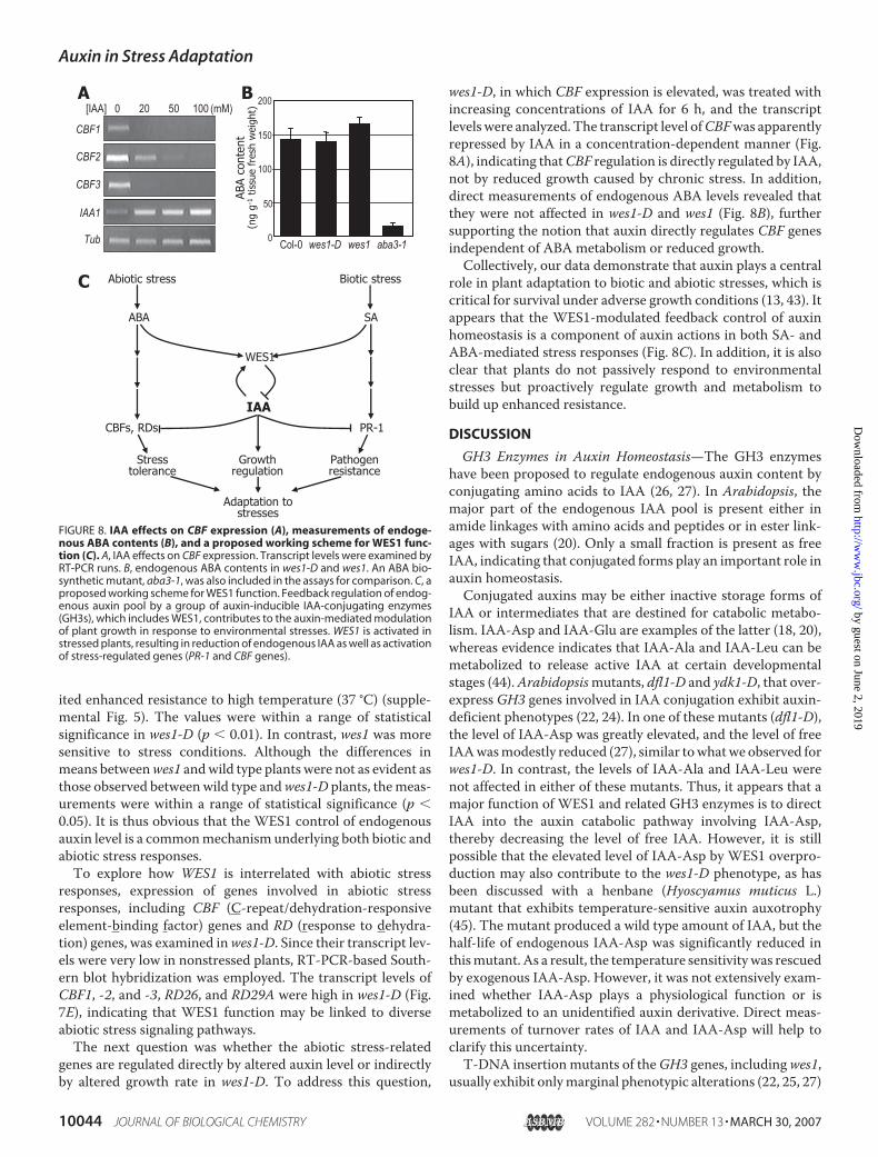
wes1-D, in which CBF expression is elevated, was treated withincreasing concentrations of IAA for 6 h, and the transcriptlevelswere analyzed. The transcript level ofCBFwas apparentlyrepressed by IAA in a concentration-dependent manner (Fig.8A), indicating thatCBF regulation is directly regulated by IAA,not by reduced growth caused by chronic stress. In addition,direct measurements of endogenous ABA levels revealed thatthey were not affected in wes1-D and wes1 (Fig. 8B), furthersupporting the notion that auxin directly regulates CBF genesindependent of ABA metabolism or reduced growth.Collectively, our data demonstrate that auxin plays a central
role in plant adaptation to biotic and abiotic stresses, which iscritical for survival under adverse growth conditions (13, 43). Itappears that the WES1-modulated feedback control of auxinhomeostasis is a component of auxin actions in both SA- andABA-mediated stress responses (Fig. 8C). In addition, it is alsoclear that plants do not passively respond to environmentalstresses but proactively regulate growth and metabolism tobuild up enhanced resistance.
DISCUSSION
GH3 Enzymes in Auxin Homeostasis—The GH3 enzymeshave been proposed to regulate endogenous auxin content byconjugating amino acids to IAA (26, 27). In Arabidopsis, themajor part of the endogenous IAA pool is present either inamide linkages with amino acids and peptides or in ester link-ages with sugars (20). Only a small fraction is present as freeIAA, indicating that conjugated forms play an important role inauxin homeostasis.Conjugated auxins may be either inactive storage forms of
IAA or intermediates that are destined for catabolic metabo-lism. IAA-Asp and IAA-Glu are examples of the latter (18, 20),whereas evidence indicates that IAA-Ala and IAA-Leu can bemetabolized to release active IAA at certain developmentalstages (44).Arabidopsismutants, dfl1-D and ydk1-D, that over-express GH3 genes involved in IAA conjugation exhibit auxin-deficient phenotypes (22, 24). In one of these mutants (dfl1-D),the level of IAA-Asp was greatly elevated, and the level of freeIAAwasmodestly reduced (27), similar towhatwe observed forwes1-D. In contrast, the levels of IAA-Ala and IAA-Leu werenot affected in either of these mutants. Thus, it appears that amajor function of WES1 and related GH3 enzymes is to directIAA into the auxin catabolic pathway involving IAA-Asp,thereby decreasing the level of free IAA. However, it is stillpossible that the elevated level of IAA-Asp by WES1 overpro-duction may also contribute to the wes1-D phenotype, as hasbeen discussed with a henbane (Hyoscyamus muticus L.)mutant that exhibits temperature-sensitive auxin auxotrophy(45). The mutant produced a wild type amount of IAA, but thehalf-life of endogenous IAA-Asp was significantly reduced inthismutant. As a result, the temperature sensitivitywas rescuedby exogenous IAA-Asp. However, it was not extensively exam-ined whether IAA-Asp plays a physiological function or ismetabolized to an unidentified auxin derivative. Direct meas-urements of turnover rates of IAA and IAA-Asp will help toclarify this uncertainty.T-DNA insertion mutants of theGH3 genes, includingwes1,
usually exhibit onlymarginal phenotypic alterations (22, 25, 27)
FIGURE 8. IAA effects on CBF expression (A), measurements of endoge-nous ABA contents (B), and a proposed working scheme for WES1 func-tion (C). A, IAA effects on CBF expression. Transcript levels were examined byRT-PCR runs. B, endogenous ABA contents in wes1-D and wes1. An ABA bio-synthetic mutant, aba3-1, was also included in the assays for comparison. C, aproposed working scheme for WES1 function. Feedback regulation of endog-enous auxin pool by a group of auxin-inducible IAA-conjugating enzymes(GH3s), which includes WES1, contributes to the auxin-mediated modulationof plant growth in response to environmental stresses. WES1 is activated instressed plants, resulting in reduction of endogenous IAA as well as activationof stress-regulated genes (PR-1 and CBF genes).
Auxin in Stress Adaptation
10044 JOURNAL OF BIOLOGICAL CHEMISTRY VOLUME 282 • NUMBER 13 • MARCH 30, 2007
by guest on June 2, 2019http://w
ww
.jbc.org/D
ownloaded from
(this work). This may be due to functional redundancy amongtheGH3enzymes.Of the 19GH3enzymes inArabidopsis, eightmembers fall into Group II, which have all been shown to beinvolved in IAAmetabolism.Alternatively, theweak phenotypemight be attributable to the expression pattern of the GH3genes. Whereas the WES1 transcript is hardly detectable innonstressed plants, it is strongly induced in stressed plants.This would explain why wes1 is phenotypically similar to wildtype under normal growth conditions yet discerniblymore sus-ceptible to biotic and abiotic stresses.TheGroup II GH3 enzymes, includingWES1 andDFL1, cer-
tainly share a common biochemical activity inactivating IAAbyforming amino acid conjugates. Accordingly, the phenotypes ofthe mutants with accelerated gene expression are similar toeach other, exhibiting dwarfed growth with small, severelycurled leaves, symptoms of reduced endogenous auxin levels.However, the WES1 and DFL1 enzymes are distinct in theirsubstrate specificities (26). Whereas DFL1 adenylates IAA,WES1 adenylates both IAA and SA. In addition, it is also pos-sible that their genes may be differentially regulated. Weobserved that WES1 is induced by SA and ABA as well as bybiotic and abiotic stress conditions. It will be interesting to seehow DFL1 and other GH3 genes are affected by external andinternal signals.Auxin Regulates both Biotic and Abiotic Stress Adaptation
Responses—Transgenic plants carrying disease resistance genesfrequently produce less biomass and fewer siliques, possiblydue to the fitness costs incurred by induced resistance (12, 43).This might explain why most attempts to develop crop plantswith enhanced resistance to fungal and bacterial pathogenshave been unsuccessful to date (12). Costs of induced cold tol-erance have also been demonstrated in transgenic Arabidopsisplants overexpressing CBF genes (46). Several growth hor-mones play essential roles in stress adaptation responses;among these, SA, JA, ethylene, and ABA are the best under-stood (3). Of particular interest is the observation that someSA- or ABA-responsive genes and those induced by patho-gen infections and abiotic stresses are also influenced byauxin (2, 6, 15).Our results indicate that feedback regulation of active auxin
content byWES1 correlates with enhanced resistance to patho-gen infections. This view is not inconsistent with the previousreports. The increased level of endogenous IAA detected ininfected cells upon pathogen infections (14) would be derivedfrom pathogen secretion of IAA (16). It has been proposed thatpathogen-secreted IAA enhances virulence, probably by weak-ening the SA-mediated defense responses in infected plants(47). It is thus likely that induction of WES1 or other relatedGH3 genes by pathogen infections is a host mechanism forregaining auxin homeostasis.WES1 is also induced by various abiotic stresses. Conse-
quently, wes1-D is resistant to abiotic stresses. Furthermore, abroad array of abiotic stress-related genes, including thoseencoding proline metabolic enzymes, are affected in wes1-D,suggesting that auxin plays a role in several abiotic stress sig-naling pathways. Consistent with theWES1 induction by vari-ous growth hormones and diverse stress conditions, scanningof cis-acting regulatory DNA elements within the WES1 pro-
moter region (�2 kbp from the start codon) using a promotersignal scan programPLACE (available on theWorldWideWebat www.dna.affrc.go.jp/PLACE/signalscan.html) revealed thattheWES1 promoter contains numerousDNAelements that arepredicted to be responsible to auxin, ABA, SA, and biotic andabiotic stresses. Examples include multiple copies of ACGTG(drought-inducible, ABRE-like element); TGTCTC (ARF-binding); TGACG (IAA/SA-inducible); TGTGA, AAAGAT,andTTGACC (disease-inducible); CACGTG (ABA-inducible);and TTGAC (SA-inducible), although it remains to be func-tionally examined.We believe that the enhanced resistance observed in wes1-D
is not simply due to indirect effects caused by chronic stress asfrequently observed in dwarfed, stressedmutants but is specificto WES1 function. The WES1 enzyme is biochemically uniqueamong theGH3 enzymes in that it possesses an SA-conjugatingactivity (26). This might represent a negative feedback loop forSA, similar to that described for IAA, and provide an additionalway of auxin-SA interactions. The different time courses forPR-1 andWES1 expression by SA (Fig. 4B) and the altered levelsof endogenous SA and SAG in wes1-D (Fig. 6B) would beexplained by such a feedback loop. In addition, althoughwes1 isphenotypically similar to wild type plants, it is discernibly moresusceptible to pathogen infections and abiotic stresses.Based on the previous observations as well as our own data,
we propose that regulation of auxin homeostasis by WES1 andrelated GH3 enzymes represents a means of modulating auxinactions in stress adaptation response. However, it should benoted that GH3-mediated auxin homeostasis is one of thediverse molecular schemes for auxin actions in this process. Ithas recently been demonstrated that a flagellin-inducedmicro-RNA represses auxin signaling by targeting auxin receptorgenes and makes host plants less susceptible to bacterial infec-tion (17). Their results provide another mechanism for auxinaction during stress adaptation response, in which TIR1-medi-ated auxin signaling is modulated by bacterial infection. Wealso observed that an auxin signalingmutant axr2-1 is resistantto pathogen infections (Fig. 5B). Further works are necessary toelucidate the genetic network of auxin responses and signalingcascades. Mutants in auxin response and signaling as well asthose in SA and ABA signalings will be of great help to answerthe question.
Acknowledgments—We thankDrs. N.-H. Chua andK. Shimamoto forscientific discussions.
REFERENCES1. Gazzarrini, S., and McCourt, P. (2003) Ann. Bot. 91, 605–6122. Katagiri, F. (2004) Curr. Opin. Plant Biol. 7, 506–5113. Kunkel, B. N., and Brooks, D.M. (2002)Curr. Opin. Plant Biol. 5, 325–3314. Dong, X. (2004) Curr. Opin. Plant Biol. 7, 547–5525. Pieterse, C. M., and Van Loon, L. C. (2004) Curr. Opin. Plant Biol. 7,
456–4646. Feys, B. J., and Parker, J. E. (2000) Trends Genet. 16, 449–4557. Reymond, P., and Farmer, E. E. (1998) Curr. Opin. Plant Biol. 1, 404–4118. Genoud, T., Millar, A. J., Nishizawa, N., Kay, S. A., Schafer, E., Nagatani,
A., and Chua, N.-H. (1998) Plant Cell 10, 889–9049. Genoud, T., Buchala, A. J., Chua, N.-H., and Metraux, J.-P. (2002) Plant J.
31, 87–95
Auxin in Stress Adaptation
MARCH 30, 2007 • VOLUME 282 • NUMBER 13 JOURNAL OF BIOLOGICAL CHEMISTRY 10045
by guest on June 2, 2019http://w
ww
.jbc.org/D
ownloaded from
10. Kim, H. J., Kim, Y. K., Park, J. Y., and Kim, J. (2002) Plant J. 29, 693–70411. Bohnert, H. J., Nelson, D. E., and Jensen, R. G. (1995) Plant Cell 7,
1099–111112. Burdon, J. J., and Thrall, P. H. (2003) Genome Biol. 4, 227.1–227.313. Heil, M., and Baldwin, I. T. (2002) Trends Plant Sci. 7, 61–6714. O’Donnell, P. J., Schmelz, E. A.,Moussatche, P., Lund, S. T., Jones, J. B., and
Klee, H. J. (2003) Plant J. 33, 245–25715. Dowd, C., Wilson, I. W., and McFadden, H. (2004) Mol. Plant Microbe
Interact. 17, 654–66716. Glickmann, E., Gardan, L., Jacquet, S., Hussain, S., Elasri, M., Petit, A., and
Dessaux, Y. (1998)Mol. Plant Microbe Interact. 11, 156–16217. Navarro, L., Dunoyer, P., Jay, F., Arnold, B., Dharmasiri, N., Estelle, M.,
Voinnet, O., and Jones, J. D. G. (2006) Science 312, 436–43918. Berleth, T., Krogan, N. T., and Scarpella, E. (2004) Curr. Opin. Plant Biol.
7, 553–56319. Hagen, G., and Guilfoyle, T. (2002) Plant Mol. Biol. 49, 373–38520. Woodward, A. W., and Bartel, B. (2005) Plant Cell Suppl. S165–S18321. Yang, T., and Poovaiah, B. W. (2000) J. Biol. Chem. 275, 3137–314322. Nakazawa, M., Yabe, N., Ichikawa, T., Yamamoto, Y. Y., Yoshizumi, T.,
Hasunuma, K., and Matsui, M. (2001) Plant J. 25, 213–22123. Takase, T.,Nakazawa,M., Ishikawa,A.,Manabe, K., andMatsui,M. (2003)
Plant Cell Physiol. 44, 1071–108024. Takase, T., Nakazawa, M., Ishikawa, A., Kawashima, M., Ichikawa, T.,
Takahashi, N., Shimada,H.,Manabe, K., andMatsui,M. (2004) Plant J. 37,471–483
25. Staswick, P. E., and Tiryaki, I. (2004) Plant Cell 16, 2117–212726. Staswick, P. E., Tiryaki, I., and Rowe, M. L. (2002) Plant Cell 14,
1405–141527. Staswick, P. E., Serban, B., Rowe, M., Tiryaki, I., Maldonado, M. T., Mal-
donado, M. C., and Suza, W. (2005) Plant Cell 17, 616–62728. Weigel, D., Ahn, J. H., Blazquez, M. A., Borevitz, J. O., Christensen, S. K.,
Fankhauser, C., Ferrandiz, C., Kardailsky, I., Malancharuvil, E. J., Neff,M. M., Nguyen, J. T., Sato, S., Wang, Z. Y., Xia, Y., Dixon, R. A., Harrison,M. J., Lamb, C. J., Yanofsky, M. F., and Chory, J. (2000) Plant Physiol. 122,
1003–101329. Liu, Y. G.,Mitsukawa, N., Oosumi, T., andWhittier, R. F. (1995) Plant J. 8,
457–46330. Koch, E., and Slusarenko, A. (1990) Plant Cell 2, 437–44531. Mengiste, T., Chen, X., Salmeron, J. M., and Dietrich, R. A. (2003) Plant
Cell 15, 2551–256532. Sakamoto, H., Maruyama, K., Sakuma, Y., Meshi, T., Iwabuchi, M., Shi-
nozaki, K., and Yamaguchi-Shinozaki, K. (2004) Plant Physiol. 136,2734–2746
33. Ristic, Z., and Ashworth, E. N. (1993) Protoplasma 172, 111–12334. Cohen, J. D. (1981) J. Label Compound. 18, 1393–139635. Epstein, E., and Cohen, J. D. (1981) J. Chromatogr. 209, 413–42036. Cohen, J. D., Baldi, B. G., and Slovin, J. P. (1986) Plant Physiol. 80, 14–1937. Bowling, S. A., Guo, A., Cao, H., Gordon, A. S., Klessig, D. F., and Dong, X.
(1994) Plant Cell 6, 1845–185738. Tanaka, S., Mochizuki, N., and Nagatani, A. (2002) Plant Physiol. 130,
887–89439. Gee, M. A., Hagen, G., and Guilfoyle, T. J. (1991) Plant Cell 3, 419–43040. Steindler, C., Matteucci, A., Sessa, G., Weimar, T., Ohgishi, M., Aoyama,
T., Morelli, G., and Ruberti, I. (1999) Development 26, 4235–424541. Kachroo, P., Shanklin, J., Shah, J., Whittle, E. J., and Klessig, D. F. (2001)
Proc. Natl. Acad. Sci. U. S. A. 98, 9448–945342. Desveaux, D., Subramaniam, R., Despres, C., Mess, J. N., Levesque, C.,
Fobert, P. R., Dangl, J. L., and Brisson, N. (2004) Dev. Cell 6, 229–24043. Hammond-Kosack, K. E., and Parker, J. E. (2003) Curr. Opin. Biotechnol.
14, 177–19344. Rampey, R. A., LeClere, S., Kowalczyk, M., Ljung, K., Sandberg, G., and
Bartel, B. (2004) Plant Physiol. 135, 978–98845. Oetiker, J. H., and Aeschbacher, G. (1997) Plant Physiol. 114, 1385–139546. Jackson, M. W., Stinchcombe, J. R., Korves, T. M., and Schmitt, J. (2004)
Mol. Ecol. 13, 3609–361547. Uppalapati, S. R., Ayoubi, P., Weng, H., Palmer, D. A., Mitchell, R. E.,
Jones, W., and Bender, C. L. (2005) Plant J. 42, 201–217
Auxin in Stress Adaptation
10046 JOURNAL OF BIOLOGICAL CHEMISTRY VOLUME 282 • NUMBER 13 • MARCH 30, 2007
by guest on June 2, 2019http://w
ww
.jbc.org/D
ownloaded from

Sun-Young Kim, Jungmook Kim, Yong-Hwan Lee and Chung-Mo ParkJung-Eun Park, Ju-Young Park, Youn-Sung Kim, Paul E. Staswick, Jin Jeon, Ju Yun,
ArabidopsisAdaptation Response in GH3-mediated Auxin Homeostasis Links Growth Regulation with Stress
doi: 10.1074/jbc.M610524200 originally published online February 1, 20072007, 282:10036-10046.J. Biol. Chem.
10.1074/jbc.M610524200Access the most updated version of this article at doi:
Alerts:
When a correction for this article is posted•
When this article is cited•
to choose from all of JBC's e-mail alertsClick here
Supplemental material:
http://www.jbc.org/content/suppl/2007/02/06/M610524200.DC1
http://www.jbc.org/content/282/13/10036.full.html#ref-list-1
This article cites 42 references, 18 of which can be accessed free at
by guest on June 2, 2019http://w
ww
.jbc.org/D
ownloaded from