Embed Size (px)
Citation preview
Cancer Therapy: Preclinical
A Novel Anti-CD22 Anthracycline-BasedAntibody–DrugConjugate (ADC)ThatOvercomesResistance to Auristatin-Based ADCsShang-Fan Yu1, Bing Zheng1, MaryAnn Go1, Jeff Lau1, Susan Spencer1, Helga Raab1,Robert Soriano1, Suchit Jhunjhunwala1, Robert Cohen1, Michele Caruso2, Paul Polakis1,John Flygare1, and Andrew G. Polson1
Abstract
Purpose:We are interested in identifying mechanisms of resis-tance to the current generation of antibody–drug conjugates(ADC) and developing ADCs that can overcome this resistance.
Experimental Design: Pinatuzumab vedotin (anti-CD22-vc-MMAE) and polatuzumab vedotin (anti-CD79b-vc-MMAE) areADCs that contain the microtubule inhibitor monomethyl aur-istatin E (MMAE) attached to the antibody by the protease-cleavable linker maleimidocaproyl-valine-citrulline-p-amino-benzoyloxycarbonyl (MC-vc-PAB). Early clinical trial data suggestthat these ADCs have promising efficacy for the treatment of non-Hodgkin lymphoma (NHL); however, some patients do notrespond or become resistant to the ADCs. Anthracyclines are veryeffective in NHL, but ADCs containing the anthracycline doxo-rubicin were not clinically efficacious probably due to the lowdrug potency and inadequate linker technology. The anthracy-cline analogue PNU-159682 is thousands of timesmore cytotoxic
than doxorubicin, so we used it to develop a new class of ADCs.We used the same MC-vc-PAB linker and antibody in pinatuzu-mab vedotin but replaced the MMAE with a derivative of PNU-159682 to make anti-CD22-NMS249 and tested it for in vivoefficacy in xenograft tumors resistant to MMAE-based ADCs.
Results:Wederived cell lines from in vivo xenograft tumors thatwere made resistant to anti-CD22-vc-MMAE and anti-CD79b-vc-MMAE.We identified P-gp (ABCB1/MDR1) as themajor driver ofresistance to the vc-MMAE–based conjugates. Anti-CD22-NMS249 was at least as effective as anti-CD22-vc-MMAE inxenograft models of the parental cell lines and maintained itsefficacy in the resistant cell lines.
Conclusions: These studies provide proof of concept foran anthracycline-based ADC that could be used to treat B-cellmalignancies that are resistant to vc-MMAE conjugates. ClinCancer Res; 21(14); 3298–306. �2015 AACR.
IntroductionAnthracyclines are one of the most widely used classes of
chemotherapy. In particular, they are the cornerstone of treatmentfor aggressive non-Hodgkin lymphoma (NHL) and acutemyeloidleukemia. Anthracyclines, such as doxorubicin, that are used insystemic therapy have not been clinically effective when used inthe context of an antibody–drug conjugate (ADC), probablybecause the drugswere not sufficiently potent. The lack of potencymay have been exacerbated by inadequate linker design (1). Thecurrent generation of ADCs that are showing clinical success usesdrugs that are much more potent (2). Because there are no novelanthracycline-based ADCs currently in clinical development andthis class of drugs has proven so effective in the treatment of a
wide variety of cancers, an anthracycline-based ADC seems tobe a promising approach to improve and broaden the utility ofADC technology. For these proof-of-concept studies, we focusedon NHL as an indication because of its responsiveness to anthra-cyclines and promising clinical data with the current generationof ADCs. There are five ADCs in clinical development for thetreatment of B-cell NHL, moxetumomab pasudotox (CAT-8015),pinatuzumab vedotin (DCDT2980S, anti-CD22-MC-vc-PAB-MMAE), polatuzumab vedotin (DCDS4501A, anti-CD79b-MC-vc-PAB-MMAE), SAR3419 (anti-CD19-SPDB-DM4), andSGN-CD19A (anti-CD19-MC-MMAF; ref. 3). Moxetumomabpasudotox is a recombinant immunotoxin composed of theFv fragment of an anti-CD22 monoclonal antibody fused to a38-kDa fragment of Pseudomonas exotoxin A, PE38 (4), and isunder investigation for hairy cell leukemia (HCL) and NHL.Pinatuzumab vedotin and polatuzumab vedotin are anti-CD22 and anti-CD79b ADCs, respectively, with protease-sensi-tive maleimidocaproyl-valine-citrulline-p-aminobenzoyloxycar-bonyl (MC-vc-PAB) linkers attached to the microtubule-disrupt-ing agent monomethyl auristatin E (MMAE; refs. 5, 6). TheseADCs have shown efficacy in heavily pretreated patients in phase Itrials (6, 7) and are currently in phase II testing in combinationwith rituximab. SAR3419 consists of a humanized anti-CD19antibody and a linker (SPDB) cleavable by disulfide reductionattached to the maytansinoid DM4 (a microtubule-disruptingagent) through lysine residues. Phase I trials of SAR3419 in
1Research and Early Development, Genentech Inc., South San Fran-cisco, California. 2Nerviano Medical Sciences, Nerviano MI, Italy.
Note: Supplementary data for this article are available at Clinical CancerResearch Online (http://clincancerres.aacrjournals.org/).
S.-F. Yu and B. Zheng contributed equally to this article.
Corresponding Author: Andrew G. Polson, Genentech Inc., 1 DNA Way, SouthSan Francisco, CA 94080. Phone: 650-225-5134; Fax: 650-225-6240; E-mail:[email protected]
doi: 10.1158/1078-0432.CCR-14-2035
�2015 American Association for Cancer Research.
ClinicalCancerResearch
Clin Cancer Res; 21(14) July 15, 20153298
on June 11, 2018. © 2015 American Association for Cancer Research. clincancerres.aacrjournals.org Downloaded from
Published OnlineFirst April 3, 2015; DOI: 10.1158/1078-0432.CCR-14-2035
relapsed and refractory NHL have also shown clinical activity(8). SGN-CD19A, another ADC targeting CD19 but with astable linker and a less membrane-permeable auristatin, hasstarted early clinical trials in NHL. Despite this progress, somepatients still do not respond to these ADCs, and others respondbut progress while on treatment. In patients with relapsed orrefractory B-cell NHLs, the objective response rates were 41%,53%, and 30% for pinatuzumab vedotin, polatuzumab vedotin,and SAR3419, respectively (6, 7, 9). Understanding this resist-ance and developingmethods to overcome it would help developthe next generation of ADCs for the treatment of NHL and othercancers.
Here, we show proof of concept for an anthracycline-basedADC for the treatment of NHL and demonstrate that our novelADC is effective in xenograft models that have innate oracquired resistance to the microtubule inhibitor–based ADCs.In addition, we identify possible drivers of resistance to thevc-MMAE–based ADCs pinatuzumab vedotin and polatuzu-mab vedotin.
Materials and MethodsAntibodies and ADCs
Pinatuzumab vedotin (DCDT2980S, anti-CD22-MC-vc-PAB-MMAE) and polatuzumab vedotin (DCDS4501A, anti-CD79b-MC-vc-PAB-MMAE) were generated as previously described(5, 10). THIOMAB version of the anti-CD22 and anti-CD79bantibodies was generated as described (11). NMS249 was syn-thesized. Before conjugation of the THIOMAB to NMS249, theblocking cysteine or glutathione that was present on the intro-duced cysteine was removed by reduction with 50-fold molarexcess dithiothreitol (DTT) in Tris buffer overnight. Reducingagent was removed by diafiltration, and the interchain disulfidebonds were reformed by incubating the THIOMAB for 3 hours in15-fold molar excess dhAA (dehydroascorbic acid; Sigma-Aldrich). The maleimide-linked NMS249 was incubated at 3- to4-fold molar excess with the activated THIOMAB for 1 hour at25�C. The antibody conjugate was purified using ion exchangechromatography (HiTrap S GE Healthcare Bio-Sciences) to
remove excess NMS249. The ADC concentration was determinedby BCA protein assay (Thermo ScientificMicro BCA Protein AssayKit). The number of conjugated linker-drug molecules per mAbwas calculated from the integrated UV peaks of the drug toantibody ratio (DAR) species resolved by analytical hydrophobicinteraction chromatography (HIC; TSK butyl-NPR 4.6 mm � 10cm, 2.5 mm; Tosoh Bioscience). The DAR was confirmed byanalyzing the reduced and intact ADC by LC-MS (Agilent 9520ESIQ-TOF, polymeric reversed-phase columnPLRPS, 1000A, 8u).Molecular masses were derived from multiply charged ionsdeconvoluted usingMassHunter software (Agilent Technologies).
Cell linesThe NHL cell lines BJAB.Luc, Granta-519, SuDHL4.Luc, and
WSU-DLCL2 were obtained from the Genentech cell line repos-itory. All cell lines were maintained in RPMI 1640 supplementedwith 10% FBS (Sigma) and 2 mmol/L L-glutamine. Each cell linewas authenticated by short tandem repeat (STR) profiling usingthe Promega PowerPlex 16 System and compared with externalSTR profiles of cell lines to determine cell line ancestry. Inaddition, an SNP fingerprint is generated from the original thawto serve as our internal master fingerprint. The SNP fingerprintingis performed each time a new batch was frozen down. Cell lineswere typically used for several months before thawing a newpassage.
Target expression levels in xenograft tumors and cell linesTo measure the target expression on xenograft tumors, recov-
ered tumors were minced and put through a 30 mm cell strainer(BD Biosciences) to achieve a single-cell suspension. The tumorcells were subsequently prepared by the standard density centri-fugation over lymphocyte separationmedium (MP Biomedicals).The resulting single-cell suspension was stained with anti-humanCD22-APC antibody (BD Biosciences; clone S-HCL-1), anti-human CD79b-PE antibody (Beckman Coulter; clone CB3-1),anti-human CD20-Pacific Blue antibody (Beckerman Coulter;clone B9E9), and 7-amino-actinomycin D (BD Biosciences).Samples were acquired on FACSCalibur or LSR II flow cytometer(BD Biosciences). Data were analyzed using FlowJo (Tree Star),and the mean fluorescent intensity (MFI) was calculated from theCD20þ and 7-amino-actinomycin D–negative population. CD22and CD79b expression on cell lines was measured with the sameset of antibodies.
Target internalizationTo measure the CD22 internalization after antibody binding,
cells were pre-equilibrated at 4�C with 10 mg/mL of same anti-CD22 antibody used in ADC for 30 minutes. Cells were thenwashedoff excess unbound antibodies andput on ice (inPBSwithazide) or incubated at 37�C (in cell culture media) for 1 hour.Afterward, cells were washed and stained with phycoerythrin(PE)-conjugated goat anti-human IgG antibody (Jackson Immu-noResearch) or rat anti-human kappa antibody (BD Biosciences).Samples were acquired on FACSCalibur, and analyzed usingFlowJo. Percentage internalization was calculated as of [geoMFI(ice) – geoMFI(37�C)] / geoMFI(ice) � 100.
Cell viability assayCells were plated in triplicates at 10 to 20 � 103 per well in
96-well plates in RPMI containing 10% FBS overnight beforetreatment with test articles. Each test article was added to
Translational Relevance
Antibody–drug conjugates (ADCs) are a technology forincreasing the safety and efficacy of traditional chemotherapy.There are three ADCs in clinical trials for the treatment of non-Hodgkin lymphoma all of which contain microtubule inhi-bitors as the drug. While promising, clinical results showsome patients do not respond or relapse during treatment.We show that an ADC based on a potent anthracycline canbe effective in xenograft tumors that have innate or acquiredresistance to the auristatin-based ADCs pinatuzumab vedotinand polatuzumab vedotin. Further, we identify P-gp as pos-sible mechanism of resistance to the auristatin-based ADCs.Oneprobable reason anthracycline-basedADCshavenot beensuccessful in the clinic is due to the use of drugs that lack thepotency required for the ADC technology. Our findings pro-vide a step forward in accessing this important class of che-motherapy as an ADC.
An Anti-CD22 Anthracycline-Based Antibody–Drug Conjugate
www.aacrjournals.org Clin Cancer Res; 21(14) July 15, 2015 3299
on June 11, 2018. © 2015 American Association for Cancer Research. clincancerres.aacrjournals.org Downloaded from
Published OnlineFirst April 3, 2015; DOI: 10.1158/1078-0432.CCR-14-2035
experimentalwells bynine-step 3-fold dilution,with controlwellsreceiving medium alone. After 4-day incubation at 37�C, cellviability was measured using the PrestoBlue Cell ViabilityReagent (Life Technologies). The concentration of test articleresulting in the 50% inhibition of cell viability was calculatedfrom a four-parameter nonlinear regression curve fit analysis byPrism (GraphPad Software).
P-gp inhibitor XR9051 (Tocris Bioscience), if used, wasadded 3 hours before treatment with test articles at a maximumconcentration (400 nmol/L) that does not affect the normal cellgrowth.
Microarray and real-time PCRThree biologic replicates were used for the parental cell
lines (BJAB.Luc and WSU-DLCL2) and for the resistant lines(BJAB.Luc-22R1.1, BJAB.Luc-22R1.2, WSU-22R1.1, and WSU-22R1.2). Total RNA was extracted using the RNeasy plus miniKit (QIAGEN). Complementary RNA was synthesized andhybridized to Affymetrix Human Genome U133 Plus 2.0arrays.
To identify genes differentially expressed across all four resis-tant lines, a simplified linear model was used:
Ygs~b0gþ b1gX1sþb2gX2sþ "gs;
where Ygs is the expression value for gene "g" in sample "s"; b0g isthe (Intercept) expression in parental BJAB.Luc line; b1g is theparental line-specific fixed effect; b2g is the treatment-specific fixedeffect; X1s is the indicator variable for parental cell line (0: BJAB.Luc, 1: WSU-DLCL2); X2s is the indicator variable for treatment(0: parental, 1: resistant); and egs is the gene and sample-specificrandom error.
The treatment-specific effect was the effect of interest. Theanalysis was done using the "limma" (10) package from Biocon-ductor. The genes were then ranked by the P value for thetreatment effect.
Real-time RT-PCR was performed on an ABI 7500 according tothe manufacturer's instruction (Applied Biosystems), using thefollowing primer sets. Mean threshold cycle (Ct) values werecalculated to determine the fold differences.
ABCB1(P-gp)-forward GTCCCAGGAGCCCATCCTABCB1(P-gp)-reverse CCCGGCTGTTGTCTCCATAABCB1(P-gp)-probe TGACTGCAGCATTGCTGAGAACATTGC
ABCC1-forward TGGTGCCCGTCAATGCTABCC1-reverse CGATTGTCTTTGCTCTTCATGTGABCC1-probe ACCTGATACGTCTTGGTCTTCATCGCCA
ABCC3-forward TCATCCTGGCGATCTACTTCCTABCC3-reverse GTGGAATCAGCAAGACCATGAAABCC3-probe CCCTCTGTCCTGGCTGGAGTCGC
ABCG2-forward TTGGCTGTCATGGCTTCAGTACABCG2-reverse CCCAAAAATTCATTATGCTGCAAABCG2-probe TCAGCATTCCACGATATGGATTTACGGC
In vivo efficacy xenograft experimentsXenograft experiments were performed as previously described
(10). Briefly, all animal studies were performed in compliancewith NIH guidelines for the care and use of laboratory animalsand were approved by the Institutional Animal Care and UseCommittee (IACUC) atGenentech, Inc. Tumor cells (2�10 7 cells
in 0.2 mL Hank's balanced salt solution) were inoculated sub-cutaneously into the flanks of female CB17 ICR SCID mice(Charles River Lab). When mean tumor size reached the desiredvolume, the mice were divided into groups of 7 to 10 mice withthe same mean tumor size and dosed i.v. via the tail vein withADCs. The results were plotted as mean tumor volume � SEM ofeach group over time. Tumor stasis was defined as tumor volumeunchanged from day 0. Partial regression was defined as tumorshrinkage of >50% but <100% of the initial tumor volumeon day 0. Complete remission was defined as 100% tumorshrinkage (i.e., no detectable tumor) during the study. Tumorgrowth inhibition (TGI) was calculated as percent area underthe tumor volume–time curve (AUC) per day of each treatmentgroup in relation to the vehicle, using the following formula:%TGI ¼ 100 � [1 � (AUCtreatment/day � AUCvehicle/day)]. Toallow for comparison between groups across tumor models,the confidence intervals (CI) for %TGI were determined, andthe 2.5 and 97.5 percentiles of CIs were reported as the lower andupper range of %TGI. The reported CIs were the values for whichthe recalculated values of %TGI would fall in this range 95% ofthe time. Groups with no overlapping CIs are considered signi-ficantly different from each other.
Development of anti-CD22-vc-MMAE–resistant cell linesTwo NHL models BJAB.Luc and WSU-DLCL2 were used to
develop acquired resistance to anti-CD22-vc-MMAE. Tumor cellswere inoculated subcutaneously into the flanks of female CB17SCID mice. When tumors reached 200 to 500 mm3 in size, micereceived an initial intravenous dose of anti-CD22-vc-MMAE todrive partial tumor regression.
Mice were dosed again when tumors regrew from the treat-ment (i.e., tumors grew back to the initial tumor volume at day0). Dose levels were slowly increased each time until it reached10� above the initial dose or MTD in mice. Frequency of dosesadministered varied over time based on the rate of tumorregrowth, but did not exceed 2 doses/week. The range of dosesadministered for BJAB.Luc model was 1.5, 2, 2.5, 3, 3.5, 4, 5, 6,8, 15, and 20 mg/kg, and the range of doses for WSU-DLCL2model was 12, 15, 18, 20, 25, and 30 mg/kg. Dosing wasdiscontinued once a tumor was no longer responding to a seriesof increasing doses. Tumors were then collected and put backin culture to generate resistant cell lines for further characteri-zation. Two resistant cell lines for each tumor model weresuccessfully established and named as BJAB.Luc-22R1.1, BJAB.Luc-22R1.2, WSU-22R1.1, and WSU-22R1.2.
Generation of P-gp overexpressing cell lineP-gp expression vector was a kind gift from Jun Guo, Genen-
tech. BJAB.Luc cells were transfected with either pIRES-hrGFP IIcontrol vector (Agilent Technologies) or the P-gp expressionvector. The transfectants were selected under Geneticin (LifeTechnologies) and sorted with anti-P-gp antibody to acquire thehighest expressing clone as determined by flow cytometry. Forsurface detection of P-gp, cells were incubated for 30 minutes onice with PE-labeled anti–P-gp antibody (eBioscience; cloneUIC2)or a PE-labeled isotype-negative control. Cells were washed withcold PBS and analyzed by standard flow cytometry.
P-gp functional assay via Rhodamine 123Approximately 106 cells were prepared in PBS buffer with 500
ng/mL of Rhodamine 123 (Life Technologies) and incubated at
Yu et al.
Clin Cancer Res; 21(14) July 15, 2015 Clinical Cancer Research3300
on June 11, 2018. © 2015 American Association for Cancer Research. clincancerres.aacrjournals.org Downloaded from
Published OnlineFirst April 3, 2015; DOI: 10.1158/1078-0432.CCR-14-2035
room temperature for 30 minutes. Subsequently, cells werewashed twice with RPMI media and divided into 3 tubes, onetube was placed immediately on ice to serve as the baselineuptake, the second tube was incubated for 2 hours at 37�C, andthe third one was pretreated with 400 nmol/L of P-gp inhibitorXR9051 before incubated at 37�C. Afterwards, test tubes were puton ice to stop the reaction; samples were washed twice with ice-cold PBS, and analyzed by flow cytometry.
ResultsGeneration and characterization of NMS249 ADCs
Anthracyclines are broadly used in the treatment of cancer, andefforts have been made to make anthracycline analogues thatincrease the effectiveness and safety for this class of drug. One ofthose analogues, Nemorubicin [(30-deamino-30-[200(S)-methoxy-400-morpholinyl]doxorubicin; MMDX], was selected for clinicalevaluation because it was not cardiotoxic at doses that wereeffective in multidrug-resistant tumor models (12). PNU-159682 was identified as a metabolic product of nemorubicinthat was 700- to 2,400-fold more potent in in vitro cytotoxicityassays than nemorubicin (13) with picomolar IC50s. Our cellviability assay data were consistent with this observation, and weobserved that PNU-159682 was more potent than MMAE on our
model NHL cell lines in two cases by an order of magnitude ormore (Table 1).
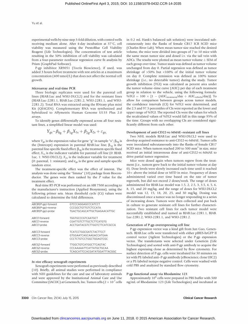
We were interested to see if we could develop an effectiveanthracycline-based ADC using PNU-159682. Given the com-plexity of ADCdevelopment,we selectedother components of theADC that have prior clinical validation and existing preclinicaltools and data. We selected the clinically validated linker mal-eimidocaproyl-valine-citrulline-p-aminobenzoyloxycarbonyl(MC-vc-PAB) used in pinatuzumab vedotin and polatuzumabvedotin and the approvedADCbrentuximab vedotin [anti-CD30-MC-vc-PAB-monomethyl auristatin E (MMAE)] and attachedit to the primary alcohol of PNU-159682 through a diethyl-amine (DEA) linkage to make the linker-drug MC-vc-PAB-DEA-PNU-159682 (referred to henceforth as NMS249; Fig. 1A). Forthe antibody, we choose the same anti-CD22 antibody inpinatuzumab that is in clinical development as an MC-vc-PAB-MMAE ADC (pinatuzumab vedotin, DCDT2980S, anti-CD22-MC-vc-PAB-MMAE). However, to control drug load, we haveengineered an cysteine residue into the IgG heavy chain (HC-A114C) that provides two reactive thiols for conjugation tomaleimide-based linkers (11). This site-specific conjugationresults in nearly homogenous conjugation of two drugs perantibody (Fig. 1B and C). This ADC, Thio-anti-CD22(10F4v3)-NMS249 (referred to from here on as anti-CD22-NMS249), was
Table 1. In vitro potency of free drugs and CD22-ADCs
IC50 (95% CI) BJAB.Luc Granta-519 SuDHL4.Luc WSU-DLCL2
MMAE (nmol/L) 0.54 (0.49–0.60) 0.25 (0.23–0.27) 1.19 (1.15–1.22) 0.25 (0.24–0.27)PNU-159682 (nmol/L) 0.10 (0.09–0.11) 0.020 (0.018–0.023) 0.055 (0.050–0.060) 0.1a
control-vc-MMAE (mg/mL) >10 >10 >10 >10anti-CD22-vc-MMAE (mg/mL) 1.1 (1.0–1.3) 0.55 (0.45–0.68) 0.95 (0.85–1.06) 0.014 (0.013–0.015)control-NMS249 (mg/mL) 2.4 (1.4–4.0) 1.96 (1.88–2.04) 2.88 (2.84–2.92) 1.9 (1.7–2.3)anti-CD22-NMS249 (mg/mL) 0.058 (0.056–0.059) 0.030 (0.027–0.032) 0.0221 (0.0217–0.0225) 0.010 (0.095–0.012)aCurve fit was ambiguous, CI value was too wide to report.
Figure 1.Description of NMS249 ADCs. A,structure of NMS249 ADCs. Only oneNMS249 attached to a cysteineengineered in to the antibody heavychain is shown for clarity. B, HICchromatogram of anti-CD22-NMS249showing relative distribution of DAR.C, deconvoluted LC-MSchromatogram of intact anti-CD22-NMS249.
An Anti-CD22 Anthracycline-Based Antibody–Drug Conjugate
www.aacrjournals.org Clin Cancer Res; 21(14) July 15, 2015 3301
on June 11, 2018. © 2015 American Association for Cancer Research. clincancerres.aacrjournals.org Downloaded from
Published OnlineFirst April 3, 2015; DOI: 10.1158/1078-0432.CCR-14-2035
active in in vitro viability assays of NHL cell lines and was 2- to 20-fold more potent than pinatuzumab vedotin (anti-CD22-MC-vc-PAB-MMAE, referred to from here on as anti-CD22-vc-MMAE)despite having a lower drug load (Table 1).
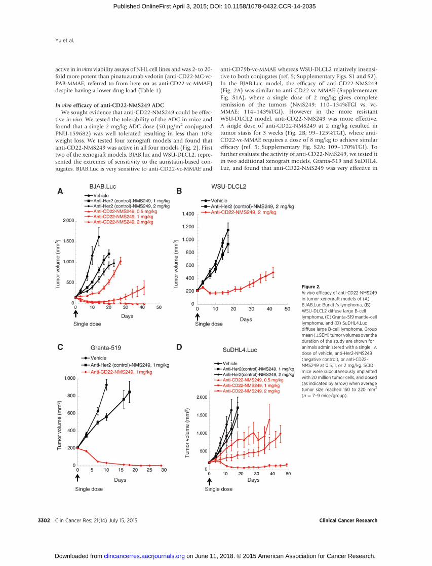
In vivo efficacy of anti-CD22-NMS249 ADCWe sought evidence that anti-CD22-NMS249 could be effec-
tive in vivo. We tested the tolerability of the ADC in mice andfound that a single 2 mg/kg ADC dose (50 mg/m2 conjugatedPNU-159682) was well tolerated resulting in less than 10%weight loss. We tested four xenograft models and found thatanti-CD22-NMS249 was active in all four models (Fig. 2). Firsttwo of the xenograft models, BJAB.luc and WSU-DLCL2, repre-sented the extremes of sensitivity to the auristatin-based con-jugates. BJAB.Luc is very sensitive to anti-CD22-vc-MMAE and
anti-CD79b-vc-MMAE whereas WSU-DLCL2 relatively insensi-tive to both conjugates (ref. 5; Supplementary Figs. S1 and S2).In the BJAB.Luc model, the efficacy of anti-CD22-NMS249(Fig. 2A) was similar to anti-CD22-vc-MMAE (SupplementaryFig. S1A), where a single dose of 2 mg/kg gives completeremission of the tumors (NMS249: 110–134%TGI vs. vc-MMAE: 114–143%TGI). However in the more resistantWSU-DLCL2 model, anti-CD22-NMS249 was more effective.A single dose of anti-CD22-NMS249 at 2 mg/kg resulted intumor stasis for 3 weeks (Fig. 2B; 99–125%TGI), where anti-CD22-vc-MMAE requires a dose of 8 mg/kg to achieve similarefficacy (ref. 5; Supplementary Fig. S2A; 109–170%TGI). Tofurther evaluate the activity of anti-CD22-NMS249, we tested itin two additional xenograft models, Granta-519 and SuDHL4.Luc, and found that anti-CD22-NMS249 was very effective in
Figure 2.In vivo efficacy of anti-CD22-NMS249in tumor xenograft models of (A)BJAB.Luc Burkitt's lymphoma, (B)WSU-DLCL2 diffuse large B-celllymphoma, (C) Granta-519mantle-celllymphoma, and (D) SuDHL4.Lucdiffuse large B-cell lymphoma. Groupmean (�SEM) tumor volumes over theduration of the study are shown foranimals administered with a single i.v.dose of vehicle, anti-Her2-NMS249(negative control), or anti-CD22-NMS249 at 0.5, 1, or 2 mg/kg. SCIDmice were subcutaneously implantedwith 20 million tumor cells, and dosed(as indicated by arrow) when averagetumor size reached 150 to 220 mm3
(n ¼ 7–9 mice/group).
Yu et al.
Clin Cancer Res; 21(14) July 15, 2015 Clinical Cancer Research3302
on June 11, 2018. © 2015 American Association for Cancer Research. clincancerres.aacrjournals.org Downloaded from
Published OnlineFirst April 3, 2015; DOI: 10.1158/1078-0432.CCR-14-2035
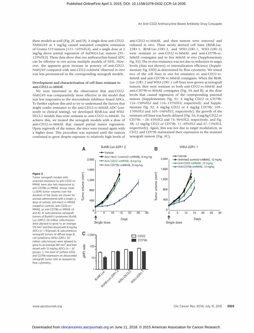
these models as well (Fig. 2C and D). A single dose anti-CD22-NMS4249 at 1 mg/kg caused sustained complete remissionof Granta-519 tumors (121–145%TGI), and a single dose at 2mg/kg drove partial regression of SuDHL4.Luc tumors (95–129%TGI). These data show that our anthracycline-based ADCcan be effective in vivo across multiple models of NHL. How-ever, the apparent great increase in potency of anti-CD22-NMS249 compared with anti-CD22-vcMMAE observed in vitrowas less pronounced in the corresponding xenograft models.
Development and characterization of cell lines resistant toanti-CD22-vc-MMAE
We were interested in the observation that anti-CD22-NMS249 was comparatively more effective in the model thatwas less responsive to the microtubule inhibitor–based ADCs.To further explore this and to try to understand the factors thatmight confer resistance to the anti-CD22-vc-MMAE ADC (cur-rently in clinical testing), we developed BJAB.Luc and WSU-DLCL2 models that were resistant to anti-CD22-vc-MMAE. Toachieve this, we treated the xenograft models with a dose ofanti-CD22-vc-MMAE that caused partial tumor regression.Upon regrowth of the tumor, the mice were treated again witha higher dose. This procedure was repeated until the tumorscontinued to grow despite exposure to relatively high levels of
anti-CD22-vc-MMAE, and then tumors were removed andcultured in vitro. These newly derived cell lines (BJAB.Luc-22R1.1, BJAB.Luc-22R1.2, and WSU-22R1.1, WSU-22R1.2)were resistant to anti-CD22-vc-MMAE and anti-CD79b-vc-MMAE conjugates and to free MMAE in vitro (SupplementaryFig. S3). The in vitro resistance was not due to reduction in targetlevels (data not shown) or internalization efficiency (Supple-mentary Fig. S3D) as determined by flow cytometry. We testedtwo of the cell lines in vivo for resistance to anti-CD22-vc-MMAE and anti-CD79b-vc-MMAE conjugates. When the BJAB.Luc-22R1.2 and WSU-22R1.1 cell lines were grown as xenografttumors, they were resistant to both anti-CD22-vc-MMAE andanti-CD79b-vc-MMAE conjugates (Fig. 3A and B); at the doselevels that caused regression of the corresponding parentaltumors (Supplementary Fig. S1; 4 mg/kg CD22 or CD79b:116–150%TGI and 116–137%TGI, respectively, and Supple-mentary Fig. S2; 8 mg/kg CD22 or 4 mg/kg CD79b: 109–170%TGI and 103–144%TGI, respectively), the growth of theresistant cell lines was barely delayed (Fig. 3A; 8 mg/kg CD22 orCD79b: �28–43%TGI and 74–96%TGI, respectively, and Fig.3B; 12 mg/kg CD22 or CD79b: 11–49%TGI and 47–73%TGI,respectively). Again, this was not due to target modulation, asCD22 and CD79b maintained their expression in the resistantxenograft tumors (Fig. 3C).
Figure 3.Tumor xenograft models withacquired resistance to anti-CD22-vc-MMAE were also less responsive toanti-CD79b-vc-MMAE. Group mean(�SEM) tumor volumes over theduration of the study are shown foranimals administered with a single i.v.dose of vehicle, anti-Her2-vc-MMAE(negative control), anti-CD22-vc-MMAE, or anti-CD79b-vc-MMAE (Aand B). A, subcutaneous xenografttumors of Burkitt's lymphoma (BJAB.Luc-22R1.2, 20 million cells/mouse)were allowed to grow to an average175mm3 and then dosedwith 8mg/kgADCs (n¼ 9/group). B, subcutaneousxenograft tumors of diffuse large B-cell lymphoma (WSU-22R1.1, 20million cells/mouse) were allowed togrow to an average 190mm3 and thendosed with 12 mg/kg ADCs (n ¼ 8/group). C, the level of surface CD22and CD79b expression on dissociatedxenograft tumor cells as assayed byflow cytometry.
An Anti-CD22 Anthracycline-Based Antibody–Drug Conjugate
www.aacrjournals.org Clin Cancer Res; 21(14) July 15, 2015 3303
on June 11, 2018. © 2015 American Association for Cancer Research. clincancerres.aacrjournals.org Downloaded from
Published OnlineFirst April 3, 2015; DOI: 10.1158/1078-0432.CCR-14-2035
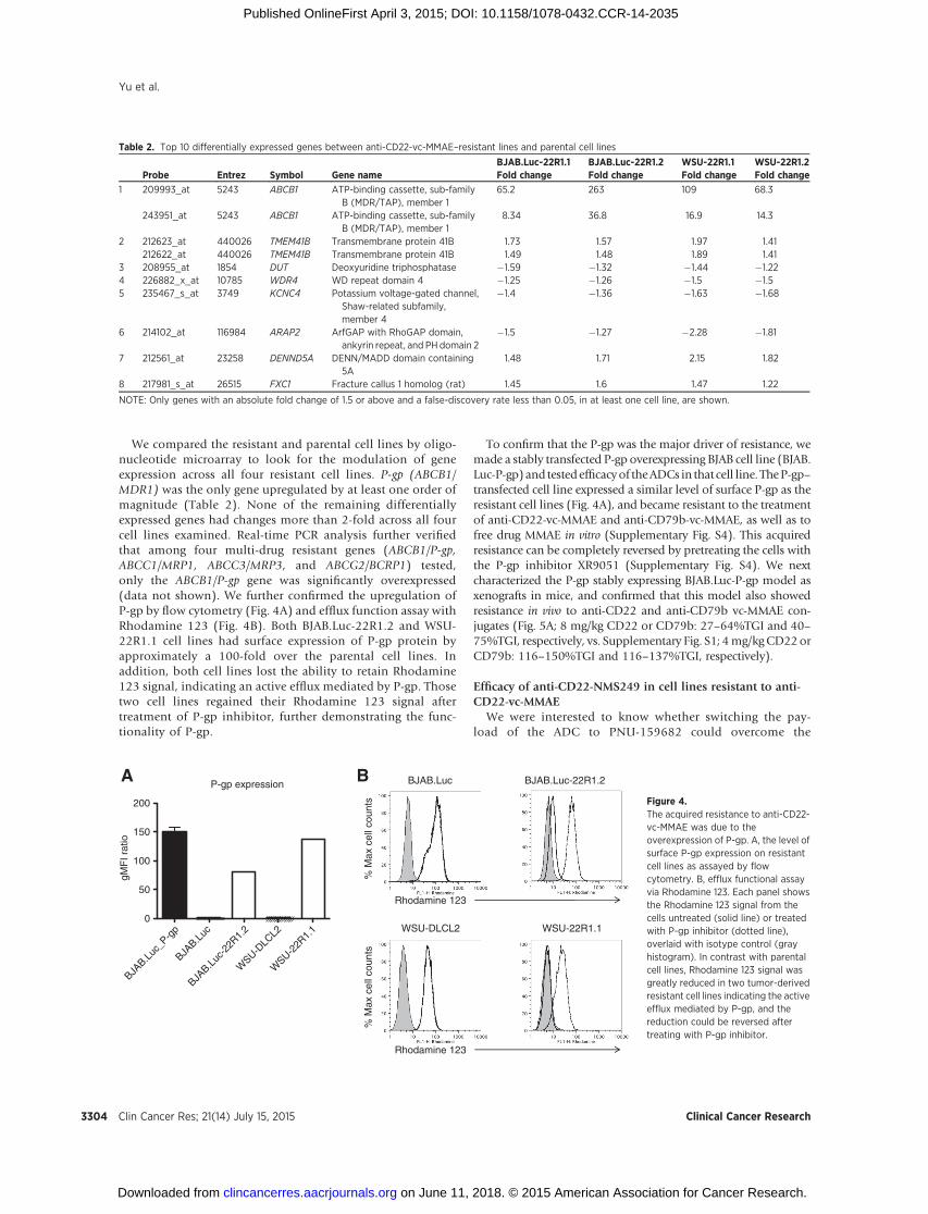
We compared the resistant and parental cell lines by oligo-nucleotide microarray to look for the modulation of geneexpression across all four resistant cell lines. P-gp (ABCB1/MDR1) was the only gene upregulated by at least one order ofmagnitude (Table 2). None of the remaining differentiallyexpressed genes had changes more than 2-fold across all fourcell lines examined. Real-time PCR analysis further verifiedthat among four multi-drug resistant genes (ABCB1/P-gp,ABCC1/MRP1, ABCC3/MRP3, and ABCG2/BCRP1) tested,only the ABCB1/P-gp gene was significantly overexpressed(data not shown). We further confirmed the upregulation ofP-gp by flow cytometry (Fig. 4A) and efflux function assay withRhodamine 123 (Fig. 4B). Both BJAB.Luc-22R1.2 and WSU-22R1.1 cell lines had surface expression of P-gp protein byapproximately a 100-fold over the parental cell lines. Inaddition, both cell lines lost the ability to retain Rhodamine123 signal, indicating an active efflux mediated by P-gp. Thosetwo cell lines regained their Rhodamine 123 signal aftertreatment of P-gp inhibitor, further demonstrating the func-tionality of P-gp.
To confirm that the P-gp was the major driver of resistance, wemade a stably transfected P-gp overexpressing BJAB cell line (BJAB.Luc-P-gp)and testedefficacyof theADCs in that cell line. TheP-gp–transfected cell line expressed a similar level of surface P-gp as theresistant cell lines (Fig. 4A), and became resistant to the treatmentof anti-CD22-vc-MMAE and anti-CD79b-vc-MMAE, as well as tofree drug MMAE in vitro (Supplementary Fig. S4). This acquiredresistance can be completely reversed by pretreating the cells withthe P-gp inhibitor XR9051 (Supplementary Fig. S4). We nextcharacterized the P-gp stably expressing BJAB.Luc-P-gp model asxenografts in mice, and confirmed that this model also showedresistance in vivo to anti-CD22 and anti-CD79b vc-MMAE con-jugates (Fig. 5A; 8 mg/kg CD22 or CD79b: 27–64%TGI and 40–75%TGI, respectively, vs. Supplementary Fig. S1; 4mg/kgCD22 orCD79b: 116–150%TGI and 116–137%TGI, respectively).
Efficacy of anti-CD22-NMS249 in cell lines resistant to anti-CD22-vc-MMAE
We were interested to know whether switching the pay-load of the ADC to PNU-159682 could overcome the
Table 2. Top 10 differentially expressed genes between anti-CD22-vc-MMAE–resistant lines and parental cell lines
BJAB.Luc-22R1.1 BJAB.Luc-22R1.2 WSU-22R1.1 WSU-22R1.2Probe Entrez Symbol Gene name Fold change Fold change Fold change Fold change
1 209993_at 5243 ABCB1 ATP-binding cassette, sub-familyB (MDR/TAP), member 1
65.2 263 109 68.3
243951_at 5243 ABCB1 ATP-binding cassette, sub-familyB (MDR/TAP), member 1
8.34 36.8 16.9 14.3
2 212623_at 440026 TMEM41B Transmembrane protein 41B 1.73 1.57 1.97 1.41212622_at 440026 TMEM41B Transmembrane protein 41B 1.49 1.48 1.89 1.41
3 208955_at 1854 DUT Deoxyuridine triphosphatase �1.59 �1.32 �1.44 �1.224 226882_x_at 10785 WDR4 WD repeat domain 4 �1.25 �1.26 �1.5 �1.55 235467_s_at 3749 KCNC4 Potassium voltage-gated channel,
Shaw-related subfamily,member 4
�1.4 �1.36 �1.63 �1.68
6 214102_at 116984 ARAP2 ArfGAP with RhoGAP domain,ankyrin repeat, and PH domain 2
�1.5 �1.27 �2.28 �1.81
7 212561_at 23258 DENND5A DENN/MADD domain containing5A
1.48 1.71 2.15 1.82
8 217981_s_at 26515 FXC1 Fracture callus 1 homolog (rat) 1.45 1.6 1.47 1.22
NOTE: Only genes with an absolute fold change of 1.5 or above and a false-discovery rate less than 0.05, in at least one cell line, are shown.
BA
Rhodamine 123
BJAB.Luc-22R1.2BJAB.Luc
WSU-DLCL2 WSU-22R1.1
P-gp expression
BJAB.L
uc_P
-gp
BJAB.L
uc
BJAB.L
uc-2
2R1.
2
WSU-D
LCL2
WSU-2
2R1.
10
50
100
150
200
gMF
I rat
io
% M
ax c
ell c
ount
s%
Max
cel
l cou
nts
Rhodamine 123
Figure 4.The acquired resistance to anti-CD22-vc-MMAE was due to theoverexpression of P-gp. A, the level ofsurface P-gp expression on resistantcell lines as assayed by flowcytometry. B, efflux functional assayvia Rhodamine 123. Each panel showsthe Rhodamine 123 signal from thecells untreated (solid line) or treatedwith P-gp inhibitor (dotted line),overlaid with isotype control (grayhistogram). In contrast with parentalcell lines, Rhodamine 123 signal wasgreatly reduced in two tumor-derivedresistant cell lines indicating the activeefflux mediated by P-gp, and thereduction could be reversed aftertreating with P-gp inhibitor.
Yu et al.
Clin Cancer Res; 21(14) July 15, 2015 Clinical Cancer Research3304
on June 11, 2018. © 2015 American Association for Cancer Research. clincancerres.aacrjournals.org Downloaded from
Published OnlineFirst April 3, 2015; DOI: 10.1158/1078-0432.CCR-14-2035
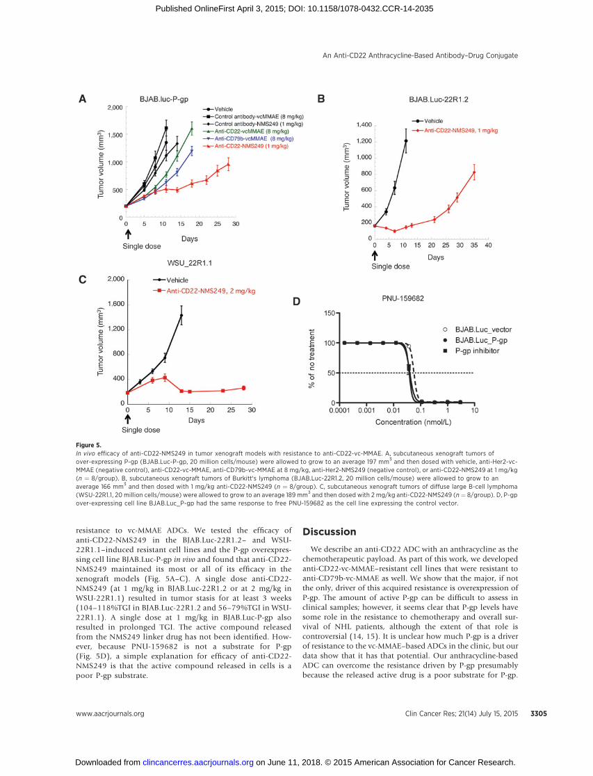
resistance to vc-MMAE ADCs. We tested the efficacy ofanti-CD22-NMS249 in the BJAB.Luc-22R1.2– and WSU-22R1.1–induced resistant cell lines and the P-gp overexpres-sing cell line BJAB.Luc-P-gp in vivo and found that anti-CD22-NMS249 maintained its most or all of its efficacy in thexenograft models (Fig. 5A–C). A single dose anti-CD22-NMS249 (at 1 mg/kg in BJAB.Luc-22R1.2 or at 2 mg/kg inWSU-22R1.1) resulted in tumor stasis for at least 3 weeks(104–118%TGI in BJAB.Luc-22R1.2 and 56–79%TGI in WSU-22R1.1). A single dose at 1 mg/kg in BJAB.Luc-P-gp alsoresulted in prolonged TGI. The active compound releasedfrom the NMS249 linker drug has not been identified. How-ever, because PNU-159682 is not a substrate for P-gp(Fig. 5D), a simple explanation for efficacy of anti-CD22-NMS249 is that the active compound released in cells is apoor P-gp substrate.
Discussion
We describe an anti-CD22 ADC with an anthracycline as thechemotherapeutic payload. As part of this work, we developedanti-CD22-vc-MMAE–resistant cell lines that were resistant toanti-CD79b-vc-MMAE as well. We show that the major, if notthe only, driver of this acquired resistance is overexpression ofP-gp. The amount of active P-gp can be difficult to assess inclinical samples; however, it seems clear that P-gp levels havesome role in the resistance to chemotherapy and overall sur-vival of NHL patients, although the extent of that role iscontroversial (14, 15). It is unclear how much P-gp is a driverof resistance to the vc-MMAE–based ADCs in the clinic, but ourdata show that it has that potential. Our anthracycline-basedADC can overcome the resistance driven by P-gp presumablybecause the released active drug is a poor substrate for P-gp.
Figure 5.In vivo efficacy of anti-CD22-NMS249 in tumor xenograft models with resistance to anti-CD22-vc-MMAE. A, subcutaneous xenograft tumors ofover-expressing P-gp (BJAB.Luc-P-gp, 20 million cells/mouse) were allowed to grow to an average 197 mm3 and then dosed with vehicle, anti-Her2-vc-MMAE (negative control), anti-CD22-vc-MMAE, anti-CD79b-vc-MMAE at 8 mg/kg, anti-Her2-NMS249 (negative control), or anti-CD22-NMS249 at 1 mg/kg(n ¼ 8/group). B, subcutaneous xenograft tumors of Burkitt's lymphoma (BJAB.Luc-22R1.2, 20 million cells/mouse) were allowed to grow to anaverage 166 mm3 and then dosed with 1 mg/kg anti-CD22-NMS249 (n ¼ 8/group). C, subcutaneous xenograft tumors of diffuse large B-cell lymphoma(WSU-22R1.1, 20 million cells/mouse) were allowed to grow to an average 189 mm3 and then dosed with 2 mg/kg anti-CD22-NMS249 (n ¼ 8/group). D, P-gpover-expressing cell line BJAB.Luc_P-gp had the same response to free PNU-159682 as the cell line expressing the control vector.
An Anti-CD22 Anthracycline-Based Antibody–Drug Conjugate
www.aacrjournals.org Clin Cancer Res; 21(14) July 15, 2015 3305
on June 11, 2018. © 2015 American Association for Cancer Research. clincancerres.aacrjournals.org Downloaded from
Published OnlineFirst April 3, 2015; DOI: 10.1158/1078-0432.CCR-14-2035
Consistent with this idea, the free drug, PNU-159682, is not aP-gp substrate (Fig. 5D).
The WSU-DLCL2 xenograft model was less sensitive to themicrotubule inhibitor–based ADCs (Supplementary Fig. S2),including both vc-MMAEandSPDB-DM4conjugates.What drivesthis unresponsiveness is unknown. It was not due to target levelsas the model has equal or higher expression of CD22 than BJAB.Luc (5), or the internalization efficiency as WSU-DLCL2 has ahigher internalization rate than BJAB.Luc (SupplementaryFig. S3D). In vitro sensitivity of these two cell lines to free drugis also very close (Table 1 ref. 5).However, we observed that in thismodel, anti-CD22-NMS249 has shown a much better improve-ment on efficacy. This could be because NMS249 is amore potentlinker drug and its potency is only revealed in resistant modelsor that the different mechanism of action changes the relativesensitivities of the cell lines. In any case, our data suggest thatregardless of P-gp expression, our anthracycline-based ADC hasthe potential to be more effective in patients who do not respondto the MMAE-based ADCs currently in the clinic.
ADCs offer the promise of increased therapeutic index forchemotherapy and possibly could replace systemic che-motherapies of the same mechanism. This strategy is beingtried for the treatment of systemic anaplastic large cell lym-phoma where the vincristine in the standard of care CHOP(cyclophosphamide, doxorubicin, vincristine, prednisone)combination chemotherapy is removed and replaced with theADC Brentuximab Vedotin (anti-CD30-vc-MMAE) that has thesame mechanism of action. Having an ADC based on ananthracycline would open up the possibility of replacing theanthracycline in cancers where it is current standard of care.
Disclosure of Potential Conflicts of InterestR. Cohen is an employee of Calico Life Sciences. J. Flygare is an employee of
and holds ownership interest (including patents) in Roche. A.G. Polson holdsownership interest (including patents) in Roche/Genentech. No potentialconflicts of interest were disclosed by the other authors.
Authors' ContributionsConception and design: S.-F. Yu, B. Zheng, R. Cohen, J. Flygare, A.G. PolsonDevelopment of methodology: S.-F. Yu, B. ZhengAcquisition of data (provided animals, acquired and managed patients,provided facilities, etc.): S.-F. Yu, B. Zheng, M. Go, J. Lau, H. RaabAnalysis and interpretation of data (e.g., statistical analysis, biostatistics,computational analysis): S.-F. Yu, B. Zheng, M. Go, J. Lau, S. Jhunjhunwala,J. Flygare, A.G. PolsonWriting, review, and/or revision of the manuscript: S.-F. Yu, B. Zheng,S. Spencer, P. Polakis, A.G. PolsonAdministrative, technical, or material support (i.e., reporting or organizingdata, constructing databases): S.-F. Yu, R. SorianoStudy supervision: S.-F. Yu, S. SpencerOther (synthesis of drug): M. Caruso
AcknowledgmentsThe authors thank the in vivo cell culture lab at Genentech for developing
resistance cell lines and preparing the cells for all the in vivo studies.The costs of publication of this article were defrayed in part by the
payment of page charges. This article must therefore be hereby markedadvertisement in accordance with 18 U.S.C. Section 1734 solely to indicatethis fact.
Received August 6, 2014; revised December 4, 2014; accepted April 2, 2015;published OnlineFirst April 3, 2015.
References1. Senter PD. Potent antibody drug conjugates for cancer therapy. Curr Opin
Chem Biol 2009;13:235–44.2. Sievers EL, Senter PD. Antibody-drug conjugates in cancer therapy. Annu
Rev Med 2013;64:15–29.3. Chu YW, Polson A. Antibody-drug conjugates for the treatment of B-cell
non-Hodgkin's lymphoma and leukemia. Future Oncol 2013;9:355–68.4. Kreitman RJ, Pastan I. Antibody fusion proteins: anti-CD22 recombi-
nant immunotoxin moxetumomab pasudotox. Clin Cancer Res 2011;17:6398–405.
5. LiD, PoonKA, Yu SF,Dere R,GoM, Lau J, et al.DCDT2980S, an anti-CD22-monomethyl auristatin E antibody-drug conjugate, is a potential treatmentfor non-Hodgkin lymphoma. Mol Cancer Ther 2013;12:1255–65.
6. Palanca-Wessels MC, Flinn IW, Sehn LH, Patel M, Sangha R, CzuczmanMS, et al. A phase I study of the anti-CD79b antibody-drug conjugate(ADC) DCDS4501A targeting CD79b in relapsed or refractory B-cellnon-Hodgkin's lymphoma (NHL). ASH Annual Meeting Abstracts2012;120:56.
7. Advani R, LebovicD, BrunvandM,ChenAI,GoyA,Chang JE, et al. A phase Istudy of DCDT2980S, an Antibody-Drug Conjugate (ADC) targetingCD22, in relapsed or refractory B-cell non-Hodgkin's lymphoma (NHL).ASH Annual Meeting Abstracts 2012;120:59.
8. Younes A, Kim S, Romaguera J, Copeland A, Farial Sde C, Kwak LW, et al.Phase I multidose-escalation study of the anti-CD19maytansinoid immu-noconjugate SAR3419 administered by intravenous infusion every 3 weeks
to patients with relapsed/refractory B-cell lymphoma. J Clin Oncol2012;30:2776–82.
9. Ribrag V, Dupuis J, Tilly H,Morschhauser F, Laine F, Houot R, et al. A dose-escalation study of SAR3419, an anti-CD19 antibody maytansinoid con-jugate, administered by intravenous infusion once weekly in patients withrelapsed/refractory B-cell non-Hodgkin lymphoma. Clin Cancer Res 2014;20:213–20.
10. Dornan D, Bennett F, Chen Y, Dennis M, Eaton D, Elkins K, et al.Therapeutic potential of an anti-CD79b antibody-drug conjugate, anti-CD79b-vc-MMAE, for the treatment of non-Hodgkin lymphoma. Blood2009;114:2721–9.
11. Junutula JR, Raab H, Clark S, Bhakta S, Leipold DD, Weir S, et al. Site-specific conjugation of a cytotoxic drug to an antibody improves thetherapeutic index. Nat Biotechnol 2008;26:925–32.
12. Sessa C, Valota O, Geroni C. Ongoing phase I and II studies of novelanthracyclines. Cardiovasc Toxicol 2007;7:75–9.
13. Quintieri L, Geroni C, Fantin M, Battaglia R, Rosato A, Speed W, et al.Formation and antitumor activity of PNU-159682, a major metaboliteof nemorubicin in human liver microsomes. Clin Cancer Res 2005;11:1608–17.
14. Rund D. Multidrug resistance in lymphoma: is it time for clinical trials?Leuk Lymphoma 2007;48:643–4.
15. Sandor V, Wilson W, Fojo T, Bates SE. The role of MDR-1 in refractorylymphoma. Leuk Lymphoma 1997;28:23–31.
Clin Cancer Res; 21(14) July 15, 2015 Clinical Cancer Research3306
Yu et al.
on June 11, 2018. © 2015 American Association for Cancer Research. clincancerres.aacrjournals.org Downloaded from
Published OnlineFirst April 3, 2015; DOI: 10.1158/1078-0432.CCR-14-2035
2015;21:3298-3306. Published OnlineFirst April 3, 2015.Clin Cancer Res Shang-Fan Yu, Bing Zheng, MaryAnn Go, et al. (ADC) That Overcomes Resistance to Auristatin-Based ADCs
Drug Conjugate−A Novel Anti-CD22 Anthracycline-Based Antibody
Updated version
10.1158/1078-0432.CCR-14-2035doi:
Access the most recent version of this article at:
Material
Supplementary
http://clincancerres.aacrjournals.org/content/suppl/2015/04/04/1078-0432.CCR-14-2035.DC1
Access the most recent supplemental material at:
Cited articles
http://clincancerres.aacrjournals.org/content/21/14/3298.full#ref-list-1
This article cites 15 articles, 6 of which you can access for free at:
Citing articles
http://clincancerres.aacrjournals.org/content/21/14/3298.full#related-urls
This article has been cited by 5 HighWire-hosted articles. Access the articles at:
E-mail alerts related to this article or journal.Sign up to receive free email-alerts
Subscriptions
Reprints and
To order reprints of this article or to subscribe to the journal, contact the AACR Publications Department at
Permissions
Rightslink site. Click on "Request Permissions" which will take you to the Copyright Clearance Center's (CCC)
.http://clincancerres.aacrjournals.org/content/21/14/3298To request permission to re-use all or part of this article, use this link
on June 11, 2018. © 2015 American Association for Cancer Research. clincancerres.aacrjournals.org Downloaded from
Published OnlineFirst April 3, 2015; DOI: 10.1158/1078-0432.CCR-14-2035