Embed Size (px)
DESCRIPTION
Ceramic nano pigments for anti corrosion coatings
Citation preview
lable at ScienceDirect
Microporous and Mesoporous Materials 218 (2015) 62e68
Contents lists avai
Microporous and Mesoporous Materials
journal homepage: www.elsevier .com/locate/micromeso
A new strategy based on thermodiffusion of ceramic nanopigmentsinto metal surfaces and formation of anti-corrosion coatings
Maryam Shaterian a, *, Asma Khoobi b, Morteza Enhessari c, Keyvan Ozaee d
a Department of Chemistry, Faculty of Science, University of Zanjan, Zanjan 45371-38791, Islamic Republic of Iranb Department of Analytical Chemistry, Faculty of Chemistry, University of Kashan, Kashan P.O. Box 87317-51167, Islamic Republic of Iranc Department of Chemistry, Naragh Branch, Islamic Azad University, Naragh, Islamic Republic of Irand Young Researchers and Elite Club, South Tehran Branch, Islamic Azad University, Tehran, Islamic Republic of Iran
a r t i c l e i n f o
Article history:Received 21 March 2015Received in revised form2 June 2015Accepted 28 June 2015Available online 11 July 2015
Keywords:Ceramic nanopigmentsThermodiffusionAnti-corrosion coatings
* Corresponding author.E-mail address: [email protected] (M. Shaterian
http://dx.doi.org/10.1016/j.micromeso.2015.06.0391387-1811/© 2015 Elsevier Inc. All rights reserved.
a b s t r a c t
The present paper is the first report about preparation of anti-corrosion coatings based on thermo-diffusion of ceramic nanopigments into metal surfaces. At first, Cr1.3Fe0.7O3 ceramic nanopigments havebeen synthesized by simple and environmentally benign solegel method. Annealing of the gels atdifferent temperatures ranging from 600 to 1000 �C yielded the pure rhombohedral structure. Thestructural evolutions and microstructural characteristics of the synthesized nanoceramics were inves-tigated through different methods containing X-ray diffraction (XRD), energy dispersive X-ray spec-troscopy (EDX), scanning electron microscopy (SEM) and transmission electron microscopy (TEM). ThenCr1.3Fe0.7O3 ceramic nanopigments were coated on mild steel surface via thermodiffusion method. Thesurface morphology and the corrosion behavior of the Cr1.3Fe0.7O3 coatings were evaluated using atomicforce microscopy (AFM), field-emission scanning electron microscopy (FE-SEM), electrochemicalimpedance spectroscopy (EIS), potentiodynamic polarization and weight loss measurements in a solu-tion of 2.0 M HCl. It is found that morphology of diffusion coatings affects the protective properties ofmild steel. On the other hand, in extra-corrosive HCl media Cr1.3Fe0.7O3 nanoceramic coatings providemild steel with high protective properties.
© 2015 Elsevier Inc. All rights reserved.
1. Introduction
Corrosion is a natural process that reduces the binding energy inmetals. It occurs at the metal/environment interfaces, and causes adeterioration of the metals and their properties. During thecorrosion process the metallic ions precipitate on the metal surfaceand by forming corrosion film separates the metal from theaggressive environment [1]. Iron and its alloys play key roles in ourdaily lives because of their excellent properties, such as high me-chanical and structural strengths [2]. These materials are used indifferent industrial and have various engineering applications. Mildsteel is now the most common form of steel because its price isrelatively low while it presents material properties that areacceptable for many applications. Low-carbon steel containsapproximately 0.05e0.15% carbon making it ductile and malleable.Mild steel has a relatively low tensile strength, but it is cheap and
).
easy to form; surface hardness can be increased through carbu-rizing [3]. Therefore, mild steel due to its excellent mechanicalproperties is the constructional material of choice in numerouschemical and petrochemical industries as well as daily life appli-cations. The main drawback of mild steel is its low corrosionresistance. On the other hand, it is easily attacked and solubilized inacidic media which are widely used to remove of undesirable rustsand scales in several industrial sectors [4]. Oil-well acidizing, acid-pickling, acid-cleaning and acid-descaling, are usual industrialprocesses for cleaning the metals surfaces. Also it is necessary toapply acid solutions to eliminate undesirable scale and corrosionproducts from metals. Sulfuric acid and hydrochloric acid aregenerally used for this purpose; but, these acids attack the metalsurface and begin corrosion process. Corrosion can cause drasticdamage to the metal surface and degrade its properties, thuslimiting its applications [5,6].
The use of corrosion inhibitors is very important method forprotecting metals from corrosion, and numerous scientists areconducting research on this area. In principle, inhibitors preventthe corrosion process of metal by interacting with themetal surface
M. Shaterian et al. / Microporous and Mesoporous Materials 218 (2015) 62e68 63
by adsorption through the p-orbitals, donor atoms, electron den-sity and the electronic structure of the molecule [7e10]. Hardcoatings with carbide, nitride, boride, or carbonitride of transitionmetals are widely used in order to increase corrosion resistance[10]. The coatings are employed in order to isolate the metal andsteel surfaces from the corrosive environment and prevent thediffusion of water vapor, oxygen, or ions, which act as a source thatbegins the corrosion [11]. Moreover, these ceramic coatingsdemonstrate high dielectric strength, which generates the forma-tion of a more noble surface [12,13]. Conventional methods formaking these coatings are thermal welding, spraying, chemicalvapor deposition (CVD), electroplating and physical vapor deposi-tion (PVD) [14,15]. All of the methods are high energy consumptiontechniques. Additionally, sometimes toxic elements such as cad-mium are used as alloy coating but now require to be replaced withmore environmental friendly materials [16,17]. Coating by com-posites or polymers is usually not suitable for high temperatureapplications because of their polymeric nature. However, oxideceramics are suitable candidates as anti-corrosion coatings to beused under harsh environmental conditions [18,19].
In this study, the surface treatment of mild steel was carried outby the thermodiffusion of Cr1.3Fe0.7O3 ceramic nanopigments forthe first time. The theory of thermodiffusion for preparation of anti-corrosive coatings was discussed. Various properties such as phasecomposition and particle size distribution of the ceramic nano-pigments have been evaluated. The effect of Cr1.3Fe0.7O3 thermo-diffusion on the corrosion inhibition of mild steel in hydrochloricacid media was investigated.
2. Experimental
2.1. Chemicals
Iron (III) nitrate decahydrate [Fe(NO3)3$10H2O], ammonium di-chromate [(NH4)2Cr2O7], stearic acid, nitric acid and hydrochloricacid were obtained from Merck company (Darmstadt, Germany).All reagents were of analytical grade and used as received withoutfurther purification. Deionized water was used throughout thisstudy.
2.2. Synthesis of the nanopowder
Cr1.3Fe0.7O3 ceramic nanopigments were synthesized via sol-egel method based on previous work [20]. Briefly, stearic acid wasmelted in a beaker at a temperature of 73 �C. Then, Fe(NO3)3$10H2Oand (NH4)2Cr2O7 were added in stoichiometric proportion to amixture of water and nitric acid (1:1%v/v). The Fe(NO3)3$10H2O/(NH4)2Cr2O7 molar ratio was 0.7:1.3. This solution was added intothe melted stearic acid. The beaker was kept in an oven at 90 �C for48 h until a homogenous light red-brown sol was obtained. The solwas cooled down to room temperature and subsequently dried inan oven for 12 h. Finally the nanopowder was calcined at 600, 800,900 and 1000 �C for 2 h.
2.3. Electrochemical cell
Corrosion experiments were performed in a three-electrode cellopened to air under stagnant conditions at a thermostated tem-perature of 25 �C. An Ag/AgCl/KCl (sat.) (Metrohm, Switzerland)and a platinum sheet (Metrohm, Switzerland) were used as thereference and counter electrodes, respectively. Corrosion inhibitiontests were performed using specimens prepared from mild steelas working electrode, having the composition (wt%): C:0.027;Si:0.0027; Mn:0.34; P:0.009; S:0.003; Cr:0.008; Ni:0.03; Cu:0.007;Al:0.068; Nb 0.003, Ti 0.003, V 0.003 and Fe balance. The test
specimens with dimension of 1 cm � 1 cm � 0.1 cm were used assubstrate. The surface of mild steel electrodes mechanicallyabraded prior to use with different emery papers up to 1200 grade.The mild steel specimens were cleaned with analytical ethanol andthen immersed in analytical acetone and finally washed withdeionized water before coating. Then Cr1.3Fe0.7O3 nanoceramiccoatings were deposited onto mild steel specimen (working elec-trode) using thermodiffusion process. The process was performedby diffusion of Cr1.3Fe0.7O3 nanopowder into the working electrodeat 900 �C for 4 h. The mechanism of thermodiffusion process isdiscussed in Result and discussion section.
In this study, hydrochloric acid (2.0 M) was selected as corrosivesolution and was prepared by dilution of analytical grade 37% HClwith deionized water.
2.4. Microstructural and electrochemical characterization
The crystal structures of the nanopigments were studied byX-ray diffraction (XRD) analysis. The diffraction patterns of thesamples were obtained with a Seifert diffractometer (Model PTS3003). The data were collected within 2q angle from 10� to 80�
using Cu Ka line (l ¼ 0.15418 nm). Scanning electron microscopy(SEM) micrographs were obtained by an LEO 1455 UP analyzer(Oxford, UK). The morphology and crystallite size of the nano-ceramics were investigated by transmission electron microscopy(TEM) using a Philips EM 208 electron microscope operated at anacceleration voltage of 100 kV.
The surface morphology of each specimen was studied using a0201/A of ARA atomic force microscopy (AFM) and field-emissionscanning electron microscopy (FE-SEM) (Mira 3 XMU) before andafter immersion in 2.0 M HCl in the absence and presence ofCr1.3Fe0.7O3 thermodiffusion.
Corrosion inhibition electrochemical tests of the Cr1.3Fe0.7O3nanoceramic coatings containing electrochemical impedancespectroscopy (EIS) and Tafel polarization were performed using anAUTOLAB model PGSTAT30. EIS studies were performed at corro-sion potentials (Ecorr) over a frequency range of 10 kHze0.1 Hz witha signal amplitude perturbation of 5.0 mV. Then impedance datawere analyzed using a Pentium IV computer and FRA software. TheTafel polarization experiments were carried out with a scan rate of0.5 mV s�1 and the data were analyzed using GPES electrochemicalsoftware.
3. Results and discussion
3.1. Characterization of the Cr1.3Fe0.7O3 nanopowder
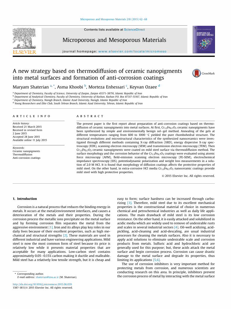
3.1.1. X-ray diffractionFig. 1 represents XRD patterns for Cr1.3Fe0.7O3 nanoceramics at
different temperatures [20]. As can be observed the crystallinity ofthe as-prepared products was continuously improved with theincrease of the calcination temperature from 600 �C to 1000 �C.Based on Fig. 1a, calcination in 1000 �C leads to formation of purerhombohedral structure (JCPDS, Card No. 35-1112). There is no peakthat could be attributed to Cr2O3 or Fe2O3.
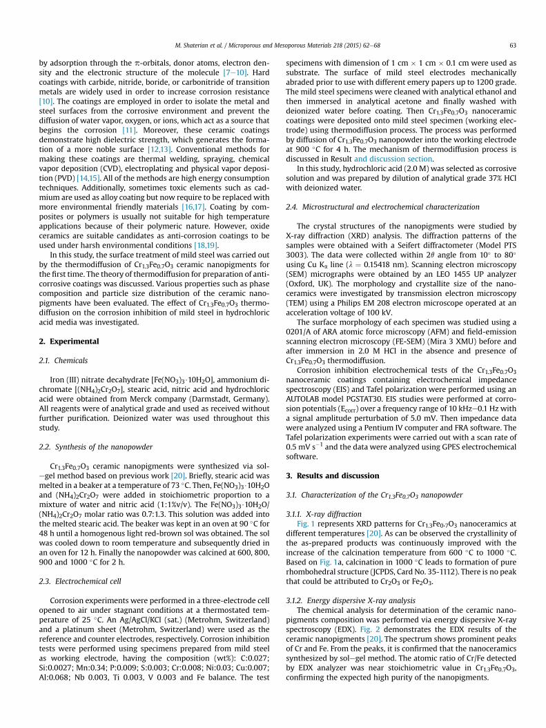
3.1.2. Energy dispersive X-ray analysisThe chemical analysis for determination of the ceramic nano-
pigments composition was performed via energy dispersive X-rayspectroscopy (EDX). Fig. 2 demonstrates the EDX results of theceramic nanopigments [20]. The spectrum shows prominent peaksof Cr and Fe. From the peaks, it is confirmed that the nanoceramicssynthesized by solegel method. The atomic ratio of Cr/Fe detectedby EDX analyzer was near stoichiometric value in Cr1.3Fe0.7O3,confirming the expected high purity of the nanopigments.
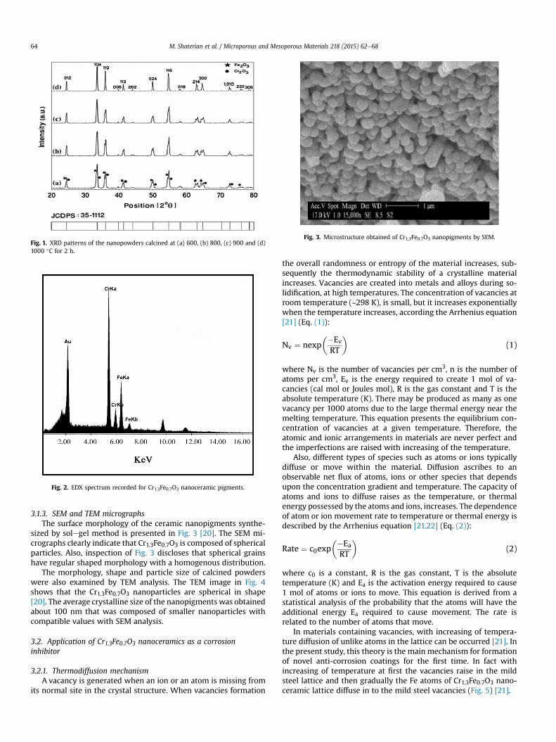
Fig. 1. XRD patterns of the nanopowders calcined at (a) 600, (b) 800, (c) 900 and (d)1000 �C for 2 h.
Fig. 2. EDX spectrum recorded for Cr1.3Fe0.7O3 nanoceramic pigments.
Fig. 3. Microstructure obtained of Cr1.3Fe0.7O3 nanopigments by SEM.
M. Shaterian et al. / Microporous and Mesoporous Materials 218 (2015) 62e6864
3.1.3. SEM and TEM micrographsThe surface morphology of the ceramic nanopigments synthe-
sized by solegel method is presented in Fig. 3 [20]. The SEM mi-crographs clearly indicate that Cr1.3Fe0.7O3 is composed of sphericalparticles. Also, inspection of Fig. 3 discloses that spherical grainshave regular shaped morphology with a homogenous distribution.
The morphology, shape and particle size of calcined powderswere also examined by TEM analysis. The TEM image in Fig. 4shows that the Cr1.3Fe0.7O3 nanoparticles are spherical in shape[20]. The average crystalline size of the nanopigments was obtainedabout 100 nm that was composed of smaller nanoparticles withcompatible values with SEM analysis.
3.2. Application of Cr1.3Fe0.7O3 nanoceramics as a corrosioninhibitor
3.2.1. Thermodiffusion mechanismA vacancy is generated when an ion or an atom is missing from
its normal site in the crystal structure. When vacancies formation
the overall randomness or entropy of the material increases, sub-sequently the thermodynamic stability of a crystalline materialincreases. Vacancies are created into metals and alloys during so-lidification, at high temperatures. The concentration of vacancies atroom temperature (~298 K), is small, but it increases exponentiallywhen the temperature increases, according the Arrhenius equation[21] (Eq. (1)):
Nv ¼ nexp��Ev
RT
�(1)
where Nv is the number of vacancies per cm3, n is the number ofatoms per cm3, Ev is the energy required to create 1 mol of va-cancies (cal mol or Joules mol), R is the gas constant and T is theabsolute temperature (K). There may be produced as many as onevacancy per 1000 atoms due to the large thermal energy near themelting temperature. This equation presents the equilibrium con-centration of vacancies at a given temperature. Therefore, theatomic and ionic arrangements in materials are never perfect andthe imperfections are raised with increasing of the temperature.
Also, different types of species such as atoms or ions typicallydiffuse or move within the material. Diffusion ascribes to anobservable net flux of atoms, ions or other species that dependsupon the concentration gradient and temperature. The capacity ofatoms and ions to diffuse raises as the temperature, or thermalenergy possessed by the atoms and ions, increases. The dependenceof atom or ion movement rate to temperature or thermal energy isdescribed by the Arrhenius equation [21,22] (Eq. (2)):
Rate ¼ c0exp��Ea
RT
�(2)
where c0 is a constant, R is the gas constant, T is the absolutetemperature (K) and Ea is the activation energy required to cause1 mol of atoms or ions to move. This equation is derived from astatistical analysis of the probability that the atoms will have theadditional energy Ea required to cause movement. The rate isrelated to the number of atoms that move.
In materials containing vacancies, with increasing of tempera-ture diffusion of unlike atoms in the lattice can be occurred [21]. Inthe present study, this theory is the main mechanism for formationof novel anti-corrosion coatings for the first time. In fact withincreasing of temperature at first the vacancies raise in the mildsteel lattice and then gradually the Fe atoms of Cr1.3Fe0.7O3 nano-ceramic lattice diffuse in to the mild steel vacancies (Fig. 5) [21].
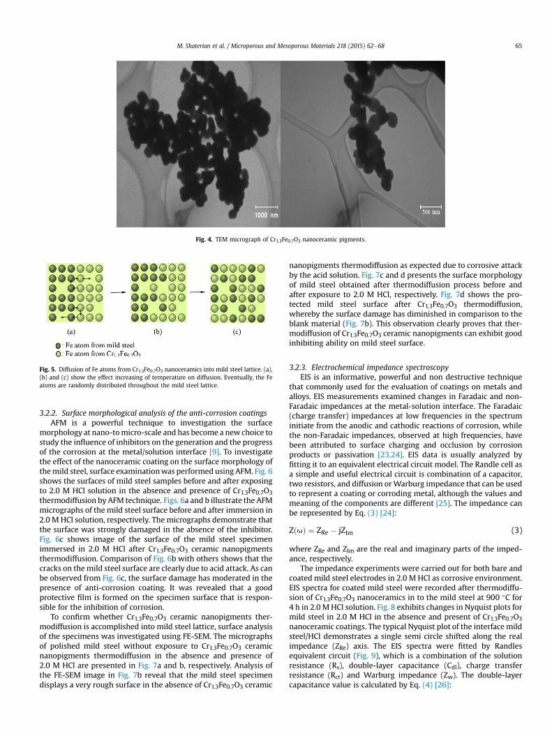
Fig. 4. TEM micrograph of Cr1.3Fe0.7O3 nanoceramic pigments.
Fig. 5. Diffusion of Fe atoms from Cr1.3Fe0.7O3 nanoceramics into mild steel lattice. (a),(b) and (c) show the effect increasing of temperature on diffusion. Eventually, the Featoms are randomly distributed throughout the mild steel lattice.
M. Shaterian et al. / Microporous and Mesoporous Materials 218 (2015) 62e68 65
3.2.2. Surface morphological analysis of the anti-corrosion coatingsAFM is a powerful technique to investigation the surface
morphology at nano-tomicro-scale and has become a newchoice tostudy the influence of inhibitors on the generation and the progressof the corrosion at the metal/solution interface [9]. To investigatethe effect of the nanoceramic coating on the surface morphology ofthemild steel, surface examinationwas performed using AFM. Fig. 6shows the surfaces of mild steel samples before and after exposingto 2.0 M HCl solution in the absence and presence of Cr1.3Fe0.7O3thermodiffusion by AFM technique. Figs. 6a and b illustrate the AFMmicrographs of the mild steel surface before and after immersion in2.0 M HCl solution, respectively. The micrographs demonstrate thatthe surface was strongly damaged in the absence of the inhibitor.Fig. 6c shows image of the surface of the mild steel specimenimmersed in 2.0 M HCl after Cr1.3Fe0.7O3 ceramic nanopigmentsthermodiffusion. Comparison of Fig. 6b with others shows that thecracks on themild steel surface are clearly due to acid attack. As canbe observed from Fig. 6c, the surface damage has moderated in thepresence of anti-corrosion coating. It was revealed that a goodprotective film is formed on the specimen surface that is respon-sible for the inhibition of corrosion.
To confirm whether Cr1.3Fe0.7O3 ceramic nanopigments ther-modiffusion is accomplished into mild steel lattice, surface analysisof the specimens was investigated using FE-SEM. The micrographsof polished mild steel without exposure to Cr1.3Fe0.7O3 ceramicnanopigments thermodiffusion in the absence and presence of2.0 M HCl are presented in Fig. 7a and b, respectively. Analysis ofthe FE-SEM image in Fig. 7b reveal that the mild steel specimendisplays a very rough surface in the absence of Cr1.3Fe0.7O3 ceramic
nanopigments thermodiffusion as expected due to corrosive attackby the acid solution. Fig. 7c and d presents the surface morphologyof mild steel obtained after thermodiffusion process before andafter exposure to 2.0 M HCl, respectively. Fig. 7d shows the pro-tected mild steel surface after Cr1.3Fe0.7O3 thermodiffusion,whereby the surface damage has diminished in comparison to theblank material (Fig. 7b). This observation clearly proves that ther-modiffusion of Cr1.3Fe0.7O3 ceramic nanopigments can exhibit goodinhibiting ability on mild steel surface.
3.2.3. Electrochemical impedance spectroscopyEIS is an informative, powerful and non destructive technique
that commonly used for the evaluation of coatings on metals andalloys. EIS measurements examined changes in Faradaic and non-Faradaic impedances at the metal-solution interface. The Faradaic(charge transfer) impedances at low frequencies in the spectruminitiate from the anodic and cathodic reactions of corrosion, whilethe non-Faradaic impedances, observed at high frequencies, havebeen attributed to surface charging and occlusion by corrosionproducts or passivation [23,24]. EIS data is usually analyzed byfitting it to an equivalent electrical circuit model. The Randle cell asa simple and useful electrical circuit is combination of a capacitor,two resistors, and diffusion orWarburg impedance that can be usedto represent a coating or corroding metal, although the values andmeaning of the components are different [25]. The impedance canbe represented by Eq. (3) [24]:
ZðuÞ ¼ ZRe � jZIm (3)
where ZRe and ZIm are the real and imaginary parts of the imped-ance, respectively.
The impedance experiments were carried out for both bare andcoated mild steel electrodes in 2.0 M HCl as corrosive environment.EIS spectra for coated mild steel were recorded after thermodiffu-sion of Cr1.3Fe0.7O3 nanoceramics in to the mild steel at 900 �C for4 h in 2.0 MHCl solution. Fig. 8 exhibits changes in Nyquist plots formild steel in 2.0 M HCl in the absence and present of Cr1.3Fe0.7O3
nanoceramic coatings. The typical Nyquist plot of the interface mildsteel/HCl demonstrates a single semi circle shifted along the realimpedance (ZRe) axis. The EIS spectra were fitted by Randlesequivalent circuit (Fig. 9), which is a combination of the solutionresistance (Rs), double-layer capacitance (Cdl), charge transferresistance (Rct) and Warburg impedance (Zw). The double-layercapacitance value is calculated by Eq. (4) [26]:
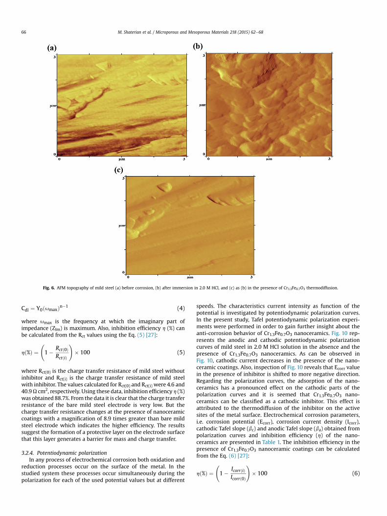
Fig. 6. AFM topography of mild steel (a) before corrosion, (b) after immersion in 2.0 M HCl, and (c) as (b) in the presence of Cr1.3Fe0.7O3 thermodiffusion.
M. Shaterian et al. / Microporous and Mesoporous Materials 218 (2015) 62e6866
Cdl ¼ Y0ðumaxÞn�1 (4)
where umax is the frequency at which the imaginary part ofimpedance (ZIm) is maximum. Also, inhibition efficiency h (%) canbe calculated from the Rct values using the Eq. (5) [27]:
hð%Þ ¼ 1� Rctð0Þ
RctðiÞ
!� 100 (5)
where Rct(0) is the charge transfer resistance of mild steel withoutinhibitor and Rct(i) is the charge transfer resistance of mild steelwith inhibitor. The values calculated for Rct(0) and Rct(i) were 4.6 and40.9U cm2, respectively. Using these data, inhibition efficiency h (%)was obtained 88.7%. From the data it is clear that the charge transferresistance of the bare mild steel electrode is very low. But thecharge transfer resistance changes at the presence of nanoceramiccoatings with a magnification of 8.9 times greater than bare mildsteel electrode which indicates the higher efficiency. The resultssuggest the formation of a protective layer on the electrode surfacethat this layer generates a barrier for mass and charge transfer.
3.2.4. Potentiodynamic polarizationIn any process of electrochemical corrosion both oxidation and
reduction processes occur on the surface of the metal. In thestudied system these processes occur simultaneously during thepolarization for each of the used potential values but at different
speeds. The characteristics current intensity as function of thepotential is investigated by potentiodynamic polarization curves.In the present study, Tafel potentiodynamic polarization experi-ments were performed in order to gain further insight about theanti-corrosion behavior of Cr1.3Fe0.7O3 nanoceramics. Fig. 10 rep-resents the anodic and cathodic potentiodynamic polarizationcurves of mild steel in 2.0 M HCl solution in the absence and thepresence of Cr1.3Fe0.7O3 nanoceramics. As can be observed inFig. 10, cathodic current decreases in the presence of the nano-ceramic coatings. Also, inspection of Fig. 10 reveals that Ecorr valuein the presence of inhibitor is shifted to more negative direction.Regarding the polarization curves, the adsorption of the nano-ceramics has a pronounced effect on the cathodic parts of thepolarization curves and it is seemed that Cr1.3Fe0.7O3 nano-ceramics can be classified as a cathodic inhibitor. This effect isattributed to the thermodiffusion of the inhibitor on the activesites of the metal surface. Electrochemical corrosion parameters,i.e. corrosion potential (Ecorr), corrosion current density (Icorr),cathodic Tafel slope (bc) and anodic Tafel slope (ba) obtained frompolarization curves and inhibition efficiency (h) of the nano-ceramics are presented in Table 1. The inhibition efficiency in thepresence of Cr1.3Fe0.7O3 nanoceramic coatings can be calculatedfrom the Eq. (6) [27]:
hð%Þ ¼ 1� IcorrðiÞ
Icorrð0Þ
!� 100 (6)
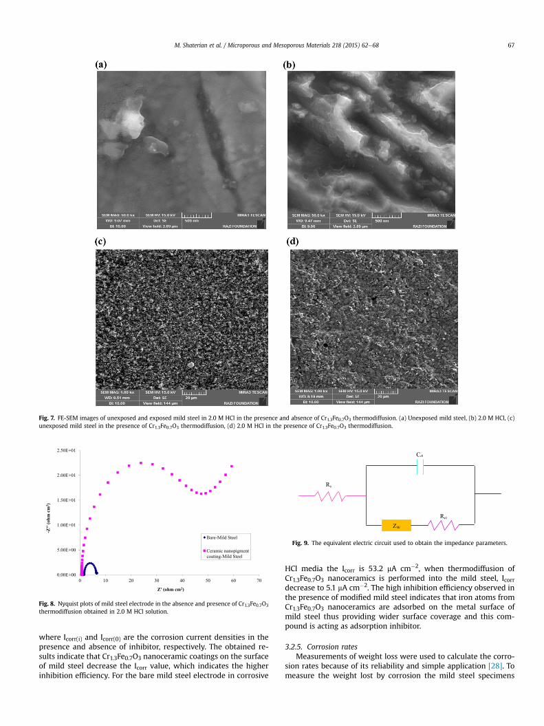
Fig. 7. FE-SEM images of unexposed and exposed mild steel in 2.0 M HCl in the presence and absence of Cr1.3Fe0.7O3 thermodiffusion. (a) Unexposed mild steel, (b) 2.0 M HCl, (c)unexposed mild steel in the presence of Cr1.3Fe0.7O3 thermodiffusion, (d) 2.0 M HCl in the presence of Cr1.3Fe0.7O3 thermodiffusion.
Fig. 8. Nyquist plots of mild steel electrode in the absence and presence of Cr1.3Fe0.7O3
thermodiffusion obtained in 2.0 M HCl solution.
Fig. 9. The equivalent electric circuit used to obtain the impedance parameters.
M. Shaterian et al. / Microporous and Mesoporous Materials 218 (2015) 62e68 67
where Icorr(i) and Icorr(0) are the corrosion current densities in thepresence and absence of inhibitor, respectively. The obtained re-sults indicate that Cr1.3Fe0.7O3 nanoceramic coatings on the surfaceof mild steel decrease the Icorr value, which indicates the higherinhibition efficiency. For the bare mild steel electrode in corrosive
HCl media the Icorr is 53.2 mA cm�2, when thermodiffusion ofCr1.3Fe0.7O3 nanoceramics is performed into the mild steel, Icorrdecrease to 5.1 mA cm�2. The high inhibition efficiency observed inthe presence of modified mild steel indicates that iron atoms fromCr1.3Fe0.7O3 nanoceramics are adsorbed on the metal surface ofmild steel thus providing wider surface coverage and this com-pound is acting as adsorption inhibitor.
3.2.5. Corrosion ratesMeasurements of weight loss were used to calculate the corro-
sion rates because of its reliability and simple application [28]. Tomeasure the weight lost by corrosion the mild steel specimens
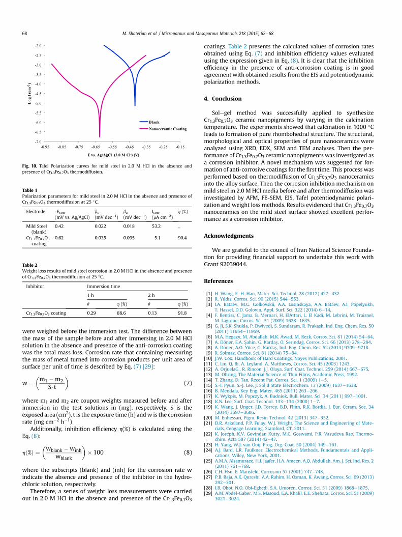
Fig. 10. Tafel Polarization curves for mild steel in 2.0 M HCl in the absence andpresence of Cr1.3Fe0.7O3 thermodiffusion.
Table 1Polarization parameters for mild steel in 2.0 M HCl in the absence and presence ofCr1.3Fe0.7O3 thermodiffusion at 25 �C.
Electrode -Ecorr(mV vs. Ag/AgCl)
bc(mV dec�1)
ba(mV dec�1)
Icorr(mA cm�2)
h (%)
Mild Steel(blank)
0.42 0.022 0.018 53.2 _
Cr1.3Fe0.7O3
coating0.62 0.035 0.095 5.1 90.4
Table 2Weight loss results of mild steel corrosion in 2.0 M HCl in the absence and presenceof Cr1.3Fe0.7O3 thermodiffusion at 25 �C.
Inhibitor Immersion time
1 h 2 h
q h (%) q h (%)
Cr1.3Fe0.7O3 coating 0.29 88.6 0.13 91.8
M. Shaterian et al. / Microporous and Mesoporous Materials 218 (2015) 62e6868
were weighed before the immersion test. The difference betweenthe mass of the sample before and after immersing in 2.0 M HClsolution in the absence and presence of the anti-corrosion coatingwas the total mass loss. Corrosion rate that containing measuringthe mass of metal turned into corrosion products per unit area ofsurface per unit of time is described by Eq. (7) [29]:
w ¼�m1 �m2
S$t
�(7)
where m1 and m2 are coupon weights measured before and afterimmersion in the test solutions in (mg), respectively, S is theexposed area (cm2), t is the exposure time (h) andw is the corrosionrate (mg cm�2 h�1)
Additionally, inhibition efficiency h(%) is calculated using theEq. (8):
hð%Þ ¼�wblank �winh
wblank
�� 100 (8)
where the subscripts (blank) and (inh) for the corrosion rate windicate the absence and presence of the inhibitor in the hydro-chloric solution, respectively.
Therefore, a series of weight loss measurements were carriedout in 2.0 M HCl in the absence and presence of the Cr1.3Fe0.7O3
coatings. Table 2 presents the calculated values of corrosion ratesobtained using Eq. (7) and inhibition efficiency values evaluatedusing the expression given in Eq. (8). It is clear that the inhibitionefficiency in the presence of anti-corrosion coating is in goodagreement with obtained results from the EIS and potentiodynamicpolarization methods.
4. Conclusion
Solegel method was successfully applied to synthesizeCr1.3Fe0.7O3 ceramic nanopigments by varying in the calcinationtemperature. The experiments showed that calcination in 1000 �Cleads to formation of pure rhombohedral structure. The structural,morphological and optical properties of pure nanoceramics wereanalyzed using XRD, EDX, SEM and TEM analyses. Then the per-formance of Cr1.3Fe0.7O3 ceramic nanopigments was investigated asa corrosion inhibitor. A novel mechanism was suggested for for-mation of anti-corrosive coatings for the first time. This process wasperformed based on thermodiffusion of Cr1.3Fe0.7O3 nanoceramicsinto the alloy surface. Then the corrosion inhibition mechanism onmild steel in 2.0 M HCl media before and after thermodiffusionwasinvestigated by AFM, FE-SEM, EIS, Tafel potentiodynamic polari-zation and weight loss methods. Results evidenced that Cr1.3Fe0.7O3nanoceramics on the mild steel surface showed excellent perfor-mance as a corrosion inhibitor.
Acknowledgments
We are grateful to the council of Iran National Science Founda-tion for providing financial support to undertake this work withGrant 92039044.
References
[1] H. Wang, E.-H. Han, Mater. Sci. Technol. 28 (2012) 427e432.[2] R. Yıldız, Corros. Sci. 90 (2015) 544e553.[3] I.A. Bataev, M.G. Golkovskii, A.A. Losinskaya, A.A. Bataev, A.I. Popelyukh,
T. Hassel, D.D. Golovin, Appl. Surf. Sci. 322 (2014) 6e14.[4] F. Bentiss, C. Jama, B. Mernari, H. ElAttari, L. El Kadi, M. Lebrini, M. Traisnel,
M. Lagrene, Corros. Sci. 51 (2009) 1628e1635.[5] G. Ji, S.K. Shukla, P. Dwivedi, S. Sundaram, R. Prakash, Ind. Eng. Chem. Res. 50
(2011) 11954e11959.[6] M.A. Hegazy, M. Abdallah, M.K. Awad, M. Rezk, Corros. Sci. 81 (2014) 54e64.[7] A. D€oner, E.A. Sahin, G. Kardas, O. Serinda�g, Corros. Sci. 66 (2013) 278e284.[8] A. D€oner, A.O. Yüce, G. Kardas, Ind. Eng. Chem. Res. 52 (2013) 9709e9718.[9] R. Solmaz, Corros. Sci. 81 (2014) 75e84.
[10] J.W. Cox, Handbook of Hard Coatings, Noyes Publications, 2001.[11] C. Liu, Q. Bi, A. Leyland, A. Matthews, Corros. Sci. 45 (2003) 1243.[12] A. OrjuelaG., R. Rinc�on, J.J. Olaya, Surf. Coat. Technol. 259 (2014) 667e675.[13] M. Ohring, The Material Science of Thin Films, Academic Press, 1992.[14] T. Zhang, D. Tan, Recent Pat. Corros. Sci. 1 (2009) 1e5.[15] S.-I. Pyun, S.-J. Lee, J. Solid State Electrochem. 13 (2009) 1637e1638.[16] B. Mendala, Key Eng. Mater. 465 (2011) 263e266.[17] K. Wykpis, M. Popczyk, A. Budniok, Bull. Mater. Sci. 34 (2011) 997e1001.[18] K.N. Lee, Surf. Coat. Technol. 133e134 (2000) 1e7.[19] K. Wang, J. Unger, J.D. Torrey, B.D. Flinn, R.K. Bordia, J. Eur. Ceram. Soc. 34
(2014) 3597e3606.[20] M. Enhessari, Pigm. Resin Technol. 42 (2013) 347e352.[21] D.R. Askeland, P.P. Fulay, W.J. Wright, The Science and Engineering of Mate-
rials, Cengage Learning, Stamford, CT, 2011.[22] K. Joseph, K.V. Govindan Kutty, M.C. Goswami, P.R. Vasudeva Rao, Thermo-
chim. Acta 587 (2014) 42e47.[23] H. Yang, W.J. van Ooij, Prog. Org. Coat. 50 (2004) 149e161.[24] A.J. Bard, L.R. Faulkner, Electrochemical Methods, Fundamentals and Appli-
cations, Wiley, New York, 2001.[25] A.M.A. Alsamuraee, H.I. Jaafer, H.A. Ameen, A.Q. Abdullah, Am. J. Sci. Ind. Res. 2
(2011) 761e768.[26] C.H. Hsu, F. Mansfeld, Corrosion 57 (2001) 747e748.[27] P.B. Raja, A.K. Qureshi, A.A. Rahim, H. Osman, K. Awang, Corros. Sci. 69 (2013)
292e301.[28] I.B. Obot, N.O. Obi-Egbedi, S.A. Umoren, Corros. Sci. 51 (2009) 1868e1875.[29] A.M. Abdel-Gaber, M.S. Masoud, E.A. Khalil, E.E. Shehata, Corros. Sci. 51 (2009)
3021e3024.