Embed Size (px)
Citation preview
JOURNAL OF COMPUTATIONAL BIOLOGYVolume 10 Numbers 3ndash4 2003copy Mary Ann Liebert IncPp 555ndash567
A New Method for Mapping DiscontinuousAntibody Epitopes to Reveal Structural
Features of Proteins
BRENDAN M MUMEY1 BRIAN W BAILEY2 BONNIE KIRKPATRICK1
ALGIRDAS J JESAITIS3 THOMAS ANGEL4 and EDWARD A DRATZ4
ABSTRACT
Antibodies that bind to protein surfaces of interest can be used to report the three-dimen-sional structure of the protein as follows Proteins are composed of linear polypeptide chainsthat fold together in complex spatial patterns to create the native protein structure Thesefolded structures form binding sites for antibodies Antibody binding sites are typicallyldquoassembledrdquo on the protein surface from segments that are far apart in the primary aminoacid sequence of the target proteins Short amino acid probe sequences that bind to theactive region of each antibody can be used as witnesses to the antibody epitope surfaceand these probes can be ef ciently selected from random sequence peptide libraries Thispaper presents a new method to align these antibody epitopes to discontinuous regions of theone-dimensional amino acid sequence of a target protein Such alignments of the epitopesindicate how segments of the protein sequence must be folded together in space and thusprovide long-range constraints for solving the 3-D protein structure This new antibody-based approach is applicable to the large fraction of proteins that are refractory to currentapproaches for structure determination and has the additional advantage of requiring verysmall amounts of the target protein The binding site of an antibody is a surface not justa continuous linear sequence so the epitope mapping alignment problem is outside thescope of classical string alignment algorithms such as SmithndashWaterman We formalize thealignment problem that is at the heart of this new approach prove that the epitope mappingalignment problem is NP-complete and give some initial results using a branch-and-boundalgorithm to map two real-life cases Initial results for two validation cases are presentedfor a graph-based protein surface neighbor mapping procedure that promises to provideadditional spatial proximity information for the amino acid residues on the protein surface
Key words protein structure antibody imprinting epitope mapping protein surface graphs
1Department of Computer Science Montana State University Bozeman MT 59717-38802NIHNIAAADICBRLMBB Fluorescence Studies Park 5 Building 12420 Parklawn Drive MSC 8115 Bethesda
MD 20892-81153Department of Microbiology Montana State University Bozeman MT 59717-35204Department of Chemistry and Biochemistry Montana State University Bozeman MT 59717-3400
555
556 MUMEY ET AL
1 ANTIBODY EPITOPE MAPPING
Proteins are nano-machines that are constructed from long chains of amino acids (typically100ndash1000 elements) using twenty different amino acids arranged in characteristic sequences Proteins
must be folded into complex 3-D shapes to create the binding pockets and active sites necessary to carryout their myriad different functions (Branden and Tooze 1999) There are at least 30000 different proteinsin human cells (Claverie 2001) and each protein has a folded functional structure Whenever the 3-Dfolded structure of linear protein sequences could be determined this information has provided importantinsights into mechanisms of action and may be extremely useful in drug design Traditional methods ofprotein structure determination require preparation of large amounts of protein in functional form whichoften may not be feasible Given suf cient protein of interest conditions are screened to seek 3-D crystalsfor structure determination by x-ray diffraction however obtaining crystals of suf cient quality may notbe possible (McPherson 1999 Michel 1990) Alternatively if the proteins are not too large are highlywater soluble and meet other criteria methods of nuclear magnetic resonance can be used for structuredetermination (Cavanagh et al 1996) It is also possible to predict 3-D structures de novo from thesequence of amino acids in the protein but the available methods for structure prediction are not veryaccurate unless a 3-D structure of a homologous protein is already known (Baker and Sali 2001) (also seepredictioncenterllnlgov)
A large fraction of protein structures of interest (50 or more) cannot be solved by the traditionalapproaches discussed above (Edwards et al 2000 Eisenstein et al 2000) Thus we are developingthe antibody imprint method to provide structural information on dif cult cases that appear refractory totraditional approaches (Burritt et al 1998 Jesaitis et al 1999 Bailey et al 2003) The antibody imprintmethod makes use of information carried in the structures of antibodies against proteins of interest to revealthe 3-D folding of target proteins (Burritt et al 1998 Jesaitis et al 1999 Demangel et al 2000 Heiskanenet al 1999 Bailey et al 2003) Antibodies tend to be highly speci c for the protein structures thatthey recognize (Janeway and Travers 1996) Antibodies can recognize either continuous or discontinuousepitopes Discontinuous epitopes provide the most useful structural information in antibody imprintingbecause they can reveal distant segments of primary sequence that are in close spatial proximity on thenative folded protein Evidence to date indicates that most antibodies recognize discontinuous epitopeson protein surfaces (Padlan 1996) Studies of a substantial number of antibodyndashprotein complexes withknown x-ray structures indicate that these complexes form in a lock and key manner with little or nostructural change induced by complex formation (Conte et al 1999) Fortunately relatively few long-distance constraints are needed to reveal the global folding of proteins (Clore et al 1993 Dandekar andArgos 1997) In addition the spatial proximity of different regions of proteins can change during functionand antibody imprinting has the potential to reveal these structural changes if appropriate antibodies canbe found that recognize the different structural shapes (Bailey et al 2003)
Brie y the antibody imprinting method is carried out by rst immobilizing antibodies (against a target ofinterest) on beads or in plastic wells The immobilized antibodies are exposed to random peptide librariesso that library members which bind to the antibodies can be captured by the surface The random peptidelibraries are carried on bacteriophage (that is called ldquophage displayrdquo of the library) as is reviewed in thefollowing reference (Barbas et al 2001) Each phage has a different peptide expressed on the surface ofone of its coat proteins and there are typically 5 cent 109 (Burritt et al 1996) and even up to 1012 differentpeptide sequences in each library (Sidhu et al 2000) These probe libraries contain linear peptides or can beconstrained with circular topology where the two ends of the probe are chemically linked with a disul debond Peptide sequences that do not stick to the antibody are washed off the immobilized antibodies andthe tightly binding phage are eluted under harsher conditions The phages that bind to the antibody aremultiplied by growth in suitable bacteria and exposed again to the immobilized antibody These cyclesof binding and enrichment of members of the random peptide library are usually repeated three times toselect the phages with the highest af nity to the antibody These enriched phages are then highly dilutedand grown as clones that arise from individual phage particles Each of the phage clones carry the DNAsequence that codes for the peptide sequence that has been selected The DNA regions of selected clonesare ampli ed by PCR with uorescent terminators and sequenced in a standard automated DNA sequencerIn this way the sequence for each epitope-mimetic peptide is discovered These individual sequences areoften highly conserved and 25ndash100 independent peptide sequences together describe a consensus sequence
MAPPING DISCONTINUOUS ANTIBODY EPITOPES 557
called the consensus epitope of the antibody The problem addressed in the present paper is to develop ameans to examine and evaluate all possible ways in which an epitope-mimetic peptide can be mapped ontothe sequence of the target protein in question to recognize discontinuous epitopes that provide proximityconstraints on the 3-D structure of the protein We adopt the terminology that a peptide epitope sequenceforms a probe that is to be aligned to the protein target sequence
In the remainder of the paper we formalize the probendashtarget alignment problem describe a branch-and-bound algorithm to nd optimal (and suboptimal) alignments prove the corresponding decision problemis NP-complete and provide some experimental results for two biologically signi cant proteins We thendescribe some initial work dispensing with a consensus epitope and using individual probendashtarget align-ments to form a surface neighbor graph apply this approach to two validation examples and comment onsome future directions being pursued in this work
2 FORMALIZING THE PROBLEM
The core idea of the antibody imprint method is that a probe that binds to the active region of aparticular antibody is expected to be highly similar to the binding site of a protein that also binds to thesame antibody We thus are faced with the problem of aligning the probe amino acid sequence s to oneor more regions of the target protein amino acid sequence t Typically s is about 8ndash20 amino acids longand t is several hundred Unlike traditional string alignment problems we allow for localized sequencerearrangements This captures the possibility that several loops of the linear protein sequence may bepinched together (possibly with sequence inversions) to form the binding site Additionally it is possiblefor local rearrangements of amino acids to occur re ecting the fact that the binding site of an antibody is asurface not just a linear sequence As such the problem is outside the scope of classical string alignmentalgorithms such as SmithndashWaterman (Smith and Waterman 1981) We have chosen an approach based ona general combinatorial alignment problem although alternative strategies such as hidden Markov modelsand stochastic free grammars have been employed for related problems and could be explored
In general we will allow any permutation of the probe sequence to align to the underlying proteinsequence1 Furthermore gaps will be permitted in both probe and target sequences Large gaps can occurwhen aligning the probe to the target sequence when the epitope is discontinuous We also allow unalignedprobe residues re ecting the possibility of nonspeci c residue insertions in the probe To be a validalignment each probe position and target position can be used at most once per mapping Formally analignment A consists of a sorted set PA D fi1 lt i2 lt cent cent cent lt ikg and another set TA D fj1 j2 jkgwith the interpretation that the ip-th probe residue sip is aligned to the jp-th target residue t jp for1 middot p middot k
We adopt a two-part scoring system to evaluate the quality of alignments The scoring system is composedof a substitution score and a epitope gap cost
scoreA D SA iexcl GA
The SA component is calculated with a substitution matrix M similar in principle to a Dayhoff matrixused in other protein alignment contexts We discuss our choice of substitution matrix in the experimentalresults section The substitution matrix is also used to score unaligned probe residues if the probe residuein position i is not aligned to any target position then it is charged a penalty according to the characterc occurring in position i of the probe sequence This cost can be found in the substitution matrix in theentry Mc ndash We have
SA DkX
pD1
Msip t jp CX
probe positions i =2 PA
Msi ndash
1In some cases eg membrane-spanning proteins it may be known or likely that certain regions of the targetprotein are inaccessible to antibodies and thus can be excluded from consideration as potential alignment positions
558 MUMEY ET AL
The epitope gap cost GA is calculated by examining the number of amino acid residues skipped alongthe target protein sequence between successive aligned probe positions
GA Dkiexcl1X
pD1
d[jjpC1 iexcl jpj]
where dx is the cost of skipping x amino acids along the target between successive mapped probepositions For circular probes we also include the term d[jjk iexcl j1j] in the above sum The computationalproblem is thus to nd an alignment A that maximizes scoreA We have evaluated different gap costmodels and discuss this point later in the results section
A branch-and-bound algorithm can be used to solve this alignment problem in practice The algorithmconstructs a search tree to nd the optimal alignment(s) Often a user may also be interested in near-optimal solutions so the algorithm is designed to nd the top r solutions where r is user speci ed Eachnode in the search tree represents a partial alignment of the probe to the protein sequence At the root allprobe positions are unaligned Nodes at level i gt 0 in the tree x the alignment of the i-th probe position(either to an available target position or to a ldquondashrdquo indicating an unmatched probe position) A leaf is reachedwhen all probe positions have been considered and each leaf represents a particular alignment Whenevera new node n is created an upper bound on the highest possible alignment score achievable in the subtreerooted at n is computed If this bound is less than the r-th best solution found so far we can immediatelyprune the node from the search Nodes that are on the boundary of the current search tree are said to beon the frontier For each frontier node n an expected score is calculated by dividing nrsquos current score byits depth in the tree A heap data structure is used to extract a node with maximal expected score from thefrontier This node is then expanded descendant child nodes are created for each possible alignment ofthe next probe position When a leaf is reached the score of the associated alignment is calculated Thisscore is compared to the current r-th best solution and if greater replaces it When such a replacementoccurs the frontier is scanned to cull out any other nodes that can now be eliminated This algorithm hasbeen implemented as a C++ program called FINDMAP The experimental results section presents someof our initial experience with FINDMAP most problems of interest run in a few minutes or less on a fastworkstation
3 PROBLEM COMPLEXITY
In this section we show that the probendashtarget sequence alignment problem is NP-complete (Garey andJohnson 1979) We rst de ne a decision version of the problem
The ALIGN decision problem
Input A probe string s a target string t (over a common alphabet) a substitution score matrix M adistance penalty function d an objective score Q
Output A decision on whether there exists an alignment with score at least Q
Lemma 1 ALIGN is NP-complete
Proof First note that ALIGN belongs to NP because the score of a given alignment can be checkedin polynomial time We will show that ALIGN is complete for NP via a polynomial time reduction from3SAT Consider an instance of 3SAT I3S consisting of a collection of clauses C D fc1 c2 cmg ona nite set of variables U D fx1 xkg We will describe a polynomial time reduction to an instanceIA D s t M d Q of ALIGN such that a truth assignment exists for U that satis es C if and only ifan alignment between s and t with score at most Q can be found We construct IA as follows The stringalphabet used is
A D U [ fx1 xkg [ fy1 ykg [ flsquoc1rsquo lsquocmrsquog [ flsquorsquo lsquocurrenrsquo lsquorsquog
MAPPING DISCONTINUOUS ANTIBODY EPITOPES 559
All entries of M are set to iexcl1 except the following Mreg ci D 0 if reg is a literal in clause ci Mxi yi DMxi yi D 0 for all 1 middot i middot n and Mcent lsquocurrenrsquo D 0 (here ldquocentrdquo represents any symbol) For each literal reglet [reg] be the multiplicity of reg among all clauses in C The probe string used is
s D B1 B2 cent cent cent Bk
where
Bi D xi cent cent cent xi| z [xi ] C 1 copies
xi cent cent cent xi| z [xi] C 1 copies
Let n D jsj iexcl m C k The target string used is
t D curren curren cent cent cent curren| z n copies
cent cent cent | z n copies
c1 c2 cent cent cent cm y1 y2 cent cent cent yk
The distance penalty function used is
dl Draquo
0 if l lt n
1 otherwise
Observe that m C k lt n so only jumps across the central gap of rsquos referred to as the bridge willcontribute to the gap cost The leading of s forces any nite-score alignment to begin on the left sideof the bridge Note that every non- letter in the target must be matched in order to completely alignthe probe (all probe positions must be matched as Mcent ndash D iexcl1) In order to match all of the yi rsquos atleast one literal from each Bi must be used Thus each Bi contributes at least one return jump across thebridge If a literal is matched against a clause symbol ci then any truth assignment that makes this literaltrue will satisfy ci We choose Q D iexcl2k to insist that each Bi contributes exactly one return jump acrossthe bridge Because the positive and negative literals in each block Bi are separated by an only literalsof a single polarity can be matched to symbols to the right of the bridge This ensures a consistent truthassignment Thus any alignment with score exactly iexcl2k will produce a satisfying assignment for I3S andvice versa
4 EXPERIMENTAL RESULTS
In this section we discuss our initial experimental results using FINDMAP and our implementation ofthe branch-and-bound alignment algorithm described above We discuss two cases a validation case wherethe 3-D structure is known and a second case where the structure has not been fully solved FINDMAPrequires an amino acid substitution probability matrix to score sequence alignments We chose the matrixshown in Fig 1 since a very similar substitution matrix was developed by Bordo and Argos (1991) forscoring substitutions of protein residues exposed to the aqueous surface Antibody binding sites on targetproteins must be exposed to the aqueous surface for antibody accessibility and so an aqueous-exposedsubstitution seems appropriate As indicated under future directions we are in the process of obtaining anexperimental substitution matrix optimized for antibody imprinting
Recently Jesaitis and coworkers carried out antibody imprinting using a polyclonal antibody against theubiquitous cytoskeletal protein actin (Jesaitis et al 1999) They reported the manual mapping of consensuspeptides derived from phage display library selection to complex epitopes on the surface of actin Thephage-display-discoveredpeptides could be mapped onto the actin surface to mimic a discontinuousepitopethat was consistent with the known 3-D x-ray structure of actin (Kabsch et al 1990) Figure 2 showsthe mapping of one of the consensus sequences VPHPTWMR onto the surface of actin and the almostidentical FINDMAP mapping It should be emphasized that this manual mapping utilized knowledge ofthe actin x-ray structure and did not use residues marked with that are not exposed on the aqueoussurface in the x-ray structure The FINDMAP alignment used only the protein primary sequence Thesingle difference from the manual mapping is FINDMAPrsquos selection of the more buried but plausible
560 MUMEY ET AL
FIG 1 Amino acid substitution scoring matrix used in FINDMAP This matrix is based on the probability ofamino acid substitutions on surface-exposed residues of proteins The Bordo and Argos (1991) substitution matrixwas modi ed so that GlyPro substitutions score 050 and ArgHis LysHis and GlySer substitutions score 025Unaligned probe positions were charged a penalty of iexcl1 The gap penalty discussed in the text was levied againstgaps in the target protein sequence that were not aligned with probe residues
Thr 103 (dark-green residue) instead of the more exposed Thr 358 (maroon-colored residue) for the Tin VPHPTWMR We viewed this result as an initial validation of the antibody imprinting technique andFINDMAP on a known protein structure Additional validation studies are described in a later section It ispossible that including an estimate for the probability of surface exposure in the overall alignment scoringfunction could be useful (Jameson and Wolf 1988) and this is being explored further
We also used the actin test case to optimize the gap cost parameters for gaps in the alignment of thetarget protein sequence to the probes We chose a simple linear penalty function up to a maximum gappenalty that does not further penalize long gaps (long gaps are expected to be frequent in discontinuousepitopes)
dn D mina cent n b
To search for suitable values of a and b we ran FINDMAP on the actin example where the 3-Dstructure is known using the probe sequence VPHPTWMR We tested 140 different combinations ofa b pairs as shown in Fig 3 The deviation from the proper mapping with parameter values that werenonoptimal were systematic When a was set too small the highest scoring epitopes found were implausiblydiscontinuous with identity matches widely spread in the mappings at the expense of any allowable aminoacid substitutions In contrast when a was too large excessively continuous local epitopes were found thatmay include large numbers of very nonfavorable amino acid substitutions In Fig 3 the best parameterchoices yielded 18 alignments that had identical optimal scores of which one agreed exactly with themanual mapping except at one residue position as described in the caption to Fig 2 A reason for theproliferation of near optimal solutions in this case is the freedom of the nal R in the consensus probe toalign to a number of positions in the target) We picked a D 05 and b D 15 from the best region for thesubsequent experiments to be discussed
The second case we considered was the integral membrane protein rhodopsin the structure of whichis not fully known Rhodopsin is the photoreceptor for dim light vision in retinal rod cells and is anarchetype for the structure and mechanism of a large superfamily of cellular G protein-coupled receptor(GPCR) proteins that respond to a wide range of hormones and neurotransmitters (Wess 1997 Marinissenand Gutkind 2001) The x-ray crystal structure of the dark-adapted resting structure of rhodopsin wasrecently published (Palczewski et al 2000 Teller et al 2001) but some of the features of the proteinon the cytoplasmic surface were poorly ordered in the crystals and not visible in the x-ray structure Acomputational model of the missing portions of the cytoplasmic surface was built and energy minimized
MAPPING DISCONTINUOUS ANTIBODY EPITOPES 561
FIG 2 Mapping of the anti-actin antibody epitope VPHPTWMR onto the surface of actin manually and by FIND-MAP Mapped residues are color coded in rainbow order from red to purple for the FINDMAP results based on theprobe peptide sequence from N to C-terminal The manual and FINDMAP mappings differ only in their alignment ofThr 358 (maroon) where FINDMAP tends to pick Thr 103 (dark green) The independent manual mapping requiredknowledge of the actin x-ray structure The top-scoring FINDMAP alignment having the best match is shown abovethe actin sequence (rsquos indicate residues known to be folded away from the aqueous surface of actin these regionswere excluded in the manual mapping)
(Bailey 2001) The cytoplasmic surface structure was uncertain in the model so antibody imprinting wasapplied to the aqueous surfaces (Bailey et al 2003)
One of the antibodies investigated (B1gN) maps to the extracellular surface of rhodopsin in a compactpatch that shows the proximity of two distant segments of sequence This mapping is in excellent agreementwith a well de ned region of the x-ray structure (Bailey et al 2003) (data not shown) One of the otherantibodies studied (4B4) targeted part of rhodopsin where the x-ray structure was not fully resolved anda model of this region is shown in Fig 4A The single most optimal mapping of the 4B4 epitope found
562 MUMEY ET AL
FIG 3 FINDMAP epitope gap penalty parameter sensitivity For the gap distance penalty function dn Dmina cent n b various combinations of a and b where tested on mapping the probe epitope VPHPTWMR to theactin sequence (see Fig 2) These alignments were ranked into three categories based on how closely they agreed withthe published manual mapping to the known 3-D structure of actin (Jesaitis et al 1999) The diamond-shaped pointsin the gure indicate parameter combinations where FINDMAP found the published mapping to within one residueposition as one of the top-scoring alignments the parameter combinations used to yield the square points missed twoor three residues and the triangular region more than three residues
with FINDMAP was unusual in that it was continuous with a segment of the rhodopsin sequence Theoptimal mapping however showed a spatial discontinuity in the proximity of two parts of the epitope asillustrated in Fig 4 The aligned epitope runs from the residue colored red in Fig 4B with a rainbow colorscheme to orange yellow and light green The dark-green residue is predicted by FINDMAP to be locatedspatially adjacent to the light-green residue but there is a large jump in the structural model as shown inFig 4A This is evidence that the surface loop folding model shown in Fig 4A is incorrect and should beadjusted to form a hairpin turn bringing A235 next to S240 as shown in Fig 4B This example supportsthat notion that the antibody imprinting technique is capable of providing new structural informationExperiments are in progress to obtain more detailed conformational information by crystallizing epitope-mimetic peptides with the active site of the antibodies to provide detailed folding information on regionsof the protein surface (Lawrence Bailey and Dratz unpublished) The x-ray crystallography appears tobe straightforward for co-crystals of peptides with antibody active sites since molecular replacement withknown antibody structures should provide the phases
Additional antibodies will be required to reveal the complete surface structure of rhodopsin and itslight-excited conformations More detailed antibody imprinting studies seeking to deduce light-stimulatedconformational changes in rhodopsin are in progress and some of these have been submitted for publication(Bailey et al 2003) Most of the antibody epitopes are found to be discontinuous and thus provideimportant long-range distance constraints on the structures If regions of the surfaces studied are exible itis anticipated that a range of conformations will be deduced by different antibody epitopes consistent withthat exible structure as would be found if other structural techniques such as NMR or x-ray diffractioncould be applied It should also be noted that it is possible to include additional information such as fromstructure prediction algorithms or experimental information if available from intramolecular cross-linksidenti ed by mass spectrometry (Young et al 2000) or from site-speci c spin labeling (Hubbell et al
MAPPING DISCONTINUOUS ANTIBODY EPITOPES 563
FIG 4 Mapping of the 4B4 antibody epitope on rhodopsin Panel A shows the epitope of the 4B4 antibody mappedon the cytoplasmic surface of a model of the dark-adapted rhodopsin 3-D structure This region is not resolved inthe x-ray crystal structure as explained in the text The 4B4 consensus probe EQQVSATAQ was best aligned usingFINDMAP to the rhodopsin residues EQQASATTQ Mapped epitope residues are shown such that they follow arainbow color scheme from red (the rst residue of the consensus epitope) to purple Different cytoplasmic regions ofthe protein are color coded salmon residues are the C-terminal segment dark gray is the loop between helices V andVI medium gray is the loop between helices III and IV and light gray is the loop between helices I and II Panel Bshows the proposed reorientation of residues 235ndash244 of the C-3 loop of rhodopsin with A235 moved next to S240based on the best-scoring FINDMAP alignment as discussed in the text
2000) to add to the information obtained from FINDMAP or to prioritize alternative FINDMAP spatialproximity mappings
5 A GRAPH-BASED APPROACH TO SURFACE EPITOPE MAPPING
An improvement to the overall process that we are currently pursuing is to eliminate the step of ndinga consensus epitope sequence Typically 25ndash100 peptide probes are sequenced that show strong af nity tothe antibody in question These sequences are often rather similar but typically not identical The centerof the antibody combining site is expected to contribute to the highest probe af nity (Conte et al 1999)whereas more peripheral binding site residues tend to make a lower contribution to the af nity and thusmay show alternative binding modes Rather than going through the step of nding a consensus sequenceFINDMAP can be run on each of the probe sequences individually to generate a family of top-scoringalignment sets one set for each probe These alignments are similar but often indicate the proximity ofadditional residues on the protein surface We using a graph-based approach to merge and visualize thecollective surface proximity information provided in the entire set of alignments In this approach eachresidue of the target protein constitutes a vertex in a weighted surface-neighbor graph Edge weightsin this graph indicate how strongly the alignment data supports the conclusion that the residues at eachendpoint are neighbors on the surface of the protein The speci c procedure employed for calculating edgeweights is as follows for each probe compute the set of top-scoring alignments Suppose there are n suchalignments and that a particular pair of residues are neighbors in k of these alignments Then k=n is addedto the weight of the edge between the two residues in question After this procedure is repeated for eachprobe edges that have comparatively high weights are most likely to link residues that are true surfaceneighbors In practice errors occur both in the experimental methods used to identify the probe sequencesas well as in cases where the top-scoring alignments are not biologically accurate Thus it appears usefulto use a weight cutoff edges are kept only if their weight is greater than a prespeci ed cutoff If the cutoffis too low it is likely that false surface neighbor relations will be included in the graph too high andtrue neighbors will be lost Another procedure that seems useful for pruning out nonepitope residues fromthe surface neighbor graph is to retain only vertices that are incident to at least one high-weighted edgeThis procedure was performed on the surface neighbor graphs shown in Fig 5 only vertices incident to
564 MUMEY ET AL
an edge of weight at least 50 of the maximum weight were kept Also a cutoff value of 1 was used toprune low weight edges
The target sequence is also scanned for multiple occurrences of tripeptide (very rare) or dipeptidesequences in the probe and hits involving these ambiguous sequences are omitted from consideration tominimize false-positive hits It is important to eliminate false-positive residue proximity information toprovide accurate structure whereas false negatives are more tolerable An example of a surface neighborgraph based on actin FINDMAP alignment data of individual probes is shown in Fig 5A The somewhatlarger protein surface mapped with this approach compared to Fig 2 is consistent with the fact that theantibody investigated is polyclonal Monoclonals that we have primarily used in this work provide surfacemaps with a smaller area coverage but it has been found feasible to map mixtures of several monoclonalsin parallel in a single experiment (Bailey and Dratz unpublished)
We wish to explore to what extent the surface neighbor graph can be used to make a map of thesurface of the protein The protein surface is two dimensional so it seems feasible to consider planarembeddings of the surface neighbor graph that place residue vertices in such a way that heavily weightededges connect neighboring vertices in the embedding Another criteria is that residues should be packedin a roughly uniform way perhaps in lattices andor proportional to their molecular volume We arecurrently experimenting with various approaches to perform this embedding including the use of somestandard graph layout packages (Fig 5 was generated using the program Graphlet available at wwwfmiuni-passaudeGraphlet) A further important constraint is that residues that are consecutive in the linearprotein sequence must also necessarily be neighbors in the embedded surface map Other related problemsthat might be useful for protein surface mapping from antibody epitope data include maximum planarsubgraph and minimum edge distance graph layout
We are also investigating a number of antibodies where the 3-D structure of the antibody-target proteincomplex is known to atomic resolution by x-ray diffraction to more thoroughly validate the accuracy ofthe surface-mapping method In these validation cases the correct antibody epitope mappings are knownThe rst case that we have investigated is hen egg lysozyme complexed with several different monoclonalantibodies The antibody contacts on the lysozyme surface have been identi ed (using the CCP4 Contactsprogram wwwccp4acukmainhtml) A collection of 50 hypothetical probe sequences were generatedby randomly connecting adjacent residues on the lysozyme contact surface on the Hyhel-10 antibodyFINDMAP alignments were found to all the probes generated and the epitope surface neighbor graphwas found using the method described above In Fig 5C we show the computed surface neighbor graphand the true epitope surface for monoclonal antibody Hyhel-10 (PDB1C08) Also shown in Fig 5D is adiagram of the experimental monoclonal antibody epitope that is seen to agree favorably with the surfaceneighbor graph edge weights
6 FUTURE WORK
The antibody imprinting approach appears to be quite general and we are applying it to a number ofadditional cases We are using antibody-protein complexes whose 3-D structure is known to optimize thevalues of the gap penalty parameters We are further validating this approach by experimentally mappingseveral known antilysozyme epitopes and are using this information to re ne the substitution matrix Thevalidation cases are also expected to be a useful guide for more systematic choices of cutoff weights forthe planar graph models of protein surfaces as shown in Fig 5 Finally we are applying the antibodyimprinting technique to several integral membrane proteins that are dif cult structural targets (Mills et al1998 Burritt et al 2001) and to reveal the nature of functional conformational changes in membraneproteins (Bailey et al 2003)
It has not escaped our attention that all the steps in the antibody imprinting process are adaptable to highthroughput enhancement As more experience is gained with this technique especially with the known testcases to aid in re ning the substitution matrix the gap penalty parameters and surface graph weights weplan to introduce high throughput enhancements to the epitope selection epitope sequencing and epitopemapping In some cases suitable antibodies are already available but in many cases suitable antibodieshave not been prepared or do not provide suf cient coverage of the protein surface or the conformationalstates of interest In the absence of available antibodies the rate-limiting step in the current process is
MAPPING DISCONTINUOUS ANTIBODY EPITOPES 565
FIG 5 Surface Neighbor Graphs and Corresponding Protein Epitopes Panels A and C Surface neighbor graphscontain numbers in rectangular boxes that indicate the residue sequence numbers mapped in the proteins consideredThe edge weights in the graphs are color coded according to their strength Panel A Actin surface neighbor graphconstructed as described in the text from the collection of top-scoring alignments for a set of 90 experimental probeepitope mimetic peptides found for a polyclonal antibody against actin (Jesaitis et al 1999) Panel B Physical locationof the residues mapped in panel A in the crystal structure of actin (PDB 1ATN) Panel C Surface neighbor graphof lysozyme constructed as described in the text Note Gap cost parameters were optimized separately for lysozymea D 03 b D 10 were found to yield the best alignments Panel D Known hyhel-10 antibody epitope on lysozymedetermined experimentally from the x-ray crystal structure of the complex (PDB 1C08) The sidechain and backboneatoms of the epitope residues that were mapped in panel C are shown with nitrogen in blue oxygen in red and carbonin green Residues in gray were either excluded from the mapping because of ambiguous multiple occurrences inthe target sequence (probe residues NT) or were not vertices in the surface neighbor graph because they were notconnected to the main graph with suf ciently heavy edges (W63 and R73)
the isolation and characterization of new antibodies Technology to express random antibody libraries onphage (Pini et al 1998 Hoogenboom et al 1998) has recently been developed and shows promise formuch more rapid identi cation and preparation of speci c antibodies Af nity maturation steps appliedto antibodies selected from random libraries to obtain subpicomolar antibody af nities (Pini et al 1998)may also be adaptable to high throughput approaches Random antibody libraries appear to be uniquelyuseful for rapid selection against transient protein conformations which are expected to reveal importantinformation on protein mechanisms
An interesting computational problem arises in the context of using mixtures of selected random antibodylibrary members avoiding the growth and screening of individual phage clones Given a set of probe-targetalignments can they be clustered into groups corresponding to unique epitopes of the target protein Thisis a natural question as the identities of the antibodies selected from a library that bind the target proteinwill not be known in general Probe-target alignments can be evaluated for each probe found that bindsat least one of these antibodies The problem is to cluster these probe-target alignments into putativeepitope groups and perhaps derive a con dence value for each epitope predicted It may be possible
566 MUMEY ET AL
to simultaneously collate all probe-target alignments to produce a surface-neighbor graph that contains(possibly disconnected) clusters for each epitope present in a way similar to the example shown in Fig 5but perhaps with a much larger surface coverage
With high throughput enhancements fully or partially in place the antibody imprinting approach maybe able to solve many otherwise intractable protein structures that are being identi ed in large numbers instructural genomics projects Perhaps most signi cantly the antibody imprinting technique described can beused to assess the accuracy of protein structure prediction algorithms for proteins with otherwise unknownstructures Ab initio protein structure predictions are typically not unique (Baker and Sali 2001 Simonset al 2001) antibody imprinting promises to be an effective method to screen out incorrect predictionsand arrive at more accurate folded protein structures
Note An initial version of this paper was presented at the RECOMB2002 conference and the expandedabstract was published in the RECOMB2002 proceedings
ACKNOWLEDGMENTS
The authors would like to thank Pat Callis for assistance with rhodopsin modeling and Jim Burritt forthe J404 phage peptide library and Paul Hargrave and Bob Molday for anti-rhodopsin antibodies Thiswork was partially supported by grants NIH R01 GM62547 (to EAD) R01 AI22735 and R01 AI26711(to AJJ)
REFERENCES
Bailey B 2001 Antibody Imprinting Studies of Rhodopsin A Model of G Protein-coupled Receptor PhD thesisMontana State University
Bailey B Mumey B Hargrave P Arendt A Ernst O Hofmann K PCallis Burritt J Jesaitis A and Dratz E2003 Structural constraints on the conformation of the cytoplasmic face of dark-adaptedand light-excited rhodopsininferred from anti-rhodopsin antibody imprints Submitted to Protein Science
Baker D and Sali A 2001 Protein structure prediction and structural genomics Science 294(5540) 93ndash96Barbas C Burton D Scott J and Silverman G 2001 Phage Display A Laboratory Manual Cold Spring Harbor
Laboratory Press Woodbury NYBordo D and Argos P 1991 Suggestions for ldquosaferdquo residue substitutions in site-directed mutagenesis J Mol Biol
217 721ndash729Branden C and Tooze C 1999 Introduction to Protein Structure Garland Publishing New York NYBurritt J Bond C Doss K and Jesaitis A 1996 Filamentous phage display of oligopeptide libraries Anal
Biochem 238 1ndash13Burritt J Busse S Gizachew D Dratz E and Jesaitis A 1998 Antibody imprint of a membrane protein surface
Phagocyte avocytochrome b J Biol Chem 273 24847ndash24852Burritt J DeLeo F McDonald C Prigge J Dinauer M Nakamura M Nauseef W and Jesaitis A 2001 Phage
display epitope mapping of human neutrophil avocytochrome b558 Identi cation of two juxtaposed extracellulardomains J Biol Chem 276 2053ndash2061
Cavanagh J Palmer A III Skelton N and Fairbrother W 1996 Protein Nmr SpectroscopyPrinciples and PracticeAcademic Press Burlington MA
Claverie J 2001 What if there are only 30000 human genes Science 291 1255ndash1257Clore G Robien M and Gronenborn A 1993 Exploring the limits of precision and accuracy of protein structures
determined by nuclear magnetic resonance spectroscopy J Mol Biol 231 82ndash102Conte L Chothia C and Janin J 1999 The atomic structure of proteinndashprotein recognition sites J Mol Biol 285
2177ndash2198Dandekar T and Argos P 1997 Applying experimental data to protein fold prediction with the genetic algorithm
Protein Eng 10 877ndash893Demangel C Maroun R Rouyre S Bon C Mazie J and Choumet V 2000 Combining phage display and
molecular modeling to map the epitope of a neutralizing antitoxin antibody Eur J Biochem 267 2345ndash2353Edwards A Arrowsmith C Christendat D Dharamsi A Friesen J Greenblatt J and Vedadi M 2000 Protein
production Feeding the crystallographers and NMR spectroscopists Nat Struct Biol 970ndash972
MAPPING DISCONTINUOUS ANTIBODY EPITOPES 567
Eisenstein E Gilliland G Herzberg O Moult J Orban J Poljak R Banerjei L Richardson D and HowardA 2000 Biological function made crystal clearmdashannotation of hypothetical proteins via structural genomics CurrOpin Biotechnol 11(1) 25ndash30
Garey M and Johnson D 1979 Computers and Intractability A Guide to the Theory of NP-CompletenessWH Freeman and Co
Heiskanen T Lundkvist A Soliymani R Koivunen E Vaheri A and Lankinen H 1999 Phage-displayedpeptides mimicking the discontinuous neutralization sites of puumala hantavirus envelope glycoproteins Virology262 321ndash332
Hoogenboom H deBruine A Hufton S Hoet R Arends J and Roovers R 1998 Antibody phage displaytechnology and its applications Immunotechnology 4 1ndash20
Hubbell W Ca so D and Altenbach C 2000 Identifying conformational changes with site-directed spin labelingNat Struct Biol 7(9) 735ndash739
Jameson B and Wolf H 1988 The antigenic index A novel algorithm for predicting antigenic determinants CABIOS4(1) 181ndash186
Janeway C and Travers P 1996 Immunobiology Current Biology Ltd San Francisco CAJesaitis AJ Gizachew D Dratz E Siemsen D Stone K and Burritt J 1999 Actin surface structure revealed
by antibody imprints Evaluation of phage-display analysis of anti-actin antibodies Protein Sci 8 760ndash770Kabsch W Mannherz H Suck D Pai E and Holmes K 1990 Atomic structure of the actindnase I complex
Nature 347 37ndash44Marinissen M and Gutkind J 2001 G-protein-coupled receptors and signaling networks Emerging paradigms
Trends Pharmacol Sci 22(7) 368ndash376McPherson A 1999 Crystallization of Biological Macromolecules Cold Springs Harbor Laboratory Press Woodbury
NYMichel H 1990 Crystallization of Membrane Proteins CRC Press Boca Raton FLMills J Miettinen H Vlases M and Jesaitis A 1998 Molecular and Cellular Basis of In ammation The structure
and function of the N-formyl peptide receptor 215ndash245 Humana Press Totowa NJPadlan E 1996 X-ray crystallography of antibodies Adv Protein Chem 49 57ndash133Palczewski K Kumasaka T Hori T Behnke C Motoshima H Fox B Le T Teller D Okada T Stenkamp
R Yamamoto M and Miyano M 2000 Crystal structure of rhodopsin A G protein-coupled receptor Science289 739ndash745
Pini A Viti F Santucci A Carnemolla B Zardi L Neri P and Neri D 1998 Design and use of a phagedisplay library Human antibodies with subnanomolar af nity against a marker of angiogenesis eluted from a two-dimensional gel J Biol Chem 273 21769ndash21776
Sidhu S Lowman H Cunningham B and Wells J 2000 Phage display for selection of novel binding peptidesMethods Enzymol 328 333ndash363
Simons K Strauss C and Baker D 2001 Prospects for ab initio protein structural genomics J Mol Biol 306(5)1191ndash1199
Smith T and Waterman M 1981 Identi cation of common molecular subsequences J Mol Biol 147 195ndash197Teller D Okada T Behnke C Palczewski K and Stenkamp R 2001 Advances in determination of a high-
resolution three-dimensional structure of rhodopsin a model of G-protein-coupled receptors (gpcrs) Biochemistry40 7761ndash7772
Wess J 1997 G-protein-coupled receptors Molecular mechanisms involved in receptor activation and selectivity ofG-protein recognition FASEB J 11(5) 346ndash354
Young M Tang N Hempel J Oshiro C Taylor E Kuntz I Gibson B and Dollinger G 2000 High through-put protein fold identi cation by using experimental constraints derived from intramolecular cross-links and massspectrometry Proc Natl Acad Sci 97(11) 5802ndash5806
Address correspondence toBrendan M Mumey
Department of Computer ScienceMontana State University
Bozeman MT 59717-3880
E-mail mumeycoemontanaedu
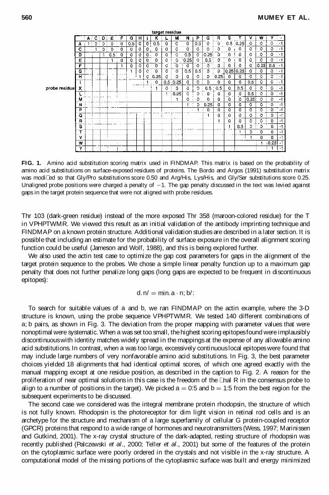
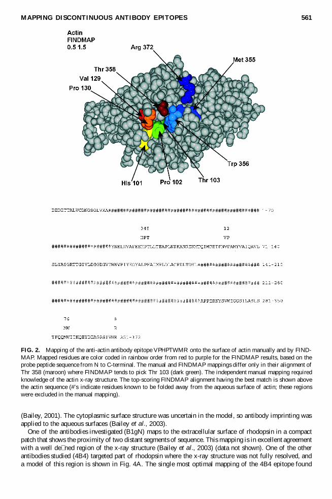
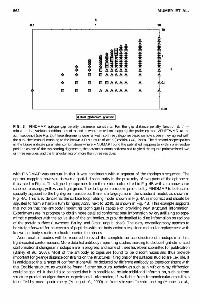
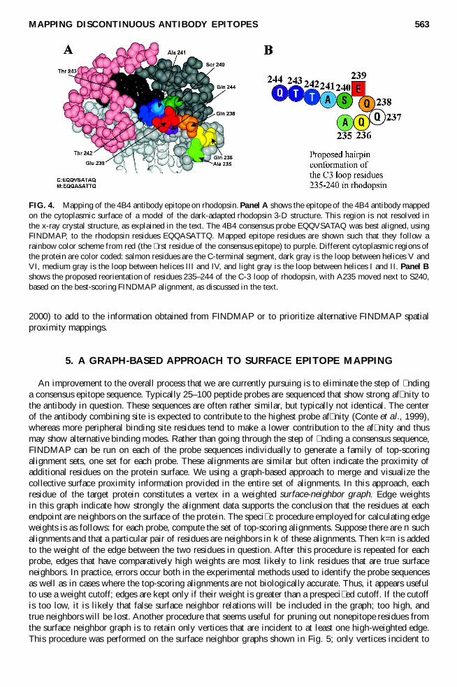
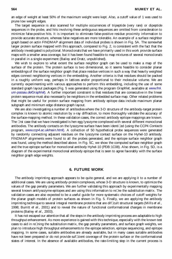
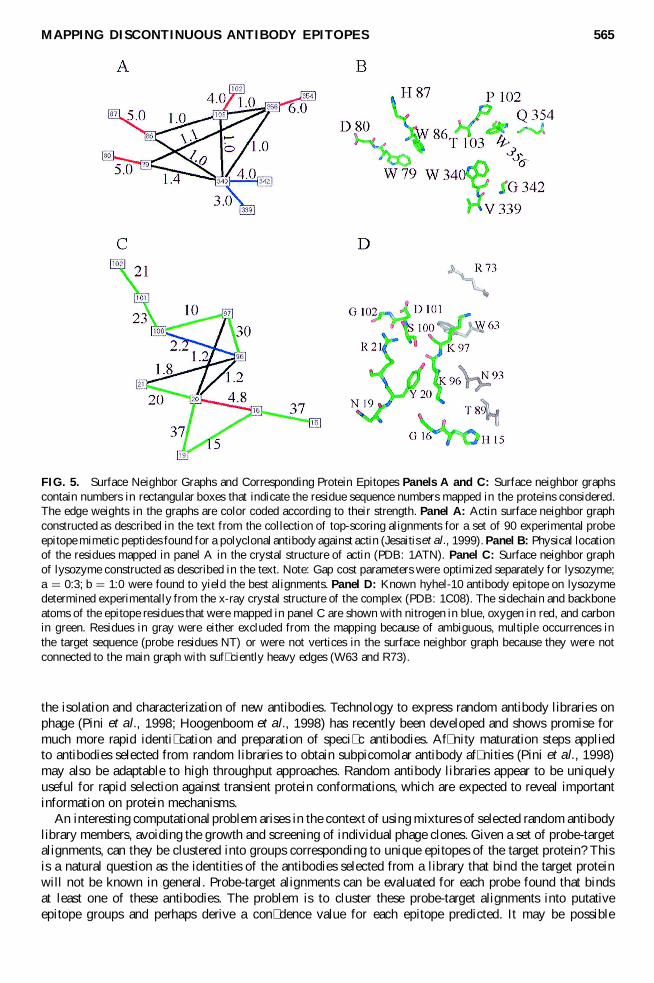


![Phosphorylation-dependentepitopes antibodies Alzheimertau · ment antibodies SMI31, SMI34, SMI35, or SMI310 (with phosphorylated epitopes) and SM133 [unphosphorylated epitopes (3)]](https://img.pdfslide.us/doc/110x75/5e62d2f4d3d32f22a55ed9e3/phosphorylation-dependentepitopes-antibodies-alzheimertau-ment-antibodies-smi31.jpg)

![A streamlined mass spectrometry-based proteomics workflow ... · [3,4]. Antibody-based histological methods are routinely performed but rely on the preservation of the relevant epitopes,](https://img.pdfslide.us/doc/110x75/5f0b51527e708231d42febe1/a-streamlined-mass-spectrometry-based-proteomics-workflow-34-antibody-based.jpg)