Embed Size (px)
Citation preview
Virus-like particles displaying conserved toxinepitopes stimulate broadly reactive, polyspeci�c,murine antibody responses capable of snakevenom recognitionStefanie K. Menzies
Liverpool School of Tropical MedicineCharlotte A. Dawson
Liverpool School of Tropical MedicineEdouard Crittenden
Liverpool School of Tropical MedicineRebecca Edge
Liverpool School of Tropical MedicineSteven R. Hall
Liverpool School of Tropical MedicineJaffer Alsolaiss
Liverpool School of Tropical MedicineMark C. Wilkinson
Liverpool School of Tropical MedicineNicholas R. Casewell
Liverpool School of Tropical MedicineRobert A. Harrison
Liverpool School of Tropical MedicineStuart Ainsworth ( [email protected] )
Liverpool School of Tropical Medicine
Research Article
Keywords: venom toxins, antivenom immunoglobulin, ELISA, lower-molecular weight, Snakebiteenvenoming (SBE)
Posted Date: November 22nd, 2021
DOI: https://doi.org/10.21203/rs.3.rs-1044937/v1

License: This work is licensed under a Creative Commons Attribution 4.0 International License. Read Full License

Virus-like particles displaying conserved toxin epitopes stimulate broadly reactive, polyspecific, murine antibody 1
responses capable of snake venom recognition 2
3
Short title: 4
Immunostimulatory potency of toxin coated virus-like particles 5
6
Authors: Stefanie K. Menzies1,2, Charlotte A. Dawson1, Edouard Crittenden1, Rebecca Edge1, Steven R. Hall1, Jaffer 7
Alsolaiss1, Mark C. Wilkinson1, Nicholas R. Casewell1,2, Robert A. Harrison1,2 and Stuart Ainsworth1* 8
9
Affiliations: 10
1 Centre for Snakebite Research and Interventions, Liverpool School of Tropical Medicine, Pembroke Place, Liverpool, 11
UK L3 5QA 12
2 Centre for Drugs and Diagnostics, Liverpool School of Tropical Medicine, Pembroke Place, Liverpool, UK, L3 5QA 13
* corresponding author – [email protected] 14
15
16
17

Abstract 18
Antivenom is currently the first-choice treatment for snakebite envenoming. However, only a low proportion of 19
antivenom immunoglobulins are specific to venom toxins, resulting in poor dose efficacy and potency. We sought to 20
investigate whether linear venom epitopes displayed on virus like particles can stimulate a robust and focused 21
antibody response capable of recognising venom toxins from diverse medically important species. Bioinformatically-22
designed epitopes, corresponding to predicted conserved regions of group I phospholipase A2 and three finger toxins, 23
were engineered for display on the surface of hepatitis B core antigen virus like particles and used to immunise female 24
CD1 mice over a 14-weeks. Antibody responses to all venom epitope virus like particles were detectable by ELISA by 25
the end of the immunisation period, although total antibody and epitope specific antibody titres were variable against 26
the different epitope immunogens. Immunoblots using pooled sera demonstrated recognition of various venom 27
components in a diverse panel of six elapid venoms, representing three continents and four genera. Finally, pooled 28
terminal sera was compared to conventional antivenom via quantitative immunoblot, and demonstrated superior 29
recognition of lower-molecular weight elapid venom toxins. This study demonstrates proof-of-principle that virus like 30
particles engineered to display conserved toxin linear epitopes can elicit specific antibody responses in mice which are 31
able to recognise a geographically broad range of elapid venoms. 32
33
34
35
36
37
38
39
40
41
42
43
44
45
46
47
48

Introduction 49
Snakebite envenoming (SBE) is a Neglected Tropical Disease estimated to result in a yearly burden of 138,000 deaths 50
and 400,000 disabilities1, and which disproportionately effects the mostly impoverished, rural-dwelling and 51
marginalised communities of the tropics and sub-tropics1,2. A key driver of the persisting high SBE mortality and 52
morbidity rate is the lack of safe and effective therapies, coupled to the inaccessibility (both physical and financial) of 53
healthcare3–6. 54
SBE is caused by a diverse number of snake species and a myriad of pathologies can be observed post envenoming1. 55
However, in very broad terms, three main envenoming syndromes are observed globally: haemotoxic envenoming 56
which disrupts haemostasis, neurotoxic envenoming which causes rapid descending neuromuscular paralysis, and 57
envenoming which results in local tissue destruction and often necrosis1. Further systemic effects of snake 58
envenoming may include nephrotoxicity and myotoxicity, depending upon the biting species1. Currently the only 59
specific therapy for envenoming is antivenom, which consists of antibodies isolated from animals hyper-immunised 60
with crude venom(s)7. Antivenom effectiveness is highly variable in neutralising different envenoming pathologies, 61
however. Generally, if an appropriate antivenom is used (i.e. an antivenom indicated for the biting snake species), it 62
is effective at restoring regular systemic haemostasis during severe haemotoxic envenoming8, even with substantial 63
delay in administration. However, the extent of antivenom effectiveness in the management of neurotoxic and local 64
envenoming effects remains contested9–12. Studies of the pharmacodynamic properties of individual, toxin specific 65
antibodies are likely able to address why antivenoms are more effective against some envenoming pathologies than 66
others13–15. Such properties include their structural format (e.g. intact immunoglobulin or fragments such as F(ab’)2 67
and Fab), their specificity and affinity for different toxins with targets in multiple tissue components and the initiation 68
of ultimately detrimental host processes (such as potential proinflammatory cascades) coupled with times to 69
treatment delivery. 70
Antivenoms are manufactured using high-cost, century-old protocols of immunising horses or sheep with crude 71
venom(s) which comprise ~20-100 components. This unguided approach, whilst clearly able to generate lifesaving 72
therapeutics, is inefficient in that it does not consider i) the diverse range of envenoming pathologies, ii) distinct venom 73
protein immunogenicities or iii) distinct toxicities of different venom components1,16–19. This is problematic as snake 74
venoms and their toxins vary at every taxonomic level20 and some of the most pathogenic and diverse toxin families, 75
particularly those responsible for neurotoxic and cytotoxic pathologies in elapids, are frequently described as poorly 76
immunogenic21,22. Consequently, antivenoms are snake-species and geographically restricted and are of very low 77
specificity and potency, with only ~15% of antivenom antibodies binding to the venom proteins used as immunogens23 78
resulting in poor clinical dose efficacy, particularly for neurotoxic snake envenoming21. As a result of these limitations 79
and the effect they have on treating snakebite globally, the World Health Organization and other organisations such 80
as Wellcome, have recently advocated the development of new envenoming therapies to overcome these deficiencies 81
and ultimately reduce mortality and morbidity24,25. 82
Research over the past two decades has increasingly focussed on overcoming these shortcomings through rationalised 83
selection and design of immunogens to improve the dose and polyspecific efficacy of antivenom18,25–28. Facilitated 84
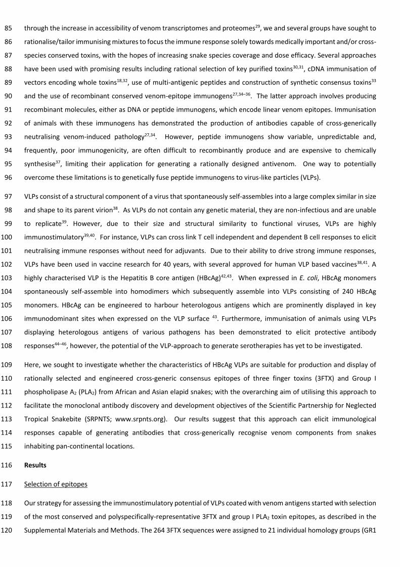
through the increase in accessibility of venom transcriptomes and proteomes29, we and several groups have sought to 85
rationalise/tailor immunising mixtures to focus the immune response solely towards medically important and/or cross-86
species conserved toxins, with the hopes of increasing snake species coverage and dose efficacy. Several approaches 87
have been used with promising results including rational selection of key purified toxins30,31, cDNA immunisation of 88
vectors encoding whole toxins18,32, use of multi-antigenic peptides and construction of synthetic consensus toxins33 89
and the use of recombinant conserved venom-epitope immunogens27,34–36. The latter approach involves producing 90
recombinant molecules, either as DNA or peptide immunogens, which encode linear venom epitopes. Immunisation 91
of animals with these immunogens has demonstrated the production of antibodies capable of cross-generically 92
neutralising venom-induced pathology27,34. However, peptide immunogens show variable, unpredictable and, 93
frequently, poor immunogenicity, are often difficult to recombinantly produce and are expensive to chemically 94
synthesise37, limiting their application for generating a rationally designed antivenom. One way to potentially 95
overcome these limitations is to genetically fuse peptide immunogens to virus-like particles (VLPs). 96
VLPs consist of a structural component of a virus that spontaneously self-assembles into a large complex similar in size 97
and shape to its parent virion38. As VLPs do not contain any genetic material, they are non-infectious and are unable 98
to replicate39. However, due to their size and structural similarity to functional viruses, VLPs are highly 99
immunostimulatory39,40. For instance, VLPs can cross link T cell independent and dependent B cell responses to elicit 100
neutralising immune responses without need for adjuvants. Due to their ability to drive strong immune responses, 101
VLPs have been used in vaccine research for 40 years, with several approved for human VLP based vaccines38,41. A 102
highly characterised VLP is the Hepatitis B core antigen (HBcAg)42,43. When expressed in E. coli, HBcAg monomers 103
spontaneously self-assemble into homodimers which subsequently assemble into VLPs consisting of 240 HBcAg 104
monomers. HBcAg can be engineered to harbour heterologous antigens which are prominently displayed in key 105
immunodominant sites when expressed on the VLP surface 43. Furthermore, immunisation of animals using VLPs 106
displaying heterologous antigens of various pathogens has been demonstrated to elicit protective antibody 107
responses44–46, however, the potential of the VLP-approach to generate serotherapies has yet to be investigated. 108
Here, we sought to investigate whether the characteristics of HBcAg VLPs are suitable for production and display of 109
rationally selected and engineered cross-generic consensus epitopes of three finger toxins (3FTX) and Group I 110
phospholipase A2 (PLA2) from African and Asian elapid snakes; with the overarching aim of utilising this approach to 111
facilitate the monoclonal antibody discovery and development objectives of the Scientific Partnership for Neglected 112
Tropical Snakebite (SRPNTS; www.srpnts.org). Our results suggest that this approach can elicit immunological 113
responses capable of generating antibodies that cross-generically recognise venom components from snakes 114
inhabiting pan-continental locations. 115
Results 116
Selection of epitopes 117
Our strategy for assessing the immunostimulatory potential of VLPs coated with venom antigens started with selection 118
of the most conserved and polyspecifically-representative 3FTX and group I PLA2 toxin epitopes, as described in the 119
Supplemental Materials and Methods. The 264 3FTX sequences were assigned to 21 individual homology groups (GR1 120

through GR21) (Table 1, Supp. Table S2). These homology groups broadly corresponded to functional sub-family 121
annotation, although notable exceptions were observed. For example, type I alpha-neurotoxins (sNTX) splitting into 122
two distinct groups: GR17 & GR15, while GR7 consisted of type IA cytotoxins (CTX) and Orphan group XV (OGXV) (Supp. 123
Table S3). 124
Of the 21 3FTX groups, six (aminergic-type [ATX] GR1, CTX and OGXV GR7, non-conventional [NCX] GR10, orphan group 125
VIII [OG8] GR13, sNTX GR15 and sNTX GR17) were selected based on their represented frequency in the data set (Supp. 126
Table S2). Hereafter, these groups will be referred to by their toxin-type only (e.g. ATX, CTX, OGXV, NCX, OG8, or SNTX) 127
(Table 1). Combined, these six groups represented 70.6% (187/264) of the 3FTX sequences analysed in the study. 128
Sequence conservation within 3FTX groups (except for NCX) was generally high, with 49-66% of AA residues being 129
≥80% conserved across all group sequences (Supp. Fig. S2 and Supp. Fig. S3). Group I PLA2 sequences were very 130
homogenous (68% and 48% AA residues at 80% and 100% conservation) across full-length sequences (Supp. Fig. S4). 131
Fifteen individual 3FTX epitopes were designed based on i) BepiPred predicted epitope regions, ii) conservation within 132
groups, iii) predicted accessibility and iv) their molecular location: either the hydrophobic core or 1st or 3rd finger of 133
3FTX, designated by “_F” or “_C”, respectively (Supp. Figs. S2, S3, S5, Supp. Table S3, Supp. File S5). It was not always 134
possible to design epitopes corresponding to regions which reflected predicted epitopes due to limited sequence 135
conservation in predicted regions. In such cases epitopes were solely designed on sequence conservation and 136
accessibility (e.g., ATX_F, NCX_F, OG8_C [Supp. Fig. S2, S3, S5]). Epitope ATX_F was near identical (1 AA greater in 137
length) to epitope Pep604-B designed to elicit antibodies against Micrurus corallinus 3FTX35. 138
Three variants of a single group I PLA2 epitope were designed (Table 1) based on conservation and accessibility (Supp. 139
Fig S4, S6). Cross-referencing this epitope region with publicly available Snake Toxin and Antivenom Binding profiles47 140
suggests this region is readily recognised by current antivenoms. The selected PLA2 epitope region resides between 141
the calcium binding loop (Tyr 28, Gly 30, Gly 32) and Asp 49 (Supp. Fig. S4, S6) residues essential for calcium ion 142
positioning for hydrolytic activity48. 143
Expression of VLPs presenting snake venom 3FTX and PLA2 epitopes 144
A sub-selection of individual designed epitopes were chosen for expression on VLPs, herein referred to as venom 145
epitope VLPs (veVLPs), and were expressed and purified as described in the Supplemental Materials and Methods. 146
Despite the resulting recombinant veVLPs expressed in Escherichia coli proving to be largely insoluble, sufficient 147
quantities of soluble material were obtained for each veVLP. Attempts to improve solubility by varying incubation 148
temperature and inducing IPTG concentration resulted in little-to-no improvement. The methods detailed for the 149
expression of veVLPs resulted in a mean yield of 9.97 mg/L soluble veVLP from a 0.6 L culture (range 1.7 – 16.4 mg/L). 150
To ensure confidence in the assembly of veVLPs (as opposed to monomers resulting from non-assembly), all affinity-151
purified veVLPs were concentrated with a 100 kDa MWCO centrifugal filter to deplete any sub-100 kDa proteins whilst 152
retaining assembled veVLPs49. All purified veVLPs were visually assessed for purity using anti-His fluorescent immuno 153
blots, comparing total protein stain (Supp. Fig. S7A) to anti-His signal (Supp. Fig. S7B). As shown in Supp. Fig. S7, the 154
major bands in purified samples contained His-tagged proteins corresponding to the individual expected molecular 155
weight of the reduced monomeric constituent proteins of each veVLP monomer. Larger bands (presumably) 156

corresponding to dimeric complexes of individual monomers were also visible for the majority of veVLPs. The veVLPs 157
were subsequently probed with SAIMR Polyvalent antivenom to determine if antibodies raised against crude venom 158
could recognise venom epitopes displayed on VLPs. Results demonstrated antivenom recognition of four veVLPs – 159
sNTX_F1 and sNTX_F2, 3FTX Core-string and 3FTX Finger-string. No recognition of the other veVLPs by SAIMR 160
Polyvalent was apparent (Supp. Fig. S7D). 161
VLPs presenting snake venom epitopes induce antibody responses in mice 162
Female CD1 mice were immunised with different veVLPs over a 14-week immunisation schedule (see Materials and 163
Methods). As described previously, we were forced to humanely euthanise 20 individuals (Table 2). This severely 164
restricted monitoring of responses in several immunogen groups. 165
The antibody response of mice to veVLP immunisation was monitored at specific points (at weeks 3, 6, 10 and at the 166
end of experiment at week 14) via ELISA using pooled sera consisting of equal volumes from each individual in each 167
experimental group (Fig. 1, Supp. File S4). Antibody responses to veVLPs were detected at week 3 for all groups (OD405 168
≥ 1 at 1/500 dilution of sera), with the exception of mice receiving Core-string (Group K) and CTX_C (Group M - without 169
adjuvant) veVLP immunogens, whose signal was indistinguishable to that of naïve serum or negative controls. The 170
OD405 for 7 of the 13 veVLP groups continued to increase until reaching a peak at week 10, which then subsequently 171
declined modestly by week 14. Due to a processing error, we unfortunately lost week 10 sera corresponding to animals 172
immunised with CTX_C (Group A – with adjuvant), CTX_C (Group M without adjuvant) and CTX_F (Group B), thus it is 173
not possible to infer if a similar response profile occurred with CTX_C or CTX_F veVLPs. 174
CTX_C , CTX_F and sNTX veVLP immunised groups (A/M, B and E) provided the lowest overall titres at terminal bleed 175
(week 14) (Fig. 1). Both CTX_C immunised groups (groups A and M with and without adjuvant, respectively) resulted 176
in near identical mean titres (1/500 OD450 1.16 and 1.15, respectively), however, CTX_C with adjuvant (Group A) titres 177
remained stable throughout the schedule, whereas CTX_C without adjuvant (Group M) titres slowly reached this titre 178
by week 14. CTX_F (Group B) titres declined after week 6, possibly reflective of the early euthanasia of 80% of the 179
individuals in this group on humane grounds (Table 2), thus this data only reflects n=1 from week 6 onwards. sNTX_C 180
(Group E) titres remained stable but relatively low throughout the immunisation period (maximum mean 1/500 OD405 181
= 0.97). 182
VLPs presenting snake venom epitopes elicit antibodies against the displayed epitope and against the VLP carrier 183
To ascertain the proportion of the antibody response directed towards the displayed epitope as opposed to the VLP 184
carrier, pooled sera from each group of veVLP immunised mice was also used to probe nativeVLPs (Fig. 1, Panel O). 185
Results broadly demonstrated three distinct profiles; i) veVLP generated sera recognised nativeVLPs at slightly lower 186
or equivalent titres compared to recognition of respective veVLPs, throughout the immunisation period (nine groups), 187
ii) veVLP sera initially recognised nativeVLP before rapid declining of nativeVLP-specific titres to baseline (3 groups), 188
or iii) veVLP generated sera displayed negligible recognition of nativeVLP (1 group). These results demonstrate that 189
the majority of veVLPs used in these immunisations are capable of eliciting polyspecific antibodies towards the carrier 190
molecule and the venom epitope. 191
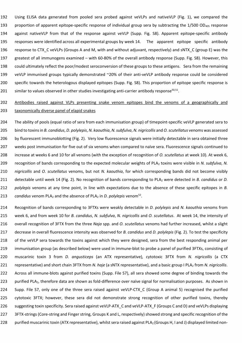
Using ELISA data generated from pooled sera probed against veVLPs and nativeVLP (Fig. 1), we compared the 192
proportion of apparent epitope-specific response of individual group sera by subtracting the 1/500 OD405 response 193
against nativeVLP from that of the response against veVLP (Supp. Fig. S8). Apparent epitope-specific antibody 194
responses were identified across all experimental groups by week 14. The apparent epitope specific antibody 195
response to CTX_C veVLPs (Groups A and M, with and without adjuvant, respectively) and sNTX_C (group E) was the 196
greatest of all immunogens examined – with 60-80% of the overall antibody response (Supp. Fig. S8). However, this 197
could ultimately reflect the poor/modest seroconversion of these groups to these antigens. Sera from the remaining 198
veVLP immunised groups typically demonstrated ~20% of their anti-veVLP antibody response could be considered 199
specific towards the heterologous displayed epitopes (Supp. Fig. S8). This proportion of epitope specific response is 200
similar to values observed in other studies investigating anti-carrier antibody response50,51. 201
Antibodies raised against VLPs presenting snake venom epitopes bind the venoms of a geographically and 202
taxonomically diverse panel of elapid snakes 203
The ability of pools (equal ratio of sera from each immunisation group) of timepoint-specific veVLP generated sera to 204
bind to toxins in B. candidus, D. polylepis, N. kaouthia, N. subfulva, N. nigricollis and O. scutellatus venoms was assessed 205
by fluorescent immunoblotting (Fig. 2). Very low fluorescence signals were initially detectable in sera obtained three 206
weeks post immunisation for five out of six venoms when compared to naïve sera. Fluorescence signals continued to 207
increase at weeks 6 and 10 for all venoms (with the exception of recognition of O. scutellatus at week 10). At week 6, 208
recognition of bands corresponding to the expected molecular weights of PLA2 toxins were visible in N. subfulva, N. 209
nigricollis and O. scutellatus venoms, but not N. kaouthia, for which corresponding bands did not become visibly 210
detectable until week 14 (Fig. 2). No recognition of bands corresponding to PLA2 were detected in B. candidus or D. 211
polylepis venoms at any time point, in line with expectations due to the absence of these specific epitopes in B. 212
candidus venom PLA2 and the absence of PLA2 in D. polylepis venom52. 213
Recognition of bands corresponding to 3FTXs were weakly detectable in D. polylepis and N. kaouthia venoms from 214
week 6, and from week 10 for B. candidus, N. subfulva, N. nigricollis and O. scutellatus. At week 14, the intensity of 215
overall recognition of 3FTX from the three Naja spp. and O. scutellatus venoms had further increased, whilst a slight 216
decrease in overall fluorescence intensity was observed for B. candidus and D. polylepis (Fig. 2). To test the specificity 217
of the veVLP sera towards the toxins against which they were designed, sera from the best responding animal per 218
immunisation group (as described below) were used in immune-blot to probe a panel of purified 3FTXs, consisting of 219
muscarinic toxin 3 from D. angusticeps (an ATX representative), cytotoxic 3FTX from N. nigricollis (a CTX 220
representative) and short chain 3FTX from N. haje (a sNTX representative), and a basic group I PLA2 from N. nigricollis. 221
Across all immune-blots against purified toxins (Supp. File S7), all sera showed some degree of binding towards the 222
purified PLA2, therefore data are shown as fold-difference over naïve signal for normalisation purposes. As shown in 223
Supp. File S7, only one of the three sera raised against veVLP-CTX_C (Group A animal 5) recognised the purified 224
cytotoxic 3FTX; however, these sera did not demonstrate strong recognition of other purified toxins, thereby 225
suggesting toxin specificity. Sera raised against veVLP-ATX_C and veVLP-ATX_F (Groups C and D) and veVLPs displaying 226
3FTX-strings (Core-string and Finger string, Groups K and L, respectively) showed strong and specific recognition of the 227
purified muscarinic toxin (ATX representative), whilst sera raised against PLA2 (Groups H, I and J) displayed limited non-228

specific recognition of other toxins. Sera raised against sNTX veVLPs (Groups E, F and G) did not demonstrate 229
recognition towards any of the toxins tested. 230
Dotblots using crude venoms showed specific recognition of venom by the veVLP sera, with no recognition by naïve 231
sera, thus demonstrating that the generated antibodies were able to recognise venom components in their native 232
conformation, as well as in reduced and denatured conditions (i.e. in immune-blot). Furthermore, the dotblots 233
demonstrated similar specific-venom recognition as observed in immune-blots; Group D (ATX_F) sera recognised five 234
of the six venoms tested in both immune-blot and dotblot, with no recognition of O. scutellatus apparent. 235
Furthermore, group D [ATX_F] sera from animals D1 and D5, which possessed the broadest and strongest binding to 236
venom components in reduced states (Table 2, Supp. File S6) demonstrated greater recognition of the five venoms in 237
their native state as determined by dotblot (Supp. Fig. S9). 238
Comparison of pooled sera to SAIMR Polyvalent antivenom 239
The venom recognition profile of pooled veVLP raised antibodies (from all immunisation groups) was compared against 240
SAIMR Polyvalent antivenom by quantitative immune-blot using a panel of sub-Saharan African elapid snake venoms 241
(Fig. 3). The venoms selected represent venoms used (alongside others) in the manufacture of SAIMR Polyvalent 242
antivenom (D. angusticeps, D. polylepis, N. subfulva and N. nivea) or are known to be preclinically neutralised by SAIMR 243
Polyvalent (N. nigricollis53). Additionally, Echis ocellatus venom, which is not used in the immunisation mixture for 244
SAIMR Polyvalent and does not contain group I PLA2 or 3FTX toxin families54 was included as a control. Terminal pooled 245
veVLP sera and SAIMR Polyvalent were diluted to equivalent protein concentrations for comparison. Results 246
demonstrate the generated veVLP mice sera and SAIMR Polyvalent both recognised components of all five elapid 247
venoms. SAIMR Polyvalent additionally recognised E. ocellatus venom (likely the result of other related viper venoms 248
being used in the immunisation process), while the veVLP sera did not, as expected. The veVLP sera only recognised 249
lower molecular weight components of the elapid venoms, highlighting the utility of the 3FTX and PLA2-specific 250
epitopes designed and implemented in this project, whilst SAIMR Polyvalent recognised both low and higher molecular 251
weight components, the latter most likely corresponding to PIII-SVMPs commonly seen in elapid venoms in low 252
abundances29. Across the immuno-blot, the veVLP sera demonstrated 1.8-fold weaker recognition (normalised 253
fluorescence measurements of 16.00 and 28.30 respectively, Supp. Fig. S10C) than SAIMR Polyvalent, skewed by the 254
recognition of higher molecular weight components and E. ocellatus venom by SAIMR Polyvalent. However, 255
promisingly, veVLP sera showed 1.2-fold stronger recognition of <20 kDa venom proteins when compared with SAIMR 256
Polyvalent (normalised fluorescence measurements of 9.59 and 7.83, respectively, Supp. Fig. S10B). This was mostly 257
due to superior recognition of bands corresponding to PLA2 in N. subfulva and N. nigricollis, as compared to SAIMR 258
Polyvalent (Fig. 3, Supp. Fig S10). 259
Immune response variation and antibody specificity in individual immunised mice 260
Immuno-blotting experiments using pooled sera are useful in providing a chronological overview of development of 261
venom component recognition; however, they do not allow resolution of: i) effectiveness of individual veVLPs to 262
generate antibodies capable of recognising specific venom proteins, or ii) whether immunisation with individual veVLP 263
leads to consistent seroconversion and antibody generation within groups. Furthermore, analysing the serological 264

response of individual animals enabled the identification of the highest-responding mice, facilitating the downstream 265
selection of splenocyte samples from individual mice to progress towards monoclonal antibody isolation in future 266
experiments. Thus, we analysed terminal (week 14) sera by ELISA and immuno-blot, as previously, for all individual 267
immunised animals. 268
Our results (Table 2, Supp. File S4, Supp. File S6) demonstrated that no individual sera in groups immunised with CTX_C 269
with adjuvant, CTX_F, sNTX_C or PLA2_2 (immunisation groups A, B, E, and I respectively) was capable of binding to 270
specific venom proteins found in our panel of elapid venoms (Table 2, Supp. File S6, Supp. File S4). This is in contrast 271
to the ELISA results, whereby PLA2_2 veVLP sera demonstrated strong recognition of PLA2_2 veVLPs (Fig. 1) and the 272
observation that group A CTX_C and group E sNTX_C have proportionately high levels of epitope-specific antibodies 273
as determined through ELISA recognition of veVLP (Supp. Fig. S8). The inability of CTX_F (group B) sera to recognise 274
venom components was unsurprising due to near-baseline levels of veVLP recognition in pooled ELISA results (Fig. 1). 275
However, the results from these groups need to be interpreted in the context of several influencing factors. Firstly, 276
ELISA results are demonstrative of veVLP recognition, not crude venom. Secondly, the low final n numbers (n=1 [group 277
B] or 2 [groups A, E]) due to animal attrition during immunisation (Table 2) in addition to variability within groups that 278
did generate venom-specific antibodies (below), suggest it is possible that the lack of toxin recognition may be the 279
result of individual variation in immune responses. 280
Sera from at least a single individual immunised with either ATX_C, ATX_F, sNTX_F1, sNTX_F2, PLA2_1, PLA2_3, Core-281
string, Finger-sting and CTX_C (without adjuvant) veVLPs (groups C, D, F, G, H, J, K, L and M, respectively) contained 282
antibodies capable of recognising specific venom proteins present in at least one venom (Table 2, Supp. File S6). The 283
results demonstrate expected specific recognition of individual toxin families (e.g. 3FTX vs PLA2) based on the known 284
approximate molecular weights of these toxin groups48,55. A notable observation was the high degree of variation in 285
the antibody response towards both the immunogen and venom observed between individual animals in the same 286
immunogen group. For example, within group C, recognition of the immunogen ATX_C veVLP at week 14, as measured 287
by ELISA, was similar for all individuals (mean 1/500 OD405 = 1.92, range 1.82-1.98) although recognition of nativeVLP 288
by group C sera was more variable (mean 1/500 OD405 = 1.69, range 1.29-1.92) (Table 2, Fig 1. Supp. File S6). Despite 289
these ELISA results suggesting overall similar recognition of ATX_C veVLPs by mice immunised with ATX_C veVLPs, only 290
sera from animal C5, which is inferred to possess highest proportion of ATX_C antibodies, was able to recognise venom 291
proteins by immuno-blot (Table 2, Supp. File S6), recognising expected 3FTX representative bands in D. polylepis, N. 292
subfulva, and N. kaouthia venoms. Unexpectedly, C5 sera also demonstrated reactivity with an additional, slightly 293
larger, protein in N. subfulva and N. kaouthia venoms (Supp. File S6). This may potentially be cross-reactivity with a 294
type II long-chain α-neurotoxin, as the ATX_C epitope contains notable similarity (≥ 7 AA alignment length, 80% 295
identity, 80% coverage, mismatches ≤ 1) with members of this toxin subclass (Table S3). 296
In veVLP immunisation groups where multiple individuals produced antibodies that could recognise venom proteins 297
(groups D, H, K and M, immunised with ATX_F, PLA2_1, Core-string and CTX_F, respectively), the number of venoms 298
recognised was either consistent across individuals (same number of venoms, similar intensity of recognition, e.g. 299
Group D) or variable (recognising different numbers of venoms with differing intensity of recognition, e.g. Group H) 300
(Table 2, Supp. File S6). Two of the three remaining animals in Group D (ATX_F), D1 and D5, possessed antibodies that 301

could recognise 3FTX bands in B. candidus, D, polylepis, N. subfulva, N. kaouthia and N. nigricollis venom with similar 302
intensities (Table 2, Supp. File S6). Conversely, all three remaining animals in Group H, immunised with PLA2_1 veVLP, 303
possessed antibodies capable of recognising bands corresponding to PLA2, in different numbers of species (H1 & H5 = 304
4 venoms, H3 = 2 venoms), though N. nigricollis and O. scutellatus PLA2 was recognised by all three (Table 2, Supp. File 305
S6). CTX_C without adjuvant (Group M) was unusual in that it appeared to show recognition of bands assumed to 306
correspond to PLA2 (e.g. M2, Supp. File S6), suggesting potential non-specific binding. 307
Discussion 308
This study demonstrates proof of principle that VLPs decorated with rationally selected conserved linear venom-309
epitopes can be used to stimulate the production of murine antibodies that are able to recognise a geographically and 310
taxonomically diverse range of elapid venoms (Table 2, Fig. 2, File. S6). Additionally, the promising results and 311
resources generated from this study enable the further progression of this research, as splenocytes isolated from the 312
best-responding individual mice identified in this study are being investigated as a resource for therapeutic anti-toxin 313
monoclonal antibody discovery. 314
Of the 10 individual epitopes designed and used in this study, six were shown to elicit antibodies capable of binding 315
specific venom components (Table 2, Supp. File S6). veVLPs decorated with 3FTX epitopes corresponding to finger 316
regions were more likely (38%, n=13) to elicit venom binding sera than core epitope veVLPs (13%, n=8) at the end of 317
the immunisation period. Additionally, sera against PLA2 epitopes PLA2_1 and PLA2_2 (immunisation groups H and I, 318
respectively) demonstrated similar abilities in recognising respective immunogens at the end of the immunisation 319
period (Fig. 1), but provided considerable differences in their ability to recognise venom components, despite differing 320
by only a single amino acid (KGTPVDLDD and KGTAVDDLD, respectively) (Table 1, Supp. File S6). Sera from all remaining 321
animals immunised with PLA2_1 (n=3) bound proteins in multiple venoms, whilst recognition of venom components 322
was not demonstrated by any of the remaining PLA2_2 immunised animals (n=4). Such stark difference in venom 323
component recognition suggests that proline may have a key role in antibody recognition of this epitope. Given highly 324
similar toxin epitope sequences can elicit drastically different antibody responses, our findings reiterates that 325
antibodies induced by toxin epitopes need to be robustly assessed for venom recognition against a diverse panel of 326
target venoms in conventional immune-assays, and not simply based on their apparent frequency in toxin sequences. 327
Antivenoms have long been reported to have poor dose efficacy in neutralising small molecular weight venom toxins, 328
including 3FTX and group I PLA2, which is frequently attributed to these toxins being poorly immunogenic21,22. Notably, 329
in this study, a pool of experimental veVLP sera demonstrated a substantial improvement in recognition of small 330
molecular weight compounds as compared to the comparative SAIMR Polyvalent antivenom at an equivalent 331
concentration (Fig. 3). While the results confidently demonstrate improved recognition of these medically important 332
toxins, we are currently unable to demonstrate if this increased recognition translates into more potent neutralising 333
efficacy. This is due to the quantities of antibodies recoverable for each animal not being sufficient to perform 334
informative in vitro neutralisation assays of toxin activity, with yields in this study ranging from approximately 45 µg 335
to 240 µg per animal. 336

Despite the success in demonstrating the ability of veVLPs to elicit anti-toxin antibodies, this study was subject to 337
several limitations. Notably, a large proportion of animals during immunisation were euthanized early due to 338
presumed adverse reactions to adjuvant (20 out of 65). Notable local inflammation and irritation (redness and local 339
swelling) were typically observed at all dosing locations in animals that received VLPs and adjuvant, which usually 340
resolved 1-2 weeks post immunisation. Animals that received veVLP without adjuvant (Group M) generally did not 341
develop any local reaction at any dose sites, which leads us to hypothesise the adjuvant, Alhydrogel (alum), was 342
contributing to the observed adverse effects. This is surprising as alum based adjuvants are routinely considered as 343
safe and are widely used in human vaccines56, and suggests that the combination of the self-adjuvating nature of 344
VLPs41 and the adjuvant might adversely exacerbate local inflammatory responses. Unfortunately, acute local 345
inflammation resulting in a non-resolving lump at the inoculation site was observed in 20 animals dosed at the rump 346
on week 2 (2nd immunisation), which necessitated euthanasia of affected animals as per our institutional and national 347
license conditions. We suspect that the tighter skin around the rump, as compared to the scruff and flanks of the mice, 348
in combination with irritation and local swelling may have exacerbated local conditions. Due to the substantial adverse 349
reactions observed in this study, we advise against immunising mice in the rump area in similar immunisation 350
experiments conducted in the future. Furthermore, the groups that ultimately were unable to recognise venom 351
components were also the groups most affected by the losses in numbers at week 2 (Table 2). For example, groups A, 352
B and E representing CTX_C, CTX_F and sNTX_C, were the most severely affected, losing 60-80% of their representative 353
animals. Based on the results obtained from groups less affected by animal loss, we currently cannot say if the inability 354
of these epitopes to elicit an immune response is due to poor candidate epitopes or individual variation in response 355
to immunisation. We speculate that the large amount of variation in venom reactivity observed between individuals 356
within a group is due in part to immunological heterogeneity in the experimental animals, as the mouse strain selected, 357
CD1, is outbred and thus reflective of antivenom manufacturing animals. Similar highly variable results in responses 358
to immunisation have been observed in antivenom manufacturing animals33 and camels57. 359
Our results demonstrate that venom epitopes fused to VLPs can induce robust anti-toxin antibody responses. 360
However, difficulties in production of veVLPS in this specific VLP format (HBcAg), encountered by ourselves and others 361
expressing heterologous antigens49,58, may prove challenging for application if this approach were to be applied to 362
producing a rationally designed antivenom at commercial scale. However, research to circumvent production 363
obstacles has been actively undertaken over the past decade. Developments include alternative methods of genetic-364
fusion for decorating VLPs with heterologous antigens, such as SpyCatcher-SpyTag ‘plug and play’ systems59, and the 365
development of computationally designed hyper-stable and soluble synthetic VLPs49,60. Use of such particles to 366
generate rationally designed antivenoms, or as a tool to rapidly discover monoclonal antibodies to specific venom 367
targets, may be a more cost-effective approach that would also increase translational viability. 368
The use of linear snake venom epitopes for rationally targeted anti-toxin antibody generation now has substantial 369
background26. Multiple different formats for delivery of linear epitopes have been used, including DNA27,34, 370
peptide34,35,61,62, and now VLP. Such studies have demonstrated the ability to generate venom-specific antibodies, and 371
several have further demonstrated the ability of epitope generated antibodies to neutralise local and systemic venom 372
pathologies. Whilst considered to be inferior to conformational epitopes in terms of potency, linear epitopes have the 373

advantage that they are easier to identify and cheaper to produce than conformational antigens – vitally important 374
considerations when proposing improvements to a therapeutic which is already prohibitively expensive to the majority 375
of people who need it most2. Furthermore, as snake venoms consist of multiple toxin families and sub-families, it is 376
highly likely that such a strategy will require multiple epitopes to ensure adequate protection against all medically 377
important venom components1. Recent publication of high-throughput antivenom-venom peptide arrays47,63, 378
alongside transcriptomic and proteomic characterisation of venoms29,64, means there is now a wealth of resources 379
available for informative venom epitope prediction. 380
However, substantial research questions remain when considering whether the approach of employing rationally 381
designed linear epitope antigens to elicit anti-toxin antibodies is a genuinely practical method for producing more 382
efficacious antivenoms. Questions include: will the results, all generated so far in mice and rabbits, be translatable in 383
manufacturing antivenom-manufacturing animals? Will antisera produced in this manner possess potencies which at 384
least match existing conventionally produced antivenoms? To date, demonstration of neutralising efficacies of anti-385
epitope antibodies have been performed against relatively low challenge doses of venom. Could alternative 386
immunisation strategies, such as combined epitope and crude venom approaches, substantially increase efficacy, 387
especially against lower molecular weight toxins? Notably in this study, the improvement in recognition of small 388
molecular weight compounds as compared to current antivenoms is promising and it may be possible to use a handful 389
of veVLPs decorated epitopes of toxins as an additive to crude venom during manufacture to improve potency of the 390
resulting antivenom. Additionally, through this work we have been able to preserve splenocytes from individual veVLP 391
immunised animals displaying the most promising antibody responses. We hope to investigate this valuable resource 392
with a view to developing mAbs, which have been raised against specific, rationally designed venom epitopes, into 393
potential next generation antivenom therapies65. 394
Materials and Methods 395
Animal ethics 396
Details and reporting of all animal experiments in this manuscript conform with ARRIVE guidelines to the best of our 397
ability. Mouse immunisations were performed using protocols approved by the Animal Welfare and Ethical Review 398
Boards of the Liverpool School of Tropical Medicine and the University of Liverpool. Experiments were conducted 399
under licence approved by the UK Home Office (Project Licence P58464F90) and in accordance with the Animal 400
(Scientific Procedures) Act 1986. 401
Murine immunisations 402
Mice were immunised with venom-epitope displaying VLPs (veVLPs) to investigate their ability to elicit anti-toxin 403
antibody responses. On the day of immunisation, aliquots of immunogen were either: (i) mixed with 2% aluminium 404
hydroxide gel (Alhydrogel, InvivoGen) at a ratio of 1:1 (v/v) and shaken at 1500 rpm for 10 minutes at room 405
temperature on a ThermoMixer (Eppendorf), or (ii) diluted with equal volume PBS when adjuvant was not used. 406
Female CD1 mice (18 – 20 g) were purchased from Charles River and allowed to acclimatise for one week before first 407
immunisation. Mice were housed in groups of five with ad libitum access to certified reference materials irradiated 408
food (Special Diet Services) and reverse osmosis water (in automatic water system), along with enrichment, and kept 409

at approximately 22 °C at 40-50 % humidity, with 12/12 hour light cycles. Mice were housed in Techniplast GM500 410
cages with Lignocel bedding (JRS, Germany) and zigzag fibres nesting material (Sizzlenest, RAJA), and cages were 411
changed fortnightly. Mice were kept in specific-pathogen-free facilities. All experiments were performed by mixed 412
gender experimenters. Humane endpoints were weight loss (>10% loss of body weight within one week, or >20% 413
within one month [despite remedial actions such as wet food]), or observation of the following animal behaviour or 414
appearance signs – reduced activity, physiological impairments, pallor, or ulceration following immunisation. 415
Mice were briefly anaesthetised with 5% isofluorane to enable shaving at the injection site for subsequent monitoring 416
of adverse reactions. Mice were subcutaneously immunised with 1 µg immunogen in a total volume of 40 µL at each 417
immunisation, according to the following schedule; week 0: Injection at one site on the scruff (with adjuvant), week 2: 418
Injection at one site on the rear, week 4: Injection over two sites (20 µL/site) on the right flank (without adjuvant), 419
week 8: Injection over two sites (20 µL/site) on the left flank, week 12: Injection over two sites (20 µL/site) at the 420
scruff. An additional group received GR7_c (Group M) immunisations which were always performed without adjuvant. 421
A total of 12 veVLP immunogens were used for immunisation, with the specific epitope immunogens assigned to each 422
group of five mice listed in Table 1. 423
Following the second immunisation at week 2, 20 animals developed large non-resolving lumps at the rear dose site 424
and were euthanised on humane grounds to prevent pain, harm and distress. Subsequent immunisations were given 425
over two dose sites in a refinement of the immunisation, from which all animals developed mild, small, self-resolving 426
lumps at the injection sites. Animals were monitored twice per week throughout the course of immunisation for 427
adverse reactions and general health, and no animals were culled due to weight loss or behavioural endpoints being 428
met. 429
Sera isolation 430
Approximately 50 µL venous blood samples were collected at week 3, 6 and 10 by tail bleed. Whole blood was allowed 431
to clot for a minimum of 2 hours at room temperature, and sera was obtained by centrifugation at 2000 x g for 10 432
minutes at 10 °C. Sera was immediately stored at -20 °C. Remaining animals were euthanised by rising concentrations 433
of carbon dioxide at week 14 (end of experiment). Following confirmation of death, cardiac punctures were performed 434
to collect whole blood and sera was processed as above. ‘Naïve’ unimmunised mouse sera controls (strain matched) 435
were sourced commercially from Charles River UK and Sigma. Additionally, splenocytes were collected and preserved 436
for future work. 437
Venoms, Antivenoms and Toxins 438
Venoms were obtained from specimens of Bungarus candidus (a historical venom stock collected from snakes of Thai 439
origin) and from Dendroaspis angusticeps (Tanzania), D. polylepis (Tanzania), Echis ocellatus (Nigeria), Naja annulifera 440
(captive bred), N. kaouthia (captive bred), N. subfulva (Uganda), N. nigricollis (Nigeria) and N. nivea (South Africa), 441
maintained in the herpetarium at the Liverpool School of Tropical Medicine. Crude venoms were immediately frozen, 442
lyophilised and stored at 4 °C until reconstitution. Oxyuranus scutellatus venom was obtained from Venom Supplies 443
Pty, Australia. Venoms were resuspended in PBS to 1 mg/mL and stored at -20 °C. SAIMR Polyvalent Snake Antivenom 444
(South African Vaccine Producers, Gauteng, South Africa; Batch BB01446, expiry date July 2015), which is an equine 445
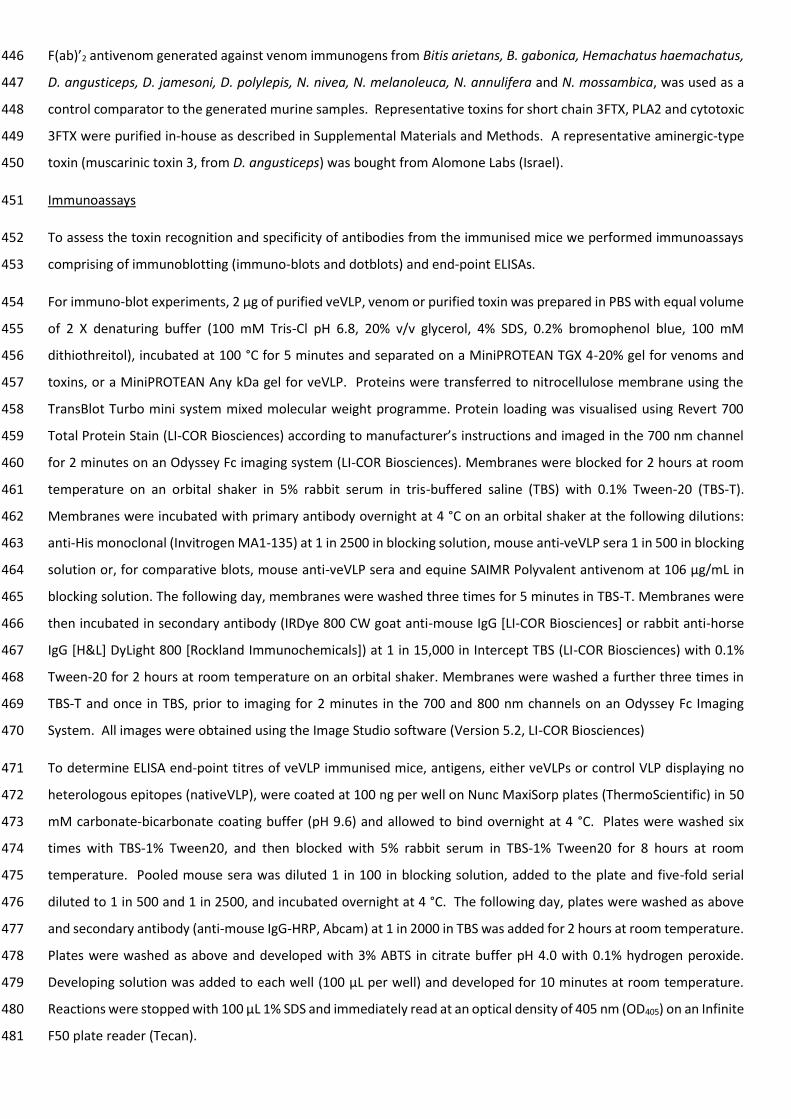
F(ab)’2 antivenom generated against venom immunogens from Bitis arietans, B. gabonica, Hemachatus haemachatus, 446
D. angusticeps, D. jamesoni, D. polylepis, N. nivea, N. melanoleuca, N. annulifera and N. mossambica, was used as a 447
control comparator to the generated murine samples. Representative toxins for short chain 3FTX, PLA2 and cytotoxic 448
3FTX were purified in-house as described in Supplemental Materials and Methods. A representative aminergic-type 449
toxin (muscarinic toxin 3, from D. angusticeps) was bought from Alomone Labs (Israel). 450
Immunoassays 451
To assess the toxin recognition and specificity of antibodies from the immunised mice we performed immunoassays 452
comprising of immunoblotting (immuno-blots and dotblots) and end-point ELISAs. 453
For immuno-blot experiments, 2 µg of purified veVLP, venom or purified toxin was prepared in PBS with equal volume 454
of 2 X denaturing buffer (100 mM Tris-Cl pH 6.8, 20% v/v glycerol, 4% SDS, 0.2% bromophenol blue, 100 mM 455
dithiothreitol), incubated at 100 °C for 5 minutes and separated on a MiniPROTEAN TGX 4-20% gel for venoms and 456
toxins, or a MiniPROTEAN Any kDa gel for veVLP. Proteins were transferred to nitrocellulose membrane using the 457
TransBlot Turbo mini system mixed molecular weight programme. Protein loading was visualised using Revert 700 458
Total Protein Stain (LI-COR Biosciences) according to manufacturer’s instructions and imaged in the 700 nm channel 459
for 2 minutes on an Odyssey Fc imaging system (LI-COR Biosciences). Membranes were blocked for 2 hours at room 460
temperature on an orbital shaker in 5% rabbit serum in tris-buffered saline (TBS) with 0.1% Tween-20 (TBS-T). 461
Membranes were incubated with primary antibody overnight at 4 °C on an orbital shaker at the following dilutions: 462
anti-His monoclonal (Invitrogen MA1-135) at 1 in 2500 in blocking solution, mouse anti-veVLP sera 1 in 500 in blocking 463
solution or, for comparative blots, mouse anti-veVLP sera and equine SAIMR Polyvalent antivenom at 106 µg/mL in 464
blocking solution. The following day, membranes were washed three times for 5 minutes in TBS-T. Membranes were 465
then incubated in secondary antibody (IRDye 800 CW goat anti-mouse IgG [LI-COR Biosciences] or rabbit anti-horse 466
IgG [H&L] DyLight 800 [Rockland Immunochemicals]) at 1 in 15,000 in Intercept TBS (LI-COR Biosciences) with 0.1% 467
Tween-20 for 2 hours at room temperature on an orbital shaker. Membranes were washed a further three times in 468
TBS-T and once in TBS, prior to imaging for 2 minutes in the 700 and 800 nm channels on an Odyssey Fc Imaging 469
System. All images were obtained using the Image Studio software (Version 5.2, LI-COR Biosciences) 470
To determine ELISA end-point titres of veVLP immunised mice, antigens, either veVLPs or control VLP displaying no 471
heterologous epitopes (nativeVLP), were coated at 100 ng per well on Nunc MaxiSorp plates (ThermoScientific) in 50 472
mM carbonate-bicarbonate coating buffer (pH 9.6) and allowed to bind overnight at 4 °C. Plates were washed six 473
times with TBS-1% Tween20, and then blocked with 5% rabbit serum in TBS-1% Tween20 for 8 hours at room 474
temperature. Pooled mouse sera was diluted 1 in 100 in blocking solution, added to the plate and five-fold serial 475
diluted to 1 in 500 and 1 in 2500, and incubated overnight at 4 °C. The following day, plates were washed as above 476
and secondary antibody (anti-mouse IgG-HRP, Abcam) at 1 in 2000 in TBS was added for 2 hours at room temperature. 477
Plates were washed as above and developed with 3% ABTS in citrate buffer pH 4.0 with 0.1% hydrogen peroxide. 478
Developing solution was added to each well (100 µL per well) and developed for 10 minutes at room temperature. 479
Reactions were stopped with 100 µL 1% SDS and immediately read at an optical density of 405 nm (OD405) on an Infinite 480
F50 plate reader (Tecan). 481

All measurements were performed in triplicate, except where indicated (due to limited amounts of sera). Control wells 482
of no protein (with mouse sera and secondary antibody), and no mouse sera (immunogen and secondary antibody) 483
were included. Raw data is available in Supp. File S4. 484
Data availability 485
All data generated or analysed during this study are either included in this published article (and its Supplementary 486
Information files) or, in the case of raw data files for fluorescent immunoblots, available from the corresponding author 487
upon request. 488
References 489
1. Gutiérrez, J. M. et al. Snakebite envenoming. Nat. Rev. Dis. Prim. 3, 17063 (2017). 490
2. Harrison, R. A., Hargreaves, A., Wagstaff, S. C., Faragher, B. & Lalloo, D. G. Snake envenoming: A disease of 491
poverty. PLoS Negl. Trop. Dis. 3, (2009). 492
3. Sharma, S. K. et al. Impact of snake bites and determinants of fatal outcomes in Southeastern Nepal. Am. J. 493
Trop. Med. Hyg. (2004) doi:10.4269/ajtmh.2004.71.234. 494
4. Iliyasu, G. et al. Effect of distance and delay in access to care on outcome of snakebite in rural north-eastern 495
Nigeria. Rural Remote Health (2015). 496
5. Visser, L. E. et al. Failure of a new antivenom to treat Echis ocellatus snake bite in rural Ghana: the 497
importance of quality surveillance. Trans. R. Soc. Trop. Med. Hyg. 102, 445–450 (2008). 498
6. Vaiyapuri, S. et al. Snakebite and its socio-economic impact on the rural population of Tamil Nadu, India. PLoS 499
One 8, 10–13 (2013). 500
7. World Health Organization (WHO). Guidelines for the production, control and regulation of snake antivenom 501
immunoglobulins. WHO (2018). 502
8. Warrell, D. A. Snake bite. Lancet 375, 77–88 (2010). 503
9. Williams, D. J., Habib, A. G. & Warrell, D. A. Clinical studies of the effectiveness and safety of antivenoms. 504
Toxicon 150, 1–10 (2018). 505
10. Silva, A., Hodgson, W. C. & Isbister, G. K. Antivenom for neuromuscular paralysis resulting from snake 506
envenoming. Toxins (Basel). 9, (2017). 507
11. Alirol, E. et al. Dose of antivenom for the treatment of snakebite with neurotoxic envenoming: Evidence from 508
a randomised controlled trial in Nepal. PLoS Negl. Trop. Dis. 11, (2017). 509
12. Ranawaka, U. K., Lalloo, D. G. & de Silva, H. J. Neurotoxicity in Snakebite-The Limits of Our Knowledge. PLoS 510
Negl. Trop. Dis. 7, (2013). 511
13. Gutiérrez, J. M., León, G. & Lomonte, B. Pharmacokinetic-pharmacodynamic relationships of immunoglobulin 512
therapy for envenomation. Clinical Pharmacokinetics vol. 42 (2003). 513

14. Knudsen, C., et al. Novel Snakebite Therapeutics Must Be Tested in Appropriate Rescue Models to Robustly 514
Assess Their Preclinical Efficacy. Toxins (Basel). 12, (2020). 515
15. Laustsen, A. H. et al. Pros and cons of different therapeutic antibody formats for recombinant antivenom 516
development. Toxicon 146, 151–175 (2018). 517
16. León, G. et al. Current technology for the industrial manufacture of snake antivenoms. Toxicon 151, (2018). 518
17. Harrison, R. A. et al. Research strategies to improve snakebite treatment: Challenges and progress. J. 519
Proteomics 74, 1768–1780 (2011). 520
18. Harrison, R. A. Development of venom toxin-specific antibodies by DNA immunisation: Rationale and 521
strategies to improve therapy of viper envenoming. Vaccine 22, 1648–1655 (2004). 522
19. Calvete, J. J. et al. Antivenomic assessment of the immunological reactivity of EchiTAb-Plus-ICP, an antivenom 523
for the treatment of snakebite envenoming in sub-Saharan Africa. Am. J. Trop. Med. Hyg. 82, 1194–1201 524
(2010). 525
20. Casewell, N. R. et al. Medically important differences in snake venom composition are dictated by distinct 526
postgenomic mechanisms. Proc. Natl. Acad. Sci. U. S. A. 111, 9205–10 (2014). 527
21. Tan, K. Y., Tan, C. H., Fung, S. Y. & Tan, N. H. Neutralization of the principal toxins from the venoms of thai 528
naja kaouthia and malaysian hydrophis schistosus: Insights into toxin-specific neutralization by two different 529
antivenoms. Toxins (Basel). 8, (2016). 530
22. Gutiérrez, J. M. et al. Preclinical evaluation of the efficacy of antivenoms for snakebite envenoming: State-of-531
the-art and challenges ahead. Toxins (Basel). 9, 1–22 (2017). 532
23. Casewell, N. R. et al. Pre-clinical assays predict Pan-African Echis viper efficacy for a species-specific 533
antivenom. PLoS Negl. Trop. Dis. 4, (2010). 534
24. Williams, D. J., et al. Strategy for a globally coordinated response to a priority neglected tropical disease: 535
Snakebite envenoming. PLoS Negl. Trop. Dis. 13, (2019). 536
25. Harrison, R. A., Casewell, N. R., Ainsworth, S. A. & Lalloo, D. G. The time is now: a call for action to translate 537
recent momentum on tackling tropical snakebite into sustained benefit for victims. Trans. R. Soc. Trop. Med. 538
Hyg. (2019) doi:10.1093/trstmh/try134. 539
26. Bermúdez-Méndez, E. et al. Innovative immunization strategies for antivenom development. Toxins vol. 10 540
(2018). 541
27. Wagstaff, S. C., Laing, G. D., Theakston, R. D. G., Papaspyridis, C. & Harrison, R. A. Bioinformatics and 542
multiepitope DNA immunization to design rational snake antivenom. PLoS Med. 3, 0832–0844 (2006). 543
28. Harrison, R. A. & Gutiérrez, J. M. Priority actions and progress to substantially and sustainably reduce the 544
mortality, morbidity and socioeconomic burden of tropical snakebite. Toxins (Basel). 8, (2016). 545

29. Tasoulis, T. & Isbister, G. K. A review and database of snake venom proteomes. Toxins (Basel). 9, (2017). 546
30. Ratanabanangkoon, K. et al. A pan-specific antiserum produced by a novel immunization strategy shows a 547
high spectrum of neutralization against neurotoxic snake venoms. Sci. Rep. (2020) doi:10.1038/s41598-020-548
66657-8. 549
31. Liu, B.-S. et al. Development of a Broad-Spectrum Antiserum against Cobra Venoms Using Recombinant 550
Three-Finger Toxins. Toxins 2021, Vol. 13, Page 556 13, 556 (2021). 551
32. Azofeifa-Cordero, G., Arce-Estrada, V., Flores-Díaz, M. & Alape-Girón, A. Immunization with cDNA of a novel 552
P-III type metalloproteinase from the rattlesnake Crotalus durissus durissus elicits antibodies which neutralize 553
69% of the hemorrhage induced by the whole venom. Toxicon 52, 302–308 (2008). 554
33. de la Rosa, G. et al. Horse immunization with short-chain consensus α-neurotoxin generates antibodies 555
against broad spectrum of elapid venomous species. Nat. Commun. 10, (2019). 556
34. Ramos, H. R. et al. A Heterologous Multiepitope DNA Prime/Recombinant Protein Boost Immunisation 557
Strategy for the Development of an Antiserum against Micrurus corallinus (Coral Snake) Venom. PLoS Negl. 558
Trop. Dis. 10, 1–19 (2016). 559
35. Castro, K. L. et al. Identification and characterization of B-cell epitopes of 3FTx and PLA2 toxins from Micrurus 560
corallinus snake venom. Toxicon 93, 51–60 (2015). 561
36. Cardoso, R. et al. Peptide mimicking antigenic and immunogenic epitope of neuwiedase from Bothrops 562
neuwiedi snake venom. Toxicon 53, 254–261 (2009). 563
37. Li, W., Joshi, M. D., Singhania, S., Ramsey, K. H. & Murthy, A. K. Peptide vaccine: Progress and challenges. 564
Vaccines vol. 2 (2014). 565
38. Tagliamonte, M., Tornesello, M. L., Buonaguro, F. M. & Buonaguro, L. Virus-Like Particles. in Micro- and 566
Nanotechnology in Vaccine Development (2017). doi:10.1016/B978-0-323-39981-4.00011-7. 567
39. Rodríguez-Limas, W. A., Sekar, K. & Tyo, K. E. J. Virus-like particles: The future of microbial factories and cell-568
free systems as platforms for vaccine development. Curr. Opin. Biotechnol. 24, 1089–1093 (2013). 569
40. Frietze, K. M., Peabody, D. S. & Chackerian, B. Engineering virus-like particles as vaccine platforms. Curr. Opin. 570
Virol. 18, 44–49 (2016). 571
41. Lua, L. H. L. et al. Bioengineering virus-like particles as vaccines. Biotechnol. Bioeng. 111, 425–440 (2014). 572
42. Whitacre, D. C., Lee, B. O. & Milich, D. R. Use of hepadnavirus core proteins as vaccine platforms. Expert 573
Review of Vaccines vol. 8 1565–1573 (2009). 574
43. Roose, K., Baets, S. De, Schepens, B. & Saelens, X. Hepatitis B core–based virus–like particles to present 575
heterologous epitopes. Expert Rev. Vaccines 12, 183–198 (2013). 576
44. Ye, X. et al. Chimeric Virus-Like Particle Vaccines Displaying Conserved Enterovirus 71 Epitopes Elicit 577

Protective Neutralizing Antibodies in Mice through Divergent Mechanisms. J. Virol. 88, (2014). 578
45. Kim, A. R. et al. Protection induced by virus-like particle vaccine containing tandem repeat gene of respiratory 579
syncytial virus G protein. PLoS One 13, (2018). 580
46. Yang, M., Lai, H., Sun, H. & Chen, Q. Virus-like particles that display Zika virus envelope protein domain III 581
induce potent neutralizing immune responses in mice. Sci. Rep. 7, (2017). 582
47. Krause, K. E. et al. An interactive database for the investigation of high-density peptide microarray guided 583
interaction patterns and antivenom cross-reactivity. PLoS Negl. Trop. Dis. 14, e0008366 (2020). 584
48. Zhou, X., Manjunatha Kini, R. & Doley, R. Snake Venom Phospholipase A2 Enzymes. in Handbook of Venoms 585
and Toxins of Reptiles (2009). doi:10.1201/9781420008661.ch8. 586
49. Bruun, T. U. J., Andersson, A. M. C., Draper, S. J. & Howarth, M. Engineering a Rugged Nanoscaffold to 587
Enhance Plug-and-Display Vaccination. ACS Nano 12, (2018). 588
50. Yenkoidiok-Douti, L., Williams, A. E., Canepa, G. E., Molina-Cruz, A. & Barillas-Mury, C. Engineering a Virus-589
Like Particle as an Antigenic Platform for a Pfs47-Targeted Malaria Transmission-Blocking Vaccine. Sci. Reports 590
2019 91 9, 1–9 (2019). 591
51. Marini, A. et al. A Universal Plug-and-Display Vaccine Carrier Based on HBsAg VLP to Maximize Effective 592
Antibody Response. Front. Immunol. 0, 2931 (2019). 593
52. Laustsen, A. H., Lomonte, B., Lohse, B., Fernández, J. & Gutiérrez, J. M. Unveiling the nature of black mamba 594
(Dendroaspis polylepis) venom through venomics and antivenom immunoprofiling: Identification of key toxin 595
targets for antivenom development. J. Proteomics 119, 126–142 (2015). 596
53. Harrison, R. A. et al. Preclinical antivenom-efficacy testing reveals potentially disturbing deficiencies of 597
snakebite treatment capability in East Africa. PLoS Negl. Trop. Dis. 11, (2017). 598
54. Wagstaff, S. C., Sanz, L., Juárez, P., Harrison, R. A. & Calvete, J. J. Combined snake venomics and venom gland 599
transcriptomic analysis of the ocellated carpet viper, Echis ocellatus. J. Proteomics 71, 609–623 (2009). 600
55. Rajagopalan, N., Manjunatha Kini, R., Doley, R. & Hegde, R. Snake Venom Three-Finger Toxins. in Handbook of 601
Venoms and Toxins of Reptiles (2009). doi:10.1201/9781420008661.sec3. 602
56. Petrovsky, N. Comparative Safety of Vaccine Adjuvants: A Summary of Current Evidence and Future Needs. 603
Drug Saf. 38, 1059 (2015). 604
57. Cook, D. A. N. et al. Analysis of camelid IgG for antivenom development: Serological responses of venom-605
immunised camels to prepare either monospecific or polyspecific antivenoms for West Africa. Toxicon 56, 606
363–372 (2010). 607
58. Mateu, M. G. Virus engineering: functionalization and stabilization. Protein Eng. Des. Sel. 24, 53–63 (2011). 608
59. Brune, K. D. et al. Plug-and-Display: decoration of Virus-Like Particles via isopeptide bonds for modular 609

immunization. Sci. Rep. (2016) doi:10.1038/srep19234. 610
60. Hsia, Y. et al. Design of a hyperstable 60-subunit protein icosahedron. Nat. 2016 5357610 535, 136–139 611
(2016). 612
61. Molina Molina, D. A. et al. Identification of a linear B-cell epitope in the catalytic domain of bothropasin, a 613
metalloproteinase from Bothrops jararaca snake venom. Mol. Immunol. 104, 20–26 (2018). 614
62. Mendes, T. M. et al. Generation and characterization of a recombinant chimeric protein (rCpLi) consisting of 615
B-cell epitopes of a dermonecrotic protein from Loxosceles intermedia spider venom. Vaccine 31, 2749–2755 616
(2013). 617
63. Engmark, M. et al. High-throughput immuno-profiling of mamba (Dendroaspis) venom toxin epitopes using 618
high-density peptide microarrays. Sci. Rep. (2016) doi:10.1038/srep36629. 619
64. Taline D Kazandjian. et al. Convergent evolution of pain-inducing defensive venom components in spitting 620
cobras. Science (80-. ). 371, 386–390 (2021). 621
65. IAVI/Wellcome. Expanding access to monoclonal antibody-based products: A global call to action. 622
https://wellcome.org/sites/default/files/expanding-access-to-monoclonal-antibody-based-products.pdf 623
(2020) 624
Acknowledgements 625
We thank Mr Paul Rowley for expert maintenance of the snakes in LSTM’s herpetarium and for the provision of 626
venoms, and the staff in the Biomedical Service Unit at the University of Liverpool for support in the maintenance and 627
care of the study mice. 628
Funding 629
This study was funded by (i) a UK Department for International Development Grant (300341-115) awarded to R.A.H. 630
and N.R.C. as part of the Scientific Research Partnership for Neglected Tropical Snakebite, (ii) a U.K. Medical Research 631
Council–funded Confidence in Concept Award (MC_PC_17167) awarded to R.A.H., S.A. and N.R.C., (iii) a UKRI Future 632
Leader Fellowship (mr/s03398x/1) awarded to S.A., (iv) a European Union Horizon 2020 FET grant (899670) awarded 633
to R.A.H. and N.R.C as part of the ADDovenom research consortium, and (v) N.R.C. was supported by a Sir Henry Dale 634
Fellowship (200517/Z/16/Z) jointly funded by the Wellcome Trust and Royal Society. 635
Author contributions 636
Conceptualisation – RAH, SA 637
Methodology – SKM, SA, CAD, EC, RE, SRH, JA, MCW, NRC, RAH 638
Investigation – SKM, CD, SA 639
Data curation – SKM, SA 640
Formal analysis – SKM, SA 641
Original draft preparation – SKM, SA, MCW, NRC, RAH 642
Editing - all authors 643
Table 1. Epitopes selected for immunisation. 644
* = species with 100% amino acid toxin-epitope matches 645
Name Homology
Group Toxin type Peptide sequence Predicted toxin target host species*
Immun.
group
CTX_C GR7 Cytotoxic 3FTX TCPEGKNL N. annulifera, N. mossambica, N. naja, N. nigricollis, N. nivea,
N. nubiae, N. oxiana, N. pallida, N. philippinensis A/M
CTX_F GR7 Cytotoxic 3FTX IDVCPKSSLL N. atra, N. melanoleuca, N. mossambica, N. nigricollis,
N. nubiae, N. oxiana, N. pallida, N. philippinensis,
N. siamensis, N. sumatrana
B
ATX_C GR1 Aminergic-type
3FTX DCPDGQNLC
D. angusticeps, D. jamesoni kaimosae, D. polylepis, D. viridis,
N. naja, N. nigricollis, N. nivea, N. nubiae, N. pallida,
W. aegyptia
C
ATX_F GR1 Aminergic-type
3FTX TRGCAATCP
A. scutatus, D. angusticeps, D. polylepis, H. haemachatus,
N. haje, N. kaouthia, N. melanoleuca, N. mossambica, N. naja,
N. nigricollis, N. nivea, N. nubiae, N. pallida, N. siamensis D
sNTX_C GR17 Short Type I
3FTX CHNQQSSQ
A. scutatus, H. haemachatus, N. atra, N. kaouthia, N. naja,
N. nivea, N. oxiana, N. pallida, N. philippinensis, N. siamensis,
W. aegyptia
E
sNTX_F1 GR17 Short Type I
3FTX DHRGTIIE
D. jamesoni kaimosae, D. jamesoni jamesoni, D. polylepis,
D. viridis, H. haemachatus, N. atra, N. haja, N. nivea,
N. oxiana, N. pallida, N. philippinensis, N. siamensis
F
sNTX_F2 GR17 Short Type I
3FTX DHRGYRTE
N. atra, N. kaouthia, N. naja, N. nigricollis, N. siamensis G
PLA2_1 - Group I PLA2 KGTPVDDLD H. haemachatus, N. haje, N. melanoleuca, N. mossambica,
N. nigricollis, N. nivea, N. pallida H
PLA2_2 - Group I PLA2 KGTAVDDLD H. haemachatus, N. mossambica, N. nigricollis, N. nubiae,
N. pallida, W. aegyptia I
PLA2_3 - Group I PLA2 SGTPVDDLD H. haemachatus, N. atra, N. kaouthia, N. melanoleuca,
N. mossambica, N. naja, N. nigricollis, N. nubiae, N. pallida,
N. philippinensis, N. siamensis, N. sumatrana, W. aegyptia
J
Core
string -
3FTx string
(core epitopes)
KKDCPDGQNLCKKCAK
TCTEEKKGCTFSCPEKK
GCTFTCPEKKTKSCEEN
SKKTTSCGDYFKKCHN
QQSSQKKTCPEGKNL
D. angusticeps, D. jamesoni jamesoni, D. jamesoni kaimosae,
D. polylepis, D. viridis, H. hemachatus, N. annulifera, N. haje,
N. kaouthia, N. melanoleuca, N. naja, N. nigricollis, N. nivea,
N. nubiae, N. pallida, N. philippinensis, N. sumatrana,
O. hannah
K
Finger
string -
3FTx string
(finger
epitopes)
KKTPATTKSCKKDHRG
TIIEKKDHRGYRTEKKID
VCPKSSLLKKTPETTEIC
PKKSGCHLKITKKTRGC
AATCPKK
A. scutatus, B. candidus, B. multicinctus, D. angusticeps,
D. jamesoni jamesoni, D. jamesoni kaimosae, D. polylepis,
D. viridis, H. haemachatus, M. fulvius, N. annulifera, N. haje,
N. kaouthia, N. melanoleuca, N. naja, N. nigricollis, N. nivea,
N. nubiae, N. oxiana, N. pallida, N. philippinensis,
N. siamensis, N. sumatrana
L

Table 2. Summary of individual animal sera recognition of veVLPs and venoms at week 14. 646
Sera from individuals that were euthanised before the end of the experiment were unable to be analysed, as 647
represented by the symbol ꝉ. Immuno-vs. venom rows denotes the venoms recognised by each animal at week 14 648
(visual inspection of blots in File S6); Bc = Bungarus candidus, Dp = Dendroapsis polylepis, Nk = Naja kaouthia, Ns = 649
Naja subfulva, Os = Oxyuranus scutellatus, 0 = no venoms recognised. ELISA vs veVLP displays absorbance values of 650
the mean 1/500 OD405 reading of each individual’s sera at week 14 (see Fig 1 for more details). Adj. = adjuvant. 651
652
653
654
655
656
657
658
Group veVLP
Epitope Toxin type Adj. Analysis
Individual
1 2 3 4 5
A CTX_C Cytotoxic
3FTx YES
Immuno-vs. venom ꝉ ꝉ ꝉ
0 0
ELISA vs. veVLP (OD405) 0.70 0.67
B CTX_F Cytotoxic
3FTx YES
Immuno-vs. venom ꝉ ꝉ ꝉ ꝉ
0
ELISA vs. veVLP (OD405) 0.41
C ATX_C Aminergic-
type 3FTx YES
Immuno-vs. venom 0 0 0 ꝉ
Dp, Nk, Ns,
ELISA vs. veVLP (OD405) 1.95 1.93 1.98 1.82
D ATX_F Aminergic-
type 3FTx YES
Immuno-vs. venom Bc, Dp, Nk,
Ns, Nn ꝉ 0
ꝉ Bc, Dp, Nk,
Ns, Nn
ELISA vs. veVLP (OD405) 1.87 1.62 1.99
E sNTX_C Short Type
I 3FTx YES
Immuno-vs. venom 0 0 ꝉ ꝉ ꝉ
ELISA vs. veVLP (OD405) 0.55 0.58
F sNTX_F1 Short Type
I 3FTx YES
Immuno-vs. venom 0 0 Ns ꝉ
Dp, Nk, Ns
ELISA vs. veVLP (OD405) 1.33 1.99 1.9 0.33
G sNTX_F2 Short Type
I 3FTx YES
Immuno-vs. venom 0 0 0 0 Bc
ELISA vs. veVLP (OD405) 1.81 2.14 2.05 0.93 1.99
H PLA2_1 Group I
PLA2 YES
Immuno-vs. venom Nk, Ns, Nn,
Os ꝉ Nn, Os
ꝉ Nk, Ns, Nn,
Os
ELISA vs. veVLP (OD405) 1.93 1.94 1.95
I PLA2_2 Group I
PLA2 YES
Immuno-vs. venom 0 0 †
0 0
ELISA vs. veVLP (OD405) 1.99 1.88 2.01 1.8
J PLA2_3 Group I
PLA2 YES
Immuno-vs. venom ꝉ
Ns, Nk,
Nn, Os 0 0
ꝉ ELISA vs. veVLP (OD405) 1.87 1.87 1.59
K Core
string
3FTx
string
(core
epitopes)
YES
Immuno-vs. venom 0 Bc, Dp,
Nk, Ns 0 0 Nk, Nn, Os
ELISA vs. veVLP (OD405) 0.21 2.1 1.1 1.62 1.02
L Finger
string
3FTx
string
(finger
epitopes)
YES Immuno-vs. venom Os Os Ns 0
ꝉ ELISA vs. veVLP (OD405) 1.79 1.54 2.23 2.04
M CTx_C Cytotoxic
3FTx NO
Immuno-vs. venom Os Os Nn, Os ꝉ
Os
ELISA vs. veVLP (OD405) 1.02 0.21 0.6 1.58

659
660
661
662
663
664
665
666
667
668
669
670
671
672
673
674
675
676
677
678
679
680
681
682
683
684
685
686
Figure 1. Panels A-N: ELISA time course of antibody responses to immunisation with veVLP antigens. OD405nm values 687
displayed are signals generated using a 1 in 500 dilution of neat sera pooled from all individuals in that group. ATX = 688
Aminergic-type , CTX = type 1A cytotoxin, sNTX = short chain neurotoxin, PLA2 = group I phospholipase A2, _C = core 689
located epitope, _F = finger located epitope. Panel O: Demonstration of proportion of pooled group terminal sera at 690
1 in 500 dilution, in recognising respective specific epitope vs the VLP, displayed as both difference in OD405 (ΔOD405) 691
between veVLP and native VLP, and as a percentage proportion of the total IgG response (specific IgG [sIgG]). Results 692
represent triplicate readings, with exceptions stated in Sup. File 4. Error bars represent ± standard deviation. 693
694

695
696
Figure 2. Western blot of elapid snake venoms probed with naïve or veVLP sera. Top: Naïve sera was compared to 697
pooled veVLP sera from all immunisation groups, collected at Weeks 3, 6, 10 and 14 during the experiment. Western 698
blots were performed and imaged for all time points in parallel. Venoms used from left to right were B. candidus, D. 699
polylepis, N. kaouthia, N. subfulva, N. nigricollis and O. scutellatus. Middle panel: Protein loading controls for all 700
blots. Bottom panel: Normalised relative fluorescence units (RFU) in the 800nm channel for each venom. Multiple 701
gels were used to obtain the five blots above, as indicated by the white space between blots. 702
703
704
705
706
707
708
709
710
711
712

713
714
715
716
717
718
719
720
721
722
723
724
725
726
727
728
729
730
731
732
733
Figure 3. Comparative Western blots of veVLP mice sera vs. SAIMR Polyvalent antivenom. Top: Western blots of 734
venoms probed with equal concentrations of SAIMR Polyvalent antivenom (left) or pooled veVLP (right), imaged at 735
800 nm for 2 minutes. Western blots were performed and imaged concurrently. Venoms used from left to right 736
were D. angusticeps, D. polylepis, N. subfulva, N. nigricollis, N. nivea and E. ocellatus. Bottom panel: Protein loading 737
controls imaged in 700 nm channel for 2 minutes. Venoms were loaded onto one gel as shown in the bottom panel, 738
which was cut in half for separate incubations with the different test sera or antivenom. The two halves were then 739
imaged side-by-side for the final image. 740
741

Supplementary Files
This is a list of supplementary �les associated with this preprint. Click to download.
FILES1.pdf
FILES2.pdf
FILES3.pdf
FileS4ELISAdata.xlsx
Supp.FileS5BEPIPREDdata.xlsx
SupplementalFileS6.pdf
SupplementalFileS7.pdf
Supplementaltablesand�gures.pdf


![Phosphorylation-dependentepitopes antibodies Alzheimertau · ment antibodies SMI31, SMI34, SMI35, or SMI310 (with phosphorylated epitopes) and SM133 [unphosphorylated epitopes (3)]](https://img.pdfslide.us/doc/110x75/5e62d2f4d3d32f22a55ed9e3/phosphorylation-dependentepitopes-antibodies-alzheimertau-ment-antibodies-smi31.jpg)