Embed Size (px)
Citation preview
Abby L Olsen MD PhDYongjie Lai MDJosep Dalmau MD PhDSteven S Scherer MD
PhDEric Lancaster MD PhD
Correspondence toDr Lancasterericlancasteruphsupennedu
Supplemental dataat Neurologyorgnn
Caspr2 autoantibodies target multipleepitopes
ABSTRACT
Objective To better understand the mechanisms of autoantibodies to the axonal proteincontactin-associated protein-like 2 (Caspr2) by studying their target epitopes
Methods A plasmid for expressing Caspr2 was modified so that the various extracellular subdo-mains were deleted individually and in groups Cultured cells were transfected to express theseconstructs and assayed by immunofluorescence staining with a commercial Caspr2 antibody anda panel of patient sera known to react with Caspr2 Western blotting was also performed The roleof glycosylation in immunogenicity was tested with tunicamycin and PNGase F treatment
Results Patient antibodies bound to the extracellular domain of Caspr2 Neither native proteinstructure nor glycosylation was required for immunoreactivity Caspr2 constructs with singleor multidomain deletions were expressed on the plasma membrane All deletion constructs wererecognized by patientsrsquo sera although reactivity was significantly reduced with deletion of thediscoidin-like subdomain and strongly reduced or abolished with larger deletions of multipleN-terminal subdomains Caspr2 with all subdomains deleted except the discoidin-like domainwas still recognized by the antibodies
Conclusion Caspr2 autoantibodies recognize multiple target epitopes in the extracellular domainof Caspr2 including one in the discoidin-like domain Reactivity for some epitopes is not depen-dent on glycosylation or native protein structure Neurol Neuroimmunol Neuroinflamm 20152e127
doi 101212NXI0000000000000127
GLOSSARYCaspr2 5 contactin-associated protein-like 2 cDNA 5 complementary DNA IRB 5 institutional review board JPN 5juxtaparanodes nAChR 5 nicotinic acetylcholine receptor NMDAR 5 NMDA receptor VGKC 5 voltage-gated potassiumchannel
Autoantibodies to the voltage-gated potassium channel (VGKC) complex are found in patientswith acquired neuromyotonia (Isaac syndrome)12 limbic encephalitis or Morvan syndrome3
Although initial reports suggested that VGKC complex antibodies bind directly to Kv11Kv12potassium channel subunits24 the antibodies primarily target the VGKC-associated proteinsleucine-rich glioma inactivated 1 (LGI1) and contactin-associated protein-like 2 (Caspr2)56
Patients with Caspr2 antibodies may have encephalitis acquired neuromyotonia or both7
Caspr2 is a transmembrane axonal protein of the Neurexin IV superfamily that organizes andconcentrates VGKCs at the juxtaparanodes (JPN) of myelinated axons by a poorly understoodmechanism8 Caspr2 and its binding partners TAG-1 and Protein 41B are each necessary toconcentrate Kv11Kv12 potassium channels at the JPN910 which is important for the properelectrical function of axons11
Caspr2 has a large extracellular domain with 8 distinct subdomains and 12 potential N-linkedglycosylation sites It is not clear what the functions of these subdomains are or how the autoanti-bodies may affect the function of Caspr2 or its interactions with other JPN proteins Autoantibodies
From the Department of Neurology (ALO YL JD SSS EL) The University of Pennsylvania Philadelphia and ICREA-IDIBAPS (JD)Hospital Unit University of Barcelona Spain
Funding information and disclosures are provided at the end of the article Go to Neurologyorgnn for full disclosure forms The Article ProcessingCharge was paid by the authors
This is an open access article distributed under the terms of the Creative Commons Attribution-NonCommercial-NoDerivatives License 40 (CCBY-NC-ND) which permits downloading and sharing the work provided it is properly cited The work cannot be changed in any way or usedcommercially
Neurologyorgnn copy 2015 American Academy of Neurology 1
to synaptic proteins including the NMDAreceptor (NMDAR) and the nicotinic acetyl-choline receptor (nAChR) may target either asingle dominant epitope or a major immuno-genic region12ndash14 For NMDAR and nAChRantibodies 3-dimensional structure is veryimportant but other autoantibodies such asto glutamic acid decarboxylase 65 (GAD65)recognize linear epitopes under denatured con-ditions (eg on Western blot) We have there-fore attempted to find a single dominant epitopefor Caspr2 antibodies and to determine whetherthe autoantibodies require intact tertiary struc-ture andor protein glycosylation
METHODS Standard protocol approvals registrationsand patient consents Serum and CSF were collected with
informed consent from patients with clinical features of Caspr2
autoimmunity (encephalitis andor peripheral nerve hyperexcit-
ability) under a tissue bank protocol approved by an institu-
tional review board (IRB) at the University of Pennsylvania
A separate protocol approved by the University of Pennsylvania
IRB was used to access the samples from the tissue bank for
testing and to access the clinical information Samples from pa-
tients found to contain Caspr2 antibodies using methods previ-
ously described7 were selected for further studies The clinical
characteristics of 8 of these patients have been published previ-
ously 2 had encephalitis only 1 had acquired peripheral nerve
hyperexcitability only and 5 had both encephalitis and periph-
eral nerve hyperexcitability7 In addition we studied samples
from 2 additional patients who were subsequently identified
1 with neuromyotonia only and 1 with both neuromyotonia
and encephalitis The number of patient samples included for
Figure 1 Patientsrsquo sera recognize surface epitopes of Caspr2
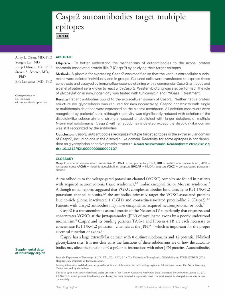
HEK293 T cells were transiently transfected to express Caspr2 stained with a series of patient sera live and then stainedwith a commercial antibody to Caspr2 after permeabilization and fixation Nuclei of transfected and untransfected cellswere stained with DAPI (blue) Surface epitopes of Caspr2 were recognized by all 10 patientsrsquo sera (4 representativesamples [S1ndashS4] are shown) Scale bar 5 10 mm
2 Neurology Neuroimmunology amp Neuroinflammation
the experiments was limited by the amount of sera available for
testing All experiments in this manuscript used a minimum of
4 patient samples to confirm results
Creation of plasmids for expressing Caspr2 mutantsComplementary DNA (cDNA) encoding full-length human
Caspr2 in a Prk5 vector was obtained (gift from Dr Elior
Figure 2 Caspr2 recognition does not depend on glycosylation or tertiary structure
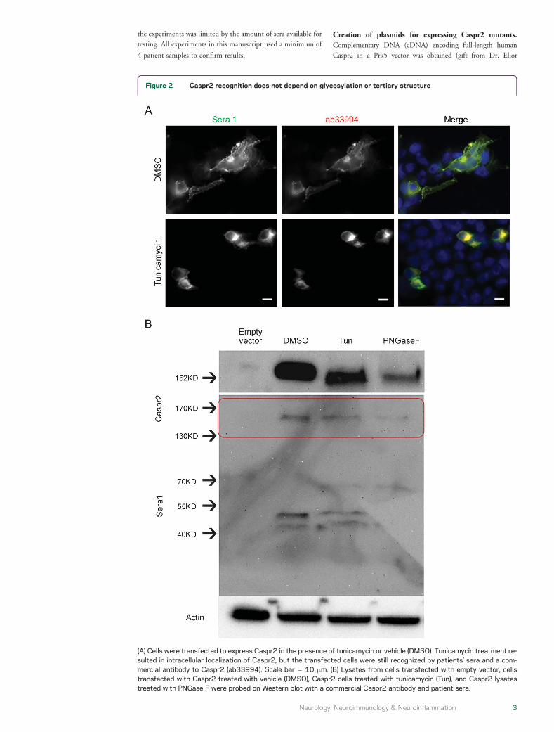
(A) Cells were transfected to express Caspr2 in the presence of tunicamycin or vehicle (DMSO) Tunicamycin treatment re-sulted in intracellular localization of Caspr2 but the transfected cells were still recognized by patientsrsquo sera and a com-mercial antibody to Caspr2 (ab33994) Scale bar 5 10 mm (B) Lysates from cells transfected with empty vector cellstransfected with Caspr2 treated with vehicle (DMSO) Caspr2 cells treated with tunicamycin (Tun) and Caspr2 lysatestreated with PNGase F were probed on Western blot with a commercial Caspr2 antibody and patient sera
Neurology Neuroimmunology amp Neuroinflammation 3
Peles Weizmann Institute of Science Rehovot Israel) This cDNA
was used as a template for deleting individual subdomains of Caspr2
using the Quikchange XL Site-Directed Mutagenesis Kit
(Stratagene Santa Clara CA) with modifications described
previously15 following an approach used previously to study
NMDAR antibodies12 Primers for the various mutations were
selected to include 27 bases before the cut site and 18 bases after
the cut site on average for both the forward and reverse primers these
primers are listed in table e-1 at Neurologyorgnn Clones were
selected using standard microbiology techniques and plasmid
DNA was purified using QiaPrep spin miniprep and maxiprep
kits (Qiagen Venlo the Netherlands) Clones were screened by
sequencing across the area with the target excision to determine
which contained the desired deletions clones with successful
deletions were verified to contain no unintended mutations by
full-length sequencing Plasmid DNA concentrations was
determined on a Nanodrop spectrophotometer (Thermo Scientific
Waltham MA)
Immunofluorescence staining HEK293 T cells were grown
on poly-D-lysinendashcoated coverslips in MEM with 5 FBS and
1 PS and were transiently transfected with full-length Caspr2
or Caspr2 mutants using JetPrime reagent (Polyplus Transfection
Illkirch France) per the manufacturerrsquos instructions Cells were
assayed 24ndash48 hours posttransfection Cells were fixed in 4
paraformaldehyde for 10 minutes permeabilized with 01 PBS-
tween or 03 triton X-100 for 15 minutes and blocked in 03 M
glycine with 10 normal goat serum and 5 BSA for 1 hour at
room temperature Cells were singly or doubly stained with a
commercial anti-Caspr2 primary antibody (ab33994 Abcam
Cambridge United Kingdom) diluted at 1200 and patient sera
diluted 11000 in blocking buffer overnight at 4degC (ab33994
targets the intracellular domain of Caspr2 and would not be
expected to compete with the human autoantibodies which
target the extracellular domain The intracellular domain was also
not modified in any of the deletion constructs) Coverslips were
washed with PBS and then secondary antibodies (Alexa Fluor 488ndash
conjugated donkey anti-human IgG and TRITC-conjugated
donkey anti-rabbit IgG at 1400) were applied for 1 hour at
room temperature Slides were mounted in Fluoromount-G with
DAPI (SouthernBiotech Birmingham AL) and visualized on a
Leica DMRBE fluorescent microscope Pictures were captured
with a Leica DFC-350F digital camera and Openlab software
(Improvision Coventry United Kingdom) Quantification of
fluorescence was performed using NIH Image J and Adobe
Photoshop software Untransfected cells and cells in which only
the secondary antibody was applied served as negative controls
Each experiment contained a minimum of 2 technical replicates
per condition and experiments were repeated a minimum of 3 times
Quantification of immunofluorescence staining Quantifi-
cation of fluorescence was performed using NIH Image J and
Adobe Photoshop software The intensity of staining of each sin-
gle domain deletion construct by patientsrsquo sera was normalized by
the amount of staining with the commercial antibody channel A
ratio was obtained for each construct
Tunicamycin treatment HEK293 T cells were cultured and
transfected as described above At 4 hours posttransfection
growth media were replaced with media containing 02 mgmL
tunicamycin (Sigma-Aldrich St Louis MO) or DMSO vehicle
control Cells were harvested 24ndash48 hours later and assayed as
described above
Western blotting HEK293 T cells were transfected as above
and lysed on ice in RIPA buffer (50 mM Tris pH 72 150
mM NaCl 01 SDS 05 Na deoxycholate 1 triton
X-100 2 mM EDTA) supplemented with complete protease
inhibitor tablet (Roche Basel Switzerland) Lysates were centri-
fuged at 10000 rpm for 10 minutes Protein concentration in
the supernatant was measured using Pierce BCA assay (Thermo
Scientific) on a plate reader The supernatant was diluted in sam-
ple buffer (Bio-Rad Hercules CA) and heated to 100degC for
5 minutes Samples were run on a Bio-Rad electrophoresis
system on 4ndash20 gels transferred to PDVF membrane
blocked in 5 milk for 1 hour and incubated in a commercial
anti-Caspr2 antibody (ab33994 Abcam) diluted 11000 or
patient sample overnight diluted 11000 at 4degC Secondary
antibodies were alkaline phosphatasendashconjugated donkey anti-
human or anti-rabbit applied for 1 hour at room temperature
at 110000 Blots were developed using Tropix CDP-Star
reagent (Applied Biosystems Waltham MA) and visualized on
a Chemi-Doc imaging system (Bio-Rad)
PNGase digestion Whole cell lysates from Caspr2-transfected
or untransfected control HEK293 T cells were diluted in
denaturing buffer and incubated at 100degC for 10 minutes after
which G7 reaction buffer 10 NP40 and PNGase F (New
England Biolabs Ipswich MA) were added per the
manufacturerrsquos instructions and incubated for 1 hour at 37degC
The samples were then prepared similarly to other lysates for
Western blotting
RESULTS Caspr2 autoantibodies consistently recognize
an extracellular epitope of Caspr2 We studied a panelof 10 sera from patients with Caspr2 antibodies
Figure 3 Single-domain deletion constructs were recognized by patientsrsquo sera
(A) Single-domain deletion constructs were generated as shown (B) Each of these con-structs was recognized by patientsrsquo sera onWestern blot probed with a commercial antibodyto Caspr2 (Caspr2) and patientsrsquo sera (sera 1)
4 Neurology Neuroimmunology amp Neuroinflammation
identified using methods previously described7 Inorder to confirm that patientsrsquo Caspr2 antibodies con-
sistently recognize a surface epitope of Caspr2
HEK293 T cells were transfected to express wild-type
Caspr2 and immunostained live with patient sera
(1500) The cells were then fixed permeabilized and
treated with a rabbit Caspr2 antibody targeting an
intracellular epitope followed by fluorescent anti-
human and anti-rabbit secondary antibodies
(4 representative cells are shown in figure 1) All 10patientsrsquo sera recognized a surface epitope ontransfected cells but not on untransfected cells
Glycosylation and tertiary structure are not required for
antibody recognition of Caspr2 Caspr2 has 12 poten-tial N-linked glycosylation sites in its extracellulardomain16 In order to determine whether glycosyla-tion of these sites is required for recognition byCaspr2 autoantibodies we transfected cultured cells
Figure 4 Some single-domain deletion constructs showed decreased labeling by patientsrsquo sera
(A) Cultured cells were transfected to express wild-type and single-domain deletion constructs All constructs were ex-pressed on the cell membrane and all were recognized by a panel of patientsrsquo sera Scale bar 5 10 mm (B) The relativestaining in the human and commercial channels was quantified for the various deletion constructs Only DDisc differedsignificantly from full-length Caspr2 using this method
Neurology Neuroimmunology amp Neuroinflammation 5
to express Caspr2 while these cells were treated withtunicamycin (figure 2) (In these experiments humanantibodies were applied after fixation and permeabili-zation since Caspr2 was no longer membrane-expressed) Tunicamycin treatment prevented cellsurface expression of Caspr2 and resulted in a shift ofthe Caspr2 band on Western blot but it did notprevent antibody recognition Protein extract fromcells transfected to express Caspr2 was alsodeglycosylated with PNGase F resulting in a shift in
the band on Western blot (figure 2B) This method ofdeglycosylation likewise did not prevent recognition ofCaspr2 by the autoantibodies even in the denaturedstate (ie on the Western blot) We further tested apanel of 6 sera for the ability to recognize Caspr2 underdenaturing conditions (on Western blot) and allsamples were able to do so (figure e-1) Caspr2autoantibodies therefore recognize the protein underdenaturing conditions and glycosylation is not requiredfor antibody recognition
Deletion of single Caspr2 subdomains does not entirely
eliminate reactivity Caspr2 has a very large extracellu-lar domain comprising more than 1200 amino acidsThe extracellular domain has previously been subdi-vided into 8 regions based on homology to various pro-tein domains We have followed a previously proposeddefinition of these domains (httpwwwuniprotorguniprotQ9UHC6section_seq) Starting from theN-terminus of the protein and moving to thetransmembrane region these domains are Discoidin(Disc) LamininG (Lam1) LamininG (Lam2) Egf(Egf1) FibrinogenC (FibC) LamininG (Lam3) Egf(Egf2) and LamininG (Lam4) There is a singletransmembrane domain and a relatively smallintracellular region with a protein 41-binding domainWe generated a series of plasmids for expressing Caspr2with deletions of each of the 8 extracellular subdomains(figure 3A) We named these constructs with a deltasymbol followed by the domain deleted DDisc forinstance has the Disc domain deleted
The single subdomain deletion constructs were allexpressed when transfected into cultured HEK293 Tcells It is interesting that patientsrsquo sera reacted witheach of these constructs on Western blot (figure 3B)and also by immunofluorescence (figure 4) All 10tested patient sera were able to immunostain living cellsexpressing each of these single-domain deletion con-structs confirming that they were all expressed on thecell membrane (figure 4) This pattern of findingsshowed that no single extracellular subdomain wasabsolutely necessary for antibody recognition and thatmultiple epitopes must exist in the extracellular domain
The relative intensity of immunostaining of someconstructs particularly DDisc was decreased by eyefor all patients studied This finding is quantified infigure 4B where the relative intensity of staining withthe human and commercial antibodies is comparedfor each of the single-domain deletion constructsUsing this method only DDisc showed significantreduction in staining with the human antibodiescompared to full-length Caspr2 These results suggestthat the Disc domain may contain a target epitope
The Disc domain is a common epitope for patients with
Caspr2 antibodies In order to more definitively deter-mine whether the Disc domain is sufficient for
Figure 5 The Disc domain contains a target epitope
(A) A construct (D182-Lam4) with all extracellular subdomains except the Disc domain wascreated (B) This construct was recognized by 9 of 10 sera in transfected cells Scale bar 510 mm (C) Western blot of lysates from cells transfected with an empty vector wild-typeCaspr2 or the D182-Lam4 construct were probed with human sera and then reprobed witha rabbit antibody to Caspr2 The D182-Lam4 protein is significantly smaller than wild-typeCaspr2 but is still recognized by both human and rabbit antibodies
6 Neurology Neuroimmunology amp Neuroinflammation
antibody recognition a construct with only the Discdomain the transmembrane domain and the intracel-lular domain was generated (figure 5A) This constructwas recognized by 9 of 10 serum samples (figure 5B)The Disc domain alone is therefore sufficient for rec-ognition by the majority of tested patients Howeverdeletion of this single Disc domain (DDisc) did notprevent recognition by any of our samples suggestingthat another epitope (or potentially multiple other epit-opes) outside the Disc domain exists This experimentwas repeated 4 times and all experiments suggesteddecreased intensity of but not absence of immunos-taining with deletion of the Disc domain comparedto the wild type (figure 4B) Thus there is a discrete
epitope within the Disc domain that is sufficient butnot necessary for reactivity
Patientsrsquo sera also recognized the construct con-taining only the Disc domain (with all other extracel-lular subdomains deleted) on Western blotsuggesting that an epitope in the Disc domain is stillrecognized under denaturing conditions (figure 5C)Taken together with the ability of patientsrsquo sera torecognize DDisc on Western blot (figure 3B) it ap-pears likely that there are multiple epitopes in theDisc domain and elsewhere that are not dependenton tertiary structure
Other target epitopes are generally in the N-terminal half
of Caspr2 In order to determine which other
Figure 6 Multiple target epitopes exist in the N-terminal domains of Caspr2
(A) Multidomain deletion constructs of Caspr2 were generated to determine which domains contain target epitopes (B)These constructs were transfected into cultured cells and immunostained with patient sera and a rabbit antibody to Caspr2(ab33994) Scale bar 5 10 mm (C) The results for 7 patient sera are shown See text for discussion
Neurology Neuroimmunology amp Neuroinflammation 7
subdomains (aside from Disc) might contain targetepitopes we constructed a new series of deletionconstructs involving multiple domains of Caspr2(figure 6A) In the case of constructs with a large dele-tion of the C-terminal domain of the protein we couldconfirm that these constructs were expressed on the cellmembrane by immunostaining live cells with patientsera (figure 6B) Although constructs with larger dele-tions of the N-terminal half of the protein werenot recognized by most patientsrsquo sera (in live orfixedpermeabilized cells) these constructs also ap-peared to be expressed on the membrane (figure 6B)Testing with a panel of 7 sera showed that target epit-opes for most patients (6 of 7) were confined entirelyto the 4 subdomains on the N-terminal of the proteinOnly 1 of 7 samples reacted (weakly) when these 4subdomains were deleted (figure 6C) Taken togetherthese results suggest that most target epitopes are con-centrated within the N-terminal half of the extracellu-lar domain and that patientsrsquo sera only rarely target anyepitope within the C-terminal half of the extracellulardomain
DISCUSSION These studies have shown that autoan-tibodies to Caspr2 target multiple epitopes on theextracellular domain of the protein The large extracel-lular domain of Caspr2 may make it a particularly avail-able target for autoimmunity compared with the othermembers of the VGKC complex A major epitope iswithin the discoidin-like domain and other epitopesappear to be concentrated within the N-terminalhalf of the protein At least some of these epitopesare not dependent on tertiary structure (ie canbe recognized on Western blot) and are notglycosylation-dependent Since our single-domaindeletions (including DDisc) were recognized onWestern blot it is likely that for most samples morethan one epitope is not dependent on tertiary structure
Other autoantibodies to neuronal surface epitopestarget a range of ionotropic neurotransmitter recep-tors (NMDAR AMPAR glycine receptor nAchR)or metabotropic neurotransmitter receptors (GABA-B-R mGluR1 mGluR5) In the case of antibodiesto the NMDAR and AMPAR the antibodies causecross-linking and internalization of the receptor andthus result in decreased receptor function17ndash19 SinceCaspr2 is a cell adhesion molecule and not a receptordifferent autoimmune mechanisms must be consid-ered for example disruption of the interactionof Caspr2 with TAG-1 disruption of the ability ofCaspr2 to cluster VGKCs or internalization ofCaspr2 (with or without associated VGKCs) Under-standing the target epitope of the antibodies is a firststep in examining these possibilities rigorously
The extracellular domain of Caspr2 is known tointeract with TAG-1 in cis (expressed on the axon)
and in trans (expressed on myelinating cellsapposed to the axon)2021 It is known that theimmunoglobulin-like domain of TAG-1 binds to theextracellular domain of Caspr222 but it is not knownwhich subdomain(s) of Caspr2 are required for theseinteractions Furthermore it is not known whether thesame area of Caspr2 is required for the cis and the transinteraction One plausible explanation for the multipleepitopes identified is that some antibodies disrupt theinteraction between Caspr2 and TAG-1 in cis whereasothers disrupt the interaction between Caspr2 in transFuture studies will be needed to determine whether theantibodies affect the interaction between Caspr2 andTAG-1 Since the interaction between these 2 proteinsis critical for the proper localization of potassium chan-nels8 this would be a plausible disease mechanism
These autoantibodies represent valuable scientifictools for further understanding the basic biology ofCaspr2 In addition understanding the pathogenicmechanism of the antibodies opens up therapeuticpossibilities Autoantibodies target multiple epitopeson Caspr2 and it is possible that antibodies to certainepitopes may be more pathogenic than antibodies toother epitopes For example antibodies to thenAChR that target the major immunogenic regionmay be more pathogenic than antibodies to otherparts of the receptor23 An important avenue of futureinvestigation is to determine which antibody-epitopeinteractions are most relevant for Caspr2 autoimmu-nity If a particular epitope is the target of disease-causing antibodies then specific immune therapies todirect the immune response away from this epitopemay be possible Luo and Lindstrom have recentlyused their knowledge of the target epitope of acetyl-choline receptor autoantibodies to create this type ofepitope-specific immunotherapy for experimentalautoimmune myasthenia gravis24
AUTHOR CONTRIBUTIONSAbby L Olsen draftingrevising the manuscript study concept or design
analysis or interpretation of data acquisition of data Yongjie Lai analysis
or interpretation of data acquisition of data Josep Dalmau draftingrevising
the manuscript contribution of vital reagentstoolspatients Steven S Scherer
draftingrevising the manuscript study concept or design analysis or interpre-
tation of data Eric Lancaster draftingrevising the manuscript study concept
or design analysis or interpretation of data acquisition of data statistical anal-
ysis study supervision obtaining funding
ACKNOWLEDGMENTThe authors thank Drs Elior Peles (Weizmann Institute Rehovot Isreal)
Domna Karagogeos (University of Crete Medical School Heraklion
Crete Greece) and Catherine Faivre-Sarrailh (Universiteacute de la Meacutediterra-
neacutee Aix-Marseille II Marseille France) for sharing plasmid constructs
related to Caspr2 used in some preliminary studies The authors also thank
the patients and their families for supporting this research by donating
biological samples
STUDY FUNDINGThis work was supported by K08 NS075142 to EL EL was also sup-
ported by a grant from the Dana Foundation
8 Neurology Neuroimmunology amp Neuroinflammation
DISCLOSUREAL Olsen and Y Lai report no disclosures J Dalmau is the editor of
Neurology Neuroimmunology amp Neuroinflammation is on the editorial
board for Neurology UpToDate holds patents for and receives royalties
from autoantibody test NMDA receptor autoantibody test has patents
pending for GABA(B) receptor autoantibody test GABA(A) receptor
autoantibody test DPPX autoantibody test and IgLON5 autoantibody
test has consulted for Advance Medical and received research support
from Euroimmun NIH NIMH Instituto Carlos III and AGUR SS
Scherer is on the scientific advisory board for the Charcot-Marie-Tooth
Association is on the editorial board for Cell and Tissue Research Exper-
imental Neurology Glia Journal of Neuroscience Research and Journal of
the Peripheral Nervous System has consulted for Lundbeck Scisive
Clarion Consulting Pfizer Guidepoint Global Advisors Cydan and
Grifols and received research support from NIHNational Institute of
Neurological Disorders and Stroke E Lancaster received speaker hono-
raria from Grifols Inc has consulted for Medimmune Inc provided
expert review for the Federal Vaccine Injury Compensation Program
and received research support from Talecris Inc Lundbeck Inc National
Institute of Neurological Disorders and Stroke and Dana Foundation
Go to Neurologyorgnn for full disclosure forms
Received October 7 2014 Accepted in final form May 22 2015
REFERENCES1 Shillito P Molenaar PC Vincent A et al Acquired neuro-
myotonia evidence for autoantibodies directed against K1
channels of peripheral nerves Ann Neurol 199538
714ndash722
2 Hart IK Waters C Vincent A et al Autoantibodies de-
tected to expressed K1 channels are implicated in neuro-
myotonia Ann Neurol 199741238ndash246
3 Tan KM Lennon VA Klein CJ Boeve BF Pittock SJ
Clinical spectrum of voltage-gated potassium channel
autoimmunity Neurology 2008701883ndash1890
4 Kleopa KA Elman LB Lang B Vincent A Scherer SS
Neuromyotonia and limbic encephalitis sera target
mature Shaker-type K1 channels subunit specificity
correlates with clinical manifestations Brain 2006129
1570ndash1584
5 Lai M Huijbers MG Lancaster E et al Investigation of
LGI1 as the antigen in limbic encephalitis previously
attributed to potassium channels a case series Lancet
Neurol 20109776ndash785
6 Irani SR Alexander S Waters P et al Antibodies to Kv1
potassium channel-complex proteins leucine-rich glioma
inactivated 1 protein and contactin-associated protein-2 in
limbic encephalitis Morvanrsquos syndrome and acquired neu-
romyotonia Brain 20101332734ndash2748
7 Lancaster E Huijbers MG Bar V et al Investigations of
caspr2 an autoantigen of encephalitis and neuromyotonia
Ann Neurol 201169303ndash311
8 Horresh I Poliak S Grant S Bredt D Rasband MN
Peles E Multiple molecular interactions determine the
clustering of Caspr2 and Kv1 channels in myelinated ax-
ons J Neurosci 20082814213ndash14222
9 Traka M Goutebroze L Denisenko N et al Association
of TAG-1 with Caspr2 is essential for the molecular
organization of juxtaparanodal regions of myelinated fi-
bers J Cell Biol 20031621161ndash1172
10 Gu C Gu Y Clustering and activity tuning of Kv1 chan-
nels in myelinated hippocampal axons J Biol Chem 2011
28625835ndash25847
11 Zhou L Messing A Chiu SY Determinants of excitability
at transition zones in Kv11-deficient myelinated nerves
J Neurosci 1999195768ndash5781
12 Gleichman AJ Spruce LA Dalmau J Seeholzer SH
Lynch DR Anti-NMDA receptor encephalitis antibody
binding is dependent on amino acid identity of a small
region within the GluN1 amino terminal domain
J Neurosci 20123211082ndash11094
13 Raju R Foote J Banga JP et al Analysis of GAD65
autoantibodies in Stiff-Person syndrome patients
J Immunol 20051757755ndash7762
14 Lindstrom J Luo J Kuryatov A Myasthenia gravis and
the tops and bottoms of AChRs antigenic structure of the
MIR and specific immunosuppression of EAMG using
AChR cytoplasmic domains Ann N Y Acad Sci 2008
113229ndash41
15 Makarova O Kamberov E Margolis B Generation of
deletion and point mutations with one primer in a single
cloning step Biotechniques 200029970ndash972
16 Poliak S Gollan L Martinez R et al Caspr2 a new
member of the neurexin superfamily is localized at the
juxtaparanodes of myelinated axons and associates with
K1 channels Neuron 1999241037ndash1047
17 Dalmau J Gleichman AJ Hughes EG et al Anti-NMDA-
receptor encephalitis case series and analysis of the effects of
antibodies Lancet Neurol 200871091ndash1098
18 Hughes EG Peng X Gleichman AJ et al Cellular and
synaptic mechanisms of anti-NMDA receptor encephalitis
J Neurosci 2010305866ndash5875
19 Lai M Hughes EG Peng X et al AMPA receptor anti-
bodies in limbic encephalitis alter synaptic receptor loca-
tion Ann Neurol 200965424ndash434
20 Chatzopoulou E Miguez A Savvaki M et al Structural
requirement of TAG-1 for retinal ganglion cell axons and
myelin in the mouse optic nerve J Neurosci 200828
7624ndash7636
21 Savvaki M Theodorakis K Zoupi L et al The expression
of TAG-1 in glial cells is sufficient for the formation of the
juxtaparanodal complex and the phenotypic rescue of tag-1
homozygous mutants in the CNS J Neurosci 201030
13943ndash13954
22 Tzimourakas A Giasemi S Mouratidou M Karagogeos D
Structure-function analysis of protein complexes involved in
the molecular architecture of juxtaparanodal regions of mye-
linated fibers Biotechnol J 20072577ndash583
23 Lindstrom J Luo J Myasthenogenicity of the main
immunogenic region Ann N Y Acad Sci 20121274
9ndash13
24 Luo J Lindstrom J Antigen-specific immunotherapeutic
vaccine for experimental autoimmune myasthenia gravis
J Immunol 20141935044ndash5055
Neurology Neuroimmunology amp Neuroinflammation 9
DOI 101212NXI000000000000012720152 Neurol Neuroimmunol Neuroinflamm
Abby L Olsen Yongjie Lai Josep Dalmau et al Caspr2 autoantibodies target multiple epitopes
This information is current as of July 2 2015
ServicesUpdated Information amp
httpnnneurologyorgcontent24e127fullhtmlincluding high resolution figures can be found at
Supplementary Material httpnnneurologyorgcontentsuppl2015080524e127DC1
Supplementary material can be found at
References httpnnneurologyorgcontent24e127fullhtmlref-list-1
This article cites 24 articles 10 of which you can access for free at
Citations httpnnneurologyorgcontent24e127fullhtmlotherarticles
This article has been cited by 3 HighWire-hosted articles
Subspecialty Collections
httpnnneurologyorgcgicollectionautoimmune_diseasesAutoimmune diseasesfollowing collection(s) This article along with others on similar topics appears in the
Permissions amp Licensing
httpnnneurologyorgmiscaboutxhtmlpermissionsits entirety can be found online atInformation about reproducing this article in parts (figurestables) or in
Reprints
httpnnneurologyorgmiscaddirxhtmlreprintsusInformation about ordering reprints can be found online
2015 American Academy of Neurology All rights reserved Online ISSN 2332-7812Published since April 2014 it is an open-access online-only continuous publication journal Copyright copy
is an official journal of the American Academy of NeurologyNeurol Neuroimmunol Neuroinflamm
to synaptic proteins including the NMDAreceptor (NMDAR) and the nicotinic acetyl-choline receptor (nAChR) may target either asingle dominant epitope or a major immuno-genic region12ndash14 For NMDAR and nAChRantibodies 3-dimensional structure is veryimportant but other autoantibodies such asto glutamic acid decarboxylase 65 (GAD65)recognize linear epitopes under denatured con-ditions (eg on Western blot) We have there-fore attempted to find a single dominant epitopefor Caspr2 antibodies and to determine whetherthe autoantibodies require intact tertiary struc-ture andor protein glycosylation
METHODS Standard protocol approvals registrationsand patient consents Serum and CSF were collected with
informed consent from patients with clinical features of Caspr2
autoimmunity (encephalitis andor peripheral nerve hyperexcit-
ability) under a tissue bank protocol approved by an institu-
tional review board (IRB) at the University of Pennsylvania
A separate protocol approved by the University of Pennsylvania
IRB was used to access the samples from the tissue bank for
testing and to access the clinical information Samples from pa-
tients found to contain Caspr2 antibodies using methods previ-
ously described7 were selected for further studies The clinical
characteristics of 8 of these patients have been published previ-
ously 2 had encephalitis only 1 had acquired peripheral nerve
hyperexcitability only and 5 had both encephalitis and periph-
eral nerve hyperexcitability7 In addition we studied samples
from 2 additional patients who were subsequently identified
1 with neuromyotonia only and 1 with both neuromyotonia
and encephalitis The number of patient samples included for
Figure 1 Patientsrsquo sera recognize surface epitopes of Caspr2
HEK293 T cells were transiently transfected to express Caspr2 stained with a series of patient sera live and then stainedwith a commercial antibody to Caspr2 after permeabilization and fixation Nuclei of transfected and untransfected cellswere stained with DAPI (blue) Surface epitopes of Caspr2 were recognized by all 10 patientsrsquo sera (4 representativesamples [S1ndashS4] are shown) Scale bar 5 10 mm
2 Neurology Neuroimmunology amp Neuroinflammation
the experiments was limited by the amount of sera available for
testing All experiments in this manuscript used a minimum of
4 patient samples to confirm results
Creation of plasmids for expressing Caspr2 mutantsComplementary DNA (cDNA) encoding full-length human
Caspr2 in a Prk5 vector was obtained (gift from Dr Elior
Figure 2 Caspr2 recognition does not depend on glycosylation or tertiary structure
(A) Cells were transfected to express Caspr2 in the presence of tunicamycin or vehicle (DMSO) Tunicamycin treatment re-sulted in intracellular localization of Caspr2 but the transfected cells were still recognized by patientsrsquo sera and a com-mercial antibody to Caspr2 (ab33994) Scale bar 5 10 mm (B) Lysates from cells transfected with empty vector cellstransfected with Caspr2 treated with vehicle (DMSO) Caspr2 cells treated with tunicamycin (Tun) and Caspr2 lysatestreated with PNGase F were probed on Western blot with a commercial Caspr2 antibody and patient sera
Neurology Neuroimmunology amp Neuroinflammation 3
Peles Weizmann Institute of Science Rehovot Israel) This cDNA
was used as a template for deleting individual subdomains of Caspr2
using the Quikchange XL Site-Directed Mutagenesis Kit
(Stratagene Santa Clara CA) with modifications described
previously15 following an approach used previously to study
NMDAR antibodies12 Primers for the various mutations were
selected to include 27 bases before the cut site and 18 bases after
the cut site on average for both the forward and reverse primers these
primers are listed in table e-1 at Neurologyorgnn Clones were
selected using standard microbiology techniques and plasmid
DNA was purified using QiaPrep spin miniprep and maxiprep
kits (Qiagen Venlo the Netherlands) Clones were screened by
sequencing across the area with the target excision to determine
which contained the desired deletions clones with successful
deletions were verified to contain no unintended mutations by
full-length sequencing Plasmid DNA concentrations was
determined on a Nanodrop spectrophotometer (Thermo Scientific
Waltham MA)
Immunofluorescence staining HEK293 T cells were grown
on poly-D-lysinendashcoated coverslips in MEM with 5 FBS and
1 PS and were transiently transfected with full-length Caspr2
or Caspr2 mutants using JetPrime reagent (Polyplus Transfection
Illkirch France) per the manufacturerrsquos instructions Cells were
assayed 24ndash48 hours posttransfection Cells were fixed in 4
paraformaldehyde for 10 minutes permeabilized with 01 PBS-
tween or 03 triton X-100 for 15 minutes and blocked in 03 M
glycine with 10 normal goat serum and 5 BSA for 1 hour at
room temperature Cells were singly or doubly stained with a
commercial anti-Caspr2 primary antibody (ab33994 Abcam
Cambridge United Kingdom) diluted at 1200 and patient sera
diluted 11000 in blocking buffer overnight at 4degC (ab33994
targets the intracellular domain of Caspr2 and would not be
expected to compete with the human autoantibodies which
target the extracellular domain The intracellular domain was also
not modified in any of the deletion constructs) Coverslips were
washed with PBS and then secondary antibodies (Alexa Fluor 488ndash
conjugated donkey anti-human IgG and TRITC-conjugated
donkey anti-rabbit IgG at 1400) were applied for 1 hour at
room temperature Slides were mounted in Fluoromount-G with
DAPI (SouthernBiotech Birmingham AL) and visualized on a
Leica DMRBE fluorescent microscope Pictures were captured
with a Leica DFC-350F digital camera and Openlab software
(Improvision Coventry United Kingdom) Quantification of
fluorescence was performed using NIH Image J and Adobe
Photoshop software Untransfected cells and cells in which only
the secondary antibody was applied served as negative controls
Each experiment contained a minimum of 2 technical replicates
per condition and experiments were repeated a minimum of 3 times
Quantification of immunofluorescence staining Quantifi-
cation of fluorescence was performed using NIH Image J and
Adobe Photoshop software The intensity of staining of each sin-
gle domain deletion construct by patientsrsquo sera was normalized by
the amount of staining with the commercial antibody channel A
ratio was obtained for each construct
Tunicamycin treatment HEK293 T cells were cultured and
transfected as described above At 4 hours posttransfection
growth media were replaced with media containing 02 mgmL
tunicamycin (Sigma-Aldrich St Louis MO) or DMSO vehicle
control Cells were harvested 24ndash48 hours later and assayed as
described above
Western blotting HEK293 T cells were transfected as above
and lysed on ice in RIPA buffer (50 mM Tris pH 72 150
mM NaCl 01 SDS 05 Na deoxycholate 1 triton
X-100 2 mM EDTA) supplemented with complete protease
inhibitor tablet (Roche Basel Switzerland) Lysates were centri-
fuged at 10000 rpm for 10 minutes Protein concentration in
the supernatant was measured using Pierce BCA assay (Thermo
Scientific) on a plate reader The supernatant was diluted in sam-
ple buffer (Bio-Rad Hercules CA) and heated to 100degC for
5 minutes Samples were run on a Bio-Rad electrophoresis
system on 4ndash20 gels transferred to PDVF membrane
blocked in 5 milk for 1 hour and incubated in a commercial
anti-Caspr2 antibody (ab33994 Abcam) diluted 11000 or
patient sample overnight diluted 11000 at 4degC Secondary
antibodies were alkaline phosphatasendashconjugated donkey anti-
human or anti-rabbit applied for 1 hour at room temperature
at 110000 Blots were developed using Tropix CDP-Star
reagent (Applied Biosystems Waltham MA) and visualized on
a Chemi-Doc imaging system (Bio-Rad)
PNGase digestion Whole cell lysates from Caspr2-transfected
or untransfected control HEK293 T cells were diluted in
denaturing buffer and incubated at 100degC for 10 minutes after
which G7 reaction buffer 10 NP40 and PNGase F (New
England Biolabs Ipswich MA) were added per the
manufacturerrsquos instructions and incubated for 1 hour at 37degC
The samples were then prepared similarly to other lysates for
Western blotting
RESULTS Caspr2 autoantibodies consistently recognize
an extracellular epitope of Caspr2 We studied a panelof 10 sera from patients with Caspr2 antibodies
Figure 3 Single-domain deletion constructs were recognized by patientsrsquo sera
(A) Single-domain deletion constructs were generated as shown (B) Each of these con-structs was recognized by patientsrsquo sera onWestern blot probed with a commercial antibodyto Caspr2 (Caspr2) and patientsrsquo sera (sera 1)
4 Neurology Neuroimmunology amp Neuroinflammation
identified using methods previously described7 Inorder to confirm that patientsrsquo Caspr2 antibodies con-
sistently recognize a surface epitope of Caspr2
HEK293 T cells were transfected to express wild-type
Caspr2 and immunostained live with patient sera
(1500) The cells were then fixed permeabilized and
treated with a rabbit Caspr2 antibody targeting an
intracellular epitope followed by fluorescent anti-
human and anti-rabbit secondary antibodies
(4 representative cells are shown in figure 1) All 10patientsrsquo sera recognized a surface epitope ontransfected cells but not on untransfected cells
Glycosylation and tertiary structure are not required for
antibody recognition of Caspr2 Caspr2 has 12 poten-tial N-linked glycosylation sites in its extracellulardomain16 In order to determine whether glycosyla-tion of these sites is required for recognition byCaspr2 autoantibodies we transfected cultured cells
Figure 4 Some single-domain deletion constructs showed decreased labeling by patientsrsquo sera
(A) Cultured cells were transfected to express wild-type and single-domain deletion constructs All constructs were ex-pressed on the cell membrane and all were recognized by a panel of patientsrsquo sera Scale bar 5 10 mm (B) The relativestaining in the human and commercial channels was quantified for the various deletion constructs Only DDisc differedsignificantly from full-length Caspr2 using this method
Neurology Neuroimmunology amp Neuroinflammation 5
to express Caspr2 while these cells were treated withtunicamycin (figure 2) (In these experiments humanantibodies were applied after fixation and permeabili-zation since Caspr2 was no longer membrane-expressed) Tunicamycin treatment prevented cellsurface expression of Caspr2 and resulted in a shift ofthe Caspr2 band on Western blot but it did notprevent antibody recognition Protein extract fromcells transfected to express Caspr2 was alsodeglycosylated with PNGase F resulting in a shift in
the band on Western blot (figure 2B) This method ofdeglycosylation likewise did not prevent recognition ofCaspr2 by the autoantibodies even in the denaturedstate (ie on the Western blot) We further tested apanel of 6 sera for the ability to recognize Caspr2 underdenaturing conditions (on Western blot) and allsamples were able to do so (figure e-1) Caspr2autoantibodies therefore recognize the protein underdenaturing conditions and glycosylation is not requiredfor antibody recognition
Deletion of single Caspr2 subdomains does not entirely
eliminate reactivity Caspr2 has a very large extracellu-lar domain comprising more than 1200 amino acidsThe extracellular domain has previously been subdi-vided into 8 regions based on homology to various pro-tein domains We have followed a previously proposeddefinition of these domains (httpwwwuniprotorguniprotQ9UHC6section_seq) Starting from theN-terminus of the protein and moving to thetransmembrane region these domains are Discoidin(Disc) LamininG (Lam1) LamininG (Lam2) Egf(Egf1) FibrinogenC (FibC) LamininG (Lam3) Egf(Egf2) and LamininG (Lam4) There is a singletransmembrane domain and a relatively smallintracellular region with a protein 41-binding domainWe generated a series of plasmids for expressing Caspr2with deletions of each of the 8 extracellular subdomains(figure 3A) We named these constructs with a deltasymbol followed by the domain deleted DDisc forinstance has the Disc domain deleted
The single subdomain deletion constructs were allexpressed when transfected into cultured HEK293 Tcells It is interesting that patientsrsquo sera reacted witheach of these constructs on Western blot (figure 3B)and also by immunofluorescence (figure 4) All 10tested patient sera were able to immunostain living cellsexpressing each of these single-domain deletion con-structs confirming that they were all expressed on thecell membrane (figure 4) This pattern of findingsshowed that no single extracellular subdomain wasabsolutely necessary for antibody recognition and thatmultiple epitopes must exist in the extracellular domain
The relative intensity of immunostaining of someconstructs particularly DDisc was decreased by eyefor all patients studied This finding is quantified infigure 4B where the relative intensity of staining withthe human and commercial antibodies is comparedfor each of the single-domain deletion constructsUsing this method only DDisc showed significantreduction in staining with the human antibodiescompared to full-length Caspr2 These results suggestthat the Disc domain may contain a target epitope
The Disc domain is a common epitope for patients with
Caspr2 antibodies In order to more definitively deter-mine whether the Disc domain is sufficient for
Figure 5 The Disc domain contains a target epitope
(A) A construct (D182-Lam4) with all extracellular subdomains except the Disc domain wascreated (B) This construct was recognized by 9 of 10 sera in transfected cells Scale bar 510 mm (C) Western blot of lysates from cells transfected with an empty vector wild-typeCaspr2 or the D182-Lam4 construct were probed with human sera and then reprobed witha rabbit antibody to Caspr2 The D182-Lam4 protein is significantly smaller than wild-typeCaspr2 but is still recognized by both human and rabbit antibodies
6 Neurology Neuroimmunology amp Neuroinflammation
antibody recognition a construct with only the Discdomain the transmembrane domain and the intracel-lular domain was generated (figure 5A) This constructwas recognized by 9 of 10 serum samples (figure 5B)The Disc domain alone is therefore sufficient for rec-ognition by the majority of tested patients Howeverdeletion of this single Disc domain (DDisc) did notprevent recognition by any of our samples suggestingthat another epitope (or potentially multiple other epit-opes) outside the Disc domain exists This experimentwas repeated 4 times and all experiments suggesteddecreased intensity of but not absence of immunos-taining with deletion of the Disc domain comparedto the wild type (figure 4B) Thus there is a discrete
epitope within the Disc domain that is sufficient butnot necessary for reactivity
Patientsrsquo sera also recognized the construct con-taining only the Disc domain (with all other extracel-lular subdomains deleted) on Western blotsuggesting that an epitope in the Disc domain is stillrecognized under denaturing conditions (figure 5C)Taken together with the ability of patientsrsquo sera torecognize DDisc on Western blot (figure 3B) it ap-pears likely that there are multiple epitopes in theDisc domain and elsewhere that are not dependenton tertiary structure
Other target epitopes are generally in the N-terminal half
of Caspr2 In order to determine which other
Figure 6 Multiple target epitopes exist in the N-terminal domains of Caspr2
(A) Multidomain deletion constructs of Caspr2 were generated to determine which domains contain target epitopes (B)These constructs were transfected into cultured cells and immunostained with patient sera and a rabbit antibody to Caspr2(ab33994) Scale bar 5 10 mm (C) The results for 7 patient sera are shown See text for discussion
Neurology Neuroimmunology amp Neuroinflammation 7
subdomains (aside from Disc) might contain targetepitopes we constructed a new series of deletionconstructs involving multiple domains of Caspr2(figure 6A) In the case of constructs with a large dele-tion of the C-terminal domain of the protein we couldconfirm that these constructs were expressed on the cellmembrane by immunostaining live cells with patientsera (figure 6B) Although constructs with larger dele-tions of the N-terminal half of the protein werenot recognized by most patientsrsquo sera (in live orfixedpermeabilized cells) these constructs also ap-peared to be expressed on the membrane (figure 6B)Testing with a panel of 7 sera showed that target epit-opes for most patients (6 of 7) were confined entirelyto the 4 subdomains on the N-terminal of the proteinOnly 1 of 7 samples reacted (weakly) when these 4subdomains were deleted (figure 6C) Taken togetherthese results suggest that most target epitopes are con-centrated within the N-terminal half of the extracellu-lar domain and that patientsrsquo sera only rarely target anyepitope within the C-terminal half of the extracellulardomain
DISCUSSION These studies have shown that autoan-tibodies to Caspr2 target multiple epitopes on theextracellular domain of the protein The large extracel-lular domain of Caspr2 may make it a particularly avail-able target for autoimmunity compared with the othermembers of the VGKC complex A major epitope iswithin the discoidin-like domain and other epitopesappear to be concentrated within the N-terminalhalf of the protein At least some of these epitopesare not dependent on tertiary structure (ie canbe recognized on Western blot) and are notglycosylation-dependent Since our single-domaindeletions (including DDisc) were recognized onWestern blot it is likely that for most samples morethan one epitope is not dependent on tertiary structure
Other autoantibodies to neuronal surface epitopestarget a range of ionotropic neurotransmitter recep-tors (NMDAR AMPAR glycine receptor nAchR)or metabotropic neurotransmitter receptors (GABA-B-R mGluR1 mGluR5) In the case of antibodiesto the NMDAR and AMPAR the antibodies causecross-linking and internalization of the receptor andthus result in decreased receptor function17ndash19 SinceCaspr2 is a cell adhesion molecule and not a receptordifferent autoimmune mechanisms must be consid-ered for example disruption of the interactionof Caspr2 with TAG-1 disruption of the ability ofCaspr2 to cluster VGKCs or internalization ofCaspr2 (with or without associated VGKCs) Under-standing the target epitope of the antibodies is a firststep in examining these possibilities rigorously
The extracellular domain of Caspr2 is known tointeract with TAG-1 in cis (expressed on the axon)
and in trans (expressed on myelinating cellsapposed to the axon)2021 It is known that theimmunoglobulin-like domain of TAG-1 binds to theextracellular domain of Caspr222 but it is not knownwhich subdomain(s) of Caspr2 are required for theseinteractions Furthermore it is not known whether thesame area of Caspr2 is required for the cis and the transinteraction One plausible explanation for the multipleepitopes identified is that some antibodies disrupt theinteraction between Caspr2 and TAG-1 in cis whereasothers disrupt the interaction between Caspr2 in transFuture studies will be needed to determine whether theantibodies affect the interaction between Caspr2 andTAG-1 Since the interaction between these 2 proteinsis critical for the proper localization of potassium chan-nels8 this would be a plausible disease mechanism
These autoantibodies represent valuable scientifictools for further understanding the basic biology ofCaspr2 In addition understanding the pathogenicmechanism of the antibodies opens up therapeuticpossibilities Autoantibodies target multiple epitopeson Caspr2 and it is possible that antibodies to certainepitopes may be more pathogenic than antibodies toother epitopes For example antibodies to thenAChR that target the major immunogenic regionmay be more pathogenic than antibodies to otherparts of the receptor23 An important avenue of futureinvestigation is to determine which antibody-epitopeinteractions are most relevant for Caspr2 autoimmu-nity If a particular epitope is the target of disease-causing antibodies then specific immune therapies todirect the immune response away from this epitopemay be possible Luo and Lindstrom have recentlyused their knowledge of the target epitope of acetyl-choline receptor autoantibodies to create this type ofepitope-specific immunotherapy for experimentalautoimmune myasthenia gravis24
AUTHOR CONTRIBUTIONSAbby L Olsen draftingrevising the manuscript study concept or design
analysis or interpretation of data acquisition of data Yongjie Lai analysis
or interpretation of data acquisition of data Josep Dalmau draftingrevising
the manuscript contribution of vital reagentstoolspatients Steven S Scherer
draftingrevising the manuscript study concept or design analysis or interpre-
tation of data Eric Lancaster draftingrevising the manuscript study concept
or design analysis or interpretation of data acquisition of data statistical anal-
ysis study supervision obtaining funding
ACKNOWLEDGMENTThe authors thank Drs Elior Peles (Weizmann Institute Rehovot Isreal)
Domna Karagogeos (University of Crete Medical School Heraklion
Crete Greece) and Catherine Faivre-Sarrailh (Universiteacute de la Meacutediterra-
neacutee Aix-Marseille II Marseille France) for sharing plasmid constructs
related to Caspr2 used in some preliminary studies The authors also thank
the patients and their families for supporting this research by donating
biological samples
STUDY FUNDINGThis work was supported by K08 NS075142 to EL EL was also sup-
ported by a grant from the Dana Foundation
8 Neurology Neuroimmunology amp Neuroinflammation
DISCLOSUREAL Olsen and Y Lai report no disclosures J Dalmau is the editor of
Neurology Neuroimmunology amp Neuroinflammation is on the editorial
board for Neurology UpToDate holds patents for and receives royalties
from autoantibody test NMDA receptor autoantibody test has patents
pending for GABA(B) receptor autoantibody test GABA(A) receptor
autoantibody test DPPX autoantibody test and IgLON5 autoantibody
test has consulted for Advance Medical and received research support
from Euroimmun NIH NIMH Instituto Carlos III and AGUR SS
Scherer is on the scientific advisory board for the Charcot-Marie-Tooth
Association is on the editorial board for Cell and Tissue Research Exper-
imental Neurology Glia Journal of Neuroscience Research and Journal of
the Peripheral Nervous System has consulted for Lundbeck Scisive
Clarion Consulting Pfizer Guidepoint Global Advisors Cydan and
Grifols and received research support from NIHNational Institute of
Neurological Disorders and Stroke E Lancaster received speaker hono-
raria from Grifols Inc has consulted for Medimmune Inc provided
expert review for the Federal Vaccine Injury Compensation Program
and received research support from Talecris Inc Lundbeck Inc National
Institute of Neurological Disorders and Stroke and Dana Foundation
Go to Neurologyorgnn for full disclosure forms
Received October 7 2014 Accepted in final form May 22 2015
REFERENCES1 Shillito P Molenaar PC Vincent A et al Acquired neuro-
myotonia evidence for autoantibodies directed against K1
channels of peripheral nerves Ann Neurol 199538
714ndash722
2 Hart IK Waters C Vincent A et al Autoantibodies de-
tected to expressed K1 channels are implicated in neuro-
myotonia Ann Neurol 199741238ndash246
3 Tan KM Lennon VA Klein CJ Boeve BF Pittock SJ
Clinical spectrum of voltage-gated potassium channel
autoimmunity Neurology 2008701883ndash1890
4 Kleopa KA Elman LB Lang B Vincent A Scherer SS
Neuromyotonia and limbic encephalitis sera target
mature Shaker-type K1 channels subunit specificity
correlates with clinical manifestations Brain 2006129
1570ndash1584
5 Lai M Huijbers MG Lancaster E et al Investigation of
LGI1 as the antigen in limbic encephalitis previously
attributed to potassium channels a case series Lancet
Neurol 20109776ndash785
6 Irani SR Alexander S Waters P et al Antibodies to Kv1
potassium channel-complex proteins leucine-rich glioma
inactivated 1 protein and contactin-associated protein-2 in
limbic encephalitis Morvanrsquos syndrome and acquired neu-
romyotonia Brain 20101332734ndash2748
7 Lancaster E Huijbers MG Bar V et al Investigations of
caspr2 an autoantigen of encephalitis and neuromyotonia
Ann Neurol 201169303ndash311
8 Horresh I Poliak S Grant S Bredt D Rasband MN
Peles E Multiple molecular interactions determine the
clustering of Caspr2 and Kv1 channels in myelinated ax-
ons J Neurosci 20082814213ndash14222
9 Traka M Goutebroze L Denisenko N et al Association
of TAG-1 with Caspr2 is essential for the molecular
organization of juxtaparanodal regions of myelinated fi-
bers J Cell Biol 20031621161ndash1172
10 Gu C Gu Y Clustering and activity tuning of Kv1 chan-
nels in myelinated hippocampal axons J Biol Chem 2011
28625835ndash25847
11 Zhou L Messing A Chiu SY Determinants of excitability
at transition zones in Kv11-deficient myelinated nerves
J Neurosci 1999195768ndash5781
12 Gleichman AJ Spruce LA Dalmau J Seeholzer SH
Lynch DR Anti-NMDA receptor encephalitis antibody
binding is dependent on amino acid identity of a small
region within the GluN1 amino terminal domain
J Neurosci 20123211082ndash11094
13 Raju R Foote J Banga JP et al Analysis of GAD65
autoantibodies in Stiff-Person syndrome patients
J Immunol 20051757755ndash7762
14 Lindstrom J Luo J Kuryatov A Myasthenia gravis and
the tops and bottoms of AChRs antigenic structure of the
MIR and specific immunosuppression of EAMG using
AChR cytoplasmic domains Ann N Y Acad Sci 2008
113229ndash41
15 Makarova O Kamberov E Margolis B Generation of
deletion and point mutations with one primer in a single
cloning step Biotechniques 200029970ndash972
16 Poliak S Gollan L Martinez R et al Caspr2 a new
member of the neurexin superfamily is localized at the
juxtaparanodes of myelinated axons and associates with
K1 channels Neuron 1999241037ndash1047
17 Dalmau J Gleichman AJ Hughes EG et al Anti-NMDA-
receptor encephalitis case series and analysis of the effects of
antibodies Lancet Neurol 200871091ndash1098
18 Hughes EG Peng X Gleichman AJ et al Cellular and
synaptic mechanisms of anti-NMDA receptor encephalitis
J Neurosci 2010305866ndash5875
19 Lai M Hughes EG Peng X et al AMPA receptor anti-
bodies in limbic encephalitis alter synaptic receptor loca-
tion Ann Neurol 200965424ndash434
20 Chatzopoulou E Miguez A Savvaki M et al Structural
requirement of TAG-1 for retinal ganglion cell axons and
myelin in the mouse optic nerve J Neurosci 200828
7624ndash7636
21 Savvaki M Theodorakis K Zoupi L et al The expression
of TAG-1 in glial cells is sufficient for the formation of the
juxtaparanodal complex and the phenotypic rescue of tag-1
homozygous mutants in the CNS J Neurosci 201030
13943ndash13954
22 Tzimourakas A Giasemi S Mouratidou M Karagogeos D
Structure-function analysis of protein complexes involved in
the molecular architecture of juxtaparanodal regions of mye-
linated fibers Biotechnol J 20072577ndash583
23 Lindstrom J Luo J Myasthenogenicity of the main
immunogenic region Ann N Y Acad Sci 20121274
9ndash13
24 Luo J Lindstrom J Antigen-specific immunotherapeutic
vaccine for experimental autoimmune myasthenia gravis
J Immunol 20141935044ndash5055
Neurology Neuroimmunology amp Neuroinflammation 9
DOI 101212NXI000000000000012720152 Neurol Neuroimmunol Neuroinflamm
Abby L Olsen Yongjie Lai Josep Dalmau et al Caspr2 autoantibodies target multiple epitopes
This information is current as of July 2 2015
ServicesUpdated Information amp
httpnnneurologyorgcontent24e127fullhtmlincluding high resolution figures can be found at
Supplementary Material httpnnneurologyorgcontentsuppl2015080524e127DC1
Supplementary material can be found at
References httpnnneurologyorgcontent24e127fullhtmlref-list-1
This article cites 24 articles 10 of which you can access for free at
Citations httpnnneurologyorgcontent24e127fullhtmlotherarticles
This article has been cited by 3 HighWire-hosted articles
Subspecialty Collections
httpnnneurologyorgcgicollectionautoimmune_diseasesAutoimmune diseasesfollowing collection(s) This article along with others on similar topics appears in the
Permissions amp Licensing
httpnnneurologyorgmiscaboutxhtmlpermissionsits entirety can be found online atInformation about reproducing this article in parts (figurestables) or in
Reprints
httpnnneurologyorgmiscaddirxhtmlreprintsusInformation about ordering reprints can be found online
2015 American Academy of Neurology All rights reserved Online ISSN 2332-7812Published since April 2014 it is an open-access online-only continuous publication journal Copyright copy
is an official journal of the American Academy of NeurologyNeurol Neuroimmunol Neuroinflamm
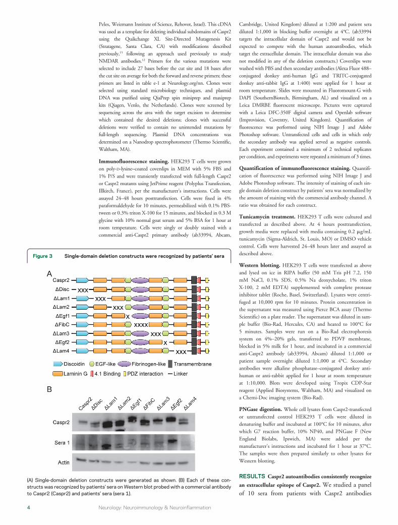
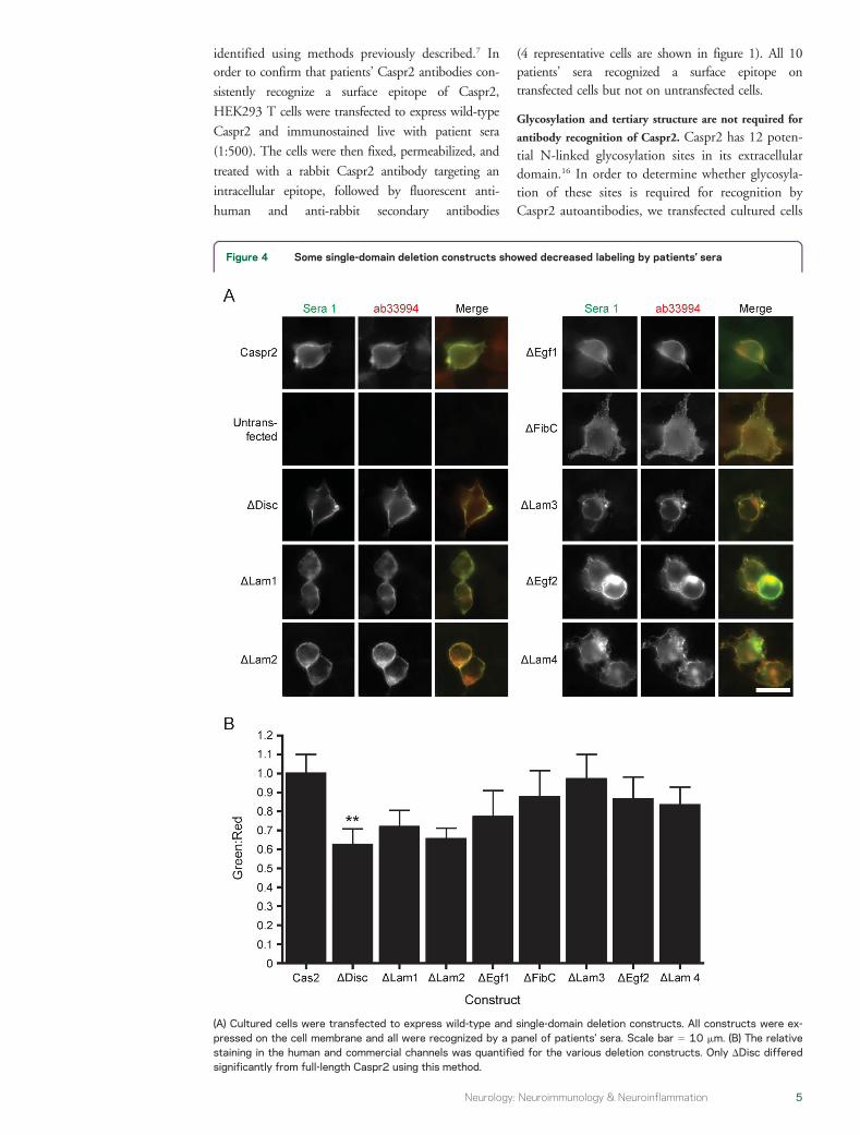
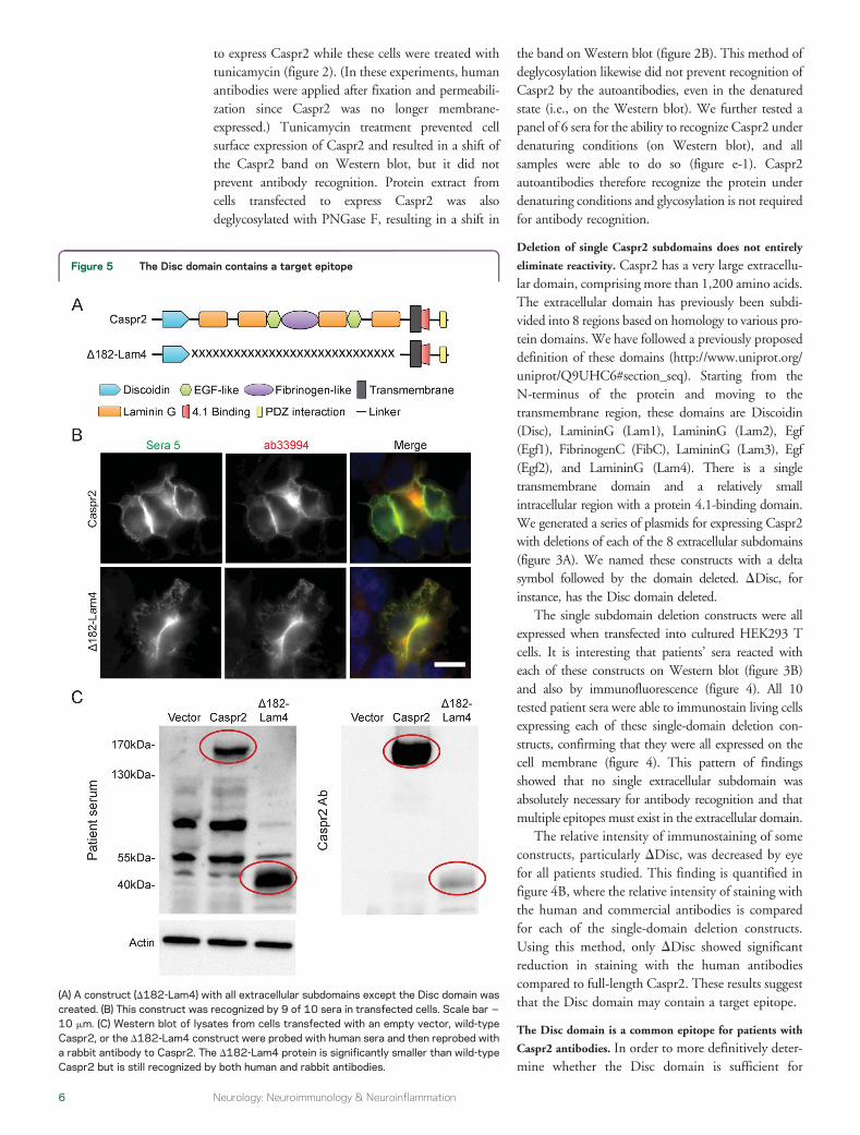
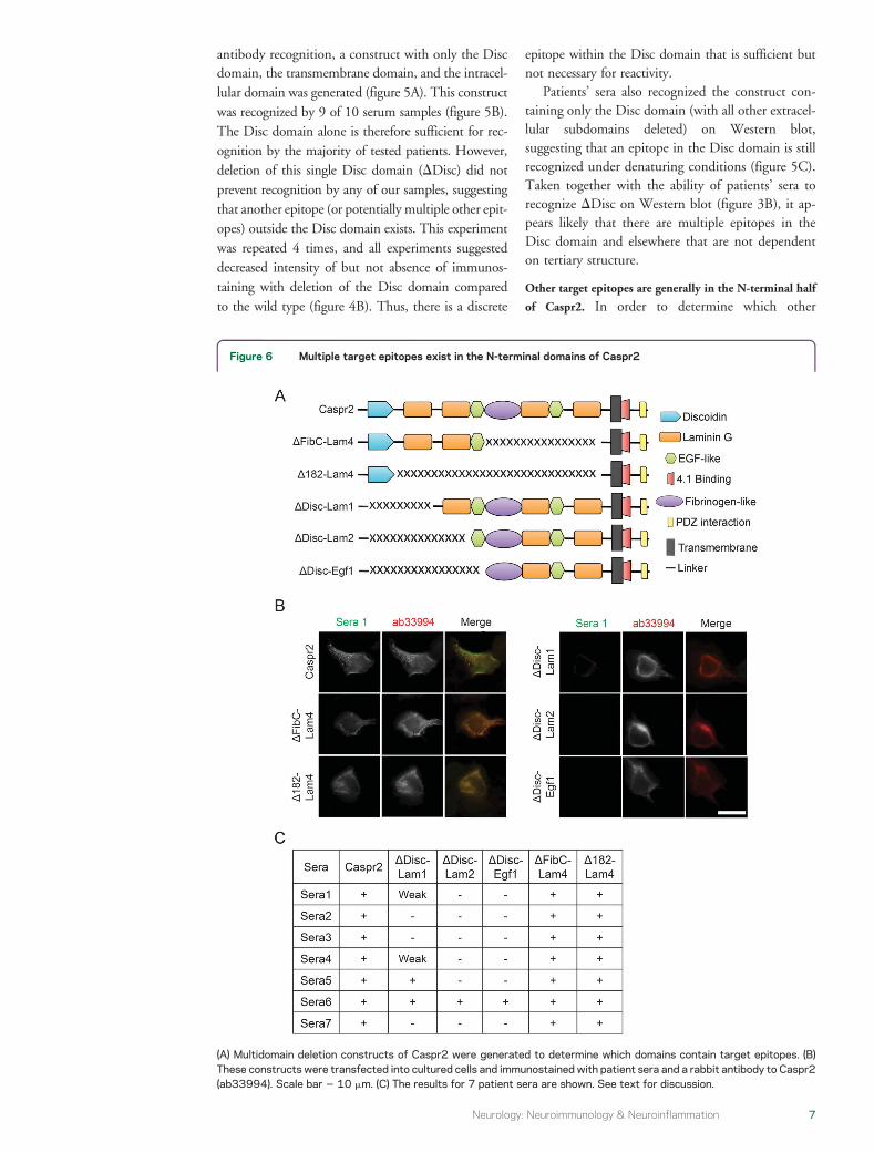