Embed Size (px)
Citation preview
Zurich Open Repository andArchiveUniversity of ZurichMain LibraryStrickhofstrasse 39CH-8057 Zurichwww.zora.uzh.ch
Year: 2018
A high throughput screening method for the nano-crystallization of salts oforganic cations
Nievergelt, Philipp P ; Babor, Martin ; Čejka, Jan ; Spingler, Bernhard
Abstract: The generation of solid salts of organic molecules is important to the chemical and pharma-ceutical industry. Commonly used salt screening methods consume a lot of resources. We employeda combination of ion exchange screening and vapour diffusion for crystallization. This technique issemi-automatic and requires just nanoliters of the solution of the analyte to be crystallized. This highthroughput screening yielded single crystals of sufficient size and quality for single-crystal X-ray structuredetermination using an in-house X-ray diffractometer. The broad scope of our method has been shown bychallenging it with 7 very different organic cations, whose aqueous solubilities vary by a factor of almost1000. At least one crystal structure for 6 out of 7 tested cations was determined; 4 out of the successful6 ones had never been crystallized before. Our method is extremely attractive for high throughput saltscreening, especially for active pharmaceutical ingredients (APIs), as about 40% of all APIs are cationicsalts. Additionally, our screening is a new and very promising procedure for the crystallization of saltsof organic cations.
DOI: https://doi.org/10.1039/C8SC00783G
Posted at the Zurich Open Repository and Archive, University of ZurichZORA URL: https://doi.org/10.5167/uzh-151075Journal ArticlePublished Version
The following work is licensed under a Creative Commons: Attribution-NonCommercial 3.0 Unported(CC BY-NC 3.0) License.
Originally published at:Nievergelt, Philipp P; Babor, Martin; Čejka, Jan; Spingler, Bernhard (2018). A high throughput screen-ing method for the nano-crystallization of salts of organic cations. Chemical Science, 9(15):3716-3722.DOI: https://doi.org/10.1039/C8SC00783G
A high throughput screening method for thenano-crystallization of salts of organic cations†
Philipp P. Nievergelt, ‡a Martin Babor, ‡ab Jan Cejka b
and Bernhard Spingler *a
The generation of solid salts of organicmolecules is important to the chemical and pharmaceutical industry.
Commonly used salt screening methods consume a lot of resources. We employed a combination of ion
exchange screening and vapour diffusion for crystallization. This technique is semi-automatic and
requires just nanoliters of the solution of the analyte to be crystallized. This high throughput screening
yielded single crystals of sufficient size and quality for single-crystal X-ray structure determination using
an in-house X-ray diffractometer. The broad scope of our method has been shown by challenging it
with 7 very different organic cations, whose aqueous solubilities vary by a factor of almost 1000. At least
one crystal structure for 6 out of 7 tested cations was determined; 4 out of the successful 6 ones had
never been crystallized before. Our method is extremely attractive for high throughput salt screening,
especially for active pharmaceutical ingredients (APIs), as about 40% of all APIs are cationic salts.
Additionally, our screening is a new and very promising procedure for the crystallization of salts of
organic cations.
Introduction
Organic salts are multi-component ionic compounds, in whichat least one of the components is an organic ion. The focus ofthis paper is the organic, cationic part of salts. Different salts ofthe same organic cation can have signicantly different physi-cochemical properties.1–6 The critical properties are solubility,7
crystal shape, hygroscopicity, melting point, and physical aswell as chemical stability. The selection of a suitable anion canavoid problems during production, storage or shipping of anorganic salt. For example, an appropriate anion can improve thepurication and ow properties of the powder and/or reducethe hygroscopicity. Therefore, salt screening is a decisive stepduring the development and optimization of the productionprocess of an organic salt.2 It can signicantly reduce theproduction cost of the chemical or pharmaceutical, as about40% of all active pharmaceutical ingredients (APIs) are cationicsalts.8 However, there are also requirements for an idealscreening. It shall require the least possible amount of material,
human work and time. At the same time, it should be aseffective as possible in discovering new salts. Additionally, theability to analyse the new crystalline forms directly from thebasic screening would be a great advantage, ideally by singlecrystal X-ray diffraction (SCXRD) structure analysis, since fastaccess to SCXRD can save a lot of material and lab work, such asimmediately identifying false positive screening hits.
Recently, several innovations have been described that, withthe help of crystallography, allow the structure determination ofcompounds, which had previously been impossible by applyingthe methods of traditional crystal growth.9,10 The ‘crystallinesponge’method was originally introduced in 2013 by Fujita andco-workers11 and has further been improved over the last fewyears.12,13 This method opened up new possibilities for deter-mining the crystal structures of apolar compounds that areavailable only in minute amounts. However, this method is a 3step procedure: (1) synthesis of the MOF host, (2) exchange ofthe previous solvent by an apolar one, and (3) soaking of theMOF crystal with the analyte. All steps are non-trivial to performand especially the last step has to be optimized for every singlecompound. Alternative sponge methods have been described,avoiding the exchange of guest molecules; however, they requiremore analyte material and synchrotron radiation.14,15 Even themost recent optimisation of the crystal sponge method is stilllabour intensive, requires the analyte to be soluble indichloromethane and can only in exceptional cases be used asan ab initio structure determination method without additionalknowledge of the analyte.16 A different MOF lattice has recentlybeen developed by the group of Yaghi for the co-crystallization
aDepartment of Chemistry, University of Zurich, Winterthurerstr. 190, 8057 Zurich,
Switzerland. E-mail: [email protected]; Web: http://www.chem.uzh.ch/en/
research/groups/spingler.htmlbDepartment of Solid State Chemistry, University of Chemistry and Technology Prague,
Prague 6, 166 28, Czech Republic
† Electronic supplementary information (ESI) available: Details of experimental,crystallographic procedures and descriptions of the crystal structures. CCDC1585747–1585765 and 1823199–1823201. For ESI and crystallographic data inCIF or other electronic format see DOI: 10.1039/c8sc00783g
‡ These authors contributed equally to this work.
Cite this: Chem. Sci., 2018, 9, 3716
Received 16th February 2018Accepted 6th March 2018
DOI: 10.1039/c8sc00783g
rsc.li/chemical-science
3716 | Chem. Sci., 2018, 9, 3716–3722 This journal is © The Royal Society of Chemistry 2018
ChemicalScience
EDGE ARTICLE
of carboxylates or alcohol containing molecules.17 Summarizingthe previous developments, a new method that could facilitatethe crystallization of cationic, polar compounds would behighly desirable.
Results and discussion
Our new method addresses this need. Our screening techniqueis based on two simple ideas and combines basic screening withsingle crystal growth directly followed by structural analysis.The rst hypothesis assumes that chloride salts of most cationshave the highest solubility in water and analogously thatsodium salts are the most common form of salts with a highsolubility in water. The second hypothesis supposes that a slowincrease of saturation might lead to the growth of single crys-tals. According to the rst idea, if we mix solutions of organicchloride salts with various sodium salts, the least solublecombination of dissolved ions should crystallize, which shouldbe a new organic salt. In addition, the mixture of the startingsalts does not have to be equimolar, because all remaining ionswill still be dissolved in water. Concerning the second idea, anincrease of analyte concentration within a drop is generated bythe method of vapour diffusion (see ESI Fig. 1†), which waspreviously developed for protein crystallization.18 This methodbasically consists of mixing 100–500 nl of the analyte in waterwith the same volume of a crystallization cocktail – in our casethis is normally a sodium salt – to generate a small drop of 200–1000 nl volume. The drop is then equilibrated via the vapour
phase against the big reservoir consisting of only the crystalli-zation cocktail. Since the crystallization cocktail in the drop wasdiluted with the analyte, the water diffuses via the gas phaseback to the reservoir. The mixing of the analyte and the sodiumsalt in the drop might already generate a supersaturated solu-tion, while the diffusion of the water to the reservoir willincrease this effect. In order to automate and miniaturize thewhole procedure, the pipetting of the reservoir and the dropsolutions, as well as the visual monitoring, is carried out bya robot. Such robots are commonly being used in mostbiochemical laboratories and pharmaceutical companies thatare engaged in structural biology projects.
Currently, the salt screening of active pharmaceuticalingredients (APIs) that can be protonated is mainly done byisolating and weighing the free base followed by addition ofdifferent acids in various solvents.19–21 This process is labourintensive and can be unreliable, as some free bases are hygro-scopic, prone to oxidation, or may otherwise be chemicallyunstable. An additional disadvantage of this method is that theratio of API to acid has a direct inuence upon the pH in thechosen solvent and therefore also upon the propensity to crys-tallize. Furthermore, different solvents might change the degreeof protonation of the API. Therefore, the amount of added acidmust be chosen carefully in advance for each solvent.
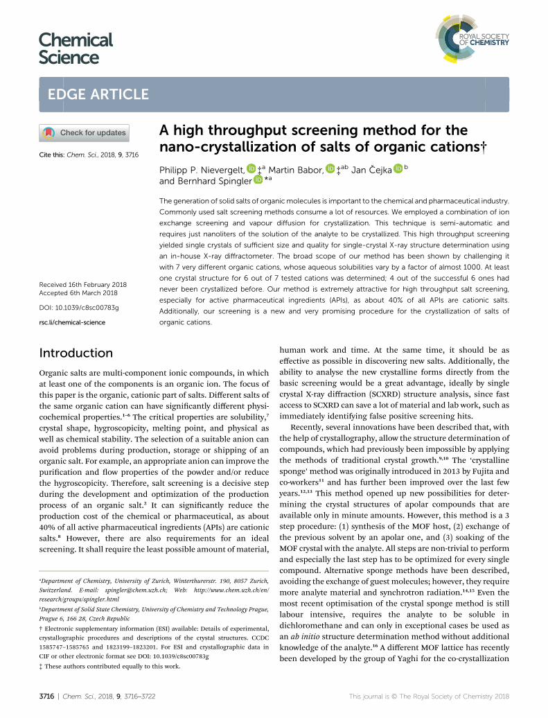
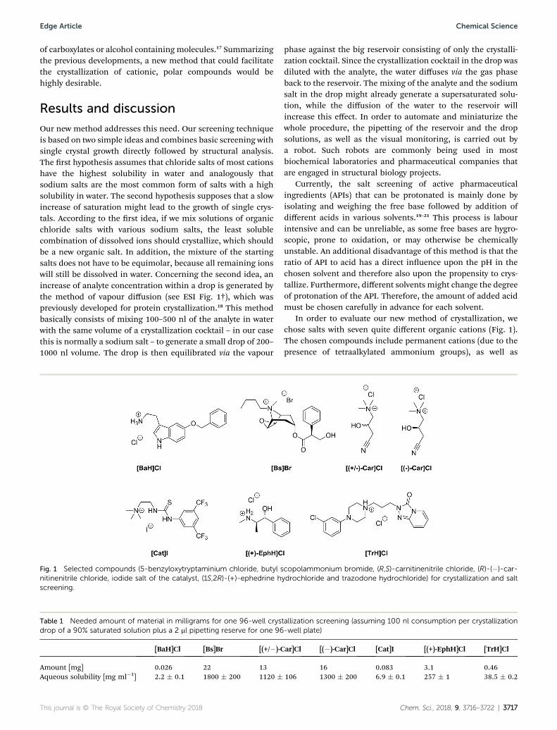
In order to evaluate our new method of crystallization, wechose salts with seven quite different organic cations (Fig. 1).The chosen compounds include permanent cations (due to thepresence of tetraalkylated ammonium groups), as well as
Fig. 1 Selected compounds (5-benzyloxytryptaminium chloride, butyl scopolammonium bromide, (R,S)-carnitinenitrile chloride, (R)-(�)-car-nitinenitrile chloride, iodide salt of the catalyst, (1S,2R)-(+)-ephedrine hydrochloride and trazodone hydrochloride) for crystallization and saltscreening.
Table 1 Needed amount of material in milligrams for one 96-well crystallization screening (assuming 100 nl consumption per crystallizationdrop of a 90% saturated solution plus a 2 ml pipetting reserve for one 96-well plate)
[BaH]Cl [Bs]Br [(+/�)-Car]Cl [(�)-Car]Cl [Cat]I [(+)-EphH]Cl [TrH]Cl
Amount [mg] 0.026 22 13 16 0.083 3.1 0.46Aqueous solubility [mg ml�1] 2.2 � 0.1 1800 � 200 1120 � 106 1300 � 200 6.9 � 0.1 257 � 1 38.5 � 0.2
This journal is © The Royal Society of Chemistry 2018 Chem. Sci., 2018, 9, 3716–3722 | 3717
Edge Article Chemical Science
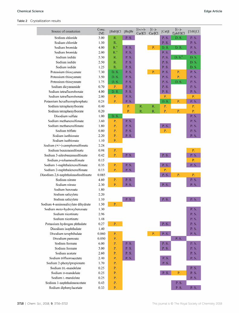
Table 2 Crystallization results
3718 | Chem. Sci., 2018, 9, 3716–3722 This journal is © The Royal Society of Chemistry 2018
Chemical Science Edge Article
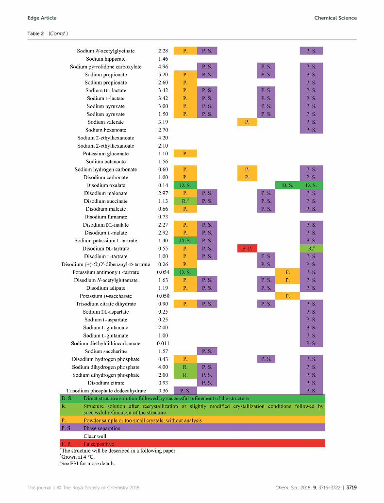
Table 2 (Contd. )
This journal is © The Royal Society of Chemistry 2018 Chem. Sci., 2018, 9, 3716–3722 | 3719
Edge Article Chemical Science
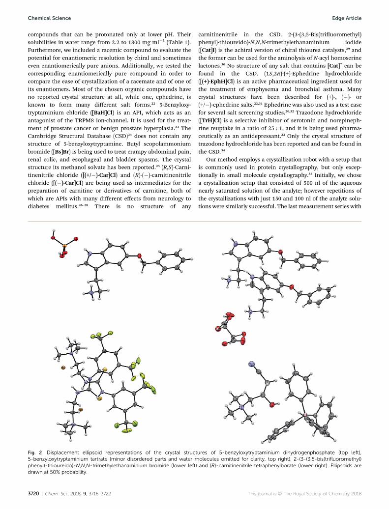
compounds that can be protonated only at lower pH. Theirsolubilities in water range from 2.2 to 1800 mg ml�1 (Table 1).Furthermore, we included a racemic compound to evaluate thepotential for enantiomeric resolution by chiral and sometimeseven enantiomerically pure anions. Additionally, we tested thecorresponding enantiomerically pure compound in order tocompare the ease of crystallization of a racemate and of one ofits enantiomers. Most of the chosen organic compounds haveno reported crystal structure at all, while one, ephedrine, isknown to form many different salt forms.22 5-Benzyloxy-tryptaminium chloride ([BaH]Cl) is an API, which acts as anantagonist of the TRPM8 ion-channel. It is used for the treat-ment of prostate cancer or benign prostate hyperplasia.23 TheCambridge Structural Database (CSD)24 does not contain anystructure of 5-benzyloxytryptamine. Butyl scopolammoniumbromide ([Bs]Br) is being used to treat crampy abdominal pain,renal colic, and esophageal and bladder spasms. The crystalstructure its methanol solvate has been reported.25 (R,S)-Carni-tinenitrile chloride ([(+/�)-Car]Cl) and (R)-(�)-carnitinenitrilechloride ([(�)-Car]Cl) are being used as intermediates for thepreparation of carnitine or derivatives of carnitine, both ofwhich are APIs with many different effects from neurology todiabetes mellitus.26–28 There is no structure of any
carnitinenitrile in the CSD. 2-(3-(3,5-Bis(triuoromethyl)phenyl)-thioureido)-N,N,N-trimethylethanaminium iodide([Cat]I) is the achiral version of chiral thiourea catalysts,29 andthe former can be used for the aminolysis of N-acyl homoserinelactones.30 No structure of any salt that contains [Cat]+ can befound in the CSD. (1S,2R)-(+)-Ephedrine hydrochloride([(+)-EphH]Cl) is an active pharmaceutical ingredient used forthe treatment of emphysema and bronchial asthma. Manycrystal structures have been described for (+)-, (�)- or(+/�)-ephedrine salts.22,31 Ephedrine was also used as a test casefor several salt screening studies.20,32 Trazodone hydrochloride([TrH]Cl) is a selective inhibitor of serotonin and norepineph-rine reuptake in a ratio of 25 : 1, and it is being used pharma-ceutically as an antidepressant.33 Only the crystal structure oftrazodone hydrochloride has been reported and can be found inthe CSD.34
Our method employs a crystallization robot with a setup thatis commonly used in protein crystallography, but only excep-tionally in small molecule crystallography.35 Initially, we chosea crystallization setup that consisted of 500 nl of the aqueousnearly saturated solution of the analyte; however repetitions ofthe crystallizations with just 150 and 100 nl of the analyte solu-tions were similarly successful. The last measurement series with
Fig. 2 Displacement ellipsoid representations of the crystal structures of 5-benzyloxytryptaminium dihydrogenphosphate (top left),5-benzyloxytryptaminium tartrate (minor disordered parts and water molecules omitted for clarity, top right), 2-(3-(3,5-bis(trifluoromethyl)phenyl)-thioureido)-N,N,N-trimethylethanaminium bromide (lower left) and (R)-carnitinenitrile tetraphenylborate (lower right). Ellipsoids aredrawn at 50% probability.
3720 | Chem. Sci., 2018, 9, 3716–3722 This journal is © The Royal Society of Chemistry 2018
Chemical Science Edge Article
100 nl analyte solution required really minute amounts ofmaterial and still yielded high quality crystals (ESI Fig. 3†) thatdirectly led to publishable structures. As can be seen in Table 1,the required amount essentially depends upon the solubility ofthe compound in water. For our method, we work with solutionsthat are 90% saturated. We used two procedures to obtain thesesolutions: one was to determine the apparent solubility bydissolution of the solid material and observing the progress withamicroscope. The othermethod wasmuch simpler and used lessmaterial: we generated a saturated stock solution, separated itfrom the solid and diluted it then to 90% saturation.
The screening was performed with the seven mentionedorganic salts and mainly sodium salts of 77 different counterions(Table 2 and ESI†). The criteria for the selection of the counterionswere a known propensity to crystallize and/or being a compoundthat is Generally Recognized As Safe (GRAS)36 for a potential lateruse in an API. Each of the chosen 7 analytes was screened under96 conditions, as some of the counterions were present in thescreening atmultiple, different concentrations. The screening wasstopped aer 16 days; however longer equilibration times couldbe applied. Quite oen the crystals formed aer a few days, seee.g. the case of 5-benzyloxytryptaminium antimony-L-tartrate, inwhich a change of crystal morphology could be observed (ESIFig. 2†). The same crystals could sometimes be observed in severaldrops containing the same counterion at different concentrations.It was possible to determine 15 high quality single crystal struc-tures of chemically different salts directly from the screeningwithout the need for any further optimisation, and 14 of themwere novel structures. Additionally, 14 lead hits consisting of toosmall crystals were manually recrystallized, which yielded in7 cases big enough crystals that could bemeasured and rened bySCXRD to give 7 novel structures (Fig. 2 and Table 2). In onefurther case, covering the drop with oil induced the crystal growthof the free base trazodone. The crystal nucleated at the oil/waterinterface and then grew by penetrating into the oil. The threecompounds – [Bs]Br, [(+/�)-Car]Cl and [(�)-Car]Cl – all having anunusually high solubility of more than one gram per milliliterwere most challenging; obviously if the chloride is already presentat such high concentrations, it is difficult to substitute it duringcrystallization from a mixed chloride/anion solution.
Conclusion
Our screening method for the growth of single crystals con-taining organic cations was very effective: six out of seven testedcationic moieties yielded at least one crystal structure. Also, theprimary screening used just a very small amount of material(12 ml of a 90% saturated aqueous solution of a salt with anorganic cation) in order to test a total of 96 different conditions,and we were able to determine at least one unit cell directly inmost of the positive crystalline hits using in-house X-raydiffractometers. Furthermore, the required manual work isquite low due to the usage of a pipetting robot and a crystalfarm, both of which are available in many biochemical insti-tutes and pharmaceutical companies. With just 26 mg of oneorganic salt employing 100 nl of analyte solution per crystalli-zation experiment, we were able to directly determine the
structure of 6 different salts and obtained the lead to another 5salts. The new screening method requires compounds that canform stable cations in water and have a water solubility of atleast 2 mg ml�1 of their salt form. We are currently working ona system, which can crystallize less water soluble cations. Incontrast to the report of Berghausen,21 we were able to performa salt screening with highly water soluble compounds ina purely aqueous environment. Performing the screening attemperatures other than room temperature is possible andyielded a second, unknown polymorph of enantiomerically pureephedrinium iodide.
Conflicts of interest
There are no conicts of interest to declare.
Acknowledgements
We thank Prof. Dr Mario Waser for kindly providing compound[Cat]I, Lonza Ltd. Basel for providing the sodium dicyanamide,and Beat Blattmann as well as Celine Stutz for setting up ourcrystallization trials on the robot. We thank Mohammad Al-Qatanani for determining the aqueous solubility of variouscompounds by HPLC and Prof. Dr Anthony Linden for a criticalreading of the manuscript. We thank the University of Zurich,the R'Equip programme of the Swiss National Science Foun-dation (project number 206021_164018) and the Czech ScienceFoundation (grant No. 16-10035S) for nancial support.
References
1 M. von Raumer, J. Dannappel and R. Hilker, Chem. Today,2006, 24, 41–44.
2 L. Kumar, A. Amin and A. K. Bansal, Drug Discovery Today,2007, 12, 1046–1053.
3 W.-Q. Tong, in Developing Solid Oral Dosage Forms, ed. Y.Chen, G. G. Z. Zhang, L. Liu and W. R. Porter, AcademicPress, San Diego, 2009, ch. 4, pp. 75–86.
4 N. Wyttenbach, B. Sutter and P. Hidber, in Handbook ofPharmaceutical Salts, ed. P. H. Stahl and C. G. Wermuth,Verlag Helvetica Chimica Acta, 2nd edn., 2011, ch. 8, pp.203–233.
5 Pharmaceutical Salts and Co-crystals, ed. J. Wouters andL. Quere, Royal Society of Chemistry, Cambridge, 2011.
6 S. R. Byrn, G. Zogra and X. Chen, J. Pharm. Sci., 2010, 99,3665–3675.
7 D. P. Elder, R. Holm and H. L. de Diego, Int. J. Pharm., 2013,453, 88–100.
8 G. S. Paulekuhn, J. B. Dressman and C. Saal, J. Med. Chem.,2007, 50, 6665–6672.
9 B. Spingler, S. Schnidrig, T. Todorova and F. Wild,CrystEngComm, 2012, 14, 751–757.
10 P. P. Nievergelt and B. Spingler, CrystEngComm, 2017, 19,142–147.
11 Y. Inokuma, S. Yoshioka, J. Ariyoshi, T. Arai, Y. Hitora,K. Takada, S. Matsunaga, K. Rissanen and M. Fujita,Nature, 2013, 495, 461–466.
This journal is © The Royal Society of Chemistry 2018 Chem. Sci., 2018, 9, 3716–3722 | 3721
Edge Article Chemical Science
12 Y. Inokuma, S. Yoshioka, J. Ariyoshi, T. Arai and M. Fujita,Nat. Protoc., 2014, 9, 246–252.
13 M. Hoshino, A. Khutia, H. Xing, Y. Inokuma and M. Fujita,IUCrJ, 2016, 3, 139–151.
14 T. R. Ramadhar, S.-L. Zheng, Y.-S. Chen and J. Clardy, ActaCrystallogr., Sect. A: Found. Adv., 2015, 71, 46–58.
15 T. R. Ramadhar, S.-L. Zheng, Y.-S. Chen and J. Clardy,CrystEngComm, 2017, 19, 4528–4534.
16 F. Sakurai, A. Khutia, T. Kikuchi andM. Fujita, Chem.–Eur. J.,2017, 23, 15035–15040.
17 S. Lee, E. A. Kapustin and O. M. Yaghi, Science, 2016, 353,808–811.
18 A. McPherson and J. A. Gavira, Acta Crystallogr., Sect. F:Struct. Biol. Commun., 2014, 70, 2–20.
19 B. M. Collman, J. M. Miller, C. Seadeek, J. A. Stambek andA. C. Blackburn, Drug Dev. Ind. Pharm., 2013, 39, 29–38.
20 M. R. Thorson, S. Goyal, B. R. Schudel, C. F. Zukoski,G. G. Z. Zhang, Y. C. Gong and P. J. A. Kenis, Lab Chip,2011, 11, 3829–3837.
21 P. B. Tarsa, C. S. Towler, G. Woollam and J. Berghausen, Eur.J. Pharm. Sci., 2010, 41, 23–30.
22 E. A. Collier, R. J. Davey, S. N. Black and R. J. Roberts, ActaCrystallogr., Sect. B: Struct. Sci., 2006, 62, 498–505.
23 J. DeFalco, D. Steiger, M. Dourado, D. Emerling andM. A. J. Duncton, Bioorg. Med. Chem. Lett., 2010, 20, 7076–7079.
24 C. R. Groom, I. J. Bruno, M. P. Lightfoot and S. C. Ward, ActaCrystallogr., Sect. B: Struct. Sci., Cryst. Eng. Mater., 2016, 72,171–179.
25 J. M. Leger, M. Gadret and A. Carpy, Acta Crystallogr., Sect. B:Struct. Crystallogr. Cryst. Chem., 1978, 34, 3705–3709.
26 H. Puetter, E. Roske and H. J. Pander, German Pat.,DE3419723A1, 1985.
27 K. Nakayama, H. Honda, Y. Ogawa, T. Ohta and T. Ozawa, USPat., US5041375, 1991.
28 M. Malaguarnera, Curr. Opin. Gastroenterol., 2012, 28, 166–176.
29 M. Tiffner, J. Novacek, A. Busillo, K. Gratzer, A. Massa andM. Waser, RSC Adv., 2015, 5, 78941–78949.
30 M. A. Bertucci, S. J. Lee and M. R. Gagne, Chem. Commun.,2013, 49, 2055–2057.
31 H. Wu, A. R. West, M. Vickers, D. C. Apperley and A. G. Jones,Chem. Eng. Sci., 2012, 77, 47–56.
32 S. N. Black, E. A. Collier, R. J. Davey and R. J. Roberts,J. Pharm. Sci., 2007, 96, 1053–1068.
33 G. Davidoff, M. Guarracini, E. Roth, J. Sliwa and G. Yarkony,Pain, 1987, 29, 151–161.
34 J. P. Fillers and S. W. Hawkinson, Acta Crystallogr., Sect. B:Struct. Crystallogr. Cryst. Chem., 1979, 35, 498–500.
35 P. K. Mandal, B. Kauffmann, H. Destecroix, Y. Ferrand,A. P. Davis and I. Huc, Chem. Commun., 2016, 52, 9355–9358.
36 SCOGS (Select Committee on GRAS Substances), https://www.accessdata.fda.gov/scripts/fdcc/?set¼SCOGS, accessed23rd October 2017.
3722 | Chem. Sci., 2018, 9, 3716–3722 This journal is © The Royal Society of Chemistry 2018
Chemical Science Edge Article
S1
Electronic supplementary information for
A high throughput screening method for the nano-crystallization of
salts of organic cations
Philipp P. Nievergelt, Martin Babor, Jan Čejka, Bernhard Spingler*
*Corresponding author: [email protected]
Content:
1. Materials and Methods
1.1 Chemicals
1.2 Stock Solutions including Supplementary Table 1
1.3 Crystallization of Compounds incl. Supplementary Figures 1-3
1.4 X-ray Single Crystal Diffraction
2. Description of the Structures including Supplementary Figures 4-26 and Supplementary Tables 2-8
S2
1. Materials and Methods:
1.1. Chemicals
5-benzyloxytryptamine hydrochloride ([BaH]Cl) was obtained from Acros Organics, Geel, Belgium. N-
Butylscopolammonium bromide (also described as butylscopolamine bromide or hyoscine butylbromide
[Bs]Br), (1S,2R)-(+)-ephedrine hydrochloride ([(+)-EphH]Cl) and trazodone hydrochloride ([TrH]Cl)
were obtained from Sigma Aldrich. (R,S)-carnitinenitrile chloride ([(+/-)-Car]Cl) was obtained from
Frontier Scientific, Logan, UT, USA. (R)-carnitinenitrile chloride ([(-)-Car]Cl) was obtained from
Angene Chemical, Hong Kong, HK. 2-(3-(3,5-bis(trifluoromethyl)phenyl)thioureido)-N,N,N-
trimethylethanammonium iodide ([Cat]I) was kindly provided by Prof. Dr. Mario Waser, Linz, Austria; it
was prepared analogously as described in 1. The used water was doubly distilled. Sodium or potassium
salts of counterions (SCI) were obtained from various commercial suppliers. If the sodium or potassium
salts were not commercially available, the corresponding sodium salts were prepared by titration of the
corresponding acids with a hydroxide sodium solution (2 M) until the pH reached 7. The solutions of
newly prepared salts were concentrated with the help of a rotary evaporator and dried by lyophilisation at
0.02 mbar. The water content was determined by elemental analysis (carbon, hydrogen and nitrogen) of
the salt.
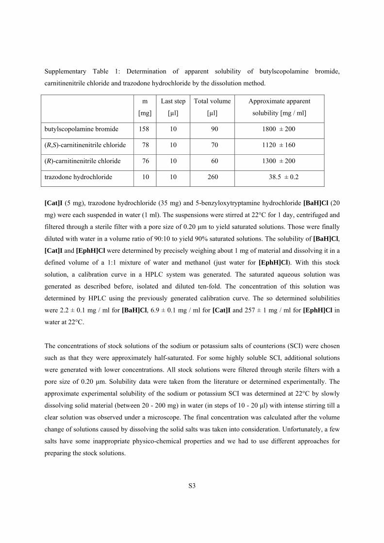
1.2. Stock Solutions
The stock solutions of (R,S)-carnitinenitrile chloride, (R)-carnitinenitrile chloride, (1S,2R)-(+)-ephedrine
hydrochloride, trazodone hydrochloride and iodide catalyst were employed at 90% concentration of the
saturated solutions. The approximate solubilities of (R,S)- and (R)-carnitinenitrile chloride were
determined by slowly dissolving solid material in water in steps of 10 μl with intense stirring until a clear
solution was observed under a microscope (see Supplementary Table 1).
S3
Supplementary Table 1: Determination of apparent solubility of butylscopolamine bromide,
carnitinenitrile chloride and trazodone hydrochloride by the dissolution method.
m
[mg]
Last step
[µl]
Total volume
[µl]
Approximate apparent
solubility [mg / ml]
butylscopolamine bromide 158 10 90 1800 ± 200
(R,S)-carnitinenitrile chloride 78 10 70 1120 ± 160
(R)-carnitinenitrile chloride 76 10 60 1300 ± 200
trazodone hydrochloride 10 10 260 38.5 ± 0.2
[Cat]I (5 mg), trazodone hydrochloride (35 mg) and 5-benzyloxytryptamine hydrochloride [BaH]Cl (20
mg) were each suspended in water (1 ml). The suspensions were stirred at 22°C for 1 day, centrifuged and
filtered through a sterile filter with a pore size of 0.20 μm to yield saturated solutions. Those were finally
diluted with water in a volume ratio of 90:10 to yield 90% saturated solutions. The solubility of [BaH]Cl,
[Cat]I and [EphH]Cl were determined by precisely weighing about 1 mg of material and dissolving it in a
defined volume of a 1:1 mixture of water and methanol (just water for [EphH]Cl). With this stock
solution, a calibration curve in a HPLC system was generated. The saturated aqueous solution was
generated as described before, isolated and diluted ten-fold. The concentration of this solution was
determined by HPLC using the previously generated calibration curve. The so determined solubilities
were 2.2 ± 0.1 mg / ml for [BaH]Cl, 6.9 ± 0.1 mg / ml for [Cat]I and 257 ± 1 mg / ml for [EphH]Cl in
water at 22°C.
The concentrations of stock solutions of the sodium or potassium salts of counterions (SCI) were chosen
such as that they were approximately half-saturated. For some highly soluble SCI, additional solutions
were generated with lower concentrations. All stock solutions were filtered through sterile filters with a
pore size of 0.20 μm. Solubility data were taken from the literature or determined experimentally. The
approximate experimental solubility of the sodium or potassium SCI was determined at 22°C by slowly
dissolving solid material (between 20 - 200 mg) in water (in steps of 10 - 20 μl) with intense stirring till a
clear solution was observed under a microscope. The final concentration was calculated after the volume
change of solutions caused by dissolving the solid salts was taken into consideration. Unfortunately, a few
salts have some inappropriate physico-chemical properties and we had to use different approaches for
preparing the stock solutions.
S4
Disodium DL-tartrate is too hygroscopic to be isolated. A saturated solution was prepared (stirring the
suspension for 48 h at 22°C) and this solution was diluted to 50%. The approximate molarity was
calculated based on weight change (0.106 g) after drying in a rotary evaporator of 1 ml 50% solution (5
hours, 60 °C and 68 mbar). The calculation of molarity was made with the molar mass of anhydrous
disodium DL-tartrate (194.051 g/mol).
Sodium pyrrolidone carboxylate, sodium hexanoate, sodium 2-ethylhexanoate and sodium octanoate all
produce gels. Suitable viscous solutions of these salts were used like stock solutions. Sodium pyrrolidone
carboxylate cannot be isolated as a powder due to its high hygroscopicity and approximate molarity was
calculated based on weight change (0.749 g) after drying in the rotary evaporator 1 ml of the stock
solution (5 hours, 60 °C and 68 mbar). The calculation of molarity was made with the molar mass of the
anhydrous sodium pyrrolidone carboxylate (151.11 g/mol).
Sodium DL-lactate and sodium L-lactate both produce gels. Solutions of sodium DL-lactate and sodium
L-lactate were prepared with a concentration of 30% m (salt) / m (water) based on the concentration of
commercially available solutions (50% solution).
1.3. Crystallization of Compounds
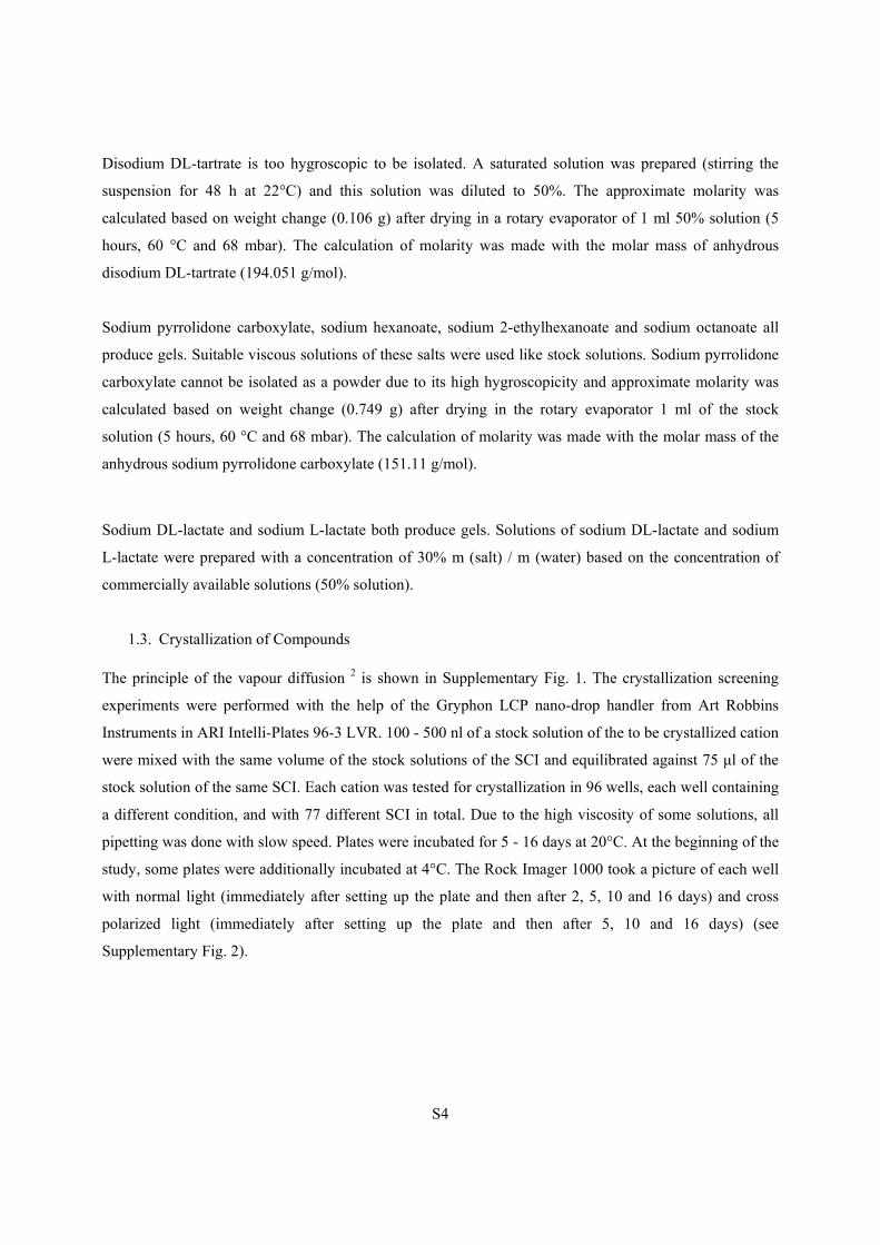
The principle of the vapour diffusion 2 is shown in Supplementary Fig. 1. The crystallization screening
experiments were performed with the help of the Gryphon LCP nano-drop handler from Art Robbins
Instruments in ARI Intelli-Plates 96-3 LVR. 100 - 500 nl of a stock solution of the to be crystallized cation
were mixed with the same volume of the stock solutions of the SCI and equilibrated against 75 μl of the
stock solution of the same SCI. Each cation was tested for crystallization in 96 wells, each well containing
a different condition, and with 77 different SCI in total. Due to the high viscosity of some solutions, all
pipetting was done with slow speed. Plates were incubated for 5 - 16 days at 20°C. At the beginning of the
study, some plates were additionally incubated at 4°C. The Rock Imager 1000 took a picture of each well
with normal light (immediately after setting up the plate and then after 2, 5, 10 and 16 days) and cross
polarized light (immediately after setting up the plate and then after 5, 10 and 16 days) (see
Supplementary Fig. 2).
S5
Supplementary Figure 1: Adopted vapour diffusion technique for the crystallization of organic cations.
S6
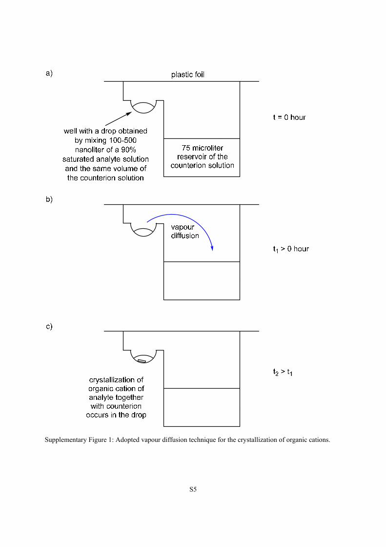
Supplementary Figure 2: Pictures of the crystallization of 5-benzyloxytryptaminium antimony-L-tartrate
from 5-benzyloxytryptaminium chloride and potassium antimony-L-tartrate (500 nl each). Pictures shown
were taken immediately after setting up the plate (top left) and then after 2 (top right), 5 (bottom left) and
16 (bottom right) days.
If single crystals were observed during this incubation period, they were removed from the wells. In order
to do so, wells with formed crystals were opened with a razor blade. Sometimes, the contents of the wells
were then covered with oil (Infineum V8512, formerly known as Paratone N). The crystals were fished
out with a nylon loop mounted on top of a CrystalCap Magnetic™ (Hampton Research), prepared on a
glass slide under oil (Infineum V8512) and measured on a single crystal X-ray diffractometer. The quality
of some crystalline material was too poor for structure determination by X-ray single crystal diffraction.
These hits were then manually re-synthesized and re-crystallized to obtain better crystals.
S7
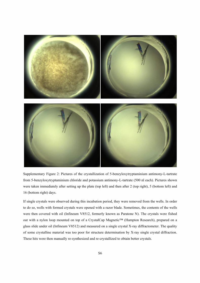
Supplementary Figure 3: Pictures of the crystallization of ephedrine hydrobromide (left) from ephedrine
hydrochloride and sodium bromide (100 nl each) and ephedrinium thiocyanide (right) from ephedrine
hydrochloride and sodium thiocyanate (100 nl each). Pictures shown were taken after 5 days.
1.4. X-ray Single Crystal Diffraction
Single-crystal X-ray diffraction was measured on two diffractometers. The first one was a Rigaku-Oxford
Diffraction XtaLAB Synergy-S dual source diffractometer: Kappa-axis four-circle goniometer with a
Dectris Pilatus3 R 200K HPC (Hybrid Photon Counting) detector and Cu and Mo PhotonJet microfocus
X-ray sources. The second one was a SuperNova dual source diffractometer: Kappa-axis four-circle
goniometer with an Atlas electronic CCD area detector and Cu and Mo microfocus X-ray sources.
Computing of measuring strategy and data reduction were performed with CrysAlisPro 3. The structures
were solved with direct methods using SIR97 4 or the charge flipping method using Superflip 5. The
structures were refined by full-matrix least-squares methods on F2 with SHELXL-2014 6 using the GUI
ShelXle 7 or refined by least-squares methods on F2 with CRYSTALS 8. Graphical output was produced
with the help of the program Mercury 9. CCDC 1585747-1585765 and 1823199-1823201 contain the
Supplementary crystallographic data for this paper. These data can be obtained free of charge from The
Cambridge Crystallographic Data Centre via www.ccdc.cam.ac.uk/structures.
S8
2. Description of the structures
[5-benzyloxytryptaminium][chloride]
Crystalline material was obtained from the screening (from a drop containing 500 nl of a 3.0 M sodium
chloride and 500 nl of a 90% saturated 5-benzyloxytryptamine hydrochloride solution equilibrating
against a reservoir of 3 M sodium chloride, as well as from a drop containing 500 nl of a 1.5 M sodium
chloride and 500 nl of a 90% saturated 5-benzyloxytryptamine hydrochloride solution equilibrating
against a reservoir of 1.5 M sodium chloride). The quality of the crystals from both experiments was too
low for structure determination by SCXRD. The crystal used for SCXRD was crystallized from a mixture
of 20 l of 3 M sodium chloride in water, 20 l of a 90% saturated solution of 5-benzyloxytryptamine
hydrochloride in water and 80 l acetone. The crystallization technique was slow evaporation in an 1.5 ml
Eppendorf vial. This complex crystallized in the monoclinic space group P21/c. The asymmetric unit
consists of one 5-benzyloxytryptaminium cation, which is protonated at the amino group, and one chloride
anion. The amino group makes three weak hydrogen bonds to three chloride anions (N1-H11 Cl21(x,1-y,-
z): 3.1834(16) Å; N1-H12 Cl21(x,1/2-y,1/2+z): 3.2310(15); N1-H13 Cl21(x,y,1+z): 3.1470(15) Å).
Supplementary Figure 4: Left: displacement ellipsoid representation of [BaH]Cl, ellipsoids are drawn at
50% probability. Right: packing diagram of [BaH]Cl.
[5-benzyloxytryptaminium][iodide]
Crystalline material was obtained from several crystallization setups: in one case, the drop obtained by
mixing 500 nl of a 2.65 M sodium iodide and 500 nl of a 90% saturated 5-benzyloxytryptamine
hydrochloride solution was equilibrated against a reservoir of 2.65 M sodium iodide. In two other setups,
half and quarter concentrations of iodide in drops and reservoirs respectively were used. All these
S9
conditions gave crystals, but their quality was too low for structure determination by SCXRD. The crystal
used for SCXRD was crystallized from a mixture of 20 l of 5.3 M sodium iodide in water, 20 l of a
90% saturated solution of 5-benzyloxytryptamine hydrochloride in water and 80 l acetone. The
crystallization technique was slow evaporation. This complex crystallized in the monoclinic space group
P21/c. The asymmetric unit consists of one 5-benzyloxytryptamininum cation, which is protonated at the
amino group, and an iodide counterion. The amino group forms three hydrogen bonds to iodide ion (N1-
H1 I21(-x,-1/2+y,1/2-z): 3.507(2) Å; N1-H12 I21: 3.562(2) Å; N1-H13 I21(x,3/2-y,1/2+z): 3.526(2) Å).
Supplementary Figure 5: Left: displacement ellipsoid representation of [BaH]I, ellipsoids are drawn at
50% probability. Right: packing diagram of [BaH]I.
[5-benzyloxytryptaminium][dihydrogen phosphate]
The crystalline material, which was obtained directly from the screening, was of insufficient quality for
structure determination by SCXRD. The setup was either a drop obtained by mixing 500 nl of a 4 M
sodium dihydrogen phosphate solution and 500 nl of a 90% saturated 5-benzyloxytryptamine
hydrochloride solution that was equilibrated against a reservoir of 4 M sodium dihydrogen phosphate, or
the same setup with half the sodium dihydrogen phosphate concentration. The crystal used for SCXRD
was crystallized from a mixture of 20 l of 4 M sodium dihydrogen phosphate in water, 20 l of a 90%
saturated solution of 5-benzyloxytryptamine hydrochloride in water and 80 l acetone. The crystallization
technique was slow evaporation. This complex crystallized in the monoclinic space group P21/c. The
asymmetric unit consists of one 5-benzyloxytryptaminium cation, which is protonated at the amino group,
and a dihydrogen phosphate anion. The amino group forms three hydrogen bonds to oxygen atoms of
dihydrogen phosphate (N1-H11 O22(x,3/2-y,-1/2+z): 3.2040(15) Å; N1-H13 O22: 2.9143(14) Å; N1-H12
O24(2-x,1-y,1-z): 2.7683(15) Å). The nitrogen of the indole creates a hydrogen bond to the oxygen atom
of the dihydrogen phosphate (N6-H61 O22: 2.9143(14) Å).
S10
Supplementary Figure 6: Left: displacement ellipsoid representation of [BaH]H2PO4, ellipsoids are drawn
at 50% probability. Right: packing diagram of [BaH]H2PO4.
[5-benzyloxytryptaminium]2[antimony L-tartrate]·2(H2O)
This crystal arose in a drop obtained by mixing 500 nl of a 54 mM potassium antimony L-tartrate and 500
nl of a 90% saturated 5-benzyloxytryptamine hydrochloride solution, and equilibrating against a reservoir
of 54 mM potassium antimony L-tartrate. This complex crystallized in the monoclinic space group C2.
The asymmetric unit consists of half an antimony L-tartrate anion, one water molecule and one 5-
benzyloxytryptaminium cation, which is protonated at the amino group. The antimony L-tartrate lies on a
two-fold axis. The amino group makes three hydrogen bonds to the antimony L-tartrate (N1-H12 O24:
2.960(6) Å, N1-H13 O25(x,1+y,z): 2.983(5) Å and N1-H13 O31(x,1+y,z): 3.003(6) Å).
Supplementary Figure 7: Left: displacement ellipsoid representation of [BaH]2[antimony L-
tartrate]·2(H2O), ellipsoids are drawn at 50% probability, for clarity only the asymmetric unit is shown.
Right: packing diagram of [BaH]2[antimony L-tartrate]·2(H2O).
S11
[5-benzyloxytryptaminium]2[L-tartrate]·3.35(H2O)
This crystal was isolated from a drop obtained by mixing of 500 nl of a 1.4 M sodium potassium L-tartrate
and 500 nl of a 90% saturated 5-benzyloxytryptamine hydrochloride solution, and equilibrating against a
reservoir of 1.4 M sodium potassium L-tartrate. The salt crystallized in the chiral space group P21. In the
asymmetric unit, there are two 5-benzyloxytryptaminium cations, one tartrate dianion and 3.35 water
molecules. The ethylammonium group of one cation is disordered in a ratio of 70:30. The non-disordered
ammonium group creates two hydrogen bonds to two carboxylate groups and one fully occupied water
molecule. The disordered ammonium group forms two hydrogen bonds to two alcohol groups of one
tartrate and one fully occupied water molecule. Because of the disorder, the hydrogen bonding pattern is
not discussed in further detail.
Supplementary Figure 8: Top: displacement ellipsoid representation of [BaH]2[L-tartrate]·3.35(H2O),
ellipsoids are drawn at 50% probability. Only the major conformer of the disordered ethylammonium
group is shown for clarity. Below: packing diagram of [BaH]2[L-tartrate]·3.35(H2O).
S12
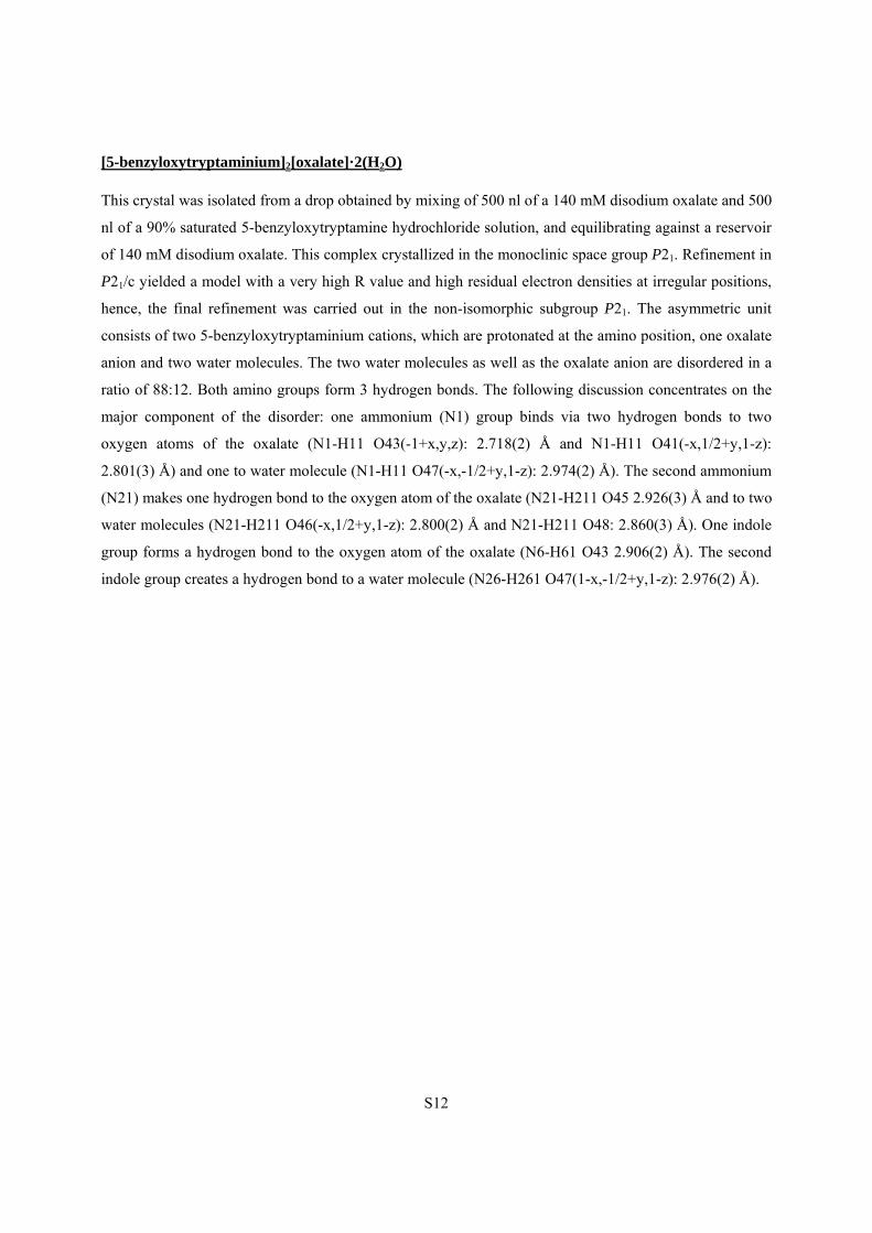
[5-benzyloxytryptaminium]2[oxalate]·2(H2O)
This crystal was isolated from a drop obtained by mixing of 500 nl of a 140 mM disodium oxalate and 500
nl of a 90% saturated 5-benzyloxytryptamine hydrochloride solution, and equilibrating against a reservoir
of 140 mM disodium oxalate. This complex crystallized in the monoclinic space group P21. Refinement in
P21/c yielded a model with a very high R value and high residual electron densities at irregular positions,
hence, the final refinement was carried out in the non-isomorphic subgroup P21. The asymmetric unit
consists of two 5-benzyloxytryptaminium cations, which are protonated at the amino position, one oxalate
anion and two water molecules. The two water molecules as well as the oxalate anion are disordered in a
ratio of 88:12. Both amino groups form 3 hydrogen bonds. The following discussion concentrates on the
major component of the disorder: one ammonium (N1) group binds via two hydrogen bonds to two
oxygen atoms of the oxalate (N1-H11 O43(-1+x,y,z): 2.718(2) Å and N1-H11 O41(-x,1/2+y,1-z):
2.801(3) Å) and one to water molecule (N1-H11 O47(-x,-1/2+y,1-z): 2.974(2) Å). The second ammonium
(N21) makes one hydrogen bond to the oxygen atom of the oxalate (N21-H211 O45 2.926(3) Å and to two
water molecules (N21-H211 O46(-x,1/2+y,1-z): 2.800(2) Å and N21-H211 O48: 2.860(3) Å). One indole
group forms a hydrogen bond to the oxygen atom of the oxalate (N6-H61 O43 2.906(2) Å). The second
indole group creates a hydrogen bond to a water molecule (N26-H261 O47(1-x,-1/2+y,1-z): 2.976(2) Å).
S13
Supplementary Figure 9: Top: displacement ellipsoid representation of [BaH]2[oxalate]·2(H2O),
ellipsoids are drawn at 50% probability. Only the major components of the disordered moieties are shown
for clarity. Below: packing diagram of [BaH]2[oxalate]·2(H2O).
S14
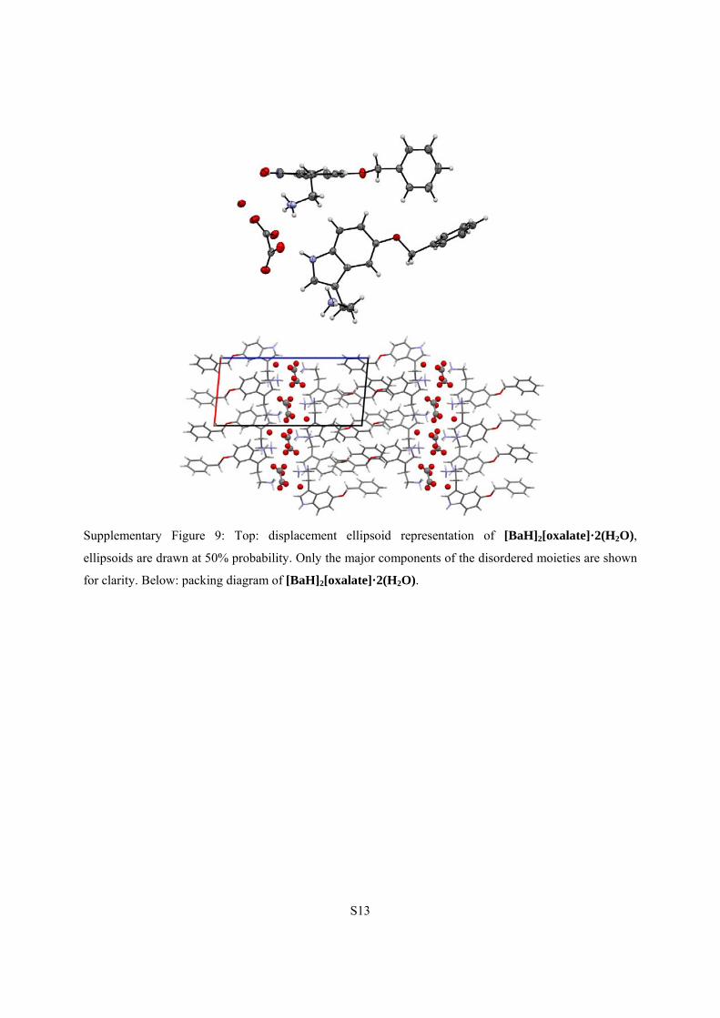
[5-benzyloxytryptaminium]2[sulfate]·2(H2O)
This crystal arose in a drop obtained by mixing of 500 nl of a 1 M sodium sulfate and 500 nl of a 90%
saturated 5-benzyloxytryptamine hydrochloride solution, and equilibrating against a reservoir of 1 M
sodium sulfate. The achiral salt crystallized in the chiral space group P21 with two 5-
benzyloxytryptaminium cations, one sulfate anion and two water molecules in the asymmetric unit. The
two 5-benzyloxytryptaminium cations are protonated at the amino group. These aminium groups form
multiple hydrogen bonds to two oxygen atoms of sulfate anions (N1-H11 O44(1-x,1/2+y,1-z): 2.769(3) Å;
N1-H13 O45: 2.784(3) Å and N21-H211 O42 (1+x,y,z): 2.756(3) Å; N21-H213 O43(1-x,-1/2+y,1-z):
2.756 (3) Å). Finally, one indole group forms a hydrogen bond to oxygen of sulfate ion (N26-H261 O42
2.834(3) Å).
Supplementary Figure 10: Top: displacement ellipsoid representation of [BaH]2[sulfate]·2(H2O),
ellipsoids are drawn at 50% probability. Below: packing diagram of [BaH]2[sulfate]·2(H2O).
S15
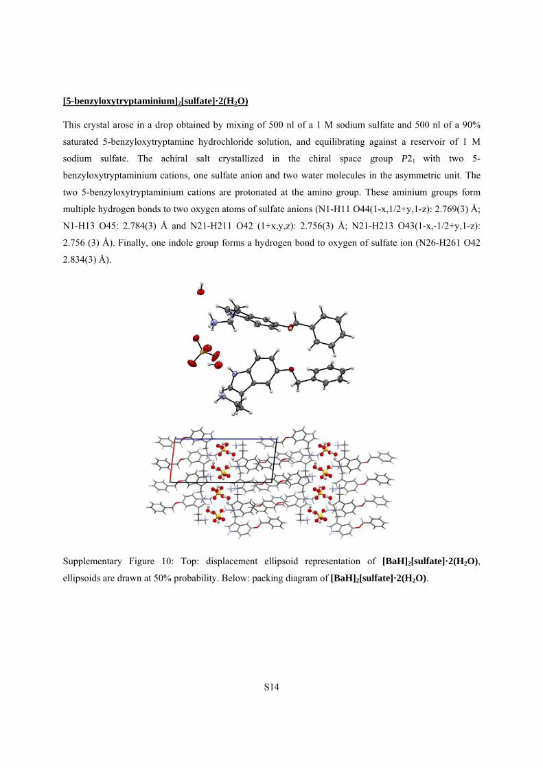
[5-benzyloxytryptaminium][tetrafluoroborate]
The single crystal was isolated from a drop obtained by mixing 500 nl of a 4 M sodium tetrafluoroborate
and 500 nl of a 90% saturated 5-benzyloxytryptamine hydrochloride solution, and equilibrating against a
reservoir of 4 M sodium tetrafluoroborate. Crystals of equal quality could be obtained by taking 100 nl of
both solutions instead. This complex crystallized in the monoclinic space group P21/c. The asymmetric
unit consists of one 5-benzyloxytryptaminium cation, which is protonated at the amino group, and one
tetrafluoroborate anion. This amino group binds to 4 fluorine atoms thus forming 2 two-centred hydrogen
bonds and a combination of a three-centered hydrogen bond with a bifurcated hydrogen bond (N1-H11
F25(2-x,1-y,1-z): 2.9192(14) Å; N1-H12 F22(x,3/2-y,1/2+z): 2.8544(15) Å; N1-H12 F25: 2.9182(14) Å;
N1-H13 F24(2-x,-1/2+y,3/2-z): 2.8619(14) Å).
Supplementary Figure 11: Left: displacement ellipsoid representation of [BaH][tetrafluoroborate],
ellipsoids are drawn at 50% probability. Right: packing diagram of [BaH][tetrafluoroborate].
S16
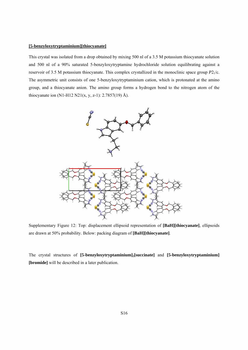
[5-benzyloxytryptaminium][thiocyanate]
This crystal was isolated from a drop obtained by mixing 500 nl of a 3.5 M potassium thiocyanate solution
and 500 nl of a 90% saturated 5-benzyloxytryptamine hydrochloride solution equilibrating against a
reservoir of 3.5 M potassium thiocyanate. This complex crystallized in the monoclinic space group P21/c.
The asymmetric unit consists of one 5-benzyloxytryptaminium cation, which is protonated at the amino
group, and a thiocyanate anion. The amino group forms a hydrogen bond to the nitrogen atom of the
thiocyanate ion (N1-H12 N21(x, y, z-1): 2.7857(19) Å).
Supplementary Figure 12: Top: displacement ellipsoid representation of [BaH][thiocyanate], ellipsoids
are drawn at 50% probability. Below: packing diagram of [BaH][thiocyanate].
The crystal structures of [5-benzyloxytryptaminium]2[succinate] and [5-benzyloxytryptaminium]
[bromide] will be described in a later publication.
S17
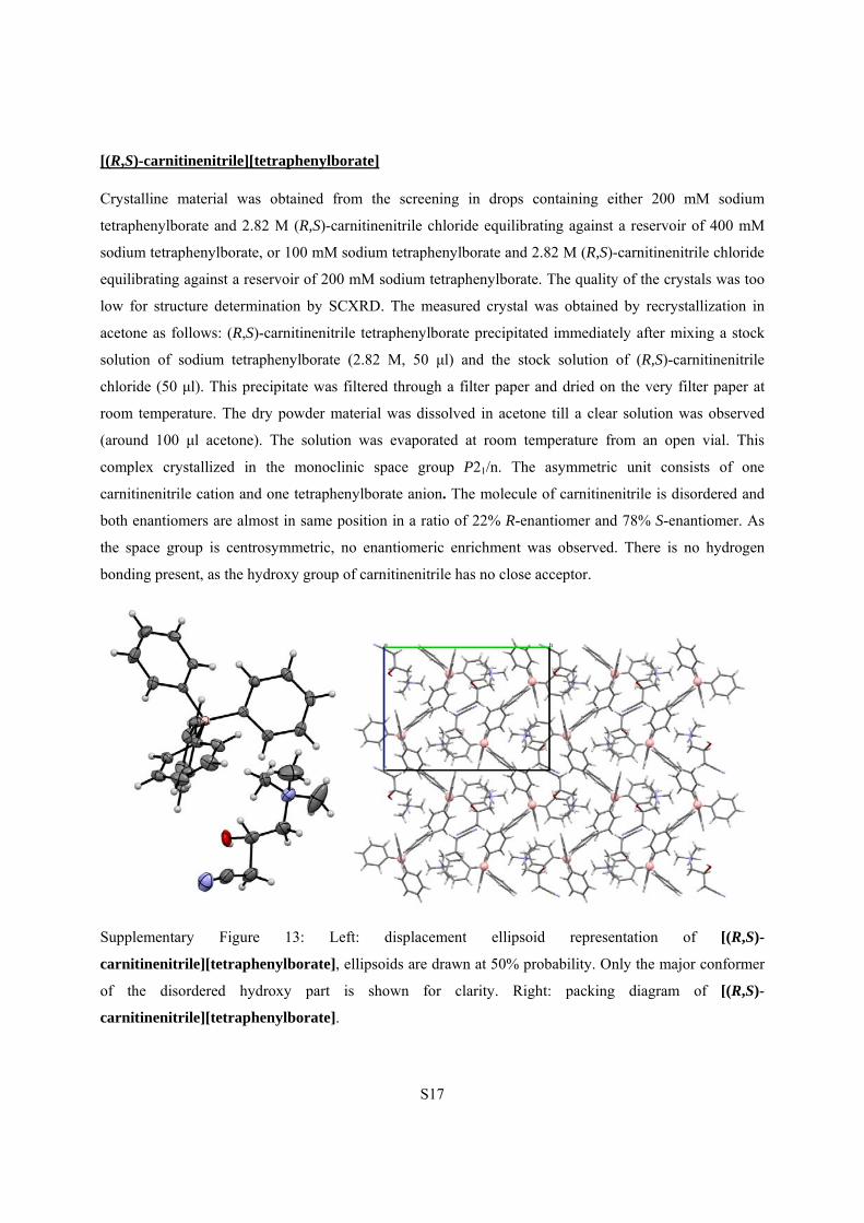
[(R,S)-carnitinenitrile][tetraphenylborate]
Crystalline material was obtained from the screening in drops containing either 200 mM sodium
tetraphenylborate and 2.82 M (R,S)-carnitinenitrile chloride equilibrating against a reservoir of 400 mM
sodium tetraphenylborate, or 100 mM sodium tetraphenylborate and 2.82 M (R,S)-carnitinenitrile chloride
equilibrating against a reservoir of 200 mM sodium tetraphenylborate. The quality of the crystals was too
low for structure determination by SCXRD. The measured crystal was obtained by recrystallization in
acetone as follows: (R,S)-carnitinenitrile tetraphenylborate precipitated immediately after mixing a stock
solution of sodium tetraphenylborate (2.82 M, 50 μl) and the stock solution of (R,S)-carnitinenitrile
chloride (50 μl). This precipitate was filtered through a filter paper and dried on the very filter paper at
room temperature. The dry powder material was dissolved in acetone till a clear solution was observed
(around 100 μl acetone). The solution was evaporated at room temperature from an open vial. This
complex crystallized in the monoclinic space group P21/n. The asymmetric unit consists of one
carnitinenitrile cation and one tetraphenylborate anion. The molecule of carnitinenitrile is disordered and
both enantiomers are almost in same position in a ratio of 22% R-enantiomer and 78% S-enantiomer. As
the space group is centrosymmetric, no enantiomeric enrichment was observed. There is no hydrogen
bonding present, as the hydroxy group of carnitinenitrile has no close acceptor.
Supplementary Figure 13: Left: displacement ellipsoid representation of [(R,S)-
carnitinenitrile][tetraphenylborate], ellipsoids are drawn at 50% probability. Only the major conformer
of the disordered hydroxy part is shown for clarity. Right: packing diagram of [(R,S)-
carnitinenitrile][tetraphenylborate].
S18
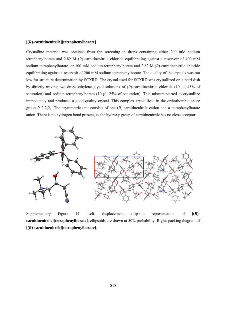
[(R)-carnitinenitrile][tetraphenylborate]
Crystalline material was obtained from the screening in drops containing either 200 mM sodium
tetraphenylborate and 2.82 M (R)-carnitinenitrile chloride equilibrating against a reservoir of 400 mM
sodium tetraphenylborate, or 100 mM sodium tetraphenylborate and 2.82 M (R)-carnitinenitrile chloride
equilibrating against a reservoir of 200 mM sodium tetraphenylborate. The quality of the crystals was too
low for structure determination by SCXRD. The crystal used for SCXRD was crystallized on a petri dish
by directly mixing two drops ethylene glycol solutions of (R)-carnitinenitrile chloride (10 μl, 45% of
saturation) and sodium tetraphenylborate (10 μl, 25% of saturation). This mixture started to crystallize
immediately and produced a good quality crystal. This complex crystallized in the orthorhombic space
group P 212121. The asymmetric unit consists of one (R)-carnitinenitrile cation and a tetraphenylborate
anion. There is no hydrogen bond present, as the hydroxy group of carnitinenitrile has no close acceptor.
Supplementary Figure 14: Left: displacement ellipsoid representation of [(R)-
carnitinenitrile][tetraphenylborate], ellipsoids are drawn at 50% probability. Right: packing diagram of
[(R)-carnitinenitrile][tetraphenylborate].
S19
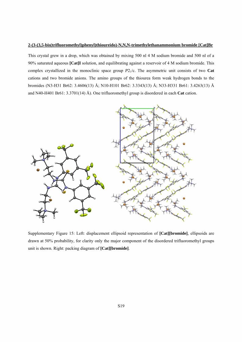
2-(3-(3,5-bis(trifluoromethyl)phenyl)thioureido)-N,N,N-trimethylethanammonium bromide [Cat]Br
This crystal grew in a drop, which was obtained by mixing 500 nl 4 M sodium bromide and 500 nl of a
90% saturated aqueous [Cat]I solution, and equilibrating against a reservoir of 4 M sodium bromide. This
complex crystallized in the monoclinic space group P21/c. The asymmetric unit consists of two Cat
cations and two bromide anions. The amino groups of the thiourea form weak hydrogen bonds to the
bromides (N3-H31 Br62: 3.4606(13) Å; N10-H101 Br62: 3.3343(13) Å; N33-H331 Br61: 3.4263(13) Å
and N40-H401 Br61: 3.3701(14) Å). One trifluoromethyl group is disordered in each Cat cation.
Supplementary Figure 15: Left: displacement ellipsoid representation of [Cat][bromide], ellipsoids are
drawn at 50% probability, for clarity only the major component of the disordered trifluoromethyl groups
unit is shown. Right: packing diagram of [Cat][bromide].
S20
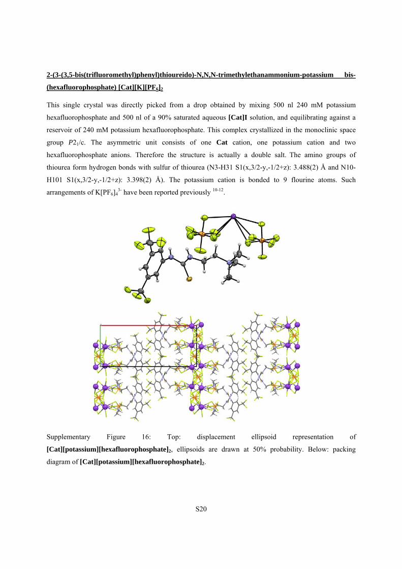
2-(3-(3,5-bis(trifluoromethyl)phenyl)thioureido)-N,N,N-trimethylethanammonium-potassium bis-
(hexafluorophosphate) [Cat][K][PF6]2
This single crystal was directly picked from a drop obtained by mixing 500 nl 240 mM potassium
hexafluorophosphate and 500 nl of a 90% saturated aqueous [Cat]I solution, and equilibrating against a
reservoir of 240 mM potassium hexafluorophosphate. This complex crystallized in the monoclinic space
group P21/c. The asymmetric unit consists of one Cat cation, one potassium cation and two
hexafluorophosphate anions. Therefore the structure is actually a double salt. The amino groups of
thiourea form hydrogen bonds with sulfur of thiourea (N3-H31 S1(x,3/2-y,-1/2+z): 3.488(2) Å and N10-
H101 S1(x,3/2-y,-1/2+z): 3.398(2) Å). The potassium cation is bonded to 9 flourine atoms. Such
arrangements of K[PF6]43- have been reported previously 10-12.
Supplementary Figure 16: Top: displacement ellipsoid representation of
[Cat][potassium][hexafluorophosphate]2, ellipsoids are drawn at 50% probability. Below: packing
diagram of [Cat][potassium][hexafluorophosphate]2.
S21
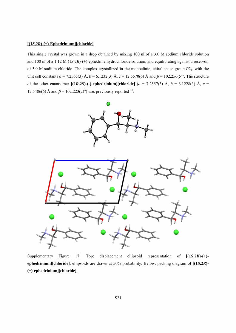
[(1S,2R)-(+)-Ephedrinium][chloride]
This single crystal was grown in a drop obtained by mixing 100 nl of a 3.0 M sodium chloride solution
and 100 nl of a 1.12 M (1S,2R)-(+)-ephedrine hydrochloride solution, and equilibrating against a reservoir
of 3.0 M sodium chloride. The complex crystallized in the monoclinic, chiral space group P21. with the
unit cell constants a = 7.2565(3) Å, b = 6.1232(3) Å, c = 12.5570(6) Å and = 102.256(5)°. The structure
of the other enantiomer [(1R,2S)-(-)-ephedrinium][chloride] (a = 7.2557(3) Å, b = 6.1228(3) Å, c =
12.5486(6) Å and = 102.223(2)°) was previously reported 13.
Supplementary Figure 17: Top: displacement ellipsoid representation of [(1S,2R)-(+)-
ephedrinium][chloride], ellipsoids are drawn at 50% probability. Below: packing diagram of [(1S,2R)-
(+)-ephedrinium][chloride].
S22
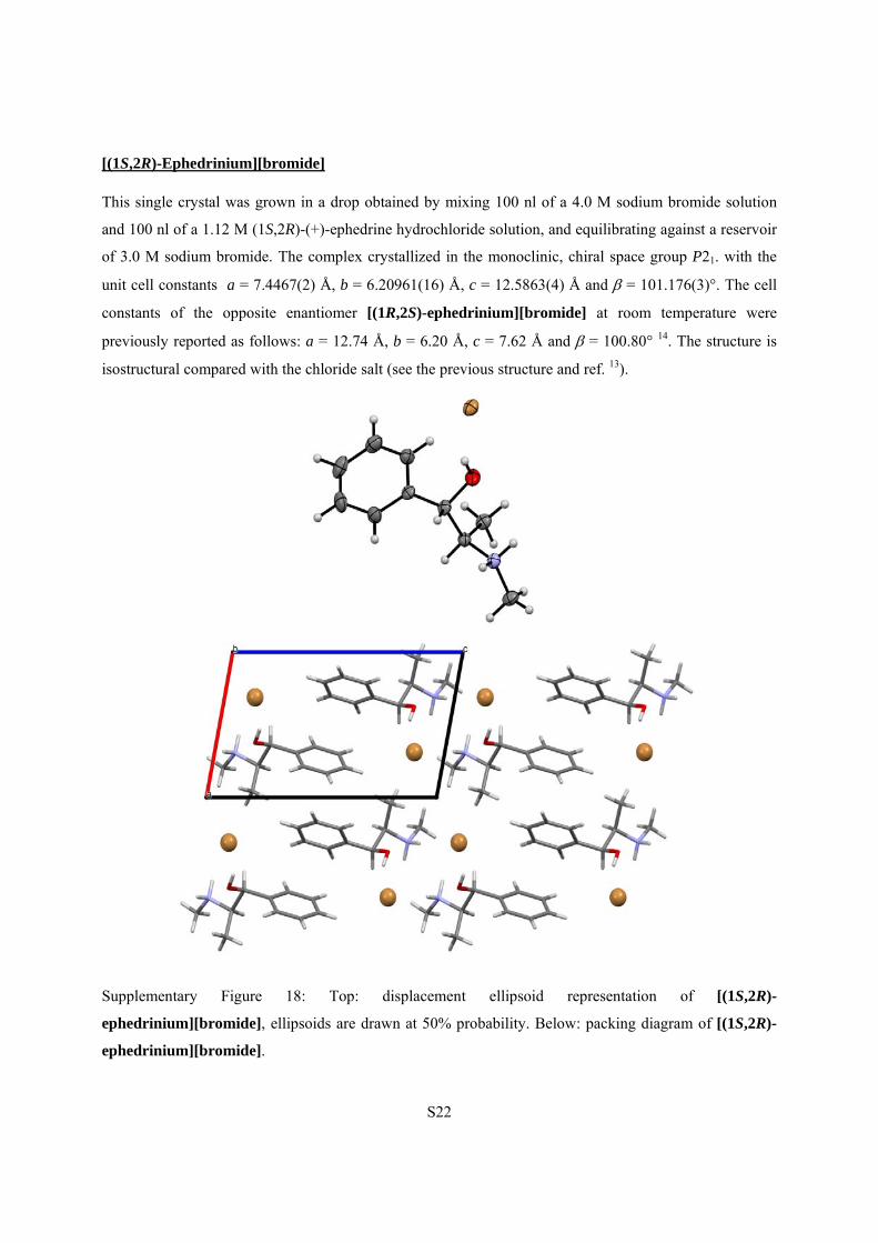
[(1S,2R)-Ephedrinium][bromide]
This single crystal was grown in a drop obtained by mixing 100 nl of a 4.0 M sodium bromide solution
and 100 nl of a 1.12 M (1S,2R)-(+)-ephedrine hydrochloride solution, and equilibrating against a reservoir
of 3.0 M sodium bromide. The complex crystallized in the monoclinic, chiral space group P21. with the
unit cell constants a = 7.4467(2) Å, b = 6.20961(16) Å, c = 12.5863(4) Å and = 101.176(3)°. The cell
constants of the opposite enantiomer [(1R,2S)-ephedrinium][bromide] at room temperature were
previously reported as follows: a = 12.74 Å, b = 6.20 Å, c = 7.62 Å and = 100.80° 14. The structure is
isostructural compared with the chloride salt (see the previous structure and ref. 13).
Supplementary Figure 18: Top: displacement ellipsoid representation of [(1S,2R)-
ephedrinium][bromide], ellipsoids are drawn at 50% probability. Below: packing diagram of [(1S,2R)-
ephedrinium][bromide].
S23
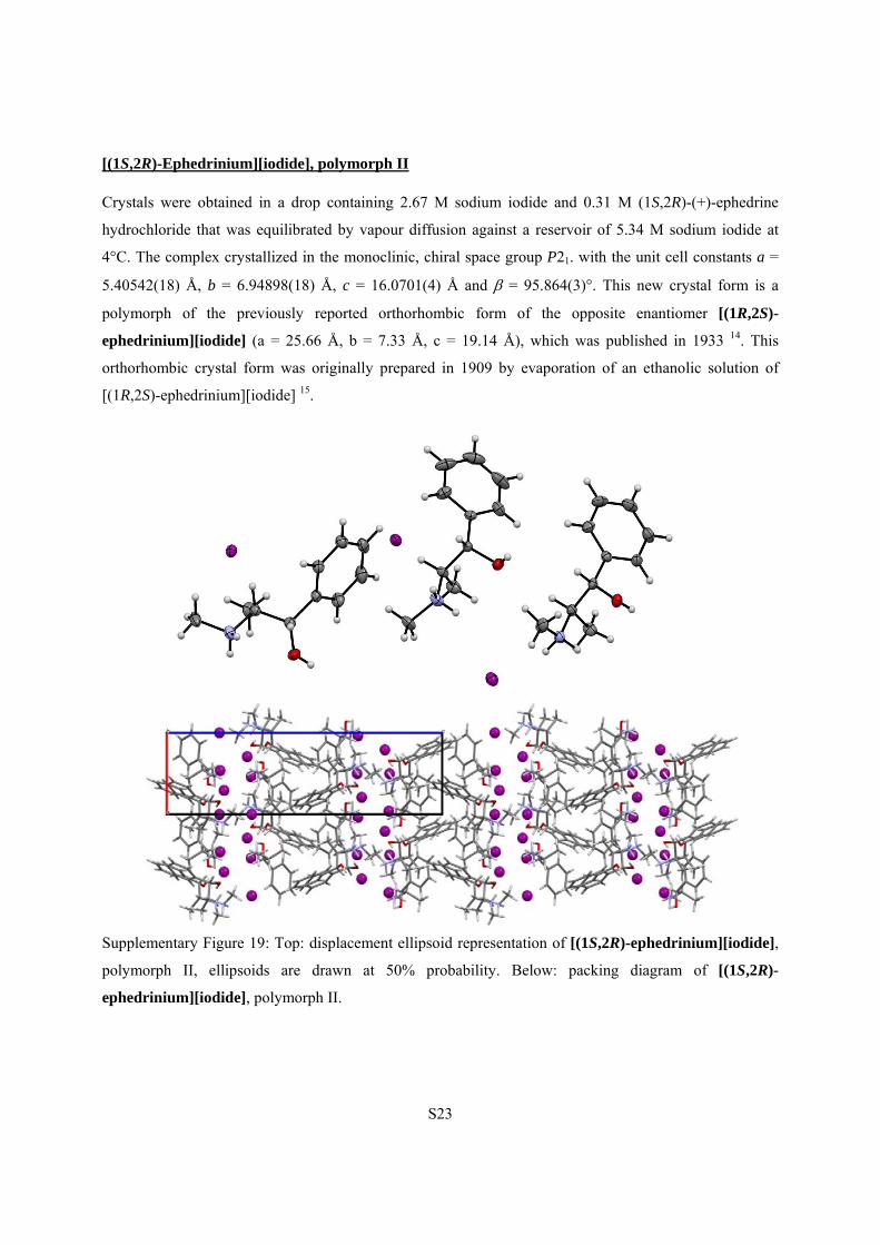
[(1S,2R)-Ephedrinium][iodide], polymorph II
Crystals were obtained in a drop containing 2.67 M sodium iodide and 0.31 M (1S,2R)-(+)-ephedrine
hydrochloride that was equilibrated by vapour diffusion against a reservoir of 5.34 M sodium iodide at
4°C. The complex crystallized in the monoclinic, chiral space group P21. with the unit cell constants a =
5.40542(18) Å, b = 6.94898(18) Å, c = 16.0701(4) Å and = 95.864(3)°. This new crystal form is a
polymorph of the previously reported orthorhombic form of the opposite enantiomer [(1R,2S)-
ephedrinium][iodide] (a = 25.66 Å, b = 7.33 Å, c = 19.14 Å), which was published in 1933 14. This
orthorhombic crystal form was originally prepared in 1909 by evaporation of an ethanolic solution of
[(1R,2S)-ephedrinium][iodide] 15.
Supplementary Figure 19: Top: displacement ellipsoid representation of [(1S,2R)-ephedrinium][iodide],
polymorph II, ellipsoids are drawn at 50% probability. Below: packing diagram of [(1S,2R)-
ephedrinium][iodide], polymorph II.
S24
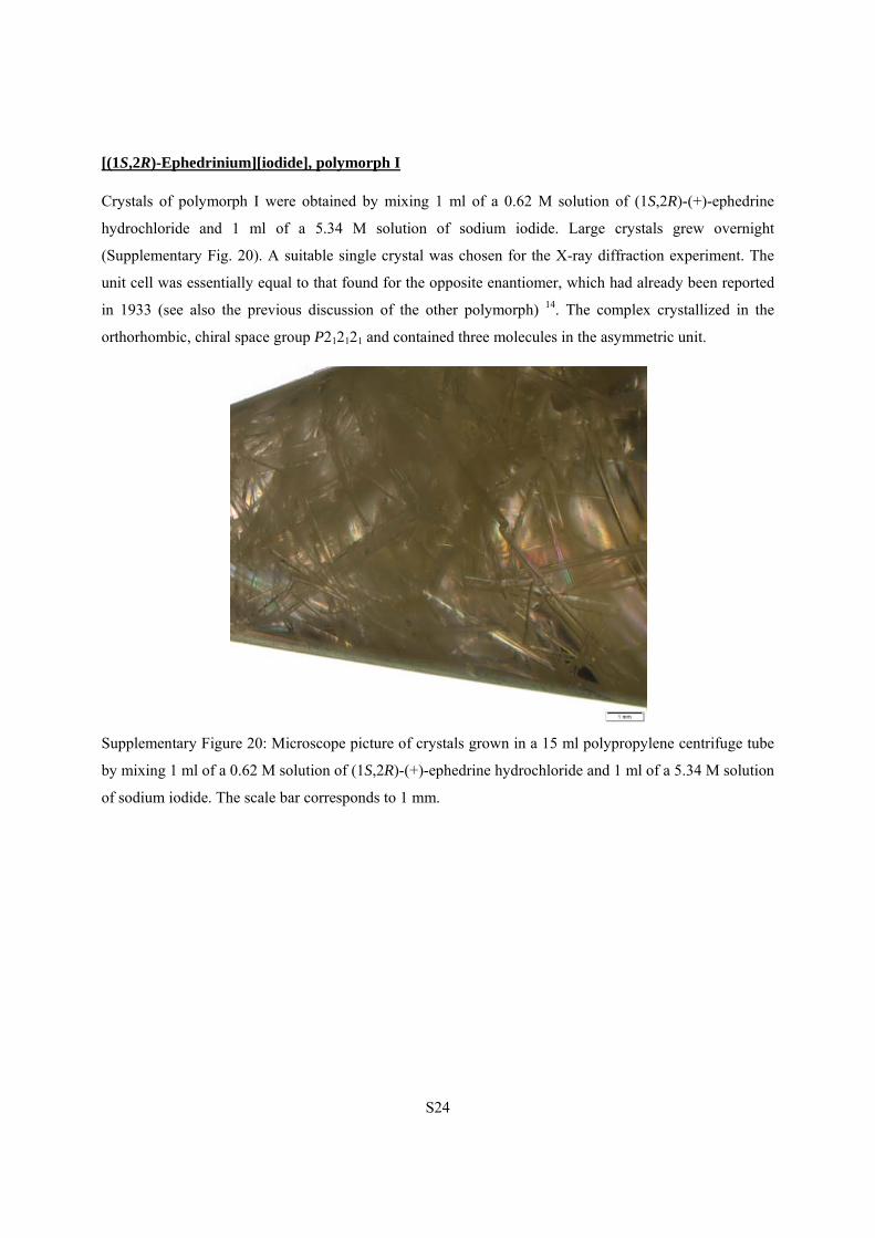
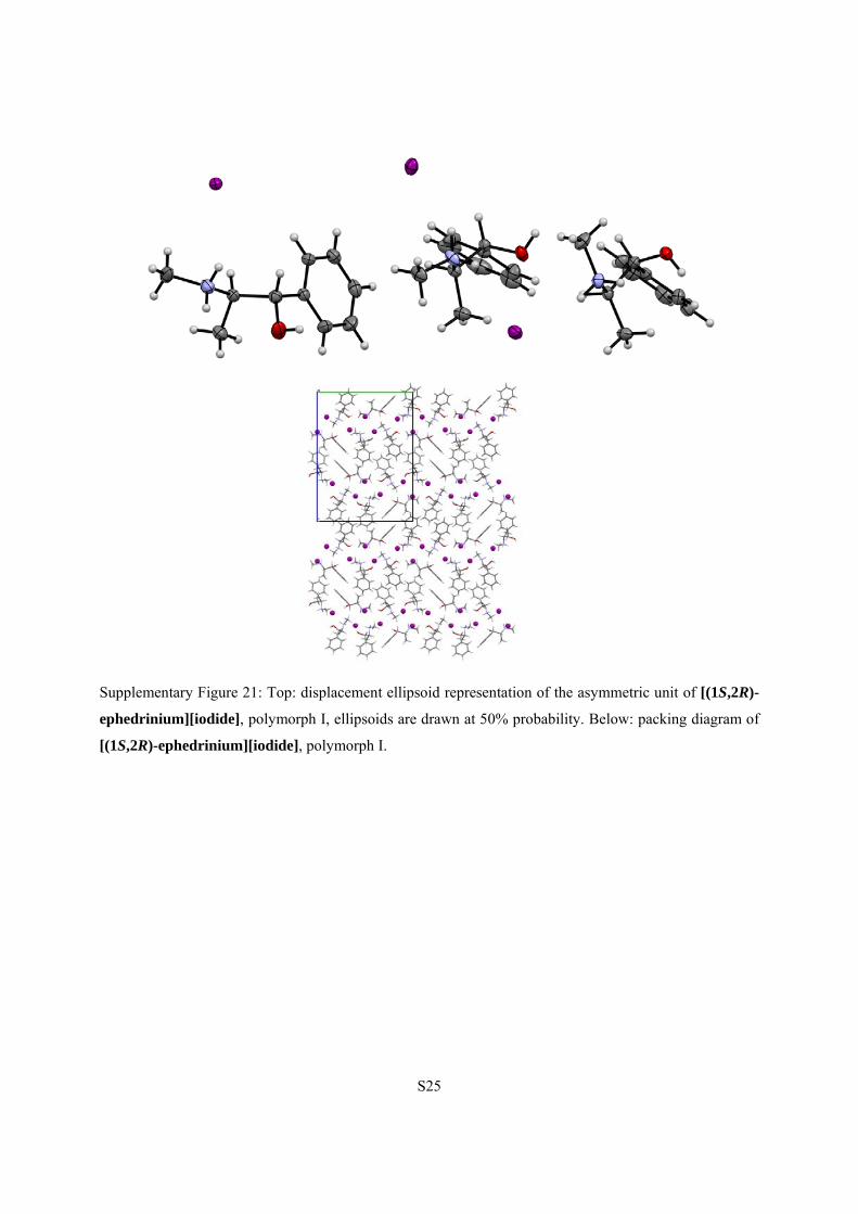
[(1S,2R)-Ephedrinium][iodide], polymorph I
Crystals of polymorph I were obtained by mixing 1 ml of a 0.62 M solution of (1S,2R)-(+)-ephedrine
hydrochloride and 1 ml of a 5.34 M solution of sodium iodide. Large crystals grew overnight
(Supplementary Fig. 20). A suitable single crystal was chosen for the X-ray diffraction experiment. The
unit cell was essentially equal to that found for the opposite enantiomer, which had already been reported
in 1933 (see also the previous discussion of the other polymorph) 14. The complex crystallized in the
orthorhombic, chiral space group P212121 and contained three molecules in the asymmetric unit.
Supplementary Figure 20: Microscope picture of crystals grown in a 15 ml polypropylene centrifuge tube
by mixing 1 ml of a 0.62 M solution of (1S,2R)-(+)-ephedrine hydrochloride and 1 ml of a 5.34 M solution
of sodium iodide. The scale bar corresponds to 1 mm.
S25
Supplementary Figure 21: Top: displacement ellipsoid representation of the asymmetric unit of [(1S,2R)-
ephedrinium][iodide], polymorph I, ellipsoids are drawn at 50% probability. Below: packing diagram of
[(1S,2R)-ephedrinium][iodide], polymorph I.
S26
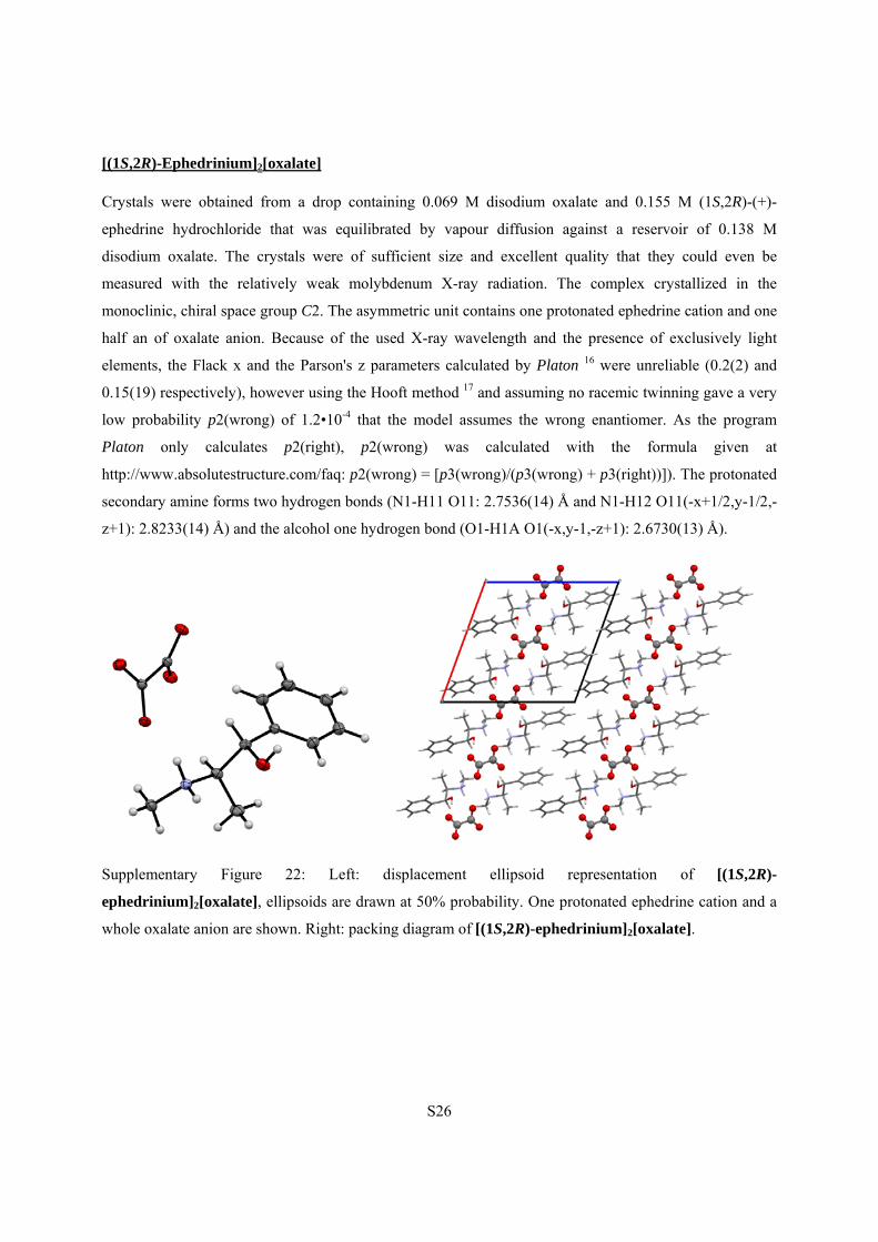
[(1S,2R)-Ephedrinium]2[oxalate]
Crystals were obtained from a drop containing 0.069 M disodium oxalate and 0.155 M (1S,2R)-(+)-
ephedrine hydrochloride that was equilibrated by vapour diffusion against a reservoir of 0.138 M
disodium oxalate. The crystals were of sufficient size and excellent quality that they could even be
measured with the relatively weak molybdenum X-ray radiation. The complex crystallized in the
monoclinic, chiral space group C2. The asymmetric unit contains one protonated ephedrine cation and one
half an of oxalate anion. Because of the used X-ray wavelength and the presence of exclusively light
elements, the Flack x and the Parson's z parameters calculated by Platon 16 were unreliable (0.2(2) and
0.15(19) respectively), however using the Hooft method 17 and assuming no racemic twinning gave a very
low probability p2(wrong) of 1.2•10-4 that the model assumes the wrong enantiomer. As the program
Platon only calculates p2(right), p2(wrong) was calculated with the formula given at
http://www.absolutestructure.com/faq: p2(wrong) = [p3(wrong)/(p3(wrong) + p3(right))]). The protonated
secondary amine forms two hydrogen bonds (N1-H11 O11: 2.7536(14) Å and N1-H12 O11(-x+1/2,y-1/2,-
z+1): 2.8233(14) Å) and the alcohol one hydrogen bond (O1-H1A O1(-x,y-1,-z+1): 2.6730(13) Å).
Supplementary Figure 22: Left: displacement ellipsoid representation of [(1S,2R)-
ephedrinium]2[oxalate], ellipsoids are drawn at 50% probability. One protonated ephedrine cation and a
whole oxalate anion are shown. Right: packing diagram of [(1S,2R)-ephedrinium]2[oxalate].
S27
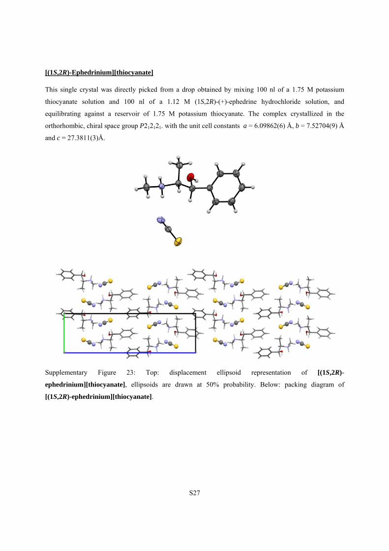
[(1S,2R)-Ephedrinium][thiocyanate]
This single crystal was directly picked from a drop obtained by mixing 100 nl of a 1.75 M potassium
thiocyanate solution and 100 nl of a 1.12 M (1S,2R)-(+)-ephedrine hydrochloride solution, and
equilibrating against a reservoir of 1.75 M potassium thiocyanate. The complex crystallized in the
orthorhombic, chiral space group P212121. with the unit cell constants a = 6.09862(6) Å, b = 7.52704(9) Å
and c = 27.3811(3)Å.
Supplementary Figure 23: Top: displacement ellipsoid representation of [(1S,2R)-
ephedrinium][thiocyanate], ellipsoids are drawn at 50% probability. Below: packing diagram of
[(1S,2R)-ephedrinium][thiocyanate].
S28
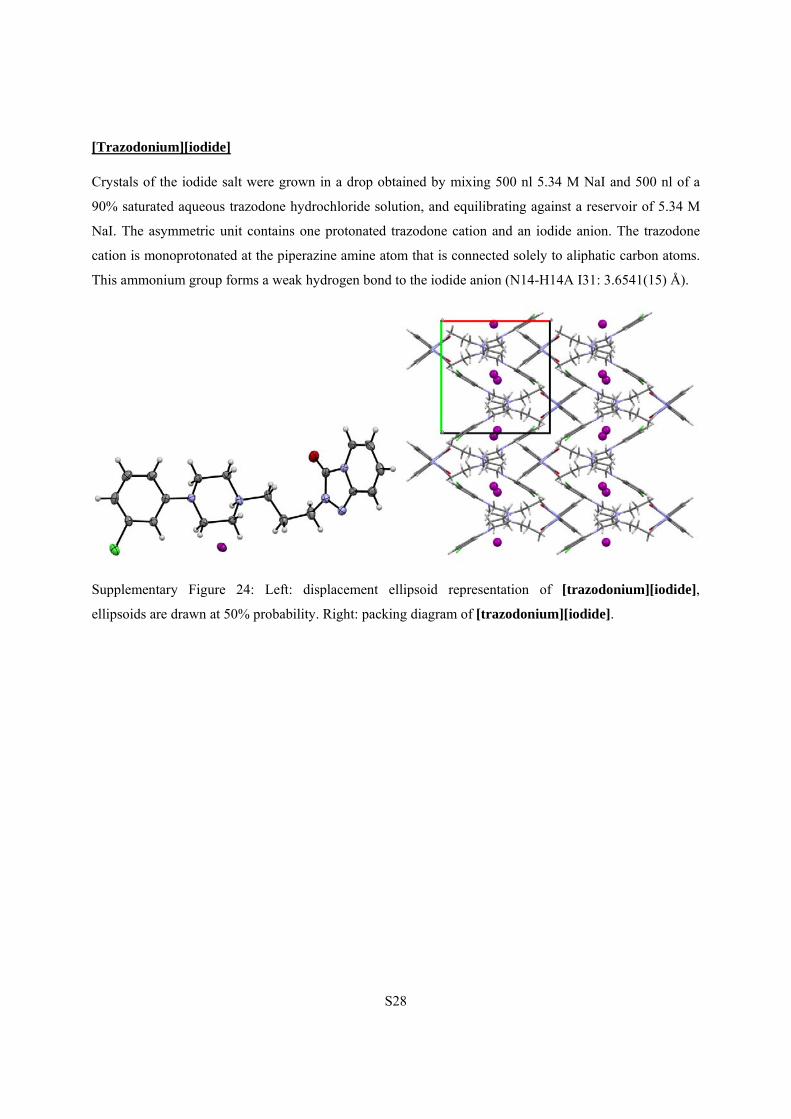
[Trazodonium][iodide]
Crystals of the iodide salt were grown in a drop obtained by mixing 500 nl 5.34 M NaI and 500 nl of a
90% saturated aqueous trazodone hydrochloride solution, and equilibrating against a reservoir of 5.34 M
NaI. The asymmetric unit contains one protonated trazodone cation and an iodide anion. The trazodone
cation is monoprotonated at the piperazine amine atom that is connected solely to aliphatic carbon atoms.
This ammonium group forms a weak hydrogen bond to the iodide anion (N14-H14A I31: 3.6541(15) Å).
Supplementary Figure 24: Left: displacement ellipsoid representation of [trazodonium][iodide],
ellipsoids are drawn at 50% probability. Right: packing diagram of [trazodonium][iodide].
S29
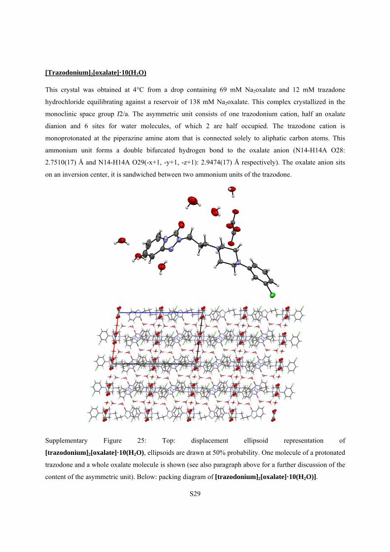
[Trazodonium]2[oxalate]·10(H2O)
This crystal was obtained at 4°C from a drop containing 69 mM Na2oxalate and 12 mM trazadone
hydrochloride equilibrating against a reservoir of 138 mM Na2oxalate. This complex crystallized in the
monoclinic space group I2/a. The asymmetric unit consists of one trazodonium cation, half an oxalate
dianion and 6 sites for water molecules, of which 2 are half occupied. The trazodone cation is
monoprotonated at the piperazine amine atom that is connected solely to aliphatic carbon atoms. This
ammonium unit forms a double bifurcated hydrogen bond to the oxalate anion (N14-H14A O28:
2.7510(17) Å and N14-H14A O29(-x+1, -y+1, -z+1): 2.9474(17) Å respectively). The oxalate anion sits
on an inversion center, it is sandwiched between two ammonium units of the trazodone.
Supplementary Figure 25: Top: displacement ellipsoid representation of
[trazodonium]2[oxalate]·10(H2O), ellipsoids are drawn at 50% probability. One molecule of a protonated
trazodone and a whole oxalate molecule is shown (see also paragraph above for a further discussion of the
content of the asymmetric unit). Below: packing diagram of [trazodonium]2[oxalate]·10(H2O)].
S30
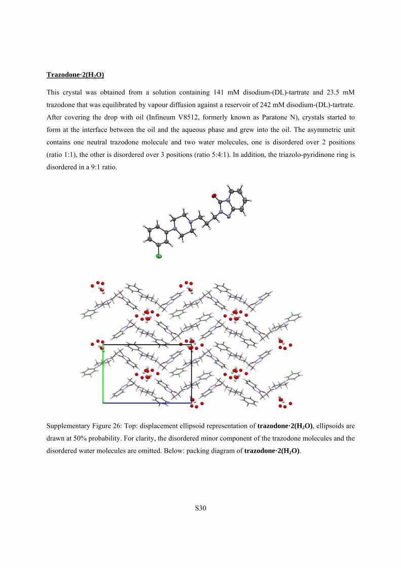
Trazodone·2(H2O)
This crystal was obtained from a solution containing 141 mM disodium-(DL)-tartrate and 23.5 mM
trazodone that was equilibrated by vapour diffusion against a reservoir of 242 mM disodium-(DL)-tartrate.
After covering the drop with oil (Infineum V8512, formerly known as Paratone N), crystals started to
form at the interface between the oil and the aqueous phase and grew into the oil. The asymmetric unit
contains one neutral trazodone molecule and two water molecules, one is disordered over 2 positions
(ratio 1:1), the other is disordered over 3 positions (ratio 5:4:1). In addition, the triazolo-pyridinone ring is
disordered in a 9:1 ratio.
Supplementary Figure 26: Top: displacement ellipsoid representation of trazodone·2(H2O), ellipsoids are
drawn at 50% probability. For clarity, the disordered minor component of the trazodone molecules and the
disordered water molecules are omitted. Below: packing diagram of trazodone·2(H2O).
S31
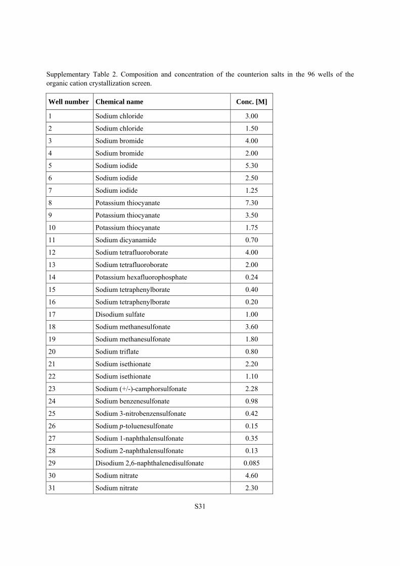
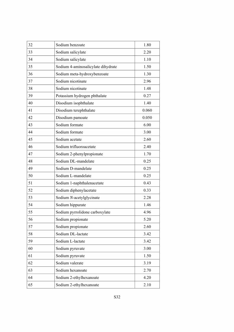
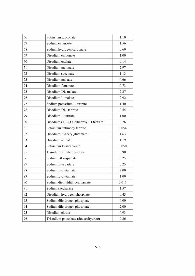
Supplementary Table 2. Composition and concentration of the counterion salts in the 96 wells of the organic cation crystallization screen.
Well number Chemical name Conc. [M]
1 Sodium chloride 3.00
2 Sodium chloride 1.50
3 Sodium bromide 4.00
4 Sodium bromide 2.00
5 Sodium iodide 5.30
6 Sodium iodide 2.50
7 Sodium iodide 1.25
8 Potassium thiocyanate 7.30
9 Potassium thiocyanate 3.50
10 Potassium thiocyanate 1.75
11 Sodium dicyanamide 0.70
12 Sodium tetrafluoroborate 4.00
13 Sodium tetrafluoroborate 2.00
14 Potassium hexafluorophosphate 0.24
15 Sodium tetraphenylborate 0.40
16 Sodium tetraphenylborate 0.20
17 Disodium sulfate 1.00
18 Sodium methanesulfonate 3.60
19 Sodium methanesulfonate 1.80
20 Sodium triflate 0.80
21 Sodium isethionate 2.20
22 Sodium isethionate 1.10
23 Sodium (+/-)-camphorsulfonate 2.28
24 Sodium benzenesulfonate 0.98
25 Sodium 3-nitrobenzensulfonate 0.42
26 Sodium p-toluenesulfonate 0.15
27 Sodium 1-naphthalensulfonate 0.35
28 Sodium 2-naphthalensulfonate 0.13
29 Disodium 2,6-naphthalenedisulfonate 0.085
30 Sodium nitrate 4.60
31 Sodium nitrate 2.30
S32
32 Sodium benzoate 1.80
33 Sodium salicylate 2.20
34 Sodium salicylate 1.10
35 Sodium 4-aminosalicylate dihydrate 1.50
36 Sodium meta-hydroxybenzoate 1.30
37 Sodium nicotinate 2.96
38 Sodium nicotinate 1.48
39 Potassium hydrogen phthalate 0.27
40 Disodium isophthalate 1.40
41 Disodium terephthalate 0.060
42 Disodium pamoate 0.050
43 Sodium formate 6.00
44 Sodium formate 3.00
45 Sodium acetate 2.60
46 Sodium trifluoroacetate 2.40
47 Sodium 2-phenylpropionate 1.70
48 Sodium DL-mandelate 0.25
49 Sodium D-mandelate 0.25
50 Sodium L-mandelate 0.25
51 Sodium 1-naphthalenacetate 0.43
52 Sodium diphenylacetate 0.33
53 Sodium N-acetylglycinate 2.28
54 Sodium hippurate 1.46
55 Sodium pyrrolidone carboxylate 4.96
56 Sodium propionate 5.20
57 Sodium propionate 2.60
58 Sodium DL-lactate 3.42
59 Sodium L-lactate 3.42
60 Sodium pyruvate 3.00
61 Sodium pyruvate 1.50
62 Sodium valerate 3.19
63 Sodium hexanoate 2.70
64 Sodium 2-ethylhexanoate 4.20
65 Sodium 2-ethylhexanoate 2.10
S33
66 Potassium gluconate 1.10
67 Sodium octanoate 1.56
68 Sodium hydrogen carbonate 0.60
69 Disodium carbonate 1.00
70 Disodium oxalate 0.14
71 Disodium malonate 2.97
72 Disodium succinate 1.13
73 Disodium maleate 0.66
74 Disodium fumarate 0.73
75 Disodium DL-malate 2.27
76 Disodium L-malate 2.92
77 Sodium potassium L-tartrate 1.40
78 Disodium DL -tartrate 0.55
79 Disodium L-tartrate 1.00
80 Disodium (+)-O,O'-dibenzoyl-D-tartrate 0.26
81 Potassium antimony tartrate 0.054
82 Disodium N-acetylglutamate 1.63
83 Disodium adipate 1.19
84 Potassium D-saccharate 0.050
85 Trisodium citrate dihydrate 0.90
86 Sodium DL-aspartate 0.25
87 Sodium L-aspartate 0.25
88 Sodium L-glutamate 2.00
89 Sodium L-glutamate 1.00
90 Sodium diethyldithiocarbamate 0.011
91 Sodium saccharine 1.57
92 Disodium hydrogen phosphate 0.43
93 Sodium dihydrogen phosphate 4.00
94 Sodium dihydrogen phosphate 2.00
95 Disodium citrate 0.93
96 Trisodium phosphate (dodecahydrate) 0.36
S34
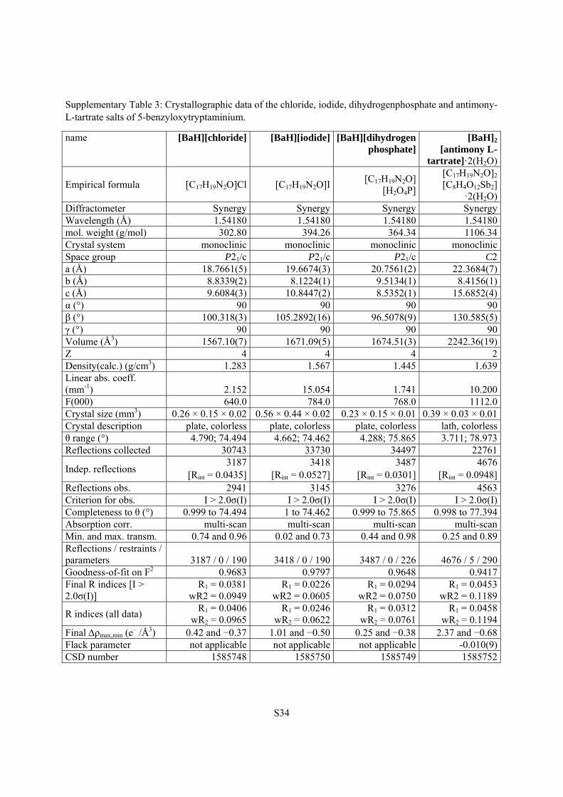
Supplementary Table 3: Crystallographic data of the chloride, iodide, dihydrogenphosphate and antimony-L-tartrate salts of 5-benzyloxytryptaminium.
name [BaH][chloride] [BaH][iodide] [BaH][dihydrogen phosphate]
[BaH]2
[antimony L-tartrate]·2(H2O)
Empirical formula [C17H19N2O]Cl [C17H19N2O]I[C17H19N2O]
[H2O4P]
[C17H19N2O]2 [C8H4O12Sb2]
·2(H2O)Diffractometer Synergy Synergy Synergy SynergyWavelength (Å) 1.54180 1.54180 1.54180 1.54180mol. weight (g/mol) 302.80 394.26 364.34 1106.34Crystal system monoclinic monoclinic monoclinic monoclinicSpace group P21/c P21/c P21/c C2a (Å) 18.7661(5) 19.6674(3) 20.7561(2) 22.3684(7)b (Å) 8.8339(2) 8.1224(1) 9.5134(1) 8.4156(1)c (Å) 9.6084(3) 10.8447(2) 8.5352(1) 15.6852(4)α (°) 90 90 90 90β (°) 100.318(3) 105.2892(16) 96.5078(9) 130.585(5)γ (°) 90 90 90 90Volume (Å3) 1567.10(7) 1671.09(5) 1674.51(3) 2242.36(19)Z 4 4 4 2Density(calc.) (g/cm3) 1.283 1.567 1.445 1.639Linear abs. coeff. (mm-1) 2.152 15.054 1.741 10.200F(000) 640.0 784.0 768.0 1112.0Crystal size (mm3) 0.26 × 0.15 × 0.02 0.56 × 0.44 × 0.02 0.23 × 0.15 × 0.01 0.39 × 0.03 × 0.01Crystal description plate, colorless plate, colorless plate, colorless lath, colorlessθ range (°) 4.790; 74.494 4.662; 74.462 4.288; 75.865 3.711; 78.973Reflections collected 30743 33730 34497 22761
Indep. reflections 3187
[Rint = 0.0435]3418
[Rint = 0.0527]3487
[Rint = 0.0301] 4676
[Rint = 0.0948]Reflections obs. 2941 3145 3276 4563Criterion for obs. I > 2.0σ(I) I > 2.0σ(I) I > 2.0σ(I) I > 2.0σ(I) Completeness to θ (°) 0.999 to 74.494 1 to 74.462 0.999 to 75.865 0.998 to 77.394Absorption corr. multi-scan multi-scan multi-scan multi-scan Min. and max. transm. 0.74 and 0.96 0.02 and 0.73 0.44 and 0.98 0.25 and 0.89Reflections / restraints / parameters 3187 / 0 / 190 3418 / 0 / 190 3487 / 0 / 226 4676 / 5 / 290Goodness-of-fit on F2 0.9683 0.9797 0.9648 0.9417Final R indices [I > 2.0σ(I)]
R1 = 0.0381wR2 = 0.0949
R1 = 0.0226wR2 = 0.0605
R1 = 0.0294 wR2 = 0.0750
R1 = 0.0453wR2 = 0.1189
R indices (all data) R1 = 0.0406
wR2 = 0.0965R1 = 0.0246
wR2 = 0.0622R1 = 0.0312
wR2 = 0.0761 R1 = 0.0458
wR2 = 0.1194Final ρmax,min (e
– /Å3) 0.42 and −0.37 1.01 and −0.50 0.25 and −0.38 2.37 and −0.68Flack parameter not applicable not applicable not applicable -0.010(9)CSD number 1585748 1585750 1585749 1585752
S35
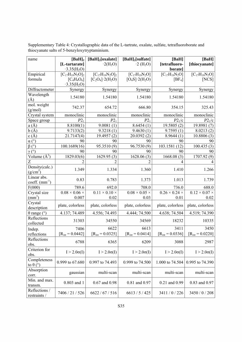
Supplementary Table 4: Crystallographic data of the L-tartrate, oxalate, sulfate, tetrafluoroborate and thiocyanate salts of 5-benzyloxytryptaminium.
name [BaH]2
[L-tartarate] ·3.35(H2O)
[BaH]2[oxalate]·2(H2O)
[BaH]2[sulfate]·2 (H2O)
[BaH] [tetrafluoro-
borate]
[BaH] [thiocyanate]
Empirical formula
[C17H19N2O]2 [C4H4O6]
·3.35(H2O)
[C17H19N2O]2 [C2O4]·2(H2O)
[C17H19N2O] [O4S]·2(H2O)
[C17H19N2O] [BF4]
[C17H19N2O][NCS]
Diffractometer Synergy Synergy Synergy Synergy SynergyWavelength (Å)
1.54180 1.54180 1.54180 1.54180 1.54180
mol. weight (g/mol)
742.37 654.72 666.80 354.15 325.43
Crystal system monoclinic monoclinic monoclinic monoclinic monoclinicSpace group P21 P21 P21 P21/c P21/ca (Å) 8.8100(1) 9.0081 (1) 8.6454 (1) 19.5805 (2) 19.8981 (7)b (Å) 9.7133(2) 9.3218 (1) 9.4630 (1) 9.7595 (1) 8.0213 (2)c (Å) 21.7147(4) 19.4957 (2) 20.0392 (2) 8.9644 (1) 10.8806 (3)α (°) 90 90 90 90 90β (°) 100.1689(16) 95.3510 (9) 96.7530 (9) 103.1581 (12) 100.435 (3)γ (°) 90 90 90 90 90Volume (Å3) 1829.03(6) 1629.95 (3) 1628.06 (3) 1668.08 (3) 1707.92 (9)Z 2 2 2 4 4Density(calc.) (g/cm3)
1.349 1.334 1.360 1.410 1.266
Linear abs. coeff. (mm-1)
0.83 0.783 1.373 1.013 1.739
F(000) 789.6 692.0 708.0 736.0 688.0Crystal size (mm3)
0.08 × 0.06 × 0.007
0.11 × 0.10 × 0.02
0.08 × 0.05 × 0.03
0.26 × 0.24 × 0.01
0.12 × 0.07 × 0.02
Crystal description
plate, colorless plate, colorless plate, colorless plate, colorless plate, colorless
θ range (°) 4.137; 74.489 4.556; 74.493 4.444; 74.500 4.638; 74.504 4.519; 74.390Reflections collected
31303 34550 34569 18232 10335
Indep. reflections
7406 [Rint = 0.0442]
6622[Rint = 0.0325]
6613[Rint = 0.0414]
3411 [Rint = 0.0336]
3450[Rint = 0.0220]
Reflections obs.
6788 6365 6209 3088 2987
Criterion for obs.
I > 2.0σ(I) I > 2.0σ(I) I > 2.0σ(I) I > 2.0σ(I) I > 2.0σ(I)
Completeness to θ (°)
0.999 to 67.680 0.997 to 74.493 0.999 to 74.500 1.000 to 74.504 0.995 to 74.390
Absorption corr.
gaussian multi-scan multi-scan multi-scan multi-scan
Min. and max. transm.
0.803 and 1 0.67 and 0.98 0.81 and 0.97 0.21 and 0.99 0.83 and 0.97
Reflections / restraints /
7406 / 21 / 526 6622 / 67 / 516 6613 / 5 / 425 3411 / 0 / 226 3450 / 0 / 208
S36
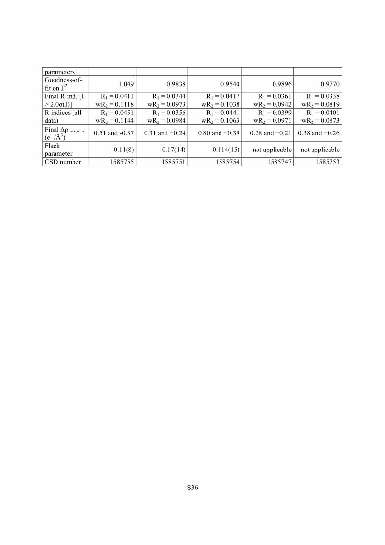
parameters Goodness-of-fit on F2
1.049 0.9838 0.9540 0.9896 0.9770
Final R ind. [I > 2.0σ(I)]
R1 = 0.0411 wR2 = 0.1118
R1 = 0.0344wR2 = 0.0973
R1 = 0.0417wR2 = 0.1038
R1 = 0.0361 wR2 = 0.0942
R1 = 0.0338wR2 = 0.0819
R indices (all data)
R1 = 0.0451 wR2 = 0.1144
R1 = 0.0356wR2 = 0.0984
R1 = 0.0441wR2 = 0.1063
R1 = 0.0399 wR2 = 0.0971
R1 = 0.0401wR2 = 0.0873
Final ρmax,min (e– /Å3)
0.51 and -0.37 0.31 and −0.24 0.80 and −0.39 0.28 and −0.21 0.38 and −0.26
Flack parameter
-0.11(8) 0.17(14) 0.114(15) not applicable not applicable
CSD number 1585755 1585751 1585754 1585747 1585753
S37
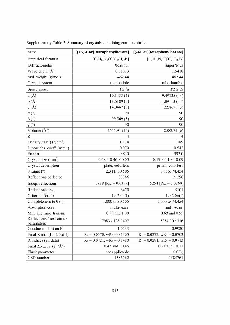
Supplementary Table 5: Summary of crystals containing carnitinenitrile
name [(+/-)-Car][tetraphenylborate] [(-)-Car][tetraphenylborate]
Empirical formula [C7H15N2O][C24H20B] [C7H15N2O][C24H20B]
Diffractometer Xcalibur SuperNova
Wavelength (Å) 0.71073 1.5418
mol. weight (g/mol) 462.44 462.44
Crystal system monoclinic orthorhombic
Space group P21/n P212121
a (Å) 10.1433 (4) 9.49835 (14)
b (Å) 18.6189 (6) 11.89113 (17)
c (Å) 14.0467 (5) 22.8675 (3)
α (°) 90 90
β (°) 99.569 (3) 90
γ (°) 90 90
Volume (Å3) 2615.91 (16) 2582.79 (6)
Z 4 4
Density(calc.) (g/cm3) 1.174 1.189
Linear abs. coeff. (mm-1) 0.070 0.542
F(000) 992.0 992.0
Crystal size (mm3) 0.48 × 0.46 × 0.05 0.43 × 0.10 × 0.09
Crystal description plate, colorless prism, colorless
θ range (°) 2.311; 30.505 3.866; 74.454
Reflections collected 33386 21298
Indep. reflections 7988 [Rint = 0.0359] 5254 [Rint = 0.0260]
Reflections obs. 6470 5101
Criterion for obs. I > 2.0σ(I) I > 2.0σ(I)
Completeness to θ (°) 1.000 to 30.505 1.000 to 74.454
Absorption corr multi-scan multi-scan
Min. and max. transm. 0.99 and 1.00 0.69 and 0.95 Reflections / restraints / parameters
7983 / 128 / 407 5254 / 0 / 316
Goodness-of-fit on F2 1.0133 0.9920
Final R ind. [I > 2.0σ(I)] R1 = 0.0578, wR2 = 0.1365 R1 = 0.0272, wR2 = 0.0703
R indices (all data) R1 = 0.0721, wR2 = 0.1480 R1 = 0.0281, wR2 = 0.0713
Final ρmax,min (e– /Å3) 0.47 and −0.46 0.21 and −0.11
Flack parameter not applicable 0.0(3)
CSD number 1585762 1585761
S38
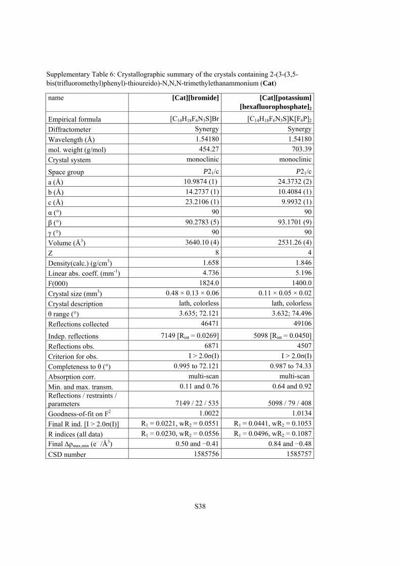
Supplementary Table 6: Crystallographic summary of the crystals containing 2-(3-(3,5-bis(trifluoromethyl)phenyl)-thioureido)-N,N,N-trimethylethanammonium (Cat)
name [Cat][bromide] [Cat][potassium] [hexafluorophosphate]2
Empirical formula [C14H18F6N3S]Br [C14H18F6N3S]K[F6P]2
Diffractometer Synergy Synergy
Wavelength (Å) 1.54180 1.54180
mol. weight (g/mol) 454.27 703.39
Crystal system monoclinic monoclinic
Space group P21/c P21/c
a (Å) 10.9874 (1) 24.3732 (2)
b (Å) 14.2737 (1) 10.4084 (1)
c (Å) 23.2106 (1) 9.9932 (1)
α (°) 90 90
β (°) 90.2783 (5) 93.1701 (9)
γ (°) 90 90
Volume (Å3) 3640.10 (4) 2531.26 (4)
Z 8 4
Density(calc.) (g/cm3) 1.658 1.846
Linear abs. coeff. (mm-1) 4.736 5.196
F(000) 1824.0 1400.0
Crystal size (mm3) 0.48 × 0.13 × 0.06 0.11 × 0.05 × 0.02
Crystal description lath, colorless lath, colorless
θ range (°) 3.635; 72.121 3.632; 74.496
Reflections collected 46471 49106
Indep. reflections 7149 [Rint = 0.0269] 5098 [Rint = 0.0450]
Reflections obs. 6871 4507
Criterion for obs. I > 2.0σ(I) I > 2.0σ(I)
Completeness to θ (°) 0.995 to 72.121 0.987 to 74.33
Absorption corr. multi-scan multi-scan
Min. and max. transm. 0.11 and 0.76 0.64 and 0.92 Reflections / restraints / parameters 7149 / 22 / 535 5098 / 79 / 408
Goodness-of-fit on F2 1.0022 1.0134
Final R ind. [I > 2.0σ(I)] R1 = 0.0221, wR2 = 0.0551 R1 = 0.0441, wR2 = 0.1053
R indices (all data) R1 = 0.0230, wR2 = 0.0556 R1 = 0.0496, wR2 = 0.1087
Final ρmax,min (e– /Å3) 0.50 and −0.41 0.84 and −0.48
CSD number 1585756 1585757
S39
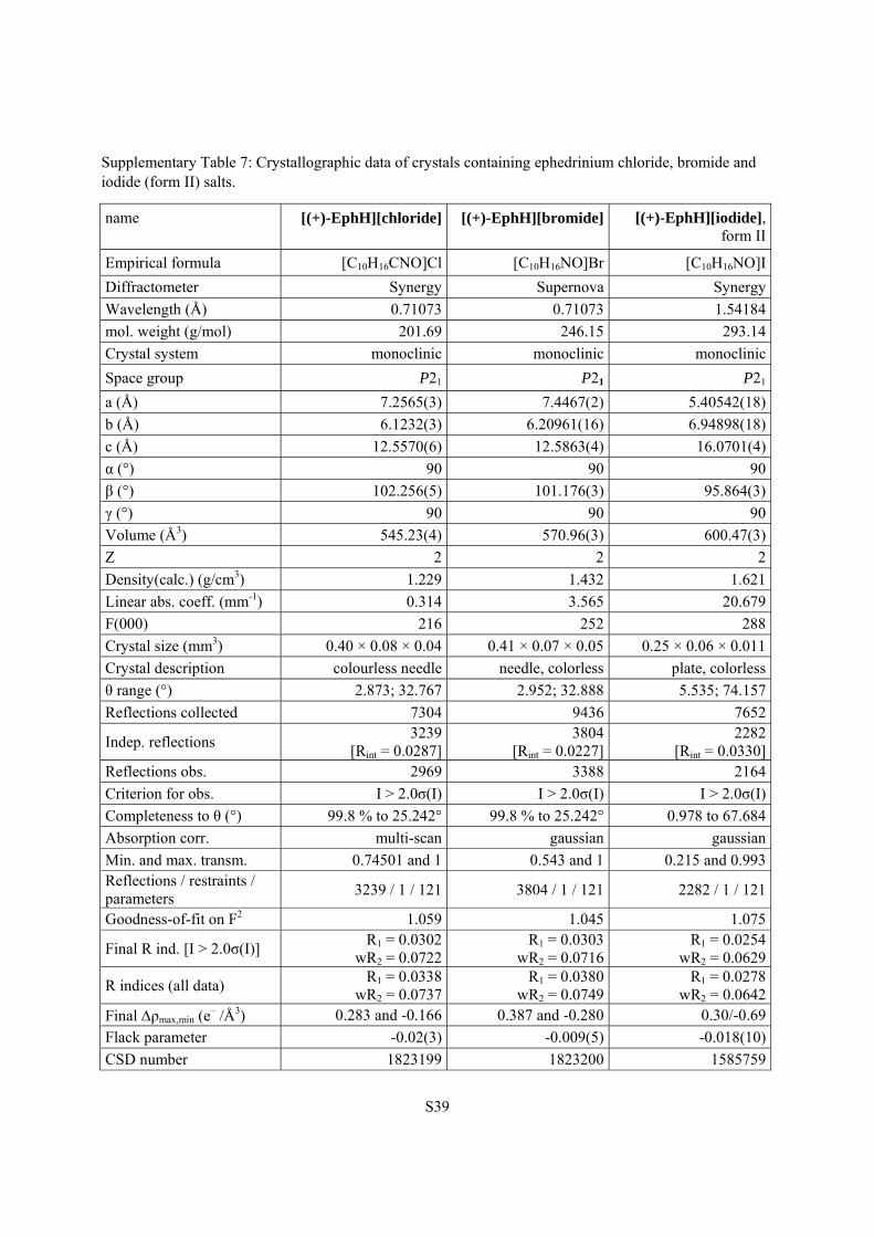
Supplementary Table 7: Crystallographic data of crystals containing ephedrinium chloride, bromide and iodide (form II) salts.
name [(+)-EphH][chloride] [(+)-EphH][bromide] [(+)-EphH][iodide], form II
Empirical formula [C10H16CNO]Cl [C10H16NO]Br [C10H16NO]I
Diffractometer Synergy Supernova Synergy
Wavelength (Å) 0.71073 0.71073 1.54184
mol. weight (g/mol) 201.69 246.15 293.14
Crystal system monoclinic monoclinic monoclinic
Space group P21 P21 P21
a (Å) 7.2565(3) 7.4467(2) 5.40542(18)
b (Å) 6.1232(3) 6.20961(16) 6.94898(18)
c (Å) 12.5570(6) 12.5863(4) 16.0701(4)
α (°) 90 90 90
β (°) 102.256(5) 101.176(3) 95.864(3)
γ (°) 90 90 90
Volume (Å3) 545.23(4) 570.96(3) 600.47(3)
Z 2 2 2
Density(calc.) (g/cm3) 1.229 1.432 1.621
Linear abs. coeff. (mm-1) 0.314 3.565 20.679
F(000) 216 252 288
Crystal size (mm3) 0.40 × 0.08 × 0.04 0.41 × 0.07 × 0.05 0.25 × 0.06 × 0.011
Crystal description colourless needle needle, colorless plate, colorless
θ range (°) 2.873; 32.767 2.952; 32.888 5.535; 74.157
Reflections collected 7304 9436 7652
Indep. reflections 3239
[Rint = 0.0287]3804
[Rint = 0.0227]2282
[Rint = 0.0330] Reflections obs. 2969 3388 2164
Criterion for obs. I > 2.0σ(I) I > 2.0σ(I) I > 2.0σ(I)
Completeness to θ (°) 99.8 % to 25.242° 99.8 % to 25.242° 0.978 to 67.684
Absorption corr. multi-scan gaussian gaussian
Min. and max. transm. 0.74501 and 1 0.543 and 1 0.215 and 0.993Reflections / restraints / parameters
3239 / 1 / 121 3804 / 1 / 121 2282 / 1 / 121
Goodness-of-fit on F2 1.059 1.045 1.075
Final R ind. [I > 2.0σ(I)] R1 = 0.0302
wR2 = 0.0722R1 = 0.0303
wR2 = 0.0716R1 = 0.0254
wR2 = 0.0629
R indices (all data) R1 = 0.0338
wR2 = 0.0737R1 = 0.0380
wR2 = 0.0749R1 = 0.0278
wR2 = 0.0642 Final ρmax,min (e
– /Å3) 0.283 and -0.166 0.387 and -0.280 0.30/-0.69
Flack parameter -0.02(3) -0.009(5) -0.018(10)
CSD number 1823199 1823200 1585759
S40
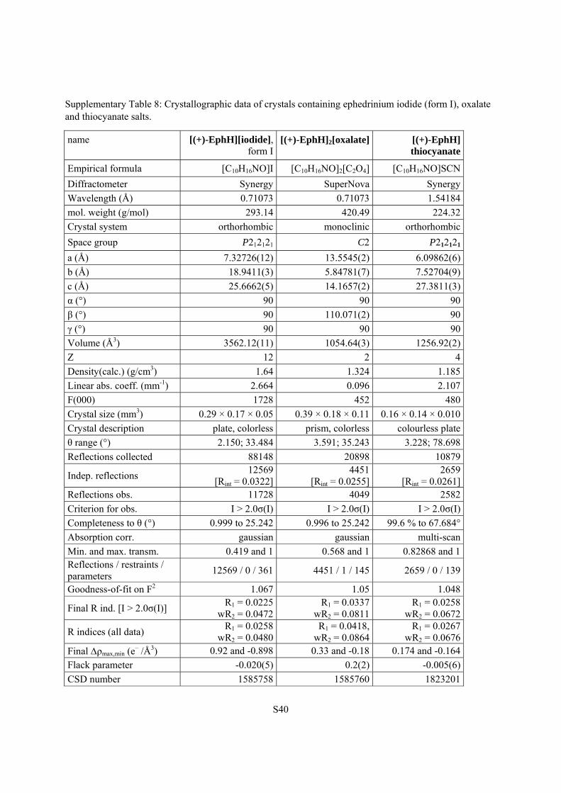
Supplementary Table 8: Crystallographic data of crystals containing ephedrinium iodide (form I), oxalate and thiocyanate salts.
name [(+)-EphH][iodide], form I
[(+)-EphH]2[oxalate] [(+)-EphH] thiocyanate
Empirical formula [C10H16NO]I [C10H16NO]2[C2O4] [C10H16NO]SCN
Diffractometer Synergy SuperNova Synergy
Wavelength (Å) 0.71073 0.71073 1.54184
mol. weight (g/mol) 293.14 420.49 224.32
Crystal system orthorhombic monoclinic orthorhombic
Space group P212121 C2 P212121
a (Å) 7.32726(12) 13.5545(2) 6.09862(6)
b (Å) 18.9411(3) 5.84781(7) 7.52704(9)
c (Å) 25.6662(5) 14.1657(2) 27.3811(3)
α (°) 90 90 90
β (°) 90 110.071(2) 90
γ (°) 90 90 90
Volume (Å3) 3562.12(11) 1054.64(3) 1256.92(2)
Z 12 2 4
Density(calc.) (g/cm3) 1.64 1.324 1.185
Linear abs. coeff. (mm-1) 2.664 0.096 2.107
F(000) 1728 452 480
Crystal size (mm3) 0.29 × 0.17 × 0.05 0.39 × 0.18 × 0.11 0.16 × 0.14 × 0.010
Crystal description plate, colorless prism, colorless colourless plate
θ range (°) 2.150; 33.484 3.591; 35.243 3.228; 78.698
Reflections collected 88148 20898 10879
Indep. reflections 12569
[Rint = 0.0322]4451
[Rint = 0.0255]2659
[Rint = 0.0261] Reflections obs. 11728 4049 2582
Criterion for obs. I > 2.0σ(I) I > 2.0σ(I) I > 2.0σ(I)
Completeness to θ (°) 0.999 to 25.242 0.996 to 25.242 99.6 % to 67.684°
Absorption corr. gaussian gaussian multi-scan
Min. and max. transm. 0.419 and 1 0.568 and 1 0.82868 and 1 Reflections / restraints / parameters
12569 / 0 / 361 4451 / 1 / 145 2659 / 0 / 139
Goodness-of-fit on F2 1.067 1.05 1.048
Final R ind. [I > 2.0σ(I)] R1 = 0.0225
wR2 = 0.0472R1 = 0.0337
wR2 = 0.0811R1 = 0.0258
wR2 = 0.0672
R indices (all data) R1 = 0.0258
wR2 = 0.0480R1 = 0.0418,
wR2 = 0.0864R1 = 0.0267
wR2 = 0.0676 Final ρmax,min (e
– /Å3) 0.92 and -0.898 0.33 and -0.18 0.174 and -0.164
Flack parameter -0.020(5) 0.2(2) -0.005(6)
CSD number 1585758 1585760 1823201
S41
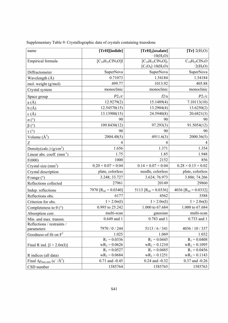
Supplementary Table 9: Crystallographic data of crystals containing trazodone
name [TrH][iodide] [TrH]2[oxalate] ·10(H2O)
[Tr]·2(H2O)
Empirical formula [C19H23ClN5O]I [C19H23ClN5O]2 [C2O4]·10(H2O)
C19H22ClN5O·2(H2O)
Diffractometer SuperNova SuperNova SuperNova
Wavelength (Å) 0.71073 1.54184 1.54184
mol. weight (g/mol) 499.77 1013.92 405.88
Crystal system monoclinic monoclinic monoclinic
Space group P21/c I2/a P21/c
a (Å) 12.9279(2) 15.1489(4) 7.10113(10)
b (Å) 12.54570(15) 13.2904(4) 13.6250(2)
c (Å) 13.13900(15) 24.5940(8) 20.6821(3)
α (°) 90 90 90
β (°) 109.8430(12) 97.293(3) 91.5054(12)
γ (°) 90 90 90
Volume (Å3) 2004.48(5) 4911.6(3) 2000.36(5)
Z 4 4 4
Density(calc.) (g/cm3) 1.656 1.371 1.354
Linear abs. coeff. (mm-1) 1.75 1.85 1.948
F(000) 1000 2152 856
Crystal size (mm3) 0.20 × 0.07 × 0.04 0.14 × 0.07 × 0.04 0.28 × 0.15 × 0.02
Crystal description plate, colorless needle, colorless plate, colorless
θ range (°) 3.248; 33.727 3.624; 76.973 3.886; 74.266
Reflections collected 27961 20149 29860
Indep. reflections 7970 [Rint = 0.0340] 5113 [Rint = 0.0336] 4036 [Rint = 0.0332]
Reflections obs. 6177 4562 3588
Criterion for obs. I > 2.0σ(I) I > 2.0σ(I) I > 2.0σ(I)
Completeness to θ (°) 0.995 to 25.242 1.000 to 67.684 1.000 to 67.684
Absorption corr. multi-scan gaussian multi-scan
Min. and max. transm. 0.649 and 1 0.783 and 1 0.733 and 1Reflections / restraints / parameters 7970 / 0 / 244 5113 / 6 / 341 4036 / 10 / 337
Goodness-of-fit on F2 1.025 1.069 1.032
Final R ind. [I > 2.0σ(I)] R1 = 0.0336
wR2 = 0.0626R1 = 0.0445
wR2 = 0.1210 R1 = 0.0408
wR2 = 0.1095
R indices (all data) R1 = 0.0527
wR2 = 0.0684R1 = 0.0485
wR2 = 0.1251 R1 = 0.0456
wR2 = 0.1143
Final ρmax,min (e– /Å3) 0.71 and -0.45 0.24 and -0.32 0.37 and -0.26
CSD number 1585764 1585765 1585763
S42
References:
1. M. Tiffner, J. Novacek, A. Busillo, K. Gratzer, A. Massa and M. Waser, RSC Adv., 2015, 5, 78941-
78949.
2. A. McPherson and J. A. Gavira, Acta Cryst., 2014, F70, 2-20.
3. Rigaku Oxford Diffraction, CrysAlisPro Software system 1.171.38, Rigaku Corporation, Oxford, UK,
2015.
4. A. Altomare, M. C. Burla, M. Camalli, G. L. Cascarano, C. Giacovazzo, A. Guagliardi, A. G. G.
Moliterni, G. Polidori and R. Spagna, J. Appl. Cryst., 1999, 32, 115-119.
5. L. Palatinus and G. Chapuis, J. Appl. Cryst., 2007, 40, 786-790.
6. G. M. Sheldrick, Acta Cryst., 2015, C71, 3-8.
7. C. B. Hübschle, G. M. Sheldrick and B. Dittrich, J. Appl. Cryst., 2011, 44, 1281-1284.
8. P. W. Betteridge, J. R. Carruthers, R. I. Cooper, K. Prout and D. J. Watkin, J. Appl. Cryst., 2003, 36,
1487-1487.
9. C. F. Macrae, I. J. Bruno, J. A. Chisholm, P. R. Edgington, P. McCabe, E. Pidcock, L. Rodriguez-
Monge, R. Taylor, J. van de Streek and P. A. Wood, J. Appl. Cryst., 2008, 41, 466-470.
10. A. V. George, L. D. Field, E. Y. Malouf, A. E. D. McQueen, S. R. Pike, G. R. Purches, T. W.
Hambley, I. E. Buys, A. H. White, D. C. R. Hockless and B. W. Skelton, J. Organomet. Chem., 1997,
538, 101-110.
11. A. Rodriguez-Fortea, P. Alemany, S. Alvarez, E. Ruiz, A. Scuiller, C. Decroix, V. Marvaud, J.
Vaissermann, M. Verdaguer, I. Rosenman and M. Julve, Inorg. Chem., 2001, 40, 5868-5877.
12. A. Peuronen, E. Lehtimäki and M. Lahtinen, Cryst. Growth Des., 2013, 13, 4615-4622.
13. E. A. Collier, R. J. Davey, S. N. Black and R. J. Roberts, Acta Cryst., 2006, B62, 498-505.
14. B. Gossner and H. Neff, Z. Kristallogr., 1933, 86, 32-41.
15. K. Schwantke, Z. Kristallogr., 1909, 46, 73-115.
16. A. L. Spek, J. Appl. Cryst., 2003, 36, 7-13.
17. R. W. W. Hooft, L. H. Straver and A. L. Spek, J. Appl. Cryst., 2008, 41, 96-103.