Embed Size (px)
Citation preview

1
DENT 655- Health Technology Assessment
A Health Technology Assessment Report
on Cone Beam Computed Tomography vs.
Conventional Two Dimensional Imaging
Techniques in Implantology
by
Aarti Angrulaa & Iris Boraschi-Diaza
with the expert assistance of
Shahrokh Esfandiarib
April 2012
Report No: S2012.02
a Masters candidate, Faculty of Dentistry, McGill University, Montreal, Canada b Associate Professor, Division of Oral Health and Society, Faculty of Dentistry, McGill University For more information contact Dr. Shahrokh Esfandiari at [email protected]

2
The views expressed in this report are those of the author(s) and do not necessarily reflect the
views of the Faculty of Dentistry, McGill University. This report was developed for the course
‘DENT 655- Health Technology Assessment’ and assumes a call from general dentists to assist
decision-making in dental offices, clinical and hospitals. All are welcome to make use of it.
However, to help us estimate the impact, it would be deeply appreciated if users could inform
us whether it has influenced policy decisions in any way.
Suggested Citation: Angrula, A.*, Boraschi-Diaz, I.*, & Esfandiari, S. (2012). A Health Technology Assessment Report on Cone Beam Computed Tomography vs. Conventional Two Dimensional Imaging Techniques in Implantology (Report no: S2012.02). Montreal, Canada: Faculty of Dentistry, McGill University. [* Equal first authors]. Retrieved from: https://www.mcgill.ca/dentistryohs/courses-and-events/dent-655/hta-reports

3
Table of Contents Page Number
Foreword……………………………………………………………………………..3
Acronyms…………………………………………………………………................4
1. Background…………………………………………………………………………..5
2. Methodology………………………………………………………………………....9
3. Results……………………………………………………………………………….11
3.1. Measurements…………………………………………………………………..11
3.2. Evaluation Time………………………………………………………………...13
3.3. Ortho-implant…………………………………………………………………...13
3.4. Implant Site……………………………………………………………………...14
3.5. Bone Assessment………………………………………………………………..15
4. Guidelines for CBCT use……………………………………………………………...17
5. Ethical and Legal Considerations……………………………………………………..19
6. Radiation………………………………………………………………………………21
7. Cost Analysis………………………………………………………………………….22
8. Discussion……………………………………………………………………………..24
9. Conclusion…………………………………………………………………………….25
Appendix # 1
References

4
Acronyms
2D: Two-dimensional space
3D: Three-dimensional space, the physical universe
AETMIS: L'Agence d'évaluation des technologies et des modes d'intervention en santé
CADTH: Canadian Agency for Drugs and Technologies in Health
CBCT: Cone Beam Computed Tomography
CT: Computed Tomography
FDA: Food and Drug Administration, agency of the United States Department of Health
and Human Services, responsible for protecting and promoting public health through the
regulation and supervision of food safety, tobacco products, etc.
INAHTA: International Network of Agencies for Health Technology Assessment
mSv: milliSievert. International System of Units (SI) derived unit of dose equivalent
radiation
NICE: National Institute for Health and Clinical Excellence
QR: Quantitative Radiology
TMJ: Temporomandibular Joint
US: United States of America
X-rays: a form of electromagnetic radiation. X-rays have a wavelength in the range of
0.01 to 10 nm.
μSv: microSievert. Unit of dose equivalent radiation.

5
1. Background
Dental radiography is an area of study that has changed a lot throughout the years. Initially called
X-rays, dentists use this tool in their offices every day in order to give better and more accurate
diagnosis. In the dental office, these images are used to diagnose cavities, cancerous or benign
masses, hidden dental structures, bone loss hidden under the gums. Furthermore, this technique
is used to check the status of a procedure and even as a follow-up of some treatments (1-3).
The images taken with these technologies are usually able to show calcified structures such as
teeth, bones, and sometimes one can visualize the surrounding soft tissues. The picture shown on
an X-ray is formed by the radiation emitted by a machine. This radiation will enter the oral
structures at different levels, depending on varying anatomical densities. For example, teeth
appear lighter because less radiation penetrates them to reach the film. In the case of cavities or
bone loss, they look usually darker because X-rays penetrate these less dense structures, so more
radiation can reach the film (1-3).
In the case of dental radiography the dosage of radiation received by a patient is typically around
0.005–0.03 mSv (miliSievert). This is considered really low, equivalent to a few days of
background environmental radiation exposure. This exposure can be reduced using different
protective lead barriers (1, 3).
In dentistry there are two types of radiographs intraoral and extraoral. Intraoral X-rays are the
most common, since they give a high level of detail, also they help the dentist in a very fast way
to diagnose anomalies like caries, check shapes and pathologies near the tooth roots, also to
check possible bone loss, stages of tooth development or even just to monitor good oral health
(1-3).

6
In the case of extraoral techniques, as the name indicates, they are radiographs which are taken
with the film outside the mouth. Generally these are considered the "big picture" X-rays. In these
we can observe teeth, bone, eventual tumors and anatomic structures. However, often the
analysis of these radiographs focuses more on the facial bones. These types of radiographs are
frequently used in areas of dentistry like orthodontics, in order to monitor growth and
development. In maxillofacial and oral surgery, these are very commonly used for the
assessment of impacted teeth or possible impacted pathologies within bone, also to examine
relationships between teeth and jaws and examining the temporomandibular joint or other bones
of the face. Within these techniques we can find panorex or panoramic X-ray, cephalometric
projections, cone beam computerized tomography, tomograms and sialographies (1, 3-5).
The panoramic x-ray is known by different names: orthopantomogram or dental panoramic
radiograph or "panorex". Basically, it consists of a dental X-ray scanning of the upper and lower
jaws, in two-dimensions which show a view of a half-circle from ear to ear. It depends on the
tomography principles, since the images of specific radiographic planes are taken to make up the
larger panoramic image. The equipment used in this technique consist of a horizontal rotating
arm which holds an X-ray source on one side and in the other side a moving film mechanism
(carrying a film). The patient is located between the x-ray source and the film. The arm rotates
around an instant center (patients head) which shifts on a dedicated trajectory. The X-ray source
is collimated toward the film, to give the least distortion possible, after crossing the patient's
skull (4, 6).
In the case of lateral or cephalometric, these are extraoral radiographs taken from the side of the
head, showing a two dimensional picture of the side of the head of the patient. This technique is
often used to gauge the size and spacial relationships of the teeth, jaws, and cranium. This

7
analysis informs treatment planning, quantifies changes during treatment, and provides data for
clinical research in the areas of orthodontics and maxillofacial and oral surgery (3).
In general all this two dimensional techniques are nowadays available as digital radiography.
This change has become the standard of care, since with these type of techniques we can reduce
the radiation, improve the quality of the image and make different analysis using different type
of dental software (5).
The general disadvantage that these techniques encounter is that all are mainly dependent on
patient positioning. Furthermore, due to the certain limitations such as lack of a third dimension
in the picture and patient discomfort, three dimensional imaging techniques like computed
tomography (CT) and similar techniques has become a necessity in the dental practice. CT scan
is the fundamental technique in modern medical radiology (7).
After the introduction of the CT scan in mid-1970, this technique has shown a major
advancement in the area of dentistry. In hospitals the spiral/helical CT scan became the standard
instrument for medical imaging since 1990’s. The introduction of multi-slice CT scan in 1998
revolutionized the field; this technique produces clearer pictures with more detail and in a lot less
time than it takes for a conventional spiral CT. The use of 3D techniques in dentistry has
increased the diagnostic information and treatment planning benefits in oral anomalies. Medical
CT scans have been used in craniofacial imaging since its introduction but it did not become very
popular until 1980s when high resolution scanners with slice thickness of 2mm were developed.
They allow the acquisition of several cross-sectional slices at the time thus reducing the scanning
time. Conventional CT scans are mainly designed for the full body scanning at high speed to
reduce the artifacts caused by lungs, heart and bowels. They are not well suited for imaging the
dentomaxillo-facial areas, where cost consideration is also important (8, 9).

8
The major disadvantage of conventional or multi-slice CT scan is the high amount of radiation
exposure required. More recently a decade ago, the cone beam computed tomography (CBCT)
has been introduced (10). In the conventional CT scanner a collimated fan shaped X-ray beam is
projected through a limited thickness slice through the human body. In this technique the X-ray
tube and detector are continuously moving around the patient. The projections are detected
through a linear array of detectors while the patient is advanced through the gantry. In the case of
CBCT, it uses a cone shaped beam and an area detector, which acquires a full volume of images
in a single rotation with no need for patient movement. The x-ray source rotates 360 degree,
around the head to obtain multiple images. The CBCT scan generates voxels which are
reassembled into a digital image (8). The voxels are the smallest subunit of a digital volume.
Unique property about voxel is its isotropic nature which means they have equal dimensions in
all the three planes (7). Therefore, the digital image produced by isotropic voxels has high spatial
resolution due to lack of any spacing between them, which contributes to the accuracy and
precision in visualizing the anatomical structures (5,10,11).
Figure 1 (12): Example of how a CBCT works. A cone-shaped x-ray beam irradiates a patient’s jaw. The
transmitted x-rays are detected by a sensor; the data is sent to a computer and reconstructed into 3-D images by
software. Hamamatsu Corp.

9
The first use of the CBCT was focus in the maxillofacial area, and it was first introduced in
1996. The FDA approved the first dental CBCT unit in March 8, 2001. The first company which
marketed this technology in the US market was QR (Verona, Italy), in May 2001. The
advantages of this technology have become one of the strongest points of marketing over other
technologies such as panorex, cephalometry, digital intraoral radiograph and even conventional
CT. Within these advantages we could find high quality 3D images, high spatial resolution, takes
less time and most importantly the radiation exposure is almost 15 times lower than the
conventional CT scan. CBCT is ideal for high quality and affordable in-house CT scanning of
the head and neck in dentomaxillofacial applications. Although CBCT has been used in all the
fields of dentistry, it has a very useful role in the implant placement because of its 3D nature and
high resolution (2, 6, 9, 13).
Radiographic analyzes of the jaws in potential implant sites depends on combinations of
periapical, panoramic, planar tomographic and CT imaging. CBCT has been recognized to
improve measuring of distances between alveolar crest and anatomic landmarks, evaluating
quality of cortical and medullary bone in the potential implant sites, visualizing inclination of the
alveolar process to increase the chances of a successful implant placement, and finally giving the
basis for treatment planning with the use of special algorithms (10).
2. Methodology
We did a systematic literature review using major databases in the area of health technology
assessment (INAHTA, NICE, AETMIS and CADTH) and also we included searches in medical
literature databases such as MEDLINE, EMBASE (searches were performed through Ovid
Online) and Cochrane library.
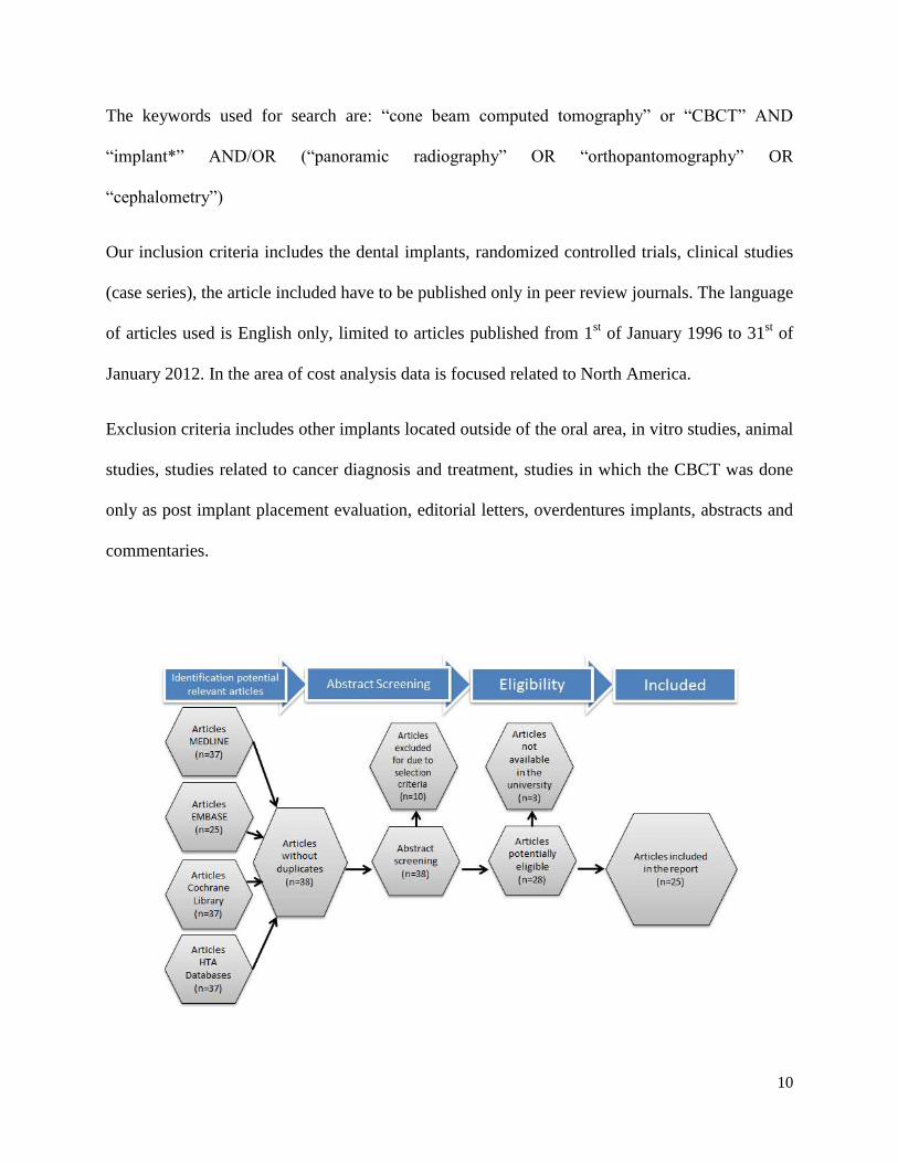
10
The keywords used for search are: “cone beam computed tomography” or “CBCT” AND
“implant*” AND/OR (“panoramic radiography” OR “orthopantomography” OR
“cephalometry”)
Our inclusion criteria includes the dental implants, randomized controlled trials, clinical studies
(case series), the article included have to be published only in peer review journals. The language
of articles used is English only, limited to articles published from 1st of January 1996 to 31
st of
January 2012. In the area of cost analysis data is focused related to North America.
Exclusion criteria includes other implants located outside of the oral area, in vitro studies, animal
studies, studies related to cancer diagnosis and treatment, studies in which the CBCT was done
only as post implant placement evaluation, editorial letters, overdentures implants, abstracts and
commentaries.

11
3. Results
3.1. Measurements
In this literature review, we found 16 studies which are discussing about the potential
measurements that can be taken with CBCT in assessing the implant planning. This explains
high benefit of CBCT in the ability to visualize different anatomical variables in three dimension
view.
Assessment of magnification ratio is very important while visualizing different anatomical areas;
most significantly in the evaluation of residual ridge. Studies have shown differences in the
magnification ratios when panoramic radiography is used at different tooth locations (1). Results
differ with different panoramic equipment used. With the use of panoramic radiograph,
magnification ratio is found to be 1.2 times. CBCT is found to reduce this difference. CBCT
showed no significant difference in the magnification ratio according to the location of tooth.
Therefore, while using CBCT one does not have to consider more than one magnification ratio
according to the location of the implant, which is not the case with panoramic radiography (3).
However, authors have accepted the possibility of differences in results due to objective
evaluation and patient positioning (1).
CBCT can be used to fabricate surgical guides in the placement of mini-implants. The software
associated helps the clinician to simulate mini-implant placement and simultaneously view the
anatomical structures such as bone, tooth, sinuses, and vessels (22). The simulated implant can
be moved in any direction until the ideal location is achieved preventing impingement to any
surrounding structures. Once the implant position is attained by computer simulation, the data
can be saved and emailed to the processing center. This is not feasible with the use of panoramic
radiography which does not allow bucco-lingual assessment of the alveolar ridge (16).

12
Also, alveolar ridge can be assessed qualitatively and quantitatively better than orthopantograph.
The measurements done in the anterior mandibular area from the root apices of anterior teeth to
mandibular base showed significant difference between two techniques (16). The differences
were not significant except in the canine region when the distance was measured from root
apices to incisive canal (16); however the incisive canal is not well visualized in orthopantograph
as compared with CBCT (15).
Ridge mapping is a common technique used to evaluate the residual ridge for implant placement.
Even though, this technique is simple and easy to apply, the information that can be obtained
from a conventional ridge mapping is only about ridge width not its shape since it is measured by
a caliper (26). Although, improved ridge mapping techniques which uses a template can be
applied to measure the topography of the alveolar ridge, its accuracy still remained to be defined
(27). In this scenario, CBCT plays an essential role to evaluate the ridge for the accurate
placement of implant. Measurements can be done after making a template and then taking CBCT
scan on which different reference points are marked for ridge measurements with CBCT
software (22). There has been found a significant difference between ridge mapping and CBCT
techniques at all the implant sites with mean difference of about 0.4mm (25). Nonetheless,
improved ridge mapping can still be used in cases with mild to moderate resorbed ridges and
when implant is placed coronal to the alveolar sulcus (25). CBCT images can also be used to
visualize alveolar crest during ridge expansion procedure, which provided better view than a
panoramic radiograph (34).
Authors have reported accuracy and reliability in the identification of condylar cortical erosion
and in general measurements can be more precise and consistent than using traditional linear
cephalometric, posteroanterior, and submentovertex radiographs (11).

13
3.2. Evaluation time
CBCT has been considered better in pre-operative evaluation of the implant site according to the
tooth location using premeasured guttapercha or metal ball as compared to the panoramic
radiograph (16). CBCT can also be used for the post-operative evaluation of the implant placed
CBCT can be very useful tool in the evaluation of placement of implant in the aesthetic area to
achieve better results (2). After the extraction of a tooth, there is a significant amount of bone
loss over a period of time. This can be more critical in the aesthetic area for the consideration of
implant placement. To avoid the bone loss, clinicians have started placing an implant
immediately in the tooth socket (8). This technique has many advantages but if the immediate
implant is not positioned accurately in the 3-D spacing, it can lead to further complications. One
of the studies used CBCT to accurately replace missing lateral incisor by evaluating the
angulation and position of the adjacent incisor so that the implant can be placed as closely to the
natural tooth position (12).
3.3. Ortho-implant
We found 4 articles which studied the placement of ortho implants with the use of CBCT
imaging. Mini implants have become a very popular skeletal anchorage system in the area of
orthodontics. However, the success of this implants is still under study, since the factors
influencing this success are still unknown (14). Failure rate of micro implants is about 11 to
30%. The failure mainly occurs due to the impingement of micro implant on the roots of the
adjacent teeth or due to the premature loosening of the implant. 2-D imaging techniques such as
panoramic radiograph are widely used for the assessment of the accurate positioning of an ortho
implant (18). But due to many disadvantages such as superimposition of the anatomical

14
structures, it is very difficult to clearly assess the implant site. Hence, CBCT can act as an
adjunct in this scenario to reconstruct 3-D images with the help of computer software provided
with the equipment, which can be further used to produce surgical guides for the accurate
placement of the micro implant. The recommended length of a micro implant is 6mm and
diameter is 1.2 to 1.6mm (11). One of the studies measured the amount of interradicular space,
cortical bone thickness and alveolar process width in maxilla and mandible to evaluate the
suitable areas for micro implant placement. They found that the appropriate site for micro
implant in maxilla is between second premolar and first molar, whereas in mandible is the
interradicular space between first premolar to second molar in the buccal alveolar process. They
also suggested that midpalatal and retromolar area are also optimal sites for micro implant
placement in maxilla. One of studies evaluated the positions of micro implants in the anterior
region and they found the area between central incisor and lateral in maxilla and the area
between lateral incisor and canine is best for micro implant positioning (29).
In the orthodontics residency programs in US and Canada, the CBCT has become essential part
of the treatment planning. In programs where the CBCT is exclusively used for diagnose 100%
of the times was for the localization of a supernumerary or an impacted teeth, the use of this
technique in the diagnostic period, has increase significantly the diagnose confidence of the
residents. Also 100% of the schools promote the use of CBCT as an evaluation tool for
craniofacial anormalies/abnotmalities (11).
3.4. Implant site
One of the studies showed the importance of evaluation of images of teeth in the anterior
maxillary area by CBCT measurements (2). One of the studies evaluated the thickness of buccal

15
and palatal bone in the mid-root and apical level in the maxillary central incisors using CBCT.
CBCT images of the fully formed, intact maxillary incisor were evaluated using software. After
the CBCT measurements, they classified the positions and angulations of the tooth roots in
reference to the alveolar process. The mean thickness of buccal bone at mid root (0.9 mm) and
apical level (2.04 ± 1.01 mm) is much lesser than the palatal counterparts (3.76 ± 1.37 mm mid
root and 8.51 ± 2.54 mm apical). Most of the central incisors were placed buccally (78. 85 %).
These can be further used for the aesthetic placement of implant in the 3-dimension position
immediately after extraction. Also by examining the angulation of the root, they showed that
CBCT imaging can be used to place implant with long term best aesthetic results (2).
CBCT imaging is very informative when implant placement is planned in the posterior
mandibular area. Not only it provides accurate information about adjacent root proximities but
also about the maxillary sinus distance which is very important to prevent perforation by an
implant (19). CBCT image helps in the preparation of surgical guides to place implant in the
posterior maxillary region. This is very efficient in the placement of orthodontic miniscrews and
mini-implants too (22). The 2-D techniques such as periapical X-rays are inefficient in the
placement of orthodontic implants because of lack of 3-D vision and one has to take several
periapical radiographs for the site evaluation (27). 3-D placement of a simulated implant with the
help of CBCT imaging provides rapid and accurate fixing of the implant.
3.5. Bone assessment
Within our literature review we have found 5 articles taking about different techniques to asses
bone in the implant sites.

16
The objective in implant dentistry is not the implant by itself but the tooth that will be placed as
replacement, true restorative driven implant dentistry must start with the theory that the implant
positioning should be consistent with the tooth that it will replace and the final implant supported
restoration that will be located at the end (15).
In general, it is known that the computerized tomography technique have revolutionized the bone
analysis and the treatment planning in implantology. This technique has become very popular
due to reasons that we will explain later on, like low radiation and possibility of analyzing bone
and soft tissues (16).
Using different interactive treatment planning software applications has become a very useful
tool for implant planning. The author Scott Ganz, has developed under that type of technology
the concept of the triangle of bone (TOB). This concept analyzes bone quality, quantity and
disposition at prospective dental implant site using CBCT scans, the objective of this technique
is to aid in the process of determining where is the best place to place the implant (15).
CBCT is a technique recommended to be used to assess bone support for dental implants, TMJ
analyzes, examination of bones and teeth for orthodontics treatment, wisdom tooth oral
surgeries, diagnose of pathologies such as cyst, tumors or infections (16).
In implants the dynamic loading communicates forces to the surrounding bone. Therefore, there
is the idea that bone density is directly proportional to the load-bearing capacity of the bone, and
an implant could fail if this density is diminished. The use of CBCT can make this assessment of
the bone quality easier, since it can determine bone quality with more accuracy than a two
dimension imaging system (17).
One of the articles included in this review evaluated the possibility of using the CBCT as an
assessment tool for a pre-implant bone graft procedure. They used the technique to evaluate

17
integration and survivability of the graft pre and post-surgery. The bone grafting is a critical
procedure that has to be done sometime previous to the implant placement, and the assessment of
the integration of the graft is vital for the success of the graft (18).
4. Guidelines for CBCT use
Several guidelines have become available throughout the internet. However initially, the
European Academy of Dentomaxillary Radiology set the ground rules for the use of the CBCT
by different professions (16). The key points of the guidelines include that CBCT imaging
should be applied in those cases where more information is needed after the appropriate clinical
examination and standard radiographic techniques have been performed (19). Also, the clinician
should decide on the basis of clinical examination that the patient requires a cross-sectional
imaging or not. The technique chosen should provide the required diagnostic information with
least radiation exposure to the patient (31). The guidelines emphasize that cross-sectional
imaging is of principle value in pre-treatment assessment and treatment planning but it should
not be used inadvertently (2). Therefore, CBCT should be used according to the diagnostic detail
required in a specific case.
In the US, the regulations are different depending on the state of practice, therefore we would
advise the practitioner to inform him/herself with the radiation safety section of their state in
order to check possible variations that have to be done. As an example, Michigan State is one
place where it is asked that the any dentist willing to buy a CBCT machine obtains a certificate
of radiation safety, also him/her will have to demonstrate that in the last 12 months at least 200
patients were candidates for dental CBCT, and finally the machine has to be registered in the
respective health department (10).

18
There are three manufacturers licensed in Canada to sell dental CBCT scanners, at the point of
March 2006. They reported for that date the installation of 5 units, one is installed in a private
dental office, another one in a dental radiology practice in Toronto, two scanners in private
radiology practices also located in Toronto, and one is in Ottawa in a dental office. For that
moment they had more request for installation however, were not approved until further notice
(19).
Interestingly, all the units were operated by “HARP certified” dental assistants, except for one
imaging center in Toronto that hires a medical radiation technologist. The need for special
qualifications or continuing education to operate a CBCT scanner are undefined (19).
Following the same instructions of traditional radiographs, the prescription of a CBCT and the
interpretation of dental CBCT images are usually completed by dentists (19).
As mentioned before, provincial, federal, and international regulations and guidelines occur to
encourage radiation safety of dental radiology. Although none of them specifically mention
dental CBCT scanners, most of those are related to intra and extraoral radiographic techniques
(19).
As an example, in Ontario, the HARP Act covers installation, use and testing of dental x-ray
machines. It does not explicitly address the use of dental CBCT scanners. It stipulates that
operators of an x-ray machine in a dental diagnostic x-ray facility must complete one of the
following(19):
1. Course in dental radiation safety approved by the Commission.
2. Program or course in dental assisting that is approved by the Commission at a College of
Applied Arts and Technology.

19
3. On and after the 1st day of January, 1981, a dental assisting program that is approved by the
Commission.
4. A program or course in dental assisting offered by the Canadian Armed Forces.
However again, these are regulations that are specific for Ontario as a province. Other provinces
have similar regulations, but none specifically for CBCT. Different universities are offering now
2 day courses or continuing educational programs in the CBCT technique in order to
communicate and instruct the operators of these machines and the professionals of dentistry.
Within this literature review only 3 articles mentioned the guidelines topic; however it should be
studied more since we couldn’t find the regulations at least for all the provinces in Canada.
5. Ethical and legal considerations
Since dental implantology is under evolution, and nowadays it is more frequent to access internet
by all the patients. It has become ethical duty of a practitioner to explain the patients the
treatment plan and the possible alternatives, prior of its implementation (16).
In this review we have found not many articles talking about legal and ethical considerations. We
could hypothesize that it could be due to the differences in regulations that are valid in each
province in the case of Canada or states in the case of US. However, this topic should be
addressed since the final diagnose and possible prognosis of a dental treatment could be
improved just if the right procedure is taken into place.
During the process of diagnosis and planning of a treatment for a patient conception and study of
the tridimensional structure of the patient oral cavity could benefit a dental practice, since it will
enrich diagnosis, risk assessment, treatment outcome, and treatment efficiency, and therefore all
this would result in reducing the possible treatment complications (17).

20
One important term in the health-legal area is “standard of care”, which generally is defined as
what a rational and judicious health professionals would do or should have done. In the area of
dentistry, it is supposed that the dentist meets or exceeds the standard of care. Presenting all the
possible alternative treatments as well as all the techniques available to the patient makes this
successful protocol that follows the standard of care principle. The failure to practice the
“standard of care” could be considered in a court of all as professional negligence, also called
malpractice. In regards of CBCT, it has been qualified as a standard of care technology (17).
In some cases an informed consent may not be a good defense in cases where a dental implant
affects a nerve or penetrates the sinus cavity, or even where orthodontic treatment is delayed due
to a non-visible mesiodens, or when we found roots of an impacted asymptomatic tooth.
However, performing a CBCT previous to the dental treatment could give the opportunity to
visualize, plan and explain more clearly the situation to the patient. Hence, the standard of care
by definition involves that, in all the cases where it is required and possible, the CBCT or some
three dimensional technique should be offered to the patients. In the case that the patient declines
after being informed of the risks, benefits, and alternatives, then informed refusal should be
obtained and documented (17).
The ethical considerations also should include the problem of who is allowed to make the
diagnosis. Since this is a specific dental technique most of the people agree on giving the dentist
or the faculty of making the final diagnose, however, on the other side many medical
professionals question this opinion and mention that dentist are not trained to read this type of
CT, therefore their opinion is that a radiologist should read a CBCT scan. We found several
articles which didn’t met the criteria talking about this; however none of the peer review journals
has published anything about it, so more information should be published about this situation, in

21
order to make a final decision of who has the ability of reading and making a diagnosis with
CBCT.
6. Radiation
The fundamental objective of diagnostic radiology should be based on the concept advocated by
ADA known as ALARA principle which implies that the diagnostic radiation dose should be
kept as low as reasonably achievable, consistent with the diagnostic goals (10, 11). The effective
irradiation dose for CBCT is between 6 to 477 μSv (16). However, there are differences between
different equipment therefore this has to be analyzed before making the investment. In the
specific case of orthodontics, several studies publish that CBCT has a radiation dosage
considerably higher than conventional radiographs, nevertheless, this radiation dose is still lower
than conventional CT scan (11).
The most relevant measurement of radiation is called the effective dose, which provides a good
way to compare the risks of biological damage induced by radiation exposure of varying types.
These doses take into account the total amount of radiation absorbed, by the tissues, the damage
caused by the types of radiation and the sensitivity of the tissues (10).
Table 1: Average radiation doses reported for different imaging techniques (10)
Field of View Technique
Min
Effective
dose
(µSv)
Max
Effective
Dose
(µSv)
Smal CBCT 5 658
Medium CBCT 48 560
Large CBCT 30 1073
Conventional Multi slice CT 474 1.410
Panoramic radiography 14,2 24,3
Bitewing 3 7
Full mouth series 34,9 388
Cephalometric radiography 5,1 5,6

22
7. Cost Analysis
In the cost analysis we have analyzed different aspects or variables that could affect the final
result. Also, several popular brands have been averaged for the different variables, however most
of the figures are guess estimates, therefore we could find differences in real life. All the prices
are evaluated in Canadian dollars and for the last fiscal trimester of year 2011.
COST
ANALYSIS
Average Amount
Initial purchase Machine 250.000 Depends on the machine
Installation 0 Included in machine price
Infrastructure 15.000 Estimated
Recurrent expenses per year
Maintenance 10.000 Average per year
Disposable items 1.250 Estimated
Operative cost
Excluded, due to variations of
(setting) in different
jurisdictions
The CBCT equipment price includes the installation fee; however, in Canada we have found that
the room where the machine is placed is designed in a way to protect from radiation the rest of
the practice. This is not required in many places around US. This special design is calculated to
cost 15.000 CAN, however this estimation could change depending on the initial clinical set up.

23
The operative cost, depends on the salary of the professional which is managing the machine,
this could be the dental hygienist, dental assistant, a technician, or the dentist. All their salaries
vary significantly, and depending on the province, those professionals might need extra
knowledge of specialty to operate the machine.
In general, we have found that life of these machines is around 10 years, therefore, we have
calculated using the approach that the machine will last for 10 years, and during this period we
will have expenses beyond the initial investment of maintenance and disposables. . This would
mean that the year cost for this technology would be $37,750 CAN per year. If we assume that
the cost for a patient of a CBCT is approximately $430 CAN.
Note: we are assuming here 2 facts, a) equipment depreciation is allowed for the same lifespan of
10 years, so we will have an equal yearly cost, and b) there is no financing, which implies no
interest charges are considered as part of the cost. Last but not least, no taxes (like income or
sales tax) are considered in these figures.
Total of the investment/year 37750.00
Cost of CBCT 430.00
# of CBCT per year 87.7907
We would need to use the equipment at least 88 CBCT scans per year to cover the expenses that
the machine is producing. It is worth to remember at this point that this estimation does not
include the salary of the operator of the machine.
The main reason, as was mentioned before, to use this type of technique is to avoid failures due
to bad positioning of the implant. We have estimated that a misplacement of an implant could
cost $2.500 CAN. Within this price we would include, the removal of the misplaced implant and

24
all the surgical procedures and materials necessary, follow up visits, also new surgical placement
of the new implant and the other materials used within the procedures and the follow up visits
related to the second implant.
Nevertheless, we know that the bad positioning of an implant is not the only reason for implant
failures, however, if we adjust for:
Age of the patient: Approx. 30 years old
Gender: Male
Health: Without any major affection
Nutrition: Normal
Care after the first implant placement: patient has good care and hygiene in the area
Non smoking
Good oral health (adjusted restorations, no periodontal disease)
No bone graft needed previous to implant surgery
Total of the investment/year 37.750
Cost of an implant failure 2.500
Avoid Implant failures 15,1
Then we could say that, by avoiding 15,1 implant failures per year could cover the year fee that
this technology has included to the dental practice.
8. Discussion
Panoramic radiographs are very well known and have several advantages, however on the other
hand, disadvantages like not illustrating width of buccolingual alveolar ridge or the angle for

25
future implants and an eventual distortion of the image, become very crucial drawbacks of the
technique and a good reason to explore new possible techniques. Some authors have classified
the orthopantomography as a technique which overestimates the space and that could influence
the possibility of a success in implantology (16).
CBCT has the advantage of having a superior spatial resolution compatible with dental implants
simulation programs, which becomes a highly important advantage in the area of implantology in
comparison with all the other techniques (16). Along with having high image quality, CBCT has
lot more benefits such as compact size, fast scan time, low radiation dose and ease of
accessibility. Another clinically valuable aspect of CBCT is the sophisticated software which can
be used to create different image portfolios for each patient. All these variables make CBCT a
valued technique in the emerging field of implantology. However, all the guidelines and specific
considerations should be followed prior to the use of CBCT in a dental clinic setting.
9. Conclusion
The development of the CBCT technology opens the door for the clinicians to obtain the highest
quality diagnostic images with an absorbed dose that is comparable to other dental surveys and
less than a conventional CT. All these advantages presented during this review can confirm that
in the case of implant placement, the use of a technique such as CBCT can improve the success
rate of the implantology in the dental practice (17).
Furthermore, this technique is still growing, and several studies have to be made in order to
assess possible disadvantages, since at this moment seems to be a technique almost without any
problem, or major problem reported.

26
The fact on the internet, we cannot find a health technology assessment report of the CBCT
demonstrates the early stage of the technique. Therefore, the authors of this review recommend
more studies in the area of possible radiation problems, as well as in the area ethical and legal
guidelines.
Specifically, in the legal area, a consensus is needed for the professional responsibility for the
final diagnose, and for the images and use of those in the clinical practice or for educational
purposes. These are recommendations since none of the articles included talked about it. In the
area of guidelines, we have found a “big black hole”, it is not clear exactly what should be the
procedure to buy a CBCT machine? Or which are the requirements for the infrastructure. This
situation is happening in all North America, and until now, there are not clear rules about it.
Despite all these recommendations, we still think that this technology has a very promising
future and eventually will revolutionize the area of dentistry, transforming the actual 2D
diagnostic image field into a 3D diagnostic area where the professionals can be more confident
of their treatment plan and the end result.

27
Appendix #1
Authors
(year)/
Journal/
Place of the
study
Study
design /
Sample
size
Age range/
pop.
charact.
Techniq
. used
Implant
site Measurem. Objectives
CBCT
Eval.
period
Conclusions
1
Yim, Jin-
hyuk, et al.
(2011)/ JCS /
Seul, Korea
(20)
CS/ 110
pxt
Pxt from
04/2005 to
09/2007
visited the
hospital were
eval., no age
specific.
OPG
and
CBCT
intra-
oral
implant
sites
(191
sites )
Vert. length of
gutta-percha or
metal ball was
measured on the
images
Eval. tooth image
measurm. according to
the tooth loc. and to
calc. the magnif. rate
for comp. and analysis
in digit. OPG and
CBCT images
Pre-tx
1. Single magnif. ratio for evaluating a
OPG is not appropriate in implant
placement planning 2. No significance
difference using CBCT
2
Lau SL, et
al.(2011)/
JOMS/ Hong
Kong,
China(21)
DS/ 170
images
Mean pxt age
was 47 years
(range 13 to
85 years)
CBCT A Mx
Mean thickness of
the B bone at the
mid-root level 0.9
± 0.4 mm and at
apical level 2.04 ±
1.01 mm.
thickness at Pa
bone was 3.76 ±
1.37 mm and 8.51
± 2.54 mm at mid-
root and apical
level respectively
apical bone height
was 9.53 ±
2.76mm
Analyze the positions
and angulations of
central maxillary
incisors with ref. to
alveolus for the
immediate implant
placement in the
aesthetic zone
Pre-tx
B bone thickness at the apical level less
than 5mm in 98.8% of the pxt, half of the
pxt thickness of less than 2mm. In
contrast, 96.5 % of pxt had a P bone
thickness of 8mm or +; 97.6% of pxt had
apical bone height 4mm or +; Eval. of
socket in 3D for good outcome
3
Kim SH, et
al.(2008)/
WJO/ South
Korea (22)
Case
study/ 1 24 years
CBCT
and
cephalo
metry
P Mx
Positioning of the
implant using
CBCT image;
Patient was
examined over a
period of 14
months after the
mini-implant
placement
Describ a clinical
application of a new
surgical guide system
that uses CBCT
images, an implant
positioning program,
stereolithography to
make a surgical guide
for the accurate
placement of
orthodontic mini-
implants
Pre/post
tx
Post CBCT demonst. accurate placement
of the mini-implant on the left and a
minor discrepancy (was slightly apically)
between the simulated mini-implant
position and clinical position on the right
4
Angelopoulo
s C, et
al.(2008)/JO
MS/ New
York,
USA(23)
CS/ 68
mand.
canals
No age
consideration
CBCT
and
OPG
Mn
Mand. canal
visualization
rating scale; 0 -
less than 25%; 1 -
more than 25%
but <50%; 2-
more than 50%
but <75%; 3-
more than 75%
Comp. CBCT
reformatted OPG
images and digital OPG
images for the
identification of Mn
canal as part of
preimplant assessment
Pre-tx
CBCT reformatted OPG images
outperformed the digital OPG images in
the identification of the Mn canal
5
Dreiseidler
T, et
al.(2009)/
OMR/
Cologne,
Germany(24)
Case
control
study/
28 pxt
mean age
was 56.04 ±
10.11 years
(range 35 to
71years);
Cases- 27;
Control - 29
OPG,
CT and
CBCT
Mn
Ranking of the
image quality;
Sign test value
frequencies
Establish a basis of
weighing of diagnostic
and therapeutic benefits
of CBCT compared
with CT and OPG
imaging in implant
dentistry
Pre-tx
Superior information about the
anatomical structures by CBCT as
compared to OPG and CT
6
Luk LC, et
al.(2011)/
TIJOMI/
Hong Kong,
China (25)
CS/ 14
pxt
Age range
24 to 56
years (mean
41 years)
CBCT
A Mx,
A Mn, P
Mn
Ridge dimension
measurements
Compare the relative
accuracy of the ridge-
mapping method
against that of CBCT
Pre-tx
Alveolar bone dimensions differ
significantly in both the techniques.
Mean difference is around 0.4mm. Ridge
mapping is indicated only for mild or
moderately resorbed ridges CBCT is
better
7
Georgescu
CE, et
al.(2010)/
RJME/
Romania(16)
CS; case
control/
132 pxt
pxt 20-77
years for
quantitative
analysis and
20-79 for
qualitative
analysis
OPG
and
CBCT
A Mn
Measurements
were made
between dental
apices, and
incisive canal and
Mn. base
Quantitatively and
qualitatively evaluate
ant. Mand. area on
CBCT comparing to
OPG
Pre-tx
Measurements on CBCT are more
accurate when compared with OPG. BD
of central incisor region is higher. More
visibility if incisive canal in CBCT
images as compared with OPG. CBCT
permits the clinician to have all
necessary information when planning
dental implants.
8
Ganz, et
al.(2008)/
CCED/ New
Jersey, USA
(15)
Case
study/ 1 17 years
CT and
CBCT A Mx
Bone quality,
Bone volume,
bone defects,
Implant length and
width
CBCT combined with
virtual treatment
planning software
enhance the diagnostic
capabilities of implant-
receptor site
Pre/post
tx
Combination of CBCT and virtual
implant placement allows the accurate
positioning of the implant

28
assessment
9
Orhan K, et
al.(2010)/
SRA/ Near
East
University,
Turkey (26)
RS/ 242
pxt
mean age
36.7 years
(range 17 to
83 years)
CBCT Mn
Course and length
of bifid
mandibular
canals; Sup. and
inf. angles
between canals
Clarify the incidence
and loc. of bifid Mn
canals in an adult
Turkish pop.
Pre-tx
CBCT uncovered a higher prevalence of
bifid mand. canals than been reported in
previous studies using conventional
radiography techniques.
10
Butura CC, et
al.(2011)/
JOMS/ USA
(27)
RS
radiogra
phic/ 10
cases
Age range
was 34 to 72
years
CBCT Mn
Bone height and
bone width
measurements
Determine the
incidence and tx of
hour glass variant
mand. for All On Four
implant placement.
The hour glass mandibular finding is a
developmental or genetic variant that
poses a significant anatomic difficulty
for dental implant surgery for All On
Four immediate function. Alveoloplasty
might be helpful in such cases
11
Bornstein
MM, et
al.(2011)/
COIR/
Berne,
Switzerland
(28)
DS and
RS/ 100
pxt
Mean age
43.09 years CBCT A Mx
B bone wall
measurements
Analyze the
dimensions and
anatomic
characteristics of the
NP canal and the
corresponding Bu bone
plate of the alveolar
process, using limited
CBCT imaging
Pre-tx
The limited CBCT scans with FOVs
varying between 4 × 4 and 8 × 8 cm are a
valid dx alternative to cross-sectional
imaging in the A Mx for dental implant
tx
12
Fayed MM,
et al.(2010)/
AO/ Cairo,
Egypt (14)
DS/ 100
patients
two age
groups - 13
to 18 years
and 19 to 27
years
CBCT Mx and
Mn
For each
interradicular
space in the
maxilla and the
mandible : MD
distance BL
thickness Cortical
bone thickness
1. the optimal sites for
mini-implant placement
in the Mx and the Mn
based on dimensional
mapping of the
interradicular spaces
and cortical bone
thickness
2. The effect of age
and sex on the studied
anatomic
measurements.
Pre-tx
The optimal site for mini-implant
placement in the A region is between the
central and lat.l incisors in the Mx and
between the lateral incisor and the canine
in the Mn at the 6-mm level from the
CEJ. The males and the age group older
than 18 years had a significantly higher
BL, Pa, and B cortical thickness at
specific levels and sites in the Mx and
Mn.
13
Nickenig HJ,
et
al.(2007)/JCS
/ Cologne,
Germany
(29)
PS/ 102
pxt
mean pxt age
was 42.4
years
CBCT -
preopera
tive
OPG -
post
operativ
e
P Mn Implant placement
measurements
implant placement after
virtual planning of
implant placement
using CBCT
Pre-tx
Implant placement after virtual planning
of implant positions using CBCT data
and surgical templates can be reliable for
preoperative assessment of implant size,
position, and anatomical complications.
15
Madrigal C,
et al.(2008)/
MOPOCB/
Spain (30)
Cohort
study/
50
subjects
14 male and
36 female,
partially or
completely
edentulous in
the ant.
Mand. area
OPG
and
CBCT
A Mn
1. Mental foramen
to midline dist
2. Dist between
mental foramina
3. Total bone
height
4. Max bone
height
5. Red osteotomy
to 5/6 mm
6. Implant height
I/II
Analyze the availability
of bone in the
interforaminal region
and to demonstrate the
variation in dx between
OPG x-ray and CBCT
Pre-tx
CBCT is an advantageous system for
interforaminal implant tx planning,
especially since the reported radiation
dose is minimal and geometric accuracy
is very high.
16
Avila G, et
al.(2010)/ JP/
Michigan,
USA (31)
Follow
up
study/20
subjects
Adult pxt in
need of sinus
augmentation
. 9 males and
12 females
with a mean
age of 57.6
years (range,
23 to 69)
CBCT,
Radiogr
aph
P Mx
BPD ranged from
5.4 to 22.7 mm,
with a mean value
of 12.5 – 3.7 mm
Assess the influence
of the distance from the
lateral to the medial
wall of the Mx sinus on
the outcomes of sinus
augmentation
procedures
Pre/post
tx
The proportion of vital bone formation
after maxillary sinus augmentation is
inversely proportional to the sinus BPD.
17
Braut V, et
al.(2011)/
TIJPRD/
Bern,
Switzerland
(32)
RS
radiogra
phic /
125
CBCT,
Sample
of 498
teeth
PXT referred
to the
department
of oral
surgery for
implant
therapy in the
ant. Max. 60
males and 65
females, with
mean age of
47.3 ± 19.5
years (range
from 17 to 84
years)
CBCT A Mx
B bone thickness:
at the crest 0.5 (0-
2.1 mm) and at the
middle of the root
0.6 (0-2.8 mm)
Analyse thickness of
facial bone wall at
various tooth positions
in the A Mx., and this
results are not affected
by age or gender.
Pre-tx
Radiographic analysis using CBCT is
recommended for the appropriated
treatment approach
18 Park J, et
al.(2009)/AJ
DS and
RS/ 60
30 men, 30
women; CBCT Ortho
Mx. and Mn. B
cortical bone
Measure interradicular
space, thickness of Pre-tx
In the alveolar process, 1mm or more
cortical bone thickness can be expected

29
ODO/
California,
USA (33)
pxt mean age,
27.1 years
thicknesses were
1.12 to 1.33 mm
and 1.25 to 2.98
mm, respectively
cortical bone, and
alveolar process width
at prospec. micro
implant placement sites
in order to understand
both safety and stability
aspects of micro
implant placement by
using CBCT
in the post. dentition area. Safe locations
for micro implant placements would be
between the second premolar and the 1st
molar in the Mx B alveolar bone,
between the molars in the maxillary
palatal alveolar bone, and interradicular
spaces from the 1st premolar to the 2nd
molar in the Mn. B alveolar bone. The
midpalatal area and the retro molar pad
area are also excellent locations for
micro implant placement. Because of
limited interradicular spaces, the
recommended diameter of a micro
implant is 1.2 to 1.6 mm for placement in
the alveolar bone, and the recommended
length is 6-7 mm.
19
Sohn DS, et
al.(2010)/JO
MS/ Daegu,
Republic of
Korea (34)
Follow
up
study/
32
subjects,
84
implants
5 men and 27
women,
mean age of
48 years
CBCT
and
OPG
P Mn
BL ridge
dimension ranging
from 2 to 4 mm,
but it does not say
how was it
measure
Report the clinical
results of a surgical
technique that expands
a narrow Mn ridge
using an immediate and
a delayed lateral
expansion technique.
Pre/post
tx
The lat ridge expansion technique is
effective for horizontal augmentation in
the severely atrophic P Mn. ridge. The
delayed lat. ridge expansion technique
can be used more safely and predictably
in pxt with high bone quality and thick
cortex and a narrower ridge in the Mn.
20
Quereshy
FA, et
al.(2010)/
Journal of
Oral and
Maxillofacial
Surgery/
Ohio, USA
(18)
Follow
up
study/ 9
subjects
mean age,
48.2 years,
with twelve
grafts (8 in
the control
group and 4
in the
experimental
group)
CBCT Mx and
Mn N/A
Efficacy of resorbable
fixation screws to
secure autologous
cortical onlay grafts to
the Mx or Mn. to
augment alveolar bone
height and/or width
before implant
placement
Pre/post
tx
Cortical onlay graft integration and
survivability are similar using 2.0-mm
resorbable or 1.5-mm titanium screw
fixation. Resorbable fixation devices in
alveolar ridge augmentation will obviate
screw removal, which may result in
screw breakage and may be difficult if
bony overgrowth occurs.
23
Curley A, et
al.(2010)/
JCDA/
California,
USA (17)
RA ___ ___ ___ ___
Uses and benefits of 3-
D imaging for dx, tx
planning and the legal
issues affecting the
stand. of case, as well
as offering risk
management tips and
use guidance
___
Accordingly, with the increasing
availability of such systems, the standard
of care has been elevated such that 3-D
imaging should be part of the pxt
discussion of options when planning
orthodontics, implant placement, surgical
extractions, and difficult orthodontics.
24
Geist JR.
(2011)/
JDMA/
Michigan,
USA (10)
RA ___ ___ ___ ___
Examine the physical
principles of CBCT,
illustrate with examples
the currently ,include
also accepted
indications and
contraindications of the
tech., and review
research into potential
future directions for its
use. The concerns
raised about possible
abuse of CBCT in the
context of the guiding
principles of X-ray
imaging and dentists’
professional judgment
___
Dentists who refer pxts for a CBCT scan
should work closely with the radiologist
to maximize the diagnostic information
with minimal radiation dose to the
patient. As the technology improves, the
effectiveness of CBCT may eventually
change. At this time, dentists should be
guided in its use by the ALARA
principle and their professional
judgment.
25
Maloney K,
et
al.(2011)/JO
MS/ Seoul,
Korea
Online
survey/2
5 pxt
Average 26
years/ 7
males and 18
females
seeking ortho
tx at
Uijeongbu St
Mary’s
Hospital
CBCT Ortho ___
Eval. the use of CBCT
in postgraduate ortho.
residency programs.
Pre-tx
Overall postgraduate ortho. residency
program CBCT accessibility, usage,
training, and interpretation are consistent
in the Eastern and Western Regions.
CBCT imaging is accesible in 83 percent
of the programs and used on a regular
basis in 75 %. Most CBCT use is for
specific diagnostic purposes only. The
majority of resident training is didactic
and practical. A qualified radiologist is
directly responsible for CBCT
interpretation.

30
CS Comparative Study
DS Descriptive Study
RS Retrospective Study
PS Prospective Study
RA Review Article
A Anterior
P Posterior
Mx Maxilla
Mn Mandible
Pxt Patients
Tx Treatment
OPG Panoramic radiography
Pa Palatal
B Buccal
BL Buccolingual
MD Mesiodistal
BPD Buccopalatal distance
CBCT Cone Beam Computed Tomography
NP Nasopalatal
BD Bone Density

31
References
1. Parks ET, Williamson GF. Digital radiography: an overview. The journal of contemporary dental practice. 2002 Nov 15;3(4):23-39. 2. Sukovic P. Cone beam computed tomography in craniofacial imaging. Orthodontics & Craniofacial Research. 2003;6:31-6. 3. Iannucci JM, Howerton LJ. Dental radiography : principles and techniques. Philadelphia: W.B. Saunders; 2000. 4. Ahlqwist M, Halling A, Hollender L. Rotational panoramic radiography in epidemiological studies of dental health. Comparison between panoramic radiographs and intraoral full mouth surveys. Swedish dental journal. 1986;10(1-2):73-84. 5. Precious DS, Miles DA. The lateral craniofacial cephalometric radiograph. Journal of Oral and Maxillofacial Surgery. 1987;45(8):737-8. 6. Ludlow J, Davies-Ludlow L, Brooks S. Dosimetry of two extraoral direct digital imaging devices: NewTom cone beam CT and Orthophos Plus DS panoramic unit. Dentomaxillofacial Radiology. 2003 July 1, 2003;32(4):229-34. 7. Hashimoto K, Kawashima S, Kameoka S, Akiyama Y, Honjoya T, Ejima K, et al. Comparison of image validity between cone beam computed tomography for dental use and multidetector row helical computed tomography. Dento maxillo facial radiology. 2007;36(8):465-71. 8. Danforth RA, Dus I, Mah J. 3-D volume imaging for dentistry: a new dimension. Journal of the California Dental Association. 2003;31(11):817-23. 9. Roberts JA, Drage NA, Davies J, Thomas DW. Effective dose from cone beam CT examinations in dentistry. British Journal of Radiology. 2009 January 1, 2009;82(973):35-40. 10. Geist JR. Cone-beam computed tomography: strengths, weaknesses, and controversies. Journal of Michigan Dental Association. [Review]. 2011;93(3):48-62. 11. Smith BR, Park JH, Cederberg RA. An Evaluation of Cone-Beam Computed Tomography Use in Postgraduate Orthodontic Programs in the United States and Canada. Journal of Dental Education. 2011 January 1, 2011;75(1):98-106. 12. Gilmore J, Weldon J, Lares M, Corp. H. CMOS technology for digital dental imaging. 2010 [April]; Available from: http://www.photonics.com/Article.aspx?AID=42008. 13. Arai Y, Tammisalo E, Iwai K, Hashimoto K, Shinoda K. Development of a compact computed tomographic apparatus for dental use. Dentomaxillofacial Radiology. 1999 July 1, 1999;28(4):245-8. 14. Fayed MMS, Pazerab P, Katsarosc C. Optimal sites for orthodontic mini-implant placement assessed by cone beam computed tomography. Angle Orthodontist. 2010 September;80(5):939-51. 15. Ganz SD. Defining new paradigms for assessment of implant receptor sites. The use of CT/CBCT and interactive virtual treatment planning for congenitally missing lateral incisors. Compendium of Continuing Education in Dentistry. 2008;29(5):256-8, 60-2, 64-7; quiz 68, 78. 16. Georgescu CE, Mihai A, Didilescu AC, Moraru R, Nimigean V, Nimigean VR, et al. Cone beam computed tomography as a method of quantitative and qualitative analysis of alveolar crest in the frontal mandibular area. Romanian Journal of Morphology & Embryology. [Comparative Study Evaluation Studies]. 2010;51(4):713-7.
17. Curley A, Hatcher DC. Cone beam CT--anatomic assessment and legal issues: the new standards of care. Today's FDA : official monthly journal of the Florida Dental Association. 2010 Jul-Aug;22(4):52-5, 7-9, 61-3.

32
18. Quereshy FA, Dhaliwal HS, El SA, Horan MP, Dhaliwal SS. Resorbable Screw Fixation for Cortical Onlay Bone Grafting: A Pilot Study With Preliminary Results. Journal of Oral and Maxillofacial Surgery. 2010;68(10):2497-502. 19. Innovation HHFGCfGe. Computed Tomography Radiation Safety Issues in Ontario. 2006 [15th, March 2012]; Available from: http://www.ehealthinnovation.org/files/CT_radiation_safety.pdf. 20. Yim JH, Ryu DM, Lee BS, Kwon YD. Analysis of digitalized panorama and cone beam computed tomographic image distortion for the diagnosis of dental implant surgery. Journal of Craniofacial Surgery. 2011 March;22(2):669-73. 21. Lau SL, Chow J, Li W, Chow LK. Classification of maxillary central incisors-implications for immediate implant in the esthetic zone. Journal of Oral & Maxillofacial Surgery. 2011;69(1):142-53. 22. Kim SH, Kang JM, Choi B, Nelson G. Clinical application of a stereolithographic surgical guide for simple positioning of orthodontic mini-implants. World Journal of Orthodontics. 2008 2008;9(4):371-82. 23. Angelopoulos C, Thomas SL, Hechler S, Parissis N, Hlavacek M. Comparison between digital panoramic radiography and cone-beam computed tomography for the identification of the mandibular canal as part of presurgical dental implant assessment.[Erratum appears in J Oral Maxillofac Surg. 2008 Dec;66(12):2657. Note: Thomas, Stephen [corrected to Thomas, Steven L]; Hechler, Stephen [corrected to Hechler, Steven]]. Journal of Oral & Maxillofacial Surgery. [Comparative Study]. 2008;66(10):2130-5. 24. Dreiseidler T, Mischkowski RA, Neugebauer J, Ritter L, Zoller JE. Comparison of cone-beam imaging with orthopantomography and computerized tomography for assessment in presurgical implant dentistry. International Journal of Oral & Maxillofacial Implants. [Comparative Study
Controlled Clinical Trial]. 2009;24(2):216-25. 25. Luk LC, Pow EH, Li TK, Chow TW. Comparison of ridge mapping and cone beam computed tomography for planning dental implant therapy. The International journal of oral & maxillofacial implants. 2011 2011;26(1):70-4. 26. Orhan K, Aksoy S, Bilecenoglu B, Sakul BU, Paksoy CS. Evaluation of bifid mandibular canals with cone-beam computed tomography in a Turkish adult population: a retrospective study. Surgical & Radiologic Anatomy. [Comparative Study
Evaluation Studies]. 2011;33(6):501-7. 27. Butura CC, Galindo DF, Cottam J, Adams M, Jensen O. Hourglass mandibular anatomic variant incidence and treatment considerations for all-on-four implant therapy: report of 10 cases. Journal of Oral & Maxillofacial Surgery. [Case Reports]. 2011;69(8):2135-43. 28. Bornstein MM, Balsiger R, Sendi P, von Arx T. Morphology of the nasopalatine canal and dental implant surgery: a radiographic analysis of 100 consecutive patients using limited cone-beam computed tomography. Clinical Oral Implants Research. 2011;22(3):295-301. 29. Nickenig H-J, Eitner S. Reliability of implant placement after virtual planning of implant positions using cone beam CT data and surgical (guide) templates. Journal of Cranio-Maxillo-Facial Surgery. [Comparative Study
Validation Studies]. 2007;35(4-5):207-11. 30. Madrigal C, Ortega R, Meniz C, Lopez-Quiles J. Study of available bone for interforaminal implant treatment using cone-beam computed tomography. Medicina Oral, Patologia Oral y Cirugia Bucal. [Comparative Study]. 2008;13(5):E307-12. 31. Avila G, Wang HL, Galindo-Moreno P, Misch CE, Bagramian RA, Rudek I, et al. The influence of the bucco-palatal distance on sinus augmentation outcomes. Journal of Periodontology. 2010 July;81(7):1041-50.

33
32. Braut V, Bornstein MM, Belser U, Buser D. Thickness of the anterior maxillary facial bone wall-a retrospective radiographic study using cone beam computed tomography. International Journal of Periodontics & Restorative Dentistry. 2011;31(2):125-31. 33. Park J, Cho HJ. Three-dimensional evaluation of interradicular spaces and cortical bone thickness for the placement and initial stability of microimplants in adults. American Journal of Orthodontics & Dentofacial Orthopedics. 2009;136(3):314.e1-12; discussion -5. 34. Sohn D-S, Lee H-J, Heo J-U, Moon J-W, Park I-S, Romanos GE. Immediate and Delayed Lateral Ridge Expansion Technique in the Atrophic Posterior Mandibular Ridge. Journal of Oral and Maxillofacial Surgery. 2010;68(9):2283-90.