Embed Size (px)
Citation preview
8/3/2019 Patel_2007_The Use of Cone Beam
http://slidepdf.com/reader/full/patel2007the-use-of-cone-beam 1/8
CLINICAL ARTICLE
The use of cone beam computed
tomography in the management of
external cervical resorption lesions
S. Patel1,2
& A. Dawood2
1Endodontic Postgraduate Unit, Guy’s, King’s & St Thomas’ Dental Institute, King’s College
London; and 245 Wimpole Street, London, UK
Abstract
Patel S, Dawood A. The use of cone beam computed tomography in the management of
external cervical resorption lesions. International Endodontic Journal .
Aim To report the use of cone beam computed tomography in the assessment of
external cervical resorption lesions.
Summary Asymptomatic external cervical resorption lesions were diagnosed radio-
graphically in two patients. Clinical examination in both cases was unremarkable. Cone
beam computed tomography scans revealed the true nature of the lesions in three
dimensions. The resorption lesion in case 1 was confined predominantly to the buccal
aspect of the root, the lesion had not perforated into the root canal. A mucoperiosteal flap
was raised to gain access to the lesion, the resorptive lesion was excavated and the
defect repaired with glass ionomer cement. In case 2 the cone beam computed
tomography scan revealed that the resorptive lesion was more extensive than it appeared
radiographically, making the prognosis of reparative treatment very poor. In this case, the
patient was advised to have the tooth extracted.Key learning points
• The true extent of external cervical resorption lesions cannot always be estimated from
conventional radiographs.
• Cone beam computed tomography can be a useful diagnostic tool in the management
of external cervical resorption lesions.
Keywords: cone beam computed tomography, external cervical resorption.
Received 7 October 2006; accepted 2 January 2007
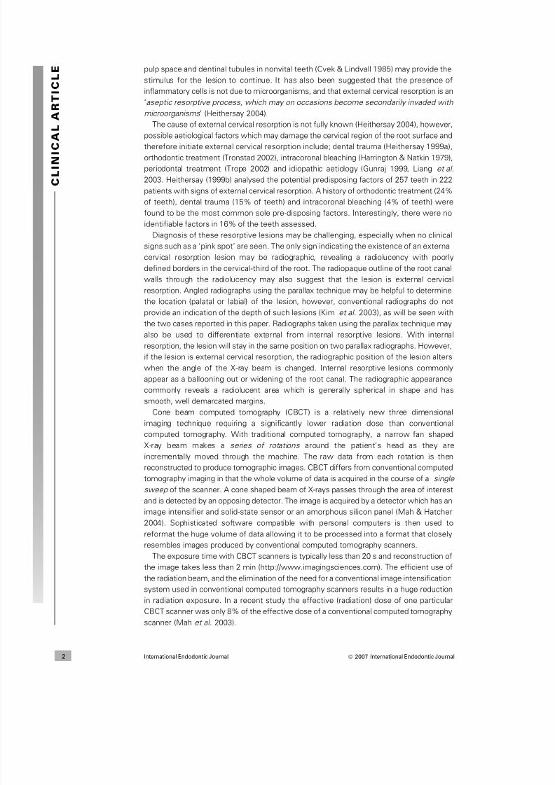
Introduction
External cervical resorption is initiated by damage to the cementum immediately belowthe epithelial attachment allowing osteoclasts to colonize the damaged portion of the root.
The pathogenesis of external cervical resorption is poorly understood. It has been reported
that microorganisms either from the gingival sulcus (Tronstad 1988, Trope 2002), or the
doi:10.1111/j.1365-2591.2007.01247.x
Correspondence: Shanon Patel, 45 Wimpole Street, London W1G 8SB, UK (Tel.: +44 20 7935
0080; fax: +44 20 7935 1181; e-mail: [email protected]).
ª 2007 International Endodontic Journal International Endodontic Journal 1
8/3/2019 Patel_2007_The Use of Cone Beam
http://slidepdf.com/reader/full/patel2007the-use-of-cone-beam 2/8
pulp space and dentinal tubules in nonvital teeth (Cvek & Lindvall 1985) may provide the
stimulus for the lesion to continue. It has also been suggested that the presence of
inflammatory cells is not due to microorganisms, and that external cervical resorption is an
‘aseptic resorptive process, which may on occasions become secondarily invaded with
microorganisms ’ (Heithersay 2004).
The cause of external cervical resorption is not fully known (Heithersay 2004), however,
possible aetiological factors which may damage the cervical region of the root surface andtherefore initiate external cervical resorption include; dental trauma (Heithersay 1999a),
orthodontic treatment (Tronstad 2002), intracoronal bleaching (Harrington & Natkin 1979),
periodontal treatment (Trope 2002) and idiopathic aetiology (Gunraj 1999, Liang et al.
2003. Heithersay (1999b) analysed the potential predisposing factors of 257 teeth in 222
patients with signs of external cervical resorption. A history of orthodontic treatment (24%
of teeth), dental trauma (15% of teeth) and intracoronal bleaching (4% of teeth) were
found to be the most common sole pre-disposing factors. Interestingly, there were no
identifiable factors in 16% of the teeth assessed.
Diagnosis of these resorptive lesions may be challenging, especially when no clinical
signs such as a ‘pink spot’ are seen. The only sign indicating the existence of an external
cervical resorption lesion may be radiographic, revealing a radiolucency with poorly
defined borders in the cervical-third of the root. The radiopaque outline of the root canal
walls through the radiolucency may also suggest that the lesion is external cervical
resorption. Angled radiographs using the parallax technique may be helpful to determine
the location (palatal or labial) of the lesion, however, conventional radiographs do not
provide an indication of the depth of such lesions (Kim et al. 2003), as will be seen with
the two cases reported in this paper. Radiographs taken using the parallax technique may
also be used to differentiate external from internal resorptive lesions. With internal
resorption, the lesion will stay in the same position on two parallax radiographs. However,
if the lesion is external cervical resorption, the radiographic position of the lesion alters
when the angle of the X-ray beam is changed. Internal resorptive lesions commonly
appear as a ballooning out or widening of the root canal. The radiographic appearance
commonly reveals a radiolucent area which is generally spherical in shape and has
smooth, well demarcated margins.
Cone beam computed tomography (CBCT) is a relatively new three dimensionalimaging technique requiring a significantly lower radiation dose than conventional
computed tomography. With traditional computed tomography, a narrow fan shaped
X-ray beam makes a series of rotations around the patient’s head as they are
incrementally moved through the machine. The raw data from each rotation is then
reconstructed to produce tomographic images. CBCT differs from conventional computed
tomography imaging in that the whole volume of data is acquired in the course of a single
sweep of the scanner. A cone shaped beam of X-rays passes through the area of interest
and is detected by an opposing detector. The image is acquired by a detector which has an
image intensifier and solid-state sensor or an amorphous silicon panel (Mah & Hatcher
2004). Sophisticated software compatible with personal computers is then used to
reformat the huge volume of data allowing it to be processed into a format that closely
resembles images produced by conventional computed tomography scanners.
The exposure time with CBCT scanners is typically less than 20 s and reconstruction of
the image takes less than 2 min (http://www.imagingsciences.com). The efficient use of
the radiation beam, and the elimination of the need for a conventional image intensification
system used in conventional computed tomography scanners results in a huge reduction
in radiation exposure. In a recent study the effective (radiation) dose of one particular
CBCT scanner was only 8% of the effective dose of a conventional computed tomography
scanner (Mah et al. 2003).
C L I N I C A
L
A R T I C L E
International Endodontic Journal ª 2007 International Endodontic Journal2
8/3/2019 Patel_2007_The Use of Cone Beam
http://slidepdf.com/reader/full/patel2007the-use-of-cone-beam 3/8
This clinical article demonstrates the use of CBCT as an effective diagnostic tool for the
assessment and management of external cervical resorption in two clinical cases.
Case 1
A 32-year-old Caucasian male was referred by his general dental practitioner for the
management of external cervical resorption. The general dental practitioner had noted aradiolucency on a dental panoramic tomograph which was taken as part of a new patient
assessment. No other radiolucent lesions were detected in the patient’s remaining teeth.
On presentation, the patient had no symptoms. He attended the dentist regularly and
had a course of orthodontic treatment in his early teenage years. His medical history was
unremarkable.
Clinical examination revealed a minimally restored dentition. Periodontal probing depths
were no greater that 2 mm. The maxillary and mandibular right posterior teeth all
responded positively to thermal and electrical sensitivity testing. The mandibular right
second premolar tooth was unrestored (Fig. 1a).
A periapical radiograph of the mandibular right quadrant revealed a 4 mm diameter
radiolucency in the cervical region of the root of the mandibular right second premolar. The
lesion had well defined borders and the root canal was visible through the lesion
suggesting that the lesion was external to the root canal (Fig. 1b).
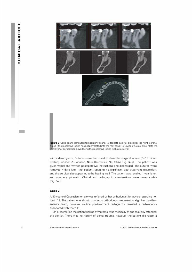
With the patient’s consent a 20 s, 120 Kv, 5 mA CBCT scan (i-CAT; Imaging Sciences
International, Hatfield, PA, USA) was taken.
The cross sectional images through the area of interest confirmed the presence of a
well defined uniformly radiolucent lesion lying on the cervical-third of the root of the
mandibular right second premolar. Interestingly, the presence of a thin buccal cortical
plate overlying the lesion was also noted. The lesion did not appear to be in
communication with the root canal (Fig. 2a–c).
After discussing the various treatment options with the patient, it was decided to
surgically repair the defect. The patient was given a preoperative chlorohexidne
mouthwash. A triangular full mucoperiosteal flap was raised with a relieving incision
mesial to the mandibular right canine. The cortical plate overlying the lesion was
removed with a surgical bur under copious irrigation to reveal a resorption cavity.Granulamatous tissue was completely excavated from the cavity, and the defect was
repaired with glass ionomer cement (Fuji LC glass ionomer cement; GC America Inc.,
Alsip, IL, USA). The margins of the restoration were polished with a fine diamond
polishing bur. The flap was repositioned and held under gentle pressure for 10 min
(a) (b)
Figure 1 (a) Buccal view of the lower right quadrant; (b) radiograph of lower right quadrant revealing a
cervical radiolucency on the lower right premolar.
CL I NI CAL
AR T I CL E
ª 2007 International Endodontic Journal International Endodontic Journal 3
8/3/2019 Patel_2007_The Use of Cone Beam
http://slidepdf.com/reader/full/patel2007the-use-of-cone-beam 4/8
with a damp gauze. Sutures were then used to close the surgical wound (5–0 Ethicon
Proline; Johnson & Johnson, New Brunswick, NJ, USA) (Fig. 3a–d). The patient was
given verbal and written postoperative instructions and discharged. The sutures were
removed 4 days later, the patient reporting no significant post-treatment discomfort,
and the surgical site appearing to be healing well. The patient was recalled 1 year later,
and was asymptomatic. Clinical and radiographic examinations were unremarkable
(Fig. 3e,f).
Case 2
A 37-year-old Caucasian female was referred by her orthodontist for advice regarding her
tooth 11. The patient was about to undergo orthodontic treatment to align her maxillary
anterior teeth, however routine pre-treatment radiographs revealed a radiolucency
associated with tooth 11.
On presentation the patient had no symptoms, was medically fit and regularly attended
the dentist. There was no history of dental trauma, however the patient did report a
(a)
(c)
(b)
Figure 2 Cone beam computed tomography scans. (a) top left, sagittal slices; (b) top right, coronalslices – the resorptive lesion has not perforated into the root canal; (c) lower left, axial slice. Note the
thin layer of cortical bone overlaying the resorptive lesion (yellow arrows).
C L I N I C A
L
A R T I C L E
International Endodontic Journal ª 2007 International Endodontic Journal4
8/3/2019 Patel_2007_The Use of Cone Beam
http://slidepdf.com/reader/full/patel2007the-use-of-cone-beam 5/8
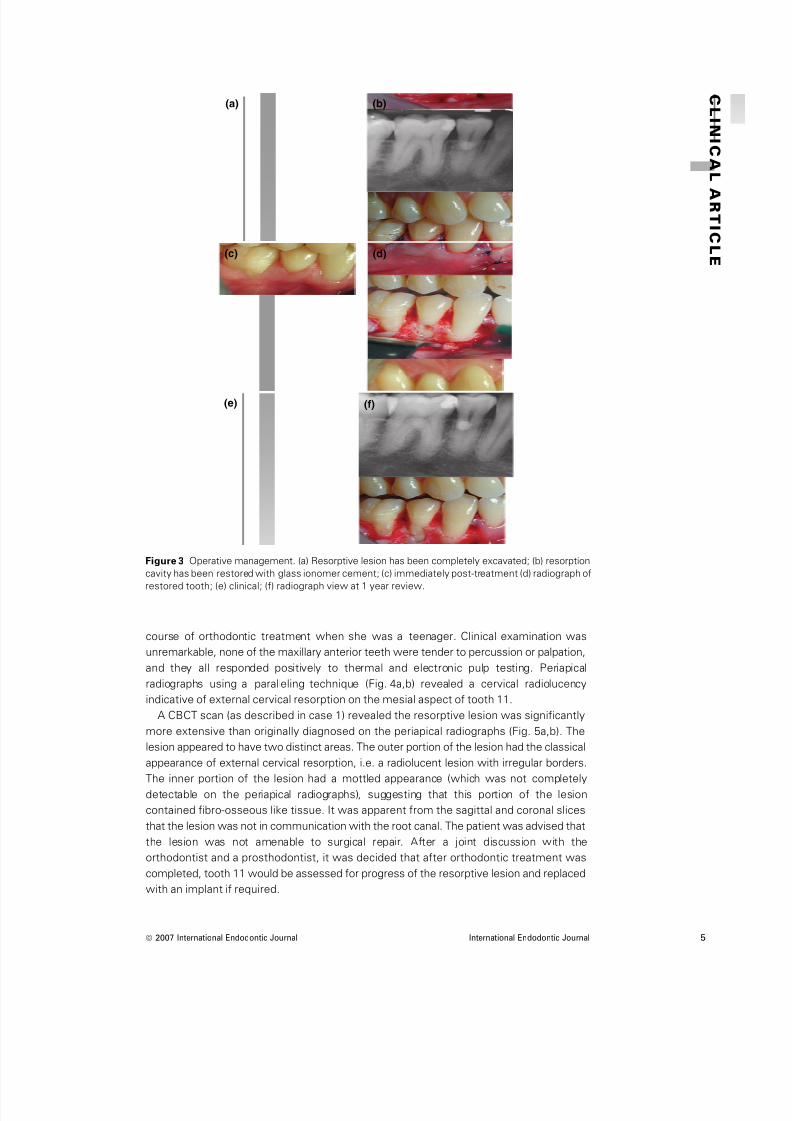
course of orthodontic treatment when she was a teenager. Clinical examination was
unremarkable, none of the maxillary anterior teeth were tender to percussion or palpation,
and they all responded positively to thermal and electronic pulp testing. Periapical
radiographs using a paralleling technique (Fig. 4a,b) revealed a cervical radiolucency
indicative of external cervical resorption on the mesial aspect of tooth 11.
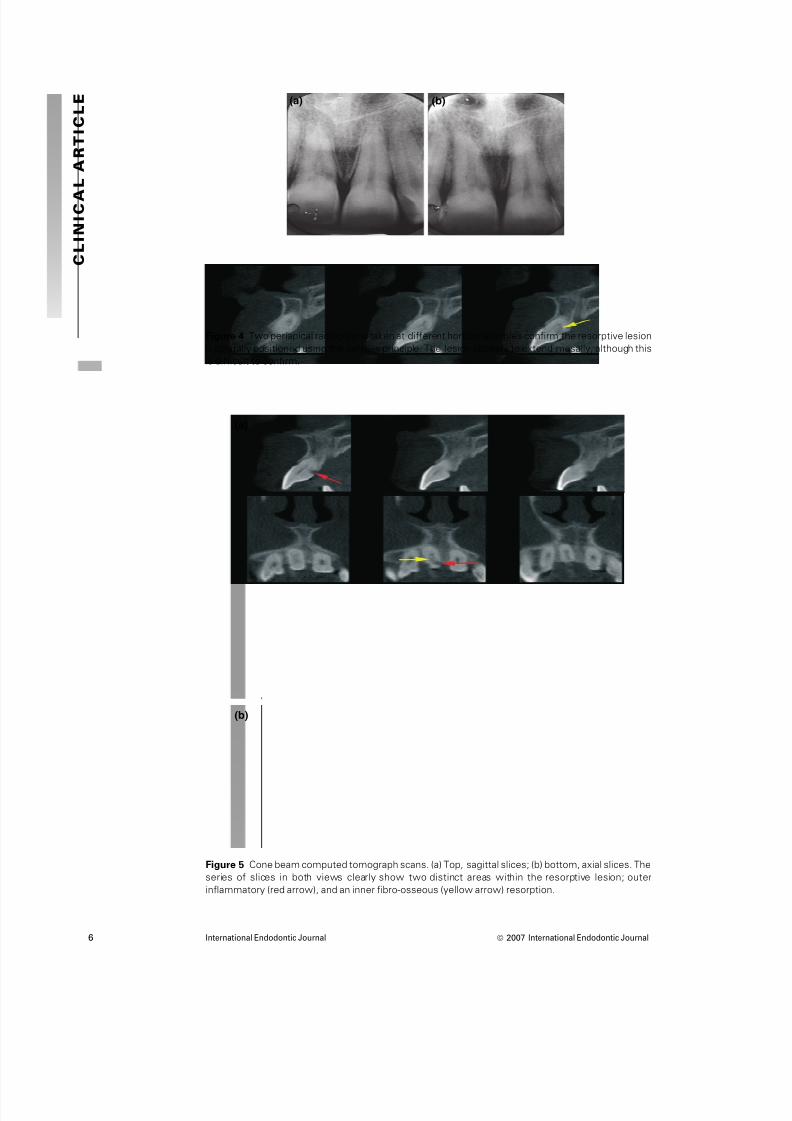
A CBCT scan (as described in case 1) revealed the resorptive lesion was significantly
more extensive than originally diagnosed on the periapical radiographs (Fig. 5a,b). The
lesion appeared to have two distinct areas. The outer portion of the lesion had the classical
appearance of external cervical resorption, i.e. a radiolucent lesion with irregular borders.
The inner portion of the lesion had a mottled appearance (which was not completely
detectable on the periapical radiographs), suggesting that this portion of the lesion
contained fibro-osseous like tissue. It was apparent from the sagittal and coronal slices
that the lesion was not in communication with the root canal. The patient was advised that
the lesion was not amenable to surgical repair. After a joint discussion with the
orthodontist and a prosthodontist, it was decided that after orthodontic treatment was
completed, tooth 11 would be assessed for progress of the resorptive lesion and replaced
with an implant if required.
CL I NI CAL
AR T I CL E
(a) (b)
(c)
(e) (f)
(d)
Figure 3 Operative management. (a) Resorptive lesion has been completely excavated; (b) resorption
cavity has been restored with glass ionomer cement; (c) immediately post-treatment (d) radiograph of
restored tooth; (e) clinical; (f) radiograph view at 1 year review.
ª 2007 International Endodontic Journal International Endodontic Journal 5
8/3/2019 Patel_2007_The Use of Cone Beam
http://slidepdf.com/reader/full/patel2007the-use-of-cone-beam 6/8
C L I N I C A
L
A R T I C L E (a) (b)
Figure 4 Two periapical radiographs taken at different horizontal angles confirm the resorptive lesion
is palatally positioned using the parallax principle. The lesion appears to extend mesally, although this
is difficult to confirm.
(a)
(b)
Figure 5 Cone beam computed tomograph scans. (a) Top, sagittal slices; (b) bottom, axial slices. The
series of slices in both views clearly show two distinct areas within the resorptive lesion; outer
inflammatory (red arrow), and an inner fibro-osseous (yellow arrow) resorption.
International Endodontic Journal ª 2007 International Endodontic Journal6

8/3/2019 Patel_2007_The Use of Cone Beam
http://slidepdf.com/reader/full/patel2007the-use-of-cone-beam 7/8
Discussion
Cone beam computed tomography was found to be particularly useful in the diagnosis of
the external cervical resorption lesions reported here. In both cases, the position, depth in
relation to the root canal and ultimately the restorability of the tooth was assessed
objectively before any treatment was carried out. This allowed the operator to be
confident of the best strategy for managing the defects and gave a better impression ofprognosis. Although the exposure time of the CBCT scan was 20 s, its pulsed nature gives
an actual equivalent exposure time of just 4.6 s. The effective dose of the i-CAT cone
beam computed tomograph scanner used for these two cases is 69 lSv (Ludlow et al.
2006). This is in the same order of magnitude as conventional radiographs (Mah et al.
2003). Of particular relevance to endodontics is the ‘limited’ CBCT scanner (3D
Accuitomo; J Morita Mfg. Corp, Kyoto, Japan) which is specifically designed to capture
information from a small region of the maxilla or mandible. The image field is similar in size
to a conventional periapical dental X-ray film. The effective dose of the 3D Accuimoto is
equivalent to 2–3 standard periapical exposures (Arai et al. 2001).
Considerable information was gained from each volumetric scan, as the acquired data
images could be easily generated in any plane (for example, coronal and axial planes) using
the available software. Both patients found the CBCT images extremely helpful in
understanding their endodontic problems.
Heithersay (1999c) described managing external cervical resorption defects using a
flapless technique. He directly applied trichloracetic acid to resorptive lesions, curetted out
the resorption tissue and then restored the cavity with a plastic restoration. In case 1, a
flapless technique could not be used as the CBCT scans clearly showed the presence of a
thin layer of cortical bony plate overlying the resorption cavity. The prognosis of case 1 is
excellent as the resorption defect was diagnosed at a relatively early stage and managed
swiftly. Left untreated, the lesion may eventually have spread horizontally and vertically
through the root and become untreatable. In case 2, it was interesting to note the true
nature of the lesion was only visible in sagittal and axial sections of the reconstructed data
from the CBCT. The scans clearly demonstrated that the resorption lesion had burrowed
extensively towards the root canal in an apical and to a lesser extent in a coronal direction
from the main (outer) lesion. This was not clearly detectable on the periapical radiographs.This previously undiagnosed inner portion of the resorptive lesion resulted in the lesion
being untreatable.
Conclusion
Cone beam computed tomography is a useful addition to the endodontist’s armamen-
tarium for the management of complex endodontic problems. The lower radiation dosages
compared with traditional CT scanners and geometric accuracy of scanned objects (Sonick
et al. 1994, Mozzo et al. 1998) makes CBCT ideal for treatment planning periapical
surgery, diagnosis of dento-alveolar trauma (Terakoda et al. 2000) and in certain
circumstances the diagnosis of radiolucent apical lesions (Simon et al. 2006, Lofthag-
Hansen et al. 2007).
Disclaimer
Whilst this article has been subjected to Editorial review, the opinions expressed, unless
specifically indicated, are those of the author. The views expressed do not necessarily
represent best practice, or the views of the IEJ Editorial Board, or of its affiliated Specialist
Societies.
CL I NI CAL
AR T I CL E
ª 2007 International Endodontic Journal International Endodontic Journal 7
8/3/2019 Patel_2007_The Use of Cone Beam
http://slidepdf.com/reader/full/patel2007the-use-of-cone-beam 8/8
Acknowledgements
Cavendish Imaging for their help and advice in the preparation of this article.
References
Arai Y, Honda K, Iwai K, Shinoda K (2001) Practical model ‘3DX’ of limited cone-beam X-ray CT fordental use. International Congress Series 1230, 713–8.
Cvek M, Lindvall A-M (1985) External root resorption following bleaching of pulpless teeth with
oxygen peroxide. Endodontics and Dental Traumatology 1, 56–60.
Gunraj MN (1999) Dental root resorption. Oral Surgery, Oral Medicine, Oral Pathology, Oral Radiology,
and Endodontics 88, 647–53.
Harrington GW, Natkin E (1979) External resorption associated with the bleaching of pulpless teeth.
Journal of Endodontics 5, 344–8.
Heithersay GS (1999a) Clinical, radiographic, and histopathologic features of invasive cervical
resorption. Quintessence 30, 27–37.
Heithersay GS (1999b) Invasive cervical resorption: an analysis of potential predisposing factors.
Quintessence 30, 83–95.
Heithersay GS (1999c) Treatment of Invasive cervical resorption: an analysis of the results
using topical application of trichloracetic acid, curettage, and restoration. Quintessence 30, 96–
110.
Heithersay GS (2004) Invasive cervical resorption. Endodontic Topics 7, 73–92.
Kim E, Kim K-D, Roh B-D, Cho Y-S, Lee S-J (2003) Computed tomography as a diagnostic aid for
extracanal invasive resorption. Journal of Endodontics 29, 463–5.
Liang H, Burkes EJ, Frederiksen NL (2003) Multiple idiopathic cervical root resorption: systematic
review and report of four cases. Dentomaxillofacial Radiology 32, 150–5.
Lofthag-Hansen S, Huumonen S, Grondahl K, Grondahl H-G (2007) Limited cone-beam CT and intraoral
radiography for the diagnosis of periapical pathology. Oral Surgery, Oral Medicine, Oral Pathology,
Oral Radiology, and Endodontology 103, 114–9.
Ludlow JB, Davies-Ludlow LE, Brooks SL, Howerton WB (2006) Dosimetry of 3 CBCT devices for oral
and maxillofacial radiology: CB Mercuray, NewTom 3G and i-CAT. Dentomaxillofacial Radiology 35,
219–26.
Mah J, Hatcher D (2004) Three-dimensional craniofacial imaging. American Journal of Orthodontics
and Dentofacial Orthopedics 126, 308–9.Mah J, Danforth RA, Bumann A, Hatcher D (2003) Radiation absorbed in maxillofacial imaging with a
new dental computed tomography device. Oral Surgery, Oral Medicine, Oral Pathology, Oral
Radiology, and Endodontics 96, 508–13.
Mozzo P, Procacci C, Tacconi A, Tinazzi Martini P, Bergamo Andreis IA (1998) A new volumetric CT
machine for dental imaging based on the cone-beam technique: preliminary results. European
Radiology 8, 1558–64.
Simon JHS, Enciso R, Malfaz J-M, Roges R, Bailey-Perry M, Patel A (2006) Differential diagnosis of
large periapical lesions using cone-beam computed tomography measurements and biopsy. Journal
of Endodontics 32, 833–7.
Sonick M, Abrahams J, Faiella RA (1994) A comparison of the accuracy of periapical, and
computerized tomographic radiographs in locating the mandibular canal. The International Journal
of Oral and Maxillofacial Implants 9, 455–60.
Terakoda M, Hashimoto K, Arai Y, Honda M, Sekiwa T, Sato H (2000) Diagnostic imaging with newly
developed ortho cubic super-high resolution computed tomography (Ortho-CT). Oral Surgery, Oral
Medicine, Oral Pathology, Oral Radiology, and Endodontology 89, 509–18.
Tronstad L (1988) Root resorption-etiology, terminology and clinical manifestations. Endodontics and
Dental Traumatology 4, 241–52.
Tronstad L (2002) Endodontic Aspects of Root Resorption. Clinical Endodontics , 2nd edn. Stuttgart,
Germany: Thieme.
Trope M (2002) Root resorption due to dental trauma. Endodontic Topics 1, 79–100.
C L I N I C A
L
A R T I C L E
International Endodontic Journal ª 2007 International Endodontic Journal8