Embed Size (px)
Citation preview

0165-4608/00/$–see front matterPII S0165-4608(99)00187-9
Cancer Genet Cytogenet 118:121–131 (2000)
Elsevier Science Inc., 2000. All rights reserved.655 Avenue of the Americas, New York, NY 10010
A FISH Study of Variant Philadelphia Rearrangements
K. S. Reddy and V. Sulcova
ABSTRACT:
A total of 39 variant Philadelphia (Ph) translocations were studied by fluorescence in situhybridization (FISH) using MBCR/ABL, mBCR/ABL, or DBCR/ABL probes. Seven cases did not have aBCR/ABL fusion signal. Of a total of 32 fusion-positive cases, 5 were simple variants involving chromo-some 22 and another chromosome apart from chromosome 9; 23 were complex variants involving chro-mosomes 22, 9, and a third chromosome (18 cases), or 22, 9, and two other chromosomes (4 cases).Masked Ph rearrangements were detected in 4 cases. One case was a Ph chromosome mimic. Fluores-cence in situ hybridization has become a widely used method for studying Ph rearrangements. The lat-est probe that is being used is the DBCR/ABL (double reciprocal BCR/ABL signals). The expected pat-tern for this probe is one green ABL signal (1G) on the normal 9, one red BCR signal (1R) on the normal22, and two fusion signals, BCR/ABL and ABL/BCR (2F), on a derivative 22 and a derivative 9, respec-tively. Deviant patterns from 1G1R2F, and sometimes 1G1R2F, were indicative of a variant, as long asthere was a fusion signal. However, in interphase analysis, it is not possible to visualize a variant rear-rangement, and when a deviant pattern involving at least one fusion signal is observed, the followingpossibilities should be contemplated. The different patterns observed in fifteen Ph variants aredescribed. The patterns observed in variants studied with the DBCR/ABL probe were 2G2R1F (40%),1G1R2F (20%), 1G1R1F (20%), 1G2R1F (13.3%), and 2G1R1F (6.66%). A single mechanism is involvedin the formation of each of these patterns. A 2G2R1F, FISH pattern in 6 cases appears to involve a sin-gle concerted event of simultaneous breaks on the participating chromosomes followed by mismatchedjoining. The three cases with 1G1R2F most probably arose by two sequential rearrangements. The1G1R1F pattern suggests that either the BCR and ABL breakpoints are different, or there are deletions atthe breakpoints, because residual signals are not observed. Two independent events appear to beinvolved in 1G2R1F with a reverse cryptic 9,22 rearrangement as the first event. In one case of 2G1R1F,the plausible explanation is an insertion of ABL next to BCR and either a simultaneous or a sequentialtranslocation with another chromosome. © Elsevier Science Inc., 2000. All rights reserved.
INTRODUCTION
The Philadelphia (Ph) chromosome [1] results from atranslocation (9;22)(q34;q11.2) [2] involving a fusion ofthe Abelson oncogene (
ABL
) from chromosome 9q34 withbreakpoint cluster region (
BCR
) on 22q11.2 [3]. In chronicmyeloid leukemia (CML) patients, the breakpoints occurin
BCR
within a 5.8-kb region known as major
BCR(MBCR)
[3, 4]. Fifty percent of acute lymphoblastic leuke-mia (ALL) cases with a Ph chromosome also have a break-point within the major
BCR
(
MBCR
) and in the other 50%,the breakpoint is in the more proximal minor
BCR
(
mBCR
)[5]. About 5% of Ph rearrangements are variants [6, 7].When a segment from 22q11.2 is translocated to anotherchromosome, it constitutes a simple variant and a com-plex variant translocation involves three or more chromo-
somes [8]. Based on the data to date, variant Ph rearrange-ments are thought to have the same diagnostic andprognostic implications as the simple t(9;22). In 5–10% ofCML cases, a Ph chromosome is not present [8]. Abouthalf of these patients have a submicroscopic cryptic rear-rangement [9]. One such case (case 30) included in thisstudy has been previously reported [10].
Reverse transcriptase-polymerase chain reaction (RT-PCR) [11] and fluorescence in situ hybridization (FISH)using ABL and BCR probes labeled in different coloredfluorophores [12, 13] have made it possible to detect a
BCR/ABL
fusion and to confirm a variant Ph rearrange-ment. Conventional FISH methods with different coloredBCR and ABL probes detect a single
BCR/ABL
fusion sig-nal on the Ph chromosome (S-FISH). This probe washighly accurate for analysis of metaphases but was impre-cise for the study of interphase cells, because
BCR
and
ABL
signals coincidentally overlap in about 4% of normalnuclei (false positive) and can be incorrectly scored [14].To overcome these limitations, a D-BCR/ABL probe thatshows double
BCR/ABL
fusion, one on the abnormal 9and one on the Ph chromosome, was commercially intro-
From the Cytogenetic Laboratory, Quest Diagnostics Inc.,Nichols Institute, San Juan Capistrano, California, USA.
Address reprint requests to: Dr. Kavita S. Reddy, CytogeneticsLaboratory, Quest Diagnostics Inc., San Juan Capistrano, CA92690.
Received June 25, 1999; accepted August 25, 1999.

122
K. S. Reddy and V. Sulcova
duced. Dewald et al. [15] tested this probe on interphasenuclei and concluded that D-FISH can detect the Ph chro-mosome and its variant translocations accurately.
In this study, FISH was performed on a large numberof variants to confirm the Ph rearrangement. The distri-bution of the various types of variants is determined. TheD-BCR/ABL probe allows us to understand the diversemechanisms that are probably involved in the formationof these variants. This understanding allows for a betterinterpretation of deviant signal pattern observed, particu-larly in interphase, and also in metaphase.
MATERIALS AND METHODS
Unstimulated bone marrow or peripheral blood cultureswere harvested after 24 and 48 hours. The slides wereGTG-banded.
Fluorescence in situ hybridization was performed withthe probes MBCR/ABL, mBCR/ABL, and DBCR/ABL (On-cor Inc.), according to the manufacturer’s instructions,with minor modifications.
BCR
was directly labeled witha red fluorophore,
ABL
with a green fluorophore, and thefusion signal was yellow (red and green). The unbandedslides were denatured at 70
8
C for 2 minutes. After over-
night hybridization at 37
8
C in a humid chamber, the slideswere washed in 0.5
3
SSC at 72
8
C for 5 minutes.In this study, the variant Ph rearrangements were
probed with commercially available MBCR/ABL ormBCR/ABL (Oncor Inc.). The MBCR/ABL probe detectedrearrangements involving the major
BCR
cluster and themBCR/ABL probe detected rearrangements involving bothmajor and the minor breakpoints. The DBCR/ABL probe(Oncor Inc.) detected fusion at
MBCR
or
mBCR
and wasformatted to show fusion signals on both the derivativechromosomes 9 and 22 in t(9;22). The FISH hybridizationpattern in cases with t(9;22)(q34;q11.2) with MBCR/ABLand mBCR/ABL probes was 1G1R1F (i.e., one green ABLsignal [1G] on a normal chromosome 9, one red BCR sig-nal [1R] on a normal 22, and one red
1
green or yellow
BCR/ABL
fusion signal [1F] on the derivative 22). TheDBCR/ABL probe gives a 1G1R2F hybridization pattern.The additional fusion signal is from the reciprocal
ABL/BCR
fusion on the derivative 9 (Fig. 1). The FISH hybridization signal pattern in each variant
case was noted. The G-banded and FISH findings werecompared to assign the signals to the chromosomes in-volved. The hybridization patterns were used to providemolecular confirmation of the chromosome rearrange-
Figure 1 A G-banded partial karyotype of a Ph translocation (9;22)(q34;q11). The FISH patterns are 1G1R1F or1W1B1B/W with the mBCR/ABL probe and 1G1R2F or 1W1B2B/W with the DBCR/ABL probe. The partial karyo-type and ideogram represents the rearrangements with the normal chromosome to the left and the rearranged chro-mosome to the right. In the ideogram, the breakpoints are shown on the normal homolog (arrow) and the abnormalchromosome shows the translocated segments (black bar with the chromosome number). The signals are shown onthe chromosomes (arrow and abbreviated signal color). The FISH metaphases show the chromosomes with thehybridization signals (arrow and chromosome number). In the black and white image, the red signal appears asblack, the green signal as white, and the fusion signal as black/white.

FISH Study of Ph Translocations
123
ments in the Ph variants. The
DBCR/ABL
cases were clas-sified based on the hybridization pattern (Tables 1 and 2).
RESULTS
Tables 1 and 2 summarize the Ph variants and masked Phrearrangement cases studied with BCR/ABL probes. Be-tween 1994 and 1998, 39 cases were studied using FISHand BCR/ABL probes to confirm a variant or masked Phrearrangement detected in cytogenetic studies or becauseof a diagnosis of CML. Thirty-two cases had a
BCR/ABL
fusion signal and 7 did not have a fusion signal. The meanage of the
BCR/ABL
fusion-positive cases was 50
6
17.35years. Twelve were female and 20 were male. There weremore males with Ph variants than females, and this differ-ence was statistically significant (chi-square
5
2 is signifi-cant at the 0.05 level). Of the seven fusion-negative cases,four involved 9q34, two involved 22q11.2, and in one case,cytogenetic analysis found 1/30 cells with a t(9;22;20)(q34;q11.2;q13) (case 15 in Table 1); yet a fusion signalcould not be detected using FISH.
Among 32 cases with a
BCR/ABL
fusion signal, 5 weresimple variants (cases 8, 9, 16, 18, and 20), 23 were com-plex variants, and 4 were masked variants. Simple varianttranslocation appeared to involve 22q11.2 and anotherchromosome other than 9. A complex variant transloca-tion appeared to involve three or more chromosomes thatincludes 9, 22, and another chromosome or chromosomes.A majority of the complex variant translocations involvedthree chromosomes (i.e. 9, 22 and another chromosome).Four variants involved four chromosomes (cases 17, 19,24 and 29). The other chromosomes in addition to 22 and/or 9 involved in the variants and their breakpoints arelisted in Table 3. Of the four masked Ph translocations(cases 2, 6, 23, and 30), one had additional material on 22,another case was a pericentric inversion of 9, inv(9)(p22q34)(case 6), and two had normal karyotypes.
The FISH pattern for M or mBCR/ABL probes was1G1R1F for normal Philadelphia t(9;22)(q34;q11.2) (Fig. 1).The first 11 cases were studied using MBCR/ABL. Twocases were negative; these cases may have involved theminor breakpoint not detected by this probe, or may lackthe Ph rearrangement. Using mBCR/ABL, 14 variants were
Table 1
Ph variant chromosomes characterized using FISH and MBCR/ABL or mBCR/ABL probes
Serialno.
Age(years) Diagnosis Karyotypes Probe(s) Hybridization pattern
1 43 Granulohyperplasia 46,XY,t(9;22)(q34;q11.2)t(9;15)(q22;q13) MBCR/ABL 1G1R1F (smear)2 74 CML 46,XY,add(22)(q11.2) MBCR/ABL 1G1R1F3 58 CML vs. MPD 46,XY,t(4;22)(q12;q11.2)/46,XY MBCR/ABL Fusion-negative4 68 Leukocytosis 46,XY,t(9;22;14)(q34;q11.2;q32) MBCR/ABL 1G1R1F5 31 CML 46,XY,?t(9;22;19)(q34;q11.2;p13) MBCR/ABL 1G1R1F[der22 or der19]6 41 CML 46,XX,inv(9)(p22q34) MBCR/ABL 1G1R1F[22]7 26 CML 46,XY,t(9;22;13)(q11;q?11.2;q11),inc/46,XY MBCR/ABL 1G[9]1R[22]1F[der22]8 60 CML 46,XY,t(11;22)(q13;q11.2) MBCR/ABL 1G[9]1R[22]1F[der22]9 46 CML 46,XX,t(10;22)(q22;q11.2) MBCR/ABL 1G[9]1R[22]1F[22]
10 82 Anemia,Thrombocytopenia
46,XY,t(?;9;12)(?;q34.1;q15) MBCR/ABL Fusion-negative 2G[9,der9]2R[22,22]
11 45 CML 46,XY,der(9;22)t(9;22;?)(q34;q11.2;?),
2
15,der(21)t(?15;21)(q22;q22),
1
marMBCR/ABL 1G[9]1R[22]1F[der22]
12 49 Lymphoma 46,XY,t(9;22;3)(q34;q11.2;p22) MBCR/ABL,mBCR/ABL
1G1R1F in interphases
13 36 AML 48,XY,
1
6,t(7;9)(q22;q34),
1
8/46,XY mBCR/ABL Fusion-negative 2G2R14 49 — 42,XX,del(9)(q13q34),del(12)(p11),
2
13,
2
13,
2
14,
2
15/46,XXmBCR/ABL Fusion-negative 1G2R
15 48 Ph Evaluation 46,XX,t(9;22;20)(q34;q11.2;q13)/46,XX mBCR/ABL Fusion-negative 2G2R16 86 MPD 46,XY,t(22;22)(q11.2;q13) mBCR/ABL 1G[9]1R[add(22)]1F[der22]17 74 — 46,XX,t(6;9;22;11)(p21.3;q34;q11.2;q13)add
(11)(p11)mBCR/ABL 1G[9]1R[22]1F[der22]
18 65 — 46,XY,t(17;22)(q25;q11.2)[10]/45,XY,der(1)t(1;4)(p11;q11),add(2)(p21),del(3)(p12),del(4)(q11),del(7)(p11),
2
12,add(13)(q32),
2
14,add(16)(q22),
2
17,
2
17,
2
18,
1
add(19)(q13),der(22)t(14;22)(q11;q13),
1
mar1,
1
mar2,
1
mar3
MBCR/ABL andmBCR/ABL
88/166 interphases 1R1G1F81/153 interphases 1R1G1F
19 46 CML 46,XY,t(9;22;15;21)(q34;q11.2;q15;q11.2) mBCR/ABL 1G[9]1R[22]1F[der22]20 33 — 46,XY,t(5;22)(q35;q11.2) mBCR/ABL 1G[9]1R[22]1F[9]21 53 — 44
,
46,XY,add(7)(p22),add(9)(q34),
2
11,
2
12,
2
16,
2
16,
2
17,
1
21,
1
mar1,
1
mar2,
1
mar3,
1
mar4/46,XY
mBCR/ABL Fusion-negative 2G2R
22 45 — 46,XX,t(9;22;12)(q34;11.2;p13) mBCR/ABL 1G[9]1R[22]1F[der22]23 63 R/o Ph 46,XX mBCR/ABL 1G[9]1R[22]1F[22]

124
K. S. Reddy and V. Sulcova
Table 2
Ph variant chromosomes characterized using FISH and DBCR/ABL probe
Serialno.
Age(years) Diagnosis Karyotypes Probe
Hybridization pattern Mechanism
2G2R1F24 51 — 46,XX,t(1;20;9;22;1)
(q25;q13;q34;q11.2;p35)
DBCR/ABL 2G[9,der9]2R[22,der1]1F[der22]
Five breaks and exchanges from 1 to 20 (G-bands), 20 to 9 (G-bands), 9 to 22 [based on a smaller green signal on der(9), and
BCR/ABL
fusion on der(22)] and 22 to 1 [a small red signal on der (1)].
25 49 — 46,XX,t(9;22;19)(q34;q11.2;q13.3)/46,XX
DBCR/ABL 2G[9,der9]2R[22,der19]1F[der22]
A 3-way translocation; 9 to 22 [based on a
BCR/ABL
fusion signal on der(22)] and a smaller green signal on der(9), 22 to 19 [based on a red signal on der (19)] and 19 to 9 (based on G-bands).
26 47 — 46,XX,t(9;22;6)(q34;q11.2;p21.3)
DBCR/ABL 2G[9,der9]2R[22,der6]1F[der22]
A 3-way translocation; 9 to 22 [based on a
BCR/ABL
fusion signal on 22 and a smaller green signal on der(9)], 22 to 6 [based on a small red signal on der(6)] and 6 to 9 (based on G-bands).
27 27 CML 46,XY,der(9;22;7)ins(7;22)(q11.2; q11q?12)t(9;22;7)(q34;q11.2;q11.2)
DBCR/ABL 2G[9,9]2R[22,der7]1F[der22]
An exchange from 9 to 22 [based on a
BCR/ABL
fusion signal on der(22) and a green signal on der(9)] and an insertion of 22 into 7 [based on a red signal on der(7)]. There was probably also a transfor of interstitial material from 7 to 9q34 (based on G-bands).
28 42 CML 46,XX,t(1;9;22)(q42;q34;q11.2)
DBCR/ABL 2G[9,der9]2R[22,1]1F[der22]
A 3-way exchange from 1 to 9 (based on G-bands), 9 to 22 [based on a
BCR/ABL
fusion signal on der(22) and a smaller green signal on der(9)] and 22 to 1 [based on a small red signal on der(1)].
29 65 — 46,XX,t(9;22;21;14)(q34;q11.2;q22.1;q32.3)/46,XX
DBCR/ABL 2G[9,der9]2R [der 21,22]1F[der22]
A 4-way translocation from 9 to 22 [based on a
BCR/ABL
fusion signal on der(22) and a green signal on der(9), 22 to 21 [based on a red signal on der(21)], 21 to 14 [based on an AML1 which maps to 21q22 probe signal on der(14)] and 14 to 9.
1G1R2F30 40 CML 46,XY mBCR/ABL
DBCR/ABL1G[9]1R[22]1F[9]
1G[9]1R[22]2F[22,9]
A Ph rearrangement was followed by a back translocation that places the
BCR/ABL
fusion signal on 9 or a cryptic exchange between 9 and 22 with reversed fusion exchange.
31 60 CML 46,XY,der(1)t(1;9;22)(p36.1;q34;q11.2)inv(1)(p36.1q12),der(9)t(1;9;22),der(22)t(1;9;22)/46,XY
DBCR/ABL 1G[9]1R[22]2F[der22,der1]
A complex rearrangement between 1;9;22 involving three events. A 9;22 translocation [shown by the
BCR/ABL
fusion signal on der(22)]. Then, a 9;1 translocation transferring the reciprocal fusion
ABL/BCR
signal from 9 to 1p36.1, followed by an inversion of the der(1) that places the reciprocal fusion signal in the q arm.
32 55 CML 46,XY,t(9;22)(q34;q11.2)/46,idem,add(4)(q31)/46,XY,t(9;22;14)(q34;q11.2;q24)
DBCR/ABL 1G[9]1R[22]2F[der22,der9]/1G [9]1R[22]2F[der22,der14]
The primary 9;22 Ph rearrangement was converted into a secondary complex variant by the transfer of the reciprocal fusion signal to the der(14).
(
continued
)

FISH Study of Ph Translocations
125
studied. Case 20 was shown to have a fusion signal onchromosome 9 and a translocation t(5;22). A case with apericentric inv(9)(p22q34) had a fusion signal on a normal22 (archived electronic pictures were corrupted) and twocases with a normal karyotype had a
BCR/ABL
fusion onan apparently normal 22 and 9 (Fig. 2).
The DBCR/ABL probe gave a 1G1R2F pattern for thenormal t(9;22)(q34;q11.2). Fifteen variants were studiedusing the DBCR/ABL probe. The patterns of hybridizationobserved in the variants studied with DBCR/ABL were2G2R1F (40%), 1G1R2F (20%), 1G1R1F (20%), 1G2R1F(13.33%), and 2G1R1F (6.66%). Table 2 gives the karyo-type and FISH hybridization pattern; the chromosomeswith signals are in parenthesis, and the mechanism bywhich they are formed is given.
For each of these patterns, there appears to be a com-mon underlying mechanism. The most frequent pattern ofhybridization, 2G2R1F, was observed in cases 24–29 (Ta-ble 3, Fig. 3A–F). The FISH pattern 2G2R1F confirmed the
G-banded rearrangement to be a complex translocationfrom one chromosome to the next. The 1G1R2F pattern ofhybridization was observed in three cases (cases 30–32)(Table 2). These variants appear to be formed by two se-quential events (Fig. 2, Fig. 4A–B). Three cases had1G1R1F (cases 33–35 in Table 2) pattern of hybridization(Fig. 5). The breakpoints in these 3 cases may be differentfrom those observed in the majority of t(9;22) because nei-ther
BCR
nor
ABL
residual signals were observed, or theremay be cryptic deletions at the breakpoints. The hybrid-ization pattern, 1G2R1F, was observed in two cases (cases36 and 37) (Table 2, Fig. 6). Case 36 appears to involve areverse 9;22 translocation. In case 37, the poor quality ofthe metaphases precluded any conclusion from beingdrawn about the mechanism by which the rearrange-ment occurred. A 2G1R1F pattern of hybridization ob-served in case 38 (Table 2) suggested that
ABL
was in-serted next to
BCR
(Fig. 7). A 2G1R pattern observed incase 39 (Table 2) was because of an interstitial deletion of
Table 2
Continued
Serialno.
Age(years) Diagnosis Karyotypes Probe
Hybridizationpattern Mechanism
1G1R1F33 53 R/o Ph 46,XX,t(9;22;17)
(q34;q11.2;q21)mBCR/ABL
DBCR/ABL1G[9]1R[22]1F[der22]1G[9]1R[22]1F[der22]
A 3-way translocation between 9, 22, and 17.The
BCR
and
ABL
breakpoints were probably different or had deletions at the breakpoints because no residual signals were observed on 9 (green) or 17 (red).
34 55 — 46,XX,t(9;22;17)(q34;q11.2;q21)
DBCR/ABL 1G[9]1R[22]1F[der22]
35 82 — 46,XY,t(1;9)(p13;q22),add(22)(q11.2)
DBCR/ABL 1G[9]1R[22]1F[add22]
No reciprocal fusion signal was observed. The additional material on the derivative 22 was due to an insertion of
ABL
. Either the breakpoint in
ABL
was different or there was a deletion, therefore the residual green signal was not detected.
1G2R1F36 41 — 46,XY,t(3;9)(p25;q34) DBCR/ABL 1G[9]2R[22,22]1F
[der3]A reverse cryptic 9,22 rearrangement
(smaller red signal on one 22). The fusion signal on 3 had probably been translocated from der(9).
37 3 Leukocytosis,30% blasts
45
z
46,XY,
2
2,
2
2,add(9)(q34),add (14)(q32),der(22)t(9;22)(q34;q11.2),
1
mar1,
1
mar2/46,XY
DBCR/ABL 1G[9]2R[22,?]1F[der22]
The poor quality of the metaphasesprecluded the assignment of the second red signal to a specific chromosome, and no conclusions could be drawn about the mechanism by which the rearrangement occurred.
2G1R1F38 35 R/o Ph 46,XY,der(9)ins(22;9)
(q11.2;q34q34)t(9;16)(q34;q22),der(22),ins(22;9)(q11.2;q34;q34),der(16)t(9;16)
DBCR/ABL 2G[9,der9]1R[22]1F[der22]
A smaller green signal on the der(9)suggests an insertion of
ABL
next to
BCR
and either a simultaneous or asequential translocation between 9and 16.
2G1R39 85 — 46,XY,
2
22,
1
mar DBCR/ABL 2G[9,9]1R[22] Fusion-negative
Probes BCR and D22S75 that map to q11.2 did not give a signal on the Ph-like chromosome, and STSW1-94 that maps to 22q13 gave a signal (Fig. 7). Therefore, there was an interstitial deletion of chromosome 22.
Abbreviations
: CML, chronic myelogenous leukemia; MPD, myeloproliferative disorder; AML, acute myeloid leukemia; r/o, rule out; –, no diagnosis given.

126
K. S. Reddy and V. Sulcova
chromosome 22 that gave the appearance of a Ph chrom-osome.
DISCUSSION
Fluorescence in situ hybridization has proven very usefulin characterizing variant and masked Ph rearrangements.
There were more males with Ph variants than females andthis difference was statistically significant (chi-square
5
2is significant at the 0.05 level), although in CML there wasno marked sex preponderance [8]. Some chromosomebands frequently involved in variants such as 11q13,12p13, and 17q25 [8] were also observed in the present co-hort of cases.
The DBCR/ABL probe has been found to be superior toMBCR or mBCR/ABL probes in deciphering variants.Cases with clinical presentation of CML and rearrange-ment involving 22q11.2 and/or 9q34 should be probedwith DBCR/ABL for the confirmation of a variant Ph trans-location. A marker that appears as a Ph chromosome incases with a normal chromosome 9 pair should be shownto have a
BCR/ABL
fusion, because it could be an intersti-tial deletion of chromosome 22. Normal karyotypes maymask Ph rearrangements. The fusion signal can be on 22or 9. In variants, the fusion can also be on other chromo-somes involved in the rearrangement apart from 9 and 22.Additional material on 22 may be an insertion of ABL,and warrants confirmation with BCR/ABL probe.
The Fitzgerald and Morris [6] study on variants con-cluded that complex translocations may arise from a sin-gle complex event, however, a minority of these translo-cations may be the result of serial translocations. TheD-FISH probe has allowed us to gain further insights intothe mechanisms involved in the formation of complex Phvariants. Complex variants formed by multiple simultaneous
Table 3
Chromosomes other than 9 and 22 involved in the Ph chromosome variants
Chromosome no. Breakpoints
1 p35, p36, p13, and q243 p22 and p255 q356 p21 and p21.37 q11.2
10 q2211 q13 (2 cases)12 p1313 q1114 q32, q24, and q32.315 q15 and q1316 q2217 q25 and q21 (2 cases)19 p13 and q13.320 q13 (2 cases)21 q11.2 and q22
Figure 2 Partial karyotypes of 46,XY,add(22)(q11.2) (case 2), 46,XX,inv(9)(p22q34), 46,XX (case 23), and 46,XY(case 30), with masked Philadelphia rearrangement. The BCR/ABL fusion signal is on 22 in cases 2, 6, and 23 andon 9 in case 30.

FISH Study of Ph Translocations
127
Figure 3
The DBCR/ABL probe hybridization pattern of 2G2R1F or 2W2B1B/W is found in cases 24–29. The Phvariants arise by simultaneous multiple breaks and mismatched fusion. The partial karyotype and ideogram repre-sent the rearrangement with the normal chromosome to the left and the rearranged chromosome to the right. Onthe ideogram the breakpoints are shown (arrow) on the normal homolog and the abnormal chromosome shows thetranslocated segment (black bar with chromosome number). The signals are shown on the chromosomes (arrowand the abbreviated signal color). The FISH metaphases show the chromosomes with the hybridization signals(arrow and chromosome number).

128
K. S. Reddy and V. Sulcova
Figure 4
The DBCR/ABL probe hybridization pattern of 1G1R2F or 1W1B2B/W is found in cases 31–32. Theserearrangements probably arise by multiple sequential events (event 1, 2, or 3). The partial karyotype and ideogramrepresent the rearrangement, with the normal chromosome to the left and the rearranged chromosome to the right.On the ideogram the breakpoints are shown (arrow) on the normal homolog and the abnormal chromosome showsthe translocated segment (black bar with chromosome number). The signals are shown on the chromosomes (arrowand the abbreviated signal color). The FISH metaphases show the chromosomes with the hybridization signals(arrow and chromosome number).

FISH Study of Ph Translocations
129
breaks and mismatched fusion is observed in about 53% ofthe variants in this study (cases 24–29, 33, and 34). Thestrongest evidence for serial stepwise rearrangements re-sulting in variant Ph rearrangements comes from the rareobservation of patients with a standard Ph translocation inone clone and a complex variant in another clone [16–18and present case 32]. Case 32 has one clone with a Phtranslocation with two FISH fusion signals for
BCR/ABL
and
ABL/BCR
on derivative 22 and 9, respectively. An-other clone has a complex Ph variant involving chromo-somes 9, 14, and 22. Cytogenetically, the variant is formedby a translocation between the derivative 9 [from thet(9;22)] and 14. The
DBCR/ABL
fusion signals on der(22)and der(9) in t(9;22) clone and on der(22) and der(14) int(9;22;14) clone suggest that the complex translocation isderived from the simple Ph translocation, and involvessuccessive steps rather than the alternative explanation ofindependent evolution of the two clones. When serial re-arrangements are involved in the formation of variants,they occur in rapid sequence, an interpretation supportedby the absence of the simple t(9;22) clone in most patientswith complex variants. If there was a time gap betweenthe t(9;22) and the formation of the complex variant, thevariant must have strong selective advantage over t(9;22)cells to be present in all abnormal cells. Because variants
are rarely formed as a clonal evolution change in blast cri-sis, and the prognosis for variants is no different then thet(9;22), significant genes may not be involved in their for-mation. Complex variant in cases 30, 32, and 36 are proba-bly two events in sequence, and in case 31, at least threeevents in close succession in one cell cycle.
The absence of residual signals in some complex trans-location (cases 33–35) may represent different breakpointinvolvement or deletion of
BCR
and
ABL
sequences adja-cent to the breakpoint, which is sometimes found in asso-ciation with the Ph translocation [19].
The Ph translocation in the vast majority of cases is asimple balanced t(9;22)(q34;q11.2). However, in rare casesvariants and masked Ph chromosome are detected. Themolecular characterization of these variants has allowedus to understand how they are formed. Cryptic Ph rear-rangements are rare in comparison to the visible balancedt(9;22)(q34;q11.2) and are observed in approximately 2.5–5% of CML cases [8]. A new finding is an inversion of 9involving q34 accompanied by cryptic Ph rearrangement(case 6).
In conclusion, any rearrangement involving 22q11.2 or9q34 should be investigated for a possible variant usingFISH. Chronic myeloid leukemia cases with a normalkaryotype should be evaluated for a masked
BCR/ABL
fu-
Figure 5 The DBCR/ABL probe hybridization pattern of 1G1R1F or 1W1B1B/W is found in cases 33–35. Theserearrangements could involve different breakpoints since BCR or ABL residual signals are not observed; or theymay involve deletions at the breakpoints. The partial karyotype and ideogram represent the rearrangement withthe normal chromosome to the left and the rearranged chromosome to the right. On the ideogram the breakpointsare shown (arrow) on the normal homolog and the abnormal chromosome shows the translocated segment (blackbar with chromosome number). The signals are shown on the chromosomes (arrow and the abbreviated signal color).The FISH metaphases show the chromosomes with the hybridization signals (arrow and chromosome number).
130
K. S. Reddy and V. Sulcova
sion by FISH or RT-PCR. The spectrum of hybridizationpatterns seen in Ph variants using the DBCR/ABL probeand the underlying mechanisms are described. A deviantinterphase FISH pattern with at least one fusion signal forthe DBCR/ABL probe is an indication of a variant Phtranslocation.
REFERENCES
1. Nowell PC, Hungerford DA (1960): A minute chromosome inhuman granulocytic leukemia. Science 132:1497.
2. Rowley JD (1990): A new consistent chromosomal abnormal-ity in chronic myelogenous leukemia identified by quina-crine fluorescence and Giemsa staining. Nature 243:290–293.
3. Heisterkamp N, Stephenson JR, Groffen J, Hansen PF, deKlien A, Bartram CR, Grosveld G (1983): Localization of thec-abl oncogene adjacent to a translocation breakpoint inchronic myelocytic leukemia. Nature 306:239–242.
4. Groffen J, Stephenson JR, Heisterkamp N, de Klien A, Bar-tram CR, Grosveld G (1984): Philadelphia chromosomal
breakpoints are clustered within a limited region, bcr, onchromosome 22. Cell 36:93–99.
5. Rodenhuis S, Smets LA, Slader RM, Behrendt H, VeermanAJP (1985): Distinguishing the Philadelphia chromosome ofacute lymphoblastic leukemia from its counterpart inchronic myelogenous leukemia. N Engl J Med 313:51–52.
6. Fitzgerald PH, Morris CM (1991): Complex chromosomaltranslocations in the Philadelphia chromosome leukemias:serial translocations or a concerted genomic rearrangement?Cancer Genet Cytogenet 57:143–151.
7. Hicks GA, Dewald GW (1995): Incidence and types of variantPh chromosomes in 378 patients with a Ph chromosome.Karyogram 11:105.
8. Heim and Mittelman (1995): Cancer Cytogenetics, 2nd Ed.Wiley-Liss, New York.
9. Van der Plan DC, Grosveld G, Hagemeijer A (1991): Reviewof clinical, cytogenetic, and molecular aspects of Ph-negativeCML. Cancer Genet Cytogenet 52:143–156.
10. Reddy KS, Grove B (1998): A Philadelphia-negative chronicmyeloid leukemia with a BCR/ABL fusion gene on chromo-some 9. Cytogenet Cell Genet 107:48–50.
11. Shtivelman E, Lifshitz B, Gale RP, Canaani E (1983): Fused
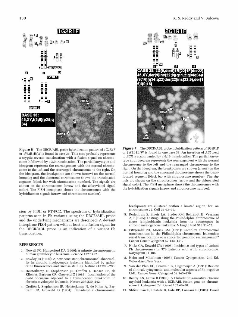
Figure 6 The DBCR/ABL probe hybridization pattern of 1G2R1For 1W2B1B/W is found in case 36. This case probably representsa cryptic reverse translocation with a fusion signal on chromo-some 9 followed by a 3,9 translocation. The partial karyotype andideogram represent the rearrangement with the normal chromo-some to the left and the rearranged chromosome to the right. Onthe ideogram, the breakpoints are shown (arrow) on the normalhomolog and the abnormal chromosome shows the translocatedsegment (black bar with chromosome number). The signals areshown on the chromosomes (arrow and the abbreviated signalcolor). The FISH metaphase shows the chromosomes with thehybridization signals (arrow and chromosome number).
Figure 7 The DBCR/ABL probe hybridization pattern of 2G1R1For 2W1B1B/W is found in one case 38. An insertion of ABL nextto BCR is accompanied by a 9;16 translocation. The partial karyo-type and ideogram represents the rearrangement with the normalchromosome to the left and the rearranged chromosome to theright. On the ideogram, the breakpoints are shown (arrow) on thenormal homolog and the abnormal chromosome shows the trans-located segment (black bar with chromosome number). The sig-nals are shown on the chromosomes (arrow and the abbreviatedsignal color). The FISH metaphase shows the chromosomes withthe hybridization signals (arrow and chromosome number).

FISH Study of Ph Translocations 131
transcript of abl and bcr genes in chronic myelogenous leu-kemia. Nature 315:550–554.
12. Arnoldus EPJ, Wiegant J, Noordermeer IA, Wessels JW, Bev-erstock GC, Grosveld GC, ven der Ploeg M, Raap AK (1990):Detection of the Philadelphia chromosome in interphasenuclei. Cytogenet Cell Genet 54:108–111.
13. Tkachuk DC, Westbrook CA, Andreeff M, Donlon TA, ClearyML, Suryanarayan K, Homge M, Redner A, Gray J, Pinkel D(1990): Detection of bcr-abl fusion in chronic myelogenousleukemia by in situ hybridization. Science 250:108–111.
14. Chase A, Grand F, Zhang Ji-Guang, Blackett N, Goldman M(1997): Factors influencing false positive and negative ratesof BCR-ABL fluorescence in situ hybridization. Genes Chro-mosom Cancer 18:246.
15. Dewald WG, Wyatt WA, Juneau AL, Carlson RO, ZinsmeisterAR, Jalal SM, Spurbeck JL, Silver RT (1998): Highly sensitivefluorescence in situ hybridization method to detect double
BCR/ABL fusion and monitor response to therapy in chronicmyeloid leukemia. Blood 19:3357–3365.
16. Lessard M, Le Prise P-Y (1982): Cytogenetic studies in 56cases with Ph1-positive hematologic disorders. Cancer GenetCytogenet 5:37–49.
17. Pedersen B (1984): Coexistence of cells with unmasked andmasked Ph1 in a case of chronic myeloid leukemia in blasticphase. Cancer Genet Cytogenet 12:129–137.
18. Weiderman LM, Karhi KK, Shivji MKK, Rayter SI, PegramSM, Dowden G, Bevan D, Will A, Galton DAG, Chan LC(1988): The correlation of breakpoint cluster region rear-rangement and p210 ph1/ab1 expression with morphologicalanalysis of Ph-negative chronic myeloid leukemia and othermyeloproliferative diseases. Blood 71:349–355.
19. Popenoe DW, Schaefer-Rego K, Mears JG, Bank A, LeibowitzD (1986): Frequent and extensive deletion during the 9;22translocation in CML. Blood 68:1123–1128.

![[3,3]-Sigmatropic rearrangements - Massey Universitygjrowlan/stereo2/lecture11.pdf · 123.702 Organic Chemistry Claisen rearrangements • One of the most useful sigmatropic rearrangements](https://img.pdfslide.us/doc/110x75/5adcada77f8b9a213e8bd8b0/33-sigmatropic-rearrangements-massey-gjrowlanstereo2lecture11pdf123702.jpg)


![34 [3,3]-sigmatropic rearrangements](https://img.pdfslide.us/doc/110x75/55503fb4b4c9058f768b4911/34-33-sigmatropic-rearrangements.jpg)


![36 [1,n]-sigmatropic rearrangements](https://img.pdfslide.us/doc/110x75/55504a55b4c9058f768b5083/36-1n-sigmatropic-rearrangements.jpg)