Embed Size (px)
Citation preview
Thorax, 1977, 32, 333-341
A computer system for processing data fromroutine pulmonary function testsA. I. PACK', ROSEMARY McCUSKER, AND F. MORAN
From the Centre for Respiratory Investigation, Glasgow Royal Infirmary
Pack, A. I., McCusker, Rosemary, and Moran, F. (1977). Thorax, 32, 333-341. A computersystem for processing data from routine pulmonary function tests. In larger pulmonary functionlaboratories there is a need for computerised techniques of data processing. A flexible computersystem, which is used routinely, is described. The system processes data from a relatively largerange of tests. Two types of output are produced-one for laboratory purposes, and one forreturn to the referring physician. The system adds an automatic interpretative report for eachset of results. In developing the interpretative system it has been necessary to utilise a numberof arbitrary definitions. The present terminology for reporting pulmonary function tests haslimitations. The computer interpretation system affords the opportunity to take account ofknown interaction between different measurements of function and different pathologicalstates.
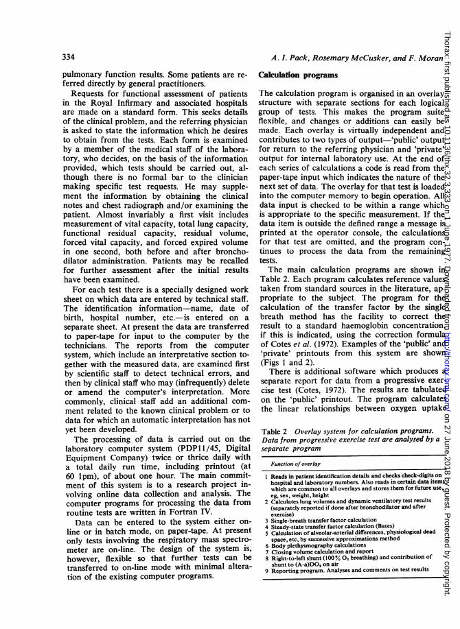
Computer systems are used more widely in bio-chemistry and haematology laboratories than inpulmonary function laboratories. In these labora-tories the need for a computer system is deter-mined by the large volume of tests which areperformed. Pulmonary function laboratories donot perform this large number of tests. There is,however, a greater amount of data processing foreach individual test. The nature of quality controlprocedures is different. Thus the main require-ments of the computer in the pulmonary functionlaboratory are different from those in other typesof clinical laboratory.A system is described for processing data from
pulmonary function tests. The system has been inoperation in the Centre for Respiratory Investiga-tion, Glasgow Royal Infirmary (CRI) for fouryears. It has been designed both to meet the in-ternal needs of the laboratory and to producereports which meet the requirements of thediverse groups of clinicians who use the service.
Table 1 Tests of function which are at presentperformed routinely in the Centre for RespiratoryInvestigation, Glasgow Royal Infirmary
Vital capacityTotal lung capacityFunctional residual capacityResidual volumeResidual volume/total lung capacity ratioForced expired volume (1 0 sec) (FEVy.0) before and afterForced vital capacity (FVC) bronchodilatorFEV5.,/FVC ratio administration;
after exerciseTransfer factor (end-tidal sampling, Bates) Rest/ExerciseTransfer factor (single-breath method)Closing volume, closing capacitySpecific conductanceAirways resistanceThoracic gas volumeArterial Po,Arterial Pco,Alveolar-arterial difference for oxygenArterial-alveolar difference for carbon dioxidePhysiological dead spaceVenous admixtureRight-to-left shunt (100% oxygen breathing)Sensitivity for carbon dioxide (rebreathing method)Progressive exercise testSteady-state exercise test (including blood gas studies)
Laboratory organisation
The service provided by the CRI is comprehensiveand includes, for example, facilities for assessmentof respiratory function (the main tests provided
'Wellcome Research Fellow
are listed in Table 1), for measurement of pul-monary haemodynamics, and for immunologicalinvestigations. Patients are referred to the Centreby physicians and surgeons in hospitals through-out the Western Region of Scotland, many ofwhom are not familiar with the interpretation of
333
on 27 June 2018 by guest. Protected by copyright.
http://thorax.bmj.com
/T
horax: first published as 10.1136/thx.32.3.333 on 1 June 1977. Dow
nloaded from
A. I. Pack, Rosemary McCusker, and F. Moran
pulmonary function results. Some patients are re-
ferred directly by general practitioners.Requests for functional assessment of patients
in the Royal Infirmary and associated hospitalsare made on a standard form. This seeks detailsof the clinical problem, and the referring physicianis asked to state the information which he desiresto obtain from the tests. Each form is examinedby a member of the medical staff of the labora-tory, who decides, on the basis of the informationprovided, which tests should be carried out, al-though there is no formal bar to the clinicianmaking specific test requests. He may supple-ment the information by obtaining the clinicalnotes and chest radiograph and/or examining thepatient. Almost invariably a first visit includesmeasurement of vital capacity, total lung capacity,functional residual capacity, residual volume,forced vital capacity, and forced expired volumein one second, both before and after broncho-dilator administration. Patients may be recalledfor further assessment after the initial resultshave been examined.For each test there is a specially designed work
sheet on which data are entered by technical staff.The identification information-name, date ofbirth, hospital number, etc.-is entered on a
separate sheet. At present the data are transferredto paper-tape for input to the computer by thetechnicians. The reports from the computersystem, which include an interpretative section to-gether with the measured data, are examined firstby scientific staff to detect technical errors, andthen by clinical staff who may (infrequently) deleteor amend the computer's interpretation. Morecommonly, clinical staff add an additional com-
ment related to the known clinical problem or todata for which an automatic interpretation has notyet been developed.The processing of data is carried out on the
laboratory computer system (PDPI1/45, DigitalEquipment Company) twice or thrice daily witha total daily run time, including printout (at60 1pm), of about one hour. The main commit-ment of this system is to a research project in-volving online data collection and analysis. Thecomputer programs for processing the data fromroutine tests are written in Fortran IV.
Data can be entered to the system either on-
line or in batch mode, on paper-tape. At presentonly tests involving the respiratory mass spectro-meter are on-line. The design of the system is,however, flexible so that further tests can betransferred to on-line mode with minimal altera-tion of the existing computer programs.
Calculation programs
The calculation program is organised in an overlaystructure with separate sections for each logicalgroup of tests. This makes the program suiteflexible, and changes or additions can easily bemade. Each overlay is virtually independent andcontributes to two types of output-'public' outputfor return to the referring physician and 'private'output for internal laboratory use. At the end ofeach series of calculations a code is read from thepaper-tape input which indicates the nature of thenext set of data. The overlay for that test is loadedinto the computer memory to begin operation. Alldata input is checked to be within a range whichis appropriate to the specific measurement. If thedata item is outside the defined range a message isprinted at the operator console, the calculationsfor that test are omitted, and the program con-tinues to process the data from the remainingtests.The main calculation programs are shown in
Table 2. Each program calculates reference valuestaken from standard sources in the literature, ap-propriate to the subject The program for thecalculation of the transfer factor by the singlebreath method has the facility to correct theresult to a standard haemoglobin concentration,if this is indicated, using the correction formulaof Cotes et al. (1972). Examples of the 'public' and'private' printouts from this system are shown(Figs 1 and 2).There is additional software which produces a
separate report for data from a progressive exer-cise test (Cotes, 1972). The results are tabulatedon the 'public' printout. The program calculatesthe linear relationships between oxygen uptake
Table 2 Overlay system for calculation programs.Data from progressive exercise test are analysed by a
separate program
Function ofoverlay
I Reads in patient identification details and checks check-digits onhospital and laboratory numbers. Also reads in certain data itemswhich are common to all overlays and stores them for future use,eg, sex, weight, height
2 Calculates lung volumes and dynamic ventilatory test results(separately reported if done after bronchodilator and afterexercise)
3 Single-breath transfer factor calculation4 Steady-state transfer factor calculation (Bates)5 Calculation of alveolar-arterial differences, physiological dead
space, etc, by successive approximations method6 Body plethysmography calculations7 Closing volume calculation and report8 Right-to-left shunt (100% 0, breathing) and contribution of
shunt to (A-a)DO, on air9 Reporting program. Analyses and comments on test results
334
on 27 June 2018 by guest. Protected by copyright.
http://thorax.bmj.com
/T
horax: first published as 10.1136/thx.32.3.333 on 1 June 1977. Dow
nloaded from

A computer system for processing data from routine pulmonary function tests
E-1N4 R"FE F-OR E LHTR YE~ r flt4' JN A I tHlF' I'RHAY
.OHNS1Ht I TH Ii1 CH FR,F,Nt-4 F,FES NIE4NTFiGE 51, HlYER NO :2741 unAI t-NC)1~,OIC7u4 flx'F_ EE (Pl,COPD HENDERS--ON E ALEV,-MB 12'5,2
+ + ~LitNl I_T's INt- USE - 1VARLUE'wS IN OIL UN1`7I' ' IEN HLNST4
OBSERVED YHLUE FFL DI' 1AUTED PREL:I> 1ES
LUN VOLUE-J' I 1.FE B{,<w -^ sL ~~.._15_
F..
PM' L o-'5 PEPS, EN!I
2,4
t, , 1 -o
4 -' ,~As -.14244
C_4 C_<
j=- *'11t s,ta-
7: ..
,:" 9.:
.'J- ..
r--,. r--,1',
1
IYN I T I L ORY TESTSPV 1.52-FEY,-'I I. 9P4FEYICF.t 1 PERCEtNT
HFTERII*-IL.LINE
F M,Lt =s 152
FEVIFY,-CF'l'-C FlPF, f"ERCEN T
-1IFi[I NOJCHHCF: 111 Pc,.t1MI N: PH.'2 ....SINLBRAT MET,)HOfLrrW'S 19 >, i &-w
; 1,,, 4., c-7;'
4I- - 4 22
6"t7. 1 - 94 "
MIN H I:F HYt - BS Tr LJcT O-4WI I tI Ei-'r RESPONSC ;t A r_1E BR Nc1 UD4i I
iN4 TrC TH H :>EE-EL-CESl .HP FP^| L G4i~ O$t'L MeaE
HE HENCIPHAL I TT' IS RE`I-- F IC I [T E VENTITLATCIRY DEFECTr.C.THE IN ..E
NETHUED IS tICLrEP:TEI '9L ['ECDUCEE LO')W TRANSFER TOP T RELATE 'LY TO 1-.4.'SB L. INK:
VO{CLL E,
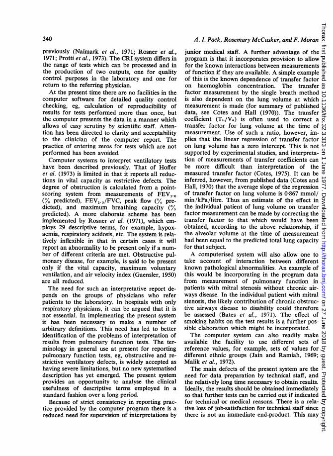
Fig. 1 Printout from computer program for return to referring physician, illustrating output ofinterpretative program.
315
-5,
1-1
t
on 27 June 2018 by guest. Protected by copyright.
http://thorax.bmj.com
/T
horax: first published as 10.1136/thx.32.3.333 on 1 June 1977. Dow
nloaded from
A. I. Pack, Rosemary McCusker, and F. Moran
-rI
F 'E'£..I 9? 7 1rJ
Lwk w xENALI
't
7271=J I. ir-z j/F7t$r= _-$or
1-'1.+_.......iC....~~ SO
.n2.2
l jI F^ C- C.A 7
VA 2=2.2 /4
FF C;-J A
OP_S~i1 A**t
D;fi??.;2W,,,-'. -<r
M'1COPPBb*C .S r ,r .r ...,TFTJJ; *g7 *7W.
-1. 7 7 ,A1 r f )1 I-;, 1: i I771>1'2>,1 -1..W
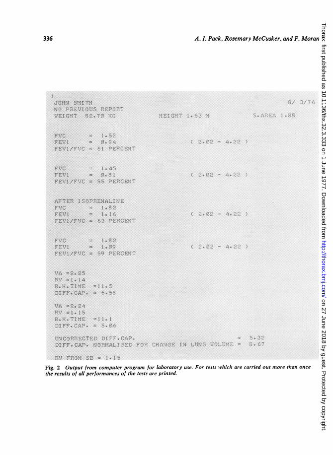
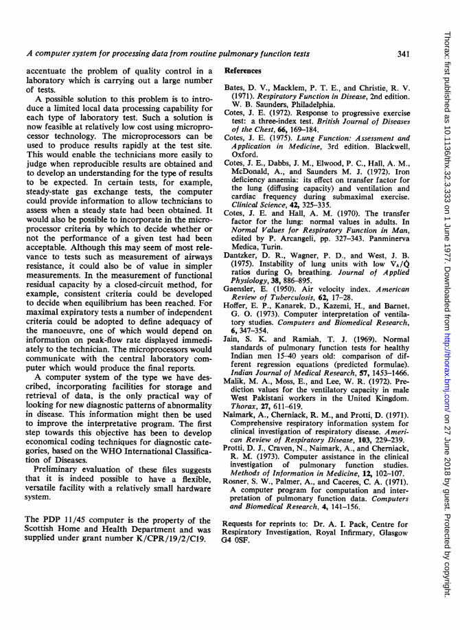
Fig. 2 Output from computer program for laboratory use. For tests which are carried out more than once
the results of all performances of the tests are printed.
336
on 27 June 2018 by guest. Protected by copyright.
http://thorax.bmj.com
/T
horax: first published as 10.1136/thx.32.3.333 on 1 June 1977. Dow
nloaded from

A computer system for processing data from routine pulmonary function tests
1--'IT~~. -..UIT4 .4.CJ- tt..4
-!-r1 i11 i04 ;4 O3 £I,!'1
*~~~~~~~~~~~~~~~~*,70 1~ __ _ < _|2053. 20 32 | '*i4 17* 1
-.-i s 1 $ s '-. ;. II . ..7 - i-;I1;yt3gft:; > t <: X c s ,S .r t tL ;~ 9 | ' s ~ 1 ~sx~e1GL4s r;
r"IT D-i1eF.H''t1,
H_s0L572CA_:. 1
-17
i- vo.71,,{Fig. 3 Printout from computer for presenting the results of a progressive submaximal exercise test. Theidentification information at the top of the printout is not shown. It is identical with that in Figure 1.
and cardiac frequency and between oxygen uptakeand ventilation (see Fig. 3). The data for oxygenuptake, etc, are given in millilitres per minute butwill soon be converted to SI units. A statisticalanalysis of the result is produced on the 'private'printout. The indices derived from the regressionlines are shown in the 'public' printout and theassociated uncertainty of the estimates on the'private' printout.A program is available to calculate the contri-
bution of any right-to-left shunt to the alveolar-arterial difference for oxygen measured during airbreathing. The assumption is made that the shuntmeasured during the breathing of 100% oxygendoes not change during air breathing. Sincebreathing 100% oxygen causes an increasein the right-to-left shunt (Dantzker et al., 1975)this program will tend to underestimate thecontribution to the alveolar-arterial difference ofmaldistribution of ventilation to perfusion and ofabnormality of diffusion. The output from thisprogram is demonstrated in Figure 4.
Interpretative program
This is based on a logical decision tree designed todescribe the type of defect of function which ispresent. At the present stage of development theinterpretation system comments only on data frommeasurements of vital capacity, total lung capa-city, functional residual capacity, residual volume,residual volume/total lung capacity ratio, forcedvital capacity, and expired volume (1 second),before and after bronchodilators, and measure-ment of transfer factor. For this the program usesthe 32 statements listed in Table 3.The program decides initially if the patient has
airways obstruction (originally an FEV1.O/FVCratio of less than 70% but the current version in-corporates the correction for age and sex) and,if so, it assigns a category to the degree of obstruc-tion. Difficulties are encountered if the subjecthas a 'mixed' type of abnormality with both re-strictive and obstructive elements. The computerrecognises the presence of a 'mixed' abnormality
337
on 27 June 2018 by guest. Protected by copyright.
http://thorax.bmj.com
/T
horax: first published as 10.1136/thx.32.3.333 on 1 June 1977. Dow
nloaded from

A. 1. Pack, Rosemary McCusker, and F. Moran
Fig. 4 Calculation of contribution of a right-to-left shunt to alveolar arterialdifference for oxygen measured during air breathing. The identification informationat the top of the printout is not shown. It is identical with that in Figure 1.
if obstruction is present and the vital capacity and/or total lung capacity are below the predictednormal range without an increase in the residualvolume. In an obstructive defect the decrease inthe FEV1.O expressed as a percentage of meanpredicted normal is used to assess the degree ofabnormality. In the 'mixed' defect the programevaluates the degree of airways obstruction presentfrom the FEVL.O/FVC ratio. In both cases >80%of predicted ratio is described as minor, 65-80% asmoderate, 45-67% moderately severe, and <45%severe. A statement is then made on the signifi-cance of the functional change produced by bron-chodilator administration. If airways obstructionis associated with an increase in residual volumewithout a corresponding increase in total lungcapacity, the airways obstruction is said to be
associated with air trapping. Increase in bothresidual volume and total lung capacity is calledhyperinflation.
In unobstructed subjects the program reportsthe presence or absence of a restrictive defect and,if present, categorises the degree of abnormality.Any reduction in transfer factor is noted next.
For abnormalities detected by the single-breathtechnique the program attempts to assess the con-tribution of an associated reduction in lung vol-ume. It is assumed in these calculations that for a1-litre change in lung volume one can anticipate,on average, a 0-867 mmol/min/kPa change intransfer factor (Cotes and Hall, 1970).
In general, the program does not make diagnos-tic comments but rather produces a verbal des-cription of the results. However, if there is no
338
on 27 June 2018 by guest. Protected by copyright.
http://thorax.bmj.com
/T
horax: first published as 10.1136/thx.32.3.333 on 1 June 1977. Dow
nloaded from
A computer system for processing data from routine pulmonary function tests
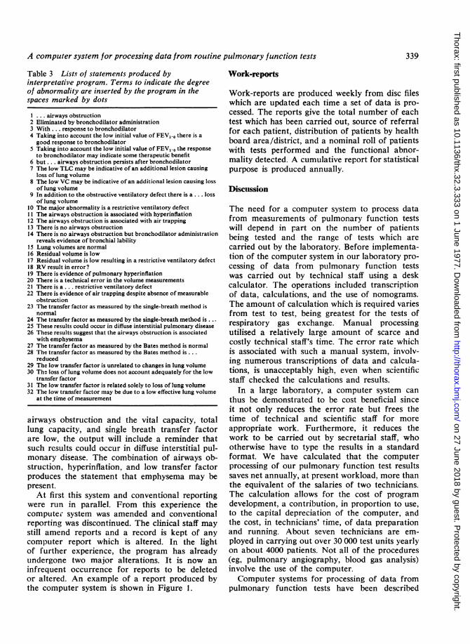
Table 3 Lists of statements produced byinterpretative program. Terms to indicate the degreeof abnormality are inserted by the program in thespaces marked by dots
I ... airways obstruction2 Eliminated by bronchodilator administration3 With ... response to bronchodilator4 Taking into account the low initial value of FEV5.0 there is agood response to bronchodilator
5 Taking into account the low initial value of FEV,.o the responseto bronchodilator may indicate some therapeutic benefit
6 but ... airways obstruction persists after bronchodilator7 The low TLC may be indicative of an additional lesion causing
loss of lung volume8 The low VC may be indicative of an additional lesion causing loss
of lung volume9 In addition to the obstructive ventilatory defect there is a ... loss
of lung volume10 The major abnormality is a restrictive ventilatory defect11 The airways obstruction is associated with hyperinflation12 The airways obstruction is associated with air trapping13 There is no airways obstruction14 There is no airways obstruction but bronchodilator administration
reveals evidence of bronchial lability15 Lung volumes are normal16 Residual volume is low17 Residual volume is low resulting in a restrictive ventilatory defect18 RV result in error?19 There is evidence of pulmonary hyperinflation20 There is a technical error in the volume measurements21 There is a ... restrictive ventilatory defect22 There is evidence of air trapping despite absence of measurable
obstruction23 The transfer factor as measured by the single-breath method is
normal24 The transfer factor as measured by the single-breath method is . ..25 These results could occur in diffuse interstitial pulmonary disease26 These results suggest that the airways obstruction is associated
with emphysema27 The transfer factor as measured by the Bates method is normal28 The transfer factor as measured by the Bates method is ...
reduced29 The low transfer factor is unrelated to changes in lung volume30 The loss of lung volume does not account adequately for the low
transfer factor31 The low transfer factor is related solely to loss of lung volume32 The low transfer factor may be due to a low effective lung volume
at the time of measurement
airways obstruction and the vital capacity, totallung capacity, and single breath transfer factorare low, the output will include a reminder thatsuch results could occur in diffuse interstitial pul-monary disease. The combination of airways ob-struction, hyperinflation, and low transfer factorproduces the statement that emphysema may bepresent.At first this system and conventional reporting
were run in parallel. From this experience thecomputer system was amended and conventionalreporting was discontinued. The clinical staff maystill amend reports and a record is kept of anycomputer report which is altered. In the lightof further experience, the program has alreadyundergone two major alterations. It is now aninfrequent occurrence for reports to be deletedor altered. An example of a report produced bythe computer system is shown in Figure 1.
Work-reports
Work-reports are produced weekly from disc fileswhich are updated each time a set of data is pro-cessed. The reports give the total number of eachtest which has been carried out, source of referralfor each patient, distribution of patients by healthboard area/district, and a nominal roll of patientswith tests performed and the functional abnor-mality detected. A cumulative report for statisticalpurpose is produced annually.
Discussion
The need for a computer system to process datafrom measurements of pulmonary function testswill depend in part on the number of patientsbeing tested and the range of tests which arecarried out by the laboratory. Before implementa-tion of the computer system in our laboratory pro-cessing of data from pulmonary function testswas carried out by technical staff using a deskcalculator. The operations included transcriptionof data, calculations, and the use of nomograms.The amount of calculation which is required variesfrom test to test, being greatest for the tests ofrespiratory gas exchange. Manual processingutilised a relatively large amount of scarce andcostly technical staff's time. The error rate whichis associated with such a manual system, involv-ing numerous transcriptions of data and calcula-tions, is unacceptably high, even when scientificstaff checked the calculations and results.
In a large laboratory, a computer system canthus be demonstrated to be cost beneficial sinceit not only reduces the error rate but frees thetime of technical and scientific staff for moreappropriate work. Furthermore, it reduces thework to be carried out by secretarial staff, whootherwise have to type the results in a standardformat. We have calculated that the computerprocessing of our pulmonary function test resultssaves net annually, at present workload, more thanthe equivalent of the salaries of two technicians.The calculation allows for the cost of programdevelopment, a contribution, in proportion to use,to the capital depreciation of the computer, andthe cost, in technicians' time, of data preparationand running. About seven technicians are em-ployed in carrying out over 30 000 test units yearlyon about 4000 patients. Not all of the procedures(eg, pulmonary angiography, blood gas analysis)involve the use of the computer.Computer systems for processing of data from
pulmonary function tests have been described
339
on 27 June 2018 by guest. Protected by copyright.
http://thorax.bmj.com
/T
horax: first published as 10.1136/thx.32.3.333 on 1 June 1977. Dow
nloaded from
340
previously (Naimark et al., 1971; Rosner et al.,1971; Protti et al., 1973). The CRI system differs inthe range of tests which can be processed and inthe production of two outputs, one for qualitycontrol purposes in the laboratory and one forreturn to the referring physician.At the present time there are no facilities in the
computer software for detailed quality controlchecking, eg, calculation of reproducibility ofresults for tests performed more than once, butthe computer presents the data in a manner whichallows of easy scrutiny by scientific staff. Atten-tion has been directed to clarity and acceptabilityto the clinician of the computer report. Thepractice of entering zeros for tests which are notperformed has been avoided.Computer systems to interpret ventilatory tests
have been described previously. That of Hofferet al. (1973) is limited in that it reports all reduc-tions in vital capacity as restrictive defects. Thedegree of obstruction is calculated from a point-scoring system from measurements of FEV1.0(% predicted), FEV1.0/FVC, peak flow (% pre-dicted), and maximum breathing capacity (%predicted). A more elaborate scheme has beenimplemented by Rosner et al. (1971), which em-ploys 29 descriptive terms, for example, hypox-aemia, respiratory acidosis, etc. The system is rela-tively inflexible in that in certain cases it willreport an abnormality to be present only if a num-ber of different criteria are met. Obstructive pul-monary disease, for example, is said to be presentonly if the vital capacity, maximum voluntaryventilation, and air velocity index (Gaensler, 1950)are all reduced.The need for such an interpretative report de-
pends on the groups of physicians who referpatients to the laboratory. In hospitals with onlyrespiratory physicians, it can be argued that it isnot essential. In implementing the present systemit has been necessary to make a number ofarbitrary definitions. This need has led to betteridentification of the problems of interpretation ofresults from pulmonary function tests. The ter-minology in general use at present for reportingpulmonary function tests, eg, obstructive and re-strictive ventilatory defects, is widely accepted ashaving severe limitations, but no new systematiseddescription has yet emerged. The present systemprovides an opportunity to analyse the clinicalusefulness of descriptive terms employed in astandard fashion over a long period.Because of strict consistency in reporting prac-
tice provided by the computer program there is areduced need for supervision of interpretations by
A. L. Pack, Rosemary McCusker, and F. Moran
junior medical staff. A further advantage of theprogram is that it incorporates provision to allowfor the known interactions between measurementsof function if they are available. A simple exampleof this is the known dependence of transfer factoron haemoglobin concentration. The transferfactor measurement by the single breath methodis also dependent on the lung volume at whichmeasurement is made (for summary of publisheddata, see Cotes and Hall (1970)). The transfercoefficient (TL/VA) is often used to correct atransfer factor for lung volume at the time ofmeasurement. Use of such a ratio, however, im-plies that the linear regression of transfer factoron lung volume has a zero intercept. This is notsupported by experimental studies, and interpreta-tion of measurements of transfer coefficients canbe more difficult than interpretation of themeasured transfer factor (Cotes, 1975). It can beinferred, however, from published data (Cotes andHall, 1970) that the average slope of the regressionof transfer factor on lung volume is 0867 mmol/min/kPa/litre. Thus an estimate of the effect inthe individual patient of lung volume on transferfactor measurement can be made by correcting thetransfer factor to that which would have beenobtained, according to the above relationship, ifthe alveolar volume at the time of measurementhad been equal to the predicted total lung capacityfor that subject.A computerised system will also allow one to
take account of interaction between differentknown pathological abnormalities. An example ofthis would be incorporating in the program datafrom measurement of pulmonary function inpatients with mitral stenosis without chronic air-ways disease. In the individual patient with mitralstenosis, the likely contribution of chronic obstruc-tive airways disease to disability could thereforebe assessed (Bates et al., 1971). The effect ofsmoking habits on the test results is a further pos-sible elaboration which might be incorporated.The computer system can also readily make
available the facility to use different sets ofreference values, for example, sets of values fordifferent ethnic groups (Jain and Ramiah, 1969;Malik et al., 1972).The main defects of the present system are the
need for data preparation by technical staff, andthe relatively long time necessary to obtain results.Ideally, the results should be obtained immediatelyso that further tests can be carried out if indicatedfor technical or medical reasons. There is a rela-tive loss of job-satisfaction for technical staff sincethere is not an immediate end-product. This may
on 27 June 2018 by guest. Protected by copyright.
http://thorax.bmj.com
/T
horax: first published as 10.1136/thx.32.3.333 on 1 June 1977. Dow
nloaded from
A computer system for processing data from routine pulmonary function tests
accentuate the problem of quality control in alaboratory which is carrying out a large numberof tests.A possible solution to this problem is to intro-
duce a limited local data processing capability foreach type of laboratory test. Such a solution isnow feasible at relatively low cost using micropro-cessor technology. The microprocessors can beused to produce results rapidly at the test site.This would enable the technicians more easily tojudge when reproducible results are obtained andto develop an understanding for the type of resultsto be expected. In certain tests, for example,steady-state gas exchange tests, the computercould provide information to allow technicians toassess when a steady state had been obtained. Itwould also be possible to incorporate in the micro-processor criteria by which to decide whether ornot the performance of a given test had beenacceptable. Although this may seem of most rele-vance to tests such as measurement of airwaysresistance, it could also be of value in simplermeasurements. In the measurement of functionalresidual capacity by a closed-circuit method, forexample, consistent criteria could be developedto decide when equilibrium has been reached. Formaximal expiratory tests a number of independentcriteria could be adopted to define adequacy ofthe manoeuvre, one of which would depend oninformation on peak-flow rate displayed immedi-ately to the technician. The microprocessors wouldcommunicate with the central laboratory com-puter which would produce the final reports.A computer system of the type we have des-
cribed, incorporating facilities for storage andretrieval of data, is the only practical way oflooking for new diagnostic patterns of abnormalityin disease. This information might then be usedto improve the interpretative program. The firststep towards this objective has been to developeconomical coding techniques for diagnostic cate-gories, based on the WHO International Classifica-tion of Diseases.
Preliminary evaluation of these files suggeststhat it is indeed possible to have a flexible,versatile facility with a relatively small hardwaresystem.
The PDP 11/45 computer is the property of theScottish Home and Health Department and wassupplied under grant number K/CPR/19/2/C19.
References
Bates, D. V., Macklem, P. T. E., and Christie, R. V.(1971). Respiratory Function in Disease, 2nd edition.W. B. Saunders, Philadelphia.
Cotes, J. E. (1972). Response to progressive exercisetest: a three-index test. British Journal of Diseasesof the Chest, 66, 169-184.
Cotes, J. E. (1975). Lung Function: Assessment andApplication in Medicine, 3rd edition. Blackwell,Oxford.
Cotes, J. E., Dabbs, J. M., Elwood, P. C., Hall, A. M.,McDonald, A., and Saunders M. J. (1972). Irondeficiency anaemia: its effect on transfer factor forthe lung (diffusing capacity) and ventilation andcardiac frequency during submaximal exercise.Clinical Science, 42, 325-335.
Cotes, J. E. and Hall, A. M. (1970). The transferfactor for the lung: normal values in adults. InNormal Values for Respiratory Function in Man,edited by P. Arcangeli, pp. 327-343. PanminervaMedica, Turin.
Dantzker, D. R., Wagner, P. D., and West, J. B.(1975). Instability of lung units with low VA/0ratios during 02 breathing. Journal of AppliedPhysiology, 38, 886-895.
Gaensler, E. (1950). Air velocity index. A mericanReview of Tuberculosis, 62, 17-28.
Hoffer, E. P., Kanarek, D., Kazemi, H., and Barnet,G. 0. (1973). Computer interpretation of ventila-tory studies. Computers and Biomedical Research,6, 347-354.
Jain, S. K. and Ramiah, T. J. (1969). Normalstandards of pulmonary function tests for healthyIndian men 15-40 years old: comparison of dif-ferent regression equations (predicted formulae).Indian Journal of Medical Research, 57, 1453-1466.
Malik, M. A., Moss, E., and Lee, W. R. (1972). Pre-diction values for the ventilatory capacity in maleWest Pakistani workers in the United Kingdom.Thorax, 27, 611-619.
Naimark, A., Cherniack, R. M., and Protti, D. (1971).Comprehensive respiratory information system forclinical investigation of respiratory disease. Ameri-can Review of Respiratory Disease, 103, 229-239.
Protti, D. J., Craven, N., Naimark, A., and Cherniack,R. M. (1973). Computer assistance in the clinicalinvestigation of pulmonary function studies.Methods of Information in Medicine, 12, 102-107.
Rosner, S. W., Palmer, A., and Caceres, C. A. (1971).A computer program for computation and inter-pretation of pulmonary function data. Computersand Biomedical Research, 4, 141-156.
Requests for reprints to: Dr. A. I. Pack, Centre forRespiratory Investigation, Royal Infirmary, GlasgowG4 OSF.
341
on 27 June 2018 by guest. Protected by copyright.
http://thorax.bmj.com
/T
horax: first published as 10.1136/thx.32.3.333 on 1 June 1977. Dow
nloaded from