Embed Size (px)
Citation preview
2011;71:6940-6947. Published OnlineFirst September 22, 2011.Cancer Res Qianzi Tang, Yiwen Chen, Clifford Meyer, et al. CistromesA Comprehensive View of Nuclear Receptor Cancer
Updated Version 10.1158/0008-5472.CAN-11-2091doi:
Access the most recent version of this article at:
MaterialSupplementary
htmlhttp://cancerres.aacrjournals.org/content/suppl/2011/09/22/0008-5472.CAN-11-2091.DC1.Access the most recent supplemental material at:
Cited Articles http://cancerres.aacrjournals.org/content/71/22/6940.full.html#ref-list-1
This article cites 20 articles, 5 of which you can access for free at:
E-mail alerts related to this article or journal.Sign up to receive free email-alerts
SubscriptionsReprints and
[email protected] Department atTo order reprints of this article or to subscribe to the journal, contact the AACR
To request permission to re-use all or part of this article, contact the AACR Publications
American Association for Cancer Research Copyright © 2011 on December 7, 2011cancerres.aacrjournals.orgDownloaded from
Published OnlineFirst September 22, 2011; DOI:10.1158/0008-5472.CAN-11-2091
Priority Report
A Comprehensive View of Nuclear Receptor CancerCistromes
Qianzi Tang1, Yiwen Chen2, Clifford Meyer2, Tim Geistlinger3, Mathieu Lupien3, Qian Wang1, Tao Liu2,
Yong Zhang1, Myles Brown3, and Xiaole Shirley Liu2
AbstractNuclear receptors comprise a superfamily of ligand-activated transcription factors that play important
roles in both physiology and diseases including cancer. The technologies of chromatin immunoprecip-itation followed by array hybridization (ChIP-chip) or massively parallel sequencing (ChIP-seq) has beenused to map, at an unprecedented rate, the in vivo genome-wide binding (cistrome) of nuclear receptors inboth normal and cancer cells. We developed a curated database of 88 nuclear receptor cistrome data setsand other associated high-throughput data sets including 121 collaborating factor cistromes, 94 epigen-omes, and 319 transcriptomes. Through integrative analysis of the curated nuclear receptor ChIP-chip/seqdata sets, we discovered novel factor-specific noncanonical motifs that may have important regulatoryroles. We also revealed a common feature of nuclear receptor pioneering factors to recognize relativelyshort and AT-rich motifs. Most nuclear receptors bind predominantly to introns and distal intergeneticregions, and binding sites closer to transcription start sites were found to be neither stronger nor moreevolutionarily conserved. Interestingly, while most nuclear receptors appear to be predominantly tran-scriptional activators, our analysis suggests that the binding of ESR1, RARA, and RARG has both activatingand repressive effects. Through meta-analysis of different omic data of the same cancer cell line modelfrom multiple studies, we generated consensus cistrome and expression profiles. We further madeprobabilistic predictions of the nuclear receptor target genes by integrating cistrome and transcriptomedata and validated the predictions using expression data from tumor samples. The final database, withcomprehensive cistrome, epigenome, and transcriptome data sets and downstream analysis results,constitutes a valuable resource for the nuclear receptor and cancer community. Cancer Res; 71(22);6940–7. �2011 AACR.
Introduction
Nuclear receptors form a large class of transcriptionfactors that can bind directly to DNA to regulate gene
expression upon ligand activation. The ligands can besteroid, hormones, or other molecules, although somenuclear receptors, called orphan receptors, have noknown ligands. The human and mouse genomes encode48 and 49 nuclear receptors, respectively. These nuclearreceptors play important roles in the development,homeostasis, and metabolism of higher organisms.
Nuclear receptors play key roles not only in normalphysiology but also in many pathologic processes, mostnotably cancer. Estrogen receptor (ESR) is overexpressedin more than 70% of breast cancers and is the archetypalmolecular therapeutic target (1). Progesterone receptorhas been shown to enhance breast cancer motility andinvasiveness (2). Androgen receptor overactivation byandrogens is essential for the initiation and progressionof prostate cancers (3, 4). Retinoic acid receptor (RAR),upon activation by retinoic acid (RA), has antiproliferativeeffects in tumor cells (5). The translocation and subse-quent oncofusion of promyelocytic leukemia (PML) withRARa in hematopoietic myeloid cells causes acute pro-myelocytic leukemia (6). Recent studies have linked cancerto lipid metabolism and cell inflammations (7, 8), and the
Authors' Affiliations: 1Department of Bioinformatics, School of LifeScience and Technology, Tongji University, Shanghai, China; andDepartments of 2Biostatistics and Computational Biology and 3MedicalOncology, Dana-Farber Cancer Institute, Harvard Medical School,Boston, Massachusetts
Note: Supplementary data for this article are available at Cancer ResearchOnline (http://cancerres.aacrjournals.org/).
Corresponding Authors: Xiaole Shirley Liu, Department of Biosta-tistics and Computational Biology, Dana-Farber Cancer Institute,Harvard School of Public Health, 44 Binney St., Boston, MA02115. Phone: 617-632-2472; Fax: 617-632-2444; E-mail:[email protected] and Myles Brown, Department of MedicalOncology, Dana-Farber Cancer Institute, Harvard Medical School,44 Binney St., Boston, MA 02115. Phone: 617-632-3948; Fax: 617-632-5417; E-mail: [email protected]
doi: 10.1158/0008-5472.CAN-11-2091
�2011 American Association for Cancer Research.
CancerResearch
Cancer Res; 71(22) November 15, 20116940
American Association for Cancer Research Copyright © 2011 on December 7, 2011cancerres.aacrjournals.orgDownloaded from
Published OnlineFirst September 22, 2011; DOI:10.1158/0008-5472.CAN-11-2091
major nuclear receptors regulating these processes includeglucocorticoid receptor (GR), PPAR, and liver X-receptor(LXR; ref. 9).Nuclear receptors often bind to DNA as homo- or
heterodimers, each recognizing a half-site of 6 nucleo-tides. Thus, their DNA-binding sequences, called hormoneresponse elements, often consist of 2 half-sites in directed,everted, or inverted configurations, separated by a vari-able gap (10). Much effort has been devoted to de novoprediction of nuclear receptor–binding sites, based solelyon genomic DNA sequence, without much success.Recently, the application of ChIP-chip/seq techniques hasenabled the accurate and effective detection of thegenome-wide in vivo binding sites or cistromes of nuclearreceptors (Supplementary Table S1). Herein, we define thecistrome as the set of cis-acting elements bound by atrans-factor at the genomic scale, that is, binding sitesidentified by ChIP-chip/seq experiments. Publicly avail-able cistrome data have been growing rapidly, and some-times multiple cistrome profiles of the same trans-factorin the same biological system are available. Meta-analysisof related cistrome profiles can often yield much morebiologically relevant insights than the examination ofsingle profiles.Previous efforts to identify nuclear receptor target genes
have mostly relied on differential expression profiles beforeand after nuclear receptor activation. However, the differ-ential expression cutoff value selected may not be ideal, andfor many genes, differential expression may be due tosecondary or tertiary effects of nuclear receptor activity.With the availability of cistrome data, target gene predic-tion based on the presence of a binding site within a certaindistance from the transcription start site (TSS) of the genehas also been used, although the distance cutoff value isoften arbitrary. In addition, many genes with nearby bind-ing sites show no differential expression upon binding, dueto the gene's promoter chromatin status, missing essentialcofactors, and other confounding effects. Intuitively, thecombination of cistrome and differential expression profilesshould allow for a much more robust prediction of thedirect target genes of nuclear receptors than either dataalone.In this study, we systematically collected and prepro-
cessed all of the publicly available genome-wide ChIP-chip/seq data for nuclear receptors, their collaboratingfactors, and histone modifications in humans and miceusing a standardized computational pipeline. We comparedthe hormone response element patterns, distance (to TSSs ofgenes) distributions, evolutionary conservation, and collab-orating partners of different nuclear receptors. We alsoconducted meta-analyses to generate consensus cistromeand expression profiles. Finally, we integrated cistrome andtranscriptome data to make probabilistic predictions ofnuclear receptor target genes, including 10 nuclear receptorsin various cancer cell line models. The resultant cistromes,epigenomes, transcriptomes, motif analyses, and target genelists are publicly available at Nuclear Receptor CancerCistromes (11).
Materials and Methods
Target gene predictionIn some systems such as ESR1 activation in the breast
cancer cell line MCF7, multiple cistrome and transcriptomedata are available from different studies using the sameor different platforms. We first used the Stouffer P valuecombination method (12) to combine different transcrip-tome data sets, giving each gene a consensus differentialexpression z-score. We also used MM-ChIP (13) to combineredundant cistrome data sets to create a consensus peaklist. On the basis of the characterization of higher orderchromatin interactions and our preliminary analysis, wecalculated the regulatory potential for a given gene, Sg, asthe sum of the nearby binding sites weighted by the distance
from each site to the TSS of the gene: Sg ¼ Pk
i¼1
e� 0:5þ4Dið Þ ,
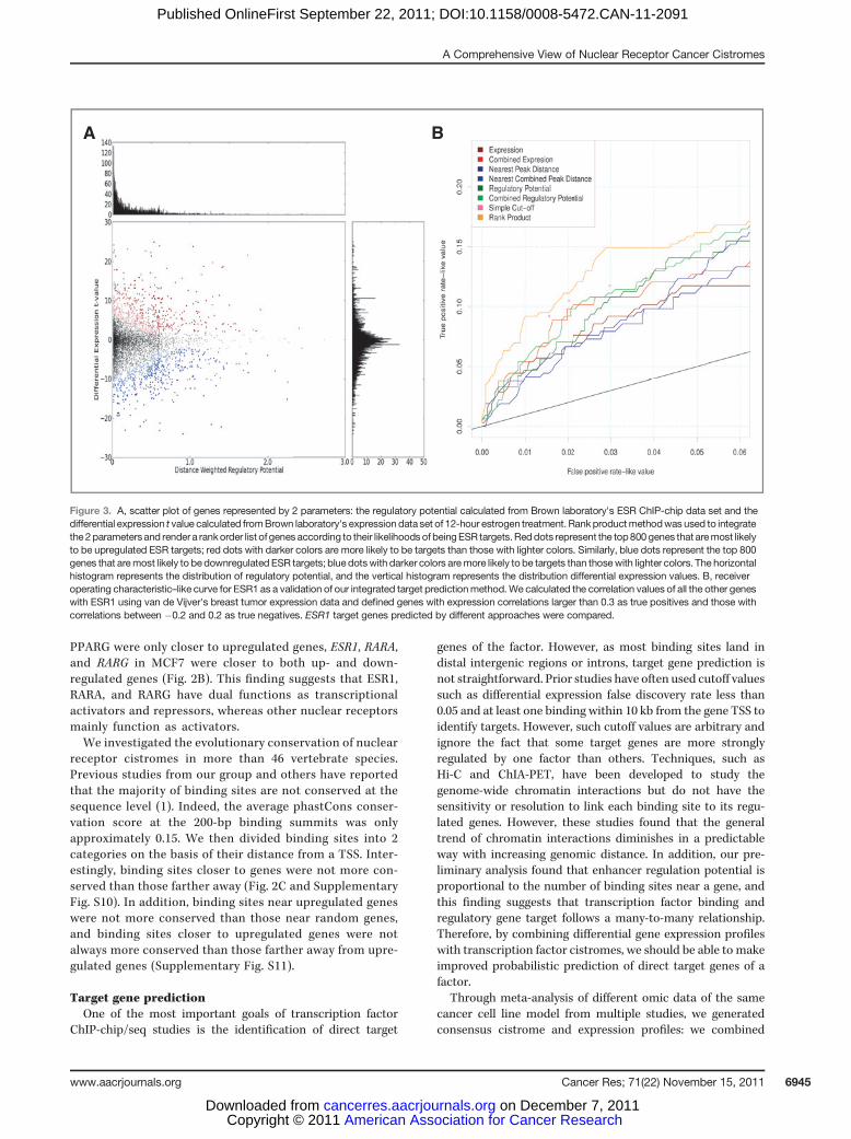
where k is the number of binding sites within 100 kb ofgene g and Di is the distance between site i and the TSS ofgene g normalized to 100 kb (e.g., 0.5 for a 50-kb distance).This equation models the influence of each binding site ongene regulation as a function that decreases monotonicallywith increasing distance from the TSS. The shape of thisfunction approximates empirical observations of the dis-tance between binding sites and differentially expressedgenes in multiple ChIP-seq experiments. The constant inthe equation enables the exponential function to adopt moreflexible shapes and 0.5 was derived to better fit ChIA-PETand Hi-C data. As rank product was finally used to predicttargets, the exact value of this constant would not changethe regulatory potential ranking of genes. Incorporatingbinding affinity into the model does not significantlyimprove the prediction power and therefore were excludedfrom the model. We represented each gene using the fol-lowing 2 parameters: (i) the differential gene expressionz-score (if multiple transcriptome data are available) ort-value (if single transcriptome data are available) and (ii)the regulatory potential. For target prediction, we onlyconsidered genes with at least one binding site within 100kb from its TSS and a differential expression z-score ort-value above the 75th percentile. We applied the Breitlingrank product method (14, 15) to combine transcriptionfactor–binding potentials with differential expression values(shown in Fig. 3A is an example of the rank product resultfrom integration of one ESR ChIP-chip data set and onedifferential expression data set of estrogen 12-hour treat-ment). The false discovery rate of each predicted target isestimated by a permutation method proposed in the work ofBreitling and colleagues (14).
Results
Data set summaryWe collected a total of 88 cistrome data sets for 13 nuclear
receptors, 121 cistrome data sets for 21 collaborating factors,and 94 genome-wide analyses of 12 histone modifications,which were profiled in the same cell systems as the nuclear
A Comprehensive View of Nuclear Receptor Cancer Cistromes
www.aacrjournals.org Cancer Res; 71(22) November 15, 2011 6941
American Association for Cancer Research Copyright © 2011 on December 7, 2011cancerres.aacrjournals.orgDownloaded from
Published OnlineFirst September 22, 2011; DOI:10.1158/0008-5472.CAN-11-2091
receptors. These data sets encompass all of the publishedgenome-wide ChIP-chip/seq studies on nuclear receptorsand their related factors in humans and mice before 2011, asfar as we are aware. We included the ESRRB ChIP-seqconducted in mouse embryonic stem cells but did notinclude other stem cell ChIP-chip/seq data because of thelarge number of such data sets that are not necessarilyrelated to the cancer focus of this study. For ChIP-chip dataconsistency, we did not include any chromosome-wide,custom tiling, or spotted cDNA arrays but did includeChIP-chip on Affymetrix whole genome or promoter tilingarrays because of their stable designs. Model-based analysisof tiling arrays (MAT; ref. 16) and model-based analysis ofChIP-Seq (MACS; ref. 17) were used for ChIP-chip and ChIP-seq peak calling, respectively. In addition, we analyzed 40gene expression data sets for 11 activation and/or deacti-vation experiments on nuclear receptors, totaling 319 micro-array profiles. A summary of the data analyzed is shown in(Table 1).
Motif analysesPrevious protein structure analysis has suggested that in
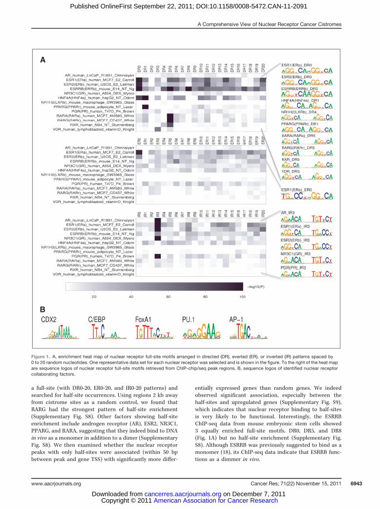
nuclear receptor dimers, one monomer often binds to DNAmuch more strongly than the other (10). However, when weapplied MDscan for de novo motif discovery in the nuclearreceptor cistrome sites, the nuclear receptor full-site motifsidentified were surprisingly symmetric between the 2 half-sites. In addition, when we collected full-site motif hits inthe cistrome sites having sufficiently good overall matches(the summation of the 2 half-site matching scores) to theconsensus sequence, the 2 half-sites were also symmetric(Fig. 1A). This suggests that the 2 monomers contributesimilarly to the in vivo binding, which may differ from invitro binding.
We then examined how the 2 monomers were arranged indirected (DR), everted (ER), or inverted (IR) configurationswith variable gaps for different nuclear receptors (see Fig. 1Aand Supplementary Figs. S1–S7). Most of the nuclear receptorsinvestigated had only one strong full-site motif, correspondingto their previously known canonical motif. Many other non-canonical motifs, although significantly enriched comparedwith the genome background, were much weaker than thecanonical ones (Fig. 1A), suggesting that the binding sites withnoncanonical motifs may be functional in a more context-dependentmanner. One interesting exceptionwas ESR1,whichhad strong enrichment of directed, everted, and invertedmotifs.
Some nuclear receptors that form heterodimers with othernuclear receptors can recognize different full-site motifs. Forexample, RXR recognizes DR5 when dimerizing with RARAin human NB4 cells and DR1 when dimerizing with PPARGin mouse adipocyte cells. Note that RXR and its dimerizationpartner in adipocytes, PPARG, show noncanonical ER14 andIR3 motifs (Supplementary Figs. S1 and S2) in preadipocytes.For PPARG, the enrichment level of ER14 and IR3 motifsbecame weaker during adipogenesis and disappeared inmature adipocytes whereas that of DR1 became stronger.For PPARG's dimerization partner RXR, ER14 and IR3enrichment was also observed in early adipogenesis andDR1 enrichment was observed in mature adipocyte. Thissuggests that PPARGmay have different interaction partnersand recognition patterns in early adipogenesis. Furtherstudies are needed to identify these factors and their tran-scriptional consequences.
Previous studies using in vitro gel-shift and protein struc-ture analysis implied that some nuclear receptors could bindhalf-site motifs and function as monomers in vivo. We tookall of the nuclear receptor cistrome sites that do not contain
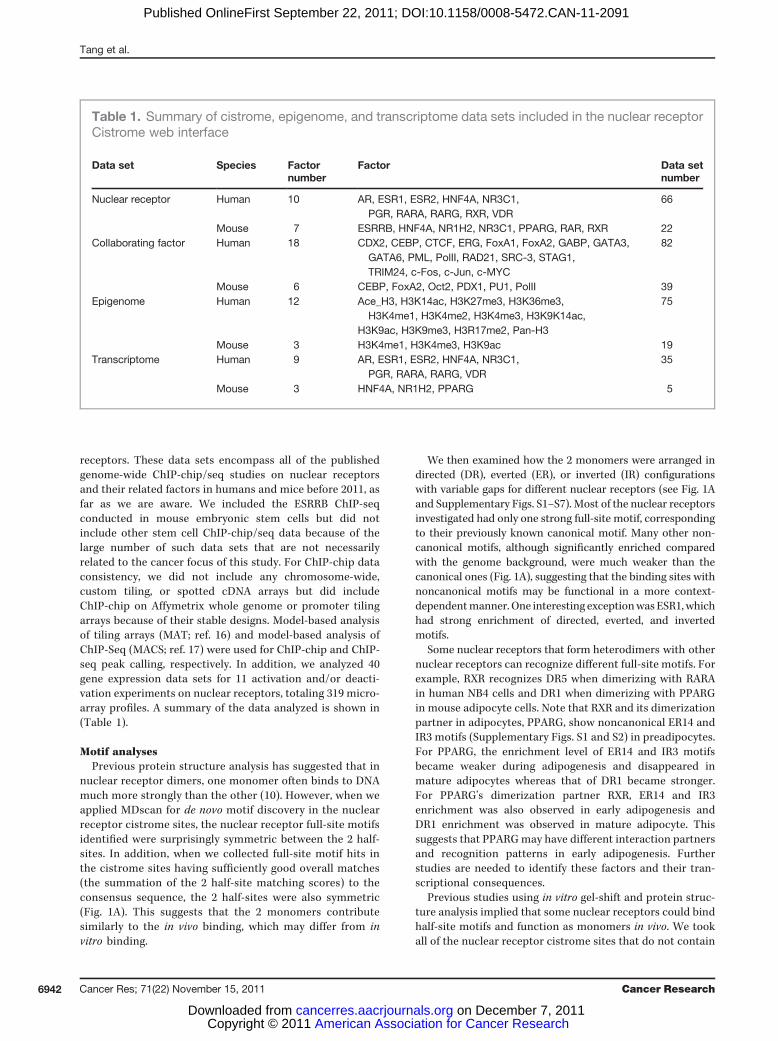
Table 1. Summary of cistrome, epigenome, and transcriptome data sets included in the nuclear receptorCistrome web interface
Data set Species Factornumber
Factor Data setnumber
Nuclear receptor Human 10 AR, ESR1, ESR2, HNF4A, NR3C1,PGR, RARA, RARG, RXR, VDR
66
Mouse 7 ESRRB, HNF4A, NR1H2, NR3C1, PPARG, RAR, RXR 22Collaborating factor Human 18 CDX2, CEBP, CTCF, ERG, FoxA1, FoxA2, GABP, GATA3,
GATA6, PML, PolII, RAD21, SRC-3, STAG1,TRIM24, c-Fos, c-Jun, c-MYC
82
Mouse 6 CEBP, FoxA2, Oct2, PDX1, PU1, PolII 39Epigenome Human 12 Ace_H3, H3K14ac, H3K27me3, H3K36me3,
H3K4me1, H3K4me2, H3K4me3, H3K9K14ac,H3K9ac, H3K9me3, H3R17me2, Pan-H3
75
Mouse 3 H3K4me1, H3K4me3, H3K9ac 19Transcriptome Human 9 AR, ESR1, ESR2, HNF4A, NR3C1,
PGR, RARA, RARG, VDR35
Mouse 3 HNF4A, NR1H2, PPARG 5
Tang et al.
Cancer Res; 71(22) November 15, 2011 Cancer Research6942
American Association for Cancer Research Copyright © 2011 on December 7, 2011cancerres.aacrjournals.orgDownloaded from
Published OnlineFirst September 22, 2011; DOI:10.1158/0008-5472.CAN-11-2091
a full-site (with DR0-20, ER0-20, and IR0-20 patterns) andsearched for half-site occurrences. Using regions 2 kb awayfrom cistrome sites as a random control, we found thatRARG had the strongest pattern of half-site enrichment(Supplementary Fig. S8). Other factors showing half-siteenrichment include androgen receptor (AR), ESR2, NR3C1,PPARG, and RARA, suggesting that they indeed bind to DNAin vivo as a monomer in addition to a dimer (SupplementaryFig. S8). We then examined whether the nuclear receptorpeaks with only half-sites were associated (within 50 bpbetween peak and gene TSS) with significantly more differ-
entially expressed genes than random genes. We indeedobserved significant association, especially between thehalf-sites and upregulated genes (Supplementary Fig. S9),which indicates that nuclear receptor binding to half-sitesis very likely to be functional. Interestingly, the ESRRBChIP-seq data from mouse embryonic stem cells showed3 equally enriched full-site motifs, DR0, DR5, and DR8(Fig. 1A) but no half-site enrichment (Supplementary Fig.S8). Although ESRRB was previously suggested to bind as amonomer (18), its ChIP-seq data indicate that ESRRB func-tions as a dimmer in vivo.
B
A
Figure 1. A, enrichment heat map of nuclear receptor full-site motifs arranged in directed (DR), everted (ER), or inverted (IR) patterns spaced by0 to 20 random nucleotides. One representative data set for each nuclear receptor was selected and is shown in the figure. To the right of the heat mapare sequence logos of nuclear receptor full-site motifs retrieved from ChIP-chip/seq peak regions. B, sequence logos of identified nuclear receptorcollaborating factors.
A Comprehensive View of Nuclear Receptor Cancer Cistromes
www.aacrjournals.org Cancer Res; 71(22) November 15, 2011 6943
American Association for Cancer Research Copyright © 2011 on December 7, 2011cancerres.aacrjournals.orgDownloaded from
Published OnlineFirst September 22, 2011; DOI:10.1158/0008-5472.CAN-11-2091
ChIP-chip/seq can pull down targets of transcriptionfactors that interact with the ChIP-ed factor of interest.We therefore conducted a motif analysis to find the mostsignificant collaborating motifs for each nuclear receptor(Fig. 1B and Supplementary Table S2). Among thesemotifs, there are previously reported and experimentallyvalidated collaborating motifs, including FoxA1 for AR,ESR1, RARA, and RARG; C/EBP for HNF4A, NR3C1,PPARG, and RXR; PU.1 for NR1H2; and CDX2 for HNF4A;there are also newly discovered collaborating motifs,including FoxA1 for PGR; AP-1 for ESR2, NR3C1, andVDR; and PU.1 for RARA and RXR. One interesting phe-nomenon we noticed is that many of the transcriptionfactors for nuclear receptors, such as FoxA1, C/EBP, PU.1,and CDX2, have relatively short and AT-rich motifs. Thesemotifs are likely the cell type–specific chromatin remo-delers that can more easily bind to nucleosome-freeregions. Once these pioneering factors pry open thechromatin, nuclear receptors can bind to the DNA and,
with their relatively longer motif patterns, convey specifictranscriptional effects.
Binding site distributionsWhen transcription factor cistromes were first published
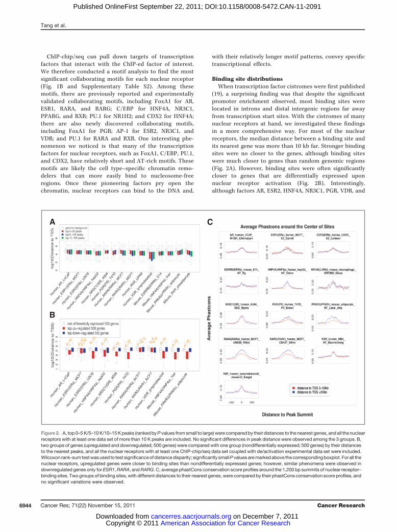
(19), a surprising finding was that despite the significantpromoter enrichment observed, most binding sites werelocated in introns and distal intergenic regions far awayfrom transcription start sites. With the cistromes of manynuclear receptors at hand, we investigated these findingsin a more comprehensive way. For most of the nuclearreceptors, the median distance between a binding site andits nearest gene was more than 10 kb far. Stronger bindingsites were no closer to the genes, although binding siteswere much closer to genes than random genomic regions(Fig. 2A). However, binding sites were often significantlycloser to genes that are differentially expressed uponnuclear receptor activation (Fig. 2B). Interestingly,although factors AR, ESR2, HNF4A, NR3C1, PGR, VDR, and
CA
B
Figure 2. A, top 0–5K/5–10K/10–15Kpeaks (rankedbyP values fromsmall to large) were comparedby their distances to the nearest genes, and all the nuclearreceptors with at least one data set of more than 10 K peaks are included. No significant differences in peak distance were observed among the 3 groups. B,two groups of genes (upregulated and downregulated; 500 genes) were compared with one group (nondifferentially expressed; 500 genes) by their distancesto the nearest peaks, and all the nuclear receptors with at least one ChIP-chip/seq data set coupled with de/activation experimental data set were included.Wilcoxon rank-sum testwasused to test significanceof distancedisparity; significantly smallP values aremarkedabove the correspondingboxplot. For all thenuclear receptors, upregulated genes were closer to binding sites than nondifferentially expressed genes; however, similar phenomena were observed indownregulated genes only for ESR1,RARA, andRARG. C, average phastCons conservation score profiles around the 1,200 bp summits of nuclear receptor–binding sites. Two groups of binding sites, with different distances to their nearest genes, were compared by their phastCons conservation score profiles, andno significant variations were observed.
Tang et al.
Cancer Res; 71(22) November 15, 2011 Cancer Research6944
American Association for Cancer Research Copyright © 2011 on December 7, 2011cancerres.aacrjournals.orgDownloaded from
Published OnlineFirst September 22, 2011; DOI:10.1158/0008-5472.CAN-11-2091
PPARG were only closer to upregulated genes, ESR1, RARA,and RARG in MCF7 were closer to both up- and down-regulated genes (Fig. 2B). This finding suggests that ESR1,RARA, and RARG have dual functions as transcriptionalactivators and repressors, whereas other nuclear receptorsmainly function as activators.We investigated the evolutionary conservation of nuclear
receptor cistromes in more than 46 vertebrate species.Previous studies from our group and others have reportedthat the majority of binding sites are not conserved at thesequence level (1). Indeed, the average phastCons conser-vation score at the 200-bp binding summits was onlyapproximately 0.15. We then divided binding sites into 2categories on the basis of their distance from a TSS. Inter-estingly, binding sites closer to genes were not more con-served than those farther away (Fig. 2C and SupplementaryFig. S10). In addition, binding sites near upregulated geneswere not more conserved than those near random genes,and binding sites closer to upregulated genes were notalways more conserved than those farther away from upre-gulated genes (Supplementary Fig. S11).
Target gene predictionOne of the most important goals of transcription factor
ChIP-chip/seq studies is the identification of direct target
genes of the factor. However, as most binding sites land indistal intergenic regions or introns, target gene prediction isnot straightforward. Prior studies have often used cutoff valuessuch as differential expression false discovery rate less than0.05 and at least one binding within 10 kb from the gene TSS toidentify targets. However, such cutoff values are arbitrary andignore the fact that some target genes are more stronglyregulated by one factor than others. Techniques, such asHi-C and ChIA-PET, have been developed to study thegenome-wide chromatin interactions but do not have thesensitivity or resolution to link each binding site to its regu-lated genes. However, these studies found that the generaltrend of chromatin interactions diminishes in a predictableway with increasing genomic distance. In addition, our pre-liminary analysis found that enhancer regulation potential isproportional to the number of binding sites near a gene, andthis finding suggests that transcription factor binding andregulatory gene target follows a many-to-many relationship.Therefore, by combining differential gene expression profileswith transcription factor cistromes, we should be able to makeimproved probabilistic prediction of direct target genes of afactor.
Through meta-analysis of different omic data of the samecancer cell line model from multiple studies, we generatedconsensus cistrome and expression profiles: we combined
BA
Figure 3. A, scatter plot of genes represented by 2 parameters: the regulatory potential calculated from Brown laboratory's ESR ChIP-chip data set and thedifferential expression t value calculated fromBrown laboratory's expression data set of 12-hour estrogen treatment. Rankproductmethodwas used to integratethe 2parametersand render a rankorder list of genes according to their likelihoodsof beingESR targets.Reddots represent the top800genes that aremost likelyto be upregulated ESR targets; red dots with darker colors are more likely to be targets than those with lighter colors. Similarly, blue dots represent the top 800genes that aremost likely to bedownregulated ESR targets; blue dotswith darker colors aremore likely to be targets than thosewith lighter colors. The horizontalhistogram represents the distribution of regulatory potential, and the vertical histogram represents the distribution differential expression values. B, receiveroperating characteristic–like curve for ESR1 as a validation of our integrated target predictionmethod.We calculated the correlation values of all the other geneswith ESR1 using van de Vijver's breast tumor expression data and defined genes with expression correlations larger than 0.3 as true positives and those withcorrelations between �0.2 and 0.2 as true negatives. ESR1 target genes predicted by different approaches were compared.
A Comprehensive View of Nuclear Receptor Cancer Cistromes
www.aacrjournals.org Cancer Res; 71(22) November 15, 2011 6945
American Association for Cancer Research Copyright © 2011 on December 7, 2011cancerres.aacrjournals.orgDownloaded from
Published OnlineFirst September 22, 2011; DOI:10.1158/0008-5472.CAN-11-2091
multiple ChIP-chip/seq data sets for the same nuclear recep-tors in the same cell line model to create a consensus peak list,andwe combinedmultiple expression data sets in the same cellline model and condition to give each gene a consensusdifferential expression z-score (see Materials and Methods).We further made probabilistic predictions of the nuclearreceptor target genes by integrating cistrome and transcrip-tome data (see Materials and Methods).
As a validation of our integrated target prediction meth-od that was applied to identify ESR1 gene targets in MCF7,we calculated the correlation of all the other genes withESR1 using van de Vijver's breast tumor expression data(20). By defining genes with expression correlations largerthan 0.3 as true positives and those with correlationsbetween �0.2 and 0.2 as true negatives, we generated areceiver operating characteristic–like curve of our predic-tions. Combining multiple expression or ChIP data gavebetter results than using single expression or ChIP data, andintegrating expression with ChIP gave better results thaneach data type alone and also better results than the simplecutoff method (Fig. 3B).
Discussion
ChIP-chip/seq methods have been increasingly adopted asa powerful approach to study transcription factor regulationin normal physiology and disease. Nuclear receptors areimportant gene regulators in many cancer systems. Wesystematically collected publicly available cistrome data fornuclear receptors in cancer cells, for their collaboratingtranscription factors, and for histone modifications. We alsointegrated the cistrome data with related differential geneexpression data to identify the direct targets of different
nuclear receptors in these cancers. Together, these integrat-ed data not only create a useful resource for the nuclearreceptor and cancer community but also provide a morecomprehensive view of the genome-wide binding character-istics and regulatory mechanisms of nuclear receptorsinvolved in cancer.
As more related cistrome and transcriptome data becomeavailable, we will add them to the current database, such asthe NR1D1 ChIP-seq data set published in 2011 (21).
We will refine the regulatory modules, including the col-laborating transcription factors and gene targets, of differentnuclear receptors in cancers. We are also working on acomprehensive data analysis pipeline (22), so researcherscan reuse the public data in combination with their owngenomic and epigenomic data to better understand generegulation in cancers.
Disclosure of Potential Conflicts of Interest
No potential conflicts of interest were disclosed.
Acknowledgments
The authors thank Mitch Lazar, Chris Glass, and Xiaopeng Cai for theirthoughtful discussions and Len Taing and Scott Taing for their assistance withthe Amazon Compute Cloud.
Grant Support
The project was supported by the National Basic Research (973) Program ofChina No. 2010CB944904 (Q. Tang, Q. Wang, and Y. Zhang) and NIH grantsDK062434 (Y. Chen, C.A.Meyer, T. Liu, andX.S. Liu) andDK074967 (T. Geistlinger,M. Lupien, and M. Brown).
Received June 25, 2011; revised September 1, 2011; accepted September 14,2011; published OnlineFirst September 22, 2011.
References1. Carroll JS, Meyer CA, Song J, Li W, Geistlinger TR, Eeckhoute J, et al.
Genome-wide analysis of estrogen receptor binding sites. Nat Genet2006;38:1289–97.
2. Fu XD, Goglia L, Sanchez AM, Flamini M, Giretti MS, Tosi V, et al.Progesterone receptor enhances breast cancer cell motility and inva-sion via extranuclear activation of focal adhesion kinase. Endocr RelatCancer 2010;17:431–43.
3. Wang Q, Li W, Zhang Y, Yuan X, Xu K, Yu J, et al. Androgen receptorregulates a distinct transcription program in androgen-independentprostate cancer. Cell 2009;138:245–56.
4. Yu J, Mani RS, Cao Q, Brenner CJ, Cao X, Wang X, et al. An integratednetwork of androgen receptor, polycomb, and TMPRSS2-ERGgene fusions in prostate cancer progression. Cancer Cell 2010;17:443–54.
5. Hua S, Kittler R, White KP. Genomic antagonism betweenretinoic acid and estrogen signaling in breast cancer. Cell 2009;137:1259–71.
6. Martens JH, Brinkman AB, Simmer F, Francoijs KJ, Nebbioso A,Ferrara F, et al. PML-RARalpha/RXR alters the epigenetic landscapein acute promyelocytic leukemia. Cancer Cell 2010;17:173–85.
7. Iliopoulos D, Jaeger SA, Hirsch HA, Bulyk ML, Struhl K. STAT3activation of miR-21 and miR-181b-1 via PTEN and CYLD are part ofthe epigenetic switch linking inflammation to cancer. Mol Cell2010;39:493–506.
8. Hirsch HA, Iliopoulos D, Joshi A, Zhang Y, Jaeger SA, Bulyk M, et al. Atranscriptional signature and common gene networks link cancer with
lipid metabolism and diverse human diseases. Cancer Cell 2010;17:348–61.
9. Glass CK, Saijo K. Nuclear receptor transrepression pathways thatregulate inflammation in macrophages and T cells. Nat Rev Immunol2010;10:365–76.
10. Kumar R, Thompson EB. The structure of the nuclear hormone recep-tors. Steroids 1999;64:310–9.
11. Nuclear Receptor Cancer Cistromes [cited 2011 Jun 10]. Availablefrom: http://cistrome.dfci.harvard.edu/NR_Cistrome.
12. Ochsner SA, Steffen DL, Hilsenbeck SG, Chen ES, Watkins C,McKenna NJ. GEMS (Gene Expression MetaSignatures), a Webresource for querying meta-analysis of expression microarraydatasets: 17beta-estradiol in MCF-7 cells. Cancer Res 2009;69:23–6.
13. Chen Y, Meyer CA, Liu T, Li W, Liu JS, Liu XS. MM-ChIP enablesintegrative analysis of cross-platform and between-laboratory ChIP-chip or ChIP-seq data. Genome Biol 2011;12:R11.
14. Breitling R, Armengaud P, Amtmann A, Herzyk P. Rank products: asimple, yet powerful, new method to detect differentially regulatedgenes in replicatedmicroarrayexperiments.FEBSLett 2004;573:83–92.
15. Klisch TJ, Xi Y, Flora A, Wang L, Li W, Zoghbi HY. In vivo Atoh1targetome reveals how a proneural transcription factor regulatescerebellar development. Proc Natl Acad Sci U S A 2011;108:3288–93.
16. JohnsonWE, LiW,MeyerCA,GottardoR,Carroll JS, BrownM.Model-based analysis of tiling-arrays for ChIP-chip. Proc Natl Acad Sci U S A2006;103:12457–62.
Tang et al.
Cancer Res; 71(22) November 15, 2011 Cancer Research6946
American Association for Cancer Research Copyright © 2011 on December 7, 2011cancerres.aacrjournals.orgDownloaded from
Published OnlineFirst September 22, 2011; DOI:10.1158/0008-5472.CAN-11-2091
17. Zhang Y, Liu T, Meyer CA, Eeckhoute J, Johnson DS, Bernstein BE.Model-based analysis of ChIP-Seq (MACS). Genome Biol 2008;9:R137.
18. Gearhart MD, Holmbeck SM, Evans RM, Dyson HJ, Wright PE. Mono-meric complex of human orphan estrogen related receptor-2 withDNA: a pseudo-dimer interface mediates extended half-site recogni-tion. J Mol Biol 2003;327:819–32.
19. Carroll JS, Liu XS, Brodsky AS, Li W, Meyer CA, Szary AJ, et al.Chromosome-wide mapping of estrogen receptor binding reveals
long-range regulation requiring the forkhead protein FoxA1. Cell2005;122:33–43.
20. vandeVijverMJ,HeYD, van't Veer LJ,DaiH,Hart AA, Voskuil DW, et al.A gene-expression signature as apredictor of survival in breast cancer.N Engl J Med 2002;347:1999–2009.
21. Feng D, Liu T, Sun Z, Bugge A, Mullican SE, Alenghat T. A circadianrhythm orchestrated by histone deacetylase 3 controls hepatic lipidmetabolism. Science 2011;331:1315–9.
22. Cistrome [cited 2010 Oct 20]. Available from: http://cistrome.org/ap/.
A Comprehensive View of Nuclear Receptor Cancer Cistromes
www.aacrjournals.org Cancer Res; 71(22) November 15, 2011 6947
American Association for Cancer Research Copyright © 2011 on December 7, 2011cancerres.aacrjournals.orgDownloaded from
Published OnlineFirst September 22, 2011; DOI:10.1158/0008-5472.CAN-11-2091