Embed Size (px)
Citation preview
Dose-Response Approaches for Nuclear Receptor- Mediated Modes of Action NIEHS Facility, RTP, NC
September 27th – 29th, 2010
h t t p : / / w w w . t e r a . o r g / p e e r / n u c l e a r r e c e p t o r /
NUCLEAR RECEPTOR BINDING DOMAIN: FIGURE FROM: HTTP://WWW.MAN.POZNAN.PL/CBB/RESEARCH.HTML
DOSE-RESPONSE APPROACHES FOR NUCLEAR RECEPTOR-
MEDIATED MODES OF ACTION WORKSHOP
SEPTEMBER 27 - 29, 2010 NATIONAL INSTITUTE FOR ENVIRONMENTAL HEALTH
SCIENCES (NIEHS) RESEARCH TRIANGLE PARK, NC
CAR/PXR Case Study
...EXPLORING THE DEVELOPMENT OF BIOLOGICALLY-BASED DOSE-RESPONSE APPROACHES FOR NUCLEAR RECEPTOR MEDIATED TOXICITY...

Page 2

Page 3
Dose-Response Approaches for Nuclear Receptor- Mediated Modes of Action NIEHS Facility, RTP, NC
September 27th – 29th, 2010
Table of Contents Tab 1 - Background ................................................................................ 5
CAR/PXR Panel Members ..........................................................................................................7 CAR/PXR Case Study Agenda ....................................................................................................9 Introduction for CAR/PXR Case Study .....................................................................................11 CAR/PXR Discussion Questions ...............................................................................................13
Tab 2 - Presentations ............................................................................ 17 CAR – Current State of Knowledge and Role in Biology/ Physiology: Curtis
Omiecinski and Wen Xie ...................................................................................................21 CAR- and PXR-Mediated Liver Growth in Rodents: Review of Key Events for
Phenobarbital-Induced Rodent Liver Tumor Formation: Brian G. Lake ..........................35 Summary of Additional Literature: Histopathology and Nomenclature: Russell
Cattley ................................................................................................................................47 Epigenetics and Carcinogenesis: Emphasis on Phenobarbital-Induced Alterations in
DNA Methylation and Gene Expression: Jay Goodman ...................................................50 CAR-Specific Data on Other Events: A Role for Oxidative Stress?: Remi Bars .....................54 Species Differences and Other Factors Impacting on Risk Assessment: Cliff
Elcombe .............................................................................................................................57 Microarray and Biological Pathway of Phenobarbital Transcriptomic Research:
Dave Geter, Amber Goetz, and Susan Hester ....................................................................58 IPCS Framework Analysis of MOA for Phenobarbital Induced Mouse Liver
Tumors: Doug Wolf and Rich Peffer .................................................................................60 Biologically Based Dose-Response Modeling for Hepatocarcinogenic Effects of
Phenobarbital: Rory Conolly .............................................................................................61
Tab 3 – Figures and Data Tables ........................................................ 63
CAR/PXR Figures ................................................................................. 67 Figure 1 - IPCS Framework Decision Tree Process for Assessing MOA and Human
Relevance (Boobis et al. (2006) .........................................................................................67 Figure 2 - Example of a graphical representation of NOAELs and LOAELs for
biological responses in rats after exposure to ammonium perchlorate and their related human equivalent dose. (USEPA, 2002) ...............................................................67
Figure 3 - Mode of Action diagram for Phenobarbital-Induced Mouse Liver Tumors. ..............................................................................................................................68
CAR/PXR Tables .................................................................................. 69 Table 1. Framework Using Hill Criteria to Determine Key Events in Phenobarbital
Mouse Liver Tumor MOA (draft)......................................................................................70 Table 2. Time Concordance Table (draft).................................................................................72 Table 3. Dose Concordance Table (draft) ..................................................................................74 Table 4. Species Concordance Table (draft) ..............................................................................76

Page 4

Page 5
Dose-Response Approaches for Nuclear Receptor- Mediated Modes of Action NIEHS Facility, RTP, NC
September 27th – 29th, 2010
Tab 1 - Background

Page 6
Dose-Response Approaches for Nuclear Receptor- Mediated Modes of Action NIEHS Facility, RTP, NC
September 27th – 29th, 2010
Tab 1 - Background

Page 7
Dose-Response Approaches for Nuclear Receptor- Mediated Modes of Action NIEHS Facility, RTP, NC
September 27th – 29th, 2010
CAR/PXR Panel Members
Co-Chairs Cliff Elcombe, PhD CXR Biosciences, University of Dundee Medical School
Douglas C. Wolf, PhD, DVM, ATS, IATP U.S. EPA
Rapporteurs
Jillian McEwan, PhD CXR Biosciences
Audrey Vardy, PhD CXR Biosciences
Panel Members
Jason Bailey, PhD Dow AgroSciences Remi Bars, PharmD, PhD Bayer CropScience David Bell, PhD European Chemicals Agency Russell Cattley, DVM, PhD Amgen, Inc. Rory Conolly, ScD, DABT U.S. EPA Kenny Crump, PhD Louisiana Tech University Stephen Ferguson, PhD CellzDirect/ Life Technologies David Geter, PhD Dow Chemical Company Amber Goetz, PhD Syngenta Crop Protection Inc.
Jay Goodman, PhD Michigan State University Susan Hester, PhD U.S. EPA Abigail Jacobs, PhD U.S. FDA-CDER Brian Lake, DSc LFR Molecular Sciences Curtis Omiecinski, PhD Penn State University Richard Peffer, PhD, DABT Syngenta Crop Protection Inc. Rita Schoeny, PhD U.S. EPA Wen Xie, PhD, MD University of Pittsburg

Page 8
Page 9
Dose-Response Approaches for Nuclear Receptor- Mediated Modes of Action NIEHS Facility, RTP, NC
September 27th – 29th, 2010
CAR/PXR Case Study Agenda
Monday, September 27th, 2010 CAR/PXR Case Study Team Opening Remarks and Introductory Topics 1:30 - 2:00 Introductions and Opening Remarks: Cliff Elcombe and Doug Wolf 2:00 - 2:45 CAR – Current State of Knowledge and Role in Biology/ Physiology:
Curtis Omiecinski and Wen Xie 2:45 - 3.00 BREAK 3:00 - 3:45 CAR- and PXR-Mediated Liver Growth in Rodents: Review of Key
Events for Phenobarbital-Induced Rodent Liver Tumor Formation: Brian Lake
3:45 - 4:10 Summary of Additional Literature: Histopathology and Nomenclature:
Russell Cattley 4:10 - 4:35 Epigenetics and Carcinogenesis: Emphasis on Phenobarbital-Induced
Alterations in DNA Methylation and Gene Expression: Jay Goodman 4:35 - 4:50 BREAK 4:50 - 5:05 CAR-Specific Data on Other Events: A Role for Oxidative Stress?: Remi
Bars 5:05 - 5:30 Species Differences and Other Factors Impacting on Risk Assessment:
Cliff Elcombe 7:00 WORKSHOP DINNER - Hotel
Tuesday, September 28th, 2010 Dose-Response Modeling Considerations 8:00 - 8:15 Review of Day 1 and Plans for Day 2 8:15 - 8:45 Microarray and Biological Pathway of Phenobarbital Transcriptomic
Research: David Geter and Susan Hester

Page 10
Dose-Response Approaches for Nuclear Receptor- Mediated Modes of Action NIEHS Facility, RTP, NC
September 27th – 29th, 2010
8:45 - 9:45 IPCS Framework Analysis of MOA for Phenobarbital-Induced Mouse
Liver Tumors (Interactive Presentation/Discussion): Douglas Wolf and Richard Peffer
9:45 - 10:00 BREAK 10:00 - 10:30 IPCS Framework Analysis of MOA for Phenobarbital-Induced Mouse
Liver Tumors (Interactive Presentation/Discussion, continued): Douglas Wolf and Richard Peffer
10:30 - 11:15 Biologically Based Dose-Response Modeling for Hepatocarcinogenic
Effects of Phenobarbital: Rory Conolly and Kenny Crump 11:15 - 12:00 Begin Case Study Discussion 12:00 - 1:00 LUNCH 1:00 - 5:30 Case Study Discussion with Breaks as Needed

Page 11
Dose-Response Approaches for Nuclear Receptor- Mediated Modes of Action NIEHS Facility, RTP, NC
September 27th – 29th, 2010
Introduction for CAR/PXR Case Study These nuclear receptor case studies will be reviewed and discussed to evaluate dose-response modeling approaches based on available data that support an understanding of key events, associative events, and modulating factors that lead to a mouse liver tumor response. The weight of evidence approach will follow the human relevance framework approach used in the US EPA Cancer Guidelines and published by WHO/IPCS. Activation of the Constitutive Androstane Receptor (CAR) by a number of chemicals and drugs, such as Phenobarbital, has been shown to result in a liver tumor response in the mouse. CAR does not require a ligand for activation and functions as a heterodimer with the Retinoid X Receptor (RXR). The CAR-RXR heterodimer binds to specific DNA response elements, which results in increased expression of multiple CAR-responsive genes including xenobiotic metabolizing enzymes. CAR can be activated by both direct ligand binding as well as indirect mechanisms. CAR activation has been associated with toxic or tumorigenic responses as well as protective responses with chemical exposure. The Pregnane X Receptor (PXR) is another xenobiotic sensing receptor that has been shown to function in a similar manner to CAR, also forming a heterodimer with RXR. PXR activation also leads to increased expression of specific genes including xenobiotic metabolizing enzymes, many of which are also CAR-responsive. Some molecules can activate both CAR and PXR, producing a combined response pattern of gene expression and functional changes. The relative contribution of the activation of CAR and PXR, in context of the evaluation of the Mode of Action (MOA) for mouse liver tumor development, will be evaluated as part of the workshop effort. However, to focus the discussions, the CAR activator Phenobarbital will be used as the model compound to investigate the current knowledge of its MOA regarding mouse liver tumors, and how the dose-responsive key events and deeper understanding of molecular mechanisms involved in that MOA can help inform what types of models are most appropriate for use in risk assessment. CAR activation is a well documented key event for Phenobarbital-induced mouse liver tumor development such that mice lacking the CAR gene do not have display the biological responses seen in Phenobarbital treated wild-type mice. For this case-study we will review and discuss the features of a CAR mediated mode of tumorigenic action. This effort will use a weight of evidence approach to describe the key events derived from traditional toxicology studies as well as molecular and genomic data. In addition the characterization of dose-response information will be evaluated to determine its value for developing a quantitative biological model. The charge questions build off the IPCS Human Relevance Framework and the modified Hill Criteria found in the US EPA Cancer Guidelines for evaluating the mode of action and its relevance for human health risk.

Page 12

Page 13
Dose-Response Approaches for Nuclear Receptor- Mediated Modes of Action NIEHS Facility, RTP, NC
September 27th – 29th, 2010
CAR/PXR Discussion Questions Introduction and Definitions This case study reviews CAR/PXR-mediated mode-of-action (MOA). The objective of this effort is to use the weight of evidence for the key events (including those derived from molecular, cellular and genomic data) and accompanying dose-response data to better characterize the likely dose-response behavior for apical outcomes (e.g., liver tumors) induced through activation of CAR/PXR. The goal is to recommend dose-response modeling approaches that most accurately reflect the underlying biology, when the data are available, or identify needed data. The discussion questions have been developed building on the IPCS Human Relevance Framework (IPCS 2007) and the modified Hill Criteria for Causality (EPA, 2005) for evaluating the MOA for CAR/PXR activation. Because the underlying mechanistic knowledge of CAR/PXR is relatively well- characterized, additional knowledge of biological processes beyond the major key events is available to refine our understanding the overall dose-response behavior. To capture the impacts of this degree of mechanistic understanding refinements to the current IPCS (2007) framework, as being developed by ILSI and others, are being used to characterize the nature of the biological steps involved. Important definitions included in the charge to the expert panel include:
Key Event:
An empirically observable causal precursor step to the adverse outcome that is itself a necessary element of the mode of action. Key events are required events for the MOA, but often are not sufficient to induce the adverse outcome in the absence of other key events.
Associative Event:
Biological processes that are themselves not causal necessary key events for the MOA, but are reliable indicators or markers for key events. Associative events can often be used as surrogate markers for a key event in a MOA evaluation or as indicators of exposure to a xenobiotic that has stimulated the molecular initiating event or a key event.
Modulating Factor:
There are many factors or biological responses that are not necessary to induce the adverse outcome, but could modulate the dose-response behavior or probability of inducing one or more key events or the adverse outcome. Such biological factors are considered modulating factors. Example: excessive body weight loss at a high dose

Page 14
Dose-Response Approaches for Nuclear Receptor- Mediated Modes of Action NIEHS Facility, RTP, NC
September 27th – 29th, 2010
Discussion Questions Step 1a: Establish Key Events in MOA for Nuclear-receptor-mediated Hepatomegaly and Tumorigenicity.). 1. What is the Mode of Action for CAR-mediated mouse liver tumors for a model CAR activator (e.g. phenobarbital or related compounds), as evaluated using the IPCS Framework for Human Relevance and the modified Hill Criteria applied to mode of action (IPCS and EPA MOA Framework)?
a. Which are key events, which are associated events that could be markers of CAR activation, and which are neither?
b. Are there key events that are not mediated via nuclear receptor activation? c. Using the IPCS Framework, what is the human relevance of each key event?
Step 1b: Receptor-Mediated Gene Changes (1st Key Events). 2. What are the fundamental biological steps in ligand-activation of the specific receptor(s) necessary to affect gene expression?
a. Is the existing molecular biology for gene regulation sufficiently understood to define it as a key event in the MOA?
i. Does this event meet the requirements of the IPCS Human Relevance and MOA Frameworks to be supported as a key event?
ii. What are the key data needs to support receptor activation as a key event; what are the data needs to establish or exclude human relevance of this key event?
b. Are the existing data sufficient to determine a dose-response relationship for this biological response?
i. Are the existing descriptions of mathematical and statistical models for characterizing the fundamental biological steps complete?
ii. Is the existing description of concentration or dose-response data for these steps sufficient for dose-response modeling?
iii. Please offer examples of dose-response data for nuclear receptor-regulated gene expression effects.
iv. What are the data needs, if any, for dose-response characterization and modeling?
c. Is there an amount of ligand that would be insufficient for activating the specific receptor for induction of changes in gene expression?
i. Are there empirical data that show an amount of ligand that is insufficient to activate a specific nuclear receptor such that there is no observable change in gene expression? Has a no effect level been demonstrated?
Step 2: Additional Biological Responses 3. Subsequent to ligand activation of the specific nuclear receptor, what are the fundamental biological changes necessary to cause biological responses?
Page 15
Dose-Response Approaches for Nuclear Receptor- Mediated Modes of Action NIEHS Facility, RTP, NC
September 27th – 29th, 2010
a. Is there sufficient understanding of these biological responses that lead to the adverse outcome (liver tumor) by the described mode of action sufficiently understood?
i. Does the proposed sequence of these biological responses meet the criteria for establishing a mode of action and its human relevance within the IPCS Framework?
ii. If not, what are the key data needs to determine the mode of action and/or the Human Relevance?
b. If these biological responses are key events, are there sufficient data to determine a dose-response relationship?
i. Is the existing description of mathematical and statistical models for characterizing these key events complete?
ii. Is the existing description of concentration or dose-response data for these key events sufficient for dose-response modeling?
iii. If not, what are the key data needed to characterize the dose-response relationship?
c. Is there an amount of ligand that would be insufficient for activating the specific nuclear receptor such that there would be no induction of these key events or associated biological responses? Does a no effect level exist?
Step 3: Adverse Outcome (Liver Tumor Response) 4. Does knowledge of Step 1 and Step 2 support the choice of an appropriate dose-response model for either precursor events or the adverse outcome of concern? If not, what are the missing data and what is needed to determine these data? Forward-looking Questions 5. When one has data for a compound that has induced liver tumors or could be reasonably expected to induce liver tumors based on its likelihood of acting as a nuclear receptor ligand:
a. What framework or guidance can be suggested that describe a minimum series of assays, tests, experiments, or studies that would specifically confirm a nuclear receptor mediated mode of action and rule out other modes.
b. If more than one nuclear receptor is activated, how does one describe the relative contribution and interactions?
6. What would be the most appropriate data to generate to inform future risk assessments for nuclear receptor activators?

Page 16

Page 17
Tab 2 - Presentations

Page 18
Tab 2 - Presentations
All abstracts and outlines are in draft form Page 19
CAR/PXR Case Study Group Draft Presentation Abstracts and Outlines
CAR – Current State of Knowledge and Role in Biology/ Physiology: Curtis Omiecinski
and Wen Xie ...........................................................................................................................21 CAR- and PXR-Mediated Liver Growth in Rodents: Review of Key Events for
Phenobarbital-Induced Rodent Liver Tumor Formation: Brian G. Lake..........................35 Summary of Additional Literature: Histopathology and Nomenclature: Russell Cattley ......47 Epigenetics and Carcinogenesis: Emphasis on Phenobarbital-Induced Alterations in
DNA Methylation and Gene Expression: Jay Goodman ......................................................50 CAR-Specific Data on Other Events: A Role for Oxidative Stress?: Remi Bars .......................54 Species Differences and Other Factors Impacting on Risk Assessment: Cliff Elcombe ..........57 Microarray and Biological Pathway of Phenobarbital Transcriptomic Research: Dave
Geter, Amber Goetz, and Susan Hester ...............................................................................58 IPCS Framework Analysis of MOA for Phenobarbital Induced Mouse Liver Tumors:
Doug Wolf and Rich Peffer ...................................................................................................60 Biologically Based Dose-Response Modeling for Hepatocarcinogenic Effects of Phenobarbital: Rory Conolly……………………………………………………………………………..61 Kenny Crump………………………………………………………………………….....62

All abstracts and outlines are in draft form Page 20

All abstracts and outlines are in draft form Page 21
CAR – Current State of Knowledge and Role in Biology/ Physiology: Curtis Omiecinski1 and Wen Xie2
1Center for Molecular Toxicology and Carcinogenesis Penn State University, University Park, PA 16802
2Depts Pharmaceutical Sciences and Pharmacology University of Pittsburgh, Pittsburgh, PA 15260
1. Overview
a. Nuclear receptor overview – overview of the nuclear receptor subfamily, different classes of receptor, common structural features and properties.
b. CAR & PXR as xenoreceptors – compare and contrast the CAR & PXR xenoreceptors; introduce the issues of cross-talk, ligand promiscuity and DNA targets.
c. Tissue Distribution – summarize information regarding the organ/cellular distribution of CAR and PXR in mouse and human.
2. CAR Structure a. Crystal Structure – mouse and human CAR have been crystallized. In this section
we will visualized the receptors and highlight important structural features that contribute to unique aspects of CAR biology, such as the nature of its constitutive activity and size/properties of its ligand binding pocket, dimerization interface with RXR and the interactions of the CAR-RXR dimer with nuclear co-regulator proteins such as co-activators and co-repressors.
b. Splice Variants – Describe the issue of alternatively spliced CARs. There are several splice variants that have been identified at the RNA transcript level; will present a diagram summarizing the variants and eliminating most from further discussion due to lack of biological relevance, e.g., production of ‘dead’ receptors. Focus on what appear to be the most abundant and biologically meaningful splice variants, including CAR2 – containing a 4 amino acid insertion in the vicinity of the ligand binding pocket of the receptor, and, CAR3 – containing a 5 amino acid insertion in the vicinity of the RXR dimerization interface. Molecular modeling graphics will be shown to illustrate the key structural features relative to CAR1, the reference form of the receptor.
3. Species Differences in CAR a. Homologies and Structural Differences – reflect back on the mouse vs. human
crystal structures, provide sequence alignments to illustrate conserved and divergent amino acids.
b. Ligand Activators/ Inverse Agonists – review differences in ligand specificities and apparent activation potential of known CAR ligands, principally from the mouse-human perspective but with comments on other species’ CAR receptors.
c. Indirect Activators, e.g., phenobarbital – discuss the important concept of CAR activation with respect to direct vs. indirect CAR activators. Direct activators are ligands for the binding pocket of the receptor, e.g., CITCO in human CAR. Indirect activators, such as phenobarbital do not directly bind within the ligand binding pocket of the receptor, rather activate the receptor through disrupting the cytosolic tethering complex that otherwise ties CAR principally to the extra-nuclear domain of the cell.
All abstracts and outlines are in draft form Page 22
d. Nuclear translocation – show fluorescence and immunolocalization micrographs illustrating the cytosolic vs. nuclear distribution of CAR in the absence and presence of activators, respectively. Summarize what appear to be the components of the cytosolic tethering complex. Summarize a listing of genes that are up- or down-regulated in the liver subsequent to CAR activation.
e. Regulation of CAR expression – briefly review studies analyzing the impact of certain substances’ ability to modulate CAR expression in the hepatocyte.
4. Biological Functions of CAR a. Lipid & Energy Metabolism – discuss the recent finding of the effect of CAR on
lipogenesis as related to hepatic steatosis and obesity. Discuss the mechanisms by which CAR inhibit lipogenesis and obesity. These include the effect of CAR the expression of genes involved in lipid and glucose metabolism and fatty acid oxidation. Discuss the functional crosstalk between CAR and LXR and the implication of this crosstalk in lipid metabolism.
b. Insulin Sensitivity – discuss the recent finding of the effect of CAR on insulin as related to type 2 diabetes. The effect of CAR on ob/ob model as well as the high fat diet induced insulin resistance model will be discussed. Effect of CAR on the expression of gluconeogenic genes will also be discussed.
c. Tumor Promotion – discuss how CAR facilitates unique phenobarbital-induced expression changes of genes involved in key pathways in precancerous liver and liver tumors.
NOTE: The following text is excerpted from a submitted review article to Toxicological Sciences that is protected by copyright to Oxford University Press. Reproduction or other use of this material requires permission from the publisher. Citation information: “Xenobiotic Metabolism/Disposition: From biochemical phenomenon to predictors of major toxicities” Curtis J. Omiecinski1, John P. Vanden Heuvel, Gary H. Perdew, Jeffrey M. Peters Center for Molecular Toxicology & Carcinogenesis, Department of Veterinary & Biomedical Sciences, Penn State University, University Park, PA 16802 1.0 Constitutive Androstane Receptor (CAR, NR1I3) 1.1 Brief History and Overview CAR was initially identified as MB67, isolated as an orphan nuclear receptor from human liver in David Moore’s laboratory (Baes et al. 1994). Mouse CAR was isolated subsequently (Choi et al. 1997). An unusual property of this receptor relative to other nuclear receptors can be inferred by its name, in that the reference form or wild-type CAR does not require a ligand for its activation. CAR functions typically as a heterodimer with the RXR and the dimer preferentially targets DNA motifs that possess 4 or 5 direct repeat elements (DR-4, DR-5), although several other DNA motifs have also been characterized as interacting elements (Baes et al. 1994; Choi et al. 1997; Sueyoshi and Negishi 2001). Anderson’s research group was the first to characterize a ‘phenobarbital-responsive element’ in the 5’-flanking region of the PB-inducible rat CYP2B2 gene (Trottier et al. 1995), followed by the Negishi laboratory’s identification of the ‘phenobarbital response enhancer module’ upstream of the PB inducible mouse gene, Cyp2b10

All abstracts and outlines are in draft form Page 23
(Honkakoski and Negishi 1997), both activated through CAR interactions. Using transgenic mouse constructs, the Omiecinski laboratory demonstrated that this modular region was required for PB responsiveness in vivo (Ramsden et al. 1999). Other PB inducible genes encoding proteins that function in all 3 phases of xenobiotic biotransformation have since been shown to possess similar modules in their upstream promoter regions (Swales and Negishi 2004). For example, CAR is known to up-regulate genes that encode the xenobiotic metabolizing enzymes CYP2B, CYP2C, CYP3A, NADPH-cytochrome P450 reductase, STs, UGTs, and GSTs (Ueda et al. 2002), as well as the xenobiotic transporters, Mrp2 and Mrp4 (Assem et al. 2004; Kast et al. 2002). In addition, results from gene expression profiling experiments have further identified a role for CAR in the regulation of many other functionally distinct genes (Ueda et al. 2002). The ensuing years since the discovery of CAR have been marked with efforts of many laboratories that have defined the important role of this receptor as a mediator of xenobiotic induction responses, its role in toxicological outcomes following xenobiotic exposures, and more recently, the impressive roles that this receptor plays in regulating energy metabolism and lipid homeostasis (Dekeyser and Omiecinski 2010; Moreau et al. 2008). These aspects of CAR function are briefly reviewed in the following sections. 1.2 Xenobiotic/Ligand Activation Specifically, CAR activation can be achieved either through direct ligand binding within the ligand binding pocket of the receptor, or through indirect activation mechanisms. Interestingly, both of these modes of interaction trigger release of the receptor from a cytoplasmic tethering complex where it is then freed to undergo nuclear translocation, followed by dimerization to its RXR nuclear partner and binding of the receptor dimer to requisite DNA motifs associated with CAR-inducible genes (Mutoh et al. 2009). For example, the prototypical inducer PB is a representative of a large class of structurally diverse xenobiotics that induce mammalian biotransformation activities. Although PB induces biotransformation largely, if not exclusively, through its interaction with CAR (Scheer et al. 2008), PB is not a direct ligand for the receptor (Moore et al. 2000b). On the other hand, agents such as 6-(4-chloropheny)imidazo[2,1- b][1,3]thiazole-5-carbaldehyde O-(3,4-dichlorobenzyl)oxime (CITCO), and 1,4-bis[2-(3,5- dichloropyridyloxy)]benzene (TCPOBOP), directly and specifically act as potent ligand- activators of the human and mouse CAR receptors, respectively [see Figure 1] (Maglich et al. 2003; Tzameli et al. 2000). Many drugs, natural product-derived substances and other xenobiotic agents have now been identified as CAR activators (Chang and Waxman 2006), establishing CAR as a critical effector of xenobiotic function and toxicity. Due to its high level of constitutive activity, a number of ligands for CAR are referred to with the unusual descriptor of ‘inverse agonists,’ reflecting their ability to bind as ligands to the receptor, but functioning to reduce the overall level of receptor activity. More recently, variants of human CAR have been identified that arise through alternative RNA splicing. Two of these variants, termed CAR2 and CAR3, contain short 4- and 5-amino acid insertions, respectively within the ligand binding domain of the receptor (Auerbach et al. 2003). Estimates of their expression levels indicate that CAR2 and CAR3 comprise ~30% and ~20% respectively, of the CAR pool in human liver (Dekeyser et al. 2009). Both of these CAR variants possess unusual functional biology in that they are not constitutively active like the reference form of the receptor, rather they are ligand activated. Using in silico modeling approaches as well as data from ligand activation studies, the 5 amino acid insertion in CAR3 appears not to

All abstracts and outlines are in draft form Page 24
interfere with the receptor’s ligand binding pocket, suggesting that CAR3 may serve as reasonable surrogate CAR in studies of ligand specificity analyses (Auerbach et al. 2005; Faucette et al. 2007). However, similar assessments of CAR2 suggest that its 4 amino acid insertion may alter the shape of the ligand binding pocket and alter its ligand specificity with respect to reference CAR (Auerbach et al. 2003). For example, the ubiquitous plasticizer and environmental contaminant, di(2ethylhexyl) phthalate, has been shown to be a highly potent and selective CAR2 activator (Dekeyser et al. 2009). It is noteworthy to point out that these human CAR variants are not present in rodents, therefore standard rodent models may not be sufficient to assess human CAR receptor function. Further studies will be required to better determine the toxicological and physiological impact in human tissues of the composite nature of CAR structural variation. 1.3 Chemical Toxicity Although CAR’s history is relatively short, CAR function has been variously associated with both protection from and a facilitator of chemical toxicities. For example, mice that are deficient for the CAR receptor are much more susceptible to hepatotoxicity resulting from acetaminophen exposure (Zhang et al. 2002), a drug that is responsible for the majority of cases of acute liver failure seen clinically in the U.S. (Lee 2003). Mechanistically, the sensitivity of CAR-null mice to acetaminophen appears due to the deficiency in these mice in the induction of the phase II protective function of GST-Pi (Zhang et al. 2002). As a parallel in CAR’s history, it was found that a traditional Chinese tea, Yin Chen, used to treat neonatal jaundice, contains a principle, 6,7-Dimethylesculetin, subsequently identified as a human CAR activator (Huang et al. 2004a). As bilirubin clearance is facilitated by UDP-glucuronosyltransferase 1A1 (UGT1A1), an enzyme function included in the battery of CAR-responsive genes, it appears that these inductive effects triggered by CAR activation may be protective for cases involving hyperbilirubinemia. Other dietary components, including certain flavinoids, have also been identified as CAR modulators (Yao et al. 2010), as have a number of medicinal chemicals such as the antimalarial drug, artemisinin (Burk et al. 2005), the antiemetic, meclizine (Huang et al. 2004b), the anti-seizure medication, phenytoin (Wang et al. 2004), and the anti-fungal agent, clotrimazole (Moore et al. 2000c). Along with PXR, CAR expression appears to offer a protective role against bile acid-induced toxicities, reflected in the hepatic toxicity initiated by exposure to lithocholic acid (Zhang et al. 2004). Thus, the role of CAR as a mediator of chemical toxicity and as a modulator of chemically-induced disease is impressive, with the examples cited reinforcing the concept that CAR functions as an integral xenobiotic sensor and a powerful biological rheostat, tuning the response of cells and organ systems to xenobiotic exposures in both humans and many other vertebrate species. 1.4 Tumor Promotion PB is non-genotoxic, yet in rodents PB has long been noted for its capacity to act as a tumor promoter in the development of hepatocellular carcinoma (HCC) in rodents. Standard initiation-promotion studies comparing tumorigenesis in wild type vs. CAR-/- mice have demonstrated clearly that CAR expression is required for the development of mouse HCC, following promotion with either the indirect CAR activator, PB, or the direct mouse CAR activator, TCPOBOP, a similarly non-genotoxic agent (Huang et al. 2005; Yamamoto et al. 2004). Hepatomegaly in these models was similarly CARdependent. Mechanistically, GADD45B (Yamamoto et al. 2010), an anti-apoptotic factor, as well as Mdm2 (Huang et al. 2005), a

All abstracts and outlines are in draft form Page 25
negative regulator of the p53 tumor suppressor, have been implicated as pathways activated by CAR and contributory to the enhanced tumorigenic response in CAR wild type animals. Recently, studies reported using connexin32 null mice determined that PB was largely ineffective in promoting hepatocarcinogenesis in these animals (Moennikes et al. 1999), effects apparently related to the inability of PB to block gap junctional intercellular communication among liver hepatocytes – an underlying promotional mechanism that has been advanced for PB and as well as other tumor promoting agents. The potential function of CAR as a regulator of intercellular communication has not yet been well explored. However, despite these intriguing results and mechanistic roles advanced for CAR in the development of liver cancer in mice, it is noteworthy that convincing results from very large retrospective epidemiological studies examining the long term effects of PB usage in human patients have revealed no increase in incidence of hepatocelluar carcinoma (Lamminpaa et al. 2002). Yet, PB appears to function similarly as a human CAR activator and gene inducer in human hepatocytes (Faucette et al. 2007; Kodama and Negishi 2006; Olsavsky et al. 2007). Further, in transgenic mice humanized for either the human CAR or PXR receptor, CAR, and not human PXR, appears to function as the primary mediator of PB activation responses (Scheer et al. 2008). These issues have interesting and likely important toxicological implications for human vs rodent tumorigenesis and raise critical risk assessment questions regarding the mode of action relevance of rodent liver tumors to human cancer risk (Holsapple et al. 2006). 1.5 Physiology In recent years, the role of CAR has expanded far beyond that of a xenobiotic sensing receptor and regulator of xenobiotic metabolism. It is now clear that CAR also contributes to physiological function, regulating processes that include glucose homeostasis, lipogenesis and energy metabolism. With respect to glucose homeostasis, wild type mice treated with the selective CAR activator, TCPOBOP, demonstrate improved insulin sensitivity and protection from developing obesity following high fat diets, in direct contrast to CAR-/- mice (Gao et al. 2009). The metabolic benefit of CAR activation was also demonstrated in the leptin deficient ob/ob mice (Gao et al. 2009). These effects are attributed to the mobilization of several pathways, impacting gluconeogenesis, inhibition of lipogenesis and enhanced peripheral fat mobilization (Gao et al. 2009). These particular effects were not observed in corresponding studies performed in PXR mouse models. Other investigators have similarly reported a role for CAR in the modulation of type II diabetes, with CAR activation in mice improving glucose tolerance, insulin sensitivity and reduction of serum glucose levels (Dong et al. 2009). Both of these recent studies are consistent with previous observations in humans that PB treatments decrease serum glucose levels in diabetic patients (Lahtela et al. 1985). Although the exact mechanisms responsible for CAR’s modulation of these physiological conditions are likely complex, one point of intersection may relate to the ability of both CAR and PXR to inactivate FoxO1 transcriptional activity (Kodama et al. 2004). FoxO1 is fork head transcription factor that positively controls genes involved in gluconeogenesis and is a target of insulin’s repressive effects on the gluconeogenic pathway (Moreau et al. 2008). CAR’s apparent role in the regulation of energy metabolism is perhaps interrelated and intriguing. At least in part, CAR appears to play a role in thyroid hormone metabolism, as wild type mice treated with TCPOBOP exhibit decreased levels of circulating thyroxine (T4), in contrast to CAR-/- mice, an effect that has been attributed, at least in part, to the CAR mediated induction of T4 metabolizing enzymes such as Ugt1a1 and several Sult pathways (Maglich et al. 2004; Qatanani et al. 2005). Finally,
All abstracts and outlines are in draft form Page 26
CAR may affect lipid metabolism by crosstalking with other NRs. A recent report suggested that CAR may inhibit lipogenesis by antagonizing the lipogenic effect of LXR (Zhai et al. 2010). For more detailed discussions of these aspects of CAR’s physiological roles, the reader is referred to several recent reviews (Gao and Xie 2010; Konno et al. 2008; Moreau et al. 2008). 2.0. Pregnane X Receptor (PXR; NR1I2) 2.1 Brief History and Overview Four years after the discovery of CAR, Kliewer and colleagues characterized a clone from a mouse liver EST database that exhibited homology to ligand-binding domains of a number of nuclear receptors (Kliewer et al. 1998). Following isolation of a full-length clone and subsequent characterization efforts, this orphan receptor was termed the pregnane X receptor (PXR), with its name derived from the observation that the receptor was activated by pregnane (21-carbon) steroids such as pregnenolone 16α–caronitrile (PCN), a synthetic glucocorticoid antagonist that had previously been recognized as an efficacious inducer of the CYP3A family of steroid hydroxylases (Kliewer et al. 1998). The human PXR, initially termed steroid and xenobiotic receptor or SXR, was first reported by Evans and colleagues (Blumberg et al. 1998). PXR is now recognized as another key xenosensing member of the NR1I nuclear receptor subfamily, functioning in parallel with CAR as a chemical sensor and gene modulator (Reschly and Krasowski 2006). Like CAR, PXR forms a heterodimer with RXR, and following ligand activation, interacts with a set of core gene promoter elements within xenobiotic responsive enhancer modules that consist typically of DR3 or ER6 motifs. CYP3A genes from various mammalian species are of particular interest as critical targets, in part due to the prominent role that CYP3A isoforms play in the metabolism of many pharmaceutical substances and other xenobiotics (Timsit and Negishi 2007). Both CAR and PXR are expressed at comparatively high level in liver tissues. Since their respective discoveries, research on PXR and CAR biology and their roles in toxicology have seen explosive growth. 2.2 Xenobiotic/Ligand Activation CAR and PXR exhibit overlap with respect to their abilities to bind multiple ligands, and each receptor’s repertoire of interacting ligands is species specific. For example, PCN binds to the rodent forms of PXR, while rifampicin is selective for human PXR and TO901317 is a ligand for both human and mouse PXR [see Figure 1] (Lehmann et al. 1998; Mitro et al. 2007). Further examples of xenobiotics that exhibit human CAR selectivity include carbamazepine, efavirenz and nevirapine, whereas selective human PXR activators include nifedipine, lovastatin and hyperforin – a component of St. John’s wort (Chang and Waxman 2006; Faucette et al. 2007; Moore et al. 2000a; Watkins et al. 2003). In addition to shared overlap among chemical ligands, PXR and CAR also share overlap with respect to their gene targets. For example, each receptor appears capable of transcriptionally activating CYP2B6 and CYP3A4 in humans, as well as activation of distinct gene targets (Maglich et al. 2002). Therefore, PXR and CAR appear to function as a dynamic and parallel set of gene regulators, casting a broad detection net that senses the intracellular chemical milleau and accordingly modulates gene expression networks in cell to accommodate the ongoing and changing patterns of chemical signaling. With respect to the evolutionary nature of these receptors, both PXR and CAR are remarkable among the nuclear receptors in that they each demonstrate marked sequence divergence across

All abstracts and outlines are in draft form Page 27
animal species, despite very little sequence variation between humans (Reschly and Krasowski 2006). In further view of this point, the respective crystal structures obtained from mouse and human reveal that both PXR and CAR show considerable sequence variation across species, even among amino acid residues that interact directly with chemical ligands (Suino et al. 2004; Watkins et al. 2002; Xu et al. 2004). The crystal structures also indicate that the ligand binding domain of reference CAR is a well-formed pocket with a volume of ~600 Å, while the pocket of PXR consists of an apo-volume of 1300 Å, with a shape that is flexible and capable of accommodating a more diverse array of potential ligands than that for CAR (Moore et al. 2006). 2.3 Physiological and Toxicological Implications Given their promiscuity in ligand specificities and overlapping gene targets, perhaps it is not surprising that PXR and CAR cross-talk with coordinate regulatory pathways that converge on toxiciological endpoints such as xenobiotic detoxication, adverse drug reactions, drug interactions and bile acid toxicity, in addition to pathophysiological conditions including energy and lipid metabolism and cholestatic liver disease. Several of these considerations have been reviewed briefly in previous sections. As a more thorough review of these topics is beyond the scope of this article, the reader is referred to a number of recent publications and references therein that discuss these aspects in impressive detail (Handschin and Meyer 2005; Kodama et al. 2004; Moore and Kliewer 2000; Stedman et al. 2005; Timsit and Negishi 2007; Wada et al. 2009). Given the first discoveries of CAR and PXR in 1994 and 1998, respectively, research into the expanding roles of these and other xenoreceptors has provided a remarkable new base for which to identify and characterize mechanisms and modes of toxicity associated with many foreign chemicals. The next years of toxicological research will undoubtedly see the intersection of current and future technologies enabling the ascertainment of genomic networks, their regulation and interplay with the metabolome as contributors to altered biological responses triggered by xenochemical exposures, together with the real world complexities embodied by factors such as interindividual variability in response. DOSE-RESPONSE ISSUES FOR CAR: The following are data excerpts from published reports that examine dose-response relationships of direct acting ligands for CAR. These data were derived from various biological model systems, including direct interactions of chemical-CAR interactions using FRET ligand sensing assays, protein-protein interaction assays with CAR together with a nuclear co-activator, SRC-1, and, transcriptional reporter assays conducted in mammalian cells in culture.

All abstracts and outlines are in draft form Page 28

All abstracts and outlines are in draft form Page 29

All abstracts and outlines are in draft form Page 30
References Assem, M., Schuetz, E. G., Leggas, M., Sun, D., Yasuda, K., Reid, G., Zelcer, N., Adachi, M., Strom, S., Evans, R. M., Moore, D. D., Borst, P., and Schuetz, J. D. (2004). Interactions between hepatic Mrp4 and Sult2a as revealed by the constitutive androstane receptor and Mrp4 knockout mice. J. Biol. Chem. 279(21), 22250-22257. Auerbach, S. S., Ramsden, R., Stoner, M. A., Verlinde, C., Hassett, C., and Omiecinski, C. J. (2003). Alternatively spliced isoforms of the human constitutive androstane receptor. Nucleic Acids Res. 31(12), 3194-3207. Auerbach, S. S., Stoner, M. A., Su, S., and Omiecinski, C. J. (2005). Retinoid X Receptor-{alpha}- Dependent Transactivation by a Naturally Occurring Structural Variant of Human Constitutive Androstane Receptor (NR1I3). Mol. Pharmacol. 68(5), 1239-1253. Baes, M., Gulick, T., Choi, H. S., Martinoli, M. G., Simha, D., and Moore, D. D. (1994). A new orphan member of the nuclear hormone receptor superfamily that interacts with a subset of retinoic acid response elements. Mol. Cell Biol. 14(3), 1544-1551. Blumberg, B., Sabbagh, W., Jr., Juguilon, H., Bolado, J., Jr., van Meter, C. M., Ong, E. S., and Evans, R. M. (1998). SXR, a novel steroid and xenobiotic-sensing nuclear receptor. Genes Dev. 12(20), 3195-3205. Burk, O., Arnold, K. A., Nussler, A. K., Schaeffeler, E., Efimova, E., Avery, B. A., Avery, M. A., Fromm, M. F., and Eichelbaum, M. (2005). Antimalarial artemisinin drugs induce cytochrome P450 and MDR1 expression by activation of xenosensors pregnane X receptor and constitutive androstane receptor. Mol. Pharmacol. 67(6), 1954-1965. Chang, T. K., and Waxman, D. J. (2006). Synthetic drugs and natural products as modulators of constitutive androstane receptor (CAR) and pregnane X receptor (PXR). Drug Metab Rev. 38(1-2), 51-73. Choi, H. S., Chung, M., Tzameli, I., Simha, D., Lee, Y. K., Seol, W., and Moore, D. D. (1997). Differential transactivation by two isoforms of the orphan nuclear hormone receptor CAR. J. Biol. Chem. 272(38), 23565-23571. Dekeyser, J. G., and Omiecinski, C. J. (2010). Constitutive androstane receptor. In Comprehensive Toxicology: Vol. 2 - Cellular and Molecular Toxicology (C.A.McQueen, Ed.), Second ed., pp. 169-181. Elsevier, New York. Dekeyser, J. G., Stagliano, M. C., Auerbach, S. S., Prabu, K. S., Jones, A. D., and Omiecinski, C. J. (2009). Di(2-ethylhexyl) phthalate is a highly potent agonist for the human constitutive androstane receptor splice variant, CAR2. Mol. Pharmacol. Dong, B., Saha, P. K., Huang, W., Chen, W., Abu-Elheiga, L. A., Wakil, S. J., Stevens, R. D., Ilkayeva, O., Newgard, C. B., Chan, L., and Moore, D. D. (2009). Activation of nuclear receptor CAR ameliorates diabetes and fatty liver disease. Proc. Natl. Acad. Sci. U. S. A 106(44), 18831-18836. Faucette, S. R., Zhang, T. C., Moore, R., Sueyoshi, T., Omiecinski, C. J., LeCluyse, E. L., Negishi, M., and Wang, H. (2007). Relative activation of human pregnane X receptor versus constitutive androstane receptor defines distinct classes of CYP2B6 and CYP3A4 inducers. J. Pharmacol. Exp. Ther. 320(1), 72-80. Gao, J., He, J., Zhai, Y., Wada, T., and Xie, W. (2009). The constitutive androstane receptor is an antiobesity nuclear receptor that improves insulin sensitivity. J. Biol. Chem. 284(38), 25984-25992. Gao, J., and Xie, W. (2010). PXR and CAR at the crossroad of drug metabolism and energy metabolism. Drug Metab Dispos. Handschin, C., and Meyer, U. A. (2005). Regulatory network of lipid-sensing nuclear receptors: roles for CAR, PXR, LXR, and FXR. Arch. Biochem. Biophys. 433(2), 387-396. Holsapple, M. P., Pitot, H. C., Cohen, S. M., Boobis, A. R., Klaunig, J. E., Pastoor, T., Dellarco, V. L., and Dragan, Y. P. (2006). Mode of action in relevance of rodent liver tumors to human cancer risk. Toxicol. Sci. 89(1), 51-56.
All abstracts and outlines are in draft form Page 31
Honkakoski, P., and Negishi, M. (1997). Characterization of a phenobarbital-responsive enhancer module in mouse P450 Cyp2b10 gene. J. Biol. Chem. 272(23), 14943-14949. Huang, W., Zhang, J., and Moore, D. D. (2004a). A traditional herbal medicine enhances bilirubin clearance by activating the nuclear receptor CAR. J. Clin. Invest 113(1), 137-143. Huang, W., Zhang, J., Washington, M., Liu, J., Parant, J. M., Lozano, G., and Moore, D. D. (2005). Xenobiotic stress induces hepatomegaly and liver tumors via the nuclear receptor constitutive androstane receptor. Mol. Endocrinol. 19(6), 1646-1653. Huang, W., Zhang, J., Wei, P., Schrader, W. T., and Moore, D. D. (2004b). Meclizine is an agonist ligand for mouse constitutive androstane receptor (CAR) and an inverse agonist for human CAR. Mol. Endocrinol. 18(10), 2402-2408. Kast, H. R., Goodwin, B., Tarr, P. T., Jones, S. A., Anisfeld, A. M., Stoltz, C. M., Tontonoz, P., Kliewer, S., Willson, T. M., and Edwards, P. A. (2002). Regulation of multidrug resistance-associated protein 2 (ABCC2) by the nuclear receptors pregnane X receptor, farnesoid X-activated receptor, and constitutive androstane receptor. J. Biol. Chem. 277(4), 2908-2915. Kliewer, S. A., Moore, J. T., Wade, L., Staudinger, J. L., Watson, M. A., Jones, S. A., McKee, D. D., Oliver, B. B., Willson, T. M., Zetterstrom, R. H., Perlmann, T., and Lehmann, J. M. (1998). An orphan nuclear receptor activated by pregnanes defines a novel steroid signaling pathway. Cell 92(1), 73-82. Kodama, S., Koike, C., Negishi, M., and Yamamoto, Y. (2004). Nuclear receptors CAR and PXR cross talk with FOXO1 to regulate genes that encode drug-metabolizing and gluconeogenic enzymes. Mol. Cell Biol. 24(18), 7931-7940. Kodama, S., and Negishi, M. (2006). Phenobarbital confers its diverse effects by activating the orphan nuclear receptor car. Drug Metab Rev. 38(1-2), 75-87. Konno, Y., Negishi, M., and Kodama, S. (2008). The roles of nuclear receptors CAR and PXR in hepatic energy metabolism. Drug Metab Pharmacokinet. 23(1), 8-13. Lahtela, J. T., Arranto, A. J., and Sotaniemi, E. A. (1985). Enzyme inducers improve insulin sensitivity in non-insulin-dependent diabetic subjects. Diabetes 34(9), 911-916. Lamminpaa, A., Pukkala, E., Teppo, L., and Neuvonen, P. J. (2002). Cancer incidence among patients using antiepileptic drugs: a long-term follow-up of 28,000 patients. Eur. J. Clin. Pharmacol. 58(2), 137-141. Lee, W. M. (2003). Acute liver failure in the United States. Semin. Liver Dis. 23(3), 217-226. Lehmann, J. M., McKee, D. D., Watson, M. A., Willson, T. M., Moore, J. T., and Kliewer, S. A. (1998). The human orphan nuclear receptor PXR is activated by compounds that regulate CYP3A4 gene expression and cause drug interactions. J. Clin. Invest 102(5), 1016-1023. Maglich, J. M., Parks, D. J., Moore, L. B., Collins, J. L., Goodwin, B., Billin, A. N., Stoltz, C. A., Kliewer, S. A., Lambert, M. H., Willson, T. M., and Moore, J. T. (2003). Identification of a novel human constitutive androstane receptor (CAR) agonist and its use in the identification of CAR target genes. J. Biol. Chem. 278(19), 17277-17283. Maglich, J. M., Stoltz, C. M., Goodwin, B., Hawkins-Brown, D., Moore, J. T., and Kliewer, S. A. (2002). Nuclear pregnane x receptor and constitutive androstane receptor regulate overlapping but distinct sets of genes involved in xenobiotic detoxification. Mol. Pharmacol. 62(3), 638-646. Maglich, J. M., Watson, J., McMillen, P. J., Goodwin, B., Willson, T. M., and Moore, J. T. (2004). The nuclear receptor CAR is a regulator of thyroid hormone metabolism during caloric restriction. J. Biol. Chem. 279, 19832-19838.
All abstracts and outlines are in draft form Page 32
Mitro, N., Vargas, L., Romeo, R., Koder, A., and Saez, E. (2007). T0901317 is a potent PXR ligand: implications for the biology ascribed to LXR. FEBS Lett. 581(9), 1721-1726. Moennikes, O., Buchmann, A., Ott, T., Willecke1 K, and Schwarz, M. (1999). The effect of connexin32 null mutation on hepatocarcinogenesis in different mouse strains. Carcinogenesis 20(7), 1379-1382. Moore, D. D., Kato, S., Xie, W., Mangelsdorf, D. J., Schmidt, D. R., Xiao, R., and Kliewer, S. A. (2006). International Union of Pharmacology. LXII. The NR1H and NR1I receptors: constitutive androstane receptor, pregnene X receptor, farnesoid X receptor alpha, farnesoid X receptor beta, liver X receptor alpha, liver X receptor beta, and vitamin D receptor. Pharmacol. Rev. 58(4), 742-759. Moore, J. T., and Kliewer, S. A. (2000). Use of the nuclear receptor PXR to predict drug interactions. Toxicology 153(1-3), 1-10. Moore, L. B., Goodwin, B., Jones, S. A., Wisely, G. B., Serabjit-Singh, C. J., Willson, T. M., Collins, J. L., and Kliewer, S. A. (2000a). St. John's wort induces hepatic drug metabolism through activation of the pregnane X receptor. Proc. Natl. Acad. Sci. U. S. A 97(13), 7500-7502. Moore, L. B., Parks, D. J., Jones, S. A., Bledsoe, R. K., Consler, T. G., Stimmel, J. B., Goodwin, B., Liddle, C., Blanchard, S. G., Willson, T. M., Collins, J. L., and Kliewer, S. A. (2000b). Orphan nuclear receptors constitutive androstane receptor and pregnane X receptor share xenobiotic and steroid ligands. J. Biol. Chem. 275(20), 15122-15127. Moore, L. B., Parks, D. J., Jones, S. A., Bledsoe, R. K., Consler, T. G., Stimmel, J. B., Goodwin, B., Liddle, C., Blanchard, S. G., Willson, T. M., Collins, J. L., and Kliewer, S. A. (2000c). Orphan nuclear receptors constitutive androstane receptor and pregnane X receptor share xenobiotic and steroid ligands. J. Biol. Chem. 275(20), 15122-15127. Moreau, A., Vilarem, M. J., Maurel, P., and Pascussi, J. M. (2008). Xenoreceptors CAR and PXR activation and consequences on lipid metabolism, glucose homeostasis, and inflammatory response. Mol. Pharm. 5(1), 35-41. Mutoh, S., Osabe, M., Inoue, K., Moore, R., Pedersen, L., Perera, L., Rebolloso, Y., Sueyoshi, T., and Negishi, M. (2009). Dephosphorylation of threonine 38 is required for nuclear translocation and activation of human xenobiotic receptor CAR (NR1I3). J. Biol. Chem. 284(50), 34785-34792. Olsavsky, K. M., Page, J. L., Johnson, M. C., Zarbl, H., Strom, S. C., and Omiecinski, C. J. (2007). Gene expression profiling and differentiation assessment in primary human hepatocyte cultures, established hepatoma cell lines, and human liver tissues. Toxicol. Appl. Pharmacol. 222, 42-56. Qatanani, M., Zhang, J., and Moore, D. D. (2005). Role of the constitutive androstane receptor in xenobiotic-induced thyroid hormone metabolism. Endocrinology 146(3), 995-1002. Ramsden, R., Beck, N. B., Sommer, K. M., and Omiecinski, C. J. (1999). Phenobarbital responsiveness conferred by the 5'-flanking region of the rat CYP2B2 gene in transgenic mice. Gene 228(1-2), 169-179. Reschly, E. J., and Krasowski, M. D. (2006). Evolution and function of the NR1I nuclear hormone receptor subfamily (VDR, PXR, and CAR) with respect to metabolism of xenobiotics and endogenous compounds. Curr. Drug Metab 7(4), 349-365. Scheer, N., Ross, J., Rode, A., Zevnik, B., Niehaves, S., Faust, N., and Wolf, C. R. (2008). A novel panel of mouse models to evaluate the role of human pregnane X receptor and constitutive androstane receptor in drug response. J. Clin. Invest 118(9), 3228-3239. Stedman, C. A., Liddle, C., Coulter, S. A., Sonoda, J., Alvarez, J. G., Moore, D. D., Evans, R. M., and Downes, M. (2005). Nuclear receptors constitutive androstane receptor and pregnane X receptor ameliorate cholestatic liver injury. Proc. Natl. Acad. Sci. U. S. A 102(6), 2063-2068.

All abstracts and outlines are in draft form Page 33
Sueyoshi, T., and Negishi, M. (2001). Phenobarbital response elements of cytochrome p450 genes and nuclear receptors. Annu. Rev. Pharmacol. Toxicol. 41, 123-143. Suino, K., Peng, L., Reynolds, R., Li, Y., Cha, J. Y., Repa, J. J., Kliewer, S. A., and Xu, H. E. (2004). The nuclear xenobiotic receptor CAR: structural determinants of constitutive activation and heterodimerization. Mol. Cell 16(6), 893-905. Swales, K., and Negishi, M. (2004). CAR, Driving into the Future. Mol. Endocrinol. Timsit, Y. E., and Negishi, M. (2007). CAR and PXR: the xenobiotic-sensing receptors. Steroids 72(3), 231- 246. Trottier, E., Belzil, A., Stoltz, C., and Anderson, A. (1995). Localization of a phenobarbital-responsive element (PBRE) in the 5'- flanking region of the rat CYP2B2 gene. Gene 158(2), 263-268. Tzameli, I., Pissios, P., Schuetz, E. G., and Moore, D. D. (2000). The xenobiotic compound 1,4-bis[2-(3,5-dichloropyridyloxy)]benzene is an agonist ligand for the nuclear receptor CAR. Mol. Cell Biol. 20(9), 2951-2958. Ueda, A., Hamadeh, H. K., Webb, H. K., Yamamoto, Y., Sueyoshi, T., Afshari, C. A., Lehmann, J. M., and Negishi, M. (2002). Diverse roles of the nuclear orphan receptor CAR in regulating hepatic genes in response to phenobarbital. Mol. Pharmacol. 61(1), 1-6. Wada, T., Gao, J., and Xie, W. (2009). PXR and CAR in energy metabolism. Trends Endocrinol. Metab 20(6), 273-279. Wang, H., Faucette, S., Moore, R., Sueyoshi, T., Negishi, M., and LeCluyse, E. (2004). Human constitutive androstane receptor mediates induction of CYP2B6 gene expression by phenytoin. J. Biol. Chem. 279(28), 29295-29301. Watkins, R. E., Maglich, J. M., Moore, L. B., Wisely, G. B., Noble, S. M., Davis-Searles, P. R., Lambert, M. H., Kliewer, S. A., and Redinbo, M. R. (2003). 2.1 A crystal structure of human PXR in complex with the St. John's wort compound hyperforin. Biochemistry 42(6), 1430-1438. Watkins, R. E., Noble, S. M., and Redinbo, M. R. (2002). Structural insights into the promiscuity and function of the human pregnane X receptor. Curr. Opin. Drug Discov. Devel. 5(1), 150-158. Xu, R. X., Lambert, M. H., Wisely, B. B., Warren, E. N., Weinert, E. E., Waitt, G. M., Williams, J. D., Collins, J. L., Moore, L. B., Willson, T. M., and Moore, J. T. (2004). A structural basis for constitutive activity in the human CAR/RXRalpha heterodimer. Mol. Cell 16(6), 919-928. Yamamoto, Y., Moore, R., Flavell, R. A., Lu, B., and Negishi, M. (2010). Nuclear receptor CAR represses TNFalpha-induced cell death by interacting with the anti-apoptotic GADD45B. PLoS. One. 5(4), e10121. Yamamoto, Y., Moore, R., Goldsworthy, T. L., Negishi, M., and Maronpot, R. R. (2004). The orphan nuclear receptor constitutive active/androstane receptor is essential for liver tumor promotion by phenobarbital in mice. Cancer Res. 64(20), 7197-7200. Yao, R., Yasuoka, A., Kamei, A., Kitagawa, Y., Tateishi, N., Tsuruoka, N., Kiso, Y., Sueyoshi, T., Negishi, M., Misaka, T., and Abe, K. (2010). Dietary flavonoids activate the constitutive androstane receptor (CAR). J. Agric. Food Chem. 58(4), 2168-2173. Zhai, Y., Wada, T., Zhang, B., Khadem, S., Ren, S., Kuruba, R., Li, S., and Xie, W. (2010). A Functional Crosstalk Between LXR{alpha} And CAR Links Lipogenesis And Xenobiotic Responses. Mol. Pharmacol. Zhang, J., Huang, W., Chua, S. S., Wei, P., and Moore, D. D. (2002). Modulation of acetaminophen-induced hepatotoxicity by the xenobiotic receptor CAR. Science 298(5592), 422-424.

All abstracts and outlines are in draft form Page 34
Zhang, J., Huang, W., Qatanani, M., Evans, R. M., and Moore, D. D. (2004). The constitutive androstane receptor and pregnane X receptor function coordinately to prevent bile acid-induced hepatotoxicity. J. Biol. Chem. 279, 49517-49522.

All abstracts and outlines are in draft form Page 35
CAR- and PXR-Mediated Liver Growth in Rodents: Review of Key Events for Phenobarbital-Induced Rodent Liver Tumor Formation: Brian G.
Lake LFR Molecular Sciences, Leatherhead, Surrey, UK and Centre for Toxicology, Faculty of Health
and Medical Sciences, University of Surrey, Guildford, Surrey, UK. Introduction A large number of non-genotoxic chemicals have been shown to increase the incidence of liver tumors in rats and/or in mice. Many of these chemicals have been shown to induce hepatic microsomal cytochrome P450 (CYP) forms. Most hepatic CYP forms are induced by receptor-mediated mechanisms resulting in an increase in gene transcription (Dickins 2004; Pelkonen et al. 2008). For example, important nuclear receptors involved in the induction of CYP1A, CYP2B, CYP3A and CYP4A forms comprise, respectively, the aryl hydrocarbon receptor (AHR), the constitutive androstane receptor (CAR), the pregnane X receptor (PXR) and the peroxisome proliferator-activated receptor alpha (PPARα). A number of other nuclear receptors (e.g. the glucocorticoid receptor) are also involved in the regulation of hepatic CYP forms (Yoshinari et al. 2008). This presentation will review the available literature on CAR- and PXR-mediated liver growth in rodents and will focus on the hepatic effects of the prototypical CYP2B form inducer phenobarbital (phenobarbitone; PB), where a mode of action (MOA) for rodent liver tumor formation has been established. CAR and PXR Activators The induction of CYP2B and CYP3A forms occurs via the activation of CAR and PXR, respectively, both CAR and PXR heterodimerise with the retinoid X receptor (RXR), followed by binding to response elements in DNA. CYP inducers can bind as ligands to nuclear receptors. However, CAR in particular can be activated (e.g. by PB) without direct ligand binding by a mechanism termed indirect or ligand-independent interaction (Lin 2006; Pelkonen et al. 2008; Yoshinari et al. 2008). For the purposes of this presentation compounds will be referred to as either CAR or PXR activators, without any consideration of the precise mechanism of receptor activation. PB is the prototypical inducer of rodent hepatic microsomal CYP2B forms. Other known CYP2B inducers include 1,4-bis[2-(3,5-dichloropyridyloxy)]benzene (TCPOBOP), 1,1,1-trichloro-2,2-bis(4-chlorophenyl)ethane (DDT), chlordane, dieldrin and oxazepam, whereas rodent CYP3A inducers include pregnenolone-16α-carbonitrile (PCN), dexamethasone, clotrimazole and troleandomycin (Dickins 2004, Martignoni et al. 2006; Maurel 1996; Nims and Lubet 1996). A number of studies have demonstrated that there is considerable cross-talk between nuclear receptors (Dickins 2004; Lin 2006). There is a high degree of similarity between CAR and PXR, with some compounds (e.g. PB, clotrimazole) being activators of both receptors (Moore et al. 2000, 2003). Both CAR and PXR can regulate distinct but overlapping sets of genes, which include genes involved in xenobiotic metabolism Maglich et al. 2002; Moore et al. 2003; Tien and Negishi 2006).

All abstracts and outlines are in draft form Page 36
Some species differences in hepatic CYP form induction have been reported (Dickins 2004; Lin 2006). For example, the chlorinated hydrocarbon TCPOBOP is a potent activator of mouse CAR, but not human CAR. In one study PB was shown to induce CYP2B forms in both the rat and mouse, whereas TCPOBOP only induced CYP2B forms in the mouse (Pustylnyak et al. 2007). However, at high doses TCPOBOP has been shown to induce CYP2B forms in the rat (Diwan et al. 1996). While rifampicin is a potent inducer and dexamethasone a moderate inducer of CYP3A4 in human hepatocytes, PCN has little effect. In contrast, PCN and dexamethasone are potent inducers of rat CYP3A forms, whereas rifampicin has little effect (Stanley et al. 2006). CAR-mediated liver growth and carcinogenicity Phenobarbital (PB) PB has been used a sedative, hypnotic and antiepileptic drug for many years (IARC 2001). It was one of the first compounds to be shown to induce hepatic xenobiotic metabolising enzymes in rodents (Parke 1968). PB induces CYP2B and other CYP forms (e.g. CYP2A, CYP2C) in rodents and in humans induces CYP2B6, CYP3A4 and other CYP forms including CYP2A6, CYP2C9 and CYP2C19 (Martignoni et al. 2006; Nims and Lubet 1996; Pelkonen et al 2008). The hepatic effects of PB (often administered as the sodium salt) in experimental animals and humans have been reviewed (IARC 2001; Lake 2009; Whysner et al. 1996) and key effects are described below, together with information on carcinogenicity, MOA for rodent tumor formation and species differences. Carcinogenicity of PB Many studies have shown that PB can promote liver tumors in rats and mice (reviewed in IARC 2001; Whysner et al. 1996). Chronic treatment with PB has been reported to produce liver tumors in a number of mouse strains. In a number of studies PB was administered either at 0.05% (500 ppm) in the drinking water or at 0.05% in the diet, giving daily intakes of around 65-70 mg/kg/day. Although marked mouse strain differences in susceptibility to spontaneous tumor formation are known to exist, PB has been shown to produce liver tumors in both high (e.g. C3H) and low (e.g. C57) spontaneous incidence strains. While changes in the diagnostic criteria for liver tumors complicate the interpretation of some of the older studies, PB does appear to be able to produce both liver adenomas and carcinomas, as was demonstrated in a recent study with C57BL/10J mice (Jones et al. 2009). This study also demonstrated a threshold with liver tumors being observed in male mice at a dose level of 113 mg/kg/day, but not at a dose level of 22 mg/kg/day. Generally, the rat appears to be more resistant than the mouse to PB-induced tumor formation, with only increased incidences of adenoma and/or altered hepatic foci being reported in some studies (Butler 1978; Hagiwara et al. 1999; Whysner et al. 1996). However, for some other CYP2B inducers which have a similar MOA for liver tumor formation to PB, such as pyrethrins and metofluthrin, liver tumors have been observed in the rat and not in the mouse (Osimitz and Lake 2009; Yamada et al. 2009). The hepatic effects of PB are more pronounced in old than in young rats and mice. Compared to 42 day old male F344 rats, the treatment of 2.4 year old rats with 0.05% PB in the drinking water resulted in an increased incidence of some types of foci and adenoma (Ward 1983), whereas compared to 6 week old male C3H/He mice, the treatment of 1 year old mice with 0.05% PB in the drinking water produced and increased incidence of foci, adenomas and carcinomas. (Ward et al. 1988).
All abstracts and outlines are in draft form Page 37
MOA for PB-induced rodent liver tumor formation In recent years frameworks for analysing the MOAs by which chemicals induce tumors in laboratory animals and the relevance of such tumor data for human risk assessment have been developed through the International Program on Chemical Safety and by the International Life Sciences Institute (ILSI) (Boobis et al. 2006; Cohen et al. 2003; Meek et al. 2003). The MOA and human relevance of PB-like rodent liver carcinogens was developed at an ILSI Workshop on rodent liver tumors held at NIEHS, RTP, NC in May 2004. The MOA was presented as part of a workshop at the 2005 Society of Toxicology meeting (Boobis et al. 2005) and was then subsequently published (Holsapple et al. 2006). The key events in the MOA for rodent liver tumor formation by PB and related compounds described by Holsapple et al. (2006) comprise: -Activation of CAR -Increased hepatocyte proliferation -Inhibition of apoptosis -Liver hypertrophy -Development of altered hepatic foci. A diagnostic effect of PB in rodent liver is the induction of CYP forms, particularly of CYP2B subfamily forms. The induction of CYP2B forms may thus serve as a surrogate for the wider pleiotropic effects of PB in rodent liver. Other effects of PB that may be associated with tumor formation or may be secondary events in the tumor process include oxidative stress (due to the production of reactive oxygen species by CYP2B forms), effect on gap junctional intercellular communication and DNA methylation (Holsapple et al. 2006; Lehman-McKeeman et al 1999; Phillips and Goodman 2009). Activation of CAR. CAR activation is clearly a key event for PB-induced liver tumor formation. Studies in transgenic mice lacking CAR have demonstrated that, unlike wild type mice, PB does not increase liver weight, does not induce CYP2B forms and does not stimulate replicative DNA synthesis in CAR knockout mice (Huang et al. 2005; Wei et al. 2000; Yamamoto et al. 2004). Moreover, although PB promoted liver tumors in normal mice initiated with the genotoxic carcinogen diethylnitrosamine (DEN), no liver adenomas or carcinomas were observed in knockout mice (Huang et al. 2005; Yamamoto et al. 2004). In a study with the more potent mouse CAR activator TCPOBOP, liver tumors were observed in wild type mice with or without DEN initiation, whereas no liver tumors were observed in CAR knockout mice (Huang et al. 2005). Increased hepatocyte proliferation. In assessing the roles of increased cell proliferation and inhibition of apoptosis as possible key events, attention needs to be given to the experimental design, the methods employed and to the time points examined. For example, replicative DNA synthesis may be investigated by the administration of a DNA precursor (e.g. 5-bromo-2′-deoxyuridine) given as a single dose which will not be as sensitive at detecting low levels of cell proliferation as when the DNA precursor is given continuously via the drinking water or osmotic pump for a number of days. Other methods include immunocytochemistry for markers such as proliferating cell nuclear antigen or Ki-67. Several studies have demonstrated that PB can induce replicative DNA synthesis in rat and mouse hepatocytes. The stimulation of cell proliferation, assessed as the labelling index (i.e. the percentage of hepatocyte nuclei undergoing replicative

All abstracts and outlines are in draft form Page 38
DNA synthesis), in rat and mouse liver is transient and not sustained, being observed after 7 and perhaps 14 or 28 days of PB treatment, but generally not at longer treatment times (IARC 2001; Kolaja et al. 1996a; Orton et al. 1996; Phillips et al. 1997; Whysner et al. 1996). However, while the hepatocyte labelling index returns to control levels with sustained PB treatment, overall cell proliferation is still enhanced due to the increase in the total number of hepatocytes per animal. For example, employing a stereological technique, an increase in the total number of hepatocytes was observed in rats treated with PB for 12 weeks (Carthew et al. 1998). At longer treatment times rates of cell proliferation are enhanced in altered hepatic foci. For example, in promotion studies in F344 rats and B6C3F1 mice where altered hepatic foci were produced by initiation with DEN, PB was found to increase replicative DNA synthesis in the foci (Kolaja et al. 1996b,c). Inhibition of apoptosis. Studies in the rat and in rat hepatocytes have demonstrated that PB can inhibit apoptosis (Foster 2000; James and Roberts 1996). Mouse hepatocytes do not appear to enter apoptosis as readily as rat hepatocytes as no increase in rates of apoptosis were seen when C3H/He, C57BL/6 and B6C3F1 mice were given PB for 7 days followed by subsequent withdrawal (Bursch et al. 2005a). In promotion studies in F344 rats and B6C3F1 mice following DEN initiation PB was reported to produce an inhibition of apoptosis in altered liver foci (Kolaja et al. 1996b,c). However, other studies suggest that the inhibition of apoptosis appears to be only a minor determinant of tumor promotion in the mouse (Bursch et al. 2005b; Goldsworthy and Fransson-Steen 2002). The lack of effect on apoptosis observed in some studies may be due to the technique employed or to a large variation between animals in rates of apoptosis. Liver hypertrophy. PB-induced liver enlargement is due to both hepatocyte hypertrophy and hyperplasia, with ultrastructural examination revealing a proliferation of the smooth endoplasmic reticulum (reviewed in Lake 2009). In the rat and mouse PB-induced hypertrophy is normally observed in the centrilobular region of the liver lobule, although some related compounds may either produce a diffuse hypertrophy or hypertrophy in other regions of the liver lobule. The treatment of both rats and mice with PB results in dose-dependent increases in relative liver weight. Development of altered hepatic foci. The chronic administration of PB results in the development of altered hepatic foci (IARC 2001; Jones et al. 2009; Thorpe and Walker, 1973; Whysner et al. 1996). In studies in aged rats (Ward 1983), C3H/He and C57CBL/6 mice (Evans et al. 1986, 1992) and in B6C3F1 mice after DEN initiation (Kolaja et al. 1996c) the liver lesions produced by PB were described as predominantly eosinophilic in nature. The MOA for PB-induced rodent liver tumor formation established by Holsapple et al. (2006) thus involves activation of the CAR, which results in a pleiotropic response leading to the stimulation of CYP forms, liver hypertrophy, increased cell proliferation and the inhibition of apoptosis. Prolonged treatment results in the formation of altered hepatic foci and subsequently of liver tumors. The key events identified by Holsapple et al. (2006) will be evaluated in other presentations. In terms of the IPCS and ILSI frameworks for analysing the relevance of a cancer mode of action for humans (Boobis et al. 2006; Cohen et al. 2003; Meek et al. 2003), having postulated the MOA, a series of key events are identified and evaluated using a weight of evidence approach based on Bradford-Hill criteria. Some of these criteria are considered below.
All abstracts and outlines are in draft form Page 39
Concordance of dose-response relationships. Unlike some other chemicals for which MOA studies have been performed for submission to regulatory agencies, most carcinogenicity studies with PB have been performed at one dose level. However, an examination of the literature demonstrates that PB produces dose-dependent effects on key events including liver weight and replicative DNA synthesis (Jones et al. 2009; Kolaja et al 1996a; Orton et al. 1996). In a recent study PB was found to produce liver tumors in male C57BL6/10J mice at a dietary level of 1000 ppm but not at 200 ppm (Jones et al. 2009). The treatment of male mice with 200 and 1000 ppm PB for periods up to one month resulted in an increase in relative liver weight, whereas replicative DNA synthesis was only increased after 3, 8 and 15 days of treatment at 1000 ppm PB. After 99 weeks treatment significant increases in relative liver weight and centrilobular hepatocyte hypertrophy were observed at both dose levels, but only altered hepatic foci and liver tumors were observed at 1000 ppm PB. This study shows that some key events were increased at non carcinogenic doses and others at just the carcinogenic dose level. Apoptosis was not investigated in this study. Dose-response relationships for PB-induced liver tumors in CD-1 and B6C3F1 mice have been reported in unpublished studies (reviewed in Whysner et al. 1996) where no observed effect levels of 10 and <10 mg/kg/day, respectively, were observed. Evidence that the effects of PB are dose-dependent is also provided by data from promotion studies in the rat and mouse following DEN initiation (Kitano et al. 1998; Kolaja et al. 1996b). Temporal association. If a key event (or events) is an essential element for carcinogenesis, it must precede the appearance of tumors. Clearly CAR activation, liver hypertrophy and increased cell proliferation are early events, whereas altered hepatic foci are only observed after chronic treatment. Increased cell proliferation is also important in the growth of altered hepatic foci. Effects on apoptosis have also been observed in some studies and possibly apoptosis may be important in the growth of altered hepatic foci. Other MOAs. In terms of excluding other possible MOAs, PB has been shown to be negative in a wide range of genotoxicity tests and does not form adducts with DNA (IARC 2001; Whysner et al. 1996). PB does not appear to be a PPARα activator and other MOAs including cytotoxicity, hormonal perturbation and porphyria can also be excluded (Holsapple et al. 2006; Meek et al. 2003). As described above there is direct experimental evidence in rodents for the key events described above, namely CAR activation, increased hepatocellular proliferation, inhibition of apoptosis, hypertrophy and development of altered hepatic foci. Studies in humans provide evidence of CAR activation (e.g. induction of CYP forms) and liver hypertrophy in subjects receiving high dose of PB. However, as described below under species differences, studies in cultured human hepatocytes and in mice containing humanised CAR/PXR demonstrate that the mitogenic effects of PB seen in rodent liver are not observed in human liver. In terms of the human relevance of PB-induced rodent liver tumors there are three questions to answer (Boobis et al. 2006). The first is: Is the weight of evidence sufficient to establish the MOA in animals? For PB the answer is clearly yes. Questions 2 and 3 involve qualitative and quantitative considerations, respectively. The second question is: Are key events in the animal MOA plausible in humans?; whereas the third question is: Taking into account kinetic and dynamic factors, are key events in the animal MOA plausible in humans? For PB it was concluded by Holsapple et al. (2006) that the answer to question 2 was yes on the basis that the

All abstracts and outlines are in draft form Page 40
actual key events could occur in humans, whereas the demonstration of no cell proliferation, no inhibition of apoptosis and no increased tumor risk in human epidemiology studies (see below) permit the answer to question 3 to be no. Thus overall, PB-induced rodent liver tumors are not considered to be relevant for humans. A similar conclusion was reached by Klaunig et al. (2003) for rodent liver tumors induced by PPARα activators (peroxisome proliferators). However, recent studies in humanised mice where mouse CAR/PXR have been replaced with their human counterparts have demonstrated that PB does not induce replicative DNA synthesis (Ross et al. 2010) suggest that the answer to question 2 could be no, hence terminating the consideration of the relevance of PB-induced rodent liver tumors to humans at this point. Species differences A number of both in vitro and in vivo studies have demonstrated species differences in the effects of PB both between experimental animals and between rodents and humans. Data from in vitro studies will largely be covered by another presentation, but are briefly summarised below. Studies with peroxisome proliferators (PPARα activators) have demonstrated species differences in effects on replicative DNA synthesis. Thus while such agents induce replicative DNA synthesis in rat and mouse liver, they appear to be largely refractory in species such as the Syrian hamster and guinea pig (reviewed in Lake 2009). Similarly, PB has been reported not to induce replicative DNA synthesis in the guinea pig (Elcombe, unpublished observations). In addition, PB has also been reported not to stimulate DNA synthesis and not to inhibit apoptosis in cultured Syrian hamster and guinea pig hepatocytes (James and Roberts 1996). PB has been reported not to produce liver tumors in the Syrian hamster when given in the drinking water at a dose level of 500 ppm (Diwan et al. 1986). Many studies have demonstrated that PB and related compounds are efficient promoters of genotoxic carcinogen-induced lesions in rat and mouse liver (IARC 2001; Whysner et al. 1996). However, while PB can induce CYP forms in the Syrian hamster, a number of studies have shown that the Syrian hamster is resistant to the promoting effects of PB and related compounds after initiation with genotoxic carcinogens (Diwan et al. 1986; Stenbäck et al. 1986; Tanaka et al. 1987). PB has been used for many years as a sedative, hypnotic and antiepileptic agent in humans and a number of epidemiological studies have been performed. The results of these studies demonstrate that in human subjects receiving PB for many years at doses producing plasma concentrations similar to those that are carcinogenic in rodents, there is no evidence of increased liver tumor risk (Holsapple et al. 2006; IARC 2001; Olsen et al. 1989, 1995; Whysner et al. 1996). While CAR is expressed in human liver, the induction of human CYP forms by PB and related compounds appears to be due more to effects on PXR than to effects on CAR (Moore et al. 2003; Holsapple et al. 2006). PB induces a number of CYP forms in human liver including CYP2B6 and CYP3A4 (Martignoni et al. 2006, Pelkonen et al. 2008). Apart from CYP form induction, some of the other effects produced by PB and related compounds in rodent liver have also been observed in human liver. Thus prolonged treatment with PB and other anticonvulsant agents has been shown to increase liver size in humans, which is associated with swelling of the hepatocytes and by ultrastructural examination the proliferation of the smooth endoplasmic reticulum (Aiges et al. 1980; Pirttiaho et al. 1978, 1982).
All abstracts and outlines are in draft form Page 41
The similarities and differences between human CAR/PXR and mouse CAR/PXR have been compared in recent studies employing double mouse CAR/PXR receptor knockout mice and in humanised mice where mouse CAR/PXR have been replaced with their human counterparts (Ross et al. 2010). The treatment of wild type mice with both PB and chlordane increased liver weight, produced hepatocellular hypertrophy, increased replicative DNA synthesis and induced CYP2B and CYP3A forms; whereas no such effects were observed in the mouse CAR/PXR receptor knockout mice. However, while liver hypertrophy and CYP form induction was also observed after PB and chlordane treatment in humanised CAR/PXR receptor mice, replicative DNA synthesis was not affected. As will be discussed in another presentation, data are available to show that while PB induces replicative DNA synthesis in cultured rodent hepatocytes, such an effect is not observed in human hepatocytes. PB has also been reported not to inhibit apoptosis in human hepatocytes (Hashmall and Roberts 1999). PXR-mediated liver growth PXR activators appear to produce similar effects in rodent liver to those produced by CAR activators, in that they can increase liver weight, stimulate replicative DNA synthesis and induce CYP forms. For example, PCN has been shown to increase liver weight, stimulate replicative DNA synthesis and to induce Cyp3a11 mRNA levels and enzyme activity in the mouse (Staudinger et al. 2001, 2003). These effects were PXR-dependent as no increase in liver weight, replicative DNA synthesis and Cyp3a11 mRNA levels and enzyme activity were observed in PXR knockout mice. A number of CYP3A inducers including PCN, clotrimazole and troleandomycin have been shown to increase liver weight and stimulate replicative DNA synthesis in rat liver (Lake et al. 1998). While all three compounds induced hepatic microsomal CYP3A protein levels, PCN and clotrimazole also increased CYP2B protein or enzyme activity. In a study with wild type mice, PXR knockout mice and humanised PXR mice (where mouse PXR has been replaced with human PXR), PCN was shown not to induce Cyp3a11 in PXR knockout mice or in humanised PXR mice, whereas rifampicin induced Cyp3a11 in humanised PXR mice, but not in wild type mice ((Ma et al. 2007). The similarities and differences between mouse and human PXR have recently been investigated employing an improved humanised PXR line, where mouse PXR has been replaced with human PXR (Scheer et al. 2010). Treatment with rifampicin produced a greater induction of Cyp3a11 in humanised PXR mice compared to wild type mice and also induced Cyp2b10 in humanised PXR mice. In contrast, dexamethasone produced a greater induction of Cyp3a11 in wild type than in humanised PXR mice, with no induction being observed in PXR knockout mice. The induction of Cyp2b10 by dexamethasone was comparable in wild type mice, PXR knockout mice and humanised PXR mice, thus suggesting that Cyp2b10 induction is independent of PXR. Conclusions Treatment with CAR and PXR activators results in a pleiotropic response in the livers of rats and mice, which includes the induction of CYP forms, increased cell proliferation and liver hypertrophy. For the CAR activator PB, a MOA for rodent liver tumor formation has been established and a number of key events identified. Studies with PB and other CAR activators

All abstracts and outlines are in draft form Page 42
have demonstrated species differences between the hepatic effects of these compounds in rats and mice compared to other experimental animals and humans. One key species difference is that while PB and related compounds are mitogenic agents in rat and mouse liver, such effects are not observed in cultured human hepatocytes and in humanised mice where mouse CAR/PXR have been replaced with their human counterparts. References Aiges HW, Daum F, Olson M, Kahn E, Teichberg S. (1980). The effects of phenobarbital and diphenylhydantoin on
liver function and morphology. J Pediatr 97: 22-26. Boobis AR, Cohen SM, Dellarco V, McGregor D, Meek ME, Vickers C, Willcocks D, Farland W. (2006). IPCS
framework for analyzing the relevance of a cancer mode of action for humans. Crit Rev Toxicol 36: 781-792. Boobis AR, Lake BG, Harpur E, Rice JM, Goodman JI. (2005). Mode of action and human relevance of
phenobarbital-like rodent liver carcinogens. Toxicol Sci 84 (Suppl. 1): 136. Bursch W, Chabicovsky M, Wastl U, Grasl-Kraupp B, Bukowska K, Taper H, Schulte-Hermann R. (2005b).
Apoptosis in early stages of mouse hepatocarcinogenesis: failure to counterbalance cell proliferation and to account for strain differences in tumor susceptibility. Toxicol Sci 85: 515-529.
Bursch W, Wastl U, Hufnagl K, Schulte-Hermann R. (2005a). No increase in apoptosis in regressing mouse liver
after withdrawal of growth stimuli or food restriction. Toxicol Sci 85: 507-514. Butler WH. (1978). Long-term effects of phenobarbitone-Na on male Fischer rats. Brit J Cancer 37: 418-423. Carthew P, Edwards RE, Nolan BM. (1998). The quantitative distinction of hyperplasia from hypertrophy in
hepatomegaly in the rat liver by phenobarbital. Toxicol Sci 44: 46-51. Cohen SM, Meek ME, Klaunig JE, Patton, DE, Fenner-Crisp PA. (2003). The human relevance of information on
carcinogenic modes of action: overview. Crit Rev Toxicol 33: 581-589. Dickins M. (2004). Induction of cytochromes P450. Curr Topics Med Chem 4:1745-1766. Diwan BA, Henneman JR, Rice JM, Nims RW. (1996). Enhancement of thyroid and hepatocarcinogenesis by 1,4-
bis[2-(3,5-dichloropyridyloxy)]benzene in rats at doses that cause maximal induction of CYP2B. Carcinogenesis 17: 37-43.
Diwan BA, Ward JM, Anderson LM, Hagiwara A, Rice JM. (1986). Lack of effect of phenobarbital on
hepatocellular carcinogenesis initiated by N-nitrosodiethylamine or methylazoxymethanol acetate in male Syrian golden hamsters. Toxicol Appl Pharmacol 86: 298-307.
Evans JG, Collins MA, Lake BG, Butler WH. (1992). The histology and development of hepatic nodules and
carcinoma in C3H/He and C57BL/6 mice following chronic phenobarbitone administration. Toxicol Pathol 20: 585-594.
Evans JG, Collins MA, Savage SA, Lake BG., Butler WH. (1986). The histology and development of hepatic
nodules in C3H/He mice following chronic administration of phenobarbitone. Carcinogenesis 7: 627-631. Foster JR. (2000). Cell death and cell proliferation in the control of normal and neoplastic tissue growth. Toxicol
Pathol 28: 441-446. Goldsworthy TL, Fransson-Steen R. (2002). Quantitation of the cancer process in C57BL/6J, B6C3F1 and C3H/HeJ
mice. Toxicol Pathol 20: 97-105.
All abstracts and outlines are in draft form Page 43
Hagiwara A, Miyata E, Tamano S, Sano M, Masuda C, Funae Y, Ito N, Fukushima S, Shirai T. (1999). Non-carcinogenicity, but dose-related increase in preneoplastic hepatocellular lesions, in a two-year feeding study of phenobarbital sodium in male F344 rats. Food Chem Toxicol 37: 869-879.
Hasmall SC, Roberts RA. (1999). The perturbation of apoptosis and mitosis by drugs and xenobiotics. Pharmacol Ther 82:63-70. Holsapple MP, Pitot HC, Cohen SM, Boobis AR, Klaunig JE, Pastoor T, Dellarco VL, Dragan YP. (2006). Mode of
action in relevance of rodent liver tumors to human cancer risk. Toxicol Sci 89: 51-56. Huang W, Zhang J, Washington M, Liu J, Parant JM, Lozano G, Moore DD. (2005). Xenobiotic stress induces
hepatomegaly and liver tumors via the nuclear receptor constitutive androstane receptor. Mol Endocrinol 19: 1646-1653.
IARC. (2001). IARC Monographs on the Evaluation of Carcinogenic Risks to Humans. Volume 79. Some
Thyrotropic Agents. Lyon: IARC Press. James NH, Roberts RA. (1996). Species differences in response to peroxisome proliferators correlate in vitro with
induction of DNA synthesis rather than suppression of apoptosis. Carcinogenesis 17: 1623-1632. Jones HB, Orton TC, Lake BG. (2009). Effect of chronic phenobarbitone administration on liver tumour formation
in the C57BL/10J mouse. Food Chem Toxicol 47: 1333-1340. Kitano M, Ichihara T, Matsuda T, Wanibuchi H, Tamano S, Hagiwara A, Imaoka S, Funae Y, Shirai T, Fukushima
S. (1998). Presence of a threshold for promoting effects of phenobarbital on diethylnitrosamine-induced hepatic foci in the rat. Carcinogenesis 19: 1475-1480.
Klaunig JE, Babich MA, Baetcke KP, Cook JC, Corton JC, David RM, DeLuca, JG, Lai DY, McKee RH, Peters
JM, Roberts RA, Fenner-Crisp PA. (2003). PPARa agonist-induced rodent tumors: modes of action and human relevance. Crit Rev Toxicol 33: 655-780.
Kolaja KL, Stevenson DE, Johnson JT, Walborg EF, Jr, Klaunig JE. (1996a). Subchronic effects of dieldrin and
phenobarbital on hepatic DNA synthesis in mice and rats. Fundam Appl Toxicol 29: 219-28. Kolaja KL, Stevenson DE, Johnson JT, Walborg EF, Jr, Klaunig JE. (1996b). Dose dependence of phenobarbital
promotion of preneoplastic hepatic lesions in F344 rats and B6C3F1 mice: effects on DNA synthesis and apoptosis. Carcinogenesis 17: 947-954.
Kolaja KL, Stevenson DE, Walborg EF, Jr, Klaunig JE. (1996c). Reversibility of promoter induced hepatic focal
lesion growth in mice. Carcinogenesis 17: 1403-1409. Lake BG. (2009). Species differences in the hepatic effects of inducers of CYP2B and CYP4A subfamily forms:
relationship to rodent liver tumour formation. Xenobiotica 39: 582-596. Lake BG, Renwick AB, Cunninghame ME, Price RJ, Surry D, Evans DC. (1998). Comparison of the effects of some
CYP3A and other enzyme inducers on replicative DNA synthesis and some cytochrome P450 isoforms in rat liver. Toxicology 131: 9-20.
Lehman-McKeeman LD, Caudill D, Vassallo JD, Fix AS. (1999). Increased spontaneous liver tumor susceptibility
in cytochrome P450 2B1 (CYP2B1) transgenic mice. Toxicol Sci 48 (Suppl. 1): 253. Lin JH. (2006). CYP induction-mediated drug interactions: in vitro assessment and clinical implications. Pharm Res
23:1089-1116.

All abstracts and outlines are in draft form Page 44
Ma X, Shah Y, Cheung C, Guo GL, Feigenbaum L, Krausz, Idle JR, Gonzalez FJ. (2007). The pregnane X receptor gene-humanized mouse: a model for investigating drug-drug interactions mediated by cytochromes P450 3A. Drug Metab Dispos 35: 194-200.
Maglich JM, Stoltz CM, Goodwin B, Hawkins-Brown D, Moore JT, Kliewer SA. (2002). Nuclear pregnane X
receptor and constitutive androstane receptor regulate overlapping but distinct sets of genes involved in xenobiotic detoxification. Mol Pharmacol 62: 638-646.
Martignoni M, Groothuis GMM, de Kanter R. (2006). Species differences between mouse, rat, dog, monkey and
human CYP-mediated drug metabolism, inhibition and induction. Expert Opin Drug Metab Toxicol 2: 875-894. Maurel P. (1996). The CYP3A family. In: Ioannides C, ed. Cytochromes P450: Metabolic and Toxicological
Aspects. Boca Raton: CRC Press, 241-270. Meek ME, Bucher JR, Cohen SM, Dellarco V, Hill RN, Lehman-McKeeman LD, Longfellow DG, Pastoor T, Seed J,
Patton DE. (2003). A framework for human relevance analysis of information on carcinogenic modes of action. Crit Rev Toxicol 33:591-653.
Moore JT, Moore LB, Maglich JM, Kliewer SA. (2003). Functional and structural comparison of PXR and CAR.
Biochim Biophys Acta 1619: 235-238. Moore LB, Parks DJ, Jones SA, Bledsoe RK, Consler TG, Stimmel JB, Goodwin B, Liddle C, Blanchard SG, Willson
TM, Collins JL, Kliewer SA. (2000). Orphan nuclear receptors constitutive androstane receptor and pregnane X receptor share xenobiotic and steroid ligands. J Biol Chem 275: 15122-15127.
Nims RW, Lubet RA. (1996). The CYP2B subfamily. In: Ioannides C, ed. Cytochromes P450: Metabolic and
Toxicological Aspects. Boca Raton: CRC Press, 135-160. Olsen, JH, Boice, JD, Jr, Jensen JPA, Fraumeni JF, Jr. (1989). Cancer among epileptic patients exposed to
anticonvulsant drugs. J Natl Cancer Inst 81: 803-808. Olsen JH, Schulgen G, Boice JD, Jr, Whysner J, Travis LB, Williams GM, Johnson FB, McGee, JO’D. (1995).
Antiepileptic treatment and risk for hepatobiliary cancer and malignant lymphoma. Cancer Res 55: 294-297. Orton TC, Doughty SE, Kalinowski AE, Lord PG, Wadsworth PF. (1996). Expression of growth factors and growth
factor receptors in the liver of C57BL/10J mice following administration of phenobarbitone. Carcinogenesis 17: 973-981.
Osimitz TG, Lake BG. (2009). Mode of action analysis for induction of rat liver tumors by Pyrethrins: relevance to
human cancer risk. Crit Rev Toxicol 39: 501-511. Parke DV. (1968). The Biochemistry of Foreign Compounds. Oxford: Pergamon Press. Pelkonen O, Turpeinen M, Hakkola J, Honkakoski P, Hukkanen J, Raunio H. (2008). Inhibition and induction of human
cytochrome P450 enzymes: current status. Arch Toxicol 82: 667-715. Phillips JC, Price RJ, Cunninghame ME, Osimitz TG, Cockburn A, Gabriel KL, Preiss F, Butler WH, Lake BG.
(1997). Effect of piperonyl butoxide on cell replication and xenobiotic metabolism in the livers of CD-1 mice and F344 rats. Fundam Appl Toxicol 38: 64-74.
Phillips JM, Goodman JI. (2009). Multiple genes exhibit phenobarbital-induced constitutive active/androstane
receptor-mediated DNA methylation changes during liver tumorigenesis and in liver tumors. Toxicol Sci 108: 273-289.
Pirttiaho HI, Sotaniemi E.A, Ahokas JT, Pitkänen U. (1978). Liver size and indices of drug metabolism in epileptics.
Br J Clin Pharmacol 6: 273-278.
All abstracts and outlines are in draft form Page 45
Pirttiaho HI, Sotaniemi EA, Pelkonen RO, Pitkänen U. (1982). Hepatic blood flow and drug metabolism in patients
on enzyme-inducing anticonvulsants. Eur J Clin Pharmacol 22: 441-445. Pustylnyak VO, Lebedev AN, Gulyaeva LF, Lyakhovich VV, Slynko NM. (2007). Comparative study of CYP2B
induction in the liver of rats and mice by different compounds. Life Sci 80: 324-328. Ross J, Plummer SM, Scheer N, Bower CC, Vogel O, Henderson CJ, Wolf CR, Elcombe CR. (2010). Human
constitutive androstane receptor (CAR) and pregnane X receptor (PXR) support the hypertrophic but not the hyperplastic response to the murine nongenotoxic hepatocarcinogens phenobarbital and chlordane in vivo. Toxicol. Sci 116: 452-466.
Scheer N, Ross J, Kapelyukh Y, Rode A, Wolf CR. (2010). In vivo responses of the human and murine pregnane X
receptor to dexamethasone in mice. Drug Metab Dispos 38: 1046-1053. Stanley LA, Horsburgh BC, Ross J, Scheer N, Wolf CR. (2006). PXR and CXR: nuclear receptors which pay a pivotal
role in drug disposition and chemical toxicity. Drug Metab Rev 38: 515-597. Staudinger J, Lui Y, Madan A, Habeebu S, Klaassen CD. (2001). Coordinate regulation of xenobiotic and bile acid
homeostasis by pregnane X receptor. Drug Metab Dispos 29: 1467-1472. Staudinger JL, Madan A, Carol KM, Parkinson A. (2003). Regulation of drug transporter gene expression by nuclear
receptors. Drug Metab Dispos. 31: 523-527. Stenbäck F, Mori H, Furuya K, Williams GM. (1986). Pathogenesis of dimethylnitrosamine-induced hepatocellular
cancer in hamster liver and lack of enhancement by phenobarbital. J Natl Cancer Inst 76: 327-333. Tanaka T, Mori H, Williams GM. (1987). Enhancement of dimethylnitrosamine-initiated hepatocarcinogenesis in
hamsters by subsequent administration of carbon tetrachloride but not by phenobarbital or p,p′-dichlorodiphenyltrichoroethane. Carcinogenesis 8: 1171-1178.
Thorpe E, Walker AIT. (1973). The toxicology of dieldrin (HEOD). II. Comparative long-term oral toxicity studies
in mice with dieldrin, DDT, phenobarbitone, β-BHC and γ-BHC. Food Chem Toxicol 11: 433-442. Tien ES, Negishi M. (2006). Nuclear receptors CAR and PXR in the regulation of hepatic metabolism. Xenobiotica 36:
1152-1163. Ward JM. (1983). Increased susceptibility of livers of aged F344/NCr rats to the effects of phenobarbital on the
incidence, morphology, and histochemistry of hepatocellular foci and neoplasms. J Natl Cancer Inst 71: 815-823. Ward JM, Lynch P, Riggs C. (1988). Rapid development of hepatocellular neoplasms in aging male C3H/HeNCr mice
given phenobarbital. Cancer Lett 39: 9-18. Wei P, Zhang J, Egan-Hafley M, Liang S, Moore DD. (2000). The nuclear receptor CAR mediates specific
xenobiotic induction of drug metabolism. Nature 407: 920-923. Whysner J, Ross PM, Williams GM. (1996). Phenobarbital mechanistic data and risk assessment : enzyme induction,
enhanced cell proliferation, and tumor promotion. Pharmacol Ther 71: 153-191. Yamada T, Uwagawa S, Okuno Y, Cohen SM, Kaneko H. (2009). Case study: an evaluation of the human relevance of
the synthetic pyrethroid metofluthrin-induced liver tumors in rats based on mode of action. Toxicol Sci 108: 59-68. Yamamoto Y, Moore R, Goldsworthy TL, Negishi M, Maronpot RR. (2004). The orphan nuclear receptor constitutive
active/androstane receptor is essential for liver tumor promotion by phenobarbital in mice. Cancer Res 64: 7197-7200.

All abstracts and outlines are in draft form Page 46
Yoshinari K, Tien E, Negishi M, Honkakoski P. (2008). Receptor-mediated regulation of cytochromes P450. In: Ioannides C, ed. Cytochromes P450: Role in the Metabolism and Toxicity of Drugs and other Xenobiotics. Cambridge: RSC Publishing, 417-448.

All abstracts and outlines are in draft form Page 47
Summary of Additional Literature: Histopathology and Nomenclature: Russell Cattley
Amgen, Inc.
This document addresses: • Nomenclature of Hepatocellular Proliferative Lesions in Rodents- Mice • Nomenclature of Hepatocellular Proliferative Lesions in Rodents- Rats • Description of hepatic effects of phenobarbital in dogs and humans
Nomenclature of hepatocellular proliferative lesions in rodents- Mice Definitions
Regenerative hyperplasia (considered not neoplastic)- usually multifocal, evidence of prior or ongoing hepatocellular damage in adjacent parenchyma, bile duct or oval cell proliferation present; hepatic architecture usually maintained, including retained portal triads and central veins Foci (considered potentially pre-neoplastic)– Distinguished by staining pattern, size of hepatocytes; minimal change in hepatic architecture; some continuity of hepatic plates and adjacent parenchyma, although limited degree of compression of adjacent parenchyma is sometimes present; sub-typed according to predominant cytoplasmic alteration (basophilic, eosinophilic, basophilic/eosinophilic mixed, clear cell) Adenoma- (considered benign neoplasia)- well-demarcated lesion with distinct compression of adjacent parenchyma; hepatic architecture not maintained Carcinoma- (considered malignant neoplasia)- well-demarcated lesion, irregular border; hepatic architecture not maintained (trabecular most common pattern, but solid and adenoid also described); varying degrees of cellular pleomorphism, mitotic figures, hemorrhage and necrosis
Historical perspective In mouse, the proliferative hepatocellular lesions have been characterized by some different nomenclature in earlier publications. For example Rueber and Ward (1979) referred to neoplastic nodule (which they considered synonymous with hepatocellular adenoma) and hyperplastic and cite a similar lesion “hyperplastic nodule” in the discussion previously reported by IRDC in 1975. Ward et al (1979) also refer to the term “type A nodule” (considered synonymous with hepatocellular adenoma). In further discussion (Ward and Vishakis, 1978), hepatocellular adenoma was equated to “nodules of hyperplasia” and “type 1 (or A) nodule”, while hepatocellular carcinoma was equated to “type B nodule”. In the same paper, the possibility that a type B lesion might arise within and type A lesion (“nodule within nodule”) was also recognized. In later papers, the present terminology (hepatocellular adenoma and carcinoma) was followed with some exceptions. Notably, Evans et al (1992) analyzed the
All abstracts and outlines are in draft form Page 48
hepatocellular proliferative lesions in C57BL/6 and C3H/He mice in phenobarbital-treated and control mice, and described the appearance of “basophilic nodules”, “eosinophilic nodules”, and carcinoma. A later study by Jones et al (2009) of C57Bl/10J mice following phenobarbital treatment reported the identification of hepatocellular adenoma that was considered “equivalent” to the “eosinophilic nodule” described by Evans et al (1992). Nomenclature of hepatocellular proliferative lesions in rodents- Rat Definitions (SSNDC Guides for Toxicologic Pathology, 1994)
Regenerative hyperplasia (considered not neoplastic)- usually multifocal, evidence of prior or ongoing hepatocellular damage in adjacent parenchyma but rarely within the lesion, bile duct or oval cell proliferation present; hepatic architecture usually maintained, including retained portal triads and central veins Foci (considered potentially pre-neoplastic)– Distinguished by staining pattern, size of hepatocytes, and nuclear features; change in hepatic architecture; variable continuity of hepatic plates and adjacent parenchyma, although limited degree of compression of adjacent parenchyma is sometimes present; sub-typed according to predominant cytoplasmic alteration (basophilic-tigroid, basophilic-homogeneous, eosinophilic, amphophilic, clear cell) Adenoma- (considered benign neoplasia)- well-demarcated lesion with distinct compression of adjacent parenchyma; hepatic architecture not maintained; lack evidence of necrosis within lesion Carcinoma- (considered malignant neoplasia)- well-demarcated lesion, irregular border; hepatic architecture not maintained (trabecular most common pattern, but solid and acinar or glandular also described); varying degrees of cellular pleomorphism, mitotic figures, hemorrhage and necrosis
Historical perspective In the rat, the proliferative hepatocellular lesions have been characterized by some different nomenclature in earlier publications. A workshop report published in 1975 (Squire and Levitt) characterized 3 hepatocellular proliferative lesions: foci of cellular alteration, neoplastic nodule, and hepatocellular carcinoma. The diagnosis of neoplastic nodule was intended to replace an earlier term, “hyperplastic nodule”. However, a subsequent publication (Maronpot et al, 1986) eliminated the term “neoplastic nodule” in favor of hepatocellular adenoma, as the two terms were considered synonymous as practically applied. Furthermore, the diagnosis of hepatocellular hyperplasia was identified and was characterized as a non-neoplastic response secondary to a primary degenerative response in the liver. This terminology was maintained with negligible change in the subsequent publication of the SSNDC Guides for Toxicologic Pathology (1994).

All abstracts and outlines are in draft form Page 49
The use of the term regenerative hyperplasia is of special interest. Hagiwara et al (1999) described causal association of regenerative hyperplasia with 500 ppm dietary CAR agonist phenobarbital in male F344 rats. However, this report did not characterize the evidence for prior or ongoing hepatocellular damage in treated rats. As phenobarbital is not usually associated with hepatocellular damage under similar conditions, the implications of the diagnosis of regenerative hyperplasia may need further investigation. Description of hepatic effects of phenobarbital in dogs and humans Gaskill et al (2005) summarized literature on phenobarbital in dogs. Elevated serum enzymes such as ALT (alanine aminotransferase) has been observed in dogs with or without other evidence of liver injury. Liver biospsies were evaluated in dogs with elevated ALT or elevated AP (alkaline phosphatase) or both. Histopathological examination of liver biopsies revealed more sever and frequent abnormalities in treated dogs compared to controls, but similar abnormalities were observed in both groups. These abnormalities included inflammation, cytoplasmic granularity, and fibrosis, as well as an overall score of injury combining a variety of observations. Evaluation of hepatic activity of ALT and AP in the biopsy samples suggested that induction was not a cause of elevated serum levels of these enzymes due to phenobarbital. The literature on effects of phenobarbital in humans includes case reports that describe effects of long term anticonvulsant therapy. DiMizio et al (2007) reported on two cases of patients with hepatonecrosis and cholangitis. There were autopsy cases and therefore causality was not definitive. In another paper by Foster et al (1991), one case of phenobarbital associated hepatic inflammation and hepatocellular degeneration and regeneration. Again, this was an autopsy case and therefore causality was not definitive.

All abstracts and outlines are in draft form Page 50
Epigenetics and Carcinogenesis: Emphasis on Phenobarbital-Induced Alterations in DNA Methylation and Gene Expression: Jay Goodman
Michigan State University Introduction The term “epigenetics” (Bird, 2007) was coined in 1942 by Conrad H. Waddington who was interested in embryonic development and what signaled cells in different regions to end-up with different sates of differentiation (Waddington, 1957). Epigenetics refers to heritable (though epigenetic alterations can occur in non-dividing cells) mechanisms controlling gene expression that do not involve changes in DNA base sequence. This includes imprinting, i.e., genes which are differentially expressed based upon their parent of origin (Wilkins and Haig, 2003). The mechanisms underlying epigenetics include: 1) DNA methylation, 5-methylcytosine content of DNA, which frequently, but always occurs at CpG sites (Bird, 2002); 2) histone code (Kouzarides, 2007); and 3) non-coding RNAs (Neilson and Sharp, 2008). These three mechanisms are actually interrelated and regulate gene expression by acting in concert (Goodman et al., 2010). Furthermore, the field of epigenetics is evolving at a rapid pace, e.g., a new DNA base, 5-hydroxymethyl cytosine was discovered recently and it might play a role in demethylation (Shinsuke et al., 2010). In light of the fundamental role that epigenetics plays in normal development (Waddington, 1957; Reik, 2007) it is not surprising that we have come to recognize the key roles that epigenetic alterations play in carcinogenesis (Jones and Baylin, 2007). Alterations in methylation can play a variety of roles in carcinogenesis (Counts and Goodman, 1995), and a methylator phenotype has been described in cancer (Issa, 2004). Rodents are more prone to tumor development than humans (Rangarajan and Weinberg, 2003). However, in my opinion, the basic genes involved in tumorigenesis are likely to be largely the same in both species. Therefore, it is reasonable to hypothesize that differences in regulation of gene activity can contribute to this species disparity in susceptibility. A fundamental difference between murine and human CAR is their responsiveness to a range of agonists and inverse agonists, which might be attributable to structural variations (Stanley et al., 2006). Thus, the relative sensitivity of mice to carcinogenesis might, in part, be due to distinct regulatory mechanisms and/or structural features of mCAR. Moreover, it has been demonstrated that methylation patterns in rodent cells are less stable than those in human cells (reviewed in Goodman and Watson, 2002), so differences in epigenetic control (e.g., DNA methylation) between the species could, in part, underlie the enhanced propensity of rodents, as compared to humans, to develop cancer. Hypothesis, Model Compound, Model Systems and Overall Goal My laboratory has a long-standing interest in discerning the mechanism(s) by which nongenotoxic compounds cause cancer. Our working hypothesis is that susceptibility to tumorigenesis is related inversely to capacity to maintain normal patterns of DNA methylation. We use Phenobarbital (PB) the classic nongenotoxic rodent liver carcinogen as our model compound. The basic experimental approach taken is to subtract effects observed in the livers of the relatively resistant mice from those seen in the sensitive in order to focus on PB’s unique effects on methylation and gene expression in liver tumor-prone mice. It is among these unique effects where we believe key events facilitating tumorigenesis lie. Two model systems are employed: 1) the liver tumor-prone B6C3F1 mouse v. the relatively resistant C57BL/6 mouse (Becker, 1982; Phillips et al., 2009a); and 2) CAR wild-type mice v. CAR knockout mice, on a C3H/He background (Yamamoto et al., 2004; Phillips et al., 2009b). Our overall goal is to
All abstracts and outlines are in draft form Page 51
elucidate progressive changes, in expression and methylation status, of genes which play key roles in phenobarbital (PB)-induced liver tumorigenesis, with an emphasis on their potential to affect signaling through critical pathways involved in the regulation of cell growth and differentiation. Phenobarbital (PB) Elicits Unique, Early Changes in the Expression of Hepatic Genes That Affect Critical Pathways in Tumor-Prone B6C3F1 Mice (Phillips et al., 2009a) At 2 and 4 weeks following treatment with Phenobarbital (PB), the classical nongenotoxic rodent liver carcinogen, we elucidated unique gene expression changes (both induction and repression) in liver tumor-susceptible B6C3F1 mice, as compared to the relatively resistant C57BL/6. Based on their cancer-related roles, we believe that altered expression of at least some of these genes might underlie PB-induced liver tumorigenesis. Putative constitutive active/androstane (CAR) response elements (CAREs), a subset of PB response elements, were present within multiple genes whose expression was uniquely altered in the B6C3F1 mice, suggesting a role for CAR in their regulation. Additionally, 3 DNA methyltransferase genes (Dnmt1, Dnmt3a, and Dnmt3b) were repressed uniquely in the tumor-prone B6C3F1 mice, and all possess putative CAREs, providing a potential direct link between PB and expression of key genes that regulate DNA methylation status. Previously, we demonstrated that PB elicited unique regions of altered methylation (RAMs) in B6C3F1 mice, as compared to the relatively resistant C57BL/6, at 2 and 4 weeks (Bachman et al., 2006), and annotation of the regions harboring these changes revealed 51 genes (Phillips and Goodman, 2008). This is extended by the current study, which employed RNA isolated from the same liver tissue used in the earlier investigations. Genes elucidated from both the methylation and expression analyses are involved in identical processes/pathways (e.g., cell cycle, apoptosis, angiogenesis, epithelialmesenchymal cell transition (EMT), invasion/metastasis, and mitogen-activated protein kinase (MAPK), transforming growth factor-beta (TGF-β), and Wingless (Wnt) signaling). Therefore, these changes might represent very early events that directly contribute to PB-induced tumorigenesis. It is instructive to consider the possibility that, in a hypothesis-driven fashion, these genes are initial candidates that could be utilized to develop a biomarker “fingerprint” of early exposure to PB and PB-like compounds. The Constitutive Active/Androstane Receptor (CAR) Facilitates Unique Phenobarbital (PB)-Induced Expression Changes of Genes Involved in Key Pathways in Precancerous Liver and Liver Tumors (Phillips et al., 2009b) PB-elicited unique expression changes of genes, including some of those identified previously as exhibiting regions of altered DNA methylation (Phillips et al., 2007; Phillips and Goodman, 2009), were discerned in precancerous liver tissue and/or individual liver tumors from susceptible constitutive active/androstane receptor (CAR) wildtype (WT) compared to resistant CAR knockout (KO) mice. Many of these function in crucial cancer-related processes, e.g., angiogenesis, apoptosis, cell cycle, DNA methylation, Hedgehog signaling, invasion/metastasis, Notch signaling, and Wnt signaling. Furthermore, a subset of the uniquely altered genes contained CAR response elements (CAREs). This included Gadd45b, a coactivator of CAR and inhibitor of apoptosis, and 2 DNA methyltransferases (Dnmt1, Dnmt3a). The presence of CAREs in Dnmts suggests a potential direct link between PB and altered DNA methylation. The current data are juxtaposed with the effects of PB on DNA methylation and gene expression which occurred uniquely in liver tumor-prone B6C3F1 mice, as compared to the resistant C57BL/6, following 2- or 4-wk of treatment (Phillips et al., 2009a). Collectively, these data reveal a
All abstracts and outlines are in draft form Page 52
comprehensive view of PB-elicited molecular alterations (i.e. changes in gene expression and DNA methylation) that can facilitate hepatocarcinogenesis. Notably, candidate genes for initial “fingerprints” of early and late stages of PB-induced tumorigenesis are proposed. References Bachman, A. N., Phillips, J. M, and Goodman J. I. (2006). Phenobarbital induces progressive patterns of GC-rich and gene-specific altered DNA methylation in the liver of tumor-prone B6C3F1 mice. Toxicol. Sci. 91: 393-405. Becker, F.F. (1982). Morphological classification of mouse liver tumors based on biological characteristics. Cancer Res. 42, 3918-3923. Bird, A. (2002). DNA methylation patterns and epigenetic memory. Genes and Development 16: 6-21. Bird, A. (2007). Perceptions of epigenetics. Nature 447: 396-398. Counts, J.L. and Goodman, J.I. (1995). Alterations in DNA methylation may play a variety of roles in carcinogenesis. Cell 83: 13-15. Goodman, J.I., Augustine, K.A., Cunningham, M.L., Dixon, D., Dragan, Y.P., Falls, J.G., Rasoulpour, R.J., Sills, R.C., Storer, Y.P., Wolf, D.C. and Pettit, S.D. (2010). What do we need to know prior to thinking about incorporating an epigenetic evaluation into safety assessments: A report based on an International Life Sciences Institute, Health and Environmental Sciences Institute (HESI) workshop on “State of the Science: Evaluating Epigenetic Changes.” Toxicol. Sci. 116: 375-381. Goodman, J. I., and Watson, R. E. (2002) Altered DNA methylation: a secondary mechanism involved in carcinogenesis. Annual Rev. Pharmacol. Toxicol. 42: 501-525. Issa, J.-P. (2004). CpG island methylator phenotype in cancer. Nature Rev. Cancer 4: 988-993. Jones, P.A. and Baylin, S.B. (2007). The epigenomics of cancer. Cell 128: 683-692. Kouzarides, T. (2007). Chromatin modifications and their function. Cell 128: 693- 705. Neilson, J.R. and Sharp, P.A. (2008). Small RNA regulators of gene expression. Cell 134 899-902. Phillips, J. M., Yamamoto. Y,, Negishi, M., Maronpot, R. R., and Goodman, J. I. (2007). Orphan nuclear receptor constitutive active/androstane receptormediated alterations in DNA methylation during phenobarbital promotion of liver tumorigenesis. Toxicol. Sci. 96: 72-82. Phillips, J.M. Burgoon, L.D. and Goodman, J.I. (2009a). Phenobarbital (PB) Elicits Unique, Early Changes in the Expression of Hepatic Genes That Affect Critical Pathways in Tumor-Prone B6C3F1 Mice. Toxicol. Sci., 109: 193-205. Phillips, J.M., Burgoon, L.D. and Goodman, J.I. (2009b). The Constitutive Active/Androstane Receptor (CAR) Facilitates Unique Phenobarbital (PB)- Induced Expression Changes of Genes Involved in Key Pathways in Precancerous Liver and Liver Tumors. Toxicol. Sci., 110: 319-333. Phillips, J. M., and Goodman, J. I. (2008). Identification of genes that may play critical roles in phenobarbital (PB)-induced liver tumorigenesis due to altered DNA methylation. Toxicol. Sci. 104: 86-99. Phillips, J. M., and Goodman, J. I. (2009). Multiple genes exhibit Phenobarbital (PB)-induced constitutive active/androstane receptor (CAR)-mediated DNA methylation changes during liver tumorigenesis and in liver tumors. Toxicol. Sci., 108: 273-289.

All abstracts and outlines are in draft form Page 53
Rangarajan, A., and Weinberg, R. A. (2003). Opinion: Comparative biology of mouse versus human cells: modeling human cancer in mice. Nat. Rev. Cancer 3: 952-959. Reik, W. (2007). Stability and flexibility of epigenetic gene regulation in mammalian development. Nature 447: 425-432. Shinsuke, I., Alessio, A.C., Taranova, O.V., Hong, K., Sowers, L.C. and Zhang, Y. (2010). Role of tet proteins in 5mC to 5hmC conversion, ES-cell self renewal and inner cell mass specification. Nature in press, 2010. Stanley, L. A., Horsburgh, B. C., Ross, J., Scheer, N., and Wolf, C. R. (2006). Drug Metab. Rev. PXR and CAR: nuclear receptors which play a pivotal role in drug disposition and chemical toxicity. 38: 515-597. Waddington, C.H., The strategy of the genes, Geo Allen and Unwin, London, 1957. Wilkins, J.F. and Haig, D. (2003). What good is genomic imprinting: The function of parent-specific gene expression. Nature Rev. Genetics 4: 1-10. Yamamoto, Y., Moore, R., Goldsworthy, T. L., Negishi, M., and Maronpot, R. R. (2004). The orphan nuclear receptor constitutive active/androstane receptor is essential for liver tumor promotion by phenobarbital in mice. Cancer Res. 64: 7197-7200.

All abstracts and outlines are in draft form Page 54
CAR-Specific Data on Other Events: A Role for Oxidative Stress?: Remi Bars
Bayer CropScience
Current hypothesis The induction of a number of hepatic enzymes (e.g. Cyp 2B and/or 3A) mediated by the activation of CAR or PXR is thought to generate reactive oxygen species (ROS) from the metabolism of xenobiotics (1-11) and possibly endogenous molecules (15) .This oxidative stress is usually compensated by the activity of another set of enzymes (phase 2) also inducible through CAR, PXR or Nrf2 activation. If compensation cannot fully counteract ROS generation evidence suggests that cytotoxicity/tissue necrosis can rapidly occur (8-11). A second possibility is that low levels and/or chronic oxidative stress leads to the generation of “initiated”/ mutated cells leading to tumour formation although evidence for this is questionable (12-14). 1: Evidence of oxidative stress induced by known CAR/PXR activators 1.1 DDT
• PXR/CAR-mediated responses (Cyp2 and Cyp3 mRNA up-regulated) were recorded in the liver of ovariectomized immature rats dosed with up to 300mg/kg/day DDT for 3 days. In addition, genes associated with oxidative stress were transiently expressed (1)
• -The mechanism of hepatocarcinogenesis due to DDT treatment has been proposed to be via chronic metabolic activation leading to oxidative stress (elevated lipid peroxide and 8-hydroxyguanosine). The subsequent oxidative DNA damage has been associated with the development of esosinophilic foci. (2)
1.2 Phenobarbital
• In vitro investigations using chicken hepatoma cells have shown that PB can increase mitochondrial reactive oxygen species generation as well as AMP-activated protein kinase (3)
• -In vitro investigations with HepaRG cells with PB led to increased gene expression of the recognized CAR- dependent responsive cytochrome P450s as well as a number of genes related to oxidative stress (4)
• -Treatment of rats with a 0.1% solution of PB in drinking water significantly enhanced hydrogen peroxide generation and NADPH oxidation in the liver (5). Further investigations conducted using the livers from this study indicated that PB reduced decreased the pyridine nucleotide-dependent systems that protect against oxidative stress (eg enhanced N-methylnicotinamide excretion by elevation of nicotinamide-N-methyl transferase activity) as well as reduced GSH peroxidase and GSSG reductase activity (6)
• -Proteomic evaluation of rat liver following 5 day treatment with Phenobarbital identified an up-regulation of proteins associated P450 induction as well as those associated with oxidative stress. In addition a strong induction of HIF1A (recently associated with angiogenesis and helping hypoxic cells (eg cancer cells) convert sugar to energy in the absence of oxygen) was recorded. (7)
All abstracts and outlines are in draft form Page 55
2: Evidence that activation of CAR or PXR leads to enhanced liver toxicity from chemicals known to produce oxidative stress / reactive metabolites (acetaminophen, paraquat). 2.1 CAR
• Activation of CAR by PB or TCPOBOP in mice co-treated with acetaminophen at a non-hepatoxic dose level? (250 mg/kg) produced hepatic necrosis in wild-type animals but not in CAR-null mice (8).
2.2 PXR • Activation of PXR by PCN in mice co-treated with acetaminophen (350 mg/kg)
produced hepatic necrosis in wild-type animals but not in PXR-null mice (9). • Activation of human PXR by rifampicin in double transgenic mice expressing hPXR
and Cyp 3A4 co-treated with acetaminophen (200 or 400 mg/kg) markedly enhanced hepatic necrosis when compared to acetaminophen administration alone (10).
• Transgenic mice with activated human PXR or wild-type mice treated with PCN showed early death and enhanced liver toxicity following paraquat administration (15 mg/kg) when compared with untreated wild-type mice. PXR-induced enhanced paraquat toxicity was associated with decrease activity of anti-oxidative enzyme system i.e. SOD and catalase (11).
3: Possible evidence of DNA damage due to oxidative stress induced by CAR/PXR activator.
• PB after 6 months treatment in BigBlue Mouse assay had mutant frequency vs control (1.37-fold) that was not stat significant , but authors report the mutation spectra is different (e.g. more tranversions). Suggest that oxidative stress may be involved. Same authors showed oxazepam positive in similar assay (12).
• PB negative in standard Comet assay using cells treated in vitro, but positive when an S9 fraction was added. Oxazepam and WY-14,643 were also tested and considered positive in the Comet assay without S9. Authors suggest oxidative stress is the source of DNA damage in these studies, but are not certain what the reason is for a positive response only in presence of S9 with PB (13).
• PB is negative in 32P-post-labeling of DNA assay indicating an absence of DNA adduct formation (14).
REFERENCES
1- Kiyosawa N, Kwekel JC, Burgoon LD, Williams KJ, Tashiro C, Chittim B and Zacharewski TR (2008). O,p’-DDT elicits PXR/CAR-, not ER- mediated responses in the immature ovariectomized rat liver. Tox. Sci. 101: 350-363
2- Harad T, Yamaguchi S, Ohtsuka R, Takeda M, Fujisawa H et al (2003). Mechanisms of promotion and progression of preneoplastic lesions in hepatocarcinogenesis by DDT in F344 rats. Tox Path., 31: 87-98
3- Blattler SM, Rencurel F, Kaufmann MR and Meyer UA (2006). In the regulation of cytochrome P450 genes, Phenobarbital targets LKB1 for necessary activation of AMP-activated protein kinase. PNAS 104: 1045-1050
4- Lambert CB, Spire C, Claude N and Guillouzo A (2009). Dose and time dependent effects of Phenobarbital on gene expression profiling in human hepatoma HepRG cells. Tox Appl Pharmacology 234: 345-360
5- Dostalek M, Brooks JD, Hardy KD, Milne GL, Moore MM, Sharma S, Morrow JD and Guengerich FP (2007). In vivo oxidative stress damage in rats is associated with barbiturate response but not other cytochrome P450 inducers. Mol Pharmacol. 72: 1419-1424

All abstracts and outlines are in draft form Page 56
6- Dostalek M, Hardy KD, Milne GL, Morrow JD, Chen C, Gonzalez FJ, Gu J et al (2008). Development of oxidative stress by cytochrome P450 induction in rodents is selective for barbiturates and related to the loss of pyridine nucleotide-dependent protective systems. JBC 283: 17147-17157
7- Dail MB, Shack LA, Chambers JE and Burgess SC (2008). Global liver proteomics of rats exposed for 5 days to phenobarbital identifies changes associated with cancer and with CYP metabolism. Tox Sci. 106: 556-569
8- Imaoka S, Osada M, Minamiyama Y, Yukimura T, Toyokuni S, Takemura S, Hiroi T, Funae Y. (2004). Role of phenobarbital-inducible cytochrome P450s as a source of active oxygen species in DNA-oxidation. Cancer Letters 203: 117–125.
9- Zhang J, Huang W, Chua S, Wei P, Moore, D. (2002). Modulation of acetaminophen-induced hepatotoxicity by the xenobiotic receptor CAR. Science 298: 422-424.
10- Guo G, Moffit J, Nicol C, Ward J, Aleksunes L, Slitt A, Kliewer S, Manautou J, Gonzales F. (2004). Enhanced acetaminophen toxicity by activation of the pregnane X receptor. Toxicol Sci 82: 374-380.
11- Cheng J, Ma X, Krausz K, Idle J, Gonzale F. (2009). Rifampicin-activated human PXR and Cyp 3A4 induction enhance acetaminophen-induced toxicity. Drug Metab Dispo. 37: 1611-1621.
12- Gong H, Singh S, Singh S, Mu Y, Lee J, Saini S, Toma D, Ren S, Kagan V, Day B, Zimniak P, Xie W. (2006). Orphan nuclear receptor pregnane X receptor sensitizes oxidative stress responses in transgenic mice and cancerous cells. Mol Endocrinol 20: 279-290.
13- Shane B,Smith-Dunn D, deBoer J, Glickman B, Cunningham M. (2000). Subchronic administration of phenobarbital alters the mutation spectrum of lacI in the livers of Big Blue transgenic mice. Mutat Res 448: 69-80.
14- Deutsch W, Kukreja A, Shane B, Hegde V. (2001). Phenobarbital, oxazepam and Wyeth 14,643 cause DNA damage as measured by the Comet assay. Mutagenesis 16: 439-442.
15- Whysner J, Montandon F, McClain M, Downing J, Verna L, Steward 3rd R, Williams G. (1998). Absence of DNA adduct formation by phenobarbital, polychlorinated biphenyls and chlordane in mouse liver using the 32P-postlabeling assay. Toxicol Appl Pharmacol 148: 14-23.
16- Jackson A, Loeb L. (2001). The contribution of endogenous sources of DNA damage to the multiple mutations in cancer. Mutat Res 477: 7-21.
All abstracts and outlines are in draft form Page 57
Species Differences and Other Factors Impacting on Risk Assessment: Cliff Elcombe CXR Biosciences
Abstract Pending
1. Expression levels, polymorphisms and splice variants of receptors 2. Ligand binding affinity/selectivity, including multiple receptor interactions
3. Key events (associated events?) Cell proliferation, apoptosis, GJIC inhibition,
etc., where data are available
These topics will be reviewed with the assistance of tabulated literature data.

All abstracts and outlines are in draft form Page 58
Microarray and Biological Pathway of Phenobarbital Transcriptomic Research: Dave Geter1, Amber Goetz2, and Susan
Hester3
1Dow Chemical Company 2Syngenta Crop Protection Inc. 3U.S. EPA
TRANSCRIPTOMICS ANALYSIS OBJECTIVES • Case Study: analyze genomic data from low dose exposure study of phenobarbital (PB)
conducted by Dave Geter (Dow Chemical): Anchor transcriptional changes with conventional toxicity endpoints. Determine whether current understanding of MOA persists across lower dose levels
(identify changes across dose). Identify whether D-R effects exist in postulated MOA key events (Figure 1).
• Identify comparable and divergent toxicity pathways; providing information on D-R implications of receptor biology with incorporation of MOA data.
• Identify data gaps in the underlying MOA data and D-R modeling for NRs. PHENOBARBITAL LOW-DOSE EXPOSURE STUDY DESIGN On July 31, 2008, Dave Geter presented a study design to a section of the Triazole Task Force (Richard Peffer, Richard Currie, and Joe Zhou) and US EPA scientists (Stephen Nesnow, Susan Hester, Doug Wolf, Chris Corton, and Bill Ward) at the US EPA ORD in RTP. The project started with a pilot exposure to PB in order to develop a dose-response of Cyp2b10 gene expression using male CD-1 (data in table below). Focus was on the lower portion of the dose-response curve. Preliminary Results from Pilot PB Study: Cyp2b10 targeted gene expression in liver.
Study Design of In-Life Phase
Based on this data the dietary dose levels of PB for the main study were 0, 0.15 (NOTEL), 1.5, 15, 75, or 150 mg/kg/day. Animals used were male and female CD-1 mice (N = 4 per exposure group) for 2 and 7 days and additional B6C3F1 and C57BL/6 male mice were exposed to 0 or 150 mg/kg/day PB for 2 and 7 days. TGx Analysis Objectives & Activities
• Review the MOA for PB, in-life and genomic information. Integrate TGx data with in-life data. Additional data to place Dow PB data into context: EPA PB (Nesnow et al., 2009)
& Syngenta PB data).

All abstracts and outlines are in draft form Page 59
Following the PB MOA model, present the triazoles and other Ag- chemicals as examples of compounds that may share this MOA (Lake, 2009). Present this MOA without the need for detailed genomic data for additional
chemicals and analyses. • Microarray data analysis Treatment groups used in this analysis included concurrent control animals and all
treated groups. Study Design:
• Sex, species and sample size: male and female CD-1 mice, n=4. • Dose levels: 0, 0.15, 1.5, 15, 75, and 150 mg/kg/day. • Time points: 2 and 7 days dietary exposure.
Quality assessment and statistical analysis of the raw transcriptomic dataset to define the DEGs. Parameters for normalization, defining the FDR, correlation and PCA and other
statistical tests: • The expert judgment of the Principle Scientist will guide the initial analyses.
Comparisons: gender|treatment|time; cross-studies comparison(s), etc. Doses – pairwise tests, combining doses into ranges (0.15 – 1.5 = low dose),
etc.? Each samples is always referenced to its concurrent control. Description of other comparisons such as Ratios, Fold-Change, FC-Ratio, etc. Time points
• Evaluation of controls to determine how time points were evaluated Overlap between analytic approaches. Commonality of the DEGs for pathway analysis.
Documentation of the microarray and compilation of DEG lists. • Pathway/Network Analysis Resulting DEGs analyzed for network building to aid in biological interpretation. Detailed documentation of the pathways analysis. The expert judgment of the Principle Scientists guided the initial analyses. From the network analyses: Were the postulated MOAs verified by the DEGs and pathways modulated by
treatment, across dose and time? Define and suggest additional objectives for continued analysis.
PROJECT MEMBERS David Geter, Ph.D. Toxicologist, Project Lead Dow Chemical Susan Hester, Ph.D. PI Systems Biology,
Analyst US EPA
Richard Currie, Ph.D. Toxicologist, Analyst Syngenta Crop Protection, Ltd. Amber Goetz, Ph.D. Toxicologist, Analyst Syngenta Crop Protection, Inc.
All abstracts and outlines are in draft form Page 60
IPCS Framework Analysis of MOA for Phenobarbital Induced Mouse Liver Tumors: Doug Wolf1 and Rich Peffer2
1U.S. EPA 2Syngenta Crop Protection Inc.
Purpose: The purpose of this presentation, and the Pre-Meeting materials that precede it, is to briefly make all CAR/PXR Team participants aware of the rigorous, stepwise criteria to be followed in the IPCS Framework for establishing: 1) that the animal MOA is sufficiently understood, and the key events are adequately defined using the analytic approach of the Hill criteria for causation; 2) whether the animal MOA is plausible in a qualitative way for humans; and 3) if so, considering kinetics and dynamics among species, whether the animal MOA is plausible ,based on knowledge of quantitative differences, in humans. This evaluation should then lead to a scientifically defensible conclusion regarding the human relevance of CAR/PXR activation in rodents. After establishing the steps needed to follow this process, the further purpose will be to efficiently gather all the data brought forth by the presenters at this Workshop ahead of time (if available), enter it into appropriate analysis tables (with references as needed) to determine which biological responses are key events using the Hill criteria. Through a facilitated discussion, the group will decided which of the observed effects in the liver after Phenobarbital treatment are key events (causal), associated biological responses that could act as markers of effect, or neither. Overall Plan: A series of tables were created as templates to populate with data related to PB-induced liver tumors. Each panel member was asked to provide data for these tables as pre-meeting materials in the established format, ahead of the meeting, and provide them to TERA where the information will be collated. The collated tables will be provided to the participants at the workshop for review. The comments in the tables will help to frame the extent and direction of discussion for the Weight of Evidence to establish key events in the mode of action and human relevance analysis. It is important for these summaries to be provided to TERA ahead of the meeting to facilitate effective discussion at the meeting so that we can successfully evaluate each key event and the associated data according to the IPCS Framework in the time available. Additional Potential Data Needs from CAR/PXR Participants or from Literature: PK and TK data on relationship of blood concentration vs. mg/kg/day dose in mice for Phenobarbital for relating in vitro dose-response to in vivo effect levels. Other CAR activators to support plausibility and coherence for specific key events with PB. (examples could be: TCPOBOP, oxazepam, cyproconazole, fenbuconazole, PFOA, PFOS). Relevance of variation in tumor outcome of different strains of rats treated with PB.

All abstracts and outlines are in draft form Page 61
Biologically Based Dose-Response Modeling for Hepatocarcinogenic Effects of Phenobarbital:
Rory Conolly U.S. EPA
1. What is a BBDR model? • Pharmacokinetics • Pharmacodynamics
2. Reasons for developing BBDR models
• Organization of information • Identification of data gaps • Input to experimental design • Analysis of existing data and of new data • Reduce uncertainty in prediction of risk outside the range of data
3. Steps in model development
• Collection of relevant information o Literature review o Targeted experimentation
• Qualitative modeling o “nodes and edges” graph representing sequences of key events
• Initial quantitative modeling with structure based on the nodes and edges graph
• Iteration of model development with collection of new data. • Use of “mature” model for risk prediction.
4. Data needs
• Good knowledge of PK mechanism to define target tissue dose o PBPK modeling preferred
• Identification of key events o Receptor activation o Downstream events
• Dose-response and time course for key events and apical effect (tumors) o Discrimination among events based on dose and time
• Careful consideration of how to describe model elements known to be important components of the exposure-dose-effect continuum but for which data are sparse of lacking.
o E.g., quantitative description of preneoplastic lesions (?) o Need to avoid creating uncertainty in predictions of dose-response and
time-course due to use of unsupportable assumptions
5. Question to debate: Where is the “tipping point” when the biologically based model becomes arguably preferable to the statistical model for prediction of low dose risk?

All abstracts and outlines are in draft form Page 62
6. Summary
• Recipe for development of BBDR model • Reduce uncertainty by maximizing use of relevant data, in contrast to
statistical modeling • Critical need for coordination of experimental design with data needs of the
BBDR model. • Cheaper in the long run!
Rory Conolly Louisiana Tech University
Abstract to be provided.

All abstracts and outlines are in draft form Page 63
Tab 3 – Figures and Tables

Page 64
Tab 3 – Figures and Tables

Page 65
CAR/PXR Figures and Data Tables
CAR/PXR Figures ................................................................................. 67 Figure 1 - IPCS Framework Decision Tree Process for Assessing MOA and Human
Relevance (Boobis et al. (2006) .........................................................................................67 Figure 2 - Example of a graphical representation of NOAELs and LOAELs for
biological responses in rats after exposure to ammonium perchlorate and their related human equivalent dose. (USEPA, 2002) ...............................................................67
Figure 3 - Mode of Action diagram for Phenobarbital-Induced Mouse Liver Tumors. ..............................................................................................................................68
CAR/PXR Tables .................................................................................. 69 Table 1. Framework Using Hill Criteria to Determine Key Events in Phenobarbital
Mouse Liver Tumor MOA (draft)......................................................................................70 Table 2. Time Concordance Table (draft).................................................................................72 Table 3. Dose Concordance Table (draft) ..................................................................................74 Table 4. Species Concordance Table (draft) ..............................................................................76

Page 66
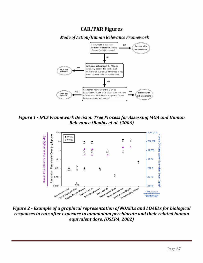
Page 67
CAR/PXR Figures
Figure 1 - IPCS Framework Decision Tree Process for Assessing MOA and Human Relevance (Boobis et al. (2006)
Figure 2 - Example of a graphical representation of NOAELs and LOAELs for biological responses in rats after exposure to ammonium perchlorate and their related human
equivalent dose. (USEPA, 2002)
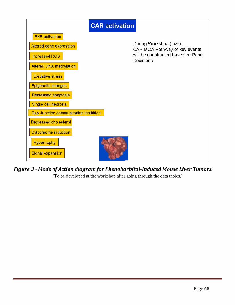
Page 68
Figure 3 - Mode of Action diagram for Phenobarbital-Induced Mouse Liver Tumors. (To be developed at the workshop after going through the data tables.)

Page 69
CAR/PXR Tables Four tables are found below. Tables 1-4 are inter-related and Tables 2-4 depend on how one fills out Table 1 – Hill criteria for key events determination. Table 1 has been populated with proposed events based on the current literature and recent research; other events may be appropriate to add. Two lines have been filled in for examples. These tables will be filled in and discussed during the workshop. The possible involvement of more than one mode of action at the tumor site should be considered. Pertinent observations that are not consistent with the hypothesized mode of action can suggest the possibility of other modes of action. Some pertinent observations can be consistent with more than one mode of action. Furthermore, different modes of action can operate in different dose ranges. If there is evidence for more than one mode of action, each should receive a separate analysis. The information on all of the modes of action should be integrated to better understand how and when each mode acts, and which mode(s) may be of interest for exposure levels relevant to human exposures of interest.
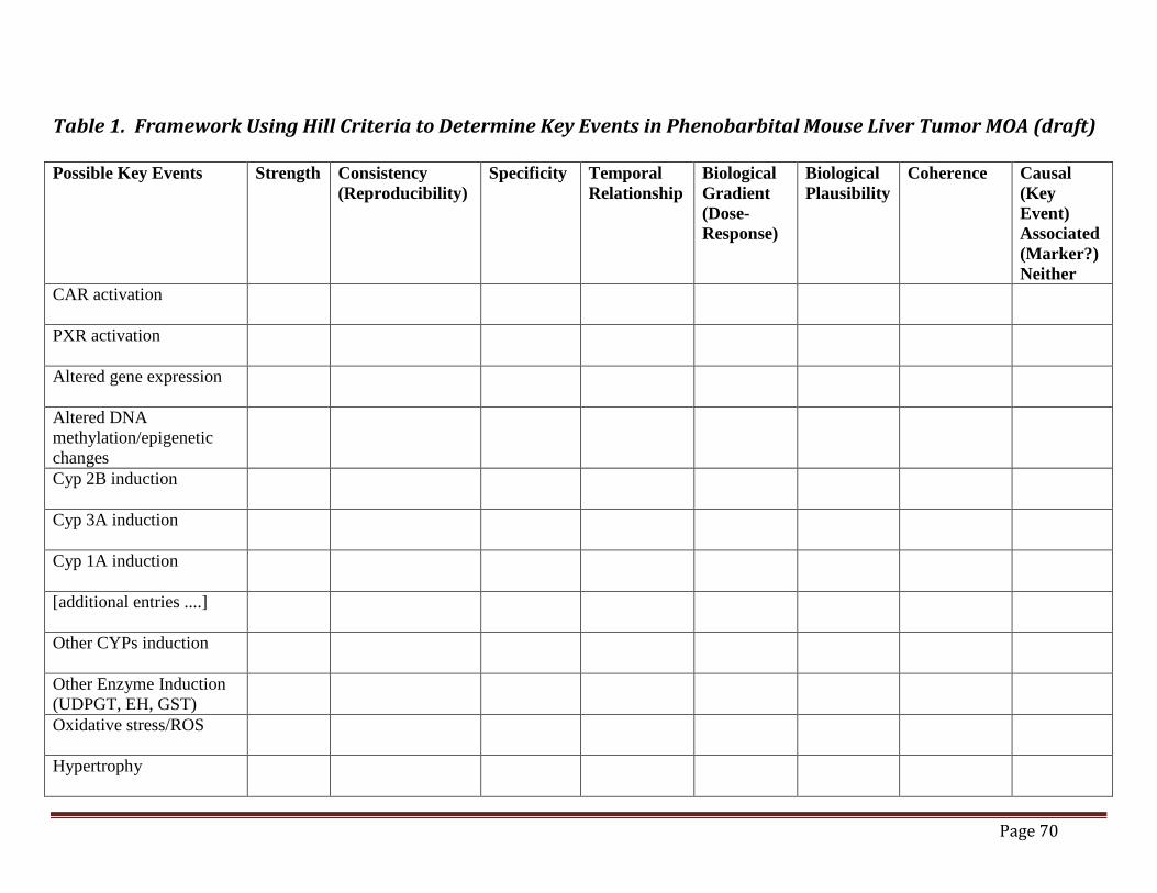
Page 70
Table 1. Framework Using Hill Criteria to Determine Key Events in Phenobarbital Mouse Liver Tumor MOA (draft) Possible Key Events Strength Consistency
(Reproducibility) Specificity Temporal
Relationship Biological Gradient (Dose-Response)
Biological Plausibility
Coherence Causal (Key Event) Associated (Marker?) Neither
CAR activation
PXR activation
Altered gene expression
Altered DNA methylation/epigenetic changes
Cyp 2B induction
Cyp 3A induction
Cyp 1A induction
[additional entries ....]
Other CYPs induction
Other Enzyme Induction (UDPGT, EH, GST)
Oxidative stress/ROS
Hypertrophy

Page 71
Possible Key Events Strength Consistency (Reproducibility)
Specificity Temporal Relationship
Biological Gradient (Dose-Response)
Biological Plausibility
Coherence Causal (Key Event) Associated (Marker?) Neither
Increased cell proliferation
Decreased apoptosis
Single cell necrosis
Gap Junction communication inhibition
Decreased cholesterol
Clonal expansion
Tumors
Others......
Page 72
Table 2. Time Concordance Table (draft) Events Species
and Strain Times tested and dose levels
In vitro data (time?)
1 day 2-6 days 7-27 days
28-90 days
91 days – 1 yr
>1 yr Ref.
CAR activation
PXR activation
Altered gene expression
Altered DNA methylation/epigenetic changes
Cyp 2B induction
Cyp 3A induction
Cyp 1A induction
Other CYPs induction
Other Enzyme Induction (UDPGT, EH, GST)
Oxidative stress/ROS
Hypertrophy
Increased cell proliferation
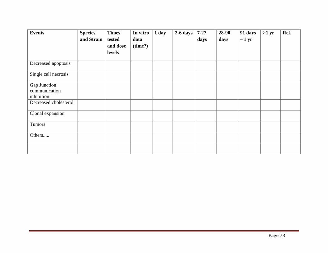
Page 73
Events Species and Strain
Times tested and dose levels
In vitro data (time?)
1 day 2-6 days 7-27 days
28-90 days
91 days – 1 yr
>1 yr Ref.
Decreased apoptosis
Single cell necrosis
Gap Junction communication inhibition
Decreased cholesterol
Clonal expansion
Tumors
Others.....

Page 74
Table 3. Dose Concordance Table (draft) Key Event or Marker Species and
Strain Doses Tested NOEL LOEL Reference
CAR activation
PXR activation
Altered gene expression
Altered DNA methylation/epigenetic changes
Cyp 2B induction
Cyp 3A induction
Cyp 1A induction
Other CYPs induction
Other Enzyme Induction (UDPGT, EH, GST)
Oxidative stress/ROS
Hypertrophy
Increased cell proliferation
Decreased apoptosis
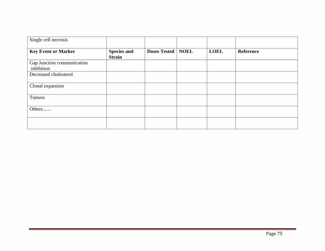
Page 75
Single cell necrosis
Key Event or Marker Species and Strain
Doses Tested NOEL LOEL Reference
Gap Junction communication inhibition
Decreased cholesterol
Clonal expansion
Tumors
Others.......

Page 76
Table 4. Species Concordance Table (draft) Key Event or Marker Mouse Rat Hamsters Primates Human
CAR activation
PXR activation
Altered gene expression
Altered DNA methylation/epigenetic changes
Cyp 2B induction
Cyp 3A induction
Cyp 1A induction
Other CYPs
Other Enzyme Induction (UDPGT, EH, GST)
Oxidative stress/ROS
Hypertrophy
Increased cell proliferation
Decreased apoptosis
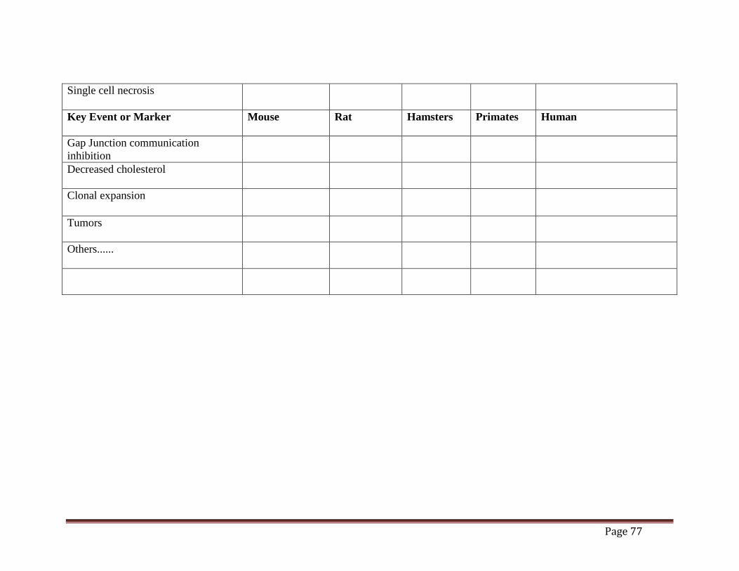
Page 77
Single cell necrosis
Key Event or Marker Mouse Rat Hamsters Primates Human
Gap Junction communication inhibition
Decreased cholesterol
Clonal expansion
Tumors
Others......


And also thanks to: DuPont; U.S. EPA Office of Chemical Safety and Pollution Prevention, U.S. EPA Office of Water