Embed Size (px)
Citation preview
© 2011 The Korean Academy of Medical Sciences.This is an Open Access article distributed under the terms of the Creative Commons Attribution Non-Commercial License (http://creativecommons.org/licenses/by-nc/3.0) which permits unrestricted non-commercial use, distribution, and reproduction in any medium, provided the original work is properly cited.
pISSN 1011-8934eISSN 1598-6357
A Case of Sheehan’s Syndrome that Manifested as Bilateral Ptosis
Hypothyroidism can cause a variety of signs and symptoms of the neuromuscular system. However, ptosis in a patient with hypothyroidism is very rare. We report here on a case of central hypothyroidism that was due to Sheehan’s syndrome and it manifested as bilateral ptosis in a 51-yr-old woman. She complained of exertional dyspnea and weakness. About 25-yr ago, she had a history of severe postpartum vaginal bleeding. The laboratory studies demonstrated hypopituitarism with secondary hypothyroidism. The ptosis was improved by replacement of thyroid hormone. Hypothyroidism should be considered in the differential diagnosis of patients who manifest with ptosis and that prompt replacement of hormone can lead to a complete recovery.
Key Words: Hypothyroidism; Hypopituitarism; Ptosis
Young Sil Lee, and Seong Su Moon
Department of Internal Medicine, College of Medicine, Dongguk University, Gyeongju, Korea
Received: 15 September 2010Accepted: 8 December 2010
Address for Correspondence:Young Sil Lee, MDDepartment of Internal Medicine, College of Medicine, Dongguk University, 307 Dongdae-ro, Gyeongju 780-350, KoreaTel: +82.54-770-8214, Fax: +82.54-770-8378E-Mail: [email protected]
DOI: 10.3346/jkms.2011.26.4.580 • J Korean Med Sci 2011; 26: 580-582
CASE REPORTEndocrinology, Nutrition & Metabolism
INTORDUCTION Ptosis may be due to myogenic, neurogenic, aponeurotic, me-chanical or traumatic causes. The most frequent disorder caus-ing myogenic ptosis is myasthenia gravis (1). Hypothyroidism can cause a variety of signs and symptoms of the neuromuscu-lar system (2). However, in the absence of other obvious etiolo-gies, ptosis in a patient with hypothyroidism is very rare (3-5). We report here on a case of central hypothyroidism that was due to Sheehan’s syndrome and the patient manifested with bi-lateral ptosis, and the patient was initially suspected as having myasthenia gravis.
CASE DESCRIPTION
The patient was a 51-yr-old woman with swelling of the extrem-ities and bilateral ptosis on March 8, 2010. She complained of exertional dyspnea and weakness. She married at the age of 25. After that time, she developed drooping of her eyes, with the left eye drooping more than the right eye. Her symptoms persisted and they had gradually increased during about the last 10 yr. About 8 yr ago, she underwent a ptosis repair operation at a lo-cal clinic, but her symptoms were not improved. She denied a history of loss of consciousness, headache, dysphagia, dysarthria and myalgia. There was no family history of similar symptoms and no history of trauma. There was no previous history of dia-betes and hypertension. On examination, she was determined to be 151 cm in height and 48 kg in weight. The initial vital signs were a blood pressure of 140/80 mmHg, a pulse rate of 68 beats/min and a respiratory rate of 20/min. She was lethargic and had facial puffiness and
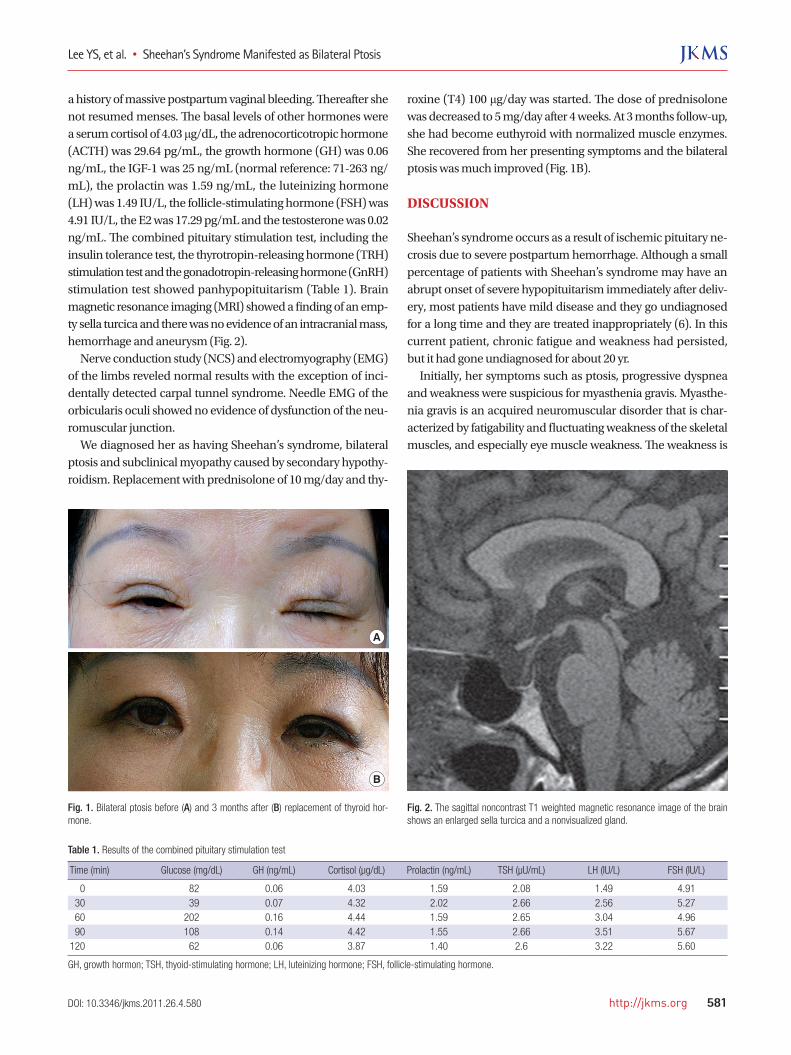
no goiter. The lung and heart examinations were unremarkable. All the extremities showed pitting. She had marked bilateral pto-sis, while the external ocular movements were normal (Fig. 1A). The other cranial nerve examinations were unremarkable. The laboratory findings were a total leucocyte count 4.75 × 10³/μL with 61.6% polymorphs, a hemoglobin level of 9.0 g/dL, the random blood glucose was 124 mg/dL, the serum sodium was 130 mM/L, the potassium was 3.7 mM/L, the blood urea nitrogen (BUN) was 13 mg/dL, the creatinine was 0.8 mg/dL, the aspartate transaminase (AST) was 81 IU/L, the alanine trans-aminase (ALT) was 29 IU/L, the total bilirubin was 0.93 mg/dL, the creatine kinase (CK) was 1,195 IU/L (normal reference: 20-180 IU/L ), the lactate dehydrogenase (LDH) was 517 IU/L (nor-mal reference: 101-218 IU/L), the total cholesterol was 256 mg/dL, the triglyceride was 121 mg/dL, the high density lipoprotein cholesterol (HDL-C) was 40 mg/dL and the low density lipopro-tein cholesterol (LDL-C) was 196 mg/dL. The urinary analysis was negative for blood and protein with using a dipstick. As the clinical findings suggested hypothyroidism and myasthenia gravis, a thyroid function test and acetylcholine receptor bind-ing antibody test were done. The serum T3 was 0.195 ng/mL (normal reference: 0.86-2.02 ng/mL), the free T4 was 0.08 ng/dL (normal reference: 0.93-1.705 ng/dL), the thyroid-stimulat-ing hormone (TSH) was 2.08 μIU/mL (normal reference: 0.27-4.2 μIU/mL), the antimicrosomal antibody was 17 IU/mL (nor-mal reference: 0-34 IU/mL), the antithyroglobulin antibody was 20.1 IU/mL (normal reference: 0-114 IU/mL) and the acetyl-choline receptor binding antibody was negative. Secondary hypothyroidism was suspected. We performed a careful history taking and other pituitary hormone evaluations. At the age of 26 the patient delivered her daughter and she had
Lee YS, et al. • Sheehan’s Syndrome Manifested as Bilateral Ptosis
http://jkms.org 581DOI: 10.3346/jkms.2011.26.4.580
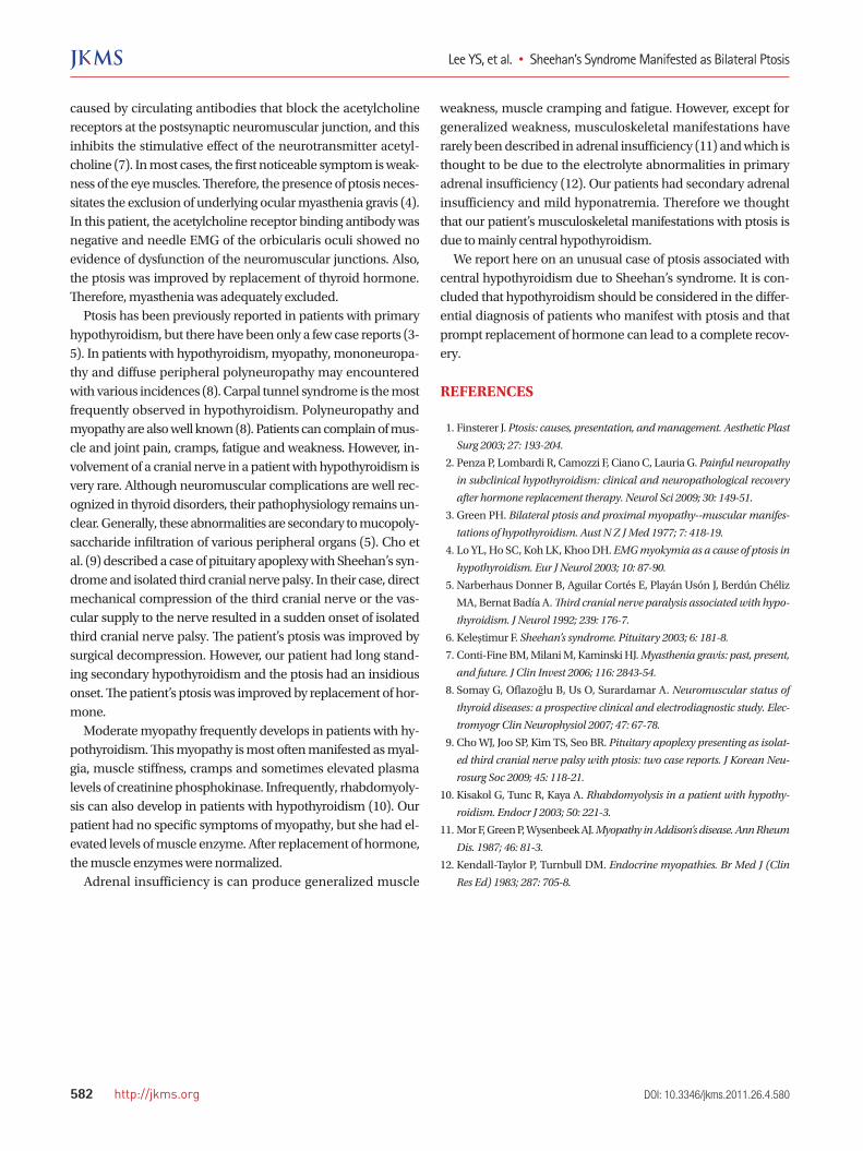
a history of massive postpartum vaginal bleeding. Thereafter she not resumed menses. The basal levels of other hormones were a serum cortisol of 4.03 μg/dL, the adrenocorticotropic hormone (ACTH) was 29.64 pg/mL, the growth hormone (GH) was 0.06 ng/mL, the IGF-1 was 25 ng/mL (normal reference: 71-263 ng/mL), the prolactin was 1.59 ng/mL, the luteinizing hormone (LH) was 1.49 IU/L, the follicle-stimulating hormone (FSH) was 4.91 IU/L, the E2 was 17.29 pg/mL and the testosterone was 0.02 ng/mL. The combined pituitary stimulation test, including the insulin tolerance test, the thyrotropin-releasing hormone (TRH) stimulation test and the gonadotropin-releasing hormone (GnRH) stimulation test showed panhypopituitarism (Table 1). Brain magnetic resonance imaging (MRI) showed a finding of an emp-ty sella turcica and there was no evidence of an intracranial mass, hemorrhage and aneurysm (Fig. 2). Nerve conduction study (NCS) and electromyography (EMG) of the limbs reveled normal results with the exception of inci-dentally detected carpal tunnel syndrome. Needle EMG of the orbicularis oculi showed no evidence of dysfunction of the neu-romuscular junction. We diagnosed her as having Sheehan’s syndrome, bilateral ptosis and subclinical myopathy caused by secondary hypothy-roidism. Replacement with prednisolone of 10 mg/day and thy-
roxine (T4) 100 μg/day was started. The dose of prednisolone was decreased to 5 mg/day after 4 weeks. At 3 months follow-up, she had become euthyroid with normalized muscle enzymes. She recovered from her presenting symptoms and the bilateral ptosis was much improved (Fig. 1B).
DISCUSSION
Sheehan’s syndrome occurs as a result of ischemic pituitary ne-crosis due to severe postpartum hemorrhage. Although a small percentage of patients with Sheehan’s syndrome may have an abrupt onset of severe hypopituitarism immediately after deliv-ery, most patients have mild disease and they go undiagnosed for a long time and they are treated inappropriately (6). In this current patient, chronic fatigue and weakness had persisted, but it had gone undiagnosed for about 20 yr. Initially, her symptoms such as ptosis, progressive dyspnea and weakness were suspicious for myasthenia gravis. Myasthe-nia gravis is an acquired neuromuscular disorder that is char-acterized by fatigability and fluctuating weakness of the skeletal muscles, and especially eye muscle weakness. The weakness is
Table 1. Results of the combined pituitary stimulation test
Time (min) Glucose (mg/dL) GH (ng/mL) Cortisol (µg/dL) Prolactin (ng/mL) TSH (µU/mL) LH (IU/L) FSH (IU/L)
0 82 0.06 4.03 1.59 2.08 1.49 4.91 30 39 0.07 4.32 2.02 2.66 2.56 5.27 60 202 0.16 4.44 1.59 2.65 3.04 4.96 90 108 0.14 4.42 1.55 2.66 3.51 5.67120 62 0.06 3.87 1.40 2.6 3.22 5.60
GH, growth hormon; TSH, thyoid-stimulating hormone; LH, luteinizing hormone; FSH, follicle-stimulating hormone.
A
B
Fig. 1. Bilateral ptosis before (A) and 3 months after (B) replacement of thyroid hor-mone.
Fig. 2. The sagittal noncontrast T1 weighted magnetic resonance image of the brain shows an enlarged sella turcica and a nonvisualized gland.
Lee YS, et al. • Sheehan’s Syndrome Manifested as Bilateral Ptosis
582 http://jkms.org DOI: 10.3346/jkms.2011.26.4.580
caused by circulating antibodies that block the acetylcholine receptors at the postsynaptic neuromuscular junction, and this inhibits the stimulative effect of the neurotransmitter acetyl-choline (7). In most cases, the first noticeable symptom is weak-ness of the eye muscles. Therefore, the presence of ptosis neces-sitates the exclusion of underlying ocular myasthenia gravis (4). In this patient, the acetylcholine receptor binding antibody was negative and needle EMG of the orbicularis oculi showed no evidence of dysfunction of the neuromuscular junctions. Also, the ptosis was improved by replacement of thyroid hormone. Therefore, myasthenia was adequately excluded. Ptosis has been previously reported in patients with primary hypothyroidism, but there have been only a few case reports (3-5). In patients with hypothyroidism, myopathy, mononeuropa-thy and diffuse peripheral polyneuropathy may encountered with various incidences (8). Carpal tunnel syndrome is the most frequently observed in hypothyroidism. Polyneuropathy and myopathy are also well known (8). Patients can complain of mus-cle and joint pain, cramps, fatigue and weakness. However, in-volvement of a cranial nerve in a patient with hypothyroidism is very rare. Although neuromuscular complications are well rec-ognized in thyroid disorders, their pathophysiology remains un-clear. Generally, these abnormalities are secondary to mucopoly-saccharide infiltration of various peripheral organs (5). Cho et al. (9) described a case of pituitary apoplexy with Sheehan’s syn-drome and isolated third cranial nerve palsy. In their case, direct mechanical compression of the third cranial nerve or the vas-cular supply to the nerve resulted in a sudden onset of isolated third cranial nerve palsy. The patient’s ptosis was improved by surgical decompression. However, our patient had long stand-ing secondary hypothyroidism and the ptosis had an insidious onset. The patient’s ptosis was improved by replacement of hor-mone. Moderate myopathy frequently develops in patients with hy-pothyroidism. This myopathy is most often manifested as myal-gia, muscle stiffness, cramps and sometimes elevated plasma levels of creatinine phosphokinase. Infrequently, rhabdomyoly-sis can also develop in patients with hypothyroidism (10). Our patient had no specific symptoms of myopathy, but she had el-evated levels of muscle enzyme. After replacement of hormone, the muscle enzymes were normalized. Adrenal insufficiency is can produce generalized muscle
weakness, muscle cramping and fatigue. However, except for generalized weakness, musculoskeletal manifestations have rarely been described in adrenal insufficiency (11) and which is thought to be due to the electrolyte abnormalities in primary adrenal insufficiency (12). Our patients had secondary adrenal insufficiency and mild hyponatremia. Therefore we thought that our patient’s musculoskeletal manifestations with ptosis is due to mainly central hypothyroidism. We report here on an unusual case of ptosis associated with central hypothyroidism due to Sheehan’s syndrome. It is con-cluded that hypothyroidism should be considered in the differ-ential diagnosis of patients who manifest with ptosis and that prompt replacement of hormone can lead to a complete recov-ery.
REFERENCES
1. Finsterer J. Ptosis: causes, presentation, and management. Aesthetic Plast
Surg 2003; 27: 193-204.
2. Penza P, Lombardi R, Camozzi F, Ciano C, Lauria G. Painful neuropathy
in subclinical hypothyroidism: clinical and neuropathological recovery
after hormone replacement therapy. Neurol Sci 2009; 30: 149-51.
3. Green PH. Bilateral ptosis and proximal myopathy--muscular manifes-
tations of hypothyroidism. Aust N Z J Med 1977; 7: 418-19.
4. Lo YL, Ho SC, Koh LK, Khoo DH. EMG myokymia as a cause of ptosis in
hypothyroidism. Eur J Neurol 2003; 10: 87-90.
5. Narberhaus Donner B, Aguilar Cortés E, Playán Usón J, Berdún Chéliz
MA, Bernat Badía A. Third cranial nerve paralysis associated with hypo-
thyroidism. J Neurol 1992; 239: 176-7.
6. Keleştimur F. Sheehan’s syndrome. Pituitary 2003; 6: 181-8.
7. Conti-Fine BM, Milani M, Kaminski HJ. Myasthenia gravis: past, present,
and future. J Clin Invest 2006; 116: 2843-54.
8. Somay G, Oflazoğlu B, Us O, Surardamar A. Neuromuscular status of
thyroid diseases: a prospective clinical and electrodiagnostic study. Elec-
tromyogr Clin Neurophysiol 2007; 47: 67-78.
9. Cho WJ, Joo SP, Kim TS, Seo BR. Pituitary apoplexy presenting as isolat-
ed third cranial nerve palsy with ptosis: two case reports. J Korean Neu-
rosurg Soc 2009; 45: 118-21.
10. Kisakol G, Tunc R, Kaya A. Rhabdomyolysis in a patient with hypothy-
roidism. Endocr J 2003; 50: 221-3.
11. Mor F, Green P, Wysenbeek AJ. Myopathy in Addison’s disease. Ann Rheum
Dis. 1987; 46: 81-3.
12. Kendall-Taylor P, Turnbull DM. Endocrine myopathies. Br Med J (Clin
Res Ed) 1983; 287: 705-8.