Embed Size (px)
Citation preview
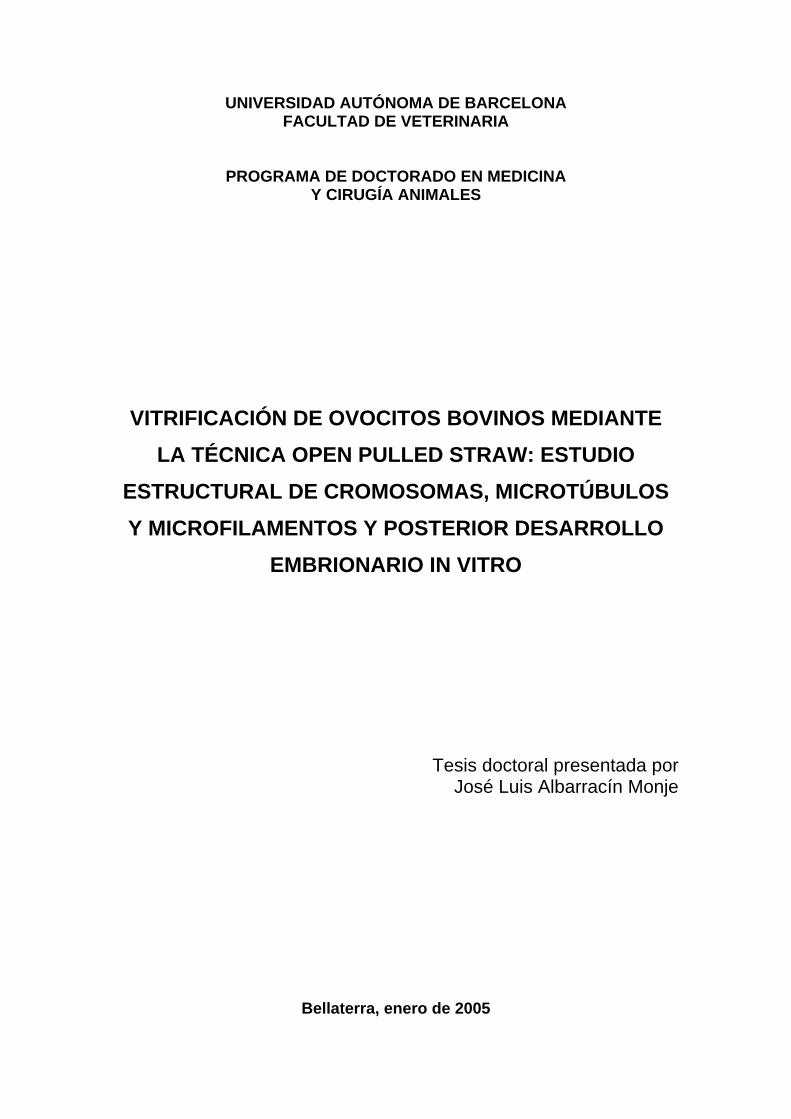
UNIVERSIDAD AUTÓNOMA DE BARCELONA FACULTAD DE VETERINARIA
PROGRAMA DE DOCTORADO EN MEDICINA Y CIRUGÍA ANIMALES
VITRIFICACIÓN DE OVOCITOS BOVINOS MEDIANTE LA TÉCNICA OPEN PULLED STRAW: ESTUDIO
ESTRUCTURAL DE CROMOSOMAS, MICROTÚBULOS Y MICROFILAMENTOS Y POSTERIOR DESARROLLO
EMBRIONARIO IN VITRO
Tesis doctoral presentada por José Luis Albarracín Monje
Bellaterra, enero de 2005
ÍNDICE
CAPÍTULO I. INTRODUCCIÓN Y OBJETIVOS............................................ 1
CAPÍTULO II. REVISIÓN BIBLIOGRÁFICA................................................... 11
2.1. Maduración nuclear y citoplasmática del ovocito.......................................... 13
2.1.1. Bloqueo meiótico y reinicio de la meiosis.......... ........................................ 14
2.1.2. Inhibición del reinicio de la meiosis. .......................................................... 16
2.2. Principios generales de crioconservación........................................................ 19
2.1. Congelación lenta............................................................................................ 20
2.2 . Congelación rápida y ultrarrápida............................................................... 22
2.3. Crioconservación de ovocitos........................................................................... 23
2.3.1. Efectos del enfriamiento y la congelación sobre las estructuras ovocitarias.. 24
2.3.1.1. Efectos de la temperatura sobre la placa metafásica.................................. 24
2.3.1.2. Efectos de la temperatura sobre el citoesqueleto ovocitario....................... 27
2.3.1.3. Efectos de la temperatura sobre otras estructuras ovocitarias.................... 28
2.4. Vitrificación........................................................................................................ 29
2.4.1. Factores que influyen en la vitrificación....................................................... 30
2.4.1.1. Velocidades de congelación y descongelación........................................... 30
2.4.1.2. Crioprotectores y aditivos crioprotectores.................................................. 30
2.4.1.2.1. Crioprotectores permeables..................................................................... 31
2.4.1.2.2. Crioprotectores no permeables. .............................................................. 32
2.4.1.3. Estadios de maduración. ............................................................................ 33
2.4.1.4. Soportes para vitrificación. ........................................................................ 35
2.5. Análisis comparativo entre ovocitos de animales prepúberes y
adultos................................................................................................................
36
2.6. Bibliografía........................................................................................................ 39
CAPÍTULO III. Effects of vitrification in open pulled straws on the cytology of
in vitro matured prepubertal and adult bovine oocytes……….
51
3.1. Abstract............................................................................................................. 53
3.2. Introduction...................................................................................................... 53
3.3. Materials and Methods................................................................................… 55
3.4. Results................................................................................…………………… 60
3.5. Discussion.......................................................................................................... 62
3.6. Bibliography...................................................................................................... 66
Capítulo I: Introducción y objetivos
CAPÍTULO IV. Effects of roscovitine on the nuclear and cytoskeletal
components of calf oocytes and their subsequent
development.................................................................................
69
4.1. Abstract............................................................................................................. 71
4.2. Introduction...................................................................................................... 71
4.3. Materials and Methods.................................................................................... 73
4.4. Results................................................................................................................ 77
4.5. Discussion.......................................................................................................... 85
4.6. Bibliography...................................................................................................... 90
CAPÍTULO V. Vitrification of calf oocytes: effect of stage of maturation and
prematuration on the nuclear and cytoskeletal components
of calf oocytes and their subsequent development...................
93
5.1. Abstract............................................................................................................. 95
5.2. Introduction...................................................................................................... 95
5.3. Materials and Methods................................................................................… 97
5.4. Results........................................................................................................….... 104
5.5. Discussion.......................................................................................................... 109
5.6. Bibliography...................................................................................................... 113
CAPÍTULO VI. DISCUSIÓN GENERAL............................................................. 119
CAPITULO VII. CONCLUSIONES........................................................................ 131
CAPÍTULO I:
INTRODUCCIÓN Y OBJETIVOS.
Capítulo I: Introducción y objetivos
3
Los ovocitos bovinos recuperados de ovarios de matadero se han convertido en una
fuente ampliamente utilizada para procedimientos tales como la fecundación in vitro, la
clonación u otras tecnologías reproductivas relacionadas. Debido a que el ovocito sólo
permanece viable un periodo limitado de tiempo y a que el número de ovocitos que
puede ser recogido en un día determinado es limitado, la capacidad para mantener a los
ovocitos crioconservados incrementaría enormemente su utilidad en la investigación
básica así como en las aplicaciones comerciales. Sin embargo, y hasta la actualidad, la
capacidad de desarrollo de los ovocitos que han sido congelados, medida como el
número de embriones viables por ovocito congelado, se mantiene baja comparada con la
obtenida a partir de ovocitos frescos.
En la actualidad ya es posible crioconservar ovocitos y embriones de algunas especies
de mamíferos mediante los protocolos de congelación lenta, congelación rápida y
vitrificación. Sin embargo, y a pesar de los esfuerzos realizados por numerosos grupos
de investigación, son pocos los trabajos que documenten gestaciones y nacidos vivos a
partir de ovocitos congelados (Fuku et al., 1992; Hamano et al., 1992; Otoi et al., 1992;
Suzuki et al., 1996; Vieira et al., 2002).
La vitrificación, descrita inicialmente en embriones por Rall y Fahy (1985), corresponde
a una técnica de congelación ultrarrápida basada en el contacto directo entre la solución
de vitrificación que contiene los agentes crioprotectores y ovocitos o embriones con el
nitrógeno líquido. La definición física de la vitrificación es la solidificación de una
solución a baja temperatura sin que ésta llegue a cristalizar debido a un enorme
incremento de la viscosidad (Fahy, 1986), manteniendo así la distribución molecular e
iónica que existía antes de la congelación (Fahy et al., 1984).
La estrategia de la vitrificación es básicamente diferente a la estrategia de la
congelación lenta. Una velocidad lenta de congelación intenta mantener un delicado
balance entre varios factores los cuales pueden resultar en lesiones celulares provocadas
por la formación de cristales de hielo, los choques osmóticos, el efecto tóxico de los
crioprotectores, la concentración de electrolitos intracelulares, los daños por
enfriamiento, las fracturas en la zona pelúcida y las alteraciones de los organelos
intracelulares, el citoesqueleto o el contacto entre células (Massip et al., 1995;
Capítulo I: Introducción y objetivos
4
Dobrinsky, 1996; Kasai, 1996; Martino et al., 1996; Saha et al., 1996). La vitrificación
elimina totalmente la formación de cristales de hielo ya que al aumentar la velocidad de
congelación disminuyen los daños causados por el enfriamiento pasando rápidamente
por la zona de mayor peligro que está situada entre los +15º y los �5ºC (Dobrinsky,
1996; Martino et al., 1996; Isachenko et al., 1998).
El proceso de vitrificación requiere la presencia de una alta concentración de
crioprotectores, por lo que es necesario minimizar el daño celular provocado por el
estrés osmótico o la toxicidad química provocada por dichas concentraciones utilizadas.
Por ello, el principal objetivo de un protocolo de vitrificación debe ser la disminución
de la toxicidad sin una pérdida de la efectividad de los agentes crioprotectores
(Liebermann et al., 2002). Para reducir la toxicidad de las soluciones crioprotectoras se
han desarrollado diferentes protocolos donde se combinan uno o varios crioprotectores
que se suplementan con azúcares, macromoléculas, etc. En el bovino, el contacto de los
ovocitos con etilenglicol (Martino et al., 1996; Saunders y Parks, 1999) provocó daños
estructurales a nivel de huso cromosómico y filamentos de actina así como una
disminución de los ovocitos que alcanzaron el estadio de blastocisto.
Otro de los factores a considerar en el proceso de vitrificación son los estadios de
maduración meiótica de los ovocitos, los cuales también pueden influir en su capacidad
para sobrevivir a la criopreservación. La congelación de ovocitos en estadio de vesícula
germinal podría tener ventajas ya que los microtúbulos aún no están organizados en
forma de huso y el material genético está dentro del núcleo protegido por la membrana
nuclear. Sin embargo, los índices de supervivencia de ovocitos vitrificados en vesícula
germinal son todavía muy bajos en el vacuno (Suzuki et al., 1996; Vieira et al., 2002) y
porcino (Didion et al., 1990; Isachenko et al., 1998) y podría ser debido a que el
proceso de congelación dañaría las membranas plasmáticas o la comunicación
intercelular entre el ovocito y las células del cúmulus perjudicando así su posterior
maduración (Hochi et al., 1998).
El índice más alto de blastocistos obtenidos tras un proceso de vitrificación es a partir
de ovocitos bovinos criopreservados en metafase de la segunda división meiótica (25%
blastocistos: Vajta et al., (1998)). Algunas de las posibles causas del bajo porcentaje de
Capítulo I: Introducción y objetivos
5
blastocistos obtenidos en los estudios de vitrificación de ovocitos en estadio de metafase
II podría deberse al daño estructural irreversible provocado por la criopreservación en la
membrana del ovocito (Arav y Zeron, 1997), en la organización del huso con las
consecuentes alteraciones de los cromosomas en los ovocitos en metafase II (Hochi et
al., 1998; Saunders y Parks, 1999; Chen et al., 2001), en la distribución de los gránulos
corticales (Hyttel et al., 2000) o en la distribución de los filamentos de actina
(Hotamisligil et al., 1996; Saunders y Parks, 1999). Existen algunos trabajos en los que
se ha planteado la posibilidad de congelar en estadios intermedios de desarrollo, como
por ejemplo, vesícula germinal rota (Men et al., 2002) o en el periodo entre metafase I y
II (Le Gal y Massip, 1999) con un relativo éxito.
Un intento para mejorar los porcentajes de supervivencia y desarrollo embrionario de
los ovocitos tras su vitrificación consistiría en premadurar citoplasmáticamente al
ovocito antes de ser crioconservado. De esta forma, el ovocito podría ser congelado en
un estadio inmaduro, estadio donde las estructuras cromosómicas y microtubulares
estarían más protegidas, mientras que su competencia citoplasmática para desarrollarse
tras la congelación se vería incrementada. Esta premaduración podría conseguirse tras
un cultivo previo de los ovocitos con inhibidores de las quinasas dependientes de
ciclinas como pueden ser la roscovitina o la butirolactona I (Mermillod et al., 2000;
Lonergan et al., 2003). Mermillod et al. (2000) bloquearon el reinicio de la meiosis de
ovocitos bovinos durante 24 horas con un concentración 50 µM de roscovitina sin que
ello provocara una disminución en la capacidad de desarrollo de los embriones
obtenidos tras el tratamiento.
Para que un proceso de vitrificación tenga éxito, las muestras deben entrar en contacto
con el nitrógeno líquido (-196ºC) lo antes posible por lo que se debe minimizar el
tiempo en que la muestra entra en contacto con los vapores de nitrógeno (-180ºC) que se
producen al introducirla en el nitrógeno líquido. Por tanto, el tamaño de la muestra debe
ser lo más pequeño posible para que la capa de vapor que se forma al contacto con el
nitrógeno líquido sea mínima y la velocidad de congelación se vea aumentada
(Liebermann y Tucker, 2002). Para minimizar el volumen de la solución de vitrificación
se han descrito diferentes tipos de soportes físicos. Entre ellos podemos citar soportes
como open pulled straw, closed pulled straw, flexipet-denuding pipette, microgotas,
Capítulo I: Introducción y objetivos
6
gradillas de cobre de microscopía electrónica, sistema hemistraw, nylon mesh y
cryoloop (Vajta et al., 1998; Chen et al., 2001; Liebermann y Tucker, 2002).
La posibilidad de utilizar los ovocitos procedentes de animales prepúberes sería de gran
utilidad en los programas de mejora genética debido a que permiten acortar el intérvalo
generacional y, en consecuencia, aumentar la presión de selección. Sin embargo, son
varios los estudios que concluyen que los ovocitos procedentes de animales prepúberes
son menos competentes para desarrollarse tras la fecundación que aquellos provenientes
de animales adultos (revisado por Gandolfi et al., 2000). Si bien los ovocitos de vacas
prepúberes presentan porcentajes de división embrionaria parecidos a los obtenidos en
vacas, el desarrollo embrionario de estos ovocitos se mantiene bajo respecto al obtenido
con ovocitos de vacas adultas (Khatir et al., 1998). Esta baja eficiencia se atribuye a
cambios en la distribución de los gránulos corticales (Duby et al., 1996), a retrasos en la
formación del aster espermático (Damiani et al., 1996), a un menor diámetro celular
(Armstrong, 2001) o a un deficiente metabolismo energético, enzimático o proteico
(revisado por Gandolfi et al., 2000), resultados que sugieren un retraso o una deficiente
maduración citoplasmática in vitro.
Así, teniendo en cuenta las consideraciones anteriores, los objetivos de esta tesis
doctoral fueron los siguientes:
1. Evaluar los efectos de vitrificar ovocitos de vaca y ternera mediante la técnica
open pulled straw sobre el huso cromosómico y el citoesqueleto.
2. Valorar el desarrollo embrionario in vitro de ovocitos de vaca y ternera
vitrificados mediante la técnica open pulled straw.
3. Analizar la efectividad de diferentes concentraciones de roscovitina para inhibir
el reinicio de la meiosis en ovocitos de ternera durante 24 horas y analizar su
efecto sobre el huso cromosómico, el citoesqueleto y el posterior desarrollo
embrionario.
4. Determinar la influencia del estadio de maduración (vesícula germinal rota vs.
metafase de la segunda división meiótica) de ovocitos de ternera en el momento
de ser vitrificados por open pulled straw sobre el huso cromosómico, el
citoesqueleto y el posterior desarrollo embrionario in vitro.
Capítulo I: Introducción y objetivos
7
5. Evaluar los efectos de una premaduración con roscovitina previa a la
vitrificación por open pulled straw de ovocitos de ternera sobre el huso
cromosómico, el citoesqueleto y el desarrollo embrionario in vitro.
Bibliografía. Arav A and Zeron Y. 1997. Vitrification of bovine oocytes using modified minimum
drop size technique (MDS) is effected by the composition and concentration of the vitrification solution and by the cooling conditions. Theriogenology 47:341.
Armstrong DT. 2001. Effects of maternal age on oocyte developmental competence. Theriogenology 55(6):1303-1322.
Chen SU, Lien YR, Cheng YU, Chen HF, Ho HN and Yang YS. 2001. Vitrification of mouse oocytes using closed pulled straws (CPS) achieves a high survival and preserves good patterns of meiotic spindles, compared with conventional straws, open pulled straws (OPS) and grids. Human Reproduction 16(11):2350-2356.
Damiani P, Fissore RA, Cibelli JB, Long CR, Balise JJ, Robl JM and Duby RT. 1996. Evaluation of developmental competence, nuclear and ooplasmic maturation of calf oocytes. Mol Reprod Dev 45(4):521-534.
Didion BA, Pomp D, Martin MJ, Homanics GE and Markert CL. 1990. Observations on the cooling and cryopreservation of pig oocytes at the germinal vesicle stage. J Anim Sci 68(9):2803-2810.
Dobrinsky JR. 1996. Cellular approach to cryopreservation of embryos. Theriogenology 45(1):17-26.
Duby RT, Damiani P, Looney CR, Fissore RA and Robl JM. 1996. Prepuberal calves as oocyte donors: Promises and problems. Theriogenology 45:121-130.
Fahy GM. 1986. Vitrification: a new approach to organ cryopreservation. Prog Clin Biol Res 224:305-335.
Fahy GM, MacFarlane DR, Angell CA and Meryman HT. 1984. Vitrification as an approach to cryopreservation. Cryobiology 21(4):407-426.
Fuku E, Kojima T, Shioya Y, Marcus GJ and Downey BR. 1992. In vitro fertilization and development of frozen-thawed bovine oocytes. Cryobiology 29(4):485-492.
Gandolfi F, Vassena R and Lauria A. 2000. The developmental competence of the oocyte before puberty: Is something missing? Reprod Domest Anim 35:66-71.
Hamano S, Koikeda A, Kuwayama M and Nagai T. 1992. Full-term development of in vitro-matured, vitrified and fertilized bovine oocytes. Theriogenology 38(6):1085-1090.
Hochi S, Ito K, Hirabayashi M, Ueda M, Kimura K and Hanada A. 1998. Effect of nuclear stages during IVM on the survival of vitrified-warmed bovine oocytes. Theriogenology 49(4):787-796.
Hotamisligil S, Toner M and Powers RD. 1996. Changes in membrane integrity, cytoskeletal structure, and developmental potential of murine oocytes after vitrification in ethylene glycol. Biol Reprod 55(1):161-168.
Hyttel P, Vajta G and Callesen H. 2000. Vitrification of bovine oocytes with the open pulled straw method: ultrastructural consequences. Mol Reprod Dev 56(1):80-88.
Capítulo I: Introducción y objetivos
8
Isachenko V, Soler C, Isachenko E, Perez-Sanchez F and Grishchenko V. 1998. Vitrification of immature porcine oocytes: effects of lipid droplets, temperature, cytoskeleton, and addition and removal of cryoprotectant. Cryobiology 36(3):250-253.
Kasai M. 1996. Simple and efficient methods for vitrification of mammalian embryos. Animal Reproduction Science 42:67-75.
Khatir H, Lonergan P, Touze JL and Mermillod P. 1998. The characterization of bovine embryos obtained from prepubertal calf oocytes and their viability after non surgical embryo transfer. Theriogenology 50(8):1201-1210.
Le Gal F and Massip A. 1999. Cryopreservation of cattle oocytes: effects of meiotic stage, cycloheximide treatment, and vitrification procedure. Cryobiology 38(4):290-300.
Liebermann J, Nawroth F, Isachenko V, Isachenko E, Rahimi G and Tucker MJ. 2002. Potential importance of vitrification in reproductive medicine. Biol Reprod 67(6):1671-1680.
Liebermann J and Tucker MJ. 2002. Effect of carrier system on the yield of human oocytes and embryos as assessed by survival and developmental potential after vitrification. Reproduction 124(4):483-489.
Lonergan P, Faerge I, Hyttel PM, Boland M and Fair T. 2003. Ultrastructural modifications in bovine oocytes maintained in meiotic arrest in vitro using roscovitine or butyrolactone. Mol Reprod Dev 64(3):369-378.
Martino A, Songsasen N and Leibo SP. 1996. Development into blastocysts of bovine oocytes cryopreserved by ultra-rapid cooling. Biol Reprod 54(5):1059-1069.
Massip A, Mermillod P and Dinnyes A. 1995. Morphology and biochemistry of in-vitro produced bovine embryos: implications for their cryopreservation. Hum Reprod 10(11):3004-3011.
Men H, Monson RL and Rutledge JJ. 2002. Effect of meiotic stages and maturation protocols on bovine oocyte's resistance to cryopreservation. Theriogenology 57(3):1095-1103.
Mermillod P, Tomanek M, Marchal R and Meijer L. 2000. High developmental competence of cattle oocytes maintained at the germinal vesicle stage for 24 hours in culture by specific inhibition of MPF kinase activity. Mol Reprod Dev 55(1):89-95.
Otoi T, Tachikawa S, Kondo S and Suzuki T. 1992. Developmental capacity of bovine oocytes cryopreserved after maturation in vitro and of frozen-thawed bovine embryos derived from frozen mature oocytes. Theriogenology 38(4):711-719.
Rall WF and Fahy GM. 1985. Ice-free cryopreservation of mouse embryos at -196 degrees C by vitrification. Nature 313(6003):573-575.
Saha S, Rajamahendran R, Boediono A, Sumantri C and Suzuki T. 1996. Viability of bovine blastocysts obtained after 7, 8 or 9 days of culture in vitro following vitrification and one-step rehydration. Theriogenology 46(2):331-343.
Saunders KM and Parks JE. 1999. Effects of cryopreservation procedures on the cytology and fertilization rate of in vitro-matured bovine oocytes. Biol Reprod 61(1):178-187.
Suzuki T, Boediono A, Takagi M, Saha S and Sumantri C. 1996. Fertilization and development of frozen-thawed germinal vesicle bovine oocytes by a one-step dilution method in vitro. Cryobiology 33(5):515-524.
Vajta G, Holm P, Kuwayama M, Booth PJ, Jacobsen H, Greve T and Callesen H. 1998. Open Pulled Straw (OPS) vitrification: a new way to reduce cryoinjuries of bovine ova and embryos. Mol Reprod Dev 51(1):53-58.

Capítulo I: Introducción y objetivos
9
Vieira AD, Mezzalira A, Barbieri DP, Lehmkuhl RC, Rubin MI and Vajta G. 2002. Calves born after open pulled straw vitrification of immature bovine oocytes. Cryobiology 45(1):91-94.
CAPÍTULO II:
REVISIÓN BIBLIOGRÁFICA.
Capítulo II: Revisión Bibliográfica
13
2.1. Maduración nuclear y citoplasmática del ovocito.
Al nacimiento, los ovocitos de las especies mamíferas están bloqueados en la fase G2 de
la profase de la primera división meiótica (Wassarman y Albertini, 1994) y tienen que
reiniciar y completar la meiosis y la maduración para que la fecundación pueda llevarse
a cabo. La competencia meiótica, es decir, la capacidad de los ovocitos para reiniciar y
completar la meiosis, se adquiere progresivamente durante el crecimiento folicular y
ovocitario y está asociada a una serie de cambios nucleares y citoplasmáticos (Sorensen
y Wassarman, 1976; Pavlok et al., 1992; Lonergan et al., 1994; De Smedt et al., 1995;
Eppig, 1996).
Los principales cambios nucleares incluyen la ruptura de la vesícula germinal (GVBD),
condensación cromosómica y progresión a metafase I (MI), extrusión del primer
corpúsculo polar y bloqueo en la metafase de la segunda división meiótica (MII). In
vivo, el reinicio de la meiosis es provocado por el pico preovulatorio de LH. In vitro, el
reinicio de la meiosis y la maduración tienen lugar de manera espontánea después de
retirar físicamente al ovocito del folículo (Pincus y Enzmann, 1935; Edwards, 1965;
Moor, 1988).
En los ovocitos de conejo (Jelinkova et al., 1994), cerdo (Hirao et al., 1995), mono
rhesus (Schramm et al., 1993) y vaca (Lonergan et al., 1994; Fair et al., 1995), se ha
observado que la competencia meiótica está relacionada con el tamaño folicular. Así, en
el bovino se ha observado que sólo aquellos ovocitos que provienen de folículos de un
diámetro superior a 2 mm son meióticamente competentes (Fair et al., 1995). Por el
contrario, aquellos ovocitos con un diámetro inferior o los ovocitos corticales no
adquieren totalmente la competencia meiótica. De hecho, algunos de estos ovocitos son
capaces de realizar una meiosis parcial llegando hasta MI, pero pocos son capaces de
llegar hasta MII y, menos aún, de desarrollarse hasta estadio de blastocisto (Arlotto et
al., 1996).
Durante la maduración citoplasmática existe una reorganización de los organelos
citoplasmáticos (Hyttel et al., 1986; Shamsuddin et al., 1993; Ducibelia et al., 1994;
Caralco, 1995), comienza la síntesis de proteínas específicas (Moor y Crosby, 1986;
Sirard et al., 1989; Tatemoto y Horiuchi, 1995; Wu et al., 1996) y se produce un
Capítulo II: Revisión Bibliográfica
14
incremento de la actividad de las quinasas, iniciándose complejas cascadas de
fosforilación y desfosforilación de proteínas específicas que involucran a numerosas
quinasas como el metaphase promoting factor (MPF), la familia de las mitogen-
activated protein kinases (MAPK, MAPKK, MAPKKK), el factor citostático (CSF), el
AMPc y el receptor del epidermal growth factor (EGFR). Se cree que estas cascadas de
fosforilación activan moléculas reguladoras nucleares y ooplasmáticas (Motlik y
Rimkevicova, 1990; Collas et al., 1993; Gall et al., 1993; Goren y Dekel, 1994; Goren
et al., 1994; Gotoh y Nishida, 1995; Lèvesque y Sirard, 1995; Tatemoto y Terada, 1995;
Fissore et al., 1996). Todos estos aspectos determinan que la maduración citoplasmática
del ovocito no pueda ser observada por técnicas sencillas, pero que pueda ser
indirectamente evaluada valorando la capacidad del ovocito para ser fecundado y
desarrollarse in vitro hasta el estadio de blastocisto.
2.1.1. Bloqueo meiótico y reinicio de la meiosis.
In vivo, el pico preovulatorio de LH libera al ovocito de su primer bloqueo meiótico
conduciéndolo a su segundo bloqueo meiótico en MII. La progresión desde el primer al
segundo bloqueo meiótico se denomina maduración ovocitaria y a partir de aquí, el
ovocito está listo para ser ovulado y fecundado. Producto de la fecundación se produce
la activación del ovocito que le provocará la salida del segundo bloqueo meiótico.
Los mecanismos por los cuales se produce el bloqueo meiótico y su reinicio no están
claramente establecidos, pero probablemente dependen de factores procedentes de las
células foliculares (Faerge et al., 2001). Entre estos factores foliculares podemos
mencionar al AMPc (Tornell y Hillensjo, 1993) y las purinas. Se ha sugerido que el
AMPc producido por las células de la granulosa y transportado al ovocito es el
responsable de la inhibición de la meiosis in vivo (Dekel, 1988). La fosforilación de las
proteínas mediante la proteína quinasa A dependiente de AMPc (PKA) parece ser el
mecanismo de acción por el cual el AMPc inhibe el reinicio de la meiosis (Heikinheimo
y Gibbons, 1998).
Durante la maduración nuclear se produce un incremento de la actividad de las
quinasas, iniciándose cascadas específicas de fosforilación y desfosforilación del MPF
(maturation-promoting factor o metaphase-promoting factor) y la actuación de la
Capítulo II: Revisión Bibliográfica
15
MAPK (mitogen-activated protein kinase), los cuales son en último término los
responsables de la progresión meiótica (Nurse, 1990; Gotoh y Nishida, 1995), siendo
esta última quinasa un factor crítico en la regulación del inicio de la meiosis (Motlik y
Rimkevicova, 1990).
El MPF es un miembro del grupo de las quinasas dependientes de ciclinas. Esta serina
treonina quinasa está formada por la unidad catalítica p34cdc2, homóloga a la proteína
quinasa cdc2 de la levadura, y por la subunidad reguladora ciclina B (Dunphy et al.,
1988; Murray et al., 1989). Durante la fase de crecimiento de los ovocitos, las dos
subunidades forman un preMPF fosforilado en los residuos treonina 161, treonina 14 y
tirosina 15 de la subunidad p34cdc2. Al inicio de la maduración nuclear, se activa el
dímero a través de la desfosforilación específica del residuo tirosina 15. Esta
desfosforilación está catalizada por la proteína fosfatasa cdc25 (Norbury y Nurse,
1992).
La actividad del MPF puede ser determinada mediante el análisis de la actividad de la
histona H1 quinasa intrínseca a esta proteína. En el bovino y el porcino, la actividad de
esta histona H1 quinasa se ve incrementada a las 6 y 20 horas de haber comenzado la
maduración, respectivamente, llegando a su primer pico de actividad durante la MI, para
posteriormente decrecer durante la anafase I. La actividad de la enzima muestra un
segundo pico máximo durante el estadio de MII (Christmann et al., 1994; Fissore et al.,
1996).
Existen discrepancias respecto al papel que juega la proteína p34cdc2 en la condensación
de la cromatina. Por un lado, Kubelka et al. (1995) sostienen que la condensación de la
cromatina, en ovocitos porcinos, ocurre independientemente de la activación del MPF.
Por otro lado, Tatemoto y Terada (1998) sugieren que la p34cdc2 ejerce una profunda
influencia en la condensación de la cromatina en ovocitos bovinos mientras que
Kubelka et al. (2000) concluyen que la condensación de la cromatina se da sin que
exista la activación del MPF.
La MAPK, una quinasa serina/treonina, es uno de los principales factores que regulan la
maduración ovocitaria (Dunphy et al., 1988; Shibuya et al., 1992; Sun et al., 1999b).
Esta enzima se activa a través de señales extracelulares específicas y mediante
Capítulo II: Revisión Bibliográfica
16
diferentes vías de transducción (factores de crecimiento y neurotransmisores). La
MAPK es activada por la MAPK-quinasa (MAPKK) la cual es, a su vez, fosforilada por
varias quinasas aunque, en los ovocitos, el producto del proto-oncogen c-mos es
probablemente el que juega el principal papel (Sagata et al., 1989; Nebreda y Hunt,
1993). La activación del MPF y la MAPK, los cuales comparten sustratos incluyendo
proteínas implicadas en la formación de la membrana nuclear, condensación de la
cromatina y formación del huso (Peter et al., 1992; Verlhac et al., 1994), provocan el
reinicio, la progresión y el bloqueo de la meiosis en MII. Una actuación inapropiada o la
presencia de bajos niveles de estas quinasas puede afectar negativamente a la capacidad
de desarrollo de los ovocitos.
En los ovocitos de ratón y rata (Sun et al., 1999b), la MAPK se activa después de la
ruptura de la vesícula germinal y por tanto, coincide con el inicio de la meiosis.
También se ha observado que la inhibición del reinicio de la meiosis por AMPc o la
proteína quinasa C (PKC) ocurre simultáneamente con la inhibición de la MAPK (Sun
et al., 1999a; Sun et al., 1999b; Lu et al., 2001). Algunos estudios muestran que, para
que se produzca la ruptura de la vesícula germinal, el citoplasma del ovocito debe tener
la habilidad de fosforilar la MAPK (Sun et al., 1999b; Lu et al., 2001).
2.1.2. Inhibición del reinicio de la meiosis.
In vivo, los ovocitos llevan a cabo la maduración citoplasmática durante el crecimiento
folicular, periodo caracterizado por una gran síntesis de RNAm y de proteínas
necesarias para el reinicio de la meiosis y primeros estadios posteriores a la fecundación
(revisado por Wassarman, 1988).
Por el contrario, en los sistemas in vitro, el reinicio de la meiosis es inducido
espontáneamente por la transferencia de ovocitos meióticamente competentes a medios
de cultivo, retirándolos así de su ambiente folicular (Pincus y Enzmann, 1935;
Staigmiller y Moor, 1984). Si bien estos ovocitos son capaces de completar la meiosis,
no son capaces de realizar la maduración citoplasmática de manera adecuada, lo cual se
verá reflejado en su menor capacidad de fecundación y desarrollo embrionario, debido a
que probablemente se ha privado a estos ovocitos de factores que se producen en etapas
tardías del crecimiento folicular.
Capítulo II: Revisión Bibliográfica
17
Por ello, se ha hipotetizado que si los ovocitos son cultivados in vitro antes de su
maduración en condiciones que mantengan el bloqueo meiótico en vesícula germinal,
éstos pueden tener la oportunidad de madurar citoplasmáticamente y adquirir así, una
mayor capacidad para el desarrollo embrionario (Fouladi Nashta et al., 1998).
En un intento para mantener el bloqueo meiótico de los ovocitos se han probado
diferentes inhibidores fisiológicos y farmacológicos. Entre los inhibidores fisiológicos
citar el fluido folicular (Sirard y First, 1988), las células tecales (Richard y Sirard,
1996b; 1996a) y también las células del cúmulus (Petr et al., 1989).
El bloqueo meiótico también puede ser provocado usando diferentes inhibidores
farmacológicos como: la ciclohexamida, un inhibidor de la síntesis proteica (Lonergan
et al., 1997; Saeki et al., 1997; Le Beux et al., 2003), la 6-dimetilaminopurina, un
inhibidor no específico de la fosforilación (Avery et al., 1998), la butirolactona I (BL-I),
un compuesto natural que inhibe las quinasas dependientes de ciclinas y que también
tiene un efecto inhibitorio sobre otras proteínas quinasas como la MAPK (Kitagawa et
al., 1993; Motlik et al., 1998; Kubelka et al., 2000; Lonergan et al., 2000) y la
roscovitina (ROS), una purina conocida por inhibir de manera específica la actividad del
MPF en numerosos sistemas celulares (Mermillod et al., 2000).
Sin embargo, diferentes estudios han señalado que durante la inhibición del reinicio de
la meiosis, tiene lugar algún tipo de progresión meiótica (Fair et al., 2002; Lonergan et
al., 2003). De hecho, Vigneron et al. (2004) demostraron que algunos eventos
relacionados con el reinicio de la meiosis (modificación de la síntesis y fosforilación de
algunas proteínas) no son inhibidos en presencia de roscovitina y que la progresión
meiótica en ovocitos tratados con roscovitina es más rápida.
La roscovitina ha sido usada en ovocitos de diferentes especies para detener la
progresión meiótica: en el bovino, Faerge et al. (2001) obtuvieron un 81% de bloqueo
meiótico usando 25 µM; Mermillod et al. (2000) obtuvieron un 88% de bloqueo y un
36% de blastocistos con la misma dosis; en el equino, Franz et al. (2003) observaron un
84% de bloqueo meiótico y un 71% de división embrionaria utilizando una
concentración de 66 µM; en el porcino, Ju et al. (2003) consiguieron un 83% de
Capítulo II: Revisión Bibliográfica
18
bloqueo meiótico y un 51% de reinicio normal de la meiosis utilizando una
concentración de 80 µM; Le Beux et al. (2003) obtuvieron un 72 % de bloqueo de la
meiosis y un 30 % de reinicio normal de la meiosis utilizando 50 µM y Krischek y
Meineke (2001), utilizando 50 µM, observaron un 74% de inhibición de la meiosis y un
76.2% de los ovocitos mostraron condensación de la cromatina a las 28 horas de
maduración sin el inhibidor; en el canino, Songsasen et al. (2003) consiguieron un 60%
de bloqueo de la meiosis utilizando 25 µM.
Donnay et al. (2004) observaron que la premaduración de ovocitos de ternera con una
combinación de butirolactona y roscovitina (12,5 µM ROS + 6,25µM BL-I) o
únicamente con roscovitina (25 µM ROS) evitó el reinicio de la meiosis y su efecto fue
reversible después de una maduración in vitro durante 17 o 24 horas. Sin embargo, y a
diferencia de Mermillod et al (2000), evidenciaron una disminución significativa del
porcentaje de blastocistos obtenidos después de la fecundación.
Varios autores observaron que la utilización de sustancias que inhiben la meiosis puede
causar modificaciones en las estructuras ovocitarias. Faerge et al. (2001) observaron
que la roscovitina, la butirolactona, el 6-DMAP y la ciclohexamida producen
variaciones morfológicas en la membrana nuclear, en la localización de la cromatina en
relación con el nucleolo y en la presencia de diferentes poblaciones de gránulos
intranucleares. Por otra parte, Ju et al. (2003) evidenciaron que entre el 15 � 21% de los
ovocitos tratados con roscovitina mostraban una MII anormal con placas metafásicas
aberrantes y/o formación aberrante de los microtúbulos citoplasmáticos.
A nivel ultraestructural, Lonergan et al. (2003) describieron que la incubación de
ovocitos bovinos con BL-1 o ROS alteraba la integridad de las células del cúmulus que
rodean al ovocito, lo cual afectaba a su expansión durante la maduración in vitro. Ya en
el citoplasma, observaron que la ROS causaba el hinchamiento de las crestas
mitocondriales, mientras que la BL-1 provocaba el aumento de poblaciones de
mitocondrias pleomórficas así como el de mitocondrias con matrices lucen. La
utilización de cualquiera de los inhibidores causó la degeneración de los gránulos
corticales reduciendo su población. Por otro lado, a nivel de núcleo, ambos tratamientos
inhibitorios provocaron un enrollamiento de la membrana nuclear y aquellos ovocitos
Capítulo II: Revisión Bibliográfica
19
tratados con ROS presentaron estructuras aberrantes dentro del nucleoplasma (Lonergan
et al., 2003).
2.2. Principios generales de crioconservación.
La crioconservación de células junto con su almacenamiento a muy bajas temperaturas
es deseable tanto por razones biológicas como por razones comerciales. Al conservar
células a temperaturas extremadamente bajas (-196 ºC) es posible detener por completo
la actividad enzimática, la respiración celular, el metabolismo, el crecimiento, la
multiplicación, etc., es decir, es posible mantener células durante un largo período de
tiempo sin afectar a su viabilidad ni causar cambios genéticos (Schneider y Mazur,
1984). No obstante, la mayoría de las células mamíferas mueren cuando se exponen a
bajas temperaturas, a menos que previamente hayan sido expuestas a una solución que
las proteja y a rangos de enfriamiento y calentamiento específicos (revisado por Shaw et
al., 2000).
La crioconservación de gametos y embriones implica el contacto inicial de éstos con
soluciones crioprotectoras, su posterior congelación y finalmente su almacenamiento a
temperaturas bajo cero. Una vez descongelados, se procede a la dilución y posterior
eliminación de los crioprotectores para luego ser transferidos a un ambiente
fisiológicamente adecuado que permita a dichas células desarrollarse adecuadamente.
Estos pasos representan un gran estrés (estrés mecánico, termal y/o químico) para las
células al cual deberán ser capaces de sobrevivir.
Contrariamente a lo que comúnmente se cree, el mayor desafío que las células deben
soportar durante la crioconservación no es el causado por el almacenamiento a bajas
temperaturas, sino la letalidad de una zona intermedia de temperatura (+15 a -5 ºC),
intervalo por el cual las células deben pasar dos veces (una durante la congelación y otra
durante la descongelación) (Mazur, 1963).
Los protocolos de crioconservación han sido clasificados como �lentos o rápidos� de
acuerdo con la velocidad de enfriamiento y el tipo y concentración de los
crioprotectores usados. Sin embargo, los principios y objetivos de la crioconservación
son aplicables a ambos: 1. Proteger de los efectos del enfriamiento y congelación, 2.
Capítulo II: Revisión Bibliográfica
20
Evitar la formación de hielo intracelular y 3. Proteger de los efectos tóxicos de los
crioprotectores tanto a temperaturas bajas como altas (revisado por Parks y Ruffing,
1992; Critser et al., 1997; Paynter et al., 1999).
2.2.1. Congelación lenta.
La congelación lenta es un técnica de crioconservación en la que existe un �equilibrio�
entre la velocidad de enfriamiento, la velocidad de deshidratación y la velocidad de
formación de núcleos de hielo. El objetivo principal de este tipo de crioconservación es
el de controlar la velocidad de enfriamiento de tal forma que a medida que descienda la
temperatura se produzca la penetración del crioprotector al interior de la célula
produciéndose un equilibrio osmótico y disminuyendo la probabilidad de formación de
cristales de hielo intracelulares.
Para prevenir la formación de hielo intracelular o minimizar el daño que éste pueda causar,
todos los protocolos de congelación están destinados a deshidratar las células. En el caso
de los protocolos de congelación lenta, este proceso se consigue colocando a las células en
una solución que contiene entre un 10 y un 11% (v/v) de crioprotector (aproximadamente
1.5M). A continuación, la temperatura disminuye y se provoca la formación de hielo (ice
seeding) dentro de esta solución. A medida que los cristales de hielo crecen, el agua de la
solución pasa de líquido a sólido y la concentración extracelular de solutos incrementa
provocando la salida de agua de la célula. Cuanto más baja es la temperatura, más cantidad
de agua puede convertirse en hielo, pero la capacidad de la célula para eliminar el agua
intracelular también disminuye a medida que la temperatura disminuye. Por lo tanto, el
éxito de un protocolo de congelación lenta se basa en alcanzar el equilibrio entre la
velocidad a la que el agua abandona la célula y la velocidad con que este agua se convierte
en hielo.
Cuando una célula es enfriada a -5 ºC, tanto la célula como el medio que la rodea se
mantienen en un estado superfrío (supercooled). Entre -5 y -10 ºC empieza la formación
de núcleos de hielo (ya sea espontáneamente o inducidos mediante seeding) en el medio
externo, mientras que el medio intracelular se mantiene descongelado. Este agua
extracelular superfría posee un alto potencial químico y provoca el eflujo de agua desde
dentro de la célula hacia afuera, agua que captan los núcleos de hielo extracelular
Capítulo II: Revisión Bibliográfica
21
(revisado por Gao et al., 1997). La célula, al perder agua, se deshidrata y,
consecuentemente, se produce la penetración de los crioprotectores para mantener el
equilibrio osmótico. Una vez se alcanza la temperatura de formación de hielo
(normalmente entre -5 y -9ºC), la temperatura debe descender hasta -33ºC / -40ºC,
temperatura a la cual las células pueden ser ya sumergidas directamente en nitrógeno
líquido. Las células congeladas según esta metodología deberán descongelarse según
protocolos de descongelación rápida (revisado por Shaw et al., 2000).
La velocidad de enfriamiento para esta técnica va de 0,2 a 0,3 ºC/minuto. Sin embargo,
para lograr una correcta velocidad de enfriamiento, se necesitan equipos costosos que se
encargan, en todo momento, de controlar la tasa de enfriamiento de manera automática.
Si una célula se enfría a velocidades de enfriamiento demasiado rápidas el agua no tiene tiempo para mantener el equilibrio osmótico, es decir, el agua intracelular no tiene tiempo suficiente para salir y formar los núcleos de hielo fuera de la célula y, por tanto, se forman núcleos de hielo intracelular que lesionaran la célula (Mazur, 1990). Por el contrario, si la tasa de enfriamiento celular es demasiado lenta, el eflujo de agua de la célula es tan lento que las células están expuestas durante demasiado tiempo a una elevada concentración de sales y otros solutos. Esta situación puede reducir la supervivencia de dichas células ya que una elevada concentración de sales y solutos puede provocar cambios en el pH intracelular, peroxidaciones lipídicas, etc. (revisado por Gao et al., 1997). Es por esto que se considera que existe un rango de enfriamiento óptimo en el cual existe un equilibrio entre el descenso de la temperatura y la eliminación de agua por parte de la célula. Son varios los protocolos de congelación lenta utilizados para la congelación de ovocitos. Al congelar ovocitos bovinos en una solución de propilenglicol de 1.5 M, Otoi et al. (1997), Lim et al. (1999) y Mogas et al. (1999) obtuvieron buenos porcentajes de supervivencia de los ovocitos tras el proceso de congelación/descongelación (67.4%, 83% y 48% respectivamente). Sin embargo, el porcentaje de blastocistos obtenidos tras la congelación fue bajo (5%, 3% y 8% respectivamente). Para intentar mejorar el porcentaje de blastocistos obtenidos por ovocito congelado, diferentes autores han introducido variaciones a los protocolos de congelación lenta. Así se han probado distintos agentes crioprotectores como el propilenglicol, el etilenglicol, el
Capítulo II: Revisión Bibliográfica
22
DMSO y el glicerol (Otoi et al., 1993; Lim et al., 1999; Mogas et al., 1999), distintas velocidades de enfriamiento como por ejemplo 0.3, 0.6 y 0.9ºC/min (Otoi et al., 1994), distintas temperaturas para la inducción del hielo como son -5.5ºC (Otoi et al., 1994) y -9 ºC (Saunders y Parks, 1999), la adición de azúcares (sacarosa) a la solución crioprotectora (Otoi et al., 1995) o la centrifugación previa de los ovocitos (Otoi et al., 1997). Sin embargo, ninguna de las variaciones introducidas en los protocolos de congelación lenta ha permitido elevar los porcentajes de blastocistos obtenidos a niveles superiores al 8% (Mogas et al., 1999). Diferentes autores han sugerido que las soluciones utilizadas durante la congelación lenta podrían ser mejoradas añadiendo EDTA (Mogas et al., 1999), citocalasina (Younis et al., 1997), pequeñas moléculas lipofílicas como el hidroxitolueno butilado (Zeron et al., 1999), proteínas anticongelantes (Arav et al., 1993a) o sales (Stachecki et al., 1998). 2.2.2. Congelación rápida y ultrarrápida. Esta técnica previene la formación de hielo intracelular mediante la deshidratación de la célula. Para conseguirlo, se expone a la célula a altas concentraciones de un crioprotector permeable y, posteriormente, a un enfriamiento rápido o ultrarrápido.
Los protocolos de congelación rápida y ultrarrápida utilizan altas concentraciones de
solutos (crioprotectores y azúcares) que eliminan rápidamente el agua de las células. En
estas soluciones, las células se deshidratan rápidamente y se favorece la entrada de los
crioprotectores, lo que permite sumergirlas directamente en nitrógeno líquido
(congelación ultrarrápida) o vapores de nitrógeno (congelación rápida). Las tasas de
congelación conseguidas con las técnicas de congelación ultrarrápidas (11000 a 14000
ºC/minuto) disminuyen drásticamente el daño por enfriamiento, permitiendo usar
soluciones crioprotectoras menos concentradas (menos tóxicas) y acortar el tiempo de
exposición del ovocito al crioprotector (Martino et al., 1996).
Los protocolos de congelación rápida se dividen en dos subcategorías dependiendo de si
existe (congelación rápida o ultrarrápida) o no (vitrificación) formación de hielo en la
solución durante la congelación. Si bien la diferenciación entre congelación ultrarrápida
y vitrificación no está bien establecido, se debería utilizar el término vitrificación sólo
para aquellas técnicas en las que no se forman cristales de hielo durante la congelación
y descongelación, ni intracelular ni extracelular. Por el contrario, si se forman aunque
Capítulo II: Revisión Bibliográfica
23
sólo sean trazas de hielo durante estos procesos, el término correcto debería ser
congelación ultrarrápida (Shaw et al., 2000).
2.3. Crioconservación de ovocitos.
El ovocito es un tipo celular único que posee un gran tamaño y una baja relación
superficie-volumen. Además, está rodeado por la zona pelúcida (ZP) y por varias capas
de células del cúmulus oophorus. La presencia de estas capas celulares que rodean al
ovocito y que juegan un papel importante durante la etapa de crecimiento del ovocito
mediante una cooperación metabólica, hacen de este tipo celular una estructura muy
difícil de crioconservar (revisado por Massip, 2003). Hasta la actualidad, se han podido
crioconservar ovocitos de varias especies animales utilizando diferentes técnicas y
crioprotectores: en el conejo, por congelación lenta (Al-Hasani et al., 1989) y rápida
(Vincent et al., 1989); en la rata, por congelación lenta (Kasai et al., 1979) y
ultrarrápida (Isachenko et al., 2000); en el ratón, por congelación lenta (Stachecki et al.,
1998), ultrarrápida (Aono et al., 2003) y vitrificación (Kasai et al., 1990; O´Neil et al.,
1997; Chen et al., 2000; Tokieda et al., 2002); en la vaca, por congelación lenta (Lim et
al., 1991; Fuku et al., 1992; Otoi et al., 1995), ultrarrápida (Martino et al., 1996; Rho et
al., 2002) y vitrificación (Vajta et al., 1998; Hyttel et al., 2000; Vieira et al., 2002); en
la yegua, por congelación lenta (Hochi et al., 1994) y por vitrificación (Hurtt et al.,
2000); en la cerda, por congelación ultrarrápida (Isachenko et al., 1998) y por
vitrificación (Nagashima et al., 1996; Rojas et al., 2004) y en la mujer, por congelación
lenta (Boiso et al., 2002) y por vitrificación (Liebermann y Tucker, 2002).
Desafortunadamente, los protocolos desarrollados para una especie en concreto son
usualmente muy difíciles de adaptar a otra especie debido a diferencias en el tamaño del
ovocito y a su sensibilidad al enfriamiento y a los crioprotectores (revisado por Shaw et
al., 2000). El factor más crítico para la crioconservación de ovocitos es su compleja
organización subcelular. Los efectos del enfriamiento afectan a los elementos del
citoesqueleto como son la placa metafásica o la integridad de los microtúbulos (Shaw et
al., 2000) y también puede afectar a los gránulos corticales produciendo el
endurecimiento de la zona pelúcida (Parks y Ruffing, 1992).
Capítulo II: Revisión Bibliográfica
24
2.3.1. Efectos del enfriamiento y la congelación sobre las estructuras ovocitarias.
La mayoría de los estudios sobre la hipotermia en ovocitos mamíferos se limitan
principalmente a los efectos de ésta sobre la morfología, la citología y la competencia
para el desarrollo embrionario de los ovocitos enfriados o crioconservados y, más
concretamente, de los ovocitos que se encuentran en estadio de MII (Parks, 1997).
Cuando se somete a un ovocito a temperaturas inferiores a las fisiológicas y estas se
elevan nuevamente a 37 ºC, el ovocito sufre una serie de cambios físicos y fisiológicos.
No obstante, los mayores daños se producen entre +15 y -5 ºC. Temperaturas entre +30
y 0 ºC pueden comprometer la integridad de la membrana, el metabolismo celular, el
citoesqueleto y la capacidad celular de controlar y reparar los daños producidos por los
radicales libres (Arav et al., 1996; Parks, 1997). Por otro lado, las temperaturas por
debajo de 0 ºC suponen un alto riesgo de formación de hielo intracelular el cual puede
provocar un daño irreparable en la célula (Trad et al., 1999).
2.3.1.1. Efectos de la temperatura sobre la placa metafásica.
El cambio más drástico que sufren los ovocitos maduros cuando son sometidos a
cambios de temperatura es el desensamblaje de la placa metafásica (Magistrini y
Szollosi, 1980). La placa metafásica está compuesta por microtúbulos que contienen
subunidades α y β de dímeros de tubulina en equilibrio dinámico entre la forma libre y
la polimerizada. Este equilibrio es extremadamente termosensible y tiende al
desensamblaje por debajo de la temperatura fisiológica. Los cromosomas se encuentran
fuertemente unidos a la placa metafásica y, en ausencia de una envoltura nuclear, se
considera que la placa metafásica es una estructura relativamente inestable (revisado por
Bernard y Fuller, 1996).
La elevada termosensibilidad de la placa metafásica ha sido observada en ovocitos
murinos (Magistrini y Szollosi, 1980; Pickering y Johnson, 1987), humanos (Pickering
et al., 1990) y bovinos (Aman y Parks, 1994) no importando el tipo de crioconservación
utilizado: congelación lenta (Vincent et al., 1989; Eroglu et al., 1998), ultrarrápida
(Aigner et al., 1992) o vitrificación (Chen et al., 2003). Magistrini y Szollosi (1980)
observaron que los microtúbulos de la placa metafásica de ovocitos de ratón se
Capítulo II: Revisión Bibliográfica
25
despolimerizaban después de 15 minutos de exposición a 0 ºC y que desaparecían
completamente después de 45 � 60 minutos de exposición. Sin embargo, un progresivo
calentamiento de los ovocitos hasta la temperatura de 37 ºC provocó la repolimerización
de los microtúbulos y la formación de la placa metafásica. Esta habilidad para la
reorganización de la placa metafásica fue confirmada por Pickering y Johnson (1987) en
ovocitos de la misma especie. Saunders y Parks (1999) estudiaron los cambios
provocados en la distribución de los cromosomas, microtúbulos y microfilamentos tras el
enfriamiento, el contacto con el crioprotector (1.5 M de etilenglicol) o la congelación de
ovocitos bovinos madurados in vitro. En los ovocitos que sólo habían sido enfriados a -9ºC
durante 12 minutos observaron cambios que incluían dispersión o agrupamiento de
cromosomas, despolimerización de los microtúbulos y la alteración de la estructura del
huso cromosómico.
Aman y Parks (1994) demostraron que el enfriamiento de ovocitos bovinos madurados
in vitro a +25 ºC o a +4 ºC daba lugar a una disgregación o disociación parcial o total
del huso cromosómico y a la dispersión de algunos cromosomas. El recalentamiento de
los ovocitos a +37ºC desde +25ºC restauraba la morfología normal del huso, mientras
que los ovocitos que habían sido enfriados a +4ºC y recalentados presentaban husos
cromosómicos anormales. Por otro lado, tras someter ovocitos bovinos en estadio de
VG a diferentes temperaturas, Wu et al. (1999) observaron que temperaturas superiores
a 24 ºC no reducen los porcentajes de placas metafásicas normales ni los porcentajes de
fecundación y división embrionaria de los ovocitos. Sin embargo, cuando se somete a
los ovocitos a temperaturas cercanas a los 4 ºC ni que solo sea por 10 minutos, se reduce
drásticamente el porcentaje de husos meióticos normales y los porcentajes de división y
fecundación. Entre los daños provocados por la refrigeración sobre la estructura de la
placa metafásica se pudo observar desorganización, descondensación y ausencia de los
cromosomas. A nivel de microtúbulos, la exposición a bajas temperaturas afectó a la
organización microtubular provocada por la desorganización de los microtúbulos o la
ausencia de algunos o todos los microtúbulos. También como consecuencia de las bajas
temperaturas, Vincent y Jonson (1992) observaron un alineamiento cromosómico
anormal y la segregación de cromátidas en ovocitos de conejo.
Diversos autores han demostrado que la congelación lenta (Eroglu et al., 1998;
Saunders y Parks, 1999), rápida (Vincent et al., 1989), ultrarrápida (Aigner et al., 1992)
Capítulo II: Revisión Bibliográfica
26
y vitrificación (Chen et al., 2000) causan una severa desorganización o desaparición de
las placas metafásicas inmediatamente después de la congelación. En la congelación
lenta de ovocitos bovinos en estadio de MII, Saunders y Parks (1999) observaron que
ésta causaba la desorganización parcial o total de los cromosomas y/o los microtúbulos,
la despolimerización de los microtúbulos y la descondensación de los cromosomas.
Similares resultados fueron observados por Boiso et al. (2002) en ovocitos humanos
madurados in vitro, quienes describieron que la congelación lenta puede llegar a
producir la desorganización o total desaparición de los microtúbulos, la aparición de
cromosomas aberrantes y la descondensación parcial de los mismos.
Sin embargo, la posterior incubación de estos ovocitos a temperatura fisiológica resultó
en la recuperación de la estructura de la placa metafásica. Así, varios autores han
observado que la recuperación de la estructura de la placa metafásica requiere un
período de incubación posterior a la descongelación y que a su vez depende del método
de congelación y de la especie animal (Aigner et al., 1992; Gook et al., 1993; Chen et
al., 2001). En el caso de los bovinos, Saunders y Parks (1999) observaron que un 64%
de los ovocitos congelados recuperaban la estructura de la placa metafásica a los 20
minutos después de ser congelados mediante congelación lenta. Eroglu et al. (1998)
observaron que los ovocitos de ratón congelados mediante congelación lenta son
capaces de recuperar completamente la estructura de la placa metafásica después de 1
hora de incubación. Este período se alargó hasta 2 o 3 horas cuando estos ovocitos
fueron congelados por congelación ultrarrápida (Aigner et al., 1992) o vitrificación
(Chen et al., 2001). En el caso de ovocitos humanos Gook et al. (1993) observaron que
un 60% de los ovocitos congelados mediante congelación lenta recuperaban la
estructura normal de la placa metafásica después de 1 hora de incubación.
Estos cambios observados en la placa metafásica debidos al proceso de
congelación/descongelación provocan cambios funcionales del ovocito durante la
fecundación y posterior desarrollo embrionario (Eroglu et al., 1998; Chen et al., 2001),
reportándose en estos ovocitos un incremento de las poliploidías después de la
fecundación (Glenister et al., 1987; Carroll et al., 1989; Bouquet et al., 1992; Eroglu et
al., 1998; Luna et al., 2001).
Capítulo II: Revisión Bibliográfica
27
2.3.1.2. Efectos de la temperatura sobre el citoesqueleto ovocitario.
Los microfilamentos están compuestos por actina polimerizada la cual se encuentra en
equilibrio dinámico con la actina libre, de manera similar a los microtúbulos. Mientras
que los microtúbulos son importantes para aquellos procesos que involucran la correcta
distribución de los cromosomas en la placa metafásica, los microfilamentos son
igualmente importantes en diferentes etapas, incluyendo la citoquinesis, la rotación de la
placa metafásica, la extrusión del corpúsculo polar y la migración pronuclear (Vincent y
Johnson, 1992).
En contraste con los microtúbulos, los microfilamentos que forman el citoesqueleto
cortical del ovocito no parecen estar modificados directamente por el enfriamiento
(revisado por Parks, 1997). Sin embargo, existe la posibilidad que éstos se vean
afectados por las diferentes etapas de la crioconservación (revisado por Bernard y
Fuller, 1996).
Varios autores observaron que la congelación causaba cambios en la organización del
citoesqueleto de actina en ratón, conejo y vaca (Vincent et al., 1989; George y Johnson,
1993; Saunders y Parks, 1999, respectivamente). En estos ovocitos se pudo observar la
desaparición de la banda de actina por despolimerización y por segmentación (Vincent
et al., 1989). Cuando los ovocitos de ratón se evaluaron inmediatamente después de la
congelación, Eroglu et al. (1998) observaron alteraciones en el citoesqueleto tales como
la disrupción de la red de microfilamentos y la aparición de núcleos de microtúbulos en
el citoplasma. Sin embargo, estos cambios no fueron dramáticos y en su mayoría fueron
reversibles después de un período de incubación posterior a la descongelación. De
hecho, Saunders y Parks (1999) observaron un bajo porcentaje de alteraciones en la banda
de actina después de congelar ovocitos bovinos por congelación lenta.
La desestructuración del citoesqueleto puede ser debida a los cambios de forma y volumen
que acompañan a los procesos de congelación/descongelación, cambios que pueden
provocar la liberación prematura de los gránulos corticales y el endurecimiento de la zona
pelúcida (revisado por Vincent y Johnson, 1992). Por este motivo, aunque la distribución
de los microfilamentos del ovocito pueda recomponerse tras la congelación o el contacto
con los agentes crioprotectores, pueden haberse dado ya alteraciones irreversibles de otros
Capítulo II: Revisión Bibliográfica
28
componentes celulares como la zona pelúcida, membrana plasmática o las mitocondrias,
debido a la asociación existente entre los microfilamentos y estas estructuras.
2.3.1.3 Efectos de la temperatura sobre otras estructuras ovocitarias.
La exocitosis de los gránulos corticales es una respuesta normal a la unión del
espermatozoide fecundante con el oolema. Las proteasas acrosomales son eliminadas al
espacio perivitelino y modifican los componentes glucoproteicos de la zona pelúcida,
responsable de la unión y penetración del espermatozoide, previniendo posteriores
fecundaciones y bloqueando así la poliespermia (Webb et al., 1986; Wassarman y
Albertini, 1994). La migración y la liberación de los gránulos corticales involucra un
correcto funcionamiento del citoesqueleto y de la organización de la membrana
plasmática (revisado por Bernard y Fuller, 1996).
El enfriamiento de los ovocitos de ratón a 4 ºC durante 5 minutos alteró dramáticamente
la zona pelúcida reduciendo el porcentaje de fecundación (Vincent et al., 1990). Estos
cambios estarían asociados a una exocitosis prematura de los gránulos corticales, lo que
provocaría el endurecimiento de la zona pelúcida (Carroll et al., 1990; Vincent y
Johnson, 1992). Fuku et al. (1995) observaron pequeños cambios en la zona pelúcida
como la presencia de irregularidades, fracturas y cambios en la densidad electrónica
cuando vitrificaron ovocitos bovinos en pajuelas de 0.25 ml. Los autores también
asociaron estos cambios a la prematura exocitosis de los gránulos corticales. Hyttel et
al. (2000) confirmaron este hecho al detectar por microscopía electrónica la ausencia de
la típica banda de gránulos corticales por debajo de la membrana plasmática al vitrificar
ovocitos bovinos mediante la técnica Open pulled straw (OPS). Además, estos autores
observaron clusters de gránulos corticales en distintas etapas de degeneración (Hyttel et
al., 2000).
La congelación también causa profundos cambios en otras estructuras ovocitarias. Fuku
et al. (1995) observaron profundas modificaciones ultraestructurales tras la vitrificación
de ovocitos bovinos. Estos autores evidenciaron la ruptura de las uniones gap o la
ausencia de microvellocidades. A nivel de mictocondrias, las anormalidades se
centraban en la presencia de extensas áreas vacuolizadas, la desaparición de la mayoría
de las crestas mitocondriales y alteraciones en la membrana mitocondrial (Fuku et al.,
Capítulo II: Revisión Bibliográfica
29
1995), así como también una marcada disminución en el número de las mitocondrias
(Rho et al., 2002). También evidenciaron la presencia de cambios en la zona pelúcida
en los ovocitos madurados in vitro, debidos a la vitrificación, los cuales fueron
asociados a una disminución en el número de gránulos corticales en el ooplasma debido
a que la congelación causa una liberación prematura de éstos.
A nivel de DNA, Men et al. (2003) observaron que un elevado porcentaje de ovocitos
que microscópicamente no presentaban daño morfológico presentaban alteraciones a
nivel de DNA tras la congelación lenta o vitrificación de ovocitos bovinos madurados in
vitro.
2.4. Vitrificación.
La vitrificación se define como la transición de las soluciones acuosas de un estado
líquido a un estado vítreo sólido sin la formación de cristales, es decir, que debido al
rápido descenso de temperatura, la �viscosidad� de la muestra aumenta hasta un punto
en que las moléculas se inmovilizan. De esta forma, se encuentran en un estado sólido
aunque su estructura molecular sea la de un líquido extremadamente viscoso (estado
vítreo) (Critser et al., 1997). Este aumento extremo de la viscosidad requiere
velocidades de enfriamiento muy rápidas (superiores a 2500 ºC/min) o elevadas
concentraciones de crioprotectores (de 5 a 7 M) (revisado por Shaw et al., 2000; Vajta,
2000).
La vitrificación presenta numerosas ventajas como la total eliminación de la formación
de hielo o la disminución del daño causado por el enfriamiento, puesto que atraviesa el
rango de temperatura de +15 a �5 ºC a velocidades de enfriamiento muy rápidas
(Dobrinsky, 1996; Martino et al., 1996; Isachenko et al., 1998; Zeron et al., 1999). Otra
gran ventaja de esta técnica es que no requiere de equipos de congelación caros o
sofisticados y puede ser realizada de manera muy sencilla.
La consecuencia negativa de esta estrategia radica en el incremento de las
probabilidades de lesionar las células debido al choque osmótico y a la toxicidad de los
crioprotectores. Sin embargo, se han aplicado diferentes protocolos para intentar
disminuir estos efectos negativos, como el uso de crioprotectores menos tóxicos o la
Capítulo II: Revisión Bibliográfica
30
combinación de crioprotectores (disminuyendo la toxicidad individual de cada uno, pero
manteniendo las propiedades osmóticas y crioprotectoras), la utilización de
crioprotectores por etapas (stepwise) y/o la utilización de soluciones concentradas
preenfriadas (revisado por Vajta, 2000).
2.4.1. Factores que influyen en la vitrificación.
2.4.1.1 Velocidades de congelación y descongelación.
Uno de los parámetros más importantes para llevar a cabo la vitrificación con éxito es la
velocidad de enfriamiento. Existe una velocidad de enfriamiento límite, que es la
velocidad límite biológica que puede soportar una célula a una concentración de
crioprotector dada. Por tanto, el equilibrio entre la máxima velocidad de congelación y
la concentración del crioprotector será de mucha importancia para conseguir resultados
óptimos (Critser et al., 1997; Liebermann et al., 2002a). La principal estrategia de la
vitrificación es pasar el rango de temperatura crítica lo más rápido posible para así
disminuir el riesgo de daño celular y llevar a la célula hasta la temperatura de -196 ºC.
Para el soporte OPS, las velocidades de enfriamiento que se consiguen son cercanas a
los 20000 ºC/minuto.
Un tejido que ha sido vitrificado y almacenado a temperaturas de nitrógeno líquido
deberá ser descongelado a temperatura ambiente antes de continuar con su cultivo y/o
desarrollo. Los protocolos de desvitrificación deberán tratar de disminuir o eliminar la
probabilidad de recristalización mediante una adecuada transferencia térmica (revisado
por Critser et al., 1997). Para descongelar una muestra vitrificada, se sumerge el soporte
de vitrificación en un medio a temperatura fisiológica (37 ºC). De esta forma, se
obtienen velocidades de calentamiento entre 3000 y 8000 ºC/minuto, dependiendo del
soporte utilizado para la vitrificación (Hochi et al., 2001).
2.4.1.2 Crioprotectores y aditivos crioprotectores.
Los crioprotectores o CPA (Cryoprotector Agent) se clasifican, desde el punto de vista
farmacológico, como drogas de acción inespecífica que permiten a las células sobrevivir
a la congelación del agua a través de diferentes mecanismos, es decir, no consiguen su
Capítulo II: Revisión Bibliográfica
31
efecto actuando directamente sobre receptores, enzimas o genes específicos. Entre los
diferentes CPA, citar los alcoholes (incluyendo los glicoles), las aminas (incluyendo las
amidas), los azúcares, las sales inorgánicas y las macromoléculas (incluyendo proteínas
y polisacáridos). A pesar de su variada naturaleza química, todos los CPA son solubles
en medios acuosos y tienen capacidad de formar puentes de hidrógeno (revisado por
Karrow, 1997). Las drogas no específicas, al igual que las específicas, también
presentan efectos adversos sumados a los efectos deseados.
Los crioprotectores tratan de evitar los daños causados por la congelación. Para
conseguir este objetivo, actúan básicamente sobre dos aspectos de la congelación
(Karrow, 1997):
1. Sobre la formación de cristales de hielo, puesto que inicialmente disminuyen la
temperatura a la cual se forman los núcleos de hielo (de 0 ºC a -4/-5 ºC), es
decir, favorecen la formación de agua �superfría� sin que se inicie la formación
de cristales de hielo.
2. Sobre la deshidratación de la célula, debido a su elevada osmolaridad. Esta
deshidratación también previene la formación de cristales de hielo intracelulares.
2.4.1.2.1. Crioprotectores permeables.
Los crioprotectores pueden ser clasificados en permeables o impermeables, según
tengan o no capacidad para atravesar la membrana plasmática. Los crioprotectores
permeables, debido a que tienen un peso molecular bajo, son capaces de atravesar la
membrana plasmática de forma activa o pasiva. Entre éstos, se encuentran los alcoholes
como el glicerol, el etilenglicol, el propilenglicol o el sorbitol (Shaw et al., 1995; Shaw
et al., 2000) y las aminas como la acetamida, la betaína, la formamida, la glutamina, la
lisina o la taurina (Karrow, 1997). De todos, el etilenglicol es el CPA permeable más
ampliamente usado para la vitrificación de embriones y ovocitos debido a su baja
toxicidad celular (Kasai et al., 1992; Ali y Shelton, 1993; Kasai, 1996) y a su rápida
capacidad de difusión a través de la membrana plasmática (Emiliani et al., 2000).
La eficacia del etilenglicol como crioprotector ha sido ampliamente descrita y se han
realizado diferentes estudios para tratar de determinar las concentraciones óptimas y los
tiempos de exposición. Hotamisligil et al. (1996) vitrificaron ovocitos de ratón con una
Capítulo II: Revisión Bibliográfica
32
concentración 6 M de etilenglicol y 0,5 M de sacarosa no encontrando diferencias
significativas entre los ovocitos vitrificados y los control en el porcentaje de blastocistos
obtenidos. Otoi et al. (1997) observaron un mayor porcentaje de supervivencia y de
blastocistos vitrificando ovocitos bovinos con un 40% de etilenglicol y una dilución en
tres pasos. Isachenko et al. (2001) obtuvieron un 22% de ovocitos en estadio de MII
después de vitrificar ovocitos porcinos en estadio de vesícula germinal utilizando un
40% de etilenglicol.
2.4.1.2.2. Crioprotectores no permeables.
Los crioprotectores no permeables no son capaces de atravesar la membrana plasmática
debido a su elevado peso molecular y a su compleja estructura. Además, no presentan
efecto crioprotector por si solos. Su principal función crioprotectora es la de elevar la
presión osmótica, disminuyendo, de esta forma, la cantidad requerida de crioprotector
permeable y su toxicidad, y favoreciendo así la deshidratación de la célula (Shaw et al.,
1997). Dentro de este grupo se encuentran los azúcares tales como la glucosa, la
fructosa, la sacarosa, la trealosa y la lactosa. Estas macromoléculas son capaces de
extraer el agua libre intracelular utilizando la diferencia de presión osmótica pero sin
penetrar en la célula. Diferentes estudios han evidenciado que estas moléculas son
capaces de encapsular al ovocito o embrión en una matriz viscosa previniendo la
cristalización intracelular durante la descongelación (Kuleshova et al., 1999) y actuando
como tampón osmótico al reducir el choque osmótico que podría resultar de la dilución
del crioprotector (Liebermann et al., 2002a). También se ha observado que la adición de
azúcares a los medios de crioconservación favorece la estabilidad de la membrana
plasmática durante los procesos de vitrificación y descongelación (Crowe et al., 1983).
Las soluciones crioprotectoras también se pueden suplementar con macromoléculas
como la albúmina o los polímeros sintéticos (ficoll, dextrano, polivinilpirrolidona,
polietilenglicol). Recientes estudios avalan los efectos potencialmente beneficiosos de
adicionar estas macromoléculas a la solución de vitrificación.(O´Neil et al., 1997) ya
que aumentan la viscosidad de la solución de vitrificación y reducen la toxicidad de las
soluciones de vitrificación al reducir las concentraciones de crioprotectores requeridas
(Liebermann y Tucker, 2002). Dichos polímeros también pueden construir una matriz
viscosa que encapsularía los ovocitos/embriones y ayudaría a prevenir la cristalización
Capítulo II: Revisión Bibliográfica
33
durante el proceso de congelación y descongelación (Kasai et al., 1990; Kuleshova et
al., 2001). O�Neil et al. (1997) observaron que la adición de polietilenglicol a la
solución de vitrificación incrementaba significativamente la supervivencia de los
ovocitos de ratón.
2.4.1.3. Estadios de maduración.
Otro de los factores a considerar en el proceso de vitrificación son los estadios de
maduración meiótica de los ovocitos, los cuales pueden influir en su capacidad para
sobrevivir a la criopreservación (revisado por Shaw et al., 2000). Desde el punto de
vista criobiológico existen importantes diferencias entre los ovocitos inmaduros que se
encuentran en los folículos primordiales, los ovocitos en vesícula germinal y aquellos en
MII (Critser et al., 1997).
La congelación de ovocitos en estadio de VG podría presentar ventajas ya que los
microtúbulos aún no están organizados en forma de huso y el material genético está
dentro del núcleo protegido por la membrana nuclear (revisado por Arav et al., 1993b).
Sin embargo, diferentes estudios revelan que los ovocitos en estadio de VG son más
sensibles a los daños por congelación que los ovocitos en otros estadios nucleares
(Parks y Ruffing, 1992; Otoi et al., 1995). Así, a pesar de que ya se han obtenido
blastocistos (Kuchenmeister y Kuwayama, 1997; Le Gal y Massip, 1999), gestaciones
(Otoi et al., 1995)) y terneros (Suzuki et al., 1996; Kubota et al., 1998; Vieira et al.,
2002) a partir de la congelación de ovocitos bovinos inmaduros, los índices de
supervivencia de estos ovocitos y el porcentaje de blastocistos obtenidos es muy bajo.
También en el porcino Didion et al (1990), Isachenko et al. (2001) y Rojas et al. (2004)
obtuvieron bajos porcentajes de supervivencia al vitrificar ovocitos inmaduros de cerdo.
Diferentes estudios han determinado que el estadio de vesícula germinal es más sensible
a la criopreservación que otros estadios nucleares debido a que el proceso de
congelación dañaría las membranas plasmáticas (Zeron et al., 1999) cuya integridad es
necesaria para la comunicación intercelular entre el ovocito y las células del cúmulus,
perjudicando así su posterior maduración (Parks y Ruffing, 1992; Otoi et al., 1995;
Fuku et al., 1995; Hochi et al., 1998). Por otra parte, Allworth y Albertini (1993)
observaron cambios en los componentes del citoesqueleto entre los ovocitos bovinos en
Capítulo II: Revisión Bibliográfica
34
estadio de VG o MII. Así, mientras que en los ovocitos en estadio de VG, los
microtúbulos y los microfilamentos aparecían rígidos y rectos, estos elementos se
mostraban ondulantes y flexibles en el estadio de MII . Park et al. (1997) observaron un
mayor número de placas metafásicas anormales en ovocitos humanos congelados en VG
y posteriormente madurados in vitro.
Un intento por mejorar los porcentajes de supervivencia y desarrollo embrionario de los
ovocitos congelados en VG consistiría en premadurar al ovocito antes de ser
crioconservado. De esta forma, el ovocito podría ser congelado en estadio de VG,
estadio donde las estructuras cromosómicas y microtubulares estarían más protegidas
mientras que su competencia citoplasmática para desarrollarse tras la congelación se
vería incrementada. Esta premaduración podría conseguirse tras una cultivo previo de
ovocitos inmaduros con inhibidores de las quinasas dependientes de ciclinas como
pueden ser la roscovitina o la butirolactona (Mermillod et al., 2000; Lonergan et al.,
2003).
El índice más alto de blastocistos obtenidos tras un proceso de vitrificación ha sido a
partir de ovocitos bovinos criopreservados en estadio de MII utilizando la técnica OPS
(25% blastocistos: Vajta et al., 1998). Sin embargo, Le Gal y Massip (1999), en bovino,
obtuvieron peores resultados utilizando también la OPS como soporte (3.3% y 1.8% en
VG y MII respectivamente). Martino et al. (1996) obtuvieron un 15% de blastocistos
utilizando gradillas de microscopía electrónica como soporte; Rho et al. (2002)
obtuvieron un 7,7 y 8.4% de blastocisitos después de vitrificar ovocitos en MII con open
pulled glass y gradillas de microscopía electrónica, respectivamente. Como posibles
causas del bajo porcentaje de blastocistos obtenidos en los estudios de vitrificación de
ovocitos en estadio de MII, citar el daño estructural irreversible provocado por la
criopreservación en la membrana del ovocito (Arav y Zeron, 1997), en la distribución
de los gránulos corticales (Fuku et al., 1995; Hyttel et al., 2000) o en la desorganización
del huso con las consecuentes alteraciones de los cromosomas en los ovocitos en MII
(Aman y Parks, 1994; Hochi et al., 1998; Saunders y Parks, 1999; Chen et al., 2001;
Boiso et al., 2002), los cuales pueden causar aneuploidía o poliploidía (Sathananthan et
al., 1987; Eroglu et al., 1998).
Capítulo II: Revisión Bibliográfica
35
Existen algunos trabajos en los que se ha planteado la posibilidad de congelar en
estadios intermedios de desarrollo, como por ejemplo, el estadio de VGBD (Men et al.,
2002) o en el periodo entre MI y MII (Hochi et al., 1998; Le Gal y Massip, 1999) con
un relativo éxito. Men et al (2002), tras vitrificar ovocitos bovinos en GVBD o MII,
obtuvieron porcentajes de división similares en ambos estadios. Sin embargo, el
porcentaje de blastocistos obtenido fue significativamente mayor para aquellos ovocitos
vitrificados en estadio de MII respecto a los de GVBD.
Luna et al. (2001) observaron que el estadio de maduración influye sobre el porcentaje
de ovocitos bovinos diploides obtenidos después de la vitrificación, obteniendo mejores
resultados al vitrificar ovocitos en estadio de MII. Le Gal y Massip (1999) no
observaron diferencias significativas en el porcentaje de división celular a las 48 horas
post-inseminación o en el porcentaje de blastocistos obtenidos tras vitrificar ovocitos
bovinos a las 0, 17 y 24 horas del inicio de la maduración. En el porcino, Rojas et al.
(2004) obtuvieron un 10.4% de embriones en estadio de 2 a 4 células tras vitrificar
ovocitos en estadio de MII mientras que cuando vitrificaron ovocitos en estadio de VG,
ninguno de los ovocitos se desarrolló tras su FIV.
2.4.1.4 Soportes para vitrificación.
Para conseguir que la velocidad de enfriamiento sea lo suficientemente rápida para
evitar la formación de cristales de hielo, es imprescindible que el volumen a vitrificar
sea lo más pequeño posible. Cuando las células son sumergidas directamente en el
nitrógeno líquido (LN2) éste se calienta por el contacto con la célula, produciéndose una
gran una capa de vapor que rodea a la célula, aislando la muestra y evitando así, el
intercambio térmico entre la muestra y el LN2. Dicho proceso provoca la disminución
de la velocidad de enfriamiento de la célula. Para prevenir esta disminución en la
velocidad de enfriamiento, la muestra a vitrificar deberá estar contenida en el menor
volumen posible para así tener una mayor superficie de contacto con el LN2.
Las diferentes técnicas de vitrificación utilizan una gran variedad de soportes para
minimizar el volumen a vitrificar. Entre estas podemos mencionar a las pajuelas open
pulled straws (Vajta et al., 1998; Vieira et al., 2002), las pajuelas closed pulled straw
(CPS) (Chen et al., 2001), las flexipet-denuding pipette (FDP) (Liebermann et al.,
Capítulo II: Revisión Bibliográfica
36
2002b), las gradillas de microscopia electrónica (Martino et al., 1996; Chen et al.,
2003), el sistema hemistraw, el nylon mesh (Matsumoto et al., 2001), los cryoloop
(Mavrides y Morroll, 2002), las microgotas (Begin et al., 2003) y las pajuelas de 0,25
mL. (Otoi et al., 1998; Chen et al., 2001). La velocidad de enfriamiento que se consigue
al vitrificar ovocitos utilizando rejillas de microscopía electrónica es de 11000 a 14000
ºC/minuto (Martino et al., 1996). Con la técnica OPS se logra una tasa de enfriamiento
de aproximadamente 20000 ºC/minuto (Vajta, 2000). La vitrificación con la técnica
OPS ha demostrado ser una de las más eficientes para la crioconservación de embriones
bovinos en estadios de precompactación y preimplantacionales (Vajta et al., 1998). Al
vitrificar ovocitos bovinos, Martino et al. (1996) y Vajta et al. (1998) obtuvieron un 12%
y un 25% de blastocistos respectivamente, vitrificando ovocitos en gradillas de
microscopía electrónica y OPS, respectivamente.
2.5. Análisis comparativo entre ovocitos de animales prepúberes y adultos.
La posibilidad de obtener embriones viables procedentes de animales prepúberes podría
permitir la disminución del intervalo generacional y, de esta forma, incrementar la
presión genética en los programas de mejora genética. Sin embargo, se ha observado
que la utilización de animales donantes juveniles o prepúberes en los programas de
transferencia embrionaria bovina no ha dado los resultados esperados, obteniéndose una
baja tasa de fecundación y un pobre potencial de desarrollo embrionario in vitro
(revisado por Gandolfi et al., 1998).
Hasta la fecha, se ha conseguido en varias ocasiones la preñez y el nacimiento de
terneros procedentes de ovocitos de animales prepúberes fecundados in vitro
(Armstrong et al., 1992; Revel et al., 1995). Sin embargo, estos ovocitos muestran una
baja eficiencia comparados con los resultados obtenidos a partir de ovocitos de animales
adultos. Así, Taneja et al. (2000) demostraron que hasta que la ternera no cumple 11
meses, los ovocitos no muestran un potencial de desarrollo similar al de los animales
adultos. Esta baja eficiencia se debe en gran medida a las diferencias estructurales y
fisiológicas que presentan los ovocitos prepúberes como son la incapacidad para
completar la maduración citoplasmática (Salamone et al., 2001), el menor diámetro
celular (Armstrong, 2001) y un menor metabolismo energético (Gandolfi et al., 1998),
enzimático y proteico (Driancourt et al., 1991).
Capítulo II: Revisión Bibliográfica
37
Las deficiencias en la maduración citoplasmática que presentan los ovocitos
procedentes de animales prepúberes pueden expresarse durante la fecundación in vitro o
en diferentes etapas del desarrollo embrionario. Estas deficiencias juegan un papel
importante en la regulación celular, incluyendo la regulación de la meiosis (Balakier y
Czolowska, 1977), y pueden expresarse como:
a) Fallo en la penetración y descondensación del espermatozoide.
b) Incapacidad para la formación del pronúcleo masculino.
c) Fallo en el bloqueo de la poliespermia.
d) Incapacidad para llevar adelante la primera división celular.
e) Incapacidad para activar el genoma embrionario.
f) Incapacidad de poder llevar adelante el desarrollo embrionario produciéndose
pérdidas embrionarias tanto en etapas preimplantacionales como
postimplantacionales (Armstrong, 2001).
Khatir et al. (1998) observaron que si bien los ovocitos procedentes de animales
prepúberes conseguían realizar la maduración nuclear y las primeras divisiones
embrionarias en un porcentaje más o menos cercano al de los ovocitos adultos, éstos no
lograban una correcta maduración citoplasmática ya que la tasa de blastocistos era
superior en ovocitos procedentes de animales adultos. Este hecho fue confirmado por
Mermillod et al. (1998) a través de la transferencia nuclear a citoplasmas de ovocitos de
animales prepúberes. En esta experiencia se transfirieron los núcleos de ovocitos de
animales adultos a citoplasmas provenientes de ovocitos de animales prepúberes y
viceversa, demostrando que el bajo potencial de desarrollo de los ovocitos estaba
directamente relacionado con el citoplasma de los ovocitos de animales prepúberes.
Damiani et al. (1996) atribuyeron la baja capacidad de desarrollo de los ovocitos de
ternera a una maduración citoplasmática incompleta o retrasada. Estos autores
evidenciaron que se produce un retraso en la migración y reorganización de los
organelos celulares tras la maduración, así como un incremento de las configuraciones
anormales de cromosomas y microtúbulos tras la fecundación in vitro, debidos
principalmente a errores en la formación del aster espermático y a una asincronía en la
formación de los pronúcleos. Estas modificaciones podrían ser compatibles con la
fecundación pero comprometerían la viabilidad embrionaria evitando que estos puedan
desarrollarse normalmente. Por otra parte Armstrong (2001) observó que los ovocitos de
Capítulo II: Revisión Bibliográfica
38
animales prepúberes presentan una zona pelúcida más delgada, vesículas
intracitoplasmáticas de pequeño tamaño y un menor número y tamaño de clusters de
gránulos corticales.
Otro factor que influye en la capacidad de los ovocitos de animales prepúberes para
llevar a cabo la maduración nuclear y citoplasmática es el tamaño del folículo del que
provienen. Así, los ovocitos bovinos procedentes de folículos de diámetro superior a 6
mm son capaces de madurar adecuadamente y desarrollarse hasta blastocistos, mientras
que aquellos ovocitos procedentes de folículos con un diámetro entre 2 y 6 mm,
presentan un menor porcentaje de formación de blastocistos (Lonergan et al., 1994). Por
otra parte, los ovocitos procedentes de folículos de menos de 2 mm de diámetro son
incapaces de desarrollarse más allá del estadio de 8 células (Pavlok et al., 1992). Con el
incremento del tamaño del ovocito, incrementa su capacidad para desarrollarse hasta
blastocisto, observándose que el tamaño óptimo de un ovocito inmaduro con capacidad
de desarrollo es de 135 µm (Fair et al., 1995; Arlotto et al., 1996; Harada et al., 1997).
La importancia de este hecho radica en que el diámetro medio de los ovocitos de ternera
(118 ± 1 µm) es ligeramente inferior al del ovocito de una vaca adulta (123 ± 1 µm), lo
que a su vez se ve reflejado en el menor tamaño de los folículos de los cuales provienen
(Armstrong, 2001). En estudios realizados por Fair et al. (1995), se observó que el
tamaño de los ovocitos de ternera tienen una correlación positiva con el tamaño
folicular. De esta forma, el potencial de desarrollo (incluyendo la capacidad para
completar la maduración meiótica y el desarrollo embrionario después de la
fecundación) aumenta con el incremento del tamaño del ovocito.
A nivel proteico, Lèvesque y Sirard (1994) evidenciaron la existencia de diferentes
perfiles proteicos entre ovocitos bovinos procedentes de animales prepúberes y adultos,
observándose que los ovocitos de terneras prepúberes presentan perfiles similares a los
obtenidos en ovocitos de folículos atrésicos. Estos datos fueron confirmados por Khatir
et al. (1996) que demostraron la existencia de pequeñas diferencias en los patrones de
síntesis de nuevas proteínas en ovocitos de terneras prepúberes. Salamone et al. (2001)
observaron que la actividad del MPF y la MAPK era menor en ovocitos de ternera que
en ovocitos de vacas, evidenciando que la síntesis y almacenaje de reguladores de la
maduración esta comprometido en los ovocitos de ternera. Por estas observaciones,
Salamone et al. (2001) hipotetizaron que la falta de capacidad para llevar a cabo el
Capítulo II: Revisión Bibliográfica
39
desarrollo embrionario por parte de los ovocitos prepúberes se debía, en gran medida, al
fallo o incapacidad de estos ovocitos para completar la maduración citoplasmática.
A nivel metabólico, se han observado diferencias significativas en el metabolismo
glucosídico y oxidativo entre ovocitos de animales prepúberes y adultos. En esta línea,
Gandolfi et al. (1998) observaron que los ovocitos de ternera presentaban un menor
metabolismo oxidativo del piruvato y la glutamina durante las primeras 3 horas de
cultivo in vitro. Así mismo, Steeves et al. (1999) observaron que los ovocitos de
animales prepúberes presentaban un menor metabolismo glucosídico durante las
primeras 12 horas de maduración in vitro.
Las evidencias descritas anteriormente sugieren que los ovocitos de terneras son más
sensibles y, por tanto, menos tolerantes a condiciones de cultivo subóptimas. Por ello,
para que la producción de embriones in vitro procedentes de animales prepúberes sea
más eficiente se requiere la optimización de las condiciones de cultivo, además de
identificar las deficiencias intrínsecas que propician su fallo en el desarrollo.
2.6. Bibliografía. Aigner S, Van der Elst J, Siebzehnrubl E, Wildt L, Lang N and Van Steirteghem AC.
1992. The influence of slow and ultra-rapid freezing on the organization of the meiotic spindle of the mouse oocyte. Hum Reprod 7(6):857-864.
Al-Hasani S, Kirsch J, Diedrich K, Blanke S, van der Ven H and Krebs D. 1989. Successful embryo transfer of cryopreserved and in-vitro fertilized rabbit oocytes. Hum Reprod 4(1):77-79.
Ali J and Shelton JN. 1993. Vitrification of preimplantation stages of mouse embryos. J Reprod Fertil 98(2):459-465.
Allworth AE and Albertini DF. 1993. Meiotic maturation in cultured bovine oocytes is accompanied by remodeling of the cumulus cell cytoskeleton. Dev Biol 158(1):101-112.
Aman RR and Parks JE. 1994. Effects of cooling and rewarming on the meiotic spindle and chromosomes of in vitro-matured bovine oocytes. Biol Reprod 50(1):103-110.
Aono N, Naganuma T, Abe Y, Hara K, Sasada H, Sato E and Yoshida H. 2003. Successful production of blastocysts following ultrarapid vitrification with step-wise equilibriation of germinal vesicle-stage mouse oocytes. J Reprod Dev 49(6):501-506.
Arav A, Rubinsky B, Fletcher G and Seren E. 1993a. Cryogenic protection of oocytes with antifreeze proteins. Mol Reprod Dev 36(4):488-493.
Arav A, Shehu D and Mattioli M. 1993b. Osmotic and cytotoxic study of vitrification of immature bovine oocytes. J Reprod Fertil 99(2):353-358.
Capítulo II: Revisión Bibliográfica
40
Arav A and Zeron Y. 1997. Vitrification of bovine oocytes using modified minimum drop size technique (MDS) is effected by the composition and concentration of the vitrification solution and by the cooling conditions. Theriogenology 47:341.
Arav A, Zeron Y, Leslie SB, Behboodi E, Anderson GB and Crowe JH. 1996. Phase transition temperature and chilling sensitivity of bovine oocytes. Cryobiology 33(6):589-599.
Arlotto T, Schwartz J-L, First NL and Leibfried-Rutledge ML. 1996. Aspects of follicle and oocyte stage that affect in vitro maturation and development of bovine oocytes. Theriogenology 45(5):943-956.
Armstrong DT. 2001. Effects of maternal age on oocyte developmental competence. Theriogenology 55(6):1303-1322.
Armstrong DT, Holm P, Irvine B, Petersen BA, Stubbings RB, McLean D, Stevens G and Seamark RF. 1992. Pregnancies and live birth born from in vitro fertilization of calf oocytes collected by laparoscopic follicular aspiration. Theriogenology 38:667-678.
Avery B, Hay-Schmidt A, Hyttel P and Greve T. 1998. Embryo development, oocyte morphology, and kinetics of meiotic maturation in bovine oocytes exposed to 6-dimethylaminopurine prior to in vitro maturation. Mol Reprod Dev 50(3):334-344.
Balakier H and Czolowska R. 1977. Cytoplasmic control of nuclear maturation in mouse oocytes. Exp Cell Res 110(2):466-469.
Begin I, Bhatia B, Baldassarre H, Dinnyes A and Keefer CL. 2003. Cryopreservation of goat oocytes and in vivo derived 2- to 4-cell embryos using the cryoloop (CLV) and solid-surface vitrification (SSV) methods. Theriogenology 59(8):1839-1850.
Bernard A and Fuller BJ. 1996. Cryopreservation of human oocytes: a review of current problems and perspectives. Hum Reprod Update 2(3):193-207.
Boiso I, Marti M, Santalo J, Ponsa M, Barri PN and Veiga A. 2002. A confocal microscopy analysis of the spindle and chromosome configurations of human oocytes cryopreserved at the germinal vesicle and metaphase II stage. Hum Reprod 17(7):1885-1891.
Bouquet M, Selva J and Auroux M. 1992. The incidence of chromosomal abnormalities in frozen-thawed mouse oocytes after in-vitro fertilization. Hum Reprod 7(1):76-80.
Caralco PG. 1995. Polarization of mitocondria in the unfertilized mouse oocyte. Dev Genet 16:503-512.
Carroll J, Depypere H and Matthews CD. 1990. Freeze-thaw-induced changes of the zona pellucida explains decreased rates of fertilization in frozen-thawed mouse oocytes. J Reprod Fertil 90(2):547-553.
Carroll J, Warnes GM and Matthews CD. 1989. Increase in digyny explains polyploidy after in-vitro fertilization of frozen-thawed mouse oocytes. J Reprod Fertil 85:489-494.
Chen SU, Lien YR, Chao KH, Ho HN, Yang YS and Lee TY. 2003. Effects of cryopreservation on meiotic spindles of oocytes and its dynamics after thawing: clinical implications in oocyte freezing--a review article. Mol Cell Endocrinol 202(1-2):101-107.
Chen SU, Lien YR, Chen HF, Chao KH, Ho HN and Yang YS. 2000. Open pulled straws for vitrification of mature mouse oocytes preserve patterns of meiotic spindles and chromosomes better than conventional straws. Hum Reprod 15(12):2598-2603.
Capítulo II: Revisión Bibliográfica
41
Chen SU, Lien YR, Cheng YU, Chen HF, Ho HN and Yang YS. 2001. Vitrification of mouse oocytes using closed pulled straws (CPS) achieves a high survival and preserves good patterns of meiotic spindles, compared with conventional straws, open pulled straws (OPS) and grids. Human Reproduction 16(11):2350-2356.
Christmann L, Jung T and Moor RM. 1994. MPF components and meiotic competence in growing pig oocytes. Mol Reprod Dev 38(1):85-90.
Collas P, Sullivan EJ and Barnes FL. 1993. Histone H1 kinase activity in bovine oocytes following calcium stimulation. Mol Reprod Dev 34(2):224-231.
Critser JK, Agca Y and Gunasena KT. 1997. The cryobiology of mammalian oocytes. In: Karrow AM, Critser JK, editors. Reproductive tissue banking. San Diego: Academic Press. pp 332-358.
Crowe JH, Crowe LM and Mouradian R. 1983. Stabilization of biological membranes at low water activities. Cryobiology 20(3):346-356.
Damiani P, Fissore RA, Cibelli JB, Long CR, Balise JJ, Robl JM and Duby RT. 1996. Evaluation of developmental competence, nuclear and ooplasmic maturation of calf oocytes. Mol Reprod Dev 45(4):521-534.
De Smedt V, Crozet N and Gall L. 1995. Morphological and functional change accompanying the acquisition of meiotic comptence in ovarian goat oocyte. J Exp Zool 269:128-131.
Dekel N. 1988. Spatial relationship of follicular cells in control of meiosis. In: Alan R, editor. Progress in clinical and biological research. New York: Haseltine, F.P. pp 87-101.
Didion BA, Pomp D, Martin MJ, Homanics GE and Markert CL. 1990. Observations on the cooling and cryopreservation of pig oocytes at the germinal vesicle stage. J Anim Sci 68(9):2803-2810.
Dobrinsky JR. 1996. Cellular approach to cryopreservation of embryos. Theriogenology 45(1):17-26.
Donnay I, Faerge I, Grondahl C, Verhaeghe B, Sayoud H, Ponderato N, Galli C and Lazzari G. 2004. Effect of prematuration, meiosis activating sterol and enriched maturation medium on the nuclear maturation and competence to development of calf oocytes. Theriogenology 62(6):1093-1107.
Driancourt MA, Thatcher WW, Terqui M and Andrieu D. 1991. Dynamics of ovarian follicular development in cattle during the estrous cycle, early pregnancy and in response to PMSG. Domest Anim Endocrinol 8(2):209-221.
Ducibelia T, Duffy P and Buetow J. 1994. Quantification and localization of cortical granules during oogenesis in the mause. Biol Reprod(50):467-473.
Dunphy WG, Brizuela L, Beach D and Newport J. 1988. The Xenopus cdc2 protein is a component of MPF, a cytoplasmic regulator of mitosis. Cell 54(3):423-431.
Edwards RG. 1965. Maturation in vitro of mause, sheep, caw, pig, rhesus monkey, and human ovaries. Nature 208:349-351.
Emiliani S, Van den Bergh M, Vannin AS, Biramane J and Englert Y. 2000. Comparison of ethylene glycol, 1,2-propanediol and glycerol for cryopreservation of slow-cooled mouse zygotes, 4-cell embryos and blastocysts. Hum Reprod 15(4):905-910.
Eppig JJ. 1996. Coordination of nuclear and cytoplasmic oocyte maturation in eutherian mammals. Reprod Fertil Dev 8:483-489.
Eroglu A, Toth TL and Toner M. 1998. Alterations of the cytoskeleton and polyploidy induced by cryopreservation of metaphase II mouse oocytes. Fertil Steril 69(5):944-957.
Capítulo II: Revisión Bibliográfica
42
Faerge I, Mayes M, Hyttel P and Sirard MA. 2001. Nuclear ultrastructure in bovine oocytes after inhibition of meiosis by chemical and biological inhibitors. Mol Reprod Dev 59(4):459-467.
Fair T, Hyttel P and Greve T. 1995. Bovine oocyte diameter in relation to maturational competence and transcriptional activity. Mol Reprod Dev 42(4):437-442.
Fair T, Hyttel P, Motlik J, Boland M and Lonergan P. 2002. Maintenance of meiotic arrest in bovine oocytes in vitro using butyrolactone I: effects on oocyte ultrastructure and nucleolus function. Mol Reprod Dev 62(3):375-386.
Fissore RA, He CL and Vande Woude GF. 1996. Potential role of mitogen-activated protein kinase during meiosis resumption in bovine oocytes. Biol Reprod 55(6):1261-1270.
Fouladi Nashta AA, Waddington D and Campbell KH. 1998. Maintenance of bovine oocytes in meiotic arrest and subsequent development In vitro: A comparative evaluation of antral follicle culture with other methods. Biol Reprod 59(2):255-262.
Franz LC, Choi YH, Squires EL, Seidel GE, Jr. and Hinrichs K. 2003. Effects of roscovitine on maintenance of the germinal vesicle in horse oocytes, subsequent nuclear maturation, and cleavage rates after intracytoplasmic sperm injection. Reproduction 125(5):693-700.
Fuku E, Kojima T, Shioya Y, Marcus GJ and Downey BR. 1992. In vitro fertilization and development of frozen-thawed bovine oocytes. Cryobiology 29(4):485-492.
Fuku E, Xia L and Downey BR. 1995. Ultrastructural changes in bovine oocytes cryopreserved by vitrification. Cryobiology 32(2):139-156.
Gall L, Le Gal F and De Smedt V. 1993. Protein phosphorylation patterns during in vitro maturation of the goat oocyte. Mol Reprod Dev 36(4):500-506.
Gandolfi F, Milanesi E, Pocar P, Luciano AM, Brevini TA, Acocella F, Lauria A and Armstrong DT. 1998. Comparative analysis of calf and cow oocytes during in vitro maturation. Mol Reprod Dev 49(2):168-175.
Gao D, Mazur P and Critser JK. 1997. Fundamental cryobiology of mammalian spermatozoa. In: Karrow AM, Critser JK, editors. Reproductive tissue banking. San Diego: Academic Press. pp 263-328.
George MA and Johnson MH. 1993. Cytoskeletal organization and zona sensitivity to digestion by chymotrypsin of frozen-thawed mouse oocytes. Hum Reprod 8(4):612-620.
Glenister PH, Wood MJ, Kirby C and Whittingham DG. 1987. Incidence of chromosome anomalies in first-cleavage mouse embryos obtained from frozen-thawed oocytes fertilized in vitro. Gamete Res 16(3):205-216.
Gook DA, Osborn SM and Johnston WI. 1993. Cryopreservation of mouse and human oocytes using 1,2-propanediol and the configuration of the meiotic spindle. Hum Reprod 8(7):1101-1109.
Goren S and Dekel N. 1994. Maintenance of meiotic arrest by a phosphorylated p34cdc2 is independent of cyclic adenosine 3',5'-monophosphate. Biol Reprod 51(5):956-962.
Goren S, Piontkewitz Y and Dekel N. 1994. Meiotic arrest in incompetent rat oocytes is not regulated by cAMP. Dev Biol 166(1):11-17.
Gotoh Y and Nishida E. 1995. Activation mechanism and function of the MAP kinase cascade. Mol Reprod Dev 42(4):486-492.
Harada M, Miyano T, Matsumura K, Osaki S, Miyake M and Kato S. 1997. Bovine oocytes from early antral follicles grow to meiotic competence in vitro: Effect of FSH and hypoxanthine. Theriogenology 48(5):743-755.
Capítulo II: Revisión Bibliográfica
43
Heikinheimo O and Gibbons WE. 1998. The molecular mechanisms of oocyte maturation and early embryonic development are unveiling new insights into reproductive medicine. Mol Hum Reprod 4(8):745-756.
Hirao Y, Tsuji Y, Miyano T, Okano A, Miyake M, Kato S and Moor RM. 1995. Association between p34cdc2 levels and meiotic arresting in pig oocyte during early growth. Zygote 3:325-332.
Hochi S, Akiyama M, Minagawa G, Kimura K and Hanada A. 2001. Effects of cooling and warming rates during vitrification on fertilization of in vitro-matured bovine oocytes. Cryobiology 42(1):69-73.
Hochi S, Fujimoto T, Choi YH, Braun J and Oguri N. 1994. Cryopreservation of equine oocytes by 2-step freezing. Theriogenology 42(7):1085-1094.
Hochi S, Ito K, Hirabayashi M, Ueda M, Kimura K and Hanada A. 1998. Effect of nuclear stages during IVM on the survival of vitrified-warmed bovine oocytes. Theriogenology 49(4):787-796.
Hotamisligil S, Toner M and Powers RD. 1996. Changes in membrane integrity, cytoskeletal structure, and developmental potential of murine oocytes after vitrification in ethylene glycol. Biol Reprod 55(1):161-168.
Hurtt AE, Landim-Alvarenga F, Seidel GE, Jr. and Squires EL. 2000. Vitrification of immature and mature equine and bovine oocytes in an ethylene glycol, ficoll and sucrose solution using open-pulled straws. Theriogenology 54(1):119-128.
Hyttel P, Callesen H and Greve T. 1986. Ultrastructure of in vitro oocyte maturation in cattle. J Reprod Fertil 78:615-625.
Hyttel P, Vajta G and Callesen H. 2000. Vitrification of bovine oocytes with the open pulled straw method: ultrastructural consequences. Mol Reprod Dev 56(1):80-88.
Isachenko V, Alabart JL, Nawroth F, Isachenko E, Vajta G and Folch J. 2001. The open pulled straw vitrification of ovine GV-oocytes: positive effect of rapid cooling or rapid thawing or both? Cryo Letters 22(3):157-162.
Isachenko V, Soler C, Isachenko E, Perez-Sanchez F and Grishchenko V. 1998. Vitrification of immature porcine oocytes: effects of lipid droplets, temperature, cytoskeleton, and addition and removal of cryoprotectant. Cryobiology 36(3):250-253.
Isachenko VV, Alabart JL, Isachenko E, Bezugly N and Michelmann HW. 2000. Ultra-rapid freezing and storage of rat embryos in an electric refrigerator at -130ºC without liquid cryo-agents, with ultra-short exposure in the freezing medium and direct rehydratation after thawing. Cryo Letters 21:13-18.
Jelinkova L, Kubelka M, Motlik J and Gutierrez P. 1994. Chromatin condensation and histone H1 kinase activity during growth and maturationof rabbit oocytes. Mol Reprod Dev 37:210-215.
Ju JC, Tsay C and Ruan CW. 2003. Alterations and reversibility in the chromatin, cytoskeleton and development of pig oocytes treated with roscovitine. Mol Reprod Dev 64(4):482-491.
Karrow AM. 1997. Pharmacological interventions in vitro. In: Karrow AM, Critser JK, editors. Reproductive tissue banking. San Diego: Academic Press. pp 167-227.
Kasai M. 1996. Simple and efficient methods for vitrification of mammalian embryos. Animal Reproduction Science 42:67-75.
Kasai M, Hamaguchi Y, Zhu SE, Miyake T, Sakurai T and Machida T. 1992. High survival of rabbit morulae after vitrification in an ethylene glycol-based solution by a simple method. Biol Reprod 46(6):1042-1046.
Capítulo II: Revisión Bibliográfica
44
Kasai M, Iritani A and Chang MC. 1979. Fertilization in vitro of rat ovarian oocytes after freezing and thawing. Biol Reprod 21(4):839-844.
Kasai M, Komi JH, Takakamo A, Tsudera H, Sakurai T and Machida T. 1990. A simple method for mouse embryo cryopreservation in a low toxicity vitrification solution, without appreciable loss of viability. J Reprod Fertil 89(1):91-97.
Khatir H, Lonergan P, Carolan C and Mermillod P. 1996. Prepubertal bovine oocyte: a negative model for studying oocyte developmental competence. Mol Reprod Dev 45(2):231-239.
Khatir H, Lonergan P and Mermillod P. 1998. Kinetics of nuclear maturation and protein profiles of oocytes from prepubertal and adult cattle during in vitro maturation. Theriogenology 50(6):917-929.
Kitagawa M, Okabe T, Ogino H, Matsumoto H, Suzuki-Takahashi I, Kokubo T, Higashi H, Saitoh S, Taya Y, Yasuda H and et al. 1993. Butyrolactone I, a selective inhibitor of cdk2 and cdc2 kinase. Oncogene 8(9):2425-2432.
Krischek C and Meinecke B. 2001. Roscovitine, a specific inhibitor of cyclin-dependent protein kinases, reversibly inhibits chromatin condensation during in vitro maturation of porcine oocytes. Zygote 9(4):309-316.
Kubelka M, Motlik J, Schultz RM and Pavlok A. 2000. Butyrolactone I reversibly inhibits meiotic maturation of bovine oocytes, without influencing chromosome condensation activity. Biol Reprod 62(2):292-302.
Kubelka M, Rimkevicova Z, Guerrier P and Motlik J. 1995. Inhibition of protein synthesis affects histone H1 kinase, but not chromosome condensation activity, during the first meiotic division of pig oocytes. Mol Reprod Dev 41(1):63-69.
Kubota C, Yang X, Dinnyes A, Todoroki J, Yamakuchi H, Mizoshita K, Inohae S and Tabara N. 1998. In vitro and in vivo survival of frozen-thawed bovine oocytes after IVF, nuclear transfer, and parthenogenetic activation. Mol Reprod Dev 51(3):281-286.
Kuchenmeister U and Kuwayama M. 1997. In vitro blastocyst formation after vitrification of immature bovine oocytes. Theriogenology 47(1):348.
Kuleshova LL, MacFarlane DR, Trouson AO and Shaw JM. 1999. Sugars exert a major influence on the vitrification propieties of ethilene glycol-based solutions and have low toxicity to embryos and oocytes. Cryobiology 38:119-130.
Kuleshova LL, Shaw JM and Trounson AO. 2001. Studies on replacing most of the penetrating cryoprotectant by polymers for embryo cryopreservation. Cryobiology 43(1):21-31.
Le Beux G, Richard FJ and Sirard MA. 2003. Effect of cycloheximide, 6-DMAP, roscovitine and butyrolactone I on resumption of meiosis in porcine oocytes. Theriogenology 60(6):1049-1058.
Le Gal F and Massip A. 1999. Cryopreservation of cattle oocytes: effects of meiotic stage, cycloheximide treatment, and vitrification procedure. Cryobiology 38(4):290-300.
Lèvesque JT and Sirard MA. 1994. Proteins in oocytes from calves and adult cows before maturation: relationship with their development capacity. Reprod Nutr Dev 34(2):133-139.
Lèvesque JT and Sirard MA. 1995. Effects of different kinases and phosphatases on nuclear and cytoplasmic maturation of bovine oocytes. Mol Reprod Dev 42(1):114-121.
Liebermann J, Nawroth F, Isachenko V, Isachenko E, Rahimi G and Tucker MJ. 2002a. Potential importance of vitrification in reproductive medicine. Biol Reprod 67(6):1671-1680.
Capítulo II: Revisión Bibliográfica
45
Liebermann J and Tucker MJ. 2002. Effect of carrier system on the yield of human oocytes and embryos as assessed by survival and developmental potential after vitrification. Reproduction 124(4):483-489.
Liebermann J, Tucker MJ, Graham JR, Han T, Davis A and Levy MJ. 2002b. Blastocyst development after vitrification of multipronuclear zygotes using the Flexipet denuding pipette. Reprod Biomed Online 4(2):146-150.
Lim JM, Fukui Y and Ono H. 1991. The post-thaw developmental capacity of frozen bovine oocytes following in vitro maturation and fertilization. Theriogenology 35:1225-1235.
Lim JM, Ko JJ, Hwang WS, Chung HM and Niwa K. 1999. Development of in vitro matured bovine oocytes after cryopreservation with different cryoprotectants. Theriogenology 51(7):1303-1310.
Lonergan P, Dinnyes A, Fair T, Yang X and Boland M. 2000. Bovine oocyte and embryo development following meiotic inhibition with butyrolactone I. Mol Reprod Dev 57(2):204-209.
Lonergan P, Faerge I, Hyttel PM, Boland M and Fair T. 2003. Ultrastructural modifications in bovine oocytes maintained in meiotic arrest in vitro using roscovitine or butyrolactone. Mol Reprod Dev 64(3):369-378.
Lonergan P, Khatir H, Carolan C and Mermillod P. 1997. Bovine blastocyst production in vitro after inhibition of oocyte meiotic resumption for 24 h. J Reprod Fertil 109(2):355-365.
Lonergan P, Monaghan P, Rizos D, Boland MP and Gordon I. 1994. Effect of follicle size on bovine oocyte quality and developmental competence following maturation, fertilization, and culture in vitro. Mol Reprod Dev 37(1):48-53.
Lu Q, Smith GD, Chen DY, Yang Z, Han ZM, Schatten H and Sun QY. 2001. Phosphorylation of mitogen-activated protein kinase is regulated by protein kinase C, cyclic 3',5'-adenosine monophosphate, and protein phosphatase modulators during meiosis resumption in rat oocytes. Biol Reprod 64(5):1444-1450.
Luna HS, Ferrari I and Rumpf R. 2001. Influence of stage of maturation of bovine oocytes at time of vitrification on the incidence of diploid metaphase II at completion of maturation. Anim Reprod Sci 68(1-2):23-28.
Magistrini M and Szollosi D. 1980. Effects of cold and of isopropyl-N-phenylcarbamate on the second meiotic spindle of mouse oocytes. Eur J Cell Biol 22(2):699-707.
Martino A, Songsasen N and Leibo SP. 1996. Development into blastocysts of bovine oocytes cryopreserved by ultra-rapid cooling. Biol Reprod 54(5):1059-1069.
Massip A. 2003. Cryopreservation of bovine oocytes: current status and recent developments. Reprod Nutr Dev 43(4):325-330.
Matsumoto H, Jiang JY, Tanaka T, Sasada H and Sato E. 2001. Vitrification of large quantities of immature bovine oocytes using nylon mesh. Cryobiology 42(2):139-144.
Mavrides A and Morroll D. 2002. Cryopreservation of bovine oocytes: is cryoloop vitrification the future to preserving the female gamete? Reprod Nutr Dev 42(1):73-80.
Mazur P. 1963. Studies on rapidly frozen suspensions of yeast cells by differential thermal analysis and conductometry. Biophys J 3:323-353.
Mazur P. 1990. Equilibrium, quasi-equilibrium, and nonequilibrium freezing of mammalian embryos. Cell Biophys 17(1):53-92.
Capítulo II: Revisión Bibliográfica
46
Men H, Monson RL, Parrish JJ and Rutledge JJ. 2003. Detection of DNA damage in bovine metaphase II oocytes resulting from cryopreservation. Mol Reprod Dev 64(2):245-250.
Men H, Monson RL and Rutledge JJ. 2002. Effect of meiotic stages and maturation protocols on bovine oocyte's resistance to cryopreservation. Theriogenology 57(3):1095-1103.
Mermillod P, Bourhis DL, Lonergan P, Khatir H and Heyman Y. 1998. Assessment of cytoplasmic competence of prepubertal calf oocytes by use of nuclear transfer. Theriogenology 49(1):187.
Mermillod P, Tomanek M, Marchal R and Meijer L. 2000. High developmental competence of cattle oocytes maintained at the germinal vesicle stage for 24 hours in culture by specific inhibition of MPF kinase activity. Mol Reprod Dev 55(1):89-95.
Mogas T, Keskintepe AI, Younis BG and Brackett BG. 1999. Effects of EGTA and slow freezing of bovine oocyte on post-thaw development in vitro. Mol Reprod Dev 52(1):86-98.
Moor RM. 1988. Regulation of meiotic cycle in oocyte of domestic animals. Ann N Y Acad Sci 541:248-258.
Moor RM and Crosby IM. 1986. Protein requirements for germinal vesicle breakdown in ovine oocytes. J Embryol Exp Morphol 94:207-220.
Motlik J, Pavlok A, Kubelka M, Kalous J and Kalab P. 1998. Interplay between CDC2 kinase and MAP kinase pathway during maturation of mammalian oocytes. Theriogenology 49(2):461-469.
Motlik J and Rimkevicova Z. 1990. Combined effects of protein synthesis and phosphorylation inhibitors on maturation of mouse oocytes in vitro. Mol Reprod Dev 27(3):230-234.
Murray AW, Solomon MJ and Kirschner MW. 1989. The role of cyclin synthesis and degradation in the control of maturation promoting factor activity. Nature 339(6222):280-286.
Nagashima H, Kuwayama M, Grupen CG, Ashman RJ and Nottle MB. 1996. Vitrification of porcine early cleavage stage embryos and oocytes after removal of cytoplasmic lipid droplets. Theriogenology 45:180.
Nebreda A and Hunt T. 1993. The c-mos proto-oncogene protein kinase turns on and maintains the activity of MAP kinase, but not MPF, in cell-free extracts of Xenopus oocytes and eggs. EMBO J 12(5):1979-1986.
Norbury C and Nurse P. 1992. Animal cell cycles and their control. Annu Rev Biochem 61:441-470.
Nurse P. 1990. Universal control mechanism regulating onset of M-phase. Nature 344(6266):503-508.
O´Neil L, Paynter SJ, Fuller BJ and Shaw RW. 1997. Vitrification of mouse oocytes: Improved results following addition of polyethylene glycol to a dimethyl sulfoxide solution. Cryobiology 34:295-301.
Otoi T, Tachikawa S, Kondo S and Suzuki T. 1993. Developmental capacity of bovine oocytes frozen in different cryoprotectants. Theriogenology 40(4):801-807.
Otoi T, Tachikawa S, Kondo S, Takagi M and Suzuki T. 1994. Developmental competence of bovine oocytes frozen at different cooling rates. Cryobiology 31(4):344-348.
Otoi T, Yamamoto K, Koyama N and Suzuki T. 1995. In vitro fertilization and development of immature and mature bovine oocytes cryopreserved by ethylene glycol with sucrose. Cryobiology 32(5):455-460.
Capítulo II: Revisión Bibliográfica
47
Otoi T, Yamamoto K, Koyama N, Tachikawa S, Murakami M, Kikkawa Y and Suzuki T. 1997. Cryopreservation of mature bovine oocytes following centrifugation treatment. Cryobiology 34(1):36-41.
Otoi T, Yamamoto K, Koyama N, Tachikawa S and Suzuki T. 1998. Cryopreservation of mature bovine oocytes by vitrification in straws. Cryobiology 37(1):77-85.
Park SE, Son WY, Lee SH, Lee KA, Ko JJ and Cha KY. 1997. Chromosome and spindle configurations of human oocytes matured in vitro after cryopreservation at the germinal vesicle stage. Fertil Steril 68(5):920-926.
Parks JE. 1997. Hypothermia and mammalian gametes. In: Karow AM, Critser JK, editors. Reproductive Tissue Banking. San Diego: Academic Press. pp 2219-2261.
Parks JE and Ruffing NA. 1992. Factors affecting low temperature survival of mammalian oocytes. Theriogenology 37:59-73.
Pavlok A, Lucas-Hahn A and Niemann H. 1992. Fertilization and developmental competence of bovine oocytes derived from different categories of antral follicles. Mol Reprod Dev 31(1):63-67.
Paynter SJ, Cooper A, Fuller BJ and Shaw RW. 1999. Cryopreservation of Bovine Ovarian Tissue: Structural Normality of Follicles after Thawing and Culturein Vitro. Cryobiology 38(4):301-309.
Peter M, Sanghera JS, Pelech SL and Nigg EA. 1992. Mitogen-activated protein kinases phosphorylate nuclear lamins and display sequence specificity overlapping that of mitotic protein kinase p34cdc2. Eur J Biochem 205(1):287-294.
Petr J, Zetova L, Fulka J, Jr. and Jilek F. 1989. Quantitative inhibitory influence of porcine cumulus cells upon the maturation of pig and cattle oocytes in vitro. Reprod Nutr Dev 29(5):541-549.
Pickering SJ, Braude PR, Johnson MH, Cant A and Currie J. 1990. Transient cooling to room temperature can cause irreversible disruption of the meiotic spindle in the human oocyte. Fertil Steril 54(1):102-108.
Pickering SJ and Johnson MH. 1987. The influence of cooling on the organization of the meiotic spindle of the mouse oocyte. Hum Reprod 2(3):207-216.
Pincus G and Enzmann EV. 1935. The comparative behavior of mammalian eggs in vitro and in vivo. J Exp Med 62:665-675.
Revel F, Mermillod P, Peynot N, Renard J and Heyman Y. 1995. Low developmental capacity of in vitro matured and fertilized oocytes from calves compared with that of cows. J Reprod Fertil 103(1):115-120.
Rho GJ, Kim S, Yoo JG, Balasubramanian S, Lee HJ and Choe SY. 2002. Microtubulin configuration and mitochondrial distribution after ultra-rapid cooling of bovine oocytes. Mol Reprod Dev 63(4):464-470.
Richard FJ and Sirard MA. 1996a. Effects of follicular cells on oocyte maturation. I: Effects of follicular hemisections on bovine oocyte maturation in vitro. Biol Reprod 54(1):16-21.
Richard FJ and Sirard MA. 1996b. Effects of follicular cells on oocyte maturation. II: Theca cell inhibition of bovine oocyte maturation in vitro. Biol Reprod 54(1):22-28.
Rojas C, Palomo MJ, Albarracin JL and Mogas T. 2004. Vitrification of immature and in vitro matured pig oocytes: study of distribution of chromosomes, microtubules, and actin microfilaments. Cryobiology In Press, Corrected Proof.
Saeki K, Nagai Y, Kishi M and Nagai M. 1997. Developmental capacity of bovine oocytes following inhibition of meiotic resumption by cycloheximide or 6-dimethylaminopurine. Theriogenology 48:1161-1172.
Capítulo II: Revisión Bibliográfica
48
Sagata N, Watanabe N, Vande Woude GF and Ikawa Y. 1989. The c-mos proto-oncogene product is a cytostatic factor responsible for meiotic arrest in vertebrate eggs. Nature 342(6249):512-518.
Salamone DF, Damiani P, Fissore JM, Robl. and Duby RT. 2001. Biochemical an developmental Evidence that ooplasmic maturation of prepubertal bovine oocyte is compromised. Biol Reprod 64:1761-1768.
Sathananthan AH, Trounson A and Freeman L. 1987. Morphology and fertilizability of frozen human oocytes. Gamete Res 16(4):343-354.
Saunders KM and Parks JE. 1999. Effects of cryopreservation procedures on the cytology and fertilization rate of in vitro-matured bovine oocytes. Biol Reprod 61(1):178-187.
Schneider U and Mazur P. 1984. Osmotic consequences of cryoprotectant permeability and its relation to the survival of frozen-thawed embryos. Theriogenology 21(1):68-79.
Schramm RD, Tennier MT, Boatman DE and Bavister BD. 1993. Chromatin configuration and meiotic competence of oocyte are related to follicular diameter in nonstimulated rhesus monkeys. Biol Reprod 48:349-356.
Shamsuddin M, Larsson B and Rodriguez - Martinez H. 1993. Maturation related changes in bovine oocytes under culture conditions. Anim Reprod Sci 31:49-60.
Shaw JM, Kuleshova LL, MacFarlane DR and Trounson AO. 1997. Vitrification properties of solutions of ethylene glycol in saline containing PVP, Ficoll, or dextran. Cryobiology 35(3):219-229.
Shaw JM, Oranratnachai A and Trounson AO. 2000. Fundamental cryobiology of mammalian oocytes and ovarian tissue. Theriogenology 53(1):59-72.
Shaw JM, Ward C and Trounson AO. 1995. Evaluation of propanediol, ethylene glycol, sucrose and antifreeze proteins on the survival of slow-cooled mouse pronuclear and 4-cell embryos. Hum Reprod 10(2):396-402.
Shibuya EK, Boulton TG, Cobb MH and Ruderman JV. 1992. Activation of p42 MAP kinase and the release of oocytes from cell cycle arrest. Embo J 11(11):3963-3975.
Sirard MA and First NL. 1988. In vitro inhibition of oocyte nuclear maturation in the bovine. Biol Reprod 39(2):229-234.
Sirard MA, Florman HM, Leibfried-Rutledge ML, Barnes FL, Sims ML and First NL. 1989. Timing of nuclear progression and protein synthesis necessary for meiotic maturation of bovine oocytes. Biol Reprod 40(6):1257-1263.
Songsasen N, Yu I, Gomez M and Leibo SP. 2003. Effects of meiosis-inhibiting agents and equine chorionic gonadotropin on nuclear maturation of canine oocytes. Mol Reprod Dev 65(4):435-445.
Sorensen RA and Wassarman PM. 1976. Relationship between growth and meiotic maturation of the mouse oocyte. Dev Biol 50(2):531-536.
Stachecki JJ, Cohen J and Willadsen SM. 1998. Cryopreservation of Unfertilized Mouse Oocytes: The Effect of Replacing Sodium with Choline in the Freezing Medium. Cryobiology 37(4):346-354.
Staigmiller RB and Moor RM. 1984. Effect of follicle cells on the maturation and deelopmental competene of ovine oocytes matured outside the follicle. Gamete Res 9:221-229.
Steeves TE, Gardner DK, Zuelke KA, Squires TS and Fry RC. 1999. In vitro development and nutrient uptake by embryos derived from oocytes of pre-pubertal and adult cows. Mol Reprod Dev 54(1):49-56.
Capítulo II: Revisión Bibliográfica
49
Sun QY, Breitbart H and Schatten H. 1999a. Role of the MAPK cascade in mammalian germ cells. Reprod Fertil Dev 11(7-8):443-450.
Sun QY, Lu Q, Breitbart H and Chen DY. 1999b. CAMP inhibits mitogen-activated protein (MAP) kinase activation and resumption of meiosis, but exerts no effects after spontaneous germinal vesicle breakdown (GVBD) in mouse oocytes. Reprod Fertil Dev 11(2):81-86.
Suzuki T, Boediono A, Takagi M, Saha S and Sumantri C. 1996. Fertilization and development of frozen-thawed germinal vesicle bovine oocytes by a one-step dilution method in vitro. Cryobiology 33(5):515-524.
Taneja M, Bols PE, Van de Velde A, Ju JC, Schreiber D, Tripp MW, Levine H, Echelard Y, Riesen J and Yang X. 2000. Developmental competence of juvenile calf oocytes in vitro and in vivo: influence of donor animal variation and repeated gonadotropin stimulation. Biol Reprod 62(1):206-213.
Tatemoto H and Horiuchi T. 1995. Requirement for protein synthesis during the onset of meiosis in bovine oocytes and its involvement in the autocatalytic amplification of maturation-promoting factor. Mol Reprod Dev 41(1):47-53.
Tatemoto H and Terada T. 1995. On the c-mos proto-oncogene product during meiotic maturation in bovine oocytes cultured in vitro. J Exp Zool 272(2):159-162.
Tatemoto H and Terada T. 1998. Involvement of cumulus cells stimulated by FSH in chromatin condensation and the activation of maturation-promoting factor in bovine oocytes. Theriogenology 49:1007-1020.
Tokieda Y, Ishiwata I, Segino M, Ishikawa H and Sato K. 2002. Establishment of a novel method for cryopreservation and thawing of the mouse ovary. Hum Cell 15(4):230-237.
Tornell J and Hillensjo T. 1993. Effect of cyclic AMP on the isolated human oocyte--cumulus complex. Hum Reprod 8(5):737-739.
Trad FS, Toner M and Biggers JD. 1999. Effects of cryoprotectants and ice-seeding temperature on intracellular freezing and survival of human oocytes. Hum Reprod 14(6):1569-1577.
Vajta G. 2000. Vitrification of the oocytes and embryos of domestic animals. Anim Reprod Sci 60-61:357-364.
Vajta G, Holm P, Kuwayama M, Booth PJ, Jacobsen H, Greve T and Callesen H. 1998. Open Pulled Straw (OPS) vitrification: a new way to reduce cryoinjuries of bovine ova and embryos. Mol Reprod Dev 51(1):53-58.
Verlhac MH, Kubiak JZ, Clarke HJ and Maro B. 1994. Microtubule and chromatin behavior follow MAP kinase activity but not MPF activity during meiosis in mouse oocytes. Development 120(4):1017-1025.
Vieira AD, Mezzalira A, Barbieri DP, Lehmkuhl RC, Rubin MI and Vajta G. 2002. Calves born after open pulled straw vitrification of immature bovine oocytes. Cryobiology 45(1):91-94.
Vigneron C, Perreau C, Dalbies-Tran R, Joly C, Humblot P, Uzbekova S and Mermillod P. 2004. Protein synthesis and mRNA storage in cattle oocytes maintained under meiotic block by roscovitine inhibition of MPF activity. Mol Reprod Dev 69(4):457-465.
Vincent C, Garnier V, Heyman Y and Renard JP. 1989. Solvent effects on cytoskeletal organization and in-vivo survival after freezing of rabbit oocytes. J Reprod Fertil 87(2):809-820.
Vincent C and Johnson MH. 1992. Cooling, cryoprotectants, and the cytoskeleton of the mammalian oocyte. Oxf Rev Reprod Biol 14:73-100.
Capítulo II: Revisión Bibliográfica
50
Vincent C, Pickering SJ, Johnson MH and Quick SJ. 1990. Dimethylsulphoxide affects the organisation of microfilaments in the mouse oocyte. Mol Reprod Dev 26(3):227-235.
Wassarman PM. 1988. Fertilization in mammals. Sci Am 259(6):78-84. Wassarman PM and Albertini DF. 1994. In The Physiology of Reproduction. In: Knobil
E, Neill JD, editors. The physiology of reproduction. 2 nd ed. New York: Raven Press. pp 79-122.
Webb M, Howlett SK and Maro B. 1986. Parthenogenesis and cytoskeletal organization in ageing mouse eggs. J Embryol Exp Morphol 95:131-145.
Wu B, Ignotz GG, Currie WB and Yang X. 1996. Temporal distinctions in the synthesis and accumulation of proteins by oocytes and cumulus cells during maturation in vitro of bovine oocytes. Mol Reprod Dev 45(4):560-565.
Wu B, Tong J and Leibo SP. 1999. Effects of cooling germinal vesicle-stage bovine oocytes on meiotic spindle formation following in vitro maturation. Mol Reprod Dev 54(4):388-395.
Younis AI, Keskintepe L, Simplicio AA, Gould K and Brackett BG. 1997. Effects of EGTA and cytochalasin-B during freezing and vitrification of immature and mature bovine and rhesus monkEY (M. mulatta) oocytes. Theriogenology 47(1):361.
Zeron Y, Pearl M, Borochov A and Arav A. 1999. Kinetic and temporal factors influence chilling injury to germinal vesicle and mature bovine oocytes. Cryobiology 38(1):35-42.
CAPÍTULO III:
EFFECTS OF VITRIFICATION IN OPEN PULLED STRAWS ON THE
CYTOLOGY OF IN VITRO MATURED PREPUBERTAL AND ADULT
BOVINE OOCYTES.
Capítulo III: Vitrificación de ovocitos bovinos prepúberes y adultos
53
3.1. Abstract.
This study was designed to evaluate the effects of the cryopreservation of oocytes
obtained from prepubertal calves or adult cows on chromosome organization, spindle
morphology, cytoskeleton structures, and the ability of fertilized oocytes to develop to
the blastocyst stage. Once in vitro matured (IVM), the oocytes were divided into 3
groups according to whether they were: 1) left untreated (control); 2) exposed to
cryoprotectant agents (CPAs); or 3) cryopreserved by the open-pulled-straw (OPS)
vitrification method. After thawing, oocyte samples were fixed, stained using specific
fluorescent probes and examined under a confocal microscope. The remaining oocytes
were fertilized, and cleavage and blastocyst rates recorded. After vitrification or CPA
exposure, significantly higher proportions of oocytes showed changes in spindle
morphology compared to the control group. The spindle structure of the adult cow IVM
oocytes was significantly more resistant to the OPS vitrification process. Vitrification of
oocytes from calves or adult cows led to significantly increased proportions of oocytes
showing discontinuous or null actin staining of the cytoskeleton compared to non-
treated controls. Oocytes only exposed to the cryoprotectants showed a similar
appearance to controls. A normal distribution of actin microfilaments was observed in
both calf and adult cow oocytes, irrespective of the treatment. Cleavage and blastocyst
rates were significantly lower for vitrified versus non-treated oocytes. Oocytes obtained
from adult cows were more sensitive to CPA exposure, while the vitrification procedure
seemed to have more detrimental effects on the calf oocytes.
3.2. Introduction.
Ovaries obtained from the slaughterhouse have become a widely used source of oocytes
for procedures such as IVF, cloning or other reproductive technologies. Given the
limited time an oocyte remains viable and the limited number of oocytes that can be
collected on any given day, the successful cryopreservation of mammalian oocytes is of
great interest for basic research and commercial applications. Thus far, the oocytes of
some mammalian species have been cryopreserved successfully through slow freezing
procedures or vitrification, but rates of subsequent fertilization and development are
much lower than those obtained using fresh oocytes. The first successful blastocysts and
offspring achieved from cryopreserved oocytes in cattle were obtained by slow-rate
Capítulo III: Vitrificación de ovocitos bovinos prepúberes y adultos
54
freezing (Lim et al., 1991). This was followed by reports of similar developmental rates
and offspring after vitrification (Hamano et al., 1992). Since these initial attempts, new
vitrification techniques for the cryopreservation of bovine oocytes have emerged
(Martino et al., 1996; Arav and Zeron, 1997; Vajta et al., 1998). The open-pulled-straw
(OPS) method developed by Vajta et al. uses a minimum amount of vitrification
solution and is reported to achieve blastocyst rates of 25% for in vitro matured/fertilized
bovine oocytes. Moreover, live offspring were produced after the transfer of embryos
OPS-vitrified a second time at the blastocyst stage (Vajta et al., 1998).
The loss of developmental potential after cryopreservation makes mammalian oocytes
probably one of the most difficult cell types to cryopreserve. Indeed, the survival and
developmental capacity of the cryopreserved oocyte are greatly impaired, probably as a
consequence of morphological and cytological damage induced by the cryopreservation
process. Ultrastructural studies on vitrified bovine oocytes have revealed that
intercellular communication between the cumulus cells and oocyte might be interrupted
and that the zona pellucida may be modified by premature cortical granule release
(Fuku et al., 1995). Changes in the structure of the cytoskeleton, mitochondria, cortical
granules and nucleoli have also been observed in bovine oocytes (Saunders and Parks,
1999; Wu et al., 1999; Hyttel et al., 2000). However, the factors most likely to affect the
success of bovine oocyte cryopreservation are the particular structural and functional
characteristics of the oocyte, such as its size (Liebermann et al., 2002), cumulus-oocyte
complex, maturation status (Hochi et al., 1998; Le Gal and Massip, 1999; Men et al.,
2002) and the dynamics of subcellular organelles during meiosis (reviewed in
Shamsuddin et al., 1996).
It is generally accepted that prepubertal oocytes are less developmentally competent
than oocytes retrieved from adult cows (reviewed in Gandolfi et al., 2000). Although
prepubertal calf oocytes give rise to similar rates of fertilization and cleavage to those
achieved using adult cow oocytes, their capacity to develop to the blastocyst stage is
relatively lower (Revel et al., 1995; Salamone et al., 2001) and it would seem that
embryos from calf oocytes are less capable of establishing pregnancies (Khatir et al.,
1998). According to Salamone et al. (Salamone et al., 2001) this reduced developmental
competence is attributable to the failure or inability of calf oocytes to complete
ooplasmic maturation.
Capítulo III: Vitrificación de ovocitos bovinos prepúberes y adultos
55
The aim of our study was to evaluate the effects of vitrification by the OPS method or
exposure to cryoprotectant agents (ethylene glycol, EG and dimethylsulfoxide, DMSO)
on in vitro matured oocytes obtained from calves and adult cows. Results were
evaluated in terms of effects on chromosome, microtubule and microfilament
distributions and the ability of oocytes to undergo fertilization and development to the
blastocyst stage.
3.3. Materials and Methods.
In vitro embryo production.
The method used for the in vitro maturation and fertilization of the oocytes has been
described elsewhere (Rizos et al., 2001). Briefly, ovaries from slaughtered prepubertal
and adult cows were transported from a local abattoir to the laboratory in PBS at 35º-
37ºC. Cumulus oocyte complexes (COCs) were obtained by aspirating 2- to 6-mm
follicles. After three washes in modified PBS (PBS supplemented with 36µg/mL
pyruvate, 50 µg/mL gentamycin and 0.5 mg/mL BSA), groups of up to 50 COCs were
placed in a 500-µL maturation medium (TCM-199 supplemented with 10% (v:v), fetal
calf serum (FCS), 10 ng/mL EGF and 50 µg/mL gentamycin) in four-well plates and
cultured for 24 h at 38.5ºC in a 5% CO2 humidified air atmosphere.
For in vitro fertilization, COCs were washed four times in PBS and then in the
fertilization medium before being transferred in groups of up to 50 into four-well plates
containing 250 µL of fertilization medium per well (Tyrode medium supplemented with
25mM bicarbonate, 22 mM Na-lactate, 1 mM Na-pyruvate, 6 mg/mL fatty acid-free
BSA and 10 µg/mL heparin-sodium salt). Motile spermatozoa were obtained by
centrifuging frozen-thawed sperm from Asturian bulls on a discontinuous Percoll
density gradient (2.5 mL 45% Percoll over 2.5 mL 90% Percoll) for 8 min at 700 x g at
room temperature. Viable spermatozoa, collected at the bottom of the 90% fraction,
were washed in Hepes-buffered Tyrode and pelleted by centrifugation at 100 x g for 5
min. Spermatozoa were counted in a hemocytometer and diluted in the appropriate
volume of fertilization medium to give a final concentration of 2 x 10⁶ spermatozoa/ml.
A 250-µL aliquot of this suspension was added to each fertilization well to obtain a final
Capítulo III: Vitrificación de ovocitos bovinos prepúberes y adultos
56
concentration of 1 x 10⁶ spermatozoa/mL. Plates were incubated for 24 h at 38.5ºC in a
5% CO2 humidified air atmosphere. Semen from the same bulls was used in all the
experiments.
Embryo culture was performed in microdroplets on a seeded layer of granulosa cells in
TCM-199 supplemented with 10% (v:v) FCS. At approximately 22 h post-insemination
(hpi), presumptive zygotes were denuded by gentle vortexing and washed four times in
PBS and twice in the culture medium before being transferred to a granulosa cell
monolayer. Co-culture was conducted for 8 days at 38.5ºC in a 5% CO2 humidified air
atmosphere. Every 48 h during culture, approximately half of the culture medium was
replaced with fresh medium. Cleavage rates were recorded at 48 hpi and the number of
blastocysts was determined on post-insemination Days 7, 8 and 9.
Oocyte vitrification.
The vitrification procedure was essentially as described by Vajta et al. (Vajta et al.,
1998). All manipulations were performed on a 41ºC hot plate in a room at 25º - 27ºC.
Twenty two hours after the onset of maturation, oocytes were denuded by gently
pipetting in PBS until only two to four layers of cumulus cells remained on the surface.
Oocytes were first equilibrated in a holding medium (TCM199-HEPES supplemented
with 20% FCS) for 5 min and initially dehydrated by 30 sec exposure to 1mL 10% EG
+ 10% DMSO in TCM199-HEPES + 20% FCS. The oocytes were then transferred to
20-µL droplets of 20% EG + 20% DMSO in TCM199-HEPES + 20% FCS + 0.5M
sucrose. Straws were loaded with three to five oocytes each by touching the surface of a
1-1.5-µL droplet of vitrification solution containing the oocytes with the narrow tip of
the OPS. The loaded straw was then directly plunged into liquid nitrogen within 25 sec.
Warming was performed by exposing the OPS straw to air for 3 sec prior to directly
immersing the narrow tip of the straw into 1.2 mL of TCM199-HEPES medium
containing 20% FCS + 0.25M sucrose. The oocytes were directly expelled into the
medium after the vitrified medium became liquid. After about 5 min, the oocytes were
transferred into 0.15 M sucrose for further rehydration. They were then washed in the
0.8 mL of holding medium for 5 min and in vitro maturation continued in the original
dish for a further 2 hours. A sample of the vitrified oocytes was fixed and stained using
Capítulo III: Vitrificación de ovocitos bovinos prepúberes y adultos
57
specific fluorescent probes before observation under a laser-scanning confocal
microscope. The remaining oocytes were fertilized, and cleavage and blastocyst rates
recorded.
Oocyte immunostaining.
Samples of oocytes from the experimental groups (see Experimental design) were fixed
in 2% formaldehyde PBS, permeabilized using Triton X-100 (0.2% in PBS) and
simultaneously immunostained for actin filaments, tubulin and chromatin detection
(Boiso et al., 2002). Fixation and subsequent incubations were performed at 37ºC.
For immunostaining, fixed oocytes were incubated with the anti-α-tubulin monoclonal
antibody (1:250) for 1.5 h, followed by incubation with anti-mouse IgG antibody-biotin
(1:5000) for 1 h, avidin-Cy5 for 30 min and falloidin-fluoroscein isothiocyanate (FICT)
conjugate (1:1000) for 1 h. Chromosomes were counterstained by incubating the
oocytes in propidium iodide (5mg/mL) for 30 min. Between incubations, the oocytes
were washed three times in pre-warmed PBS for 5 min.
Groups of five oocytes were mounted on poly-L-lysine-treated coverslips fitted with a
self-adhesive reinforcement ring and then covered with an antifade mounting medium
(glycerol-n-propyl-gallate and sodium azide in PBS). The preparation was sealed with
nail varnish and stored refrigerated and protected from light until observation within the
following two days. The actin filaments, tubulin and chromatin, stained with FICT, Cy5
and propidium iodide, respectively, were examined under a laser-scanning confocal
microscope (Leica TCS-SP2 - AOBS) provided with an argon-krypton laser. Images
were recorded on a host computer.
Spindle morphology was regarded as normal when the structure was symmetric, barrel-
shaped, lacked astral microtubules and the diameter of the metaphase plate was longer
than the pole-pole distance. Chromosome organization was regarded as normal when
the chromosomes were arranged on a compact metaphase plate at the equator of the
structure. Microtubules could also be seen in the first polar body of the oocyte. Spindle
structure was regarded as abnormal when there was microtubule disruption, partial or
total disorganization, or a complete lack of microtubules. Chromosome organization
Capítulo III: Vitrificación de ovocitos bovinos prepúberes y adultos
58
was regarded as abnormal when chromosomes were dispersed or had an aberrant, less
condensed appearance. Details of the abnormal patterns found are provided in Figure 1.
The cytoskeleton actin band was considered normal when an evenly stained layer of
actin was observed immediately beneath the plasma membrane. Changes in the
appearance of the cortical actin band were classified as abnormal when diffuse or
discontinuous actin staining was observed, and the band was considered as missing
when no staining was seen (Fig.1).
Experimental design.
Oocytes (from adult cows/calves) were randomly assigned to one of three experimental
groups: (1) control oocytes matured in vitro for 24 h (N = 133/106); (2) oocytes matured
for 22 h and exposed to CPAs (N = 138/106); (3) oocytes matured for 22 h and vitrified
by the OPS method in the presence of CPAs (N = 150/86). Oocytes in groups 2 and 3
were left to mature for an additional 2 hours after treatment. The experiment was
performed as three replicates.
Statistical analysis.
The χ2-test was used to compare data among the ex
perimental groups. The level of statistical significance was set at P < 0.05.
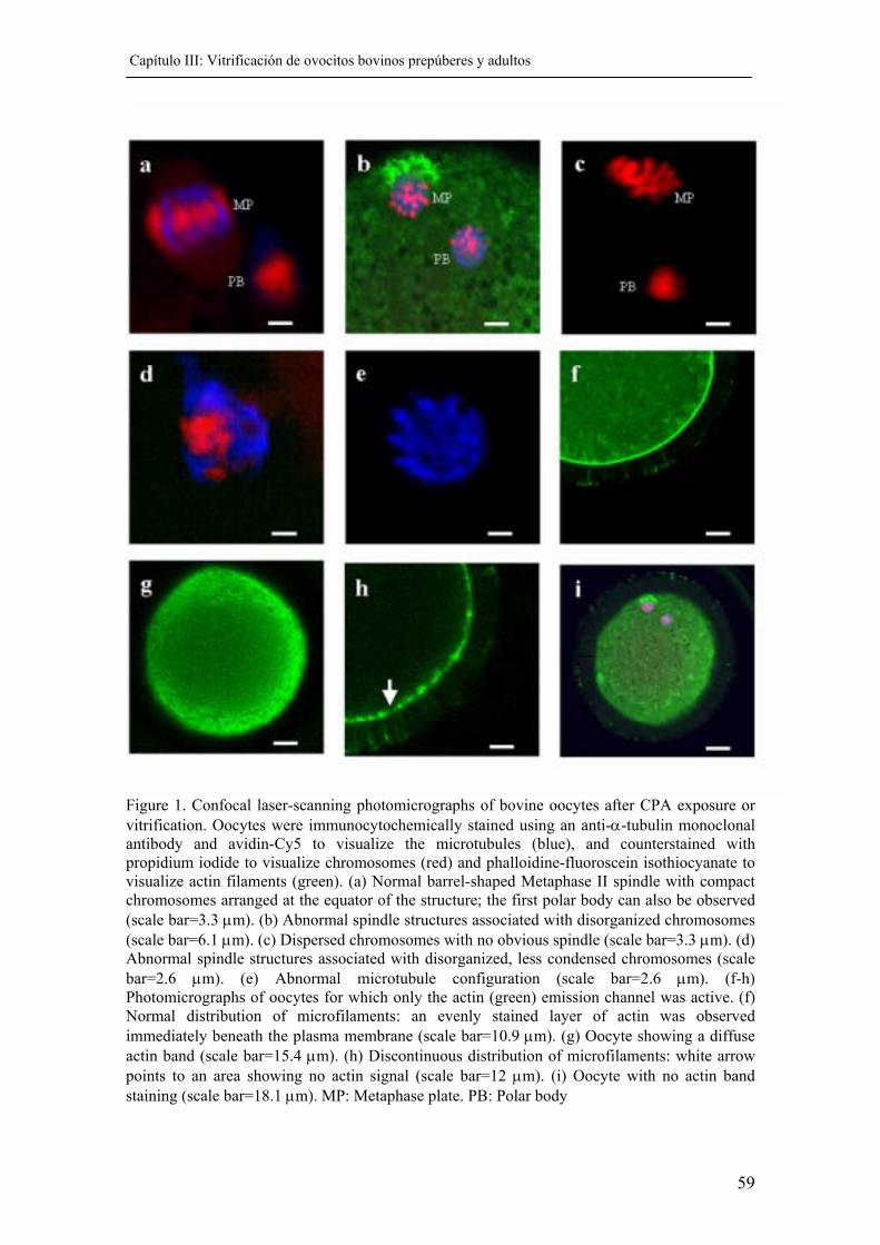
Capítulo III: Vitrificación de ovocitos bovinos prepúberes y adultos
59
Figure 1. Confocal laser-scanning photomicrographs of bovine oocytes after CPA exposure or vitrification. Oocytes were immunocytochemically stained using an anti-α-tubulin monoclonal antibody and avidin-Cy5 to visualize the microtubules (blue), and counterstained with propidium iodide to visualize chromosomes (red) and phalloidine-fluoroscein isothiocyanate to visualize actin filaments (green). (a) Normal barrel-shaped Metaphase II spindle with compact chromosomes arranged at the equator of the structure; the first polar body can also be observed (scale bar=3.3 µm). (b) Abnormal spindle structures associated with disorganized chromosomes (scale bar=6.1 µm). (c) Dispersed chromosomes with no obvious spindle (scale bar=3.3 µm). (d) Abnormal spindle structures associated with disorganized, less condensed chromosomes (scale bar=2.6 µm). (e) Abnormal microtubule configuration (scale bar=2.6 µm). (f-h) Photomicrographs of oocytes for which only the actin (green) emission channel was active. (f) Normal distribution of microfilaments: an evenly stained layer of actin was observed immediately beneath the plasma membrane (scale bar=10.9 µm). (g) Oocyte showing a diffuse actin band (scale bar=15.4 µm). (h) Discontinuous distribution of microfilaments: white arrow points to an area showing no actin signal (scale bar=12 µm). (i) Oocyte with no actin band staining (scale bar=18.1 µm). MP: Metaphase plate. PB: Polar body
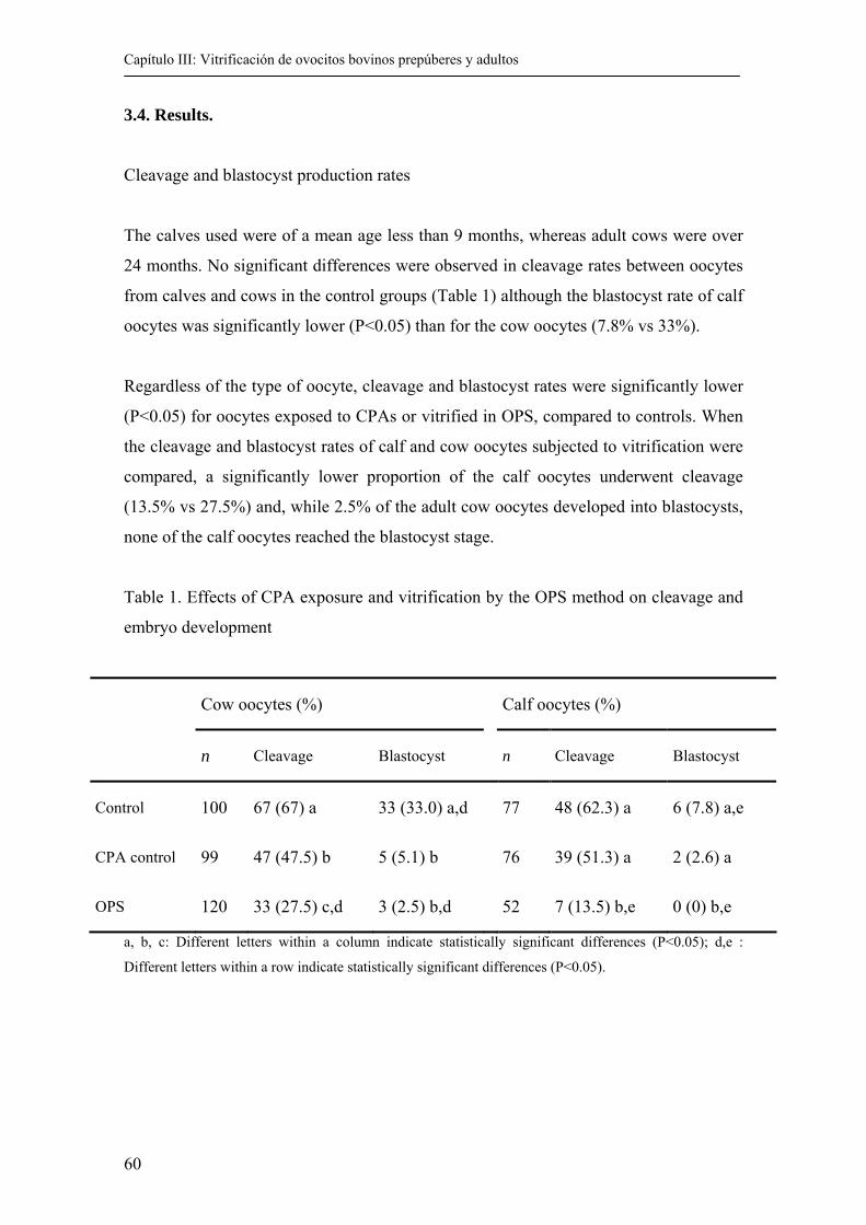
Capítulo III: Vitrificación de ovocitos bovinos prepúberes y adultos
60
3.4. Results.
Cleavage and blastocyst production rates
The calves used were of a mean age less than 9 months, whereas adult cows were over
24 months. No significant differences were observed in cleavage rates between oocytes
from calves and cows in the control groups (Table 1) although the blastocyst rate of calf
oocytes was significantly lower (P<0.05) than for the cow oocytes (7.8% vs 33%).
Regardless of the type of oocyte, cleavage and blastocyst rates were significantly lower
(P<0.05) for oocytes exposed to CPAs or vitrified in OPS, compared to controls. When
the cleavage and blastocyst rates of calf and cow oocytes subjected to vitrification were
compared, a significantly lower proportion of the calf oocytes underwent cleavage
(13.5% vs 27.5%) and, while 2.5% of the adult cow oocytes developed into blastocysts,
none of the calf oocytes reached the blastocyst stage.
Table 1. Effects of CPA exposure and vitrification by the OPS method on cleavage and
embryo development
Cow oocytes (%) Calf oocytes (%)
n Cleavage Blastocyst n Cleavage Blastocyst
Control 100 67 (67) a 33 (33.0) a,d 77 48 (62.3) a 6 (7.8) a,e
CPA control 99 47 (47.5) b 5 (5.1) b 76 39 (51.3) a 2 (2.6) a
OPS 120 33 (27.5) c,d 3 (2.5) b,d 52 7 (13.5) b,e 0 (0) b,e
a, b, c: Different letters within a column indicate statistically significant differences (P<0.05); d,e :
Different letters within a row indicate statistically significant differences (P<0.05).
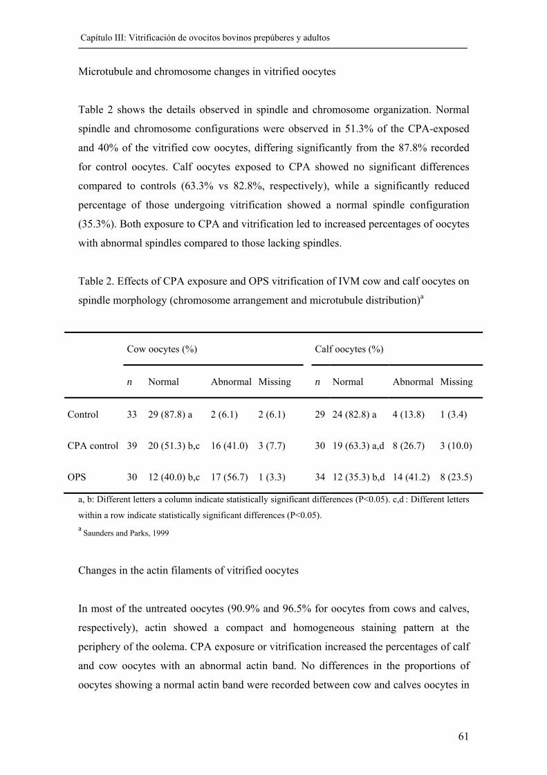
Capítulo III: Vitrificación de ovocitos bovinos prepúberes y adultos
61
Microtubule and chromosome changes in vitrified oocytes
Table 2 shows the details observed in spindle and chromosome organization. Normal
spindle and chromosome configurations were observed in 51.3% of the CPA-exposed
and 40% of the vitrified cow oocytes, differing significantly from the 87.8% recorded
for control oocytes. Calf oocytes exposed to CPA showed no significant differences
compared to controls (63.3% vs 82.8%, respectively), while a significantly reduced
percentage of those undergoing vitrification showed a normal spindle configuration
(35.3%). Both exposure to CPA and vitrification led to increased percentages of oocytes
with abnormal spindles compared to those lacking spindles.
Table 2. Effects of CPA exposure and OPS vitrification of IVM cow and calf oocytes on
spindle morphology (chromosome arrangement and microtubule distribution)a
Cow oocytes (%) Calf oocytes (%)
n Normal Abnormal Missing n Normal Abnormal Missing
Control 33 29 (87.8) a 2 (6.1) 2 (6.1) 29 24 (82.8) a 4 (13.8) 1 (3.4)
CPA control 39 20 (51.3) b,c 16 (41.0) 3 (7.7) 30 19 (63.3) a,d 8 (26.7) 3 (10.0)
OPS 30 12 (40.0) b,c 17 (56.7) 1 (3.3) 34 12 (35.3) b,d 14 (41.2) 8 (23.5)
a, b: Different letters a column indicate statistically significant differences (P<0.05). c,d : Different letters
within a row indicate statistically significant differences (P<0.05). a Saunders and Parks, 1999
Changes in the actin filaments of vitrified oocytes
In most of the untreated oocytes (90.9% and 96.5% for oocytes from cows and calves,
respectively), actin showed a compact and homogeneous staining pattern at the
periphery of the oolema. CPA exposure or vitrification increased the percentages of calf
and cow oocytes with an abnormal actin band. No differences in the proportions of
oocytes showing a normal actin band were recorded between cow and calves oocytes in
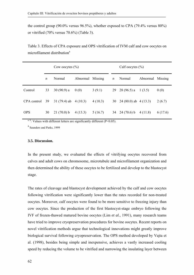
Capítulo III: Vitrificación de ovocitos bovinos prepúberes y adultos
62
the control group (90.0% versus 96.5%), whether exposed to CPA (79.4% versus 80%)
or vitrified (70% versus 70.6%) (Table 3).
Table 3. Effects of CPA exposure and OPS vitrification of IVM calf and cow oocytes on
microfilament distributiona
Cow oocytes (%) Calf oocytes (%)
n Normal Abnormal Missing n Normal Abnormal Missing
Control 33 30 (90.9) a 0 (0) 3 (9.1) 29 28 (96.5) a 1 (3.5) 0 (0)
CPA control 39 31 (79.4) ab 4 (10.3) 4 (10.3) 30 24 (80.0) ab 4 (13.3) 2 (6.7)
OPS 30 21 (70.0) b 4 (13.3) 5 (16.7) 34 24 (70.6) b 4 (11.8) 6 (17.6)
a, b: Values with different letters are significantly different (P<0.05). a Saunders and Parks, 1999
3.5. Discussion.
In the present study, we evaluated the effects of vitrifying oocytes recovered from
calves and adult cows on chromosome, microtubule and microfilament organization and
then determined the ability of these oocytes to be fertilized and develop to the blastocyst
stage.
The rates of cleavage and blastocyst development achieved by the calf and cow oocytes
following vitrification were significantly lower than the rates recorded for non-treated
oocytes. Moreover, calf oocytes were found to be more sensitive to freezing injury than
cow oocytes. Since the production of the first blastocyst-stage embryo following the
IVF of frozen-thawed matured bovine oocytes (Lim et al., 1991), many research teams
have tried to improve cryopreservation procedures for bovine oocytes. Recent reports on
novel vitrification methods argue that technological innovations might greatly improve
biological survival following cryopreservation. The OPS method developed by Vajta et
al. (1998), besides being simple and inexpensive, achieves a vastly increased cooling
speed by reducing the volume to be vitrified and narrowing the insulating layer between
Capítulo III: Vitrificación de ovocitos bovinos prepúberes y adultos
63
the cooling agent and the vitrification solution. Using this method, these authors have
reported rates of up to 50% cleavage and up to 25% blastocyst development after
vitrification. Our cleavage rates and blastocyst yields after the vitrification of cow and
calf oocytes are lower than those obtained by Vajta et al. (1998), but similar to those
reported by other authors (Men et al., 2002). Moreover, in accordance with the results
of Martino et al. (1996) and Rho et al. (2002), control oocytes exposed to CPA without
further cooling showed reduced development over controls, suggesting that osmotic
shock plays an important role in the success of cryopreservation procedures.
Calf oocytes are less competent to develop than those harvested from adult cows
(reviewed in Gandolfi et al., 2000). Although fertilization and cleavage rates do not
differ greatly for calf and cow oocytes, blastocyst yields are significantly reduced for
calf oocytes (Revel et al., 1995; Khatir et al., 1996). This deficient developmental
capacity of calf oocytes is most likely due to the abnormal cytoplasmic maturation of
these oocytes. Both our cow and calf oocytes were matured in vitro, such that the
differences observed were not attributable to our IVM system but more likely the result
of an intrinsic deficiency of calf oocytes. It is also possible, however, that calf oocytes,
because of their immaturity at the time of collection, require a modified medium to
achieve developmental competence. Damiani et al. (1996) reported that calf oocytes
show a delay in organelle migration (mainly cortical granules) following in vitro
maturation, as well as abnormal chromatin and microtubule configurations.
Nevertheless, in the present study, similar percentages of untreated calf and cow oocytes
showed a normal spindle configuration and distribution of actin microfilaments.
Exposure to the cryoprotectants EG and DMSO had a drastic effect on the arrangement
of microtubules and chromosomes in cow oocytes, while the vitrification procedure
seemed to more severely affect the spindle configuration of calf oocytes. Approximately
64% of the vitrified calf oocytes and 60% of the vitrified cow oocytes appeared to have
abnormal or missing spindles compared to control oocytes, while in the manipulated
controls, abnormal spindle patterns were detected in ~ 37% of the calf and ~ 48% of the
cow oocytes. Several authors have described that the main hurdle in developing
successful protocols for the cryopreservation of mammalian oocytes is being able to
preserve the integrity of the meiotic spindle when the oocytes are cooled (Eroglu et al.,
1998). Temperature fluctuations directly affect the cytoskeletal and chromosome
Capítulo III: Vitrificación de ovocitos bovinos prepúberes y adultos
64
organization of mature bovine (Aman and Parks, 1994; Saunders and Parks, 1999) and
human oocytes (Almeida and Bolton, 1995). The main consequence of cooling is
pronounced depolymerization and the disappearance of microtubule organizing centers
(Webb et al., 1986). Chilling leads to the disassembly of spindle fibers within minutes,
followed by an equally rapid reassembly of the spindle after the return to normal
temperatures (Inoue, 1981). Magistrini and Szöllösi (1980) reported that the meiotic
spindles of mouse oocytes were sensitive to cooling, with complete disassembly
occurring after 45-60 min at 0ºC. The effects of cooling on the spindle appeared to be
reversible in the mouse oocyte, with normal spindle formation occurring after step-wise
re-warming. Pickering et al. (Pickering et al., 1990) found that the meiotic spindle of
human oocytes completely disassembled, and this was accompanied by chromosomal
dispersion in 60% of the oocytes after 30 min at room temperature. This effect appeared
to be reversible in only 25%-50% of the oocytes. The meiotic spindle becomes
completely disassembled when in vitro-matured bovine oocytes are maintained for 10-
20 min at 4ºC (Richardson and Parks, 1992). When pig oocytes were kept for 5 min at
4ºC, microtubules in the spindles of most oocytes partially or completely disassembled
(Liu et al., 2003).
Cryoprotectants are known to induce changes in microtubule organization in several
species, including the mouse (Vincent and Johnson, 1992; Cooper et al., 1996), rabbit
(Vincent et al., 1989), human (Sathananthan et al., 1987) and cow (Hyttel et al., 2000).
The exposure of bovine oocytes to EG did not affect spindle arrangement after 20 min
re-warming, but a lower percentage of oocytes with abnormal spindle configuration was
seen after 1 h and 3 h (Saunders and Parks, 1999). In our study, effects on the normal
spindle pattern were similarly observed after 2 h re-warming in oocytes exposed to EG
+ DMSO. At room temperature, the addition of a cryoprotectant (propanediol or
DMSO) to rabbit oocytes led to disorganization of spindle microtubules (Vincent et al.,
1989). In mouse oocytes, a similar effect of DMSO (Jonhson and Pickering, 1987; Van
der Elst et al., 1988) and propanediol (Van der Elst et al., 1988) has been noted, and a
consequent dispersal of chromosomes is often seen in both species.
In this study, exposure to cryoprotectants led to no substantial disruption of
microfilament organization in either cow or calf oocytes. Williams et al. (Williams et
al., 1992) observed that microfilament organization in bovine oocytes loaded with EG
Capítulo III: Vitrificación de ovocitos bovinos prepúberes y adultos
65
or PrOH was similar to that in untreated oocytes and Saunders and Parks (Saunders and
Parks, 1999) described the normal structure of the actin band within 1 h to 3 h post-
thawing of oocytes exposed to EG. These results confirm our observations of only a
discrete effect of EG +DMSO on the distribution of actin. Vitrification was, however,
found to modify the organization of actin filaments. Freezing has also been described to
causes changes in the organization of cytoskeleton actin in rabbit and mouse oocytes
(Vincent et al., 1989; George and Johnson, 1993), although these changes were often
reversible upon thawing. Dramatic effects on cytoskeletal actin were observed after
freezing bovine oocytes (Saunders and Parks, 1999). Because of the association of
microfilaments with other structures, it is possible that their disruption is the result of
damage to another cell component such as the plasma membrane or mitochondria
(Didion et al., 1990; Fuku et al., 1995). Disruption of the cytoskeleton may be intrinsic
to the changes in shape and shrinkage related to cryopreservation procedures, which in
turn may lead to irreversibly changes in the structure of the plasma membrane or
cytoskeleton so that it can no longer adjust to changing conditions, including those
associated with fertilization. Thus, even when the normal microfilament distribution of
oocytes is restored after CPA exposure and vitrification, irreversible alterations to other
cell components may already have occurred such as the early release of cortical granule
enzymes and zona hardening. These changes may either prevent fertilization completely
or incompletely block polyspermy, both leading to decreased cleavage rates after
insemination.
In conclusion, the vitrification of bovine oocytes at the MII stage by the OPS method
produces biological changes in the oocytes after thawing, reflected by subsequently
impaired fertilization and embryo development. Oocytes retrieved from adult cows were
found to be more sensitive to exposure to CPAs, while vitrification seemed to have
worse effects on calf oocytes. In spite of these alterations, however, a certain proportion
of the vitrified oocytes were capable of developing into embryos.
Capítulo III: Vitrificación de ovocitos bovinos prepúberes y adultos
66
3.6. Bibliography. Almeida PA and Bolton VN. 1995. The effect of temperature fluctuations on the
cytoskeletal organisation and chromosomal constitution of the human oocyte. Zygote 3(4):357-365.
Aman RR and Parks JE. 1994. Effects of cooling and rewarming on the meiotic spindle and chromosomes of in vitro-matured bovine oocytes. Biol Reprod 50(1):103-110.
Arav A and Zeron Y. 1997. Vitrification of bovine oocytes using modified minimum drop size technique (MDS) is effected by the composition and concentration of the vitrification solution and by the cooling conditions. Theriogenology 47:341.
Boiso I, Marti M, Santalo J, Ponsa M, Barri PN and Veiga A. 2002. A confocal microscopy analysis of the spindle and chromosome configurations of human oocytes cryopreserved at the germinal vesicle and metaphase II stage. Hum Reprod 17(7):1885-1891.
Cooper A, O`Neil L, Bernard A, Fuller BJ and Shaw RW. 1996. The effect of protein source and temperature on the damage to mouse oocytes cytoskeleton after exposure to a vitrification solution. Cryo Letters 17:149-156.
Damiani P, Fissore RA, Cibelli JB, Long CR, Balise JJ, Robl JM and Duby RT. 1996. Evaluation of developmental competence, nuclear and ooplasmic maturation of calf oocytes. Mol Reprod Dev 45(4):521-534.
Didion BA, Pomp D, Martin MJ, Homanics GE and Markert CL. 1990. Observations on the cooling and cryopreservation of pig oocytes at the germinal vesicle stage. J Anim Sci 68(9):2803-2810.
Eroglu A, Toth TL and Toner M. 1998. Alterations of the cytoskeleton and polyploidy induced by cryopreservation of metaphase II mouse oocytes. Fertil Steril 69(5):944-957.
Fuku E, Xia L and Downey BR. 1995. Ultrastructural changes in bovine oocytes cryopreserved by vitrification. Cryobiology 32(2):139-156.
Gandolfi F, Vassena R and Lauria A. 2000. The developmental competence of the oocyte before puberty: Is something missing? Reprod Domest Anim 35:66-71.
George MA and Johnson MH. 1993. Cytoskeletal organization and zona sensitivity to digestion by chymotrypsin of frozen-thawed mouse oocytes. Hum Reprod 8(4):612-620.
Hamano S, Koikeda A, Kuwayama M and Nagai T. 1992. Full-term development of in vitro-matured, vitrified and fertilized bovine oocytes. Theriogenology 38(6):1085-1090.
Hochi S, Ito K, Hirabayashi M, Ueda M, Kimura K and Hanada A. 1998. Effect of nuclear stages during IVM on the survival of vitrified-warmed bovine oocytes. Theriogenology 49(4):787-796.
Hyttel P, Vajta G and Callesen H. 2000. Vitrification of bovine oocytes with the open pulled straw method: ultrastructural consequences. Mol Reprod Dev 56(1):80-88.
Inoue S. 1981. Cell division and the meiotic spindle. J Cell Biol 91:131-147. Jonhson MH and Pickering SJ. 1987. The effect of dimethylsulfoxide on the
microtubular system of mouse oocyte. Development 100:313-324. Khatir H, Lonergan P, Carolan C and Mermillod P. 1996. Prepubertal bovine oocyte: a
negative model for studying oocyte developmental competence. Mol Reprod Dev 45(2):231-239.
Capítulo III: Vitrificación de ovocitos bovinos prepúberes y adultos
67
Khatir H, Lonergan P, Touze JL and Mermillod P. 1998. The characterization of bovine embryos obtained from prepubertal calf oocytes and their viability after non surgical embryo transfer. Theriogenology 50(8):1201-1210.
Le Gal F and Massip A. 1999. Cryopreservation of cattle oocytes: effects of meiotic stage, cycloheximide treatment, and vitrification procedure. Cryobiology 38(4):290-300.
Liebermann J, Nawroth F, Isachenko V, Isachenko E, Rahimi G and Tucker MJ. 2002. Potential importance of vitrification in reproductive medicine. Biol Reprod 67(6):1671-1680.
Lim JM, Fukui Y and Ono H. 1991. The post-thaw developmental capacity of frozen bovine oocytes following in vitro maturation and fertilization. Theriogenology 35:1225-1235.
Liu RH, Sun QY, Li YH, Jiao LH and Wang WH. 2003. Effects of cooling on meiotic spindle structure and chromosome alignment within in vitro matured porcine oocytes. Mol Reprod Dev 65(2):212-218.
Magistrini M and Szollosi D. 1980. Effects of cold and of isopropyl-N-phenylcarbamate on the second meiotic spindle of mouse oocytes. Eur J Cell Biol 22(2):699-707.
Martino A, Pollard JW and Leibo SP. 1996. Effect of chilling bovine oocytes on their developmental competence. Mol Reprod Dev 45(4):503-512.
Men H, Monson RL and Rutledge JJ. 2002. Effect of meiotic stages and maturation protocols on bovine oocyte's resistance to cryopreservation. Theriogenology 57(3):1095-1103.
Pickering SJ, Braude PR, Johnson MH, Cant A and Currie J. 1990. Transient cooling to room temperature can cause irreversible disruption of the meiotic spindle in the human oocyte. Fertil Steril 54(1):102-108.
Revel F, Mermillod P, Peynot N, Renard J and Heyman Y. 1995. Low developmental capacity of in vitro matured and fertilized oocytes from calves compared with that of cows. J Reprod Fertil 103(1):115-120.
Rho GJ, Kim S, Yoo JG, Balasubramanian S, Lee HJ and Choe SY. 2002. Microtubulin configuration and mitochondrial distribution after ultra-rapid cooling of bovine oocytes. Mol Reprod Dev 63(4):464-470.
Richardson RR and Parks JE. 1992. Effects of chilling on the meiotic spindle and chromosomes of bovine ova. Theriogenology 37(1):284.
Rizos D, Ward F, Boland MP and Lonergan P. 2001. Effect of culture system on the yield and quality of bovine blastocysts as assessed by survival after vitrification. Theriogenology 56(1):1-16.
Salamone DF, Damiani P, Fissore JM, Robl. and Duby RT. 2001. Biochemical an developmental Evidence that ooplasmic maturation of prepubertal bovine oocyte is compromised. Biol Reprod 64:1761-1768.
Sathananthan AH, Trounson A and Freeman L. 1987. Morphology and fertilizability of frozen human oocytes. Gamete Res 16(4):343-354.
Saunders KM and Parks JE. 1999. Effects of cryopreservation procedures on the cytology and fertilization rate of in vitro-matured bovine oocytes. Biol Reprod 61(1):178-187.
Shamsuddin M, Niwa K, Larsson B and Rodriguez-Martinez H. 1996. In vitro Maturation and fertilization of bovine oocytes. Reproduction Domestically Animals 31:613-622.
Vajta G, Holm P, Kuwayama M, Booth PJ, Jacobsen H, Greve T and Callesen H. 1998. Open Pulled Straw (OPS) vitrification: a new way to reduce cryoinjuries of bovine ova and embryos. Mol Reprod Dev 51(1):53-58.
Capítulo III: Vitrificación de ovocitos bovinos prepúberes y adultos
68
Van der Elst J, Van den Abbeel E, Jacobs R, Wisse E and Van Steirteghem A. 1988. Effect of 1,2-propanediol and dimethylsulphoxide on the meiotic spindle of the mouse oocyte. Hum Reprod 3(8):960-967.
Vincent C, Garnier V, Heyman Y and Renard JP. 1989. Solvent effects on cytoskeletal organization and in-vivo survival after freezing of rabbit oocytes. J Reprod Fertil 87(2):809-820.
Vincent C and Johnson MH. 1992. Cooling, cryoprotectants, and the cytoskeleton of the mammalian oocyte. Oxf Rev Reprod Biol 14:73-100.
Webb M, Howlett SK and Maro B. 1986. Parthenogenesis and cytoskeletal organization in ageing mouse eggs. J Embryol Exp Morphol 95:131-145.
Williams MR, Richardson RR and Parks JE. 1992. Affects of cryoprotective agents (CPAs) on cytoskeleton and meiotic spindle apparatus of bovine oocytes. Cryobiology 29:757.
Wu B, Tong J and Leibo SP. 1999. Effects of cooling germinal vesicle-stage bovine oocytes on meiotic spindle formation following in vitro maturation. Mol Reprod Dev54(4):388-395.
CAPÍTULO IV:
EFFECTS OF ROSCOVITINE ON THE NUCLEAR AND CYTOSKELETAL
COMPONENTS OF CALF OOCYTES AND THEIR SUBSEQUENT
DEVELOPMENT
Capítulo IV: Inhibición de la meiosis en ovocitos de terneras
71
4.1. Abstract .
Roscovitine, a potent inhibitor of M-phase promoting factor kinase activity, was used to
maintain calf oocytes at the germinal vesicle stage for a 24-h culture period. Cumulus-
oocyte complexes were aspirated from slaughterhouse calf ovaries and cultured for 24 h
in TCM199 containing different levels of roscovitine (12.5, 25, 50 and 100 µM). After
this culture period, the oocytes were either fixed immediately or cultured for a further
24 h in conditions permissive of maturation. After fixing a sample of these oocytes, the
remaining oocytes were subsequently fertilized and cleavage and blastocyst rates were
recorded. Oocytes cultured in the presence of roscovitine, at all the concentrations
tested, were significantly blocked at the germinal vesicle stage. The inhibitory effect
varied according to the dose, with 50 µM and 100 µM roscovitine being the most
efficient concentrations, producing developmental arrest at the GV stage in over 60.0%
of oocytes. However, this inhibitory effect of roscovitine was fully reversible since over
73% of the oocytes cultured for 24 h in the presence of 50 µM roscovitine reached the
metaphase II stage after a further 24 h of culture in a permissive medium. Cleavage
rates and blastocyst yields were not significantly different for oocytes cultured under 50
µM roscovitine inhibition compared to oocytes not subjected to prematuration culture
(rates of 76.7% cleavage and 8.7% blastocysts for control oocytes compared to 69.8%
and 6.3% respectively for oocytes pretreated with 50 µM roscovitine). The morphology
of the meiotic spindle was typical of metaphase II in 75.8% and 82.1% of the oocytes
reaching the metaphase II stage after pretreatment with 50 µM roscovitine compared to
control, respectively. A normal distribution of actin filaments was observed in 97.0%
and 98.2% of oocytes exposed to 50 µM roscovitine compared to control, respectively.
These results demonstrate the feasibility of maintaining calf oocytes in artificial meiotic
arrest without compromising their subsequent developmental competence.
4.2. Introduction.
The in vitro maintenance of oocytes at the germinal vesicle (GV) stage helps them
acquire developmental competence. The capacity to preserve oocytes at this stage
without producing damage serves to synchronize the resumption of meiosis such that all
collected oocytes can complete the cytoplasmic maturation process, maximizing embryo
production.
Capítulo IV: Inhibición de la meiosis en ovocitos de terneras
72
In vivo, meiotically competent oocytes are kept at the GV stage by follicular factors
until the preovulatory gonadotropin surge. This gives them time to differentiate before
ovulation. In vitro, all meiotically competent oocytes spontaneously re-enter the meiotic
process as soon as they are removed from their follicles. Nevertheless, nuclear meiotic
maturation of the oocytes in itself is insufficient to ensure subsequent embryo
development. Thus, we hypothesized that if oocytes are cultured before maturation in
vitro under conditions that arrest meiosis at the GV stage, this might offer them the
opportunity to acquire greater developmental competence.
Calf oocytes have been reported to lack developmental competence compared to adult
cow oocytes (Damiani et al., 1996; Duby et al., 1996; Khatir et al., 1996; Presicce et al.,
1997). Although fertilization and cleavage rates do not differ greatly between calf and
cow oocytes, blastocyst yields using calf oocytes are significantly reduced (Khatir et al.,
1996). This reduced developmental competence may be due to a failure or inability of
calf oocytes to undergo both nuclear and cytoplasmic maturation.
Attempts have been made to establish for pharmacological (cycloheximide, 6-
dimethylaminopurine, butyrolactone I, roscovitine) or physiological (theca cells)
inhibitors aimed at achieving meiotic arrest (Richard et al., 1997; Saeki et al., 1997;
Avery et al., 1998; Kubelka et al., 2000; Mermillod et al., 2000). Roscovitine (ROS), a
specific cdc2 kinase inhibitor, has been shown capable of reversibly inhibiting meiotic
resumption in cow oocytes for 24 h with no negative effects on subsequent development
to the blastocyst stage (Mermillod et al., 2000). Similar results have been reported for
pig oocytes (Krischek and Meinecke, 2001; Marchal et al., 2001). Donnay et al. (2004),
on the other hand, observed that a prematuration treatment with ROS prevented the
progression of meiosis in calf oocytes but that this treatment led to a dramatic decrease
in embryo development after fertilization.
Lonergan et al. (2003) noted ultrastructural changes in bovine oocytes maintained in
meiotic arrest in vitro using ROS. These authors observed that incubation with ROS
leads to swelling of mitochondrial cristae, degeneration of cortical granules and
convolution of the nuclear membrane. Chromatin and cytoskeleton alterations in pig
oocytes treated with ROS were assessed by Ju et al. (2003), who reported that the
Capítulo IV: Inhibición de la meiosis en ovocitos de terneras
73
resumption of meiosis and effects on GV chromatin and the cytoskeleton were
reversible once the drug was removed.
The present study was designed to evaluate: the effectiveness of ROS in maintaining
calf oocytes at the GV stage for 24 h; subsequent nuclear maturation rates after removal
of the inhibitor; and the resulting developmental potential of these oocytes kept at the
GV stage for 24 h. We also examined the changes experienced by both nuclear and
cytoskeletal components, including the chromosomes, microtubules and microfilaments,
of oocytes treated with ROS during in vitro maturation.
4.3. Materials and methods .
Oocyte collection and inhibition of meiotic resumption.
The method used for the in vitro maturation and fertilization of the oocytes has been
described elsewhere (Rizos et al., 2001). Briefly, ovaries from slaughtered prepubertal
calves were transported from a local abattoir to the laboratory in PBS at 35ºC-37ºC.
Cumulus oocyte complexes (COCs) were obtained by aspirating 2- to 10-mm follicles.
After three washes in modified PBS (PBS supplemented with 36 µg/mL pyruvate, 50
µg/mL gentamicin and 0.5 mg/mL BSA), groups of up to 50 COCs were placed in 500
µL of maturation medium containing different levels of ROS (12.5, 25, 50 and 100 µM)
in four-well plates and cultured for 24 h at 38.5ºC in a 5% CO2 humidified air
atmosphere. The maturation medium was comprised of TCM-199 supplemented with
10% (v:v), fetal calf serum (FCS), 10 ng/mL EGF and 50 µg/mL gentamicin. ROS was
prepared as a 5mM stock solution in dimethyl sulfoxide and stored in aliquots at � 20 ºC
until use.
In vitro maturation
To allow the arrested oocytes to resume meiotic maturation, COCs were rinsed several
times in modified PBS to avoid carry-over of ROS into the oocyte maturation medium
and then cultured in maturation medium for a further 24 hours at 38.5ºC in a 5% CO2
humidified air atmosphere.
Capítulo IV: Inhibición de la meiosis en ovocitos de terneras
74
In vitro fertilization
For in vitro fertilization, the COCs were washed four times in PBS and then in the
fertilization medium before being transferred in groups of up to 50 to four-well plates
containing 250 µL of fertilization medium per well (Tyrode�s medium supplemented
with 25mM bicarbonate, 22 mM Na-lactate, 1 mM Na-pyruvate, 6 mg/mL fatty acid-
free BSA and 10 µg/mL heparin-sodium salt). Motile spermatozoa were obtained by
centrifuging frozen-thawed sperm from Asturian bulls on a discontinuous Percoll
density gradient (2.5 mL 45% Percoll over 2.5 mL 90% Percoll) for 8 min at 700 x g at
room temperature. Viable spermatozoa, collected at the bottom of the 90% fraction,
were washed in Hepes-buffered Tyrode�s and pelleted by centrifugation at 100 x g for 5
min. Spermatozoa were counted in a hemocytometer and diluted in an appropriate
volume of fertilization medium to give a final concentration of 2 x 10⁶ spermatozoa/mL.
A 250-µL aliquot of this suspension was added to each fertilization well to obtain a final
concentration of 1 x 10⁶ spermatozoa/mL. Plates were incubated for 24 h at 38.5ºC in a
5% CO2 humidified air atmosphere. Semen from the same bulls was used in all the
experiments.
In vitro culture
Embryo culture was performed in microdroplets on a seeded layer of granulosa cells in
TCM-199 supplemented with 10% (v:v) FCS. At approximately 20 h post-insemination
(hpi), presumptive zygotes were denuded by gentle vortexing and washed four times in
PBS and twice in the culture medium before being transferred to a granulosa cell
monolayer. Co-culture was conducted for 8 days at 38.5ºC in a 5% CO2 humidified air
atmosphere. Every 48 h during culture, approximately half of the culture medium was
replaced with fresh medium. Cleavage rates were recorded at 48 hpi and numbers of
blastocysts were determined on post-insemination Days 7, 8 and 9.
Experiment 1
COCs were aspirated from slaughterhouse calf ovaries and cultured for 24 h in TCM199
containing different levels of ROS (12.5, 25, 50 and 100 µM). After 24 h of inhibition, a
Capítulo IV: Inhibición de la meiosis en ovocitos de terneras
75
third of the oocytes were denuded, fixed overnight, lacmoid stained and observed under
a phase contrast microscope to determine their nuclear status. The remaining oocytes
(two-thirds) that had been incubated in ROS-containing medium were washed and
transferred to 500 µL of maturation medium for a further 24 h, after which another third
of the original number of oocytes was fixed and stained as above. The remaining
oocytes (final third) were fertilized and cleavage and blastocyst rates were recorded.
Another batch of oocytes not exposed to ROS before IVM was also fixed and stained
after 24 h of maturation (Control oocytes). The experiment was replicated four times.
Experiment 2
COCs were aspirated from slaughterhouse ovaries and cultured for 24 h in TCM199
containing different levels of ROS (12.5, 25, 50 and 100 µM). Half of the oocytes in
each group were fixed at 24 h in fixative for immunocytochemical labeling of
chromosomes, microtubules and microfilaments. The remaining oocytes that had been
incubated in ROS-containing medium were washed and transferred to 500 µL of
maturation medium for further 24 h and then fixed and examined with the aid of a
confocal microscope to evaluate the reversibility of cytoskeletal and nuclear alterations
following the removal of ROS. A batch of oocytes not exposed to ROS before IVM was
also fixed and stained after 24 h of maturation (Control oocytes). The experiment was
replicated four times.
Evaluation of nuclear maturation stage
Oocytes were freed from their surrounding cumulus cells by repeated pipetting and
washed three times in PBS. To evaluate meiotic progression, the oocytes were fixed in
ethanol:acetic acid (3:1) overnight. The oocytes were mounted on microscope slides
with vaseline, covered with a glass coverslip and stained with lacmoid. The stage of
nuclear maturation was assessed by phase contrast microscopy (magnification, x 40).
Oocytes were scored as being at the: germinal vesicle (GV) stage, when their chromatin,
either filamentous or slightly condensed, was enclosed in a nuclear membrane; germinal
vesicle breakdown (GVBD) stage, when there was no visible nuclear membrane and
Capítulo IV: Inhibición de la meiosis en ovocitos de terneras
76
chromatin was condensed; metaphase I (MI) stage, when the chromatin was distributed
at the equator of a large spindle; anaphase I stage, when homologous chromosomes
could be seen to move away from each other and there was a visible spindle; telophase I
stage, when homologous chromosomes were apart; and metaphase II (MII) stage, when
the chromatin was located at the spindle and abstriction of the first polar body was
detected.
Oocyte immunostaining
Samples of oocytes from the experimental groups (see above) were fixed in 2%
formaldehyde PBS, permeabilized using Triton X-100 (0.2% in PBS), and
simultaneously immunostained for actin filaments, tubulin and chromatin detection
(Boiso et al., 2002). Fixation and subsequent incubations were performed at 37ºC.
For immunostaining, fixed oocytes were incubated with the anti-α-tubulin monoclonal
antibody (1:250) for 1.5 h, followed by incubation with anti-mouse IgG antibody-biotin
(1:5000) for 1 h, avidin-Cy5 for 30 min and falloidin-fluorescein isothiocyanate (FICT)
conjugate (1:1000) for 1 h. Between incubations, the oocytes were washed three times
in pre-warmed PBS for 5 min.
Groups of five oocytes were mounted on poly-L-lysine-treated coverslips fitted with a
self-adhesive reinforcement ring and then stained with DAPI (125 ng/mL). The
preparation was sealed with nail varnish and stored refrigerated and protected from light
until observation within the following two days. A laser-scanning confocal microscope
(Leica TCS-SP2 - AOBS) was used to examine actin filaments (FICT; excitation 488
nm), tubulin (Cy5; excitation 633 nm) and chromatin (DAPI; excitation 405 nm).
Images were recorded on a computer.
Statistical analysis
The χ2-test was used to compare data among the experimental groups. The level of
statistical significance was set at P < 0.05.
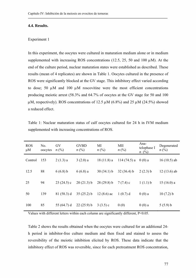
Capítulo IV: Inhibición de la meiosis en ovocitos de terneras
77
4.4. Results.
Experiment 1
In this experiment, the oocytes were cultured in maturation medium alone or in medium
supplemented with increasing ROS concentrations (12.5, 25, 50 and 100 µM). At the
end of the culture period, nuclear maturation states were established as described. These
results (mean of 4 replicates) are shown in Table 1. Oocytes cultured in the presence of
ROS were significantly blocked at the GV stage. This inhibitory effect varied according
to dose; 50 µM and 100 µM roscovitine were the most efficient concentrations
producing meiotic arrest (58.3% and 64.7% of oocytes at the GV stage for 50 and 100
µM, respectively). ROS concentrations of 12.5 µM (6.8%) and 25 µM (24.5%) showed
a reduced effect.
Table 1: Nuclear maturation status of calf oocytes cultured for 24 h in IVM medium
supplemented with increasing concentrations of ROS.
Values with different letters within each column are significantly different, P<0.05.
Table 2 shows the results obtained when the oocytes were cultured for an additional 24-
h period in inhibitor-free culture medium and then fixed and stained to assess the
reversibility of the meiotic inhibition elicited by ROS. These data indicate that the
inhibitory effect of ROS was reversible, since for each pretreatment ROS concentration,
ROS µM
No. oocytes
GV n (%)
GVBD n (%)
MI n (%)
MII n (%)
Ana-telophase I n (%)
Degenerated n (%)
Control 153 2 (1.3) a 3 (2.0) a 18 (11.8) a 114 (74.5) a 0 (0) a 16 (10.5) ab
12.5 88 6 (6.8) b 6 (6.8) a 30 (34.1) b 32 (36.4) b 2 (2.3) b 12 (13.6) ab
25 94 23 (24.5) c 20 (21.3) b 28 (29.8) b 7 (7.4) c 1 (1.1) b 15 (16.0) a
50 139 81 (58.3) d 35 (25.2) b 12 (8.6) ac 1 (0.7) d 0 (0) a 10 (7.2) b
100 85 55 (64.7) d 22 (25.9) b 3 (3.5) c 0 (0) 0 (0) a 5 (5.9) b
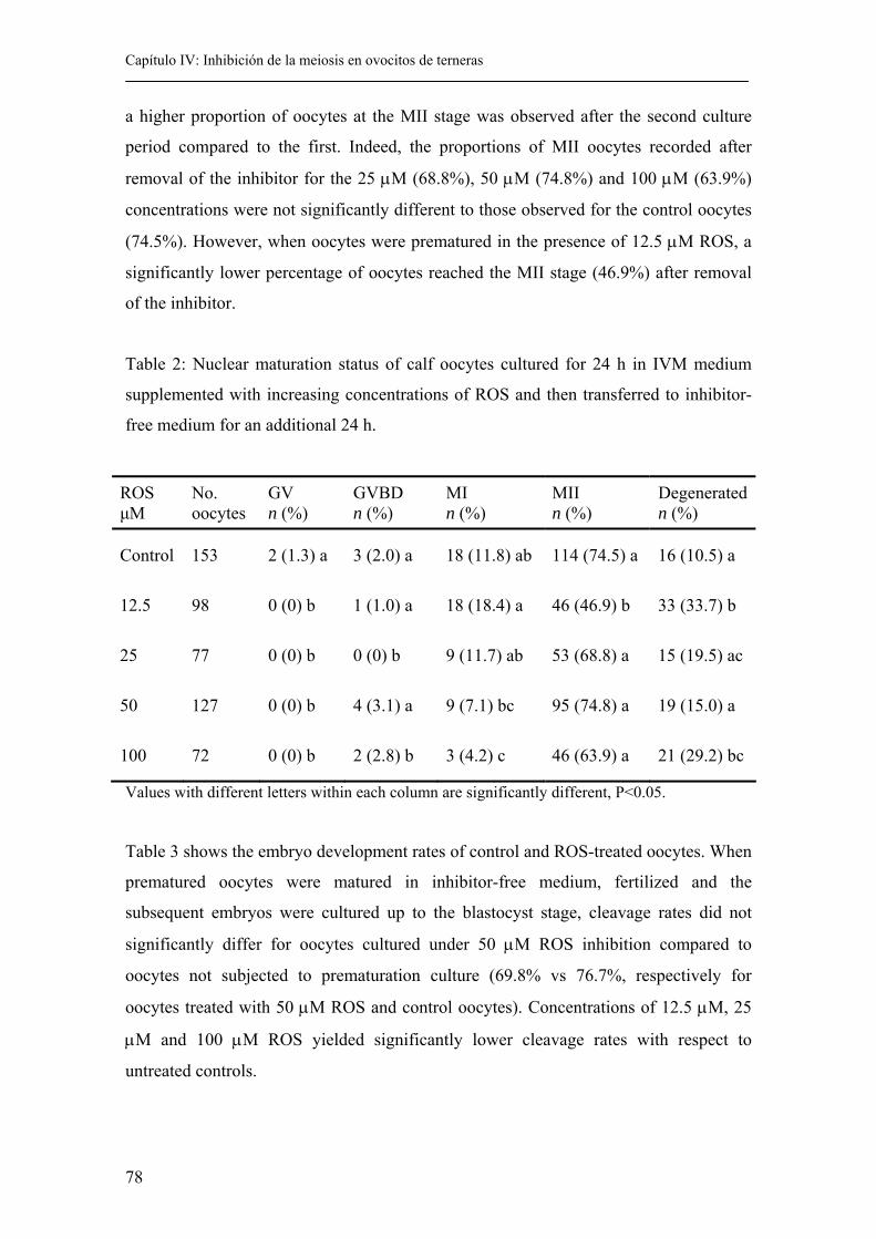
Capítulo IV: Inhibición de la meiosis en ovocitos de terneras
78
a higher proportion of oocytes at the MII stage was observed after the second culture
period compared to the first. Indeed, the proportions of MII oocytes recorded after
removal of the inhibitor for the 25 µM (68.8%), 50 µM (74.8%) and 100 µM (63.9%)
concentrations were not significantly different to those observed for the control oocytes
(74.5%). However, when oocytes were prematured in the presence of 12.5 µM ROS, a
significantly lower percentage of oocytes reached the MII stage (46.9%) after removal
of the inhibitor.
Table 2: Nuclear maturation status of calf oocytes cultured for 24 h in IVM medium
supplemented with increasing concentrations of ROS and then transferred to inhibitor-
free medium for an additional 24 h.
Values with different letters within each column are significantly different, P<0.05.
Table 3 shows the embryo development rates of control and ROS-treated oocytes. When
prematured oocytes were matured in inhibitor-free medium, fertilized and the
subsequent embryos were cultured up to the blastocyst stage, cleavage rates did not
significantly differ for oocytes cultured under 50 µM ROS inhibition compared to
oocytes not subjected to prematuration culture (69.8% vs 76.7%, respectively for
oocytes treated with 50 µM ROS and control oocytes). Concentrations of 12.5 µM, 25
µM and 100 µM ROS yielded significantly lower cleavage rates with respect to
untreated controls.
ROS µM
No. oocytes
GV n (%)
GVBD n (%)
MI n (%)
MII n (%)
Degenerated n (%)
Control 153 2 (1.3) a 3 (2.0) a 18 (11.8) ab 114 (74.5) a 16 (10.5) a
12.5 98 0 (0) b 1 (1.0) a 18 (18.4) a 46 (46.9) b 33 (33.7) b
25 77 0 (0) b 0 (0) b 9 (11.7) ab 53 (68.8) a 15 (19.5) ac
50 127 0 (0) b 4 (3.1) a 9 (7.1) bc 95 (74.8) a 19 (15.0) a
100 72 0 (0) b 2 (2.8) b 3 (4.2) c 46 (63.9) a 21 (29.2) bc
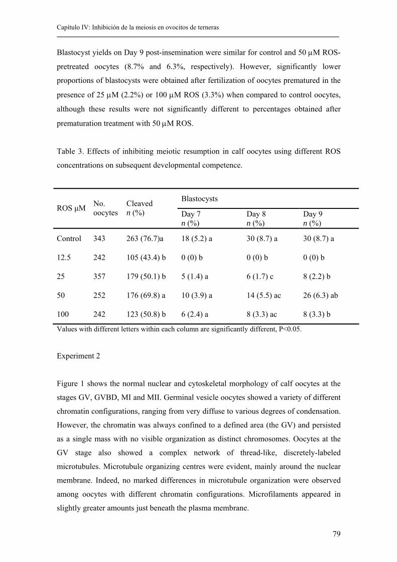
Capítulo IV: Inhibición de la meiosis en ovocitos de terneras
79
Blastocyst yields on Day 9 post-insemination were similar for control and 50 µM ROS-
pretreated oocytes (8.7% and 6.3%, respectively). However, significantly lower
proportions of blastocysts were obtained after fertilization of oocytes prematured in the
presence of 25 µM (2.2%) or 100 µM ROS (3.3%) when compared to control oocytes,
although these results were not significantly different to percentages obtained after
prematuration treatment with 50 µM ROS.
Table 3. Effects of inhibiting meiotic resumption in calf oocytes using different ROS
concentrations on subsequent developmental competence.
Values with different letters within each column are significantly different, P<0.05.
Experiment 2
Figure 1 shows the normal nuclear and cytoskeletal morphology of calf oocytes at the
stages GV, GVBD, MI and MII. Germinal vesicle oocytes showed a variety of different
chromatin configurations, ranging from very diffuse to various degrees of condensation.
However, the chromatin was always confined to a defined area (the GV) and persisted
as a single mass with no visible organization as distinct chromosomes. Oocytes at the
GV stage also showed a complex network of thread-like, discretely-labeled
microtubules. Microtubule organizing centres were evident, mainly around the nuclear
membrane. Indeed, no marked differences in microtubule organization were observed
among oocytes with different chromatin configurations. Microfilaments appeared in
slightly greater amounts just beneath the plasma membrane.
Blastocysts ROS µM No.
oocytes Cleaved n (%)
Day 7 n (%)
Day 8 n (%)
Day 9 n (%)
Control 343 263 (76.7)a 18 (5.2) a 30 (8.7) a 30 (8.7) a
12.5 242 105 (43.4) b 0 (0) b 0 (0) b 0 (0) b
25 357 179 (50.1) b 5 (1.4) a 6 (1.7) c 8 (2.2) b
50 252 176 (69.8) a 10 (3.9) a 14 (5.5) ac 26 (6.3) ab
100 242 123 (50.8) b 6 (2.4) a 8 (3.3) ac 8 (3.3) b
Capítulo IV: Inhibición de la meiosis en ovocitos de terneras
80
As the oocytes reenter the meiotic process, germinal vesicles break down (GVBD stage)
and the chromatin condenses into short, thick, distinct chromosomes. At this point,
densely stained radial-like microtubules could be observed in close association with the
recently condensed and distinguishable chromosomes. Microfilaments clearly
concentrated in the cortical region of the oocyte.
During the metaphase stage of the first meiotic division, these highly condensed
chromosomes aligned themselves along the center of the spindle, radially elongated
microtubules formed the meiotic spindle and microfilaments concentrated in the cortical
region of the oocyte.
In oocytes at the metaphase stage of the second meiotic division, the spindle structure
always appeared at the periphery, oriented perpendicularly to the plasma membrane.
Oocytes that reached the MII stage were classified as follows:
A) The spindle morphology of MII oocytes was classified as three categories: (1)
Normal spindles. These were barrel-shaped with chromosomes clustered as a discrete
bundle at the metaphase plate and microtubules crosssing the length of the spindle from
pole to pole or extending from the spindle poles to the chromosomes; microtubules
within the polar body had no discernable organization and instead appeared as an
amorphous mass intertwined with chromatin occupying the perivitelline space. (2)
Abnormal spindles. Microtubules were not organized as typical spindles or some
microtubules were disassembled. (3) Absence of the spindle. No microtubules could be
observed around the chromosomes. Detailed images of these normal and abnormal
patterns are provided in Figure 1.
B) Chromosomal organization was classified as: (1) Dispersed chromosomes. These
were scattered in the cytoplasm or dispersed in a few zones of the cytoplasm. (2)
Decondensed chromosomes. Chromosomes with an aberrant, less condensed
appearance. (3) Absence of chromosomes. No chromosomes were observed (Figure 1).
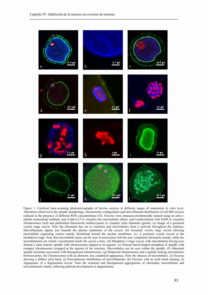
Capítulo IV: Inhibición de la meiosis en ovocitos de terneras
81
Figure 1. Confocal laser-scanning photomicrographs of bovine oocytes at different stages of maturation in vitro (a-e). Alterations observed in the spindle morphology, chromosome configuration and microfilament distribution of calf MII oocytes cultured in the presence of different ROS concentrations (f-l). Oocytes were immunocytochemically stained using an anti-α-tubulin monoclonal antibody and avidin-Cy5 to visualize the microtubules (blue), and counterstained with DAPI to visualize chromosomes (red) and phalloidine-fluoroscein isothiocyanate to visualize actin filaments (green). (a) Image of a germinal vesicle stage oocyte. Note the chromatin has yet to condense and microtubules form a network throughout the ooplasm. Microfilaments appear just beneath the plasma membrane of the oocyte. (b) Germinal vesicle stage oocyte showing microtubule organizing centers mainly distributed around the nuclear membrane. (c) A germinal vesicle oocyte at the breakdown stage. Note that microtubule asters can be seen in association with the now condensed chromatin strands, while the microfilaments are clearly concentrated inside the oocyte cortex. (d) Metaphase I stage oocyte with microtubules having now formed a clear meiotic spindle with chromosomes aligned at its equator. (e) Normal barrel-shaped metaphase II spindle with compact chromosomes arranged at the equator of the structure. Microtubules can be seen within the spindle. (f) Abnormal spindle structures associated with disorganized chromosomes. (g) Dispersed chromosomes and a spindle lacking microtubules between poles. (h) Chromosomes with an aberrant, less condensed appearance. Note the absence of microtubules. (i) Oocytes showing a diffuse actin band. (j) Discontinuous distribution of microfilaments. (k) Oocytes with no actin band staining. (l) Appearance of a degenerated oocyte. Note the scattered and disorganized aggregations of chromatin, microtubules and microfilaments clearly reflecting aberrant development or degeneration.
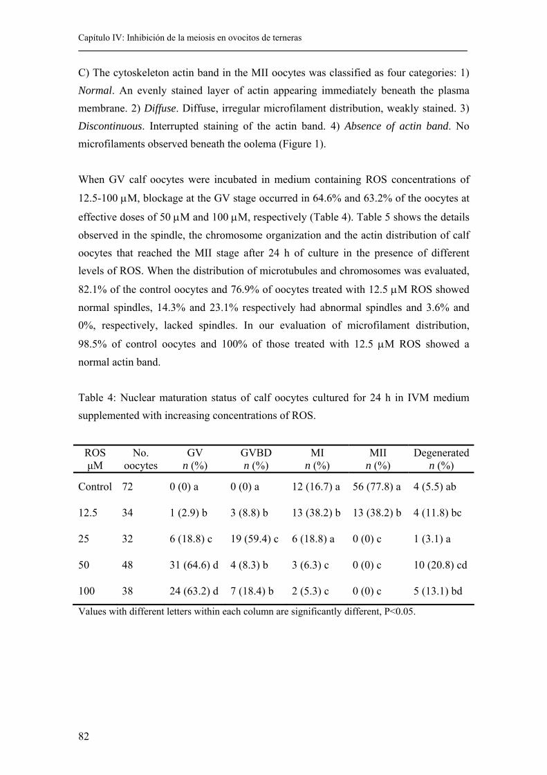
Capítulo IV: Inhibición de la meiosis en ovocitos de terneras
82
C) The cytoskeleton actin band in the MII oocytes was classified as four categories: 1) Normal. An evenly stained layer of actin appearing immediately beneath the plasma membrane. 2) Diffuse. Diffuse, irregular microfilament distribution, weakly stained. 3) Discontinuous. Interrupted staining of the actin band. 4) Absence of actin band. No microfilaments observed beneath the oolema (Figure 1). When GV calf oocytes were incubated in medium containing ROS concentrations of
12.5-100 µM, blockage at the GV stage occurred in 64.6% and 63.2% of the oocytes at
effective doses of 50 µM and 100 µM, respectively (Table 4). Table 5 shows the details observed in the spindle, the chromosome organization and the actin distribution of calf oocytes that reached the MII stage after 24 h of culture in the presence of different levels of ROS. When the distribution of microtubules and chromosomes was evaluated,
82.1% of the control oocytes and 76.9% of oocytes treated with 12.5 µM ROS showed normal spindles, 14.3% and 23.1% respectively had abnormal spindles and 3.6% and 0%, respectively, lacked spindles. In our evaluation of microfilament distribution,
98.5% of control oocytes and 100% of those treated with 12.5 µM ROS showed a normal actin band.
Table 4: Nuclear maturation status of calf oocytes cultured for 24 h in IVM medium supplemented with increasing concentrations of ROS.
Values with different letters within each column are significantly different, P<0.05.
ROS µM
No. oocytes
GV n (%)
GVBD n (%)
MI n (%)
MII n (%)
Degenerated n (%)
Control 72 0 (0) a 0 (0) a 12 (16.7) a 56 (77.8) a 4 (5.5) ab
12.5 34 1 (2.9) b 3 (8.8) b 13 (38.2) b 13 (38.2) b 4 (11.8) bc
25 32 6 (18.8) c 19 (59.4) c 6 (18.8) a 0 (0) c 1 (3.1) a
50 48 31 (64.6) d 4 (8.3) b 3 (6.3) c 0 (0) c 10 (20.8) cd
100 38 24 (63.2) d 7 (18.4) b 2 (5.3) c 0 (0) c 5 (13.1) bd
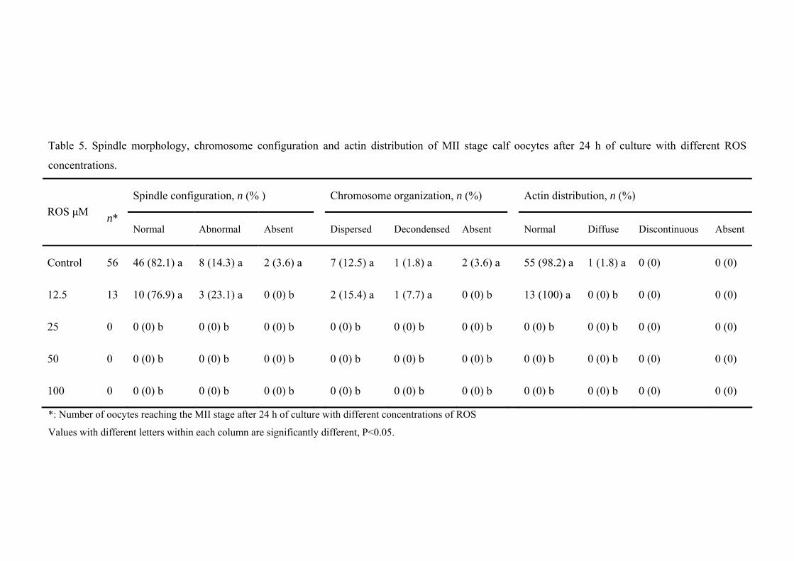
Table 5. Spindle morphology, chromosome configuration and actin distribution of MII stage calf oocytes after 24 h of culture with different ROS
concentrations.
*: Number of oocytes reaching the MII stage after 24 h of culture with different concentrations of ROS
Values with different letters within each column are significantly different, P<0.05.
Spindle configuration, n (% ) Chromosome organization, n (%) Actin distribution, n (%) ROS µM
n* Normal Abnormal Absent Dispersed Decondensed Absent Normal Diffuse Discontinuous Absent
Control 56 46 (82.1) a 8 (14.3) a 2 (3.6) a 7 (12.5) a 1 (1.8) a 2 (3.6) a 55 (98.2) a 1 (1.8) a 0 (0) 0 (0)
12.5 13 10 (76.9) a 3 (23.1) a 0 (0) b 2 (15.4) a 1 (7.7) a 0 (0) b 13 (100) a 0 (0) b 0 (0) 0 (0)
25 0 0 (0) b 0 (0) b 0 (0) b 0 (0) b 0 (0) b 0 (0) b 0 (0) b 0 (0) b 0 (0) 0 (0)
50 0 0 (0) b 0 (0) b 0 (0) b 0 (0) b 0 (0) b 0 (0) b 0 (0) b 0 (0) b 0 (0) 0 (0)
100 0 0 (0) b 0 (0) b 0 (0) b 0 (0) b 0 (0) b 0 (0) b 0 (0) b 0 (0) b 0 (0) 0 (0)
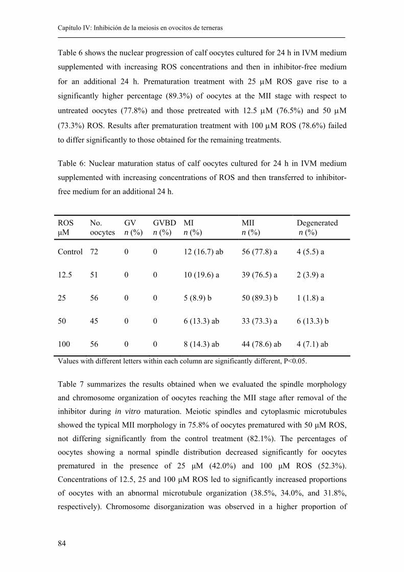
Capítulo IV: Inhibición de la meiosis en ovocitos de terneras
84
Table 6 shows the nuclear progression of calf oocytes cultured for 24 h in IVM medium
supplemented with increasing ROS concentrations and then in inhibitor-free medium
for an additional 24 h. Prematuration treatment with 25 µM ROS gave rise to a
significantly higher percentage (89.3%) of oocytes at the MII stage with respect to
untreated oocytes (77.8%) and those pretreated with 12.5 µM (76.5%) and 50 µM
(73.3%) ROS. Results after prematuration treatment with 100 µM ROS (78.6%) failed
to differ significantly to those obtained for the remaining treatments.
Table 6: Nuclear maturation status of calf oocytes cultured for 24 h in IVM medium
supplemented with increasing concentrations of ROS and then transferred to inhibitor-
free medium for an additional 24 h.
Values with different letters within each column are significantly different, P<0.05.
Table 7 summarizes the results obtained when we evaluated the spindle morphology
and chromosome organization of oocytes reaching the MII stage after removal of the
inhibitor during in vitro maturation. Meiotic spindles and cytoplasmic microtubules
showed the typical MII morphology in 75.8% of oocytes prematured with 50 µM ROS,
not differing significantly from the control treatment (82.1%). The percentages of
oocytes showing a normal spindle distribution decreased significantly for oocytes
prematured in the presence of 25 µM (42.0%) and 100 µM ROS (52.3%).
Concentrations of 12.5, 25 and 100 µM ROS led to significantly increased proportions
of oocytes with an abnormal microtubule organization (38.5%, 34.0%, and 31.8%,
respectively). Chromosome disorganization was observed in a higher proportion of
ROS µM
No. oocytes
GV n (%)
GVBD n (%)
MI n (%)
MII n (%)
Degenerated n (%)
Control 72 0 0 12 (16.7) ab 56 (77.8) a 4 (5.5) a
12.5 51 0 0 10 (19.6) a 39 (76.5) a 2 (3.9) a
25 56 0 0 5 (8.9) b 50 (89.3) b 1 (1.8) a
50 45 0 0 6 (13.3) ab 33 (73.3) a 6 (13.3) b
100 56 0 0 8 (14.3) ab 44 (78.6) ab 4 (7.1) ab
Capítulo IV: Inhibición de la meiosis en ovocitos de terneras
85
oocytes pretreated with all the ROS concentrations tested than in untreated controls
(33.3%, 42.0%, 24.2% and 43.2% vs 12.5% for 12.5, 25, 50 and 100 µM ROS vs
control, respectively). When oocytes were prematured with 25 µM ROS, a significantly
higher percentage of chromosomal decondensation was observed when compared to the
other treatments.
Our analysis of the cytoskeleton actin band in oocytes that reached the MII stage
revealed a normal distribution of microfilaments in 98.2% of control oocytes and 97.0%
of oocytes treated with 50 µM ROS. Concentrations of 12.5, 25 and 100 µM ROS
produced a significant decrease in the proportions of oocytes with a normal
microfilament distribution while these ROS levels led to a significant increase in the
number of oocytes showing diffuse actin labeling (38.5%, 18.0% and 25.0% for 12.5,
25 and 100 µM ROS, respectively).
Degenerated Oocytes
After 24 h of ROS treatment, some of the oocytes were classified as degenerated. The
proportion of degenerated oocytes ranged from 3.1% to 20.8%, depending on the
treatment and was lowest (1.8% to 13.3%) after IVM in the inhibitor-free medium.
Oocytes classified as degenerated showed aberrant chromatin and cytoskeletal patterns.
In these degenerated oocytes, DNA was either not visible or visible only as hair-like
strands or scattered drops. Similarly, the microtubules and microfilaments were either
non visible or aggregated in small clusters scattered throughout the ooplasm.
4.5. Discussion.
Our findings demonstrate that calf oocytes can be kept in vitro at the germinal vesicle
stage for 24 h without affecting their meiotic or developmental competence. Exposure
to concentrations of ROS greater than 50 µM prevented meiotic resumption in ~ 60%
of oocytes. This meiotic inhibition was fully reversible for oocytes prematured in the
presence of 50 µM ROS, since GVBD occurred in all cases and > 70% of oocytes
reached the metaphase II stage after a further 24 h of culture in the absence of the
inhibitor. Further, these artificially arrested oocytes maintained their developmental
competence as indicated by their cleavage rates and blastocyst development following
IVM/IVF/IVC.
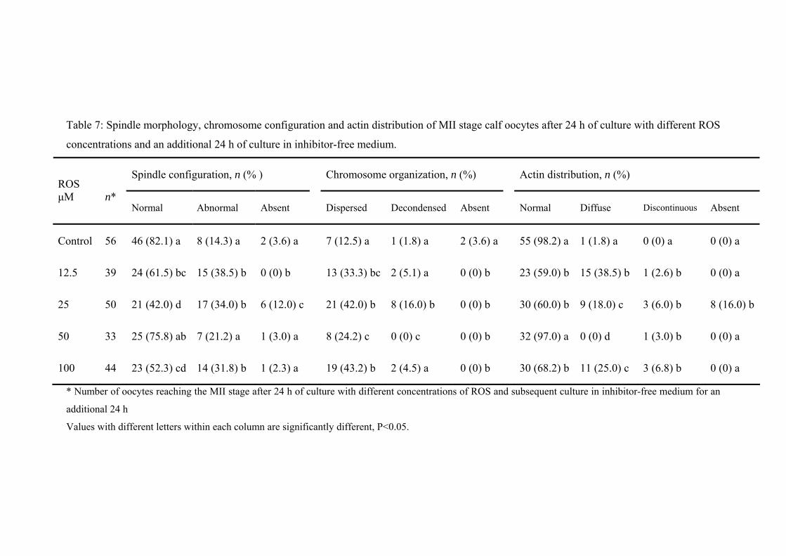
Table 7: Spindle morphology, chromosome configuration and actin distribution of MII stage calf oocytes after 24 h of culture with different ROS
concentrations and an additional 24 h of culture in inhibitor-free medium.
* Number of oocytes reaching the MII stage after 24 h of culture with different concentrations of ROS and subsequent culture in inhibitor-free medium for an
additional 24 h
Values with different letters within each column are significantly different, P<0.05.
Spindle configuration, n (% ) Chromosome organization, n (%) Actin distribution, n (%) ROS µM
n*
Normal Abnormal Absent Dispersed Decondensed Absent Normal Diffuse Discontinuous Absent
Control 56 46 (82.1) a 8 (14.3) a 2 (3.6) a 7 (12.5) a 1 (1.8) a 2 (3.6) a 55 (98.2) a 1 (1.8) a 0 (0) a 0 (0) a
12.5 39 24 (61.5) bc 15 (38.5) b 0 (0) b 13 (33.3) bc 2 (5.1) a 0 (0) b 23 (59.0) b 15 (38.5) b 1 (2.6) b 0 (0) a
25 50 21 (42.0) d 17 (34.0) b 6 (12.0) c 21 (42.0) b 8 (16.0) b 0 (0) b 30 (60.0) b 9 (18.0) c 3 (6.0) b 8 (16.0) b
50 33 25 (75.8) ab 7 (21.2) a 1 (3.0) a 8 (24.2) c 0 (0) c 0 (0) b 32 (97.0) a 0 (0) d 1 (3.0) b 0 (0) a
100 44 23 (52.3) cd 14 (31.8) b 1 (2.3) a 19 (43.2) b 2 (4.5) a 0 (0) b 30 (68.2) b 11 (25.0) c 3 (6.8) b 0 (0) a
Capítulo IV: Inhibición de la meiosis en ovocitos de terneras
87
Herein, we also demonstrate that the ability of ROS to inhibit the resumption of meiosis
is dose-dependent, a concentration of 50 µM being the minimum dose at which ~ 60%
of the oocytes are inhibited.
In vivo, the oocyte reaches cytoplasmic maturity (capacitation) and the competence to
resume meiosis (maturation) after a long series of processes, in which it is prepared for
fertilization and further embryo development. The resumption of meiosis in vitro is
spontaneously induced by the transfer of meiotically competent oocytes from ovarian
follicles into a suitable culture medium. Nevertheless, nuclear meiotic maturation of the
oocyte in itself is not enough to ensure subsequent embryo development. Many authors
(Lonergan et al., 1997; Avery et al., 1998; Mermillod et al., 2000; Motlik et al., 2000)
have tried to mimic the in vivo oocyte capacitation process by including a period of
prematuration in the presence of meiotic inhibitors. This prematuration interval could
enable the oocyte to construct, modify and store newly synthesized proteins and
ribonucleoproteins and so enhance its developmental competence.
Several attempts have been made to maintain oocytes at the GV stage, including
treatment with cycloheximide (Lonergan et al., 1997; Saeki et al., 1997), 6-
dimethylaminopurine (Lonergan et al., 1997; Avery et al., 1998), butyrolactone I (BL-I)
(Kitagawa et al., 1993; Motlik et al., 1998; Kubelka et al., 2000; Lonergan et al., 2000),
roscovitine (Mermillod et al., 2000), roscovitine plus retinoic acid (Duque et al., 2002)
and BL-I plus ROS (Ponderato et al., 2001).
Roscovitine is a selective cdc2 inhibitor, which has been reported to arrest cells in late
G1 and at G2/M cell cycle transition. It acts as a competitive inhibitor for ATP and
when complexed with cdk2, it binds to the ATP-binding pocket of cdk2. Roscovitine
has been successfully used to prevent the resumption of meiosis or GVBD in cow
(Mermillod et al., 2000; Donnay et al., 2004), pig (Krischek and Meinecke, 2001; Ju et
al., 2003; Le Beux et al., 2003; Schoevers et al., 2004) and horse oocytes (Franz et al.,
2003).
The extent of meiotic suppression of calf oocytes achieved by ROS in the present study
was lower than described for bovine oocytes (88% blockage using 50 µM ROS;
Capítulo V: Vitrificación en distintos estadios de maduración
88
(Mermillod et al., 2000), porcine oocytes (86% using 50 µM ROS; (Krischek and
Meinecke, 2001); 81% using 50 µM ROS; (Le Beux et al., 2003); 83% using 80 µM
ROS; (Ju et al., 2003)) and equine oocytes (84% using 66 µM ROS; (Franz et al.,
2003)).
In general, calf oocytes show reduced developmental competence compared to results
obtainable with adult cow oocytes, although this difference has not been clearly
established. Indeed, the capacity to form blastocysts at a rate equivalent to that of adult
oocytes has been reported for young calves treated with FSH (Armstrong et al., 1992;
Irvine et al., 1993; Armstrong et al., 1994). On the contrary, the defective
developmental competence of prepubertal oocytes has been suggested by several other
studies involving a larger number of animals, a wider age range and the use or lack of
use of gonadotropins (Palma, 1994; Looney et al., 1995; Damiani et al., 1996; Duby et
al., 1996; Khatir et al., 1996; Presicce et al., 1997). Donnay et al. (2004) reported
similar results to ours when they prematured calf oocytes, with 70% of the oocytes
remaining at earlier stages to metaphase I after a prematuration treatment using 25 µM
ROS and 60% reaching the MII stage after removal of the inhibitor. However, while
Mermillod et al. (2000) maintained cattle oocytes at the GV stage for 24 h without
decreasing their embryo developmental competence, Donnay et al. (2004) described a
considerable decrease in blastocyst rate with respect to non-prematured oocytes (14% vs
4%, control and 25 µM ROS treatments, respectively).
The results obtained in experiment 1 indicate that roscovitine prevented meiotic
resumption in around 60% of the oocytes during 24 h of culture at an inhibitor
concentration of 50 µM. This concentration is higher than the level of 25 µM used by
Mermillod et al. (2000) and Donnay et al. (2004). The factors accounting for this
difference are not clear although potential contributing factors are the source and batch
of ROS used, source of calf ovaries, the IVM system or the procedure used to assess
results. However, the fact that we need a highest ROS concentration (50 µM) to inhibit
meiotic resumption of calf oocytes and Salamone et al. (Salamone et al., 2001) reported
that the activity of MPF and MAPK was lower in calf oocytes that in oocytes of adult
cattle, it could suggest that ROS have also effect over other cytoplasmic factors rather
that only the MPF.
Capítulo IV: Inhibición de la meiosis en ovocitos de terneras
89
In the present study, nuclear and cytoskeletal changes were carefully determined
through immunocytochemical staining, defining the three factors: nuclear,
microfilament and microtubular status. When GV stage calf oocytes were incubated
with roscovitine for 24 h, no effects on spindle formation were observed after IVM in
oocytes reaching the MII stage in the presence of 50 µM ROS. However, when oocytes
where preincubated with lower (12.5 and 25 µM) or higher (100 µM) concentrations of
roscovitine, significantly lower proportions of oocytes showed a normal metaphase
spindle after IVM. Moreover, a normal distribution of actin filaments was observed in
85% and 93.2% of oocytes exposed to 50 µM ROS compared to control, respectively.
These results are similar to those obtained by Shoevers et al. (2004) in the pig. These
authors observed similar proportions of oocytes with normal MII spindles after a
prematuration treatment in the presence or absence of ROS. Others, however, have
reported chromatin and spindle abnormalities in a substantial proportion of MII stage
pig oocytes prematured using a ROS concentration of 80 µM (Ju et al., 2003).
Lonergan et al. (2003) noted morphological changes occurring in immature and in vitro
matured bovine oocytes following exposure to ROS. These authors demonstrated that a
prematuration treatment with 125 µM ROS caused disruption of the integrity of the
surrounding cumulus cells, swelling of the mitochondrial cristae and degeneration of the
cortical granules. Moreover, convolution of the nuclear membrane and aberrant
structures were observed within the nucleoplasm of ROS-treated oocytes. Here, we used
ROS concentrations ≤ 100 µM and recorded increases in the percentage of abnormal
spindles or abnormal actin distribution at this highest concentration.
In conclusion, our findings indicate the capacity of ROS to maintain calf oocytes in
meiotic arrest for 24 h without seriously compromising their potential to develop into
blastocysts. The resumption of meiosis and alterations in the chromosome configuration
of the meiotic spindle and cytoskeleton distribution were all reversible after removal of
the drug.
Capítulo V: Vitrificación en distintos estadios de maduración
90
4.6. Bibliography.. Armstrong DT, Holm P, Irvine B, Petersen BA, Stubbings RB, McLean D, Stevens G
and Seamark RF. 1992. Pregnancies and live birth born from in vitro fertilization of calf oocytes collected by laparoscopic follicular aspiration. Theriogenology 38:667-678.
Armstrong DT, Irvine BJ, Earl CR, McLean D and Seamark RF. 1994. Gonadotropin stimulation regimens for follicular aspiration and in vitro embryo production from calf oocytes. Theriogenology 42:1227-1236.
Avery B, Hay-Schmidt A, Hyttel P and Greve T. 1998. Embryo development, oocyte morphology, and kinetics of meiotic maturation in bovine oocytes exposed to 6-dimethylaminopurine prior to in vitro maturation. Mol Reprod Dev 50(3):334-344.
Boiso I, Marti M, Santalo J, Ponsa M, Barri PN and Veiga A. 2002. A confocal microscopy analysis of the spindle and chromosome configurations of human oocytes cryopreserved at the germinal vesicle and metaphase II stage. Hum Reprod 17(7):1885-1891.
Damiani P, Fissore RA, Cibelli JB, Long CR, Balise JJ, Robl JM and Duby RT. 1996. Evaluation of developmental competence, nuclear and ooplasmic maturation of calf oocytes. Mol Reprod Dev 45(4):521-534.
Donnay I, Faerge I, Grondahl C, Verhaeghe B, Sayoud H, Ponderato N, Galli C and Lazzari G. 2004. Effect of prematuration, meiosis activating sterol and enriched maturation medium on the nuclear maturation and competence to development of calf oocytes. Theriogenology 62(6):1093-1107.
Duby RT, Damiani P, Looney CR, Fissore RA and Robl JM. 1996. Prepuberal calves as oocyte donors: Promises and problems. Theriogenology 45:121-130.
Duque P, Diez C, Royo L, Lorenzo PL, Carneiro G, Hidalgo CO, Facal N and Gomez E. 2002. Enhancement of developmental capacity of meiotically inhibited bovine oocytes by retinoic acid. Hum Reprod 17(10):2706-2714.
Franz LC, Choi YH, Squires EL, Seidel GE, Jr. and Hinrichs K. 2003. Effects of roscovitine on maintenance of the germinal vesicle in horse oocytes, subsequent nuclear maturation, and cleavage rates after intracytoplasmic sperm injection. Reproduction 125(5):693-700.
Irvine BJ, Armstrong DT, Earl CR, McLean D and Seamark RF. 1993. Follicle development and oocyte recovery from calves with repeated gonadotropin stimulation and follicular aspitarion. Theriogenology 39:237(abstract).
Ju JC, Tsay C and Ruan CW. 2003. Alterations and reversibility in the chromatin, cytoskeleton and development of pig oocytes treated with roscovitine. Mol Reprod Dev 64(4):482-491.
Khatir H, Lonergan P, Carolan C and Mermillod P. 1996. Prepubertal bovine oocyte: a negative model for studying oocyte developmental competence. Mol Reprod Dev 45(2):231-239.
Kitagawa M, Okabe T, Ogino H, Matsumoto H, Suzuki-Takahashi I, Kokubo T, Higashi H, Saitoh S, Taya Y, Yasuda H and et al. 1993. Butyrolactone I, a selective inhibitor of cdk2 and cdc2 kinase. Oncogene 8(9):2425-2432.
Krischek C and Meinecke B. 2001. Roscovitine, a specific inhibitor of cyclin-dependent protein kinases, reversibly inhibits chromatin condensation during in vitro maturation of porcine oocytes. Zygote 9(4):309-316.
Capítulo IV: Inhibición de la meiosis en ovocitos de terneras
91
Kubelka M, Motlik J, Schultz RM and Pavlok A. 2000. Butyrolactone I reversibly inhibits meiotic maturation of bovine oocytes, without influencing chromosome condensation activity. Biol Reprod 62(2):292-302.
Le Beux G, Richard FJ and Sirard MA. 2003. Effect of cycloheximide, 6-DMAP, roscovitine and butyrolactone I on resumption of meiosis in porcine oocytes. Theriogenology 60(6):1049-1058.
Lonergan P, Dinnyes A, Fair T, Yang X and Boland M. 2000. Bovine oocyte and embryo development following meiotic inhibition with butyrolactone I. Mol Reprod Dev 57(2):204-209.
Lonergan P, Faerge I, Hyttel PM, Boland M and Fair T. 2003. Ultrastructural modifications in bovine oocytes maintained in meiotic arrest in vitro using roscovitine or butyrolactone. Mol Reprod Dev 64(3):369-378.
Lonergan P, Khatir H, Carolan C and Mermillod P. 1997. Bovine blastocyst production in vitro after inhibition of oocyte meiotic resumption for 24 h. J Reprod Fertil 109(2):355-365.
Looney CR, Damiani P, Lindsey BR, Long CR, Gonseth CL, Johnson DL and Duby RT. 1995. Use of prepuberal heifers as oocytes donors for IVF: Effect of age and gonadotrphin treatement. Theriogenology 43:269 (abstract).
Marchal R, Tomanek M, Terqui M and Mermillod P. 2001. Effects of cell cycle dependent kinases inhibitor on nuclear and cytoplasmic maturation of porcine oocytes. Mol Reprod Dev 60(1):65-73.
Mermillod P, Tomanek M, Marchal R and Meijer L. 2000. High developmental competence of cattle oocytes maintained at the germinal vesicle stage for 24 hours in culture by specific inhibition of MPF kinase activity. Mol Reprod Dev 55(1):89-95.
Motlik J, Pavlok A, Kubelka M, Kalous J and Kalab P. 1998. Interplay between CDC2 kinase and MAP kinase pathway during maturation of mammalian oocytes. Theriogenology 49(2):461-469.
Motlik J, Pavlok A, Lapathitis G and Kubelka M. 2000. Impact of two-step in vitro culture system on developmental potency of oocytes. Reprod Domest Anim 35:267-271.
Palma GA. 1994. Effect of FSH and estradiol-17beta for maturation of calf oocytes on the in vitro development to blastocysts. Theriogenology 41:267(abstract).
Ponderato N, Lagutina I, Crotti G, Turini P, Galli C and Lazzari G. 2001. Bovine oocytes treated prior to in vitro maturation with a combination of butyrolactone I and roscovitine at low doses maintain a normal developmental capacity. Mol Reprod Dev 60(4):579-585.
Presicce GA, Jiang S, Simkin M, Zhang L, Looney CR, Godke RA and Yang X. 1997. Age and hormonal dependence of acquisition of oocyte competence for embryogenesis in prepubertal calves. Biol Reprod 56(2):386-392.
Richard FJ, Fortier MA and Sirard MA. 1997. Role of the cyclic adenosine monophosphate-dependent protein kinase in the control of meiotic resumption in bovine oocytes cultured with thecal cell monolayers. Biol Reprod 56(6):1363-1369.
Rizos D, Ward F, Boland MP and Lonergan P. 2001. Effect of culture system on the yield and quality of bovine blastocysts as assessed by survival after vitrification. Theriogenology 56(1):1-16.
Saeki K, Nagai Y, Kishi M and Nagai M. 1997. Developmental capacity of bovine oocytes following inhibition of meiotic resumption by cycloheximide or 6-dimethylaminopurine. Theriogenology 48:1161-1172.
Capítulo V: Vitrificación en distintos estadios de maduración
92
Salamone DF, Damiani P, Fissore JM, Robl. and Duby RT. 2001. Biochemical an developmental Evidence that ooplasmic maturation of prepubertal bovine oocyte is compromised. Biol Reprod 64:1761-1768.
Schoevers EJ, Bevers MM, Roelen BAJ and Colenbrander B. 2004. Nuclear and cytoplasmic maturation of sow oocytes are not synchronized by specific meiotic inhibition with roscovitine during in vitro maturation. Theriogenology:(in press).

CAPÍTULO V:
VITRIFICATION OF CALF OOCYTES: EFFECTS OF MATURATION STAGE
AND PREMATURATION TREATMENT ON THE NUCLEAR AND
CYTOSKELETAL COMPONENTS OF OOCYTES AND THEIR SUBSEQUENT
DEVELOPMENT
Capítulo V: Vitrificación en distintos estadios de maduración
95
5.1. Abstract.
This study was designed to establish the effects of the meiotic stage of bovine oocytes
and of a prematuration treatment with roscovitine (ROS) on their resistance to
cryopreservation. Calf oocytes at the stages germinal vesicle breakdown (GVBD) and
metaphase II (MII) were vitrified by the open pulled straw (OPS) method. In another
experiment, GVBD oocytes were prematured with 50 µM ROS before vitrification.
After thawing, oocytes in the GVBD and ROS groups underwent an additional 18 h of
maturation, while those in the MII group were only subjected to a further 2 h of
maturation. After this post-thaw maturation period, some oocyte samples were fixed,
stained using specific fluorescent probes and examined under a confocal microscope.
The remaining oocytes were fertilized, and cleavage and blastocyst rates recorded.
Significantly lower cleavage rates were obtained for the vitrified GVBD and MII
oocytes (9.9% and 12.6%, respectively) compared to control oocytes (73.9%).
Significantly worse results in terms of cleavage rates were obtained when GVBD calf
oocytes were exposed to cryoprotectant (CPA) (13.1%) or vitrified (1.6%) after a
prematuration treatment with ROS, when compared to untreated control oocytes
(68.7%) or ROS-control oocytes (56.6%). None of the vitrification procedures yielded
blastocysts, irrespective of the initial meiotic stage or previous prematuration treatment.
Significantly lower proportions of oocytes showing a normal spindle configuration were
observed after CPA exposure or vitrification of either GVBD or MII calf oocytes,
compared to controls. When GVBD oocytes were vitrified, high percentages of
dispersed chromosomes were observed, whereas a prematuration treatment before
vitrification increased the proportions of decondensed chromosomes. These results
indicate that the vitrification protocol has a deleterious effect on the meiotic spindle
organization of calf oocytes cryopreserved at both the GVBD and MII stage, which
impairs the capacity for further development of the embryos derived from these vitrified
oocytes. Prematuration treatment with ROS has no beneficial effect on the outcome of
vitrification by the OPS method.
5.2. Introduction.
Bovine oocytes are much more difficult to cryopreserve than bovine cleavage-stage
embryos (Vincent and Johnson, 1992; Aman and Parks, 1994; Martino et al., 1996a;
Capítulo V: Vitrificación en distintos estadios de maduración
96
Martino et al., 1996b; Barnes et al., 1997; Shaw et al., 2000). This sensitive nature of
oocytes to cryopreservation may be explained by their unique morphological and
functional features, such as their size and the dynamic nature of their subcellular
organelles, active biochemical processes and meiosis (Hyttel, 1987; Thibault et al.,
1987; Leibfried-Rutledge et al., 1989; Sirard et al., 1989; Niimura and Hosoe, 1995).
Although many protocols, such as slow freezing (Lim et al., 1992; Suzuki et al., 1996),
rapid cooling (Hochi et al., 1998) or ultrarapid cooling (Martino et al., 1996b) have
been attempted, there is no ideal procedure that can produce consistent results. Vajta
and co-workers recently developed a vitrification technique for the cryopreservation of
bovine oocytes. Using this method, in which the oocytes are deposited in a minimal
amount of vitrification solution in open pulled straws (OPS), these authors reported that
25% in vitro matured OPS-vitrified bovine oocytes developed to the blastocyst stage
after in vitro fertilization and culture, and live offspring were produced after the transfer
of embryos that were OPS vitrified a second time at the blastocyst stage (Vajta et al.,
1998).
Besides the cryopreservation protocol itself, the meiotic stage of the oocytes will also
affect the ability of oocytes to survive cryopreservation. Until recently, all efforts were
focused on the cryopreservation of mature oocytes, but at this stage, the process induces
disorganization of the spindle and the consequent disruption of chromosomes
(Magistrini and Szollosi, 1980; Aman and Parks, 1994; Saunders and Parks, 1999) and
microfilaments (Saunders and Parks, 1999), an altered distribution of cortical granules
(Fuku et al., 1995; Hyttel et al., 2000) and increased polyploidy at fertilization
(Glenister et al., 1987; Carroll et al., 1989; Bouquet et al., 1992; Eroglu et al., 1998).
Immature oocytes do not have an organized meiotic spindle, and so cryopreservation at
this stage may be an alternative approach. Indeed, frozen immature bovine oocytes have
given rise to blastocysts (Küchenmeister and Kuwayama, 1997; Le Gal and Massip,
1998), pregnancies (Otoi et al., 1995; Yang et al., 1998) and newborns (Suzuki et al.,
1996; Vieira et al., 2002), but rates of in vitro embryo development are still very low.
This could be explained by the fact that the cells immediately adjacent to the oocyte (the
corona radiate) have long cytoplasmic extensions that penetrate the zona pellucida and
terminate in bulbous swellings closely associated with the oocyte membrane (Heller et
al., 1981; Szollosi et al., 1988). These processes and gap junctions play an important
role in the metabolic cooperation that exists between oocytes and cumulus cells during
Capítulo V: Vitrificación en distintos estadios de maduración
97
the growth and final maturation of the oocytes. Some studies have described irreversible
structural damage to the oocyte membrane (Arav et al., 1996) or impaired intercellular
communication between the oocyte and the cumulus cells after cryopreservation of
germinal vesicle (GV) oocytes (Fuku et al., 1995). Therefore, choosing an intermediate
stage, such as germinal vesicle breakdown (GVBD), might circumvent some of the
problems associated with the cryopreservation of GV or metaphase II (MII) oocytes.
Keeping oocytes in reversible meiotic arrest in vitro at the GV stage using a variety of
chemicals that interfere with the cAMP transduction pathway has proved feasible in a
number of species, particularly in the cow (Mermillod et al., 2000). This so-called
prematuration period may provide the necessary time for cytoplasmic maturation to
occur, making the oocytes developmentally more competent (Fouladi Nashta et al.,
1998). Recently, it has been possible to culture adult and prepubertal calf oocytes under
meiotic inhibition without decreasing their developmental potential (Mermillod et al.,
2000; Albarracin et al., 2004). The authors involved used roscovitine (ROS), a potent
inhibitor of M-phase promoting factor (MPF) kinase activity, for a 24 h culture period
before continuing with in vitro maturation and fertilization.
In this paper, we describe a set of experiments in which the effects of the oocyte meiotic
stage (GVBD vs MII) and the influence of a prematuration treatment with ROS prior to
vitrification of GVBD oocytes were evaluated in terms of chromosome, microtubule
and microfilament distributions and the ability of the calf oocytes to undergo
fertilization and development to the blastocyst stage.
5.3. Materials and methods.
Oocyte collection.
The methods used for the in vitro maturation and fertilization of the oocytes have been
described elsewhere (Rizos et al., 2001). Briefly, ovaries from slaughtered prepubertal
calves were transported from a local abattoir to the laboratory in PBS at 35-37ºC.
Cumulus oocyte complexes (COCs) were obtained by aspirating 2- to 10-mm follicles.
After three washes in modified PBS (PBS supplemented with 36 µg/mL pyruvate, 50
Capítulo V: Vitrificación en distintos estadios de maduración
98
µg/mL gentamicin and 0.5 mg/mL BSA), groups of up to 50 COCs were placed in 500
µL of maturation medium in four-well plates and cultured for 24 h at 38.5ºC in a 5%
CO2 humidified air atmosphere. The maturation medium was comprised of TCM-199
supplemented with 10% (v:v) fetal calf serum (FCS), 10 ng/mL epidermal growth factor
(EGF) and 50 µg/mL gentamicin.
ROS pretreatment.
ROS was prepared as a 5mM stock solution in dimethyl sulfoxide (DMSO), aliquoted
and stored at � 20ºC until required. Immature oocytes were incubated in TCM-199
supplemented with 10% (v:v) FCS, 10 ng/mL EGF, 50 µg/mL gentamicin and 50 µM
ROS for 24 h at 38.5ºC under an atmosphere of 5% CO2 in air with maximum humidity.
After the incubation period, the oocytes were rinsed several times in modified PBS to
avoid carry-over of ROS into the oocyte maturation medium and then cultured in
maturation medium for a further 6 h at 38.5ºC in a 5% CO2 humidified air atmosphere.
In vitro fertilization.
For in vitro fertilization, the COCs were washed four times in PBS and then in the
fertilization medium before being transferred, in groups of up to 50, to four-well plates
containing 250 µL of fertilization medium per well (Tyrode�s medium supplemented
with 25 mM bicarbonate, 22 mM Na-lactate, 1 mM Na-pyruvate, 6 mg/mL fatty acid-
free BSA and 10 µg/mL heparin-sodium salt). Motile spermatozoa were obtained by
centrifuging frozen-thawed sperm from Asturian bulls on a discontinuous Percoll
density gradient (2.5 mL 45% Percoll over 2.5 mL 90% Percoll) for 8 min at 700 x g at
room temperature. Viable spermatozoa, collected from the bottom of the 90% fraction,
were washed in Hepes-buffered Tyrode�s and pelleted by centrifugation at 100 x g for 5
min. Spermatozoa were counted in a hemocytometer and diluted in an appropriate
volume of fertilization medium to give a final concentration of 2 x 10⁶ spermatozoa/mL.
A 250-µL aliquot of this suspension was then added to each fertilization well to obtain a
final concentration of 1 x 10⁶ spermatozoa/mL. Plates were incubated for 24 h at 38.5ºC
in a 5% CO2 humidified air atmosphere. Semen from the same bulls was used in all the
experiments.
Capítulo V: Vitrificación en distintos estadios de maduración
99
In vitro development.
At approximately 22 h post-insemination (hpi), presumptive zygotes were denuded by
gentle vortexing and washed four times in PBS and twice in the culture medium before
being transferred to 25 µl culture droplets (1 embryo/µl) under mineral oil. Culture was
conducted in synthetic oviductal fluid (SOF) (Holm et al., 1999) for 8 days at 38.5ºC in
a 5% CO2, 5% O2, 90% N2 humidified atmosphere. Cleavage rates were recorded at 48
hpi and numbers of blastocysts were determined on post-insemination Days 7, 8 and 9.
Oocyte vitrification.
The vitrification procedure was essentially as described by Vajta et al. (1998). All
manipulations were performed on a 41ºC hot plate in a room at 25-27ºC. Six hours (for
GVBD stage oocytes) or 22 h (for MII stage oocytes) after the onset of maturation,
oocytes were denuded by gently pipetting in PBS until only two to four layers of
cumulus cells remained on the surface. Oocytes were first equilibrated in a holding
medium (TCM199-HEPES supplemented with 20% FCS) for 5 min and initially
dehydrated by 30 s exposure to 1 mL 10% ethylene glycol (EG) + 10% DMSO in
TCM199-HEPES + 20% FCS. The oocytes were then transferred to 20-µL droplets of
20% EG + 20% DMSO in TCM199-HEPES + 20% FCS + 0.5 M sucrose. Straws were
loaded with three to five oocytes each, by touching the surface of a 1-1.5-µL droplet of
vitrification solution containing the oocytes with the narrow tip of the OPS. The loaded
straw was then directly plunged into liquid nitrogen within 25 s. Warming was
performed by exposing the OPS straw to air for 3 s prior to directly immersing the
narrow tip of the straw into 0.8 mL of TCM199-HEPES medium containing 20% FCS +
0.25 M sucrose. The oocytes were directly expelled into the medium after the vitrified
medium became liquid. After about 5 min, the oocytes were transferred to 0.15 M
sucrose for further rehydration. They were then washed in 1.2 mL holding medium for 5
min and in vitro maturation continued in the original dish for 18 (GVBD) or 2 (MII)
additional hours.
Capítulo V: Vitrificación en distintos estadios de maduración
100
Experiment 1
In this experiment, calf oocytes were exposed to CPA (EG+DMSO), or vitrified at the
GVBD or MII stage. Oocytes were randomly assigned to one of 5 experimental groups:
(1) control, oocytes matured in vitro for 24 h; (2) oocytes matured for 6 h (GVBD stage)
and exposed to different concentrations of CPA without vitrification; (3) oocytes
matured for 6 h (GVBD stage) and vitrified by the OPS method; (4) oocytes matured for
22 h (MII stage) and exposed to different concentrations of CPA without vitrification;
(5) oocytes matured for 22 h (MII stage) and vitrified by the OPS method. Oocytes in
groups 2 and 3 were in vitro matured for an additional 18 hours and those in groups 4
and 5 matured for 2 further hours. The experiment was repeated four times.
After 24 h of in vitro maturation, a sample of oocytes from each experimental group
was fixed and evaluated using specific fluorescent probes before visualization using a
laser-scanning confocal microscope. The remaining oocytes were fertilized and
cleavage rates were recorded.
Experiment 2
In this experiment, calf oocytes were treated with 50 µM ROS for 24 h and
subsequently exposed to CPA or vitrified by the OPS method at the GVBD stage.
Oocytes were randomly assigned to one of 6 experimental groups: (1) control oocytes
matured in vitro for 24 h; (2) oocytes matured for 6 h and exposed to different
concentrations of CPA without vitrification; (3) oocytes matured for 6 h and vitrified by
the OPS method; (4) oocytes prematured for 24 h in the presence of 50 µM ROS and
then matured in vitro for 24 additional hours; (5) oocytes prematured for 24 h in the
presence of 50 µM ROS, matured for 6 h and exposed to different concentrations of
CPA without vitrification; (6) oocytes prematured for 24 h in the presence of 50 µM
ROS, matured for 6 h and vitrified by the OPS method. Oocytes in experimental groups
2, 3, 5 and 6 were subjected to a further 18 hours of in vitro maturation. The experiment
was repeated four times.
Capítulo V: Vitrificación en distintos estadios de maduración
101
After 24 h of in vitro maturation, a sample of oocytes from each experimental group
was fixed and evaluated using specific fluorescent probes before visualization using a
laser-scanning confocal microscope. The remaining oocytes were fertilized and
cleavage rates were recorded.
Oocyte immunostaining.
Samples of oocytes from the experimental groups (see above) were fixed in 2%
formaldehyde PBS, permeabilized using Triton X-100 (0.2% in PBS) and
simultaneously immunostained for actin filaments, tubulin and chromatin detection
(Boiso et al., 2002). Fixation and subsequent incubations were performed at 37ºC.
For immunostaining, fixed oocytes were incubated with the anti-α-tubulin monoclonal
antibody (1:250) for 1.5 h, followed by incubation with anti-mouse IgG antibody-biotin
(1:5000) for 1 h, avidin-Cy5 for 30 min and falloidin-fluorescein isothiocyanate (FICT)
conjugate (1:1000) for 1 h. Between incubations, the oocytes were washed three times
in pre-warmed PBS for 5 min.
Groups of five oocytes were mounted on poly-L-lysine-treated coverslips fitted with a
self-adhesive reinforcement ring and then stained with DAPI (125 ng/mL). The
preparation was sealed with nail varnish and stored refrigerated and protected from light
until observation within the following two days. A laser-scanning confocal microscope
(Leica TCS-SP2-AOBS) was used to examine actin filaments (FICT; excitation 488
nm), tubulin (Cy5; excitation 633 nm) and chromatin (DAPI; excitation 405 nm).
Images were recorded on a computer.
Figure 1 shows the normal nuclear and cytoskeletal morphology of calf oocytes at the
stages GV, GVBD, MI and MII. In oocytes at the metaphase stage of the second meiotic
division, the spindle structure always appears at the periphery, oriented perpendicularly
to the plasma membrane. Oocytes that reached the MII stage were classified as follows:
A) The spindle morphology of MII oocytes was classified as three categories: (1)
Normal spindles. These were barrel-shaped with chromosomes clustered as a discrete
bundle at the metaphase plate and microtubules crossing the length of the spindle from
Capítulo V: Vitrificación en distintos estadios de maduración
102
pole to pole, or extending from the spindle poles to the chromosomes; microtubules
within the polar body had no discernable organization and instead appeared as an
amorphous mass intertwined with chromatin occupying the perivitelline space; (2)
Abnormal spindles. Microtubules were not organized as typical spindles or some
microtubules were disassembled; (3) Absence of the spindle. No microtubules could be
observed around the chromosomes. Detailed images of these normal and abnormal
patterns are provided in Figure 1.
B) Chromosomal organization was classified as: (1) Dispersed chromosomes. These
were scattered in the cytoplasm or dispersed in a few zones of the cytoplasm; (2)
Decondensed chromosomes. Chromosomes with an aberrant, less condensed
appearance; (3) Absence of chromosomes. No chromosomes were observed (Figure 1).
C) The cytoskeleton actin band in the MII oocytes was classified as four categories: 1)
Normal. An evenly stained layer of actin appearing immediately beneath the plasma
membrane; 2) Diffuse. Diffuse, irregular microfilament distribution, weakly stained; 3)
Discontinuous. Interrupted staining of the actin band; 4) Absence of actin band. No
microfilaments observed beneath the oolema (Figure 1).
Statistical analysis.
The χ2-test was used to compare data among the experimental groups. The level of
statistical significance was set at P < 0.05.
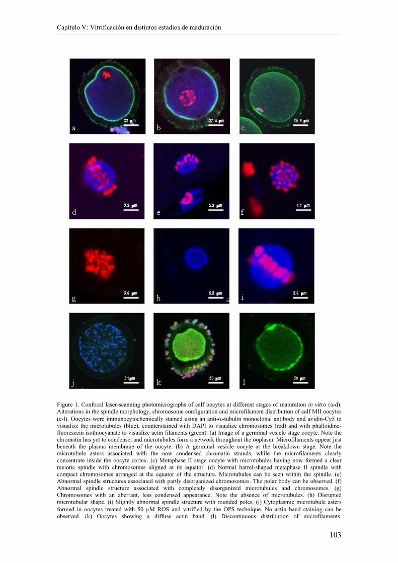
Capítulo V: Vitrificación en distintos estadios de maduración
103
Figure 1. Confocal laser-scanning photomicrographs of calf oocytes at different stages of maturation in vitro (a-d). Alterations in the spindle morphology, chromosome configuration and microfilament distribution of calf MII oocytes (e-l). Oocytes were immunocytochemically stained using an anti-α-tubulin monoclonal antibody and avidin-Cy5 to visualize the microtubules (blue), counterstained with DAPI to visualize chromosomes (red) and with phalloidine-fluorescein isothiocyanate to visualize actin filaments (green). (a) Image of a germinal vesicle stage oocyte. Note the chromatin has yet to condense, and microtubules form a network throughout the ooplasm. Microfilaments appear just beneath the plasma membrane of the oocyte. (b) A germinal vesicle oocyte at the breakdown stage. Note the microtubule asters associated with the now condensed chromatin strands, while the microfilaments clearly concentrate inside the oocyte cortex. (c) Metaphase II stage oocyte with microtubules having now formed a clear meiotic spindle with chromosomes aligned at its equator. (d) Normal barrel-shaped metaphase II spindle with compact chromosomes arranged at the equator of the structure. Microtubules can be seen within the spindle. (e) Abnormal spindle structures associated with partly disorganized chromosomes. The polar body can be observed. (f) Abnormal spindle structure associated with completely disorganized microtubules and chromosomes. (g) Chromosomes with an aberrant, less condensed appearance. Note the absence of microtubules. (h) Disrupted microtubular shape. (i) Slightly abnormal spindle structure with rounded poles. (j) Cytoplasmic microtubule asters formed in oocytes treated with 50 µM ROS and vitrified by the OPS technique. No actin band staining can be observed. (k) Oocytes showing a diffuse actin band. (l) Discontinuous distribution of microfilaments.
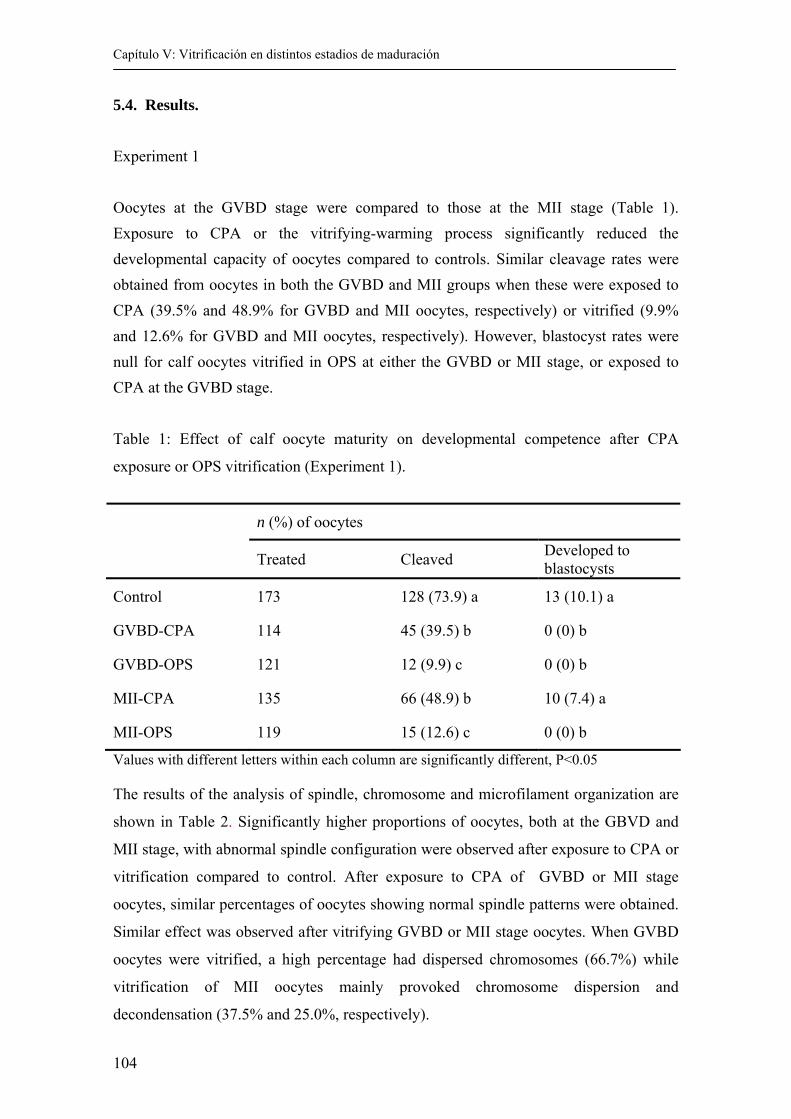
Capítulo V: Vitrificación en distintos estadios de maduración
104
5.4. Results. Experiment 1 Oocytes at the GVBD stage were compared to those at the MII stage (Table 1). Exposure to CPA or the vitrifying-warming process significantly reduced the developmental capacity of oocytes compared to controls. Similar cleavage rates were obtained from oocytes in both the GVBD and MII groups when these were exposed to CPA (39.5% and 48.9% for GVBD and MII oocytes, respectively) or vitrified (9.9% and 12.6% for GVBD and MII oocytes, respectively). However, blastocyst rates were null for calf oocytes vitrified in OPS at either the GVBD or MII stage, or exposed to CPA at the GVBD stage. Table 1: Effect of calf oocyte maturity on developmental competence after CPA
exposure or OPS vitrification (Experiment 1).
n (%) of oocytes
Treated Cleaved Developed to blastocysts
Control 173 128 (73.9) a 13 (10.1) a
GVBD-CPA 114 45 (39.5) b 0 (0) b
GVBD-OPS 121 12 (9.9) c 0 (0) b
MII-CPA 135 66 (48.9) b 10 (7.4) a
MII-OPS 119 15 (12.6) c 0 (0) b
Values with different letters within each column are significantly different, P<0.05 The results of the analysis of spindle, chromosome and microfilament organization are
shown in Table 2. Significantly higher proportions of oocytes, both at the GBVD and
MII stage, with abnormal spindle configuration were observed after exposure to CPA or
vitrification compared to control. After exposure to CPA of GVBD or MII stage
oocytes, similar percentages of oocytes showing normal spindle patterns were obtained.
Similar effect was observed after vitrifying GVBD or MII stage oocytes. When GVBD
oocytes were vitrified, a high percentage had dispersed chromosomes (66.7%) while
vitrification of MII oocytes mainly provoked chromosome dispersion and
decondensation (37.5% and 25.0%, respectively).
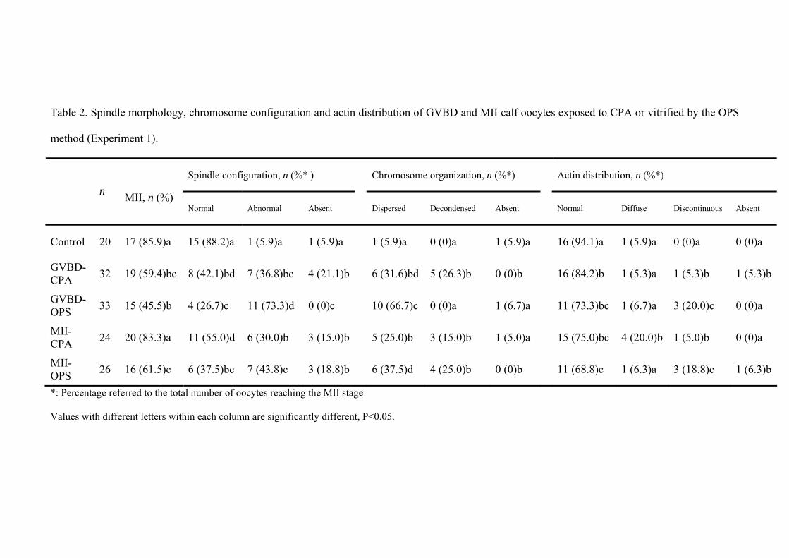
Table 2. Spindle morphology, chromosome configuration and actin distribution of GVBD and MII calf oocytes exposed to CPA or vitrified by the OPS
method (Experiment 1).
*: Percentage referred to the total number of oocytes reaching the MII stage
Values with different letters within each column are significantly different, P<0.05.
Spindle configuration, n (%* ) Chromosome organization, n (%*) Actin distribution, n (%*) n
MII, n (%) Normal Abnormal Absent Dispersed Decondensed Absent Normal Diffuse Discontinuous Absent
Control 20 17 (85.9)a 15 (88.2)a 1 (5.9)a 1 (5.9)a 1 (5.9)a 0 (0)a 1 (5.9)a 16 (94.1)a 1 (5.9)a 0 (0)a 0 (0)a
GVBD-CPA 32 19 (59.4)bc 8 (42.1)bd 7 (36.8)bc 4 (21.1)b 6 (31.6)bd 5 (26.3)b 0 (0)b 16 (84.2)b 1 (5.3)a 1 (5.3)b 1 (5.3)b
GVBD-OPS 33 15 (45.5)b 4 (26.7)c 11 (73.3)d 0 (0)c 10 (66.7)c 0 (0)a 1 (6.7)a 11 (73.3)bc 1 (6.7)a 3 (20.0)c 0 (0)a
MII-CPA 24 20 (83.3)a 11 (55.0)d 6 (30.0)b 3 (15.0)b 5 (25.0)b 3 (15.0)b 1 (5.0)a 15 (75.0)bc 4 (20.0)b 1 (5.0)b 0 (0)a
MII-OPS 26 16 (61.5)c 6 (37.5)bc 7 (43.8)c 3 (18.8)b 6 (37.5)d 4 (25.0)b 0 (0)b 11 (68.8)c 1 (6.3)a 3 (18.8)c 1 (6.3)b
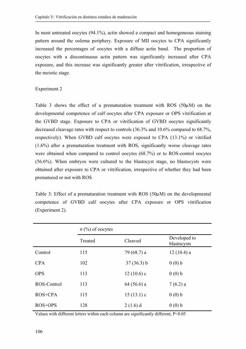
Capítulo V: Vitrificación en distintos estadios de maduración
106
In most untreated oocytes (94.1%), actin showed a compact and homogeneous staining pattern around the oolema periphery. Exposure of MII oocytes to CPA significantly increased the percentages of oocytes with a diffuse actin band. The proportion of oocytes with a discontinuous actin pattern was significantly increased after CPA exposure, and this increase was significantly greater after vitrification, irrespective of the meiotic stage.
Experiment 2
Table 3 shows the effect of a prematuration treatment with ROS (50µM) on the developmental competence of calf oocytes after CPA exposure or OPS vitrification at the GVBD stage. Exposure to CPA or vitrification of GVBD oocytes significantly decreased cleavage rates with respect to controls (36.3% and 10.6% compared to 68.7%, respectively). When GVBD calf oocytes were exposed to CPA (13.1%) or vitrified (1.6%) after a prematuration treatment with ROS, significantly worse cleavage rates were obtained when compared to control oocytes (68.7%) or to ROS-control oocytes (56.6%). When embryos were cultured to the blastocyst stage, no blastocysts were obtained after exposure to CPA or vitrification, irrespective of whether they had been prematured or not with ROS.
Table 3: Effect of a prematuration treatment with ROS (50µM) on the developmental competence of GVBD calf oocytes after CPA exposure or OPS vitrification (Experiment 2).
n (%) of oocytes
Treated Cleaved Developed to blastocysts
Control 115 79 (68.7) a 12 (10.4) a
CPA 102 37 (36.3) b 0 (0) b
OPS 113 12 (10.6) c 0 (0) b
ROS-Control 113 64 (56.6) a 7 (6.2) a
ROS+CPA 115 15 (13.1) c 0 (0) b
ROS+OPS 128 2 (1.6) d 0 (0) b
Values with different letters within each column are significantly different, P<0.05
Capítulo V: Vitrificación en distintos estadios de maduración
107
Table 4 shows a summary of the observations made of the spindles. The maturation rate
achieved was significantly higher for control oocytes (88.9%) with respect to GVBD
oocytes exposed to CPA (60.0%) or vitrified (45.5%). When GVBD calf oocytes were
vitrified (27.9%) after prematuration treatment with ROS, significantly lower
maturation rates were observed compared to ROS-control oocytes (75.5%). The
maturation rates of oocytes exposed to CPA after prematuration (63.6%) did not differ
significantly from those recorded for ROS-control oocytes (75.5%). Normal spindle
structures were observed in 90.0% of the control oocytes, whereas this percentage
decreased for GVBD oocytes exposed to CPA (40.0%) or vitrified (25%).
Prematuration treatment with ROS did not significantly improve the percentage of
normal spindles observed, with values of 45.7% or 16.7% recorded after CPA exposure
or OPS vitrification, respectively.
When GVBD oocytes were exposed to CPA or vitrified, the microtubules in the spindle
of most oocytes appeared not to be organized in a defined spindle shape or were
partially or completely disassembled. When oocytes were exposed to CPA, with or
without prematuration treatment, the proportions of abnormal or missing spindles
increased significantly with respect to control oocytes. When GVBD oocytes were
vitrified by the OPS technique, significantly higher percentages of abnormal
microtubule patterns were observed, either with or without prematuration. However, a
novel abnormal microtubular pattern was observed when oocytes were pretreated with
ROS and then OPS vitrified. Six oocytes (out of 12) showed numerous microtubular
asters and a microtubular network formed by radiating arrays of these asters, as shown
in Figure 1J.
On the other hand, the proportions of oocytes with dispersed chromosomes in the
cytoplasm increased significantly after all the treatments tested. The highest percentage
of oocytes with dispersed chromosomes was observed in the group of GVBD oocytes
that had been vitrified (65.0%), while oocytes prematured with ROS and vitrified
yielded the highest proportions with decondensed chromosomes (50.0%).
Oocytes vitrified either with or without a prematuration treatment showed significantly
lower percentages of normal microfilament patterns while CPA exposure did not
modify actin distribution when compared to control oocytes.
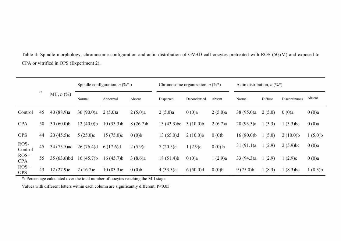
Table 4: Spindle morphology, chromosome configuration and actin distribution of GVBD calf oocytes pretreated with ROS (50µM) and exposed to
CPA or vitrified in OPS (Experiment 2).
*: Percentage calculated over the total number of oocytes reaching the MII stage
Values with different letters within each column are significantly different, P<0.05.
Spindle configuration, n (%* ) Chromosome organization, n (%*) Actin distribution, n (%*) n
MII, n (%) Normal Abnormal Absent Dispersed Decondensed Absent Normal Diffuse Discontinuous Absent
Control 45 40 (88.9)a 36 (90.0)a 2 (5.0)a 2 (5.0)a 2 (5.0)a 0 (0)a 2 (5.0)a 38 (95.0)a 2 (5.0) 0 (0)a 0 (0)a
CPA 50 30 (60.0)b 12 (40.0)b 10 (33.3)b 8 (26.7)b 13 (43.3)bc 3 (10.0)b 2 (6.7)a 28 (93.3)a 1 (3.3) 1 (3.3)bc 0 (0)a
OPS 44 20 (45.5)c 5 (25.0)c 15 (75.0)c 0 (0)b 13 (65.0)d 2 (10.0)b 0 (0)b 16 (80.0)b 1 (5.0) 2 (10.0)b 1 (5.0)b
ROS-Control 45 34 (75.5)ad 26 (76.4)d 6 (17.6)d 2 (5.9)a 7 (20.5)e 1 (2.9)c 0 (0) b 31 (91.1)a 1 (2.9) 2 (5.9)bc 0 (0)a
ROS+ CPA 55 35 (63.6)bd 16 (45.7)b 16 (45.7)b 3 (8.6)a 18 (51.4)b 0 (0)a 1 (2.9)a 33 (94.3)a 1 (2.9) 1 (2.9)c 0 (0)a
ROS+ OPS 43 12 (27.9)e 2 (16.7)c 10 (83.3)c 0 (0)b 4 (33.3)c 6 (50.0)d 0 (0)b 9 (75.0)b 1 (8.3) 1 (8.3)bc 1 (8.3)b
Capítulo V: Vitrificación en distintos estadios de maduración
109
5.5. Discussion.
The results of Experiment 1 indicate that the meiotic stage has no effect on the ability of
calf oocytes to survive cryopreservation, with both GVBD and MII stage oocytes being
similarly able to withstand the cryopreservation process. Further, handling control
oocytes, which were only exposed to CPA without further cooling, showed a reduced
capacity for development compared to untreated controls, suggesting that osmotic shock
compromises the success of cryopreservation procedures.
There have been a number of attempts over the last few decades to cryopreserve oocytes
at various stages (Lim et al., 1992; Otoi et al., 1995; Eroglu et al., 1998; Hochi et al.,
1998; Men et al., 2002). These investigations have established that oocytes at different
stages show differential responses to temperature shock. In fact, several authors have
reported that GV oocytes are more sensitive to chilling or cryopreservation than MII
oocytes (Lim et al., 1992; Fuku et al., 1995; Zeron et al., 1999; Goud et al., 2000).
Bovine oocytes at the GVBD stage have been described as more resistant to cooling
than GV or MII oocytes (Barnes et al., 1997). However, when cryopreservation was
attempted, Men at al. (2002) observed that a significantly higher proportion of cleaved
bovine embryos from vitrified MII oocytes developed into blastocysts than those
derived from vitrified GVBD oocytes. In addition to GVBD oocytes, attempts have also
been made at vitrifying oocytes at other maturation stages. Thus, when bovine oocytes
were vitrified 0, 6, 12 or 24 h after the onset of maturation, Hochi et al. (1998) found
the best stage for vitrification was that of oocytes matured for 12 h. Le Gal and Massip
(1999) reported similar percentages of blastocysts following the vitrification of bovine
oocytes at 0, 17 or 24 h after the start of the maturation process.
In the present study, nuclear and cytoskeletal changes were carefully determined
through immunocytochemical staining, defining the three factors: nuclear,
microfilament and microtubular status. When calf oocytes were exposed to CPA or
vitrified either at the GVBD or MII stage, they showed significantly lower percentages
of both normal spindle and chromosome configurations than untreated oocytes.
Temperature fluctuations are known to directly affect the cytoskeletal organization and
chromosome configuration of mouse (Pickering and Johnson, 1987), human (Pickering
et al., 1990; Almeida and Bolton, 1995), bovine (Richardson and Parks, 1992; Aman
Capítulo V: Vitrificación en distintos estadios de maduración
110
and Parks, 1994; Saunders and Parks, 1999; Wu et al., 1999) and porcine (Liu et al.,
2003; Rojas et al., 2005) oocytes. The partial or complete disassembly of microtubules
in the spindle has been observed after cooling oocytes to room temperature or lower
(Pickering and Johnson, 1987; Pickering et al., 1990; Aman and Parks, 1994; Almeida
and Bolton, 1995). When returned to normal temperature, the spindle recovers although
there are species-specific differences. The spindles of mouse oocytes undergo complete
disassembly after a 45 min to 60 min period at 0ºC, but the spindles of most oocytes
reassemble after rewarming (Magistrini and Szollosi, 1980). However, the spindles of
human (Almeida and Bolton, 1995; Wang et al., 2001), cow (Richardson and Parks,
1992; Aman and Parks, 1994), sheep (Moor and Crosby, 1985) or pig (Liu et al., 2003)
oocytes only undergo limited recovery after cooling-rewarming treatment. The meiotic
spindle becomes completely disassembled when in vitro matured bovine oocytes are
maintained for 10 to 20 min at 4ºC (Richardson and Parks, 1992). Similar findings were
reported by Wu et al. (1999), who noted a drastic decrease in the formation of normal
meiotic spindles when immature bovine oocytes were chilled to 4ºC for as little as 10
min. The fact that cooling immature oocytes, with no microtubular spindle present,
affects subsequent meiotic spindle formation and polar body extrusion implies that
cooling may lead to the damage of key regulatory factors, such as MPF, mitogen-
activated protein kinase (MAPK) or others, affecting microtubule organization (Vincent
and Johnson, 1992; Wu et al., 1996; Wu et al., 1997).
Previous studies have shown that cryoprotectants disrupt the cortical microfilament
network and cause depolymerization and disorganization of the spindle microtubules,
which in turn results in chromosomal scattering (Magistrini and Szollosi, 1980; Jonhson
and Pickering, 1987; Vincent et al., 1989; Vincent et al., 1990). EG may affect the
dynamic assembly and disassembly of microtubules, as has been shown for DMSO or
propanediol (Vincent et al., 1989). At room temperature, the addition of cryoprotectant
(propanediol or DMSO) to rabbit oocytes leads to the disorganization of microtubule
and microfilament distribution (Vincent et al., 1989). In mouse oocytes, a similar effect
of DMSO (Johnson and Pickering, 1987; Van der Elst et al., 1988) and propanediol
(Van der Elst et al., 1988) has also been noted and the consequent dispersal of
chromosomes is often seen in both species. The exposure of bovine oocytes to 1.5M EG
for 10 min has been observed to increase abnormal spindle formation, although this
treatment did not affect normal microfilament distribution (Saunders and Parks, 1999).
Capítulo V: Vitrificación en distintos estadios de maduración
111
The factors likely to contribute to the different sensitivity of immature oocytes
compared to mature oocytes are the specific structure of the zona pellucida, the
cytoskeleton, the cytoplasmic membranes and their composition (Sathananthan, 1994; Ji
et al., 1997) along with the connections of cumulus-cell projections and the oocyte
cytoskeleton through their junctions with the oolema (Allworth and Albertini, 1993). It
is important that these connections remain intact for the completion of normal
cytoplasmic maturation in vitro. In bovine oocytes, active transcription and translation
occur at the GVBD and later stages of meiotic maturation (Hunter and Moor, 1987;
Leibfried-Rutledge et al., 1989; Sirard et al., 1989). Therefore, in addition to
detrimental effects on cytology, cryopreservation may also disrupt the biochemical
processes occurring within the oocytes with the consequent negative effects on
cytoplasmic maturation.
Some authors report a loss of cumulus cells from the COC following cryopreservation
and thawing (Van der Elst et al., 1993; Cooper et al., 1998; Goud et al., 2000). By
examining the ultrastructure of immature bovine (Fuku et al., 1995) and equine (Hochi
et al., 1996) oocytes after vitrification, some have suggested that freezing damage is
associated with the destruction of these intercellular contacts between cumulus cells and
the oocyte. The three-dimensional COC is likely to be particularly prone to the physical
disruption caused by ice crystal formation. Even when ice crystal formation is avoided
during vitrification, the vast difference in size between the oocyte and its associated
cumulus cells means that they are likely to react very differently to the stresses in play
during cryopreservation. Ruppert-Lingham et al. (2003) tried to overcome this problem
by co-culturing cryopreserved mouse GV oocytes with fresh disassociated cumulus
cells, although no improvement on embryo development was observed.
Men et al. (2002) achieved blastocysts after vitrifying GVBD and MII cow oocytes by
the OPS method. However, we were unable to obtain blastocysts from calf oocytes
vitrified either at the GVBD or MII stage. Our previous studies described in Chapter III
revealed that calf oocytes were more sensitive to freezing injury than adult cow oocytes,
yielding lower percentages of cleavage and blastocyst formation after vitrification.
Oocytes collected form prepubertal animals are known to be less competent after in
vitro maturation, fertilization and embryo culture than oocytes collected from adult
animals (Revel et al., 1995; Damiani et al., 1996; Duby et al., 1996; Khatir et al., 1996;
Capítulo V: Vitrificación en distintos estadios de maduración
112
Presicce et al., 1997; Taneja et al., 2000). This deficient developmental capacity of calf
oocytes is most probably attributable to their abnormal cytoplasmic maturation
(Salamone et al., 2001).
Several authors have hypothesized that if oocytes are cultured before maturation in vitro
under conditions that arrest meiosis at the GV stage, this might offer them the
opportunity to acquire greater developmental competence (Lonergan et al., 2000;
Mermillod et al., 2000). In the present study, we subjected calf oocytes to a
prematuration treatment with ROS before vitrification, in an effort to improve oocyte
cytoplasmic maturation and increase the developmental capacity of the thawed oocytes.
Our results, however, indicate no such improvement and lower cleavage rates were
obtained after CPA exposure or vitrification of prematured oocytes compared to non-
prematured oocytes. On the other hand, similar percentages of oocytes showing a
normal spindle and cytoskeleton configuration were observed after CPA exposure or
vitrification, irrespective of the prematuration treatment. Morató et al. (2004) reported
similar proportions of oocytes with normal MII spindles for non-prematured oocytes
and those treated with 50 µM ROS. In contrast, Lonergan et al. (2003) described the
disruption of the integrity of the surrounding cumulus cells, swelling of the
mitochondrial cristae and degeneration of cortical granules in ROS-prematured bovine
oocytes. In addition to the possible detrimental effects of prematuration treatment, the
vitrification process is known to induce intense ultrastructural modifications to the
microvilli, mitochondria, vesicle formation and the ooplasm of immature bovine
oocytes (Fuku et al., 1995). Hence, although ROS prematuration treatment failed to
alter normal spindle and actin configurations, our significantly lowered cleavage rates
might have been a consequence of both the ultrastructural changes induced by ROS and
the detrimental effects of crypreservation.
Collectively, our findings suggest that the vitrification protocol used has deleterious
effects on the organization of the meiotic spindle of calf oocytes cryopreserved at both
the GVBD and MII stage, impairing the capacity for further development of the
embryos derived from these vitrified oocytes. A prematuration treatment with ROS had
no beneficial effect on oocytes vitrified by the OPS method. The cryoinjury suffered by
the oocytes was the main determinant for the low cleavage rates and poor
Capítulo V: Vitrificación en distintos estadios de maduración
113
developmental potential of these frozen oocytes. Further research will help clarify the
cellular and molecular mechanisms of cryopreservation-induced injury.
5.6 Bibliography. Albarracin JL, Morató R, Izquierdo MD and Mogas T. 2004. Embryo developmental
capacity of calf oocytes meiotically inhibited by roscovitine. Reproduction in domestic animals 39(4):273.
Allworth AE and Albertini DF. 1993. Meiotic maturation in cultured bovine oocytes is accompanied by remodeling of the cumulus cell cytoskeleton. Dev Biol 158(1):101-112.
Almeida PA and Bolton VN. 1995. The effect of temperature fluctuations on the cytoskeletal organisation and chromosomal constitution of the human oocyte. Zygote 3(4):357-365.
Aman RR and Parks JE. 1994. Effects of cooling and rewarming on the meiotic spindle and chromosomes of in vitro-matured bovine oocytes. Biol Reprod 50(1):103-110.
Arav A, Zeron Y, Leslie SB, Behboodi E, Anderson GB and Crowe JH. 1996. Phase transition temperature and chilling sensitivity of bovine oocytes. Cryobiology 33(6):589-599.
Barnes FL, Damiani P, Looney CR and Duby RT. 1997. The meiotic stage affects subsequent development of cooled bovine oocytes. Theriogenology 47(1):183.
Boiso I, Marti M, Santalo J, Ponsa M, Barri PN and Veiga A. 2002. A confocal microscopy analysis of the spindle and chromosome configurations of human oocytes cryopreserved at the germinal vesicle and metaphase II stage. Hum Reprod 17(7):1885-1891.
Bouquet M, Selva J and Auroux M. 1992. The incidence of chromosomal abnormalities in frozen-thawed mouse oocytes after in-vitro fertilization. Hum Reprod 7(1):76-80.
Carroll J, Warnes GM and Matthews CD. 1989. Increase in digyny explains polyploidy after in-vitro fertilization of frozen-thawed mouse oocytes. J Reprod Fertil 85:489-494.
Cooper A, Paynter SJ, Fuller BJ and Shaw RW. 1998. Differential effects of cryopreservation on nuclear or cytoplasmic maturation in vitro in immature mouse oocytes from stimulated ovaries. Hum Reprod 13(4):971-978.
Damiani P, Fissore RA, Cibelli JB, Long CR, Balise JJ, Robl JM and Duby RT. 1996. Evaluation of developmental competence, nuclear and ooplasmic maturation of calf oocytes. Mol Reprod Dev 45(4):521-534.
Duby RT, Damiani P, Looney CR, Fissore RA and Robl JM. 1996. Prepuberal calves as oocyte donors: Promises and problems. Theriogenology 45:121-130.
Eroglu A, Toth TL and Toner M. 1998. Alterations of the cytoskeleton and polyploidy induced by cryopreservation of metaphase II mouse oocytes. Fertil Steril 69(5):944-957.
Fouladi Nashta AA, Waddington D and Campbell KH. 1998. Maintenance of bovine oocytes in meiotic arrest and subsequent development In vitro: A comparative evaluation of antral follicle culture with other methods. Biol Reprod 59(2):255-262.
Fuku E, Xia L and Downey BR. 1995. Ultrastructural changes in bovine oocytes cryopreserved by vitrification. Cryobiology 32(2):139-156.
Capítulo V: Vitrificación en distintos estadios de maduración
114
Glenister PH, Wood MJ, Kirby C and Whittingham DG. 1987. Incidence of chromosome anomalies in first-cleavage mouse embryos obtained from frozen-thawed oocytes fertilized in vitro. Gamete Res 16(3):205-216.
Goud A, Goud P, Qian C, Van der Elst J, Van Maele G and Dhont M. 2000. Cryopreservation of human germinal vesicle stage and in vitro matured M II oocytes: influence of cryopreservation media on the survival, fertilization, and early cleavage divisions. Fertil Steril 74(3):487-494.
Heller DT, Cahill DM and Schultz RM. 1981. Biochemical studies of mammalian oogenesis: Metabolic cooperativity between granulosa cells and growing mouse oocytes. Dev Biol 84:455-464.
Hochi S, Ito K, Hirabayashi M, Ueda M, Kimura K and Hanada A. 1998. Effect of nuclear stages during IVM on the survival of vitrified-warmed bovine oocytes. Theriogenology 49(4):787-796.
Hochi S, Kozawa M, Fujimoto T, Hondo E, Yamada J and Oguri N. 1996. In vitro maturation and transmission electron microscopic observation of horse oocytes after vitrification. Cryobiology 33(3):300-310.
Holm P, Booth PJ, Schmidt MH, Greve T and Callesen H. 1999. High Bovine Blastocyst Development in a Static in Vitro Production System Using SOFaa Medium Supplemented with Sodium Citrate and Myo-Inositol With or Without Serum-Proteins. Theriogenology 52(4):683-700.
Hunter AG and Moor RM. 1987. Stage-dependent effects of inhibiting ribonucleic acids and protein synthesis on meiotic maturation of bovine oocytes in vitro. J Dairy Sci 70(8):1646-1651.
Hyttel P. 1987. Bovine cumulus-oocyte disconnection in vitro. Anat Embryol (Berl) 176(1):41-44.
Hyttel P, Vajta G and Callesen H. 2000. Vitrification of bovine oocytes with the open pulled straw method: ultrastructural consequences. Mol Reprod Dev 56(1):80-88.
Ji YZ, Bomsel M, Jouannet P and Wolf JP. 1997. Modifications of the human oocyte plasma membrane protein pattern during preovulatory maturation. Mol Reprod Dev 47(1):120-126.
Johnson MH and Pickering SJ. 1987. The effect of dimethylsulphoxide on the microtubular system of the mouse oocyte. Development 100(2):313-324.
Jonhson MH and Pickering SJ. 1987. The effect of dimethylsulfoxide on the microtubular system of mouse oocyte. Development 100:313-324.
Khatir H, Lonergan P, Carolan C and Mermillod P. 1996. Prepubertal bovine oocyte: a negative model for studying oocyte developmental competence. Mol Reprod Dev 45(2):231-239.
Küchenmeister U and Kuwayama M. 1997. In vitro blastocysts formation, after vitrification of immature bovine oocytes. Theriogenology 47:348.
Le Gal F and Massip A. 1998. Development of thawed oocytes fertilized in vitro after vitrification by the open pulled straw method before or after in vitro maturation. In: Lauria A, Gandolfi F, Enne E, Gianaroli L, editors. Gametes: Development and Function. Rome: Serono Symposia. p 554.
Le Gal F and Massip A. 1999. Cryopreservation of cattle oocytes: effects of meiotic stage, cycloheximide treatment, and vitrification procedure. Cryobiology 38(4):290-300.
Leibfried-Rutledge ML, Florman HM and First NL. 1989. The molecular biology of mammalian oocyte maturation. In: Schatten H, Schatten G, editors. The molecular biology of fertilization. New York: Academic Press. pp 259-275.
Capítulo V: Vitrificación en distintos estadios de maduración
115
Lim JM, Fukui Y and Ono H. 1992. Developmental competence of bovine oocytes frozen at various maturation stages followed by in vitro maturation and fertilization. Theriogenology 37:351-361.
Liu RH, Sun QY, Li YH, Jiao LH and Wang WH. 2003. Effects of cooling on meiotic spindle structure and chromosome alignment within in vitro matured porcine oocytes. Mol Reprod Dev 65(2):212-218.
Lonergan P, Dinnyes A, Fair T, Yang X and Boland M. 2000. Bovine oocyte and embryo development following meiotic inhibition with butyrolactone I. Mol Reprod Dev 57(2):204-209.
Lonergan P, Faerge I, Hyttel PM, Boland M and Fair T. 2003. Ultrastructural modifications in bovine oocytes maintained in meiotic arrest in vitro using roscovitine or butyrolactone. Mol Reprod Dev 64(3):369-378.
Magistrini M and Szollosi D. 1980. Effects of cold and of isopropyl-N-phenylcarbamate on the second meiotic spindle of mouse oocytes. Eur J Cell Biol 22(2):699-707.
Martino A, Pollard JW and Leibo SP. 1996a. Effect of chilling bovine oocytes on their developmental competence. Mol Reprod Dev 45(4):503-512.
Martino A, Songsasen N and Leibo SP. 1996b. Development into blastocysts of bovine oocytes cryopreserved by ultra-rapid cooling. Biol Reprod 54(5):1059-1069.
Men H, Monson RL and Rutledge JJ. 2002. Effect of meiotic stages and maturation protocols on bovine oocyte's resistance to cryopreservation. Theriogenology 57(3):1095-1103.
Mermillod P, Tomanek M, Marchal R and Meijer L. 2000. High developmental competence of cattle oocytes maintained at the germinal vesicle stage for 24 hours in culture by specific inhibition of MPF kinase activity. Mol Reprod Dev 55(1):89-95.
Moor RM and Crosby IM. 1985. Temperature-induced abnormalities in sheep oocytes during maturation. J Reprod Fertil 75(2):467-473.
Morató R, Albarracin JL, Izquierdo MD and Mogas T. Alterations and reversibility in the chromatin and cytoskeleton of calf oocytes treated with roscovitine; 2004; Lyon (France). p 156.
Niimura S and Hosoe M. 1995. Changes in cortical granule distribution within bovine oocytes during maturation and fertilization in vitro. J Reprod Dev 41:103-108.
Otoi T, Yamamoto K, Koyama N and Suzuki T. 1995. In vitro fertilization and development of immature and mature bovine oocytes cryopreserved by ethylene glycol with sucrose. Cryobiology 32(5):455-460.
Pickering SJ, Braude PR, Johnson MH, Cant A and Currie J. 1990. Transient cooling to room temperature can cause irreversible disruption of the meiotic spindle in the human oocyte. Fertil Steril 54(1):102-108.
Pickering SJ and Johnson MH. 1987. The influence of cooling on the organization of the meiotic spindle of the mouse oocyte. Hum Reprod 2(3):207-216.
Presicce GA, Jiang S, Simkin M, Zhang L, Looney CR, Godke RA and Yang X. 1997. Age and hormonal dependence of acquisition of oocyte competence for embryogenesis in prepubertal calves. Biol Reprod 56(2):386-392.
Revel F, Mermillod P, Peynot N, Renard J and Heyman Y. 1995. Low developmental capacity of in vitro matured and fertilized oocytes from calves compared with that of cows. J Reprod Fertil 103(1):115-120.
Richardson RR and Parks JE. 1992. Effects of chilling on the meiotic spindle and chromosomes of bovine ova. Theriogenology 37(1):284.
Capítulo V: Vitrificación en distintos estadios de maduración
116
Rizos D, Ward F, Boland MP and Lonergan P. 2001. Effect of culture system on the yield and quality of bovine blastocysts as assessed by survival after vitrification. Theriogenology 56(1):1-16.
Rojas C, Palomo MJ, Albarracin JL and Mogas T. 2005. Vitrification of Immature and In Vitro Matured Pig Oocytes: Study of Distribution of Chromosomes, Microtubules and Microfilaments. Cryobiology:In press.
Ruppert-Lingham CJ, Paynter SJ, Godfrey J, Fuller BJ and Shaw RW. 2003. Developmental potential of murine germinal vesicle stage cumulus-oocyte complexes following exposure to dimethylsulphoxide or cryopreservation: loss of membrane integrity of cumulus cells after thawing. Hum Reprod 18(2):392-398.
Salamone DF, Damiani P, Fissore JM, Robl. and Duby RT. 2001. Biochemical an developmental Evidence that ooplasmic maturation of prepubertal bovine oocyte is compromised. Biol Reprod 64:1761-1768.
Sathananthan AH. 1994. Ultrastructural changes during meiotic maturation in mammalian oocytes: unique aspects of the human oocyte. Microsc Res Tech 27(2):145-164.
Saunders KM and Parks JE. 1999. Effects of cryopreservation procedures on the cytology and fertilization rate of in vitro-matured bovine oocytes. Biol Reprod 61(1):178-187.
Shaw JM, Oranratnachai A and Trounson AO. 2000. Fundamental cryobiology of mammalian oocytes and ovarian tissue. Theriogenology 53(1):59-72.
Sirard MA, Florman HM, Leibfried-Rutledge ML, Barnes FL, Sims ML and First NL. 1989. Timing of nuclear progression and protein synthesis necessary for meiotic maturation of bovine oocytes. Biol Reprod 40(6):1257-1263.
Suzuki T, Boediono A, Takagi M, Saha S and Sumantri C. 1996. Fertilization and development of frozen-thawed germinal vesicle bovine oocytes by a one-step dilution method in vitro. Cryobiology 33(5):515-524.
Szollosi D, Desmedt V, Crozet N and Brender C. 1988. In vitro maturation of sheep ovarian oocytes. Reprod Nutr Dev 28(4B):1047-1080.
Taneja M, Bols PE, Van de Velde A, Ju JC, Schreiber D, Tripp MW, Levine H, Echelard Y, Riesen J and Yang X. 2000. Developmental competence of juvenile calf oocytes in vitro and in vivo: influence of donor animal variation and repeated gonadotropin stimulation. Biol Reprod 62(1):206-213.
Thibault C, Szollosi D and Gerard M. 1987. Mammalian oocyte maturation. Reprod Nutr Dev 27(5):865-896.
Vajta G, Holm P, Kuwayama M, Booth PJ, Jacobsen H, Greve T and Callesen H. 1998. Open Pulled Straw (OPS) vitrification: a new way to reduce cryoinjuries of bovine ova and embryos. Mol Reprod Dev 51(1):53-58.
Van der Elst J, Van den Abbeel E, Jacobs R, Wisse E and Van Steirteghem A. 1988. Effect of 1,2-propanediol and dimethylsulphoxide on the meiotic spindle of the mouse oocyte. Hum Reprod 3(8):960-967.
Van der Elst JC, Nerinckx SS and Van Steirteghem AC. 1993. Slow and ultrarapid freezing of fully grown germinal vesicle-stage mouse oocytes: optimization of survival rate outweighed by defective blastocyst formation. J Assist Reprod Genet 10(3):202-212.
Vieira AD, Mezzalira A, Barbieri DP, Lehmkuhl RC, Rubin MI and Vajta G. 2002. Calves born after open pulled straw vitrification of immature bovine oocytes. Cryobiology 45(1):91-94.
Capítulo V: Vitrificación en distintos estadios de maduración
117
Vincent C, Garnier V, Heyman Y and Renard JP. 1989. Solvent effects on cytoskeletal organization and in-vivo survival after freezing of rabbit oocytes. J Reprod Fertil 87(2):809-820.
Vincent C and Johnson MH. 1992. Cooling, cryoprotectants, and the cytoskeleton of the mammalian oocyte. Oxf Rev Reprod Biol 14:73-100.
Vincent C, Pickering SJ, Johnson MH and Quick SJ. 1990. Dimethylsulphoxide affects the organisation of microfilaments in the mouse oocyte. Mol Reprod Dev 26(3):227-235.
Wang WH, Meng L, Hackett RJ, Odenbourg R and Keefe DL. 2001. Limited recovery of meiotic spindles in living human oocytes after cooling-rewarming observed using polarized light microscopy. Hum Reprod 16(11):2374-2378.
Wu B, Ignotz G, Currie WB and Yang X. 1997. Dynamics of maturation-promoting factor and its constituent proteins during in vitro maturation of bovine oocytes. Biol Reprod 56(1):253-259.
Wu B, Ignotz GG, Currie WB and Yang X. 1996. Temporal distinctions in the synthesis and accumulation of proteins by oocytes and cumulus cells during maturation in vitro of bovine oocytes. Mol Reprod Dev 45(4):560-565.
Wu B, Tong J and Leibo SP. 1999. Effects of cooling germinal vesicle-stage bovine oocytes on meiotic spindle formation following in vitro maturation. Mol Reprod Dev 54(4):388-395.
Yang BC, Ryu LS, Kirn IH, Yeon SH, Choi SS, Son DS, Im KS and Hwang WS. Effect of freezing of immature and mature bovine oocytes on their development to blastocyst in vitro; 1998; Seoul, Korea. p 950-951.
Zeron Y, Pearl M, Borochov A and Arav A. 1999. Kinetic and temporal factors influence chilling injury to germinal vesicle and mature bovine oocytes. Cryobiology 38(1):35-42.

CAPÍTULO VI:
DISCUSIÓN GENERAL.
Capítulo VI: Discusión general
121
El primer capítulo experimental de esta tesis doctoral se centró en el estudio de los
efectos de la técnica de vitrificación mediante OPS sobre las estructuras de la placa
metafásica, del citoesqueleto y el desarrollo embrionario en ovocitos de vacas
prepúberes y adultas. En este estudio, se observó que tanto la exposición a los
crioprotectores como el proceso de vitrificación provocaba daños a nivel estructural y
por tanto, una disminución en los porcentajes de ovocitos que llegaban al estadio de
blastocisto, tanto en ovocitos procedentes de ovarios de vaca como de ternera. Además,
se determinó que los ovocitos de ternera son más resistentes a la exposición a las
soluciones crioprotectoras pero más sensibles a la vitrificación puesto que se observó
una disminución significativa del porcentaje de ovocitos de ternera que presentaban
husos y/o distribuciones de actina normales en comparación con los ovocitos
procedentes de ovarios de vaca. Asimismo, el desarrollo embrionario in vitro de los
ovocitos de ternera tras su vitrificación y fecundación fue inferior al de los ovocitos de
vaca. La técnica de OPS desarrollada por Vajta et al. (1998) para la vitrificación de
ovocitos bovinos madurados in vitro es la que mejores porcentajes de desarrollo
embrionario obtiene comparada con otras técnicas de vitrificación para el mismo estadio
de maduración, utilizando diferentes soportes y/o crioprotectores (Martino et al., 1996;
Le Gal y Massip, 1999; Rho et al., 2002). Estos resultados nos llevaron a pensar que
esta técnica sería la más adecuada para llevar a cabo la vitrificación de ovocitos
procedentes de terneras. En nuestro laboratorio, el porcentaje de blastocistos obtenido
tras vitrificar ovocitos de vaca y ternera mediante esta técnica fue inferior al alcanzado
Vajta et al. (1998), pero similar al obtenido por otros autores (Le Gal y Massip, 1999).
Nuestros resultados confirmaron el hecho de que los ovocitos de ternera, aún
presentando una división celular similar a la ovocitos adultos, poseen una menor
capacidad de desarrollo embrionario. Este hecho es explicado por varios autores como
una deficiencia o retraso en la capacidad de maduración citoplasmática de los ovocitos
procedentes de animales prepúberes (Khatir et al., 1998; Mermillod et al., 1998;
Armstrong, 2001; Salamone et al., 2001).
Los resultados de desarrollo embrionario obtenidos en este estudio indicaron que los
ovocitos de ternera poseen una capacidad similar a los ovocitos procedentes de animales
adultos para resistir el daño osmótico causado por los crioprotectores. Sin embargo,
estos mismos ovocitos son más susceptibles al daño causado por la congelación
Capítulo VI: Discusión general
122
comparado con sus similares procedentes de adulto. Minimizar el daño osmótico es un
punto importante en las técnicas de vitrificación, ya que, para conseguir velocidades de
enfriamiento lo suficientemente altas para evitar la formación de cristales de hielo, es
necesario exponer al ovocito a concentraciones multimolares del crioprotector (revisado
por Shaw et al., 2000; Vajta, 2000). Ello provoca que el ovocito sufra alteraciones
estructurales derivadas de este cambio osmótico, expresadas en el cambio de volumen
que sufre la célula en contacto con el crioprotector hiperosmótico y que afecta al
citoesqueleto del ovocito.
Diversos autores sugieren que el principal objetivo a desarrollar en los protocolos de
crioconservación es el de tratar de preservar la integridad de la placa metafásica durante
el enfriamiento (Eroglu et al., 1998). La principal consecuencia del enfriamiento sobre
la placa metafásica es la despolimerización y la desaparición de los microtúbulos (Webb
et al., 1986). El reensamblaje de esta estructura es posible una vez es restituida la
temperatura normal (Inoue, 1981), aunque no siempre se da de manera correcta,
observándose que un porcentaje de estas estructuras puede presentar alteraciones en los
patrones de distribución de los cromosomas o los microtúbulos (Richardson y Parks,
1992; Liu et al., 2003). La exposición de los ovocitos maduros de distintas especies
como el ratón (Vincent y Johnson, 1992; Cooper et al., 1996), la coneja (Vincent et al.,
1989), la mujer (Sathananthan et al., 1987) y la vaca (Saunders y Parks, 1999) a los
crioprotectores también presentó un efecto adverso sobre la estructura de los
microtúbulos. De hecho, el contacto del propanodiol o el DMSO con ovocitos de conejo
madurados in vitro provocó la desorganización de los microtúbulos de la placa
metafásica (Vincent et al., 1989). Similar efecto fue observado en ovocitos de ratón
expuestos al propanodiol (Van der Elst et al., 1988).
La disrupción del citoesqueleto constituye un efecto intrínseco a los cambios de
volumen que sufre el ovocito como consecuencia de los procedimientos de
crioconservación. Sin embargo, en este estudio no se observó que la exposición de los
ovocitos de ternera y vaca a concentraciones multimolares de crioprotector tuviera un
efecto significativo sobre la disrupción de los filamentos de actina. Williams et al.
(1992) observaron que la organización de los microfilamentos de ovocitos bovinos
expuestos a EG o a propanodiol fue parecida a la de los ovocitos control. Saunders y
Parks (1999) evidenciaron la presencia de bandas normales de distribución de la actina
Capítulo VI: Discusión general
123
después de 1-3 horas post-incubación de ovocitos bovinos que habían sido expuestos a
EG.
Por otra parte, el proceso de vitrificación por OPS llevado a cabo en este estudio
provocó una disminución significativa de los ovocitos que presentaban un patrón de
distribución normal de los filamentos de actina, tanto en ovocitos procedentes de vaca
como de ternera. Similares efectos fueron observados en ovocitos de ratón y conejo
(Vincent et al., 1989; George y Johnson, 1993) aunque estos cambios fueron reversibles
tras la descongelación. Por otra parte, la congelación lenta de ovocitos maduros de vaca
comportó una disminución significativa en el porcentaje de ovocitos con una
distribución normal de los microfilamentos (Saunders y Parks, 1999). Los cambios
observados en la organización de los filamentos de actina podrían provocar cambios en
otros componentes celulares como las mitocondrias o la membrana plasmática debido a
la asociación de los microfilamentos a éstas y a otras estructuras del citoplasma (Didion
et al., 1990; Fuku et al., 1995).
La presencia de la placa metafásica en los ovocitos maduros hace de éstos un tipo
celular muy difícil de crioconservar. Esta estructura posee microtúbulos polimerizados
íntimamente relacionados con los cromosomas condensados. Ambas estructuras son
altamente susceptibles a la despolimerización y/o desorganización por descensos de
temperatura (revisado por Aman y Parks, 1994; Bernard y Fuller, 1996). Por este
motivo, diferentes autores hipotetizaron que la crioconservación de ovocitos en estadios
inmaduros de desarrollo favorecería la preservación de estas estructuras ya que este tipo
de ovocitos mantiene los cromosomas descondensados y protegidos por una membrana
nuclear, y los microtúbulos despolimerizados y distribuidos por el citoplasma (revisado
por Arav et al., 1993). Sin embargo, los resultados obtenidos por Didion et al.(1990),
Isachenko et al. (2001), Otoi et al. (1995), Parks y Ruffing (1992) y Rojas et al. (2004)
demostraron que la crioconservación de ovocitos en este estadio de maduración daba
lugar a peores resultados que la crioconservación de ovocitos maduros. Éstos resultados
difieren de lo observado en nuestros estudios en terneras utilizando la vitrificación por
OPS ya que tan sólo se observó una pequeña disminución no significativa en el
porcentaje de división embrionaria a las 48 horas post-inseminación tras vitrificar
ovocitos en estadio de GVBD comparado con aquellos vitrificados en estadio de MII.
Estos resultados pueden ser explicados como una consecuencia de vitrificar ovocitos en
Capítulo VI: Discusión general
124
un estadio donde se suman las ventajas y desventajas de congelar en un estadio de
maduración intermedio (Hochi et al., 1998; Le Gal y Massip, 1999; Men et al., 2002).
Así, en nuestro estudio, la vitrificación se llevó a cabo a las seis horas de haber iniciado
la maduración, lo que permitiría al ovocito llevar a cabo la síntesis de proteínas
necesarias para la fecundación y posterior desarrollo embrionario. Además, el hecho de
que la GVBD no presente aún una organización estricta de los cromosomas ni de los
microtúbulos nos llevó a suponer que este estadio de maduración sería menos sensible a
la vitrificación. Los resultados obtenidos en nuestros experimentos demostraron que si
bien los ovocitos en GVBD eran capaces de completar la primera división meiótica tras
su vitrificación, el porcentaje de placas metafásicas normales no mejoró. Por tanto,
podemos suponer que la vitrificación en estadio de GVBD provocó suficientes daños en
los cromosomas y los microtúbulos como para que la placa metafásica resultante de la
primera división meiótica presentase ya patrones de distribución de cromosomas y
microtúbulos anormales.
La premaduración de los ovocitos con un inhibidor del reinicio de la meiosis permitiría
que los ovocitos tuvieran una mayor oportunidad para concluir la maduración
citoplasmática y así, mejorar su potencial de desarrollo embrionario (Lonergan et al.,
1997; Avery et al., 1998; Mermillod et al., 2000; Motlik et al., 2000). Para tratar de
mejorar el potencial de desarrollo embrionario, la literatura describe diferentes
inhibidores del reinicio de la meiosis como la ciclohexamida (Lonergan et al., 1997;
Saeki et al., 1997), la dimetilaminopurina (Lonergan et al., 1997; Avery et al., 1998), la
butirolactona I (Kitagawa et al., 1993; Motlik et al., 1998; Kubelka et al., 2000;
Lonergan et al., 2000), la roscovitina (Mermillod et al., 2000), la roscovitina con ácido
retinoico (Duque et al., 2002) y la roscovitina con butirolactona (Ponderato et al.,
2001). Tras la inhibición del reinicio de la meiosis de ovocitos de ternera con
roscovitina, observamos que la dosis mínima necesaria para el bloqueo meiótico era de
50 µM, con la que cerca de un 60% de los ovocitos permaneció en estadio de VG
después de 24 horas de inhibición y un 74,8% de ovocitos alcanzó el estadio en MII
después de 24 horas de maduración in vitro en ausencia del inhibidor. Este resultado fue
inferior al obtenido por Mermillod et al. (2000) usando una concentración inferior de
inhibidor (88% de los ovocitos en VG con 25 µM de roscovitina).
Capítulo VI: Discusión general
125
A nivel estructural, observamos que la premaduración de ovocitos de ternera con 50 µM
de roscovitina no afectó significativamente a la estructura de la placa metafásica
después de la maduración in vitro de los ovocitos. Sin embargo, a concentraciones
superiores o inferiores (12.5, 25 y 100 µM), disminuyó significativamente el porcentaje
de ovocitos que presentaban un patrón de placa metafásica normal. Estos resultados
fueron similares a los obtenidos por Schoevers et al. (2004) quienes obtuvieron una
proporción similar de ovocitos de cerdo que presentaban placas metafásicas anormales
tras un proceso de premaduración con roscovitina (50µM). Por el contrario, Ju et al.
(2003) observaron que el porcentaje de placas metafásicas anormales aumentaba tras
premadurar ovocitos de cerdo con 80 µM de roscovitina. Lonergan et al. (2003)
describieron cambios morfológicos en ovocitos premadurados con 125 µM de
roscovitina, evidenciando la disrupción de la integridad de las células del cúmulus, el
hinchamiento de las crestas mitocondriales y la degeneración de los gránulos corticales.
Los resultados del tercer capítulo experimental mostraron que ninguno de los dos
estadios meióticos estudiados influenció la capacidad de supervivencia de los ovocitos
de ternera a la crioconservación, puesto que tanto el estadio de VGBD como el de MII
proporcionaron porcentajes similares de división embrionaria a las 48 horas post-
inseminación. Por otro lado, la exposición de ambos estadios de maduración a los
crioprotectores disminuyó el porcentaje de desarrollo embrionario obtenido. Varios
autores han evidenciado que el estadio de VG es más sensible a la crioconservación
comparado con los estadios maduros (Lim et al., 1992; Fuku et al., 1995; Zeron et al.,
1999; Goud et al., 2000). Barnes et al. (1997) observaron que los ovocitos bovinos en
VGBD eran más resistentes al enfriamiento que los ovocitos en estadio de GV o MII.
Sin embargo, Men et al. (2002) obtuvieron un mayor desarrollo embrionario tras
vitrificar ovocitos bovinos en estadio de MII en relación a los ovocitos vitrificados en
GVBD.
A nivel estructural, pudimos observar que los ovocitos procedentes de animales
prepúberes muestran un ligero descenso no significativo en el número de placas
metafásicas normales después de la vitrificación y/o exposición a los crioprotectores en
estadio de VGBD, comparados con aquellos en estadio de MII. El hecho de que el
proceso de vitrificación de ovocitos en estadio de VGBD, por tanto con microtúbulos
Capítulo VI: Discusión general
126
descondensados, afecte a la formación de la placa metafásica después de la maduración,
nos hace suponer que la crioconservación también pueda afectar a otros factores que
intervienen en la formación de esta estructura (Vincent y Johnson, 1992; Wu et al.,
1996; Wu et al., 1997).
Men et al. (2002) obtuvieron blastocistos tras vitrificar ovocitos de vaca en estadio de
VGBD y MII mediante la técnica OPS. En este estudio, el porcentaje de blastocistos
obtenido tras vitrificar ovocitos procedentes de ovarios de ternera en estadio de GVBD
o MII fue nulo. Estos resultados pueden explicarse por la mayor sensibilidad que tienen
los ovocitos de terneras prepúberes a la vitrificación, lo cual ya fue descrito en el primer
capítulo experimental de este trabajo. Por otra parte y como ya ha sido comentado
anteriormente, los ovocitos procedentes de animales prepúberes son menos competentes
para el desarrollo embrionario in vitro que sus correspondientes de animales adultos
(Revel et al., 1995; Damiani et al., 1996; Duby et al., 1996; Khatir et al., 1996; Presicce
et al., 1997; Taneja et al., 2000).
La hipótesis de que la premaduración en bloqueo meiótico permitía a los ovocitos
completar la maduración citoplasmática (Lonergan et al., 2000; Mermillod et al., 2000)
nos llevó a pensar que al someter a los ovocitos procedentes de animales prepúberes a
un proceso de premaduración con roscovitina permitiría a estos ovocitos mejorar su
potencial de desarrollo embrionario y así, la vitrificación en estadio de VGBD podría
ser más eficiente, debido a las ventajas estructurales anteriormente mencionadas para
este estadio. Sin embargo, los resultados obtenidos demostraron que una premaduración
con roscovitina no logró mejorar los porcentajes de división embrionaria a las 48 horas
post-inseminación comparado con ovocitos vitrificados en estadio de MII.
La vitrificación de ovocitos de ternera tras una premaduración con 50 µM de roscovitina
no incrementó el porcentaje de placas metafásicas anormales respecto a los ovocitos
control pero sí disminuyó significativamente el porcentaje de división embrionaria a las
48 horas post-inseminación. Lonergan et al (2003) describieron daños ultraestructurales
tras premadurar ovocitos bovinos con roscovitina lo que nos hace suponer que son
precisamente estos daños a nivel ultraestructural causados por una premaduración con
roscovitina, junto con aquellos causados por el proceso de vitrificación, los responsables
de los bajos resultados en el desarrollo embrionario.
Capítulo VI: Discusión general
127
Bibliography. Aman RR and Parks JE. 1994. Effects of cooling and rewarming on the meiotic spindle
and chromosomes of in vitro-matured bovine oocytes. Biol Reprod 50(1):103-110.
Arav A, Shehu D and Mattioli M. 1993. Osmotic and cytotoxic study of vitrification of immature bovine oocytes. J Reprod Fertil 99(2):353-358.
Armstrong DT. 2001. Effects of maternal age on oocyte developmental competence. Theriogenology 55(6):1303-1322.
Avery B, Hay-Schmidt A, Hyttel P and Greve T. 1998. Embryo development, oocyte morphology, and kinetics of meiotic maturation in bovine oocytes exposed to 6-dimethylaminopurine prior to in vitro maturation. Mol Reprod Dev 50(3):334-344.
Barnes FL, Damiani P, Looney CR and Duby RT. 1997. The meiotic stage affects subsequent development of cooled bovine oocytes. Theriogenology 47(1):183.
Bernard A and Fuller BJ. 1996. Cryopreservation of human oocytes: a review of current problems and perspectives. Hum Reprod Update 2(3):193-207.
Cooper A, O`Neil L, Bernard A, Fuller BJ and Shaw RW. 1996. The effect of protein source and temperature on the damage to mouse oocytes cytoskeleton after exposure to a vitrification solution. Cryo Letters 17:149-156.
Damiani P, Fissore RA, Cibelli JB, Long CR, Balise JJ, Robl JM and Duby RT. 1996. Evaluation of developmental competence, nuclear and ooplasmic maturation of calf oocytes. Mol Reprod Dev 45(4):521-534.
Didion BA, Pomp D, Martin MJ, Homanics GE and Markert CL. 1990. Observations on the cooling and cryopreservation of pig oocytes at the germinal vesicle stage. J Anim Sci 68(9):2803-2810.
Duby RT, Damiani P, Looney CR, Fissore RA and Robl JM. 1996. Prepuberal calves as oocyte donors: Promises and problems. Theriogenology 45:121-130.
Duque P, Diez C, Royo L, Lorenzo PL, Carneiro G, Hidalgo CO, Facal N and Gomez E. 2002. Enhancement of developmental capacity of meiotically inhibited bovine oocytes by retinoic acid. Hum Reprod 17(10):2706-2714.
Eroglu A, Toth TL and Toner M. 1998. Alterations of the cytoskeleton and polyploidy induced by cryopreservation of metaphase II mouse oocytes. Fertil Steril 69(5):944-957.
Fuku E, Xia L and Downey BR. 1995. Ultrastructural changes in bovine oocytes cryopreserved by vitrification. Cryobiology 32(2):139-156.
George MA and Johnson MH. 1993. Cytoskeletal organization and zona sensitivity to digestion by chymotrypsin of frozen-thawed mouse oocytes. Hum Reprod 8(4):612-620.
Goud A, Goud P, Qian C, Van der Elst J, Van Maele G and Dhont M. 2000. Cryopreservation of human germinal vesicle stage and in vitro matured M II oocytes: influence of cryopreservation media on the survival, fertilization, and early cleavage divisions. Fertil Steril 74(3):487-494.
Hochi S, Ito K, Hirabayashi M, Ueda M, Kimura K and Hanada A. 1998. Effect of nuclear stages during IVM on the survival of vitrified-warmed bovine oocytes. Theriogenology 49(4):787-796.
Inoue S. 1981. Cell division and the meiotic spindle. J Cell Biol 91:131-147.
Capítulo VI: Discusión general
128
Isachenko V, Alabart JL, Nawroth F, Isachenko E, Vajta G and Folch J. 2001. The open pulled straw vitrification of ovine GV-oocytes: positive effect of rapid cooling or rapid thawing or both? Cryo Letters 22(3):157-162.
Ju JC, Tsay C and Ruan CW. 2003. Alterations and reversibility in the chromatin, cytoskeleton and development of pig oocytes treated with roscovitine. Mol Reprod Dev 64(4):482-491.
Khatir H, Lonergan P, Carolan C and Mermillod P. 1996. Prepubertal bovine oocyte: a negative model for studying oocyte developmental competence. Mol Reprod Dev 45(2):231-239.
Khatir H, Lonergan P, Touze JL and Mermillod P. 1998. The characterization of bovine embryos obtained from prepubertal calf oocytes and their viability after non surgical embryo transfer. Theriogenology 50(8):1201-1210.
Kitagawa M, Okabe T, Ogino H, Matsumoto H, Suzuki-Takahashi I, Kokubo T, Higashi H, Saitoh S, Taya Y, Yasuda H and et al. 1993. Butyrolactone I, a selective inhibitor of cdk2 and cdc2 kinase. Oncogene 8(9):2425-2432.
Kubelka M, Motlik J, Schultz RM and Pavlok A. 2000. Butyrolactone I reversibly inhibits meiotic maturation of bovine oocytes, without influencing chromosome condensation activity. Biol Reprod 62(2):292-302.
Le Gal F and Massip A. 1999. Cryopreservation of cattle oocytes: effects of meiotic stage, cycloheximide treatment, and vitrification procedure. Cryobiology 38(4):290-300.
Lim JM, Fukui Y and Ono H. 1992. Developmental competence of bovine oocytes frozen at various maturation stages followed by in vitro maturation and fertilization. Theriogenology 37:351-361.
Liu RH, Sun QY, Li YH, Jiao LH and Wang WH. 2003. Effects of cooling on meiotic spindle structure and chromosome alignment within in vitro matured porcine oocytes. Mol Reprod Dev 65(2):212-218.
Lonergan P, Dinnyes A, Fair T, Yang X and Boland M. 2000. Bovine oocyte and embryo development following meiotic inhibition with butyrolactone I. Mol Reprod Dev 57(2):204-209.
Lonergan P, Faerge I, Hyttel PM, Boland M and Fair T. 2003. Ultrastructural modifications in bovine oocytes maintained in meiotic arrest in vitro using roscovitine or butyrolactone. Mol Reprod Dev 64(3):369-378.
Lonergan P, Khatir H, Carolan C and Mermillod P. 1997. Bovine blastocyst production in vitro after inhibition of oocyte meiotic resumption for 24 h. J Reprod Fertil 109(2):355-365.
Martino A, Songsasen N and Leibo SP. 1996. Development into blastocysts of bovine oocytes cryopreserved by ultra-rapid cooling. Biol Reprod 54(5):1059-1069.
Men H, Monson RL and Rutledge JJ. 2002. Effect of meiotic stages and maturation protocols on bovine oocyte's resistance to cryopreservation. Theriogenology 57(3):1095-1103.
Mermillod P, Bourhis DL, Lonergan P, Khatir H and Heyman Y. 1998. Assessment of cytoplasmic competence of prepubertal calf oocytes by use of nuclear transfer. Theriogenology 49(1):187.
Mermillod P, Tomanek M, Marchal R and Meijer L. 2000. High developmental competence of cattle oocytes maintained at the germinal vesicle stage for 24 hours in culture by specific inhibition of MPF kinase activity. Mol Reprod Dev 55(1):89-95.
Capítulo VI: Discusión general
129
Motlik J, Pavlok A, Kubelka M, Kalous J and Kalab P. 1998. Interplay between CDC2 kinase and MAP kinase pathway during maturation of mammalian oocytes. Theriogenology 49(2):461-469.
Motlik J, Pavlok A, Lapathitis G and Kubelka M. 2000. Impact of two-step in vitro culture system on developmental potency of oocytes. Reprod Domest Anim 35:267-271.
Otoi T, Yamamoto K, Koyama N and Suzuki T. 1995. In vitro fertilization and development of immature and mature bovine oocytes cryopreserved by ethylene glycol with sucrose. Cryobiology 32(5):455-460.
Parks JE and Ruffing NA. 1992. Factors affecting low temperature survival of mammalian oocytes. Theriogenology 37:59-73.
Ponderato N, Lagutina I, Crotti G, Turini P, Galli C and Lazzari G. 2001. Bovine oocytes treated prior to in vitro maturation with a combination of butyrolactone I and roscovitine at low doses maintain a normal developmental capacity. Mol Reprod Dev 60(4):579-585.
Presicce GA, Jiang S, Simkin M, Zhang L, Looney CR, Godke RA and Yang X. 1997. Age and hormonal dependence of acquisition of oocyte competence for embryogenesis in prepubertal calves. Biol Reprod 56(2):386-392.
Revel F, Mermillod P, Peynot N, Renard J and Heyman Y. 1995. Low developmental capacity of in vitro matured and fertilized oocytes from calves compared with that of cows. J Reprod Fertil 103(1):115-120.
Rho GJ, Kim S, Yoo JG, Balasubramanian S, Lee HJ and Choe SY. 2002. Microtubulin configuration and mitochondrial distribution after ultra-rapid cooling of bovine oocytes. Mol Reprod Dev 63(4):464-470.
Richardson RR and Parks JE. 1992. Effects of chilling on the meiotic spindle and chromosomes of bovine ova. Theriogenology 37(1):284.
Rojas C, Palomo MJ, Albarracin JL and Mogas T. 2004. Vitrification of immature and in vitro matured pig oocytes: study of distribution of chromosomes, microtubules, and actin microfilaments. Cryobiology In Press, Corrected Proof.
Saeki K, Nagai Y, Kishi M and Nagai M. 1997. Developmental capacity of bovine oocytes following inhibition of meiotic resumption by cycloheximide or 6-dimethylaminopurine. Theriogenology 48:1161-1172.
Salamone DF, Damiani P, Fissore JM, Robl. and Duby RT. 2001. Biochemical an developmental Evidence that ooplasmic maturation of prepubertal bovine oocyte is compromised. Biol Reprod 64:1761-1768.
Sathananthan AH, Trounson A and Freeman L. 1987. Morphology and fertilizability of frozen human oocytes. Gamete Res 16(4):343-354.
Saunders KM and Parks JE. 1999. Effects of cryopreservation procedures on the cytology and fertilization rate of in vitro-matured bovine oocytes. Biol Reprod 61(1):178-187.
Schoevers EJ, Bevers MM, Roelen BAJ and Colenbrander B. 2004. Nuclear and cytoplasmic maturation of sow oocytes are not synchronized by specific meiotic inhibition with roscovitine during in vitro maturation. Theriogenology:(in press).
Shaw JM, Oranratnachai A and Trounson AO. 2000. Fundamental cryobiology of mammalian oocytes and ovarian tissue. Theriogenology 53(1):59-72.
Taneja M, Bols PE, Van de Velde A, Ju JC, Schreiber D, Tripp MW, Levine H, Echelard Y, Riesen J and Yang X. 2000. Developmental competence of juvenile calf oocytes in vitro and in vivo: influence of donor animal variation and repeated gonadotropin stimulation. Biol Reprod 62(1):206-213.
Capítulo VI: Discusión general
130
Vajta G. 2000. Vitrification of the oocytes and embryos of domestic animals. Anim Reprod Sci 60-61:357-364.
Vajta G, Holm P, Kuwayama M, Booth PJ, Jacobsen H, Greve T and Callesen H. 1998. Open Pulled Straw (OPS) vitrification: a new way to reduce cryoinjuries of bovine ova and embryos. Mol Reprod Dev 51(1):53-58.
Van der Elst J, Van den Abbeel E, Jacobs R, Wisse E and Van Steirteghem A. 1988. Effect of 1,2-propanediol and dimethylsulphoxide on the meiotic spindle of the mouse oocyte. Hum Reprod 3(8):960-967.
Vincent C, Garnier V, Heyman Y and Renard JP. 1989. Solvent effects on cytoskeletal organization and in-vivo survival after freezing of rabbit oocytes. J Reprod Fertil 87(2):809-820.
Vincent C and Johnson MH. 1992. Cooling, cryoprotectants, and the cytoskeleton of the mammalian oocyte. Oxf Rev Reprod Biol 14:73-100.
Webb M, Howlett SK and Maro B. 1986. Parthenogenesis and cytoskeletal organization in ageing mouse eggs. J Embryol Exp Morphol 95:131-145.
Williams MR, Richardson RR and Parks JE. 1992. Affects of cryoprotective agents (CPAs) on cytoskeleton and meiotic spindle apparatus of bovine oocytes. Cryobiology 29:757.
Wu B, Ignotz G, Currie WB and Yang X. 1997. Dynamics of maturation-promoting factor and its constituent proteins during in vitro maturation of bovine oocytes. Biol Reprod 56(1):253-259.
Wu B, Ignotz GG, Currie WB and Yang X. 1996. Temporal distinctions in the synthesis and accumulation of proteins by oocytes and cumulus cells during maturation in vitro of bovine oocytes. Mol Reprod Dev 45(4):560-565.
Zeron Y, Pearl M, Borochov A and Arav A. 1999. Kinetic and temporal factors influence chilling injury to germinal vesicle and mature bovine oocytes. Cryobiology 38(1):35-42.

CAPÍTULO VII:
CONCLUSIONES.
Capítulo VII: Conclusiones
133
1. La vitrificación de ovocitos procedentes de ovarios de ternera y vaca provocó
cambios estructurales a nivel de cromosomas, microtúbulos y microfilamentos, lo
cual se tradujo en una disminución de su potencial de desarrollo in vitro.
2. Los ovocitos procedentes de vacas resultaron más sensibles al contacto con los
crioprotectores mientras que la vitrificación afectó en mayor medida a los ovocitos
de ternera.
3. Una concentración 50 µM de roscovitina permitió bloquear el reinicio de la meiosis
de los ovocitos de ternera durante 24 horas sin afectar a su posterior potencial de
desarrollo in vitro.
4. El bloqueo del reinicio de la meiosis con roscovitina (50 µM) durante 24 horas no
provocó modificaciones significativas a nivel de la configuración de la placa
metafásica y del citoesqueleto.
5. El estadio de maduración de los ovocitos de ternera (VGBD o MII) no tuvo
influencia en los porcentajes de placas metafásicas normales, la distribución normal
de los microfilamentos o el posterior desarrollo embrionario in vitro tras su
vitrificación.
6. La vitrificación de ovocitos de ternera tras una premaduración con roscovitina no
modificó los porcentajes de placas metafásicas normales ni la distribución normal de
los microfilamentos, pero sí disminuyó el porcentaje de embriones in vitro
obtenidos respecto a los ovocitos vitrificados.