Embed Size (px)
Citation preview
Transworld Research Network
37/661 (2), Fort P.O.
Trivandrum-695 023
Kerala, India
Taurine in Health and Disease, 2012: 167-190 ISBN: 978-81-7895-520-9
Editors: A. El Idrissi and W. L’Amoreaux
8. Taurine and metabolic disease
Svend Høime Hansen1
and Ole Hartvig Mortensen2
1Department of Clinical Biochemistry, 3-01-1, Rigshospitalet, Copenhagen University Hospital
Blegdamsvej 9, DK-2100 Copenhagen, Denmark; 2Department of Biomedical Sciences
University of Copenhagen, Blegdamsvej 3, DK-2100 Copenhagen, Denmark
1.1. Introduction
Taurine is a sulfur-containing amino acid, which does not enter protein synthesis, and is traditionally considered as an inert molecule without any reactive groups. Besides the well-known conjugation with bile acids, taurine has a number of other physiological functions such as, intracellular osmolyte for volume regulation and some antioxidant properties (Bouckenooghe et al., 2006; Hansen, 2001; Huxtable, 1992; Jacobsen and Smith, 1968; Schuller- Levis and Park, 2003). Taurine is thus not expected to be directly involved in metabolic pathways. Nevertheless, taurine deficiency in the cat results in a marked dysfunction of energy demanding tissues (i.e. retina and heart) (Sturman, 1993), and similar observations are seen in the taurine-transporter knockout mouse (Warskulat et al., 2007).
1.2. Taurine and glucose metabolism - diabetes
Taurine was first shown to have an effect upon glucose homeostasis
in the 1930s (Ackermann and Heinsen, 1935), where it was reported to have a Correspondence/Reprint request: Dr. Ole Hartvig Mortensen, Department of Biomedical Sciences, Blegdamsvej
3, DK-2200 Copenhagen, Denmark. E-mail: [email protected]; Svend Høime Hansen, Rigshospitalet
Copenhagen University Hospital, Blegdamsvej 9, Dk-2100 Copenhagen, Denmark. E-mail: [email protected]
Svend Høime Hansen & Ole Hartvig Mortensen 168
hypoglycemic effect. Since then, a lot of studies have shown a clear
interaction between taurine and diabetes, as described in recent literature
reviews (Franconi et al., 2006; Hansen, 2001; Kim et al., 2007). Several
hypotheses as to how this interplay is orchestrated have been proposed, as
taurine has shown both developmental, osmoregulatory, anti-apoptotic, anti-
inflammatory, and anti-oxidant effects as well as effects on lipid, cholesterol,
and calcium homeostasis (Della Corte et al., 2002; Franconi et al., 2004; Lee
et al., 2005; Schaffer et al., 2009).
1.2.1. Humans
In diabetic patients, taurine homeostasis is dysregulated, as type 1
diabetic patients have an increased urinary taurine excretion (Hermansson
and Mårtensson, 1984) and both type 1 and type 2 diabetic patients have a
decreased plasma taurine concentration (De Luca et al., 2001b, 2001a;
Franconi et al., 1995). Furthermore, diabetes seem to cause long term
changes in taurine homeostasis, as non-diabetic women who had gestational
diabetes, but recovered, had a lower plasma taurine concentration several
years after giving birth (Seghieri et al., 2007).
Relatively few studies have examined the effect of taurine
supplementation upon glucose homeostasis in diabetic patients or patients
predisposed for developing diabetes. Elizarova et al. found that taurine
supplementation (0.5 g twice a day for 30 days) markedly decreased the
average daily plasma glucose levels and glycosuria independently of insulin
in type 1 diabetic patients (Elizarova and Nedosugova, 1996). In direct
contradiction to this study, Chauncey et al. found no effect of taurine
supplementation (1.5 g twice a day for 4 months) upon fasting glucose or
HaemoglobinA1c (HbA1c) levels in type 2 diabetic patients (Chauncey et al.,
2003). This finding is further corroborated by another study showing no
effect of taurine supplementation (0.75 g twice a day for 8 weeks) on fasting
glucose, glucose tolerance, fasting insulin and insulin sensitivity (as
measured by euglycemic hyperinsulinemic clamp) in obese healthy subjects
with a family history of type 2 diabetes (Brøns et al., 2004). Last, taurine
supplementation (1 g three times a day for 2 weeks) of obese non-diabetic
men did not have an effect on fasting plasma glucose, but taurine did prevent
intralipid-infusion-induced insulin resistance (Xiao et al., 2008). However,
these studies suffer from different limitations in their design making their
interpretation difficult: All studies lack a control group of completely healthy
subjects who receive only taurine, two studies has a very limited
characterization of the subjects (Elizarova and Nedosugova, 1996; Chauncey
et al., 2003), and one study lacks a placebo group (Elizarova and Nedosugova,

Metabolic disease 169
1996). The study with the best design (double-blinded, randomized crossover
study) examines subjects with a predominantly normal glucose tolerance
(Brøns et al., 2004). Furthermore, in several taurine supplementation studies
examining diabetic complications, differences in fasting plasma glucose,
insulin or HbA1c after taurine supplementation were not reported (Franconi
et al., 1996; Nakamura et al., 1999). Thus, whether or not taurine
supplementation has a positive effect upon glucose homeostasis in human
diabetic patients is at present impossible to ascertain and more clinical studies
examining the effect of taurine upon glucose homeostasis in diabetic patients
are needed.
1.2.2. Animal models
Whereas taurine homeostasis seems largely unaffected in streptozotocin
induced type 1 diabetes in rats (Trachtman et al., 1992), a genetic model of
type 2 diabetes, the Zucker fatty rat, shows increased taurine excretion in
urine (Williams et al., 2005) as well as an increase in the plasma taurine
concentration (Wijekoon et al., 2004). Furthermore, in high-fructose fed rats,
a well-known model of type 2 diabetes, a decrease in plasma and liver taurine
has been observed (Nandhini et al., 2005).
Several studies have shown that taurine protects β-cells against
destruction induced by streptozotocin in rodents, with most studies showing a
glucose lowering effect of taurine (Alvarado-Vásquez et al., 2003; Odetti
et al., 2003; Di Leo et al., 2004; Song et al., 2003; Hansen, 2001). The same
protective and glucose lowering effect was seen in studies on alloxan induced
type 1 diabetes in both mice (Lim et al., 1998), rats (Gavrovskaya et al.,
2008), and rabbits (Tenner et al., 2003; Winiarska et al., 2009). However, the
exact mechanisms by which taurine protects the β-cells against destruction
and lowers plasma glucose are largely unknown, but may be related to a
general mitochondrial protective effect asserted by taurine (Han et al., 2004)
or the recently reported taurine mediated remodeling of pancreatic islets
(El Idrissi et al., 2009).
Taurine has shown a beneficial effect in several animal models of type 2
diabetes. In the Otsuka Long-Evans Tokushima Fatty (OLETF) rats and in
high-fructose fed rats, taurine increased insulin sensitivity and reduced
hyperglycemia (El Mesallamy et al., 2010; Harada et al., 2004a; Nakaya
et al., 2000; Nandhini et al., 2005). However, taurine supplementation does
not show an effect in all animal models of type 2 diabetes, as in the GK rats,
taurine supplementation had no effect on plasma glucose levels (Nishimura
et al., 2002). As in type 1 diabetes, the exact mechanism by which taurine
exerts its effects upon glucose homeostasis in type 2 diabetes is largely
Svend Høime Hansen & Ole Hartvig Mortensen 170
unknown, although the effect may be either tied together with the lipid
lowering effect of taurine (as increased plasma lipids are associated with
insulin resistance), by the effect of taurine upon insulin signaling (Nandhini
et al., 2005; Takatani et al., 2004) or by a mitochondrial protective effect, as
mitochondrial dysfunction seems to be associated with decreased insulin
secretion (Mulder and Ling, 2009; Maechler et al., 2010) and insulin
resistance (Kelley et al., 2002; Lowell and Shulman, 2005; Pagel-
Langenickel et al., 2010). It may also be possible that the effect of taurine is
mediated through its conjugation with bile acids, as a recent study showed
that tauro-ursodeoxycholic acid, a chemical chaperone and a derivative of
bile acids was found to protect against diet-induced insulin resistance by
relieving ER stress (Ozcan et al., 2006). Furthermore, taurine may also be an
important factor in regulation of the inflammatory response, as its normally
occuring derivative, taurine chloramine, is a potent anti-inflammatory agent
(Park and Schuller-Levis, 2003). Incidently, low-grade inflammation is
thought to play a key role in the development of obesity induced type 2
diabetes (Lee and Pratley, 2005).
1.2.3. The C57BL/6J mouse
The mouse strain C57BL/6J (or just B6) is one of the most widely-used
animal models for understanding metabolic diseases, especially concerning
lipid metabolism and atherosclerosis and when reporting the mouse genome,
this mouse strain was chosen as the reference strain. C57BL/6J is also very
susceptible to development of obesity and atherosclerosis when using a high
fat diet (Nishina et al., 1990, 1993; Paigen et al., 1985). Based on data from
July 2010 approximately 50% of all genetically modified mouse strains in the
Mouse Genome Informatics database (http://www.informatics.jax.org) are
based on this strain.
In the 1950s, a defect in the taurine renal reabsorption was reported for
the C57BL/6J mouse strain (Harris and Searle, 1953), and further
characterisation of the transporter defect was done in the 1970s and 1980s
(Chesney et al., 1976; Jean et al., 1984; Mandla et al., 1988; Rozen et al.,
1983). Later genetic studies have not been reported. When interpreting the
results of studies using transgenic mice, the effect of taurine is only discussed
in specific taurine supplementation studies, but taurine could play the role as
silent partner, especially for metabolic studies in transgenic models back
crossed to C57BL/6J mice. In terms of supplementation studies in C57BL/6J
mice, the taurine effects could possibly be interpreted as compensation for the
renal absorption defect. Whether or not the difference in taurine homeostasis
between mice strains is important for glucose homeostasis remains to be
Metabolic disease 171
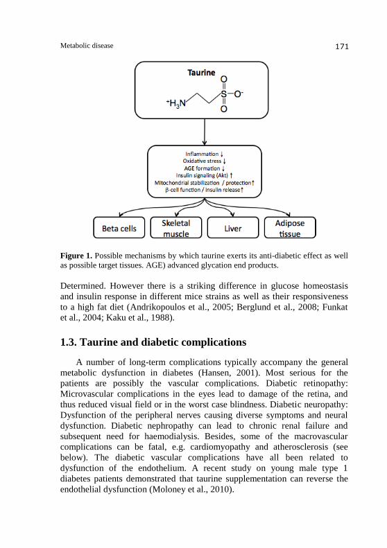
Figure 1. Possible mechanisms by which taurine exerts its anti-diabetic effect as well
as possible target tissues. AGE) advanced glycation end products.
Determined. However there is a striking difference in glucose homeostasis
and insulin response in different mice strains as well as their responsiveness
to a high fat diet (Andrikopoulos et al., 2005; Berglund et al., 2008; Funkat
et al., 2004; Kaku et al., 1988).
1.3. Taurine and diabetic complications
A number of long-term complications typically accompany the general
metabolic dysfunction in diabetes (Hansen, 2001). Most serious for the
patients are possibly the vascular complications. Diabetic retinopathy:
Microvascular complications in the eyes lead to damage of the retina, and
thus reduced visual field or in the worst case blindness. Diabetic neuropathy:
Dysfunction of the peripheral nerves causing diverse symptoms and neural
dysfunction. Diabetic nephropathy can lead to chronic renal failure and
subsequent need for haemodialysis. Besides, some of the macrovascular
complications can be fatal, e.g. cardiomyopathy and atherosclerosis (see
below). The diabetic vascular complications have all been related to
dysfunction of the endothelium. A recent study on young male type 1
diabetes patients demonstrated that taurine supplementation can reverse the
endothelial dysfunction (Moloney et al., 2010).

Svend Høime Hansen & Ole Hartvig Mortensen 172
One of the suggested hypotheses for understanding the microvascular
diabetic late complications is based on the so-called sorbitol pathway and
subsequent osmolyte depletion hypothesis. The hypothesis is based on the
fact that high glucose levels lead to intracellular sorbitol accumulation due to
aldose reductase. Sorbitol cannot in itself be transported across the cellular
membrane, and thus more labile osmolytes like taurine or myo-inositol will
gradually be depleted from the intracellular environment. Finally, the
sorbitol-producing cells will swell due to impaired volume regulation and
dysregulation of the taurine transporter (Askwith et al., 2009; Hansen, 2001;
Stevens et al., 1993). Furthermore additional taurine depletion can be caused
by intracellular scavenging of reactive carbonyl compounds and thus
prevention of AGE formation (Hansen, 2001).
The weakness of this hypothesis based on the sorbitol accumulation and
taurine depletion is the fact that it gives no direct biochemical link to the
cellular dysfunction observed in the diabetic complications. It should be noted
Figure 2. Simplified model for development of diabetic complications. Hyper-
glycemia causes sorbitol accumulation in the aldose reductase containing cells. This
causes a gradual depletion of transportable osmolytes like taurine. Consequently, cell
volume regulation becomes dysfunctional in the tissue. The process is accompanied
by a slow intracellular depletion of taurine from the mitochondrial compartment
resulting in increasing mitochondrial dysfunction.

Metabolic disease 173
that the types of tissue and organs involved in diabetic complications are all
very energy-demanding, and when comparing the clinical manifestations of
diabetic complications with those found in patients with mitochondrial
diseases (Kisler et al., 2010), major correspondence can be found. However,
it must be reasonable to assume that a gradual taurine depletion in the
intracellular environment will be expected to be accompanied by
mitochondrial depletion of taurine. Accepting taurine as an necessary
compound for mitochondrial function (see below), either as matrix pH buffer
(Hansen et al., 2006, 2010) and/or as a requirement for mitochondrial
translation being found in mitochondrial tRNA (Suzuki et al., 2002; Schaffer
et al., 2009), taurine depletion can possibly be a direct cause of mitochondrial
dysfunction. This mitochondrial role of taurine should thus be included in the
suggested viewpoint (Brownlee, 2005) of mitochondrial dysfunction as a
possible unifying hypothesis for diabetic complications.
1.4. Taurine and lipid metabolism – obesity
1.4.1. Cholesterol catabolism and bile acids
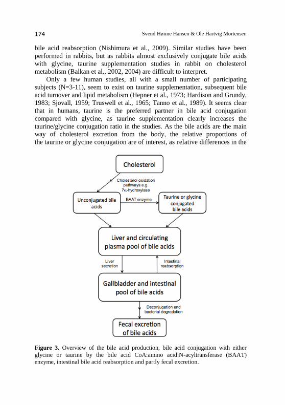
In most physiology and biochemistry text books, taurine is mentioned
solely as a component of the bile acids. Cholesterol catabolism and
subsequent excretion from the body occurs through the bile acids as the
major metabolic pathway (se Figure 3). Cholesterol is oxidized in the liver to
cholic acid by a complex enzyme framework (Russell, 2003). Cholic acid is
subsequently conjugated with either taurine or glycine predominantly in the
hepatocyte peroxisomes (Ferdinandusse et al., 2009; Solaas et al., 2000) by
the enzyme bile acid-CoA:amino acid N-acyltransferase (BAAT) (Falany
et al., 1994; He et al., 2003; Sfakianos et al., 2002). Species differences in the
amount of bile acids conjugated to glycine or taurine exist. Thus, in rat,
hamster, pig, and human the enzyme is capable of performing taurine as well as
glycine conjugation. In cat and rat, taurine conjugation is almost exclusively
performed, and in mouse and dog, only taurine conjugation occurs (Falany
et al., 1997; He et al., 2003; Kwakye et al., 1991; Rabin et al., 1976; Sfakianos
et al., 2002; Trautwein et al., 1999). In the rabbit only glycine conjugation has
traditionally been reported (Wildgrube et al., 1986), but findings in a more
recent study raises doubt about this fact (Hagey et al., 1998). Animal experiments in rats have demonstrated that taurine
supplementation can alleviate the consequences of a high cholesterol diet by
enhancing the excretion of bile acids, either due to enhanced activity of the
hepatic 7α-hydroxylase (Nishimura et al., 2003), enhancement by taurine of
bile acid conjugation (Sugiyama et al., 1989), and/or by inhibiting the ileal
Svend Høime Hansen & Ole Hartvig Mortensen 174
bile acid reabsorption (Nishimura et al., 2009). Similar studies have been
performed in rabbits, but as rabbits almost exclusively conjugate bile acids
with glycine, taurine supplementation studies in rabbit on cholesterol
metabolism (Balkan et al., 2002, 2004) are difficult to interpret.
Only a few human studies, all with a small number of participating
subjects (N=3-11), seem to exist on taurine supplementation, subsequent bile
acid turnover and lipid metabolism (Hepner et al., 1973; Hardison and Grundy,
1983; Sjovall, 1959; Truswell et al., 1965; Tanno et al., 1989). It seems clear
that in humans, taurine is the preferred partner in bile acid conjugation
compared with glycine, as taurine supplementation clearly increases the
taurine/glycine conjugation ratio in the studies. As the bile acids are the main
way of cholesterol excretion from the body, the relative proportions of
the taurine or glycine conjugation are of interest, as relative differences in the
Figure 3. Overview of the bile acid production, bile acid conjugation with either
glycine or taurine by the bile acid CoA:amino acid:N-acyltransferase (BAAT)
enzyme, intestinal bile acid reabsorption and partly fecal excretion.
Metabolic disease 175
reabsorption of the conjugates are expected, as the water solubility is better
for taurocholate due to the sulfonic acid group in taurine.
Taurine-conjugated bile acids are readily reabsorbed in the intestine by
an active ileal transport (Krag and Phillips, 1974), so a larger circulating bile
acid pool becomes the first result of taurine supplementation. However, it is
reasonable to assume that the reabsorption becomes down-regulated through a
feedback mechanism from the circulating bile acids as in the rat (Nishimura
et al., 2009) and then enhanced excretion of cholesterol will ensue. Alternately,
the human studies could indicate that the increased circulating levels of bile
acids (including taurocholate) would downregulate the cholesterol biosynthesis.
Furthermore, bile acids and bile acid receptors have been found to participate
in metabolic regulation. Such effects of bile acids can be found reviewed
elsewhere (Lefebvre et al., 2009; Staels et al., 2010; Trauner et al., 2010).
Finally, it should be noted that a reported stimulation by tauroconjugation on
fecal bacterial degradation of cholic acid (Van Eldere et al., 1996) could
actually cause an increase of excretion of cholic acid by taurine.
1.5. Lipid metabolism
The direct involvement of taurine in cholesterol catabolism seems to
have made the majority of taurine studies to concentrate on cholesterol levels,
lipid accumulation and atherosclerotic lesions in the vessels (Kondo et al.,
2001; Militante and Lombardini, 2004; Murakami et al., 1999). A few studies
in mouse, hamster, rat or recently quail have included quantitative
determinations of lipids and/or triglycerides in plasma, liver tissue or fat
deposition (Gandhi et al., 1992; Harada et al., 2004b; Kondo et al., 2001;
Murakami et al., 2002, 2010; Mochizuki et al., 1999; Nakaya et al., 2000;
Sethupathy et al., 2002; Tsuboyama-Kasaoka et al., 2006; Yokogoshi and
Oda, 2002; Yan et al., 1993). Generally taurine supplementation causes a
decrease in plasma lipids and triglycerides. It should specifically be noticed
that taurine supplementation could reverse obesity and increase energy
expenditure in C57BL/6J mice fed a high-fat diet (Tsuboyama-Kasaoka et al.,
2006).
A clinical study with taurine supplementation to a minor group of
overweight or obese non-diabetic subjects found a minor decrease in plasma
triglycerides, but no change in plasma cholesterol (Zhang et al., 2004). In one
study (Mochizuki et al., 1999) no effect of taurine supplementation was
found on the liver lipids, but a minor decrease was observed on liver
cholesterol. In all the other studies, taurine supplementation demonstrated a
clear decrease in lipid and triglyceride levels. Finally, an epidemiologic
Svend Høime Hansen & Ole Hartvig Mortensen 176
cohort study has shown that urinary taurine excretion (due to intake of fish
and shellfish) was inversely correlated to the risk of developing
artherosclerosis (Yamori, 2004, 2006). No explanation was presented in any
of the studies for the possible biochemical role of taurine.
As discussed in the previous section taurine supplementation must be
expected to increase the bile acid pool and thus improve bile acid-assisted
lipid transport mechanisms and associated metabolic regulation. Alternately,
taurine supplementation could be interpreted to enhance the mitochondrial
oxidation of fatty acids. Following this argumentation, the observed
improvements of lipid metabolism could be considered as support for the
recently presented hypothesis of taurine as mitochondrial matrix pH buffer.
Taurine supplementation will increase matrix pH buffering capacity and thus
stabilise the oxidative environment in the mitochondrial matrix to reduce
release of reactive oxygen species (ROS), and perhaps even more important
stabilise the very pH-dependent beta-oxidation of the fatty acids by the acyl-
CoA dehydrogenase enzymes (Hansen et al., 2006, 2010).
1.6. Taurine and fetal programming of metabolic disease
During the last two decades, it has become apparent that nutrition and
environment during pregnancy and early life have a lasting effect upon the
metabolic phenotype in adult life. The idea that there is a link between early
life conditions and subsequent disease was already discovered in the 1930s
(Smith and Kuh, 2001). However, it was not until Barker in 1986 reported a
correlation between childhood nutrition and ischemic heart disease (Barker
and Osmond, 1986) and in subsequent studies Barker and Hales during the
early 1990s coined the “thrifty phenotype” hypothesis (Hales and Barker,
1992) that the correlation was rediscovered. The hypothesis suggests that
early pre- and postnatal life is a critical period during which environmental
exposures that hinder growth will lead to an adaptation of metabolism to a
limited supply of nutrients, or other types of growth restraints. This
adaptation will contribute to increased risk for disease in adult life if
sufficient nutrients are provided. The term “fetal programming” can be used
to describe the hypothesized, yet unknown, mechanism behind the “thrifty
phenotype” hypothesis (Desai and Hales, 1997; Hales and Barker, 2001).
Several human studies have thus convincingly shown that fetal
malnutrition during pregnancy, which often leads to low birth weight or a
small for gestational age fetus, confers an increased risk of obesity, insulin
resistance, type 2 diabetes and a general low life expectancy (Jones and
Ozanne, 2009; Poulsen et al., 1997; Ravelli et al., 1999; Roseboom et al.,
2001a, 2001b). Several animal models in species ranging from rodents to
Metabolic disease 177
monkeys and of fetal malnutrition or low birth weight has been used to study
fetal programming, or intrauterine growth retardation (IUGR), with some of
the most popular ones being protein or dietary restriction during gestation,
intrauterine artery ligation and dexamethasone treatment of the pregnant dam
(Martin-Gronert and Ozanne, 2007). Human IUGR exhibit decreased taurine
levels in the fetus (Cetin et al., 1990; Economides et al., 1989), something
which is reflected in animal models of IUGR as well (Reusens et al., 1995;
Wu et al., 1998).
Taurine is considered to be an essential amino acid during development,
as the endogenous synthesis of taurine is inadequate in the fetus (Hibbard
et al., 1990). Thus, the fetus is dependent on the maternal supply of taurine.
Taurine deficiency leads to a smaller birth weight in both cats (Sturman,
1991) and rodents (Ejiri et al., 1987). The offspring of cats (which are unable
to synthesize taurine) reared on a taurine free diet, exhibit profound
developmental abnormalities, among these being: Smaller body weight,
smaller brain weight, abnormal hind leg development as well as a
degeneration or abnormal development of the retina and visual cortex
(Sturman, 1991). Furthermore, mice deficient in the taurine transporter gene
(TauT), show a smaller overall size, however no information regarding
birthweight is available (Warskulat et al., 2007). TauT knockout mice also
show defects in heart and skeletal muscle development, most likely due to
mitochondrial effects (Ito et al., 2008; Warskulat et al., 2004). In humans, a
low plasma taurine concentration in the infant has been linked to detrimental
mental development (Heird, 2004; Wharton et al., 2004), something which
has been corroborated by animal studies (Sturman, 1993). Furthermore,
taurine supplementation in mice has shown that the exact timing of taurine
supplementation during brain development influence the learning ability,
with taurine sufficiency being most important during the perinatal and
postnatal period (Suge et al., 2007). Experimental animal studies suggest that
taurine may be a marker of fetal well being (de Boo and Harding, 2007).
Several studies have documented that taurine ameliorates some of the
harmful effects that detrimental fetal programming may confer upon the
offspring in terms of the risk of developing metabolic disease and notably,
taurine is able to at least partially prevent an experimental induced decrease
in birth weight in several animal models of fetal programming. Thus, taurine
supplementation has been shown to normalize proliferation and
vascularization of the pancreas following gestational protein restriction
(Boujendar et al., 2002, 2003) and to decrease the sensitivity of the pancreas
towards cytokines (Merezak et al., 2001, 2004). In fact, taurine prevented all
changes in mRNA expression levels in the pancreas in newborns caused by
gestational protein restriction (Reusens et al., 2008). Likewise, taurine
Svend Høime Hansen & Ole Hartvig Mortensen 178
prevented a large portion of the changes in mRNA expression levels in both
skeletal muscle and liver caused by gestational protein restriction (Mortensen
et al., 2010). Interestingly these studies of the fetal gene expression profile in
both pancreas, liver and skeletal muscle suggest that the rescue effect taurine
exerts may have a mitochondrial component. This may also be important in
human development, as a reduced activity of the placental taurine
transporters has been observed in low birth weight in humans (Norberg et al.,
1998), something which may explain the low taurine concentrations in fetal
plasma often observed in this pregnancy complication (Cetin et al., 1990). A
recent study suggest that excessive taurine during gestation may also have
detrimental effects later in life, as taurine supplementation of pregnant rats
resulted in increased obesity and insulin resistance in the offspring (Hultman
et al., 2007). Collectively these studies suggest that taurine has a programming
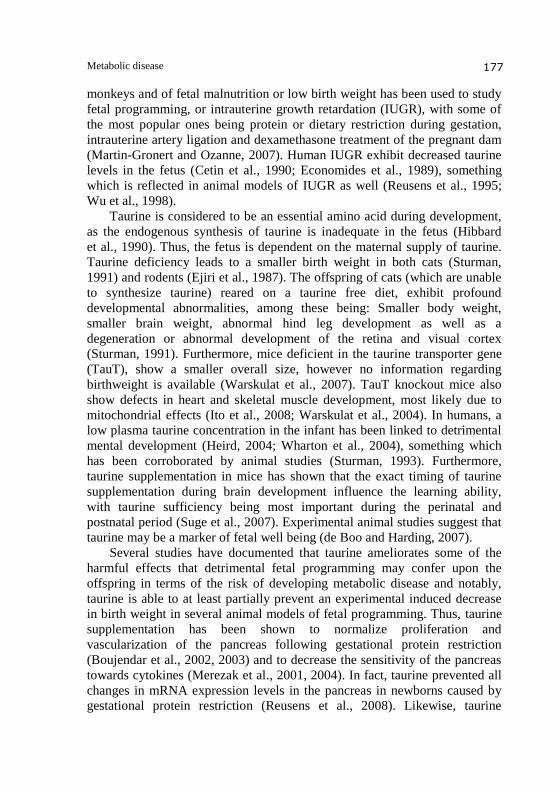
Figure 4. The different pathways by which taurine supplementation influences fetal
programming mediated development of type 2 diabetes.
Metabolic disease 179
or rescuing effect during fetal development, perhaps via epigenetic and/or
organogenesis related mechanisms.
1.7. Taurine, mitochondrial function and metabolic disease –
the missing link?
Recently, taurine has been suggested to have an important role in the
mitochondria as it has been suggested as a pH buffer in the mitochondrial
matrix to stabilize mitochondrial beta-oxidation of fatty acids. This oxidative
process requires mildly alkaline conditions in the mitochondrial matrix with
taurine as optimal pH buffer (Hansen et al., 2006, 2010). In addition, taurine
could have a direct role in the metabolic regulation of the pyruvate
dehydrogenase (Lombardini, 1998). Besides, taurine may be required for
optimal mitochondrial protein synthesis through taurine modified tRNAs
(Suzuki et al., 2002; Schaffer et al., 2009). High mitochondrial taurine
concentrations immediately explains the pivotal requirement for taurine
during fetal development (Suzuki et al., 2002), especially for the strongly
oxidative and thus mitochondria-rich tissues like liver and skeletal muscle as
well as pancreatic -cells. Consequently, taurine deficiency either during
development or adult life may cause impaired mitochondrial oxidation, fatty
acid oxidation and altered mitochondrial protein synthesis.
The role of mitochondrial dysfunction in insulin resistance and type 2
diabetes is debated and may actually be a consequence of insulin resistance
rather than a causal factor (Abdul-Ghani and DeFronzo, 2010; Dumas et al.,
2009; Holloszy, 2009; Schiff et al., 2009; Schrauwen et al., 2010; Turner and
Heilbronn, 2008). However, many studies have found a correlation between
decreased mitochondrial function or amount, or gene expression patterns and
type 2 diabetes. In addition, a recent viewpoint (Brownlee, 2005) has
suggested mitochondrial dysfunction as a possible unifying hypothesis for
diabetic complications. Hence, taurine deficiency as seen in diabetes may
increase the mitochondrial dysfunction and/or be involved in a vicious cycle
that ultimately lead to a worsening of the diabetic condition and
mitochondrial function. Thus it will be important in to examine the TauT
knockout mice in terms of susceptibility to diet induced type 2 diabetes.
1.7.1. Therapeutic perspectives for taurine as supplementation
Several animal studies have demonstrated positive effect on cholesterol
and lipid metabolism with subsequent prevention in the development of
atherosclerosis. These studies strongly support the idea of using taurine for
alleviating impaired lipid metabolism in atherosclerosis, type 2 diabetes and
Svend Høime Hansen & Ole Hartvig Mortensen 180
obesity. The arguments for using taurine to prevent diabetic complication are
also convincing, possibly in combination with aldose reductase inhibitors, as
taurine uptake in the affected cells might be prevented due to sorbitol
accumulation, which can be prevented with these inhibitors.
Taurine is today available as supplementation or in high concentrations
in energy drinks like the Austrian brand Red Bull or the Japanese brand
Lipovitan. However, the availability as commercial consumer products also
seems to represent a hindrance for establishing health recommendations with
regards to daily intake of taurine, as the number of associated clinical
metabolism studies is very limited. Even a recent meta-analysis on the role of
taurine in development and growth from Cochrane (Verner et al, 2007) does
not include any convincing biochemical arguments.
We hope that this review encourages future research, as the therapeutic
perspectives of the presented hypotheses need to be tested.
1.8. References
1. Abdul-Ghani, M. A., and DeFronzo, R. A. (2010). Pathogenesis of insulin
resistance in skeletal muscle. J. Biomed. Biotechnol. 2010, 476279.
2. Ackermann, D., and Heinsen, H. (1935). Über die physiologische Wirkung des
Asterubins und anderer, zum Teil neu dargestellter schwelfelhaltiger
Guanidinderivate. Hoppe Seyles Z. Physiol. Chemie. 235, 115-121.
3. Alvarado-Vásquez, N., Zamudio, P., Cerón, E., Vanda, B., Zenteno, E., and
Carvajal-Sandoval, G. (2003). Effect of glycine in streptozotocin-induced
diabetic rats. Comp. Biochem. Physiol. C. Toxicol. Pharmacol. 134, 521-527.
4. Andrikopoulos, S., Massa, C. M., Aston-Mourney, K., Funkat, A., Fam, B. C.,
Hull, R. L., Kahn, S. E., and Proietto, J. (2005). Differential effect of inbred
mouse strain (C57BL/6, DBA/2, 129T2) on insulin secretory function in response
to a high fat diet. J. Endocrinol. 187, 45-53.
5. Askwith, T., Zeng, W., Eggo, M. C., and Stevens, M. J. (2009). Oxidative stress
and dysregulation of the taurine transporter in high-glucose-exposed human
Schwann cells: implications for pathogenesis of diabetic neuropathy. Am. J.
Physiol. Endocrinol. Metab. 297, E620-E628.
6. Balkan, J., Kanbağli, O., Hatipoğlu, A., Kücük, M., Cevikbaş, U., Aykaç-Toker,
G., and Uysal, M. (2002). Improving effect of dietary taurine supplementation on
the oxidative stress and lipid levels in the plasma, liver and aorta of rabbits fed on
a high-cholesterol diet. Biosci. Biotechnol. Biochem. 66, 1755-1758.
7. Balkan, J., Oztezcan, S., Hatipoglu, A., Cevikbas, U., Aykac-Toker, G., and
Uysal, M. (2004). Effect of a taurine treatment on the regression of existing
atherosclerotic lesions in rabbits fed on a high-cholesterol diet. Biosci.
Biotechnol. Biochem. 68, 1035-1039. 8. Barker, D. J., and Osmond, C. (1986). Infant mortality, childhood nutrition, and
ischaemic heart disease in England and Wales. Lancet. 327, 1077-1081.
Metabolic disease 181
9. Berglund, E. D., Li, C. Y., Poffenberger, G., Ayala, J. E., Fueger, P. T., Willis, S. E.,
Jewell, M. M., Powers, A. C., and Wasserman, D. H. (2008). Glucose metabolism in
vivo in four commonly used inbred mouse strains. Diabetes. 57, 1790-1799.
10. Bouckenooghe, T., Remacle, C., and Reusens, B. (2006). Is taurine a functional
nutrient? Curr. Opin. Clin. Nutr. Metab. Care 9, 728-33.
11. Boujendar, S., Reusens, B., Merezak, S., Ahn, M., Arany, E., Hill, D., and
Remacle, C. (2002). Taurine supplementation to a low protein diet during foetal
and early postnatal life restores a normal proliferation and apoptosis of rat
pancreatic islets. Diabetologia 45, 856-66.
12. Boujendar, S., Arany, E., Hill, D., Remacle, C., and Reusens, B. (2003). Taurine
supplementation of a low protein diet fed to rat dams normalizes the
vascularization of the fetal endocrine pancreas. J. Nutr. 133, 2820-5.
13. Brownlee, M. (2005). The pathobiology of diabetic complications: a unifying
mechanism. Diabetes 54, 1615-1625.
14. Brøns, C., Spohr, C., Storgaard, H., Dyerberg, J., and Vaag, A. (2004). Effect of
taurine treatment on insulin secretion and action, and on serum lipid levels in
overweight men with a genetic predisposition for type II diabetes mellitus. Eur. J.
Clin. Nutr. 58, 1239-1247.
15. Cetin, I., Corbetta, C., Sereni, L. P., Marconi, A. M., Bozzetti, P., Pardi, G., and
Battaglia, F. C. (1990). Umbilical amino acid concentrations in normal and
growth-retarded fetuses sampled in utero by cordocentesis. Am. J. Obstet.
Gynecol. 162, 253-261.
16. Chauncey, K. B., Tenner, T. E., Lombardini, J. B., Jones, B. G., Brooks, M. L.,
Warner, R. D., Davis, R. L., and Ragain, R. M. (2003). The effect of taurine
supplementation on patients with type 2 diabetes mellitus. Adv. Exp. Med. Biol.
526, 91-96.
17. Chesney, R. W., Scriver, C. R., and Mohyuddin, F. (1976). Localization of the
membrane defect in transepithelial transport of taurine by parallel studies in vivo
and in vitro in hypertaurinuric mice. J. Clin. Invest. 57, 183-193.
18. de Boo, H. A., and Harding, J. E. (2007). Taurine as a marker for foetal
wellbeing? Neonatology 91, 145-54
19. De Luca, G., Calpona, P. R., Caponetti, A., Macaione, V., Di Benedetto, A.,
Cucinotta, D., and Di Giorgio, R. M. (2001a). Preliminary report: Amino acid
profile in platelets of diabetic patients. Metab. Clin. Exp 50, 739-741.
20. De Luca, G., Calpona, P. R., Caponetti, A., Romano, G., Di Benedetto, A.,
Cucinotta, D., and Di Giorgio, R. M. (2001b). Taurine and osmoregulation:
platelet taurine content, uptake, and release in type 2 diabetic patients. Metab.
Clin. Exp. 50, 60-64.
21. Della Corte, L., Crichton, R. R., Duburs, G., Nolan, K., Tipton, K. F., Tirzitis, G.,
and Ward, R. J. (2002). The use of taurine analogues to investigate taurine
functions and their potential therapeutic applications. Amino Acids 23, 367-379.
22. Desai, M., and Hales, C. N. (1997). Role of fetal and infant growth in
programming metabolism in later life. Biol. Rev. Camb. Philos. Soc. 72,
329-348.

Svend Høime Hansen & Ole Hartvig Mortensen 182
23. Di Leo, M. A. S., Santini, S. A., Silveri, N. G., Giardina, B., Franconi, F., and
Ghirlanda, G. (2004). Long-term taurine supplementation reduces mortality rate
in streptozotocin-induced diabetic rats. Amino Acids 27, 187-191.
24. Dumas, J., Simard, G., Flamment, M., Ducluzeau, P., and Ritz, P. (2009). Is
skeletal muscle mitochondrial dysfunction a cause or an indirect consequence of
insulin resistance in humans? Diabetes Metab. 35, 159-167.
25. Economides, D. L., Nicolaides, K. H., Gahl, W. A., Bernardini, I., Evans, M. I.
(1989). Plasma amino acids in appropriate- and small-for-gestational-age fetuses.
Am. J. Obstet. Gynecol. 161, 1219-1227.
26. Ejiri, K., Akahori, S., Kudo, K., Sekiba, K., and Ubuka, T. (1987). Effect of
guanidinoethyl sulfonate on taurine concentrations and fetal growth in pregnant
rats. Biol. Neonate. 51, 234-240.
27. El Idrissi, A., Boukarrou, L., and L'Amoreaux, W. (2009). Taurine
supplementation and pancreatic remodeling. Adv. Exp. Med. Biol. 643, 353-358.
28. El Mesallamy, H. O., El-Demerdash, E., Hammad, L. N., and El Magdoub, H. M.
(2010). Effect of taurine supplementation on hyperhomocysteinemia and markers
of oxidative stress in high fructose diet induced insulin resistance. Diabetol.
Metab. Syndr. 2, 46.
29. Elizarova, E. P., and Nedosugova, L. V. (1996). First experiments in taurine
administration for diabetes mellitus. The effect on erythrocyte membranes. Adv.
Exp. Med. Biol. 403, 583-588.
30. Falany, C. N., Fortinberry, H., Leiter, E. H., and Barnes, S. (1997). Cloning,
expression, and chromosomal localization of mouse liver bile acid CoA:amino
acid N-acyltransferase. J. Lipid Res. 38, 1139-1148.
31. Falany, C. N., Johnson, M. R., Barnes, S., and Diasio, R. B. (1994). Glycine and
taurine conjugation of bile acids by a single enzyme. Molecular cloning and
expression of human liver bile acid CoA:amino acid N-acyltransferase. J. Biol.
Chem. 269, 19375-19379.
32. Ferdinandusse, S., Denis, S., Faust, P. L., and Wanders, R. J. A. (2009). Bile
acids: the role of peroxisomes. J. Lipid Res. 50, 2139-2147.
33. Franconi, F., Bennardini, F., Mattana, A., Miceli, M., Ciuti, M., Mian, M.,
Gironi, A., Anichini, R., and Seghieri, G. (1995). Plasma and platelet taurine are
reduced in subjects with insulin-dependent diabetes mellitus: effects of taurine
supplementation. Am. J. Clin. Nutr. 61, 1115-1119.
34. Franconi, F., Miceli, M., Fazzini, A., Seghieri, G., Caputo, S., DiLeo, M. A.,
Lepore, D., and Ghirlanda, G. (1996). Taurine and diabetes. Humans and
experimental models. Adv. Exp. Med. Biol. 403, 579-582.
35. Franconi, F., Di Leo, M. A. S., Bennardini, F., and Ghirlanda, G. (2004). Is
taurine beneficial in reducing risk factors for diabetes mellitus? Neurochem. Res.
29, 143-150.
36. Franconi, F., Loizzo, A., Ghirlanda, G., and Seghieri, G. (2006). Taurine
supplementation and diabetes mellitus. Curr. Opin. Clin. Nutr. Metab. Care 9, 32-36.
37. Funkat, A., Massa, C. M., Jovanovska, V., Proietto, J., and Andrikopoulos, S.
(2004). Metabolic adaptations of three inbred strains of mice (C57BL/6, DBA/2,
and 129T2) in response to a high-fat diet. J. Nutr. 134, 3264-3269.

Metabolic disease 183
38. Gandhi, V. M., Cherian, K. M., and Mulky, M. J. (1992). Hypolipidemic action
of taurine in rats. Indian J. Exp. Biol. 30, 413-417.
39. Gavrovskaya, L. K., Ryzhova, O. V., Safonova, A. F., Matveev, A. K., and
Sapronov, N. S. (2008). Protective effect of taurine on rats with experimental
insulin-dependent diabetes mellitus. Bull. Exp. Biol. Med. 146, 226-228.
40. Hagey, L. R., Schteingart, C. D., Rossi, S. S., Ton-Nu, H. T., and Hofmann, A. F.
(1998). An N-acyl glycyltaurine conjugate of deoxycholic acid in the biliary bile
acids of the rabbit. J. Lipid Res. 39, 2119-2124.
41. Hales, C. N., and Barker, D. J. (2001). The thrifty phenotype hypothesis. Br.
Med. Bull. 60, 5-20.
42. Hales, C. N., and Barker, D. J. (1992). Type 2 (non-insulin-dependent) diabetes
mellitus: the thrifty phenotype hypothesis. Diabetologia 35, 595-601.
43. Han, J., Bae, J. H., Kim, S., Lee, H., Jang, B., Lee, I., Cho, C., Lim, J., Suh, S.,
Kwon, T., et al. (2004). Taurine increases glucose sensitivity of UCP2-
overexpressing beta-cells by ameliorating mitochondrial metabolism. Am. J.
Physiol. Endocrinol. Metab. 287, E1008-1018.
44. Hansen, S. H. (2001). The role of taurine in diabetes and the development of
diabetic complications. Diabetes Metab. Res. Rev. 17, 330-46.
45. Hansen, S., Andersen, M., Cornett, C., Gradinaru, R., and Grunnet, N. (2010). A
role for taurine in mitochondrial function. Journal of Biomedical Science. J.
Biomed. Sci. 17 Suppl. 1:S23 (8 pages).
46. Hansen, S. H., Andersen, M. L., Birkedal, H., Cornett, C., and Wibrand, F.
(2006). The important role of taurine in oxidative metabolism. Adv. Exp. Med.
Biol. 583, 129-135.
47. Harada, N., Ninomiya, C., Osako, Y., Morishima, M., Mawatari, K., Takahashi,
A., and Nakaya, Y. (2004a). Taurine alters respiratory gas exchange and nutrient
metabolism in type 2 diabetic rats. Obes. Res. 12, 1077-1084.
48. Harada, N., Ninomiya, C., Osako, Y., Morishima, M., Mawatari, K., Takahashi,
A., and Nakaya, Y. (2004b). Taurine alters respiratory gas exchange and nutrient
metabolism in type 2 diabetic rats. Obes. Res. 12, 1077-1084.
49. Hardison, W. G., and Grundy, S. M. (1983). Effect of bile acid conjugation
pattern on bile acid metabolism in normal humans. Gastroenterology 84, 617-620.
50. Harris, H., and Searle, A. G. (1953). Urinary amino-acids in mice of different
genotypes. Ann. Eugen. 17, 165-167.
51. He, D., Barnes, S., and Falany, C. N. (2003). Rat liver bile acid CoA:amino acid
N-acyltransferase: expression, characterization, and peroxisomal localization. J.
Lipid Res. 44, 2242-2249.
52. Heird, W. C. (2004). Taurine in neonatal nutrition--revisited. Arch. Dis. Child.
Fetal Neonatal Ed. 89, F473-474.
53. Hepner, G. W., Sturman, J. A., Hofmann, A. F., and Thomas, P. J. (1973).
Metabolism of steroid and amino acid moieties of conjugated bile acids in man.
3. Cholyltaurine (taurocholic acid). J. Clin. Invest. 52, 433-440.
54. Hibbard, J. U., Pridjian, G., Whitington, P. F., and Moawad, A. H. (1990).
Taurine transport in the in vitro perfused human placenta. Pediatr. Res. 27, 80-84.

Svend Høime Hansen & Ole Hartvig Mortensen 184
55. Holloszy, J. O. (2009). Skeletal muscle "mitochondrial deficiency" does not
mediate insulin resistance. Am. J. Clin. Nutr. 89, 463S-6S.
56. Hultman, K., Alexanderson, C., Mannerås, L., Sandberg, M., Holmäng, A., and
Jansson, T. (2007). Maternal taurine supplementation in the late pregnant rat
stimulates postnatal growth and induces obesity and insulin resistance in adult
offspring. J. Physiol. (Lond.) 579, 823-833.
57. Huxtable, R. J. (1992). Physiological actions of taurine. Physiol. Rev 72, 101-163.
58. Ito, T., Kimura, Y., Uozumi, Y., Takai, M., Muraoka, S., Matsuda, T., Ueki, K.,
Yoshiyama, M., Ikawa, M., Okabe, M., et al. (2008). Taurine depletion caused by
knocking out the taurine transporter gene leads to cardiomyopathy with cardiac
atrophy. J. Mol. Cell. Cardiol. 44, 927-937.
59. Jacobsen, J. G., and Smith, L. H. (1968). Biochemistry and physiology of taurine
and taurine derivatives. Physiol. Rev. 48, 424-511.
60. Jean, T., Poujeol, P., and Ripoche, P. (1984). Taurine transport in brush border
membrane vesicles isolated from hyper- and normotaurinuric mouse kidney. Ren
Physiol. 7, 349-356.
61. Jones, R. H., and Ozanne, S. E. (2009). Fetal programming of glucose-insulin
metabolism. Mol. Cell. Endocrinol. 297, 4-9.
62. Kaku, K., Fiedorek, F. T., Province, M., and Permutt, M. A. (1988). Genetic
analysis of glucose tolerance in inbred mouse strains. Evidence for polygenic
control. Diabetes 37, 707-713.
63. Kelley, D. E., He, J., Menshikova, E. V., and Ritov, V. B. (2002). Dysfunction of
mitochondria in human skeletal muscle in type 2 diabetes. Diabetes 51, 2944-2950.
64. Kim, S., Gupta, R. C., and Lee, H. W. (2007). Taurine-diabetes interaction: from
involvement to protection. Curr. Diabetes Rev. 3, 165-75.
65. Kisler, J. E., Whittaker, R. G., and McFarland, R. (2010). Mitochondrial diseases
in childhood: a clinical approach to investigation and management. Dev Med
Child Neurol. 52, 422-433.
66. Kondo, Y., Toda, Y., Kitajima, H., Oda, H., Nagate, T., Kameo, K., and
Murakami, S. (2001). Taurine inhibits development of atherosclerotic lesions in
apolipoprotein E-deficient mice. Clin. Exp. Pharmacol. Physiol. 28, 809-815.
67. Krag, E., and Phillips, S. F. (1974). Active and passive bile acid absorption in
man. Perfusion studies of the ileum and jejunum. J. Clin. Invest. 53, 1686-1694.
68. Kwakye, J. B., Johnson, M. R., Barnes, S., and Diasio, R. B. (1991). A
comparative study of bile acid CoA:amino acid:N-acyltransferase (BAT) from
four mammalian species. Comp. Biochem. Physiol., B 100, 131-136.
69. Lee, Y., and Pratley, R. E. (2005). The evolving role of inflammation in obesity
and the metabolic syndrome. Curr. Diab. Rep. 5, 70-75.
70. Lee, Y. Y., Park, K. S., Pak, Y. K., and Lee, H. K. (2005). The role of
mitochondrial DNA in the development of type 2 diabetes caused by fetal
malnutrition. J. Nutr. Biochem. 16, 195-204.
71. Lefebvre, P., Cariou, B., Lien, F., Kuipers, F., and Staels, B. (2009). Role of bile
acids and bile acid receptors in metabolic regulation. Physiol. Rev. 89, 147-191.
72. Lim, E., Park, S., and Kim, H. (1998). Effect of taurine supplementation on the
lipid peroxide formation and the activities of glutathione-related enzymes in the

Metabolic disease 185
liver and islet of type I and II diabetic model mice. Adv. Exp. Med. Biol. 442,
99-103.
73. Lombardini, J. B. (1998). Increased phosphorylation of specific rat cardiac and
retinal proteins in taurine-depleted animals: isolation and identification of the
phosphoproteins. Adv. Exp. Med. Biol. 442, 441-447.
74. Lowell, B. B., and Shulman, G. I. (2005). Mitochondrial dysfunction and type 2
diabetes. Science 307, 384-387.
75. Maechler, P., Li, N., Casimir, M., Vetterli, L., Frigerio, F., and Brun, T. (2010).
Role of mitochondria in beta-cell function and dysfunction. Adv. Exp. Med. Biol.
654, 193-216.
76. Mandla, S., Scriver, C. R., and Tenenhouse, H. S. (1988). Decreased transport in
renal basolateral membrane vesicles from hypertaurinuric mice. Am. J. Physiol.
255, F88-95.
77. Martin-Gronert, M. S., and Ozanne, S. E. (2007). Experimental IUGR and later
diabetes. J. Intern. Med. 261, 437-52.
78. Merezak, S., Hardikar, A. A., Yajnik, C. S., Remacle, C., and Reusens, B. (2001).
Intrauterine low protein diet increases fetal beta-cell sensitivity to NO and IL-1
beta: the protective role of taurine. J. Endocrinol. 171, 299-308.
79. Merezak, S., Reusens, B., Renard, A., Goosse, K., Kalbe, L., Ahn, M. T.,
Tamarit-Rodriguez, J., and Remacle, C. (2004). Effect of maternal low-protein
diet and taurine on the vulnerability of adult Wistar rat islets to cytokines.
Diabetologia 47, 669-75.
80. Militante, J., and Lombardini, J. (2004). Dietary taurine supplementation:
hypolipidemic and antiatherogenic effects. Nutrition Research 24, 787-801.
81. Mochizuki, H., Takido, J., Oda, H., and Yokogoshi, H. (1999). Improving effect
of dietary taurine on marked hypercholesterolemia induced by a high-cholesterol
diet in streptozotocin-induced diabetic rats. Biosci. Biotechnol. Biochem. 63,
1984-1987.
82. Moloney, M. A., Casey, R. G., O' Donnell, D. H., Fitzgerald, P., Thompson, C.,
and Bouchier-Hayes, D. J. (2010). Two weeks taurine supplementation reverses
endothelial dysfunction in young male type 1 diabetics. Diab. Vasc. Dis. Res. 7,
300-10.
83. Mortensen, O. H., Olsen, H. L., Frandsen, L., Nielsen, P. E., Nielsen, F. C.,
Grunnet, N., and Quistorff, B. (2010). Gestational protein restriction in mice has
pronounced effects on gene expression in newborn offspring's liver and skeletal
muscle; protective effect of taurine. Pediatr. Res. 67, 47-53.
84. Mulder, H., and Ling, C. (2009). Mitochondrial dysfunction in pancreatic beta-
cells in Type 2 diabetes. Mol. Cell. Endocrinol. 297, 34-40.
85. Murakami, S., Kondo-Ohta, Y., and Tomisawa, K. (1999). Improvement in
cholesterol metabolism in mice given chronic treatment of taurine and fed a high-
fat diet. Life Sci. 64, 83-91.
86. Murakami, S., Kondo, Y., Toda, Y., Kitajima, H., Kameo, K., Sakono, M., and
Fukuda, N. (2002). Effect of taurine on cholesterol metabolism in hamsters:
up-regulation of low density lipoprotein (LDL) receptor by taurine. Life Sci. 70,
2355-2366.
Svend Høime Hansen & Ole Hartvig Mortensen 186
87. Murakami, S., Sakurai, T., Tomoike, H., Sakono, M., Nasu, T., and Fukuda, N.
(2010). Prevention of hypercholesterolemia and atherosclerosis in the
hyperlipidemia- and atherosclerosis-prone Japanese (LAP) quail by taurine
supplementation. Amino Acids 38, 271-278.
88. Mårtensson, J., and Hermansson, G. (1984). Sulfur amino acid metabolism in
juvenile-onset nonketotic and ketotic diabetic patients. Metab. Clin. Exp 33, 425-428.
89. Nakamura, T., Ushiyama, C., Suzuki, S., Shimada, N., Ohmuro, H., Ebihara, I.,
and Koide, H. (1999). Effects of taurine and vitamin E on microalbuminuria,
plasma metalloproteinase-9, and serum type IV collagen concentrations in
patients with diabetic nephropathy. Nephron 83, 361-362.
90. Nakaya, Y., Minami, A., Harada, N., Sakamoto, S., Niwa, Y., and Ohnaka, M.
(2000). Taurine improves insulin sensitivity in the Otsuka Long-Evans
Tokushima Fatty rat, a model of spontaneous type 2 diabetes. Am. J. Clin. Nutr.
71, 54-58.
91. Nandhini, A. T. A., Thirunavukkarasu, V., and Anuradha, C. V. (2005). Taurine
modifies insulin signaling enzymes in the fructose-fed insulin resistant rats.
Diabetes Metab. 31, 337-44.
92. Nishimura, N., Umeda, C., Oda, H., and Yokogoshi, H. (2003). The effect of
taurine on the cholesterol metabolism in rats fed diets supplemented with
cholestyramine or high amounts of bile acid. J. Nutr. Sci. Vitaminol. 49, 21-26.
93. Nishimura, N., Umeda, C., Ona, H., and Yokogoshi, H. (2002). The effect of
taurine on plasma cholesterol concentration in genetic type 2 diabetic GK rats. J.
Nutr. Sci. Vitaminol. 48, 483-490.
94. Nishimura, N., Yamamoto, T., and Ota, T. (2009). Taurine feeding inhibits bile
acid absorption from the ileum in rats fed a high cholesterol and high fat diet.
Adv. Exp. Med. Biol. 643, 285-291.
95. Nishina, P. M., Verstuyft, J., and Paigen, B. (1990). Synthetic low and high fat
diets for the study of atherosclerosis in the mouse. J. Lipid Res 31, 859-869.
96. Nishina, P. M., Wang, J., Toyofuku, W., Kuypers, F. A., Ishida, B. Y., and
Paigen, B. (1993). Atherosclerosis and plasma and liver lipids in nine inbred
strains of mice. Lipids 28, 599-605.
97. Norberg, S., Powell, T. L., and Jansson, T. (1998). Intrauterine growth restriction
is associated with a reduced activity of placental taurine transporters. Pediatr.
Res. 44, 233-238.
98. Odetti, P., Pesce, C., Traverso, N., Menini, S., Maineri, E. P., Cosso, L.,
Valentini, S., Patriarca, S., Cottalasso, D., Marinari, U. M., et al. (2003).
Comparative trial of N-acetyl-cysteine, taurine, and oxerutin on skin and kidney
damage in long-term experimental diabetes. Diabetes 52, 499-505.
99. Ozcan, U., Yilmaz, E., Ozcan, L., Furuhashi, M., Vaillancourt, E., Smith, R. O.,
Görgün, C. Z., and Hotamisligil, G. S. (2006). Chemical chaperones reduce ER
stress and restore glucose homeostasis in a mouse model of type 2 diabetes.
Science 313, 1137-40.
100. Pagel-Langenickel, I., Bao, J., Pang, L., and Sack, M. N. (2010). The role of
mitochondria in the pathophysiology of skeletal muscle insulin resistance.
Endocr. Rev. 31, 25-51.
Metabolic disease 187
101. Paigen, B., Morrow, A., Brandon, C., Mitchell, D., and Holmes, P. (1985).
Variation in susceptibility to atherosclerosis among inbred strains of mice.
Atherosclerosis 57, 65-73.
102. Poulsen, P., Vaag, A. A., Kyvik, K. O., Møller Jensen, D., and Beck-Nielsen, H.
(1997). Low birth weight is associated with NIDDM in discordant monozygotic
and dizygotic twin pairs. Diabetologia 40, 439-446. 103. Rabin, B., Nicolosi, R. J., and Hayes, K. C. (1976). Dietary influence on bile acid
conjugation in the cat. J. Nutr. 106, 1241-1246. 104. Ravelli, A. C., van Der Meulen, J. H., Osmond, C., Barker, D. J., and Bleker, O.
P. (1999). Obesity at the age of 50 y in men and women exposed to famine prenatally. Am. J. Clin. Nutr. 70, 811-816.
105. Reusens, B., Sparre, T., Kalbe, L., Bouckenooghe, T., Theys, N., Kruhøffer, M., Orntoft, T. F., Nerup, J., and Remacle, C. (2008). The intrauterine metabolic environment modulates the gene expression pattern in fetal rat islets: prevention by maternal taurine supplementation. Diabetologia 51, 836-845.
106. Reusens, B., Dahri, S., Snoech, A., Bennis-Taleb, N., Remacle, C., and Hoet, J. (1995). Long-term Consequences of Diabetes and Its Complications May Have a Fetal Origin: Experimental and Epidemiological Evidence. Nestle Nutrition Workshop Series 35, 187-198.
107. Roseboom, T. J., van der Meulen, J. H., Osmond, C., Barker, D. J., Ravelli, A. C., and Bleker, O. P. (2001a). Adult survival after prenatal exposure to the Dutch famine 1944--45. Paediatr Perinat Epidemiol 15, 220-225.
108. Roseboom, T. J., van der Meulen, J. H., Ravelli, A. C., Osmond, C., Barker, D.
J., and Bleker, O. P. (2001b). Effects of prenatal exposure to the Dutch famine on
adult disease in later life: an overview. Mol. Cell. Endocrinol 185, 93-98.
109. Rozen, R., Scriver, C. R., and Mohyuddin, F. (1983). Hypertaurinuria in the
C57BL/6J mouse: altered transport at the renal basolateral membrane. Am. J.
Physiol 244, F150-155.
110. Russell, D. W. (2003). The enzymes, regulation, and genetics of bile acid
synthesis. Annu. Rev. Biochem. 72, 137-174.
111. Schaffer, S. W., Azuma, J., and Mozaffari, M. (2009). Role of antioxidant
activity of taurine in diabetes. Can. J. Physiol. Pharmacol. 87, 91-99.
112. Schiff, M., Loublier, S., Coulibaly, A., Bénit, P., de Baulny, H. O., and Rustin, P.
(2009). Mitochondria and diabetes mellitus: untangling a conflictive relationship?
J. Inherit. Metab. Dis 32, 684-698.
113. Schrauwen, P., Schrauwen-Hinderling, V., Hoeks, J., and Hesselink, M. K. C.
(2010). Mitochondrial dysfunction and lipotoxicity. Biochim. Biophys. Acta.
1801, 266-271.
114. Schuller-Levis, G. B., and Park, E. (2003). Taurine: new implications for an old
amino acid. FEMS Microbiol. Lett. 226, 195-202.
115. Seghieri, G., Tesi, F., Bianchi, L., Loizzo, A., Saccomanni, G., Ghirlanda, G.,
Anichini, R., and Franconi, F. (2007). Taurine in women with a history of
gestational diabetes. Diabetes Res. Clin. Pract. 76, 187-192.
116. Sethupathy, S., Elanchezhiyan, C., Vasudevan, K., and Rajagopal, G. (2002).
Antiatherogenic effect of taurine in high fat diet fed rats. Indian J. Exp. Biol. 40,
1169-1172.
Svend Høime Hansen & Ole Hartvig Mortensen 188
117. Sfakianos, M. K., Wilson, L., Sakalian, M., Falany, C. N., and Barnes, S. (2002).
Conserved residues in the putative catalytic triad of human bile acid Coenzyme
A:amino acid N-acyltransferase. J. Biol. Chem. 277, 47270-47275.
118. Sjovall, J. (1959). Dietary glycine and taurine on bile acid conjugation in man;
bile acids and steroids 75. Proc. Soc. Exp. Biol. Med. 100, 676-678.
119. Smith, G. D., and Kuh, D. (2001). Commentary: William Ogilvy Kermack and
the childhood origins of adult health and disease. Int. J. Epidemiol. 30, 696-703.
120. Solaas, K., Ulvestad, A., Söreide, O., and Kase, B. F. (2000). Subcellular
organization of bile acid amidation in human liver: a key issue in regulating the
biosynthesis of bile salts. J. Lipid Res. 41, 1154-1162.
121. Song, X., Chen, C., Dong, B., Shi, Y., Zhang, W., Yan, L., and Luo, G. (2003).
[Study on the intervening mechanism of taurine on streptozotocin-induced
diabetic cataracts]. Zhonghua Yan Ke Za Zhi 39, 605-609.
122. Staels, B., Handelsman, Y., and Fonseca, V. (2010). Bile acid sequestrants for
lipid and glucose control. Curr. Diab. Rep. 10, 70-77.
123. Stevens, M. J., Lattimer, S. A., Kamijo, M., Van Huysen, C., Sima, A. A., and
Greene, D. A. (1993). Osmotically-induced nerve taurine depletion and the
compatible osmolyte hypothesis in experimental diabetic neuropathy in the rat.
Diabetologia 36, 608-614.
124. Sturman, J. A. (1991). Dietary taurine and feline reproduction and development.
J. Nutr. 121, S166-170.
125. Sturman, J. A. (1993). Taurine in development. Physiol. Rev. 73, 119-147.
126. Suge, R., Hosoe, N., Furube, M., Yamamoto, T., Hirayama, A., Hirano, S., and
Nomura, M. (2007). Specific timing of taurine supplementation affects learning
ability in mice. Life Sci 81, 1228-1234.
127. Sugiyama, K., Ohishi, A., Ohnuma, Y., and Muramatsu, K. (1989). Comparison
between the Plasma Cholesterol-lowering Effects of Glycine and Taurine in Rats
Fed on High Cholesterol Diets. Agric. Biol. Chem. 53, 1647-1652.
128. Suzuki, T., Suzuki, T., Wada, T., Saigo, K., and Watanabe, K. (2002). Taurine as
a constituent of mitochondrial tRNAs: new insights into the functions of taurine
and human mitochondrial diseases. EMBO J. 21, 6581-6589.
129. Takatani, T., Takahashi, K., Uozumi, Y., Matsuda, T., Ito, T., Schaffer, S. W.,
Fujio, Y., and Azuma, J. (2004). Taurine prevents the ischemia-induced apoptosis
in cultured neonatal rat cardiomyocytes through Akt/caspase-9 pathway.
Biochem. Biophys. Res. Commun. 316, 484-489.
130. Tanno, N., Oikawa, S., Koizumi, M., Fujii, Y., Hori, S., Suzuki, N., Sakuma, E.,
Kotake, H., Namai, K., and Toyota, T. (1989). Effect of taurine administration on
serum lipid and biliary lipid composition in man. Tohoku J. Exp. Med 159, 91-99.
131. Tenner, T. E., Zhang, X. J., and Lombardini, J. B. (2003). Hypoglycemic effects
of taurine in the alloxan-treated rabbit, a model for type 1 diabetes. Adv. Exp.
Med. Biol. 526, 97-104.
132. Trachtman, H., Futterweit, S., and Sturman, J. A. (1992). Cerebral taurine
transport is increased during streptozocin-induced diabetes in rats. Diabetes 41,
1130-1140.

Metabolic disease 189
133. Trauner, M., Claudel, T., Fickert, P., Moustafa, T., and Wagner, M. (2010). Bile
acids as regulators of hepatic lipid and glucose metabolism. Dig Dis 28, 220-224.
134. Trautwein, E. A., Kunath-Rau, A., and Erbersdobler, H. F. (1999). Increased
fecal bile acid excretion and changes in the circulating bile acid pool are involved
in the hypocholesterolemic and gallstone-preventive actions of psyllium in
hamsters. J. Nutr. 129, 896-902.
135. Truswell, A. S., McVeigh, S., Mitchell, W. D., and Bronte-Stewart, B. (1965).
Effect in man of feeding taurine on bile acid conjugation and serum cholesterol
levels. J Atheroscler Res. 5, 526-529.
136. Tsuboyama-Kasaoka, N., Shozawa, C., Sano, K., Kamei, Y., Kasaoka, S.,
Hosokawa, Y., and Ezaki, O. (2006). Taurine (2-aminoethanesulfonic acid)
deficiency creates a vicious circle promoting obesity. Endocrinology 147,
3276-3284.
137. Turner, N., and Heilbronn, L. K. (2008). Is mitochondrial dysfunction a cause of
insulin resistance? Trends Endocrinol. Metab. 19, 324-330.
138. Van Eldere, J., Celis, P., De Pauw, G., Lesaffre, E., and Eyssen, H. (1996).
Tauroconjugation of cholic acid stimulates 7 alpha-dehydroxylation by fecal
bacteria. Appl. Environ. Microbiol. 62, 656-661.
139. Warskulat, U., Flögel, U., Jacoby, C., Hartwig, H., Thewissen, M., Merx, M. W.,
Molojavyi, A., Heller-Stilb, B., Schrader, J., and Häussinger, D. (2004). Taurine
transporter knockout depletes muscle taurine levels and results in severe skeletal
muscle impairment but leaves cardiac function uncompromised. FASEB J 18,
577-579.
140. Warskulat, U., Heller-Stilb, B., Oermann, E., Zilles, K., Haas, H., Lang, F., and
Häussinger, D. (2007). Phenotype of the taurine transporter knockout mouse.
Meth. Enzymol. 428, 439-458.
141. Wharton, B. A., Morley, R., Isaacs, E. B., Cole, T. J., and Lucas, A. (2004). Low
plasma taurine and later neurodevelopment. Arch. Dis. Child. Fetal Neonatal Ed
89, F497-498.
142. Wijekoon, E. P., Skinner, C., Brosnan, M. E., and Brosnan, J. T. (2004). Amino
acid metabolism in the Zucker diabetic fatty rat: effects of insulin resistance and
of type 2 diabetes. Can. J. Physiol. Pharmacol. 82, 506-514.
143. Wildgrube, H. J., Stockhausen, H., Petri, J., Füssel, U., and Lauer, H. (1986).
Naturally occurring conjugated bile acids, measured by high-performance liquid
chromatography, in human, dog, and rabbit bile. J. Chromatogr. 353, 207-213.
144. Williams, R. E., Lenz, E. M., Evans, J. A., Wilson, I. D., Granger, J. H., Plumb,
R. S., and Stumpf, C. L. (2005). A combined (1)H NMR and HPLC-MS-based
metabonomic study of urine from obese (fa/fa) Zucker and normal Wistar-
derived rats. J. Pharm. Biomed. Anal. 38, 465-471.
145. Winiarska, K., Szymanski, K., Gorniak, P., Dudziak, M., and Bryla, J. (2009).
Hypoglycaemic, antioxidative and nephroprotective effects of taurine in alloxan
diabetic rabbits. Biochimie. 91, 261-270.
146. Wu, G., Pond, W. G., Ott, T., and Bazer, F. W. (1998). Maternal dietary protein
deficiency decreases amino acid concentrations in fetal plasma and allantoic fluid
of pigs. J. Nutr. 128, 894-902.
Svend Høime Hansen & Ole Hartvig Mortensen 190
147. Xiao, C., Giacca, A., and Lewis, G. F. (2008). Oral taurine but not
N-acetylcysteine ameliorates NEFA-induced impairment in insulin sensitivity
and beta cell function in obese and overweight, non-diabetic men. Diabetologia
51, 139-46.
148. Yamori, Y. (2006). Food factors for atherosclerosis prevention: Asian perspective
derived from analyses of worldwide dietary biomarkers. Exp Clin Cardiol 11,
94-98.
149. Yamori, Y. (2004). Worldwide epidemic of obesity: hope for Japanese diets.
Clin. Exp. Pharmacol. Physiol. 31 Suppl. 2, S2-4.
150. Yan, C. C., Bravo, E., and Cantàfora, A. (1993). Effect of taurine levels on liver
lipid metabolism: an in vivo study in the rat. Proc. Soc. Exp. Biol. Med. 202, 88-96.
151. Yokogoshi, H., and Oda, H. (2002). Dietary taurine enhances cholesterol
degradation and reduces serum and liver cholesterol concentrations in rats fed a
high-cholesterol diet. Amino Acids 23, 433-439.
152. Zhang, M., Bi, L. F., Fang, J. H., Su, X. L., Da, G. L., Kuwamori, T., and
Kagamimori, S. (2004). Beneficial effects of taurine on serum lipids in
overweight or obese non-diabetic subjects. Amino Acids 26, 267-271.