Embed Size (px)
Citation preview
Page 1 of 18
Taurine treatment of retinal degeneration and cardiomyopathy in a consanguineous family
with SLC6A6 taurine transporter deficiency
Muhammad Ansar1,2,§,*, Emmanuelle Ranza1,3,4,§, Madhur Shetty5, Sohail A. Paracha6, Maleeha
Azam7, Ilse Kern8, Justyna Iwaszkiewicz9, Omer Farooq10, Constantin J. Pournaras11, Ariane
Malcles12, Mateusz Kecik12, Carlo Rivolta13,14,15, Waqar Muzaffar16, Aziz Qurban16, Liaqat Ali7,
Yacine Aggoun17, Federico A. Santoni1,18, Periklis Makrythanasis1,19, Jawad Ahmed6, Raheel
Qamar7, Muhammad T. Sarwar6, L. Keith Henry5, Stylianos E. Antonarakis1,3,20,*
1Department of Genetic Medicine and Development, University of Geneva, Geneva, Switzerland;
2Current address, Clinical Research Center, Institute of Molecular and Clinical Ophthalmology
Basel (IOB), Basel, Switzerland;
3Service of Genetic Medicine, University Hospitals of Geneva, Geneva, Switzerland;
4Current address, Medigenome, Swiss Institute of Genomic Medicine, Geneva, Switzerland;
5Dept. of Biomedical Sciences, School of Medicine and Health Sciences, University of North
Dakota, Grand Forks, ND, United States;
6Institute of Basic Medical Sciences, Khyber Medical University, Peshawar, Pakistan;
7Department of Biosciences, Faculty of Science, COMSATS University Islamabad, Pakistan;
8Pediatric Nephrology and Metabolism Unit, Pediatric Subspecialties Service, Children's Hospital,
Geneva University Hospitals, Geneva, Switzerland;
9Swiss Institute of Bioinformatics, Molecular Modeling Group, University of Lausanne, Lausanne,
Switzerland;
Page 2 of 18
10Bahria University Medical and Dental College, Karachi, Pakistan;
11Hirslanden Clinique La Colline, Geneva, Switzerland ;
12Department of Ophthalmology, University Hospitals of Geneva, Geneva, Switzerland;
13Clinical Research Center, Institute of Molecular and Clinical Ophthalmology Basel (IOB), Basel,
Switzerland;
14Department of Ophthalmology, University Hospital Basel, Switzerland;
15Department of Genetics and Genome Biology, University of Leicester, Leicester, United
16Armed Forces Institute of Ophthalmology, Rawalpindi, Pakistan;
17Pediatric Cardiology, Geneva University Hospitals, Geneva, Switzerland;
18Current address, Department of Endocrinology Diabetes and Metabolism, University Hospital
of Lausanne, Lausanne, Switzerland;
19Current address, Biomedical Research Foundation of the Academy of Athens, Athens, Greece;
Kingdom;
20iGE3 Institute of Genetics and Genomics of Geneva, Geneva, Switzerland;
§These authors contributed equally
*Corresponding authors;
Muhammad Ansar
University of Geneva Medical School, 1 Rue Michel Servet, 1211 Geneva, Switzerland
Phone: +41 78 805 5480
Fax: +41-22-379-5706

Page 3 of 18
Current address, Clinical Research Center, Institute of Molecular and Clinical Ophthalmology
Basel (IOB), Basel, Switzerland.
Stylianos Antonarakis
University of Geneva Medical Faculty, 1 Rue Michel Servet, 1211 Geneva, Switzerland
Phone: +41 22 37 95708
Fax: +41-22-379-5706

Page 4 of 18
Abstract
In a consanguineous Pakistani family with 2 affected individuals, a homozygous variant
Gly399Val in the 8th transmembrane domain of the taurine transporter SLC6A6 was identified
resulting in a hypomorph transporting capacity of ~15% compared to normal. 3D modeling of this
variant has indicated that it likely causes displacement of the Tyr138 (TM3) side chain, important
for transport of taurine. The affected individuals presented with rapidly progressive childhood
retinal degeneration, cardiomyopathy, and almost undetectable plasma taurine levels. Oral taurine
supplementation of 100mg/kg/day resulted in maintenance of normal blood taurine levels.
Following approval by the ethics committee, a long-term supplementation treatment was
introduced. Remarkably, after 24-months, the cardiomyopathy was corrected in both affected
siblings, and in the 6 y.o. the retinal degeneration was arrested, and the vision was clinically
improved. Similar therapeutic approaches could be employed in mendelian phenotypes caused by
the dysfunction of the hundreds of other molecular transporters.
Page 5 of 18
Introduction
Transporters constitute a large number of cell membrane proteins involved in the uptake of small
molecules into cells(1). The current human gene catalogue contains 423 protein-coding genes
classified as solute carrier (SLC)(1) (https://www.genenames.org/data/genegroup/#!/group/752).
Members of this large gene family are involved in a number of human disorders; OMIM contains
268 entries with dominant, recessive, and X-linked mendelian phenotypes caused by pathogenic
variants in these genes (24sep19 search https://www.omim.org/).
Genome or exome sequencing provided the opportunity to identify novel causative links between
pathogenic variants of genes and mendelian phenotypes. This is particularly important for the
discovery of novel autosomal recessive genes since the majority of those are still unknown.
Consanguinity, which is practiced in a considerable fraction of world’s populations, provides a
means to identify novel recessive genes because of the large genomic regions of homozygosity in
the offspring of closely related parents(2). The identification of pathogenic variants in novel genes
causing recessive disorders provide a better understanding of the molecular pathophysiology of
the resulting phenotype, and the opportunity for improved diagnostic services to the affected
families and populations; occasionally, new therapeutic options could be offered based on the
underlying molecular mechanism.
As part of the Swiss-Pakistani consanguinity project, to identify novel genes for visual impairment
or intellectual disability(3, 4), we present a novel gene for childhood progressive retinal
degeneration and cardiomyopathy in one family. The taurine transport defect identified due to a
hypomorph homozygous pathogenic variant in the SLC6A6 gene, has provided the opportunity
for treatment with long-term taurine supplementation. After 24 months of treatment the

Page 6 of 18
cardiomyopathy was corrected in both affected siblings, and in the 6 y.o. the retinal degeneration
was arrested and the vision was clinically improved.
Page 7 of 18
Results
Genetic analysis
We used exome sequencing and genotyping of more than 200 Pakistani consanguineous families
with multiple affected individuals to identify candidate genes and high impact variants responsible
for recessive visual impairment. In one of these consanguineous families, F315, from the Kohat
region of Pakistan with 2 affected individuals, we have identified a homozygous deleterious
variant Gly399Val (NM_003043.5:c.1196G>T) in the taurine transporter SLC6A6 (MIM:186854)
that segregated with the phenotype of progressive retinal degeneration (Figure 1A, 1B). Gly399 of
SLC6A6 is well-conserved in all vertebrates (Figure 1C), and 3D molecular modeling (Figure 1D)
predicted that the Val399 substitution causes a displacement of Tyr138 side chain, important for
the recognition and transport of the ligand. Blood taurine levels in the two affected individuals
IV:1 and IV:3 were almost undetected (6-7 μmol/l).
Functional analysis
Transient and stable transfection of the Gly399Val variant in HEK-293 cells resulted in a
hypomorph with transport capacity of ~15% compared to normal as determined by single point
(Figure S2) and saturation (Figure S3 and Table S6) radioactive taurine uptake analyses.
Fibroblasts from affected and carrier individuals of the family showed similar results where single
point uptake revealed significant taurine uptake deficits in the affected individuals compared to
carriers (Figure S4) consistent with kinetic data showing a significant reduction in VMAX (Figure
2A and Table S7). Plasma membrane expression analysis in HEK-293 cells and fibroblasts
strongly support that the transport deficit is functional rather than a result of decreased surface
protein (Figure S5 and Figure 2 panels B and C). In HEK-293 cells, taurine transport KM values
Page 8 of 18
were 3.4-fold lower in SLC6A6 Gly399Val compared to the normal transporter (Table S5) and
whereas this could contribute to decreased transport capacity in HEK cells, KM values were
unchanged between affected and carrier fibroblasts (Table S7) indicating the reduced taurine
uptake in the affected patients originates from reduced transporter cycling and not substrate
recognition.
Clinical evaluation
Clinical examinations in the University Hospitals of Geneva revealed a cone-rod retinopathy and
cardiomyopathy. The older 15 y.o. male IV:1 had light perception vision due to advanced macular
atrophy with severe peripheral alterations including pseudoosteoblast formation and peripheral
atrophy. There was no retinal response in the electroretinogram (ERG) and extensive loss of
photoreceptors was noted in the optical coherence tomography (OCT). The younger 6 y.o. female
IV:3 had vision of counting fingers due to foveal-spearing macular atrophy. There were less
marked peripheral retinal changes (salt-and-pepper fundus pigmentation), no response in global
ERG, and minimal electrical focal macular response in the multifocal ERG. The OCT showed
atrophy of the photoreceptors with persistence of residual photoreceptors in the central area (Figure
S1). Echocardiography showed mild hypokinetic cardiomyopathy in both affected individuals with
systolic dysfunction (shortening fraction 24-27%) and systolic dilatation of the left ventricle
(Figure 3B); the effort test was however within normal limits. In both patients, brain MRI and
hepatic ultrasound was normal (Table S1). Plasma amino acids showed very low levels of taurine
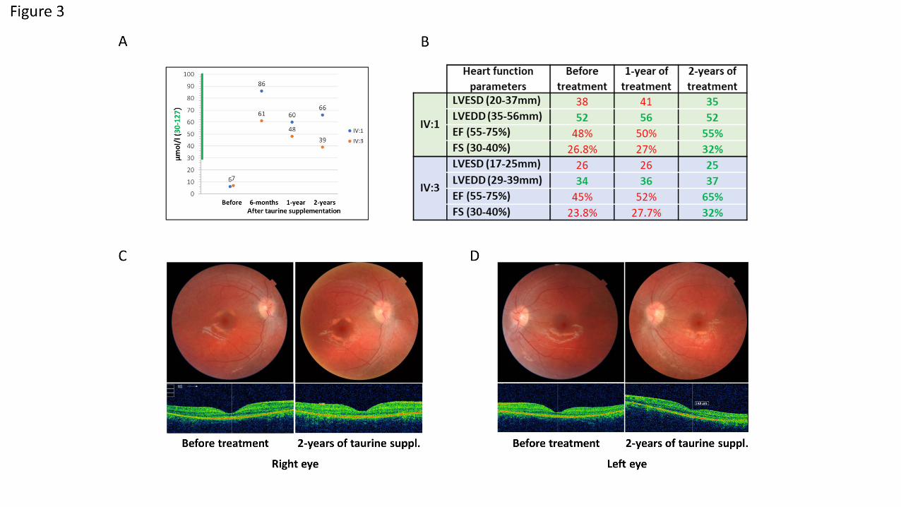
in the affecteds and intermediate levels in the carrier parents (Figure 3A).
Page 9 of 18
Taurine supplementation treatment
The oral taurine loading test and subsequent supplementation at 100mg/kg/day resulted in
maintenance of normal blood taurine levels in both affected individuals (more than 40 μmol/l in
each patient) (Figure S6, Figure 3A). We hypothesized that taurine administration may be
beneficial in this family. Following approval by the ethics committee of the University Hospitals
of Geneva (protocol #CER 11-036), a long-term oral supplementation treatment of 100mg/kg/day
taurine divided in 3 doses was introduced upon return of the family to Pakistan. Remarkably, after
24 months of treatment, the cardiomyopathy was corrected in both affected siblings. The heart
function objectified by echocardiography measurements were within normal limits; indicatively,
the fractional shortening of the left ventricle in both affected children was 32% (normal range of
30-40%) (Figure 3B). In the female IV:3 (now 8 y.o.) we have noted an improved visual
performance with the visual acuity of 20/100 in the right eye and 20/160 in the left eye, while the
ophthalmological exams showed stability of the anatomy of the central retina, suggesting an arrest
in the further degeneration of the retina (Figure 1C, 1D). Note that the elder brother (IV:1) had a
complete visual loss at the age of 8-years. No side effects from the taurine supplementation were
noted, as previously reported(5).
Page 10 of 18
Discussion
Taurine is the most abundant amino acid in the retina, important in photoreceptor survival and
protection from oxidative stress and light damage(6, 7). Mice with targeted disruption of the
Taut/Slc6a6 gene develop degenerative retinal disease(8) similar to that observed in the family
F315. Furthermore, chemically-induced taurine deficiency in mice following a treatment with a
taurine transporter inhibitor guanidoethane sulfonate, resulted in photoreceptor degeneration and
retinal ganglion cell loss(7, 9, 10). In addition, taurine deficiency in cats and dogs(11) causes
cardiomyopathy(12). Dogs diagnosed with taurine deficiency and dilated cardiomyopathy had
significant improvement in their echocardiographic parameters and normalization of taurine
concentrations following diet change and taurine supplementation(11).
In this study we present a family with taurine deficiency due to a homozygous amino acid
substitution in third transmembrane domain (TM3) of the taurine transporter SLC6A6. The
identification of the functional defect of the taurine transporter SLC6A6 in this consanguineous
family which altered taurine homeostasis provided an opportunity for treatment. Two years of oral
taurine supplementation resulted in complete reversal of the systolic cardiomyopathy in both
affected children, and non-progression of the retinopathy in the younger sibling. We propose the
continuation of the taurine supplementation with the objective to stabilize the retinal damage.
Additional families with this novel SLC6A6 retinopathy and cardiomyopathy are necessary to
establish the therapeutic value of oral taurine supplementation. This study emphasizes the
contribution of each novel mendelian gene in the understanding of disease etiology, and provides
the opportunity to investigate nutritional or pharmaceutical therapy for severe mendelian disorders
due to the large family of 423 transporter-encoding genes(1). In addition, the identification of

Page 11 of 18
novel genes for autosomal recessive disorders provides the opportunity for carrier detection in the
extended pedigree and family planning.
Page 12 of 18
Materials and Methods
Family ascertainment
Family (F315) was ascertained and sampled by the Institute of Basic Medical Sciences (IBMS),
Khyber Medical University, Peshawar, Pakistan and was studied at the Department of Genetic
Medicine and Development, University of Geneva, Switzerland. The study was approved by the
ethical committee of the Khyber Medical University, Peshawar, Pakistan and by the Bioethics
Committee of the University Hospitals of Geneva (Protocol number: CER 11-036). Informed
consent was signed by the guardians of this family. Blood samples were obtained from all
individuals of the family including affecteds, unaffected siblings and both parents. Genomic DNA
was extracted from blood samples.
Genetic analysis
Exome sequencing of the proband (IV:3) was performed as described previously(13). All the
family members including affecteds (IV:1 and IV:3), unaffected siblings (IV:4 and IV:5) and both
parents (III:3 and III:4) were genotyped to identify the runs of homozygosity (ROH). Screening of
all the ROHs segregating with the disease phenotype, identifying variants from the exome
sequencing present in the segregating ROHs, and filtering of the selected variants were performed
by using CATCH(14). Filtering and prioritization of likely pathogenic variants was performed as
described previously(4, 13). All candidate variants were validated by Sanger sequencing (Figure
1B).
[3H Taurine] Uptake Assay and Surface Biotinylation

Page 13 of 18
Human embryonic kidney (HEK-293) cell lines were transfected with standard TransIT-LT1
protocol to stably express either wildtype taurine transporter SLC6A6 (formerly called TauT) or a
SLC6A6 containing the SNP Glu399Val. HEK-293 cells transiently transfected or stably
expressing SLC6A6 or the Glu399Val mutation (100,000 cells/well) or primary fibroblasts
(50,000 cells/well) were plated in a 24 well culture plate (culturplate-24, Perkin-Elmer, Inc) and
grown for 24 h. Single point uptake of 30 nM or 5 µM [3H] taurine (Perkin Elmer) with vehicle
or indicated concentrations of cold taurine was carried out for 10 minutes at 37˚C. 5 µM was
obtained by mixing [3H] taurine with non-labeled taurine. Uptake was terminated with three
washes of ice-cold KR buffer of pH 7.4. Radioactivity remaining in cells was measured by liquid
scintillation. Non-specific uptake was determined for HEK-293 cells by subtracting radioactivity
obtained with parental, non-transfected cells. Non-specific uptake for primary fibroblasts was
determined by uptake in the presence of 25 mM non-labeled β-alanine. For saturation analysis,
[3H] taurine was diluted into non-labeled taurine to obtain the necessary concentrations. Statistical
significance was determined using unpaired T-Test and one-way ANOVA (post-Tukey Test) with
significance set at p < 0.05.
The protocol for surface biotinylation is described in the supplementary data. SLC6A6 expression
was normalized to TFRC for each sample.
Taurine loading test and supplementation
A taurine loading test was performed in the Pediatrics clinical research unit of the University
hospitals of Geneva after approval by the ethics committee. We have administered an oral bolus
dose of 100mg/kg of Taurine to the two affected individuals and their heterozygous parents on day
2, as this dose was recommended by the literature as non-toxic(5). Taurine was provided in tablets,
Page 14 of 18
commercialized by Burgerstein Pharmaceuticals. Repeated measurements of taurine in blood and
urine were subsequently performed. On days 3 and 4, the patients received a 100mg/kg/day,
administered in 3 doses (33mg/kg/q8hours).
Page 15 of 18
Acknowledgements
We thank the grants from the ProVisu foundation and ERC 219968 to S.E.A., and NIH DA027845
to L.K.H. We thank the family members for their contribution and Mr. M. Akbar for assistance.
We thank F. Sloan Bena, J. Fluss, H. Cao Van, S. Hanquinet, F. Marechal and M. Rodriguez, for
clinical care and laboratory assistance.
Competing interests
The authors declare no competing interests.

Page 16 of 18
References
1 Hediger, M.A., Clemencon, B., Burrier, R.E. and Bruford, E.A. (2013) The ABCs of membrane transporters in health and disease (SLC series): introduction. Mol Aspects Med, 34, 95-107. 2 Hamamy, H., Antonarakis, S.E., Cavalli-Sforza, L.L., Temtamy, S., Romeo, G., Kate, L.P., Bennett, R.L., Shaw, A., Megarbane, A., van Duijn, C. et al. (2011) Consanguineous marriages, pearls and perils: Geneva International Consanguinity Workshop Report. Genet Med, 13, 841-847. 3 Ansar, M., Chung, H.L., Taylor, R.L., Nazir, A., Imtiaz, S., Sarwar, M.T., Manousopoulou, A., Makrythanasis, P., Saeed, S., Falconnet, E. et al. (2018) Bi-allelic Loss-of-Function Variants in DNMBP Cause Infantile Cataracts. Am J Hum Genet, 103, 568-578. 4 Ansar, M., Ullah, F., Paracha, S.A., Adams, D.J., Lai, A., Pais, L., Iwaszkiewicz, J., Millan, F., Sarwar, M.T., Agha, Z. et al. (2019) Bi-allelic Variants in DYNC1I2 Cause Syndromic Microcephaly with Intellectual Disability, Cerebral Malformations, and Dysmorphic Facial Features. Am J Hum Genet, 104, 1073-1087. 5 Ghandforoush-Sattari, M., Mashayekhi, S., Krishna, C.V., Thompson, J.P. and Routledge, P.A. (2010) Pharmacokinetics of oral taurine in healthy volunteers. J Amino Acids, 2010, 346237. 6 Froger, N., Moutsimilli, L., Cadetti, L., Jammoul, F., Wang, Q.P., Fan, Y., Gaucher, D., Rosolen, S.G., Neveux, N., Cynober, L. et al. (2014) Taurine: the comeback of a neutraceutical in the prevention of retinal degenerations. Prog Retin Eye Res, 41, 44-63. 7 Froger, N., Jammoul, F., Gaucher, D., Cadetti, L., Lorach, H., Degardin, J., Pain, D., Dubus, E., Forster, V., Ivkovic, I. et al. (2013) Taurine is a crucial factor to preserve retinal ganglion cell survival. Adv Exp Med Biol, 775, 69-83. 8 Warskulat, U., Heller-Stilb, B., Oermann, E., Zilles, K., Haas, H., Lang, F. and Haussinger, D. (2007) Phenotype of the taurine transporter knockout mouse. Methods Enzymol, 428, 439-458. 9 Gaucher, D., Arnault, E., Husson, Z., Froger, N., Dubus, E., Gondouin, P., Dherbecourt, D., Degardin, J., Simonutti, M., Fouquet, S. et al. (2012) Taurine deficiency damages retinal neurones: cone photoreceptors and retinal ganglion cells. Amino Acids, 43, 1979-1993. 10 Hadj-Said, W., Froger, N., Ivkovic, I., Jimenez-Lopez, M., Dubus, E., Degardin-Chicaud, J., Simonutti, M., Quenol, C., Neveux, N., Villegas-Perez, M.P. et al. (2016) Quantitative and Topographical Analysis of the Losses of Cone Photoreceptors and Retinal Ganglion Cells Under Taurine Depletion. Invest Ophthalmol Vis Sci, 57, 4692-4703. 11 Kaplan, J.L., Stern, J.A., Fascetti, A.J., Larsen, J.A., Skolnik, H., Peddle, G.D., Kienle, R.D., Waxman, A., Cocchiaro, M., Gunther-Harrington, C.T. et al. (2018) Taurine deficiency and dilated cardiomyopathy in golden retrievers fed commercial diets. PLoS One, 13, e0209112. 12 Pion, P.D., Kittleson, M.D., Rogers, Q.R. and Morris, J.G. (1987) Myocardial failure in cats associated with low plasma taurine: a reversible cardiomyopathy. Science, 237, 764-768. 13 Makrythanasis, P., Nelis, M., Santoni, F.A., Guipponi, M., Vannier, A., Bena, F., Gimelli, S., Stathaki, E., Temtamy, S., Megarbane, A. et al. (2014) Diagnostic exome sequencing to elucidate the genetic basis of likely recessive disorders in consanguineous families. Hum Mutat, 35, 1203-1210. 14 Santoni, F.A., Makrythanasis, P. and Antonarakis, S.E. (2015) CATCHing putative causative variants in consanguineous families. BMC Bioinformatics, 16, 310.
Page 17 of 18
Figure Legends
Figure1. The segregation, conservation and 3D modeling of the SLC6A6 pathogenic variant
Gly399Val in family F315 (A) Pedigree of consanguineous family F315 showing the segregation
of the homozygous pathogenic variant NM_003043.5:c.1196G>T:p.(Gly399Val) in the SLC6A6
gene. (B) Chromatograms of Sanger sequencing showing the segregation of the SLC6A6 variant
c.1196G>T:p.(Gly399Val) in all family members tested. (C) Amino acid alignment in various
species showing that Gly399 is well conserved. (D) Molecular modeling of the SLC6A6 variant
Gly399Val indicating that Val399 is predicted to cause the displacement of the Tyr138 (TM3) side
chain, which is important for recognition and transport of the ligand.
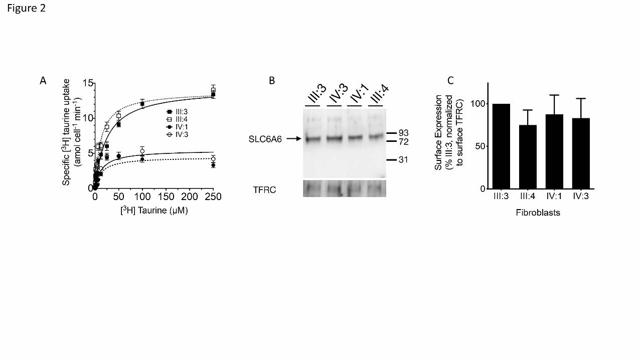
Figure2. Functional characterization of SLC6A6 activity from patient-derived fibroblasts.
(A) Specific Taurine Saturation Uptake Analysis in Patient-derived Fibroblasts. Fibroblasts of
affected individuals IV:1 (filled circle), IV:3(open circle), and parents III:3 (closed square) and
III:4 (open square) were incubated with [3H] taurine concentrations from 0.05 to 250 µM for 10
min. Non-specific counts were determined by inhibition of SLC6A6 with β-alanine (25 mM) and
subtracted from total counts. (B) Western Blot detection of SLC6A6 in the plasma membrane from
patient-derived fibroblasts. Surface protein was purified through biotinylation with a cell
impermeant crosslinker and detected using anti-SLC6A6 antibody. (C) Percent surface expression
was calculated from band densitometry for each sample and normalized to TFRC and plotted. *
P<0.05
Figure3. Results of taurine supplementation therapy for 24-months (A) Taurine levels in the
blood of both affected individuals (IV:1 and IV:3) before and after 6-months, 1-year and 2-years
of taurine supplementation. (B) Results of the echocardiography show that the cardiomyopathy of
Page 18 of 18
both affected individuals (IV:1 and IV:3) has been corrected after 2-years of taurine
supplementation. LVESD: left ventricular end-systolic diameter, LVEDD: left ventricular end
diastolic diameter, EF: ejection fraction, FS: fractional shortening. Green and red numbers
represent normal and abnormal values respectively. (C, D) Fundus photographs and macular OCT
of the right eye (C) and left eye (D) of the patient IV:3 at baseline and after 24 months of taurine
supplementation; anatomical stability with preservation of foveal photoreceptors can be noted.

Page 1 of 31
Taurine treatment of retinal degeneration and cardiomyopathy in a consanguineous family
with SLC6A6 taurine transporter deficiency
Muhammad Ansar1,2,*,§, Emmanuelle Ranza1,3,4,*, Madhur Shetty5, Sohail A. Paracha6, Maleeha
Azam7, Ilse Kern8, Justyna Iwaszkiewicz9, Omer Farooq10, Constantin J. Pournaras11, Ariane
Malcles12, Mateusz Kecik12, Carlo Rivolta13,14,15, Waqar Muzaffar16, Aziz Qurban16, Liaqat Ali7,
Yacine Aggoun17, Federico A. Santoni1,18, Periklis Makrythanasis1,19, Jawad Ahmed6, Raheel
Qamar7, Muhammad T. Sarwar6, L. Keith Henry5, Stylianos E. Antonarakis1,3,20,§
1Department of Genetic Medicine and Development, University of Geneva, Geneva, Switzerland;
2Current address, Clinical Research Center, Institute of Molecular and Clinical Ophthalmology
Basel (IOB), Basel, Switzerland;
3Service of Genetic Medicine, University Hospitals of Geneva, Geneva, Switzerland;
4Current address, Medigenome, Swiss Institute of Genomic Medicine, Geneva, Switzerland;
5Dept. of Biomedical Sciences, School of Medicine and Health Sciences, University of North
Dakota, Grand Forks, ND, United States;
6Institute of Basic Medical Sciences, Khyber Medical University, Peshawar, Pakistan;
7Department of Biosciences, Faculty of Science, COMSATS University Islamabad, Pakistan;
8Pediatric Nephrology and Metabolism Unit, Pediatric Subspecialties Service, Children's Hospital,
Geneva University Hospitals, Geneva, Switzerland;
9Swiss Institute of Bioinformatics, Molecular Modeling Group, University of Lausanne, Lausanne,
Switzerland;
Page 2 of 31
10Bahria University Medical and Dental College, Karachi, Pakistan;
11Hirslanden Clinique La Colline, Geneva, Switzerland ;
12Department of Ophthalmology, University Hospitals of Geneva, Geneva, Switzerland;
13Clinical Research Center, Institute of Molecular and Clinical Ophthalmology Basel (IOB), Basel,
Switzerland;
14Department of Ophthalmology, University Hospital Basel, Switzerland;
15Department of Genetics and Genome Biology, University of Leicester, Leicester, United
16Armed Forces Institute of Ophthalmology, Rawalpindi, Pakistan;
17Pediatric Cardiology, Geneva University Hospitals, Geneva, Switzerland;
18Current address, Department of Endocrinology Diabetes and Metabolism, University Hospital
of Lausanne, Lausanne, Switzerland;
19Current address, Biomedical Research Foundation of the Academy of Athens, Athens, Greece;
Kingdom;
20iGE3 Institute of Genetics and Genomics of Geneva, Geneva, Switzerland;
*These authors contributed equally
§Corresponding authors;
Muhammad Ansar, [email protected]
Stylianos Antonarakis, [email protected]
Page 3 of 31
Supplementary Appendix
Table of Contents
Supplementary Appendix ............................................................................................................................ 3
Methods ........................................................................................................................................................ 5
Genetic analysis .............................................................................................................................5
Molecular modeling of the SLC6A6 missense variant .......................................................................5
[3H Taurine] Uptake Assay ..............................................................................................................6
Surface Biotinylation ......................................................................................................................7
Taurine loading test and supplementation ......................................................................................8
Results........................................................................................................................................................... 9
Genetic analyses reveal a missense variant in the taurine transporter SLC6A6 .................................9
Clinical evaluation ........................................................................................................................ 10
Figure S1. Fundus photographs and macular OCT (optical coherence tomography) before taurine supplementation ................................................................................................................................. 10
Functional analysis ....................................................................................................................... 14
Figure S2. Taurine uptake in HEK-293 cells expressing the SLC6A6 G399V variant at high and low substrate concentrations .................................................................................................................... 14
Figure S3. Taurine Saturation Uptake Analysis of SLC6A6 G399V Mutant in HEK-293 Cells. ............. 15
Table S6. Kinetic Analysis of Taurine uptake by SLC6A6 G399V Mutant ............................................ 16
Figure S4. Taurine Uptake in Patient-derived Fibroblasts. ................................................................. 17
Table S7. Kinetic Analysis of Taurine Uptake by Patient-derived Fibroblasts ..................................... 18
Figure S5. SLC6A6 Surface Expression from Transfected Tissue Culture Cells ................................... 19
Taurine loading test ..................................................................................................................... 20
Taurine supplementation and 2 year follow up ............................................................................. 22
Figure S8. Fundus photographs and OCT of boy (IV:1) after two years of taurine supplementation. 23
Figure S9. Fundus photographs and OCT of the affected girl (IV:3) before the taurine supplementation. ................................................................................................................................ 24
Figure S10. Fundus photographs and OCT of the affected girl (IV:3) 1-year after the taurine supplementation. ................................................................................................................................ 25
Figure S11. Fundus photographs and OCT of the affected girl (IV:3) 2-year after the taurine supplementation. ................................................................................................................................ 26
Figure S12. Multifocal ERG before and after one and two-years of taurine supplementation. ........ 27
Page 4 of 31
References .................................................................................................................................................. 28
Table S1: Clinical features of affected individuals with recessive SLC6A6 variants. ................................... 29
Page 5 of 31
Methods
Genetic analysis
The initial analysis was done by performing the exome sequencing of the proband (IV:3) by using
SureSelect Human All Exon v6 reagents (Agilent Technologies, Santa Clara, CA, USA). The
sequencing was performed on an Illumina HiSeq4000 platform. A customized pipeline was used
to analyze the exome sequencing data, that includes the published algorithms, Burrows-Wheeler
aligner tool (BWA)(1), SAMtools(1), PICARD and the Genome Analysis Toolkit (GATK)(2), and the
sequenced reads were aligned to the GRCh37/hg19(3) reference human genome. The filtering
and interpretation of the filtered variants was performed as described in previous studies(4, 5).
All the family members including affecteds (IV:1 and IV:3), unaffected siblings (IV:4 and IV:5) and
both parents (III:3 and III:4) were genotyped by using the Illumina 720K SNP array
(HumanOmniExpress Bead Chip by Illumina Inc®, San Diego, CA, USA) (Figure 1). PLINK(6) was
used to analyze the genotyping data and to calculate the run of homozygosity (ROH). A ROH was
defined as the region of 50 consecutive homozygous SNPs, allowing a maximum of one mismatch
and bordered by the first heterozygous SNP at the edge.
Molecular modeling of the SLC6A6 missense variant
To predict the potential structural consequences of the SLC6A6 Gly399Val variant, the dopamine
transporter structure with bound cocaine molecule (PDB 4xp4) template was used. The UCSF
Chimera software(7) was used to model SLC6A6 protein with (Gly399Val) or without (Gly399)
variants, and to visualize the resulting protein structures.
Page 6 of 31
Cell and fibroblast Culture
Human embryonic kidney (HEK-293) cell lines were transfected with standard TransIT-LT1
protocol to stably express either wildtype taurine transporter SLC6A6 (formerly called TauT) or a
SLC6A6 containing the SNP G399V. Cells and primary fibroblasts were maintained in Dulbecco's
Modified Eagle's medium (DMEM), 10% Fetal Bovine Serum (FBS), 1% PSA Antibiotic solution
(10,000 units of penicillin, 10,000 µg of streptomycin, and 25 µg of Amphotericin B per mL) at
37˚C and 5% CO2 and were typically used for experiments after reaching 60%-70% confluency.
[3H Taurine] Uptake Assay
HEK-293 cells transiently transfected or stably expressing SLC6A6 or the G399V mutation
(100,000 cells/well) or primary fibroblasts (50,000 cells/well) were plated in a 24 well culture
plate (culturplate-24, Perkin-Elmer, Inc) and grown for 24 h. Cells were washed twice with 0.5 mL
KR buffer (130 mM NaCl, 1.3 mM KCl, 2.2 mM CaCl2, 1.2 mM MgSO4, 1.2 mM KH2PO4, 10 mM
HEPES, and 10 mM glucose, pH 7.4.) KR buffer of pH 5.5 was used to perform uptakes as SLC6A6
is more active under acidic conditions. Single point uptake of 30 nM or 5 µM [3H] taurine (Perkin
Elmer) with vehicle or indicated concentrations of cold taurine was carried out for 10 minutes at
37˚C. 5 µM was obtained by mixing [3H] taurine with non-labeled taurine. Uptake was terminated
with three washes of ice-cold KR buffer of pH 7.4. Radioactivity remaining in cells was measured
by liquid scintillation. Non-specific uptake was determined for HEK-293 cells by subtracting
radioactivity obtained with parental, non-transfected cells. Non-specific uptake for primary
fibroblasts was determined by uptake in the presence of 25 mM non-labeled β-alanine. For

Page 7 of 31
saturation analysis, [3H] taurine was diluted into non-labeled taurine to obtain the necessary
concentrations. Uptake was performed for 10 min followed by wash with cold-KR. Non-specific
counts were determined as above. Statistical significance was determined using unpaired T-Test
and one-way ANOVA (post-Tukey Test) with significance set at p < 0.05.
Surface Biotinylation
HEK-293 cells stably expressing SLC6A6 and G399V mutation (100,000 cells/plate) or primary
fibroblasts (100,000 cells/plate) were plated in a 100 mM culture plate. At 70% confluency,
primary fibroblasts were treated with 50 mM NaCl (final concentration) to enhance SLC6A6
expression. After 12-17 h incubation, all remaining steps were performed on ice to prevent
trafficking. Surface SLC6A6 levels were isolated by treatment with 0.5 mg/mL Sulfo-NHS-SS-biotin
(ThermoFisher, USA) for 25 minutes upon gentle shaking or rocking. Unreacted biotin was
quenched by incubation with 100 mM glycine for 25 minutes with gentle rocking. Cells were
solubilized with 500 µL RIPA buffer plus protease inhibitors for 15 minutes on ice. Lysed cells
were spun for 20 minutes at 4˚C at 16,000 x g followed by collection of the supernatant. Cells
were washed in between biotinylation, quenching and lysing twice with 1X Phosphate-buffered
saline with Ca and Mg (PBS/CM) that contained (in mM): 137 mM NaCl, 2.7 mM KCl, 10.1 mM
Na2HPO4, 1.8 mM KH2PO4, 0.1 mM CaCl2, 1.0 mM MgCl2, and 10 mM glucose, pH 7.4. 100 µg
protein was reacted with 50% slurry neutrAvidin agarose beads (ThermoFisher) and rotated
overnight at 4˚C. Beads are then pelleted at 300 x g for 2 minutes and washed with RIPA buffer
three times. Beads are incubated with 2X loading dye for 30 minutes at 37˚C, followed by elution
of proteins by pelleting the beads at 5,000 x g for 2 minutes. Purified proteins were resolved on
Page 8 of 31
NEXTGEL® 10% acrylamide gels and transferred to 0.45 µM PVDF membranes (Immobilon-P,
Millipore). Membranes were blocked for 2 hours at 5% bovine serum albumin (BSA) followed by
incubation with SLC6A6 anti-rabbit polyclonal antibody (PA5-37460 Thermo Fisher Scientific) or
chicken polyclonal anti-transferrin receptor (TFRC) primary antibody overnight at 4˚C.
Membranes were incubated with HRP-conjugated goat anti-rabbit or goat anti-chicken
secondary antibody for 1 hour at room temperature. Membranes were imaged on a LiCor C-Digit
using WesternSure® PREMIUM Chemiluminescent Substrate (LiCor). SLC6A6 was normalized to
TFRC for each membrane sample. Statistical significance was determined using one-way ANOVA
(post-Tukey Test) with significance set at p < 0.05.
Taurine loading test and supplementation
A taurine loading test was performed in the Pediatrics clinical research unit of the University
hospitals of Geneva after approval by the ethics committee. We have administered an oral bolus
dose of 100mg/kg of Taurine to the two affected individuals and their heterozygous parents on
day 2, this dose was recommended by the literature as non-toxic. Taurine was provided in tablets,
commercialized by Burgerstein Pharmaceuticals. Repeated measurements of taurine in blood
and urine were subsequently performed. On days 3 and 4, the patients received a 100mg/kg/day,
administered in 3 doses (33mg/kg/q8hours).

Page 9 of 31
Results
Genetic analyses reveal a missense variant in the taurine transporter SLC6A6
The genetic analysis was initiated by performing exome sequencing of proband (IV:3) of family
F315. More than 95% of the sequenced region was covered at 20x with a total mean coverage of
~100x. An in-house pipeline and prioritization algorithm(4, 5) was used to analyze the high
throughput sequencing data. None of the genes known to be implicated in retinal degeneration
and all types of visual impairment has yielded any pathogenic variant. Then by combining the
exome sequencing data of the proband and genotyping data of all family members, variants
present in the regions of homozygosity (ROH) that segregated with the disease phenotype in
recessive manner were selected and filtered as described previously(5, 8). By using this approach
a homozygous missense variant (NM_003043.5:c.1196G>T:p.(Gly399Val) in SLC6A6 was
identified (Figure 1). The variant SLC6A6:p.(Gly399Val) is not present in gnomAD, Bravo database
(https://bravo.sph.umich.edu/freeze5/hg38/) or our local cohort of 300 controls from the same
Pakistani ethnicity. As shown in the chromatograms (figure 1B), both the affected individuals
(IV:1 and IV:3) are homozygous, both parents (III:3 and III:4) and one unaffected sibling (IV:4) are
heterozygous for the variant (Gly399Val) and another unaffected sibling (IV:5) has both normal
alleles; confirming the recessive inheritance of the variant. By sharing our findings through
GeneMatcher(9), genetic conferences and meetings, we have not found any second case having
pathogenic or likely pathogenic variants in the SLC6A6 gene. However, by publishing this article
we hope to identify similar cases.
Page 10 of 31
Clinical evaluation
The two affected individuals and their parents were clinically evaluated at the University Hospital of
Geneva. The results of the evaluation are described in the table 1.
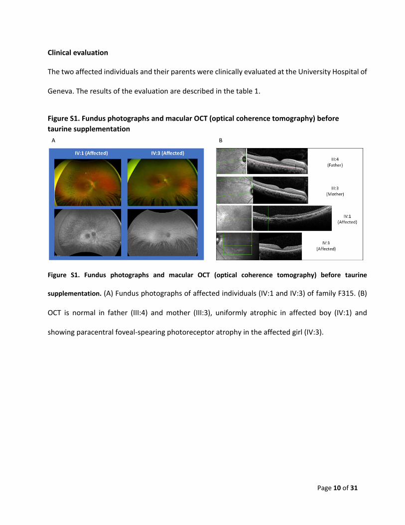
Figure S1. Fundus photographs and macular OCT (optical coherence tomography) before taurine supplementation
Figure S1. Fundus photographs and macular OCT (optical coherence tomography) before taurine
supplementation. (A) Fundus photographs of affected individuals (IV:1 and IV:3) of family F315. (B)
OCT is normal in father (III:4) and mother (III:3), uniformly atrophic in affected boy (IV:1) and
showing paracentral foveal-spearing photoreceptor atrophy in the affected girl (IV:3).
Page 11 of 31
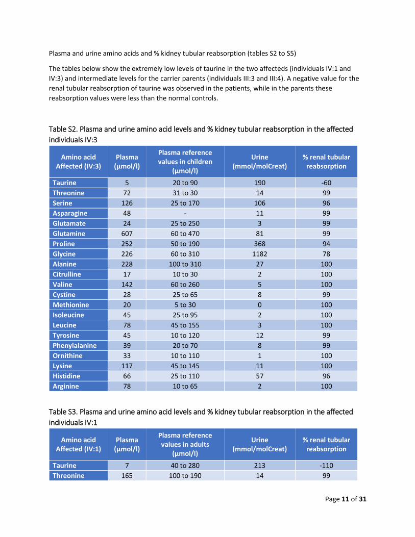
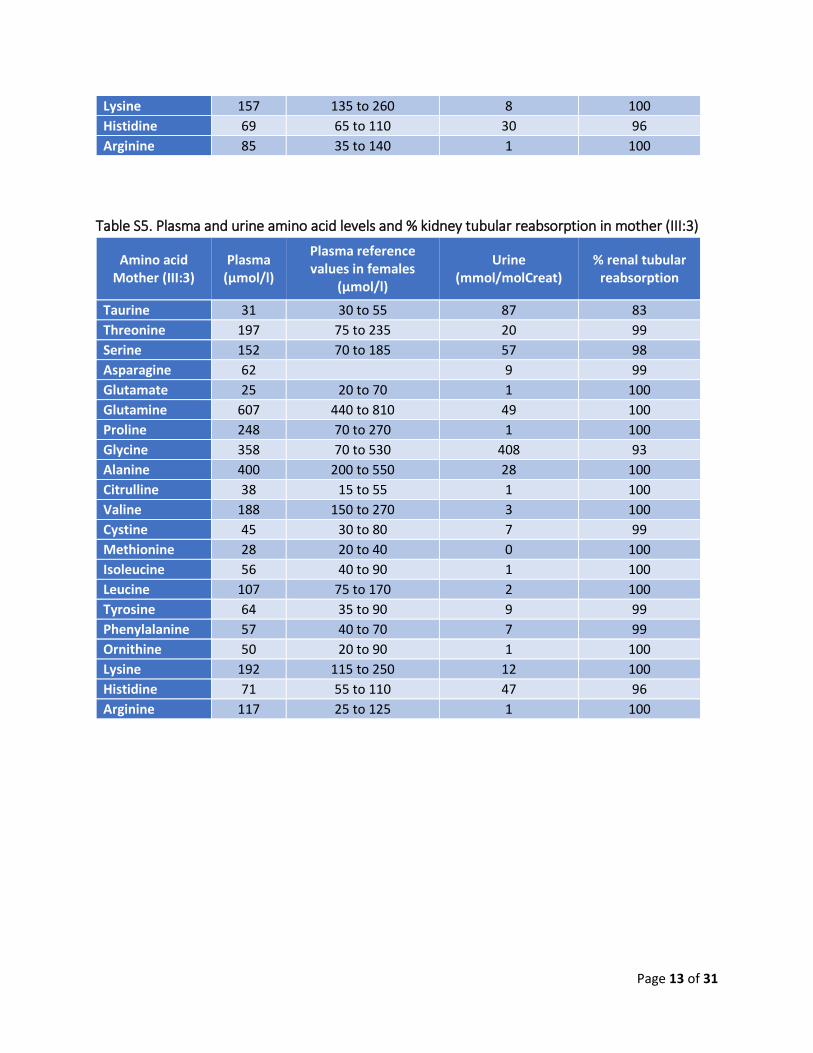
Plasma and urine amino acids and % kidney tubular reabsorption (tables S2 to S5)
The tables below show the extremely low levels of taurine in the two affecteds (individuals IV:1 and IV:3) and intermediate levels for the carrier parents (individuals III:3 and III:4). A negative value for the renal tubular reabsorption of taurine was observed in the patients, while in the parents these reabsorption values were less than the normal controls.
Table S2. Plasma and urine amino acid levels and % kidney tubular reabsorption in the affected individuals IV:3
Amino acid Affected (IV:3)
Plasma (µmol/l)
Plasma reference values in children
(µmol/l)
Urine (mmol/molCreat)
% renal tubular reabsorption
Taurine 5 20 to 90 190 -60 Threonine 72 31 to 30 14 99 Serine 126 25 to 170 106 96 Asparagine 48 - 11 99 Glutamate 24 25 to 250 3 99 Glutamine 607 60 to 470 81 99 Proline 252 50 to 190 368 94 Glycine 226 60 to 310 1182 78 Alanine 228 100 to 310 27 100 Citrulline 17 10 to 30 2 100 Valine 142 60 to 260 5 100 Cystine 28 25 to 65 8 99 Methionine 20 5 to 30 0 100 Isoleucine 45 25 to 95 2 100 Leucine 78 45 to 155 3 100 Tyrosine 45 10 to 120 12 99 Phenylalanine 39 20 to 70 8 99 Ornithine 33 10 to 110 1 100 Lysine 117 45 to 145 11 100 Histidine 66 25 to 110 57 96 Arginine 78 10 to 65 2 100
Table S3. Plasma and urine amino acid levels and % kidney tubular reabsorption in the affected individuals IV:1
Amino acid Affected (IV:1)
Plasma (µmol/l)
Plasma reference values in adults
(µmol/l)
Urine (mmol/molCreat)
% renal tubular reabsorption
Taurine 7 40 to 280 213 -110 Threonine 165 100 to 190 14 99
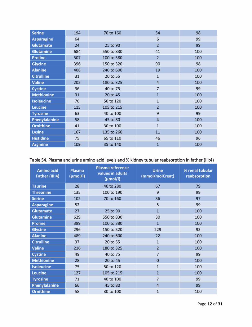
Page 12 of 31
Serine 194 70 to 160 54 98 Asparagine 64 6 99 Glutamate 24 25 to 90 2 99 Glutamine 684 550 to 830 41 100 Proline 507 100 to 380 2 100 Glycine 396 150 to 320 90 98 Alanine 408 240 to 600 19 100 Citrulline 31 20 to 55 1 100 Valine 202 180 to 325 4 100 Cystine 36 40 to 75 7 99 Methionine 31 20 to 45 1 100 Isoleucine 70 50 to 120 1 100 Leucine 115 105 to 215 2 100 Tyrosine 63 40 to 100 9 99 Phenylalanine 58 45 to 80 4 100 Ornithine 41 30 to 100 1 100 Lysine 167 135 to 260 11 100 Histidine 75 65 to 110 46 96 Arginine 109 35 to 140 1 100
Table S4. Plasma and urine amino acid levels and % kidney tubular reabsorption in father (III:4)
Amino acid Father (III:4)
Plasma (µmol/l)
Plasma reference values in adults
(µmol/l)
Urine (mmol/molCreat)
% renal tubular reabsorption
Taurine 28 40 to 280 67 79 Threonine 135 100 to 190 9 99 Serine 102 70 to 160 36 97 Asparagine 52 5 99 Glutamate 27 25 to 90 1 100 Glutamine 629 550 to 830 30 100 Proline 389 100 to 380 1 100 Glycine 296 150 to 320 229 93 Alanine 489 240 to 600 22 100 Citrulline 37 20 to 55 1 100 Valine 216 180 to 325 2 100 Cystine 49 40 to 75 7 99 Methionine 28 20 to 45 0 100 Isoleucine 75 50 to 120 1 100 Leucine 127 105 to 215 1 100 Tyrosine 71 40 to 100 7 99 Phenylalanine 66 45 to 80 4 99 Ornithine 58 30 to 100 1 100
Page 13 of 31
Lysine 157 135 to 260 8 100 Histidine 69 65 to 110 30 96 Arginine 85 35 to 140 1 100
Table S5. Plasma and urine amino acid levels and % kidney tubular reabsorption in mother (III:3)
Amino acid Mother (III:3)
Plasma (µmol/l)
Plasma reference values in females
(µmol/l)
Urine (mmol/molCreat)
% renal tubular reabsorption
Taurine 31 30 to 55 87 83 Threonine 197 75 to 235 20 99 Serine 152 70 to 185 57 98 Asparagine 62 9 99 Glutamate 25 20 to 70 1 100 Glutamine 607 440 to 810 49 100 Proline 248 70 to 270 1 100 Glycine 358 70 to 530 408 93 Alanine 400 200 to 550 28 100 Citrulline 38 15 to 55 1 100 Valine 188 150 to 270 3 100 Cystine 45 30 to 80 7 99 Methionine 28 20 to 40 0 100 Isoleucine 56 40 to 90 1 100 Leucine 107 75 to 170 2 100 Tyrosine 64 35 to 90 9 99 Phenylalanine 57 40 to 70 7 99 Ornithine 50 20 to 90 1 100 Lysine 192 115 to 250 12 100 Histidine 71 55 to 110 47 96 Arginine 117 25 to 125 1 100
Page 14 of 31
Functional analysis
Functional analysis were performed to observe the effect of the SLC6A6 G399V variant on the taurine uptake and surface expression of the transporter(10).
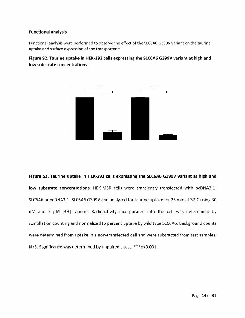
Figure S2. Taurine uptake in HEK-293 cells expressing the SLC6A6 G399V variant at high and low substrate concentrations
Figure S2. Taurine uptake in HEK-293 cells expressing the SLC6A6 G399V variant at high and
low substrate concentrations. HEK-MSR cells were transiently transfected with pcDNA3.1-
SLC6A6 or pcDNA3.1- SLC6A6 G399V and analyzed for taurine uptake for 25 min at 37˚C using 30
nM and 5 µM [3H] taurine. Radioactivity incorporated into the cell was determined by
scintillation counting and normalized to percent uptake by wild type SLC6A6. Background counts
were determined from uptake in a non-transfected cell and were subtracted from test samples.
N>3. Significance was determined by unpaired t-test. ***p<0.001.
Page 15 of 31
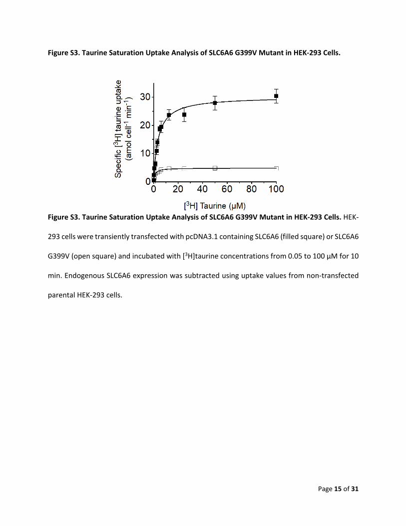
Figure S3. Taurine Saturation Uptake Analysis of SLC6A6 G399V Mutant in HEK-293 Cells.
Figure S3. Taurine Saturation Uptake Analysis of SLC6A6 G399V Mutant in HEK-293 Cells. HEK-
293 cells were transiently transfected with pcDNA3.1 containing SLC6A6 (filled square) or SLC6A6
G399V (open square) and incubated with [3H]taurine concentrations from 0.05 to 100 µM for 10
min. Endogenous SLC6A6 expression was subtracted using uptake values from non-transfected
parental HEK-293 cells.
Page 16 of 31
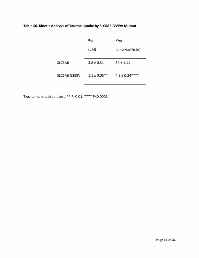
Table S6. Kinetic Analysis of Taurine uptake by SLC6A6 G399V Mutant
KM
(µM)
Vmax
(amol/cell/min)
SLC6A6 3.8 ± 0.31 30 ± 1.11
SLC6A6 G399V 1.1 ± 0.05** 4.9 ± 0.24****
Two-tailed unpaired t test, ** P<0.01, **** P<0.0001.

Page 17 of 31
Figure S4. Taurine Uptake in Patient-derived Fibroblasts.
Figure S4. Taurine Uptake in Patient-derived Fibroblasts. Percent uptake is plotted after
normalization to IV:1. One-way ANOVA with Tukey’s multiple comparison post-hoc. All
differences within fibroblast samples were significant at * p<0.05, *** p<0.001. Nonsignificant
differences were indicated by ns. HEK-293 cells were used as a positive control as transfection
with SLC6A6 plasmid shows significant ~12-fold increase in taurine transport over the
endogenous SLC6A6 transport. 30 nM of [3H] taurine was used for the uptake analysis.
Page 18 of 31
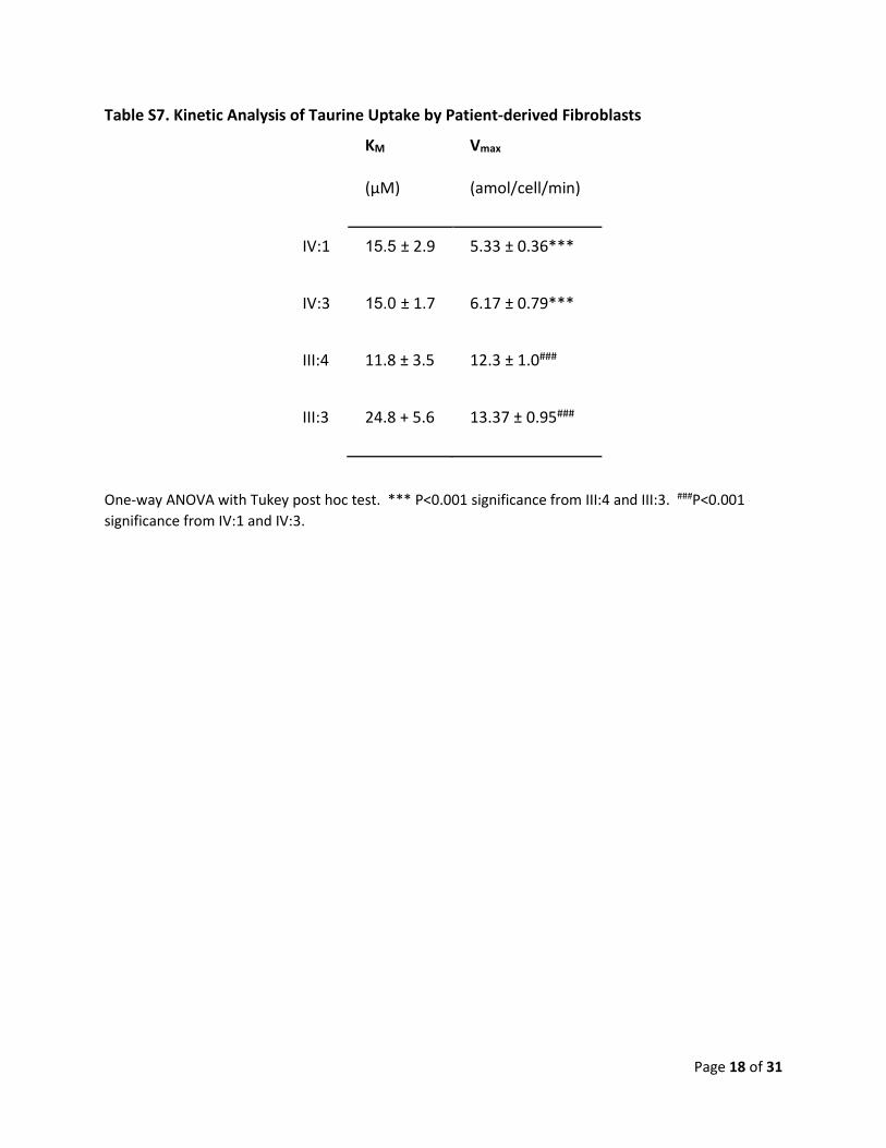
Table S7. Kinetic Analysis of Taurine Uptake by Patient-derived Fibroblasts
KM
(µM)
Vmax
(amol/cell/min)
IV:1 15.5 ± 2.9 5.33 ± 0.36***
IV:3 15.0 ± 1.7 6.17 ± 0.79***
III:4 11.8 ± 3.5 12.3 ± 1.0###
III:3 24.8 + 5.6 13.37 ± 0.95###
One-way ANOVA with Tukey post hoc test. *** P<0.001 significance from III:4 and III:3. ###P<0.001 significance from IV:1 and IV:3.

Page 19 of 31
Figure S5. SLC6A6 Surface Expression from Transfected Tissue Culture Cells A B
SLC6A6 Surface Expression Analysis in Transiently Transfected HEK-293 Cells. (A) Western blot
detection of SLC6A6 from purified plasma membrane biotinylated proteins extracted from
parental HEK-293 cells (Par) and cells transiently transfected with 200 ng of plasmid DNA allowing
expression of SLC6A6 or SLC6A6 G399V. Blots were striped and probed for the plasma membrane
transferrin receptor which was used to normalize SLC6A6 expression. (B) Plot of densitometry
values for SLC6A6 normalized to percent of transferrin receptor (TFRC). ** P<0.01

Page 20 of 31
Taurine loading test
A baseline profile of taurine levels in blood and urine was first measured in both affected children
and their parents; serial measurements without taurine supplementation were obtained during
day 1. The results showed extremely low levels of taurine in the two affected individuals (around
5 µmol/l), while their heterozygous parents had intermediate levels. The standard daily diet did
not increase the blood taurine levels in the patients or their parents (see Figure S9, day 1).
The results were as follows (figure S9):
• In the two affected children and their parents, blood taurine levels reached a high peak
(400-900 µmol/l) after 4 hours; after 8 hours, the levels were still elevated (> 100 µmol/l).
• The kinetics of blood taurine levels were similar between the patients and their parents.
Moreover, the time of the peak of blood Taurine levels after the bolus administration was
comparable to that of normal volunteers.
• On days 3 and 4, we observed that the taurine supplementation 3 times a day in the
affecteds, resulted in a peak of around 250 µmol/l. More importantly, even though the
last administration of taurine was at 8pm, the fasting levels in the next morning were
between 53 and 78 µmol/l, which correspond to the usual heterozygous levels without
supplementation or the low normal range.

Page 21 of 31
Figure S6. Taurine loading test.
Figure S6. Taurine loading test. Blood Taurine levels in the 4 members of the family, before and
after the Taurine loading test, and the Taurine supplementation, performed at the University
Hospitals of Geneva. For discussion of the results see text.

Page 22 of 31
Figure S7. Plasma taurine levels in healthy volunteers.
Figure S7. Plasma taurine levels in healthy volunteers. Linear plot of mean plasma taurine levels
(mmoL) in eight healthy volunteers following administration of 4 g (32 mmoL) oral taurine (this
dose is about half of that used in the members of the F315 family). Data from Ghandforoush-
Sattari M et al(11).
These results show that daily supplementation of taurine is sufficient to increase taurine to low
normal blood levels: thus, since the mutation in these patients allows a 10-18% intracellular
transport of taurine, the increased levels in blood could provide sufficient taurine transport in
the retina cells and prevent further destruction of photoreceptors in the affected female.
Moreover, this supplementation could avoid progression of the cardiomyopathy in both affected
children.
Taurine supplementation and 2 year follow up
Page 23 of 31
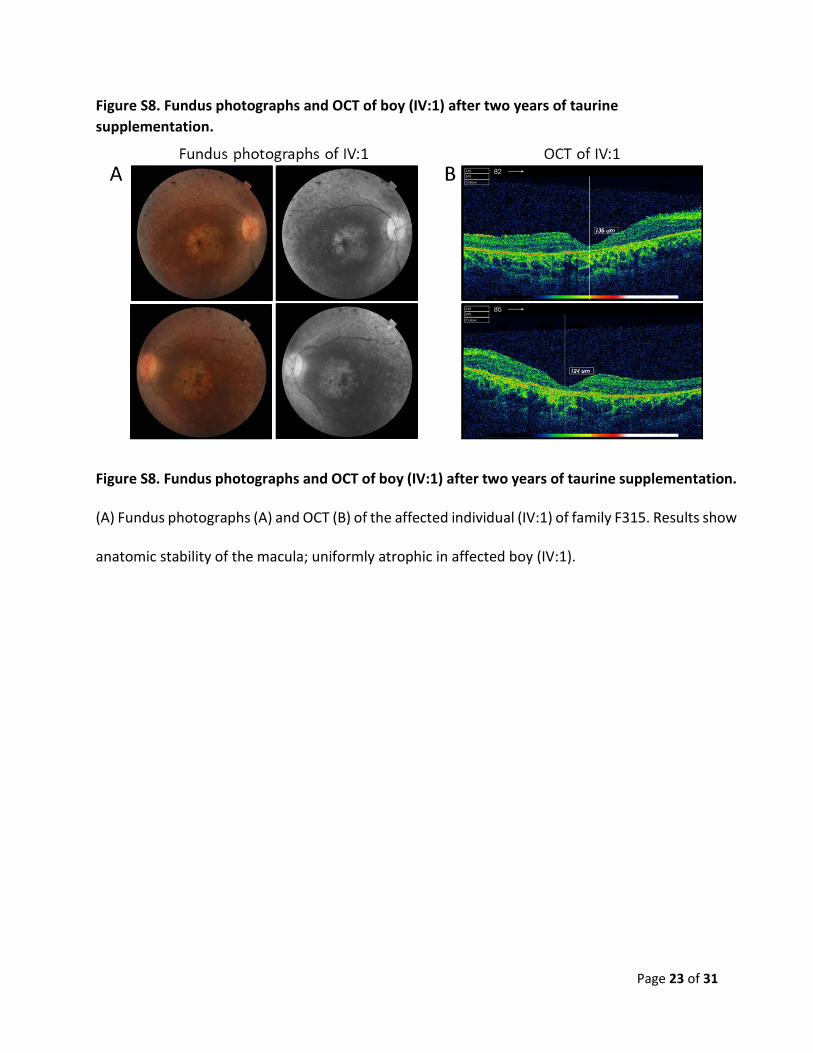
Figure S8. Fundus photographs and OCT of boy (IV:1) after two years of taurine supplementation.
Figure S8. Fundus photographs and OCT of boy (IV:1) after two years of taurine supplementation.
(A) Fundus photographs (A) and OCT (B) of the affected individual (IV:1) of family F315. Results show
anatomic stability of the macula; uniformly atrophic in affected boy (IV:1).

Page 24 of 31
Figure S9. Fundus photographs and OCT of the affected girl (IV:3) before the taurine supplementation.
Figure S9. Fundus photographs and OCT of the affected girl (IV:3) before the taurine
supplementation. Fundus photographs and OCT of affected girl (IV:3) of family F315 show a
paracentral foveal-spearing photoreceptor atrophy in the affected girl (IV:3)

Page 25 of 31
Figure S10. Fundus photographs and OCT of the affected girl (IV:3) 1-year after the taurine supplementation.
Figure S10. Fundus photographs and OCT of the affected girl (IV:3) 1-year after the taurine
supplementation. Fundus photographs and OCT of affected girl (IV:3) show the anatomical stability
with preservation of foveal photoreceptors.

Page 26 of 31
Figure S11. Fundus photographs and OCT of the affected girl (IV:3) 2-year after the taurine supplementation.
Figure S11. Fundus photographs and OCT of the affected girl (IV:3) 2-year after the taurine
supplementation. Fundus photographs and OCT of affected girl (IV:3) show the anatomical stability
with preservation of foveal photoreceptors. Her brother (IV:1) had complete visual loss at this age.
Page 27 of 31
Normal Before treatment 1-year of taurine suppl. 2-years of taurine suppl.
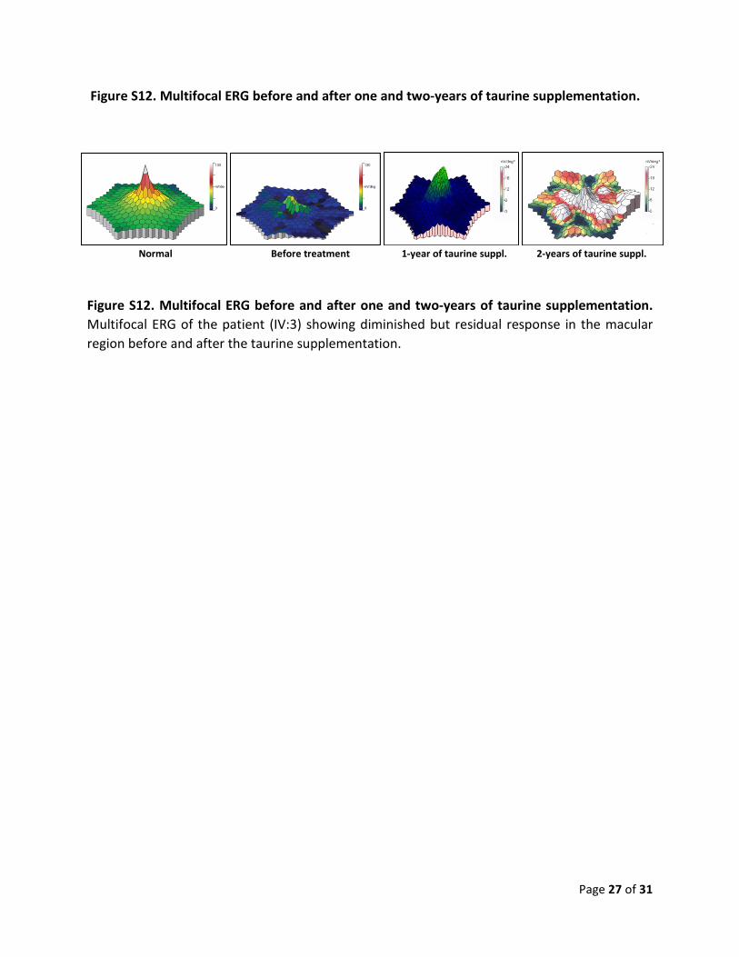
Figure S12. Multifocal ERG before and after one and two-years of taurine supplementation.
Figure S12. Multifocal ERG before and after one and two-years of taurine supplementation. Multifocal ERG of the patient (IV:3) showing diminished but residual response in the macular region before and after the taurine supplementation.
Page 28 of 31
References
1 Li, H. and Durbin, R. (2009) Fast and accurate short read alignment with Burrows-Wheeler transform. Bioinformatics, 25, 1754-1760. 2 DePristo, M.A., Banks, E., Poplin, R., Garimella, K.V., Maguire, J.R., Hartl, C., Philippakis, A.A., del Angel, G., Rivas, M.A., Hanna, M. et al. (2011) A framework for variation discovery and genotyping using next-generation DNA sequencing data. Nat Genet, 43, 491-498. 3 Pruitt, K.D., Tatusova, T. and Maglott, D.R. (2007) NCBI reference sequences (RefSeq): a curated non-redundant sequence database of genomes, transcripts and proteins. Nucleic acids research, 35, D61-65. 4 Makrythanasis, P., Nelis, M., Santoni, F.A., Guipponi, M., Vannier, A., Bena, F., Gimelli, S., Stathaki, E., Temtamy, S., Megarbane, A. et al. (2014) Diagnostic exome sequencing to elucidate the genetic basis of likely recessive disorders in consanguineous families. Hum Mutat, 35, 1203-1210. 5 Ansar, M., Chung, H., Waryah, Y.M., Makrythanasis, P., Falconnet, E., Rao, A.R., Guipponi, M., Narsani, A.K., Fingerhut, R., Santoni, F.A. et al. (2018) Visual impairment and progressive phthisis bulbi caused by recessive pathogenic variant in MARK3. Hum Mol Genet, in press. 6 Purcell, S., Neale, B., Todd-Brown, K., Thomas, L., Ferreira, M.A., Bender, D., Maller, J., Sklar, P., de Bakker, P.I., Daly, M.J. et al. (2007) PLINK: a tool set for whole-genome association and population-based linkage analyses. Am J Hum Genet, 81, 559-575. 7 Pettersen, E.F., Goddard, T.D., Huang, C.C., Couch, G.S., Greenblatt, D.M., Meng, E.C. and Ferrin, T.E. (2004) UCSF Chimera--a visualization system for exploratory research and analysis. J Comput Chem, 25, 1605-1612. 8 Ansar, M., Ullah, F., Paracha, S.A., Adams, D.J., Lai, A., Pais, L., Iwaszkiewicz, J., Millan, F., Sarwar, M.T., Agha, Z. et al. (2019) Bi-allelic Variants in DYNC1I2 Cause Syndromic Microcephaly with Intellectual Disability, Cerebral Malformations, and Dysmorphic Facial Features. Am J Hum Genet, 104, 1073-1087. 9 Sobreira, N., Schiettecatte, F., Valle, D. and Hamosh, A. (2015) GeneMatcher: a matching tool for connecting investigators with an interest in the same gene. Hum Mutat, 36, 928-930. 10 Tomi, M., Tajima, A., Tachikawa, M. and Hosoya, K. (2008) Function of taurine transporter (Slc6a6/TauT) as a GABA transporting protein and its relevance to GABA transport in rat retinal capillary endothelial cells. Biochim Biophys Acta, 1778, 2138-2142. 11 Ghandforoush-Sattari, M., Mashayekhi, S., Krishna, C.V., Thompson, J.P. and Routledge, P.A. (2010) Pharmacokinetics of oral taurine in healthy volunteers. J Amino Acids, 2010, 346237.
Page 29 of 31
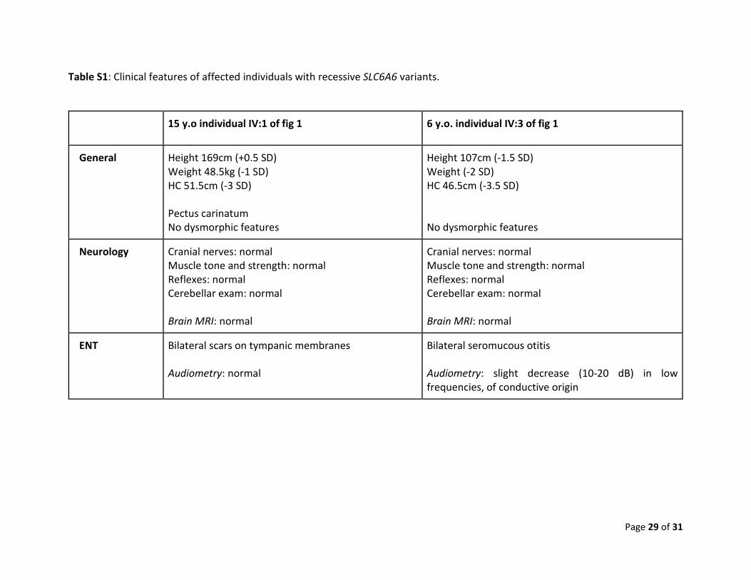
Table S1: Clinical features of affected individuals with recessive SLC6A6 variants.
15 y.o individual IV:1 of fig 1 6 y.o. individual IV:3 of fig 1
General Height 169cm (+0.5 SD) Weight 48.5kg (-1 SD) HC 51.5cm (-3 SD) Pectus carinatum No dysmorphic features
Height 107cm (-1.5 SD) Weight (-2 SD) HC 46.5cm (-3.5 SD) No dysmorphic features
Neurology Cranial nerves: normal Muscle tone and strength: normal Reflexes: normal Cerebellar exam: normal Brain MRI: normal
Cranial nerves: normal Muscle tone and strength: normal Reflexes: normal Cerebellar exam: normal Brain MRI: normal
ENT Bilateral scars on tympanic membranes Audiometry: normal
Bilateral seromucous otitis Audiometry: slight decrease (10-20 dB) in low frequencies, of conductive origin

Page 30 of 31
Cardiology BP: 127/70 mmHg Heart rate: 102/min Saturation: 100% AA No heart murmurs Echocardiography: mild hypokinetic cardiomyopathy with systolic dysfunction (shortening fraction 26-27 %) and slight systolic dilatation of left ventricle (Z score +3.2). Effort test: normal
BP: 103/73 mmHg Heart rate: 135/min Saturation: 100% AA No heart murmurs Echocardiography: mild hypokinetic cardiomyopathy with systolic dysfunction (shortening fraction 24 %) and slight systolic dilatation of left ventricle (Z score +2.5). Effort test: normal
Ophthalmology Visual acuity: Light perception ODS Fundus: advanced cone-rod dystrophy, severe peripheral alterations and central atrophy Autofluorescence: severe peripheral and central hypoautofluorescence with a preserved paracentral ring of isoautofluorescence Global electroretinogram (ERG): extinguished Multifocal ERG: not possible to perform due to complete visual loss Visual field: not possible to perform
Visual acuity: Counting fingers ODS Fundus: cone-rod dystrophy, salt-and-peper peripheral alterations and paracentral macular atrophy Autofluorescence: central area of isoautofluorescence with a ring of hypoautofluorescence and outer ring of hyperautofluorescence; normal autofluorescence in retinal periphery Global electroretinogramm (ERG): extinguished Multifocal ERG: minimal foveal macular response Visual field: abnormal, paracentral scotoma with a relatively preserved peripheral visual field
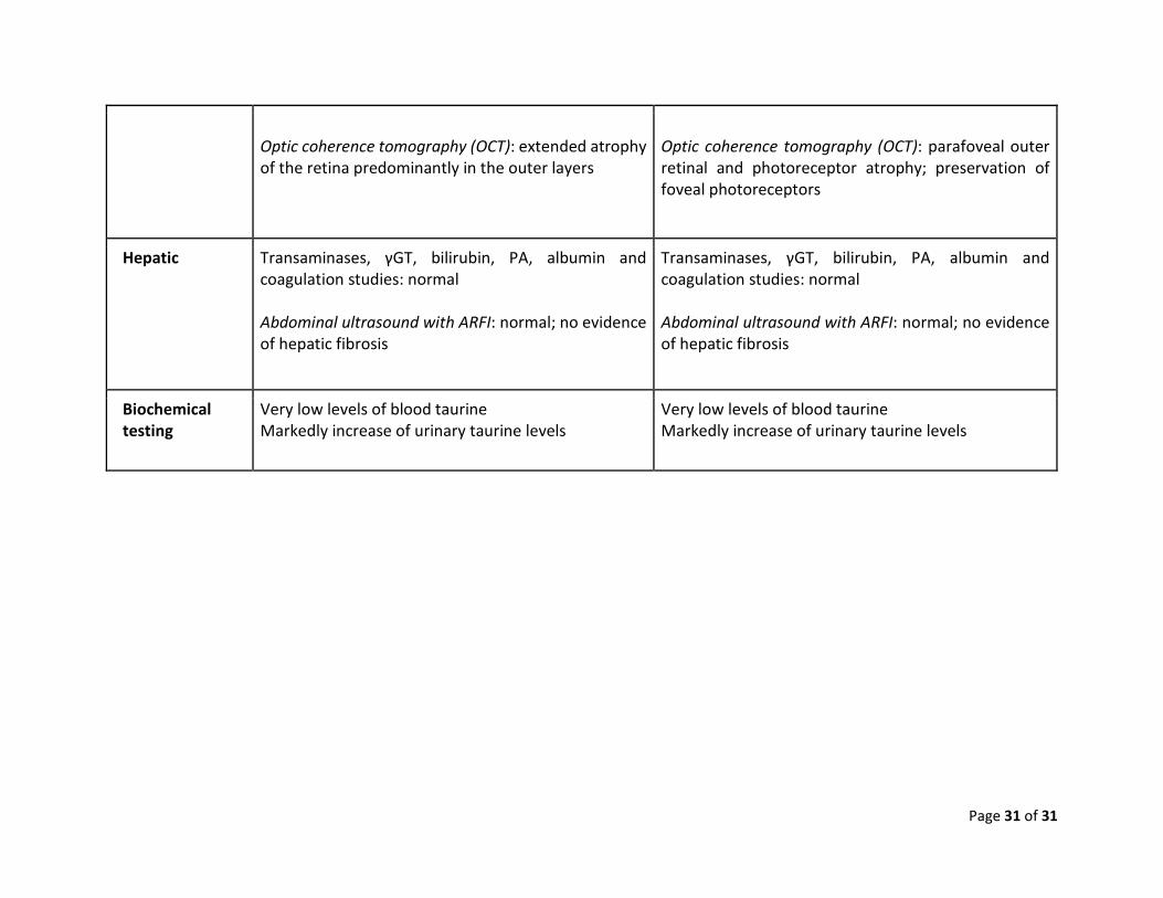
Page 31 of 31
Optic coherence tomography (OCT): extended atrophy of the retina predominantly in the outer layers
Optic coherence tomography (OCT): parafoveal outer retinal and photoreceptor atrophy; preservation of foveal photoreceptors
Hepatic Transaminases, γGT, bilirubin, PA, albumin and coagulation studies: normal Abdominal ultrasound with ARFI: normal; no evidence of hepatic fibrosis
Transaminases, γGT, bilirubin, PA, albumin and coagulation studies: normal Abdominal ultrasound with ARFI: normal; no evidence of hepatic fibrosis
Biochemical testing
Very low levels of blood taurine Markedly increase of urinary taurine levels
Very low levels of blood taurine Markedly increase of urinary taurine levels