Embed Size (px)
Citation preview
8/14/2019 5.2remodelacion Asma
http://slidepdf.com/reader/full/52remodelacion-asma 1/8
"
Review article
#$%&'$()*) +,$- .//0122(03(45)464/('3$7,&('85$,65 9$, 0*,8$&(' 78* $&':5 ;$ $/.*, 78*8 %4/.$7/ 0*,-4884$&5
Airway remodelling in asthma and novel therapy
Wiparat Manuyakorn,1 Peter H Howarth
2and Stephen T Holgate
2
Summary
Asthma is an airway inflammatory disease
with functional and structural changes, leading
to bronchial hyperresponsiveness (BHR) and
airflow obstruction. Airway structural changes
or airway remodelling consist of epithelial injury,
goblet cell hyperplasia, subepithelial layer
thickening, airway smooth muscle hyperplasia
and angiogenesis. These changes were previously
considered as a consequence of chronic airway
inflammation. However, several studies havedemonstrated that inflammation and remodelling
can occur as separate but parallel aspects of the
asthmatic process. As such there is increasing
evidence for the role of mechanocompressive
forces within the asthmatic airway contributing
to airway structural changes. Furthermore, it is
unclear what is the best treatment to modify
remodelling and which component to target.
There is also a need to identify asthma phenotype
that might specifically respond to novel therapies
such as anti-IL5, anti-IL13 and tyrosine kinase
inhibitors. (Asian Pac J Allergy Immunol 2013;31:3-
10)
Key words: Asthma, remodelling, physical forces,
asthma therapy, airway structural changes
Introduction
Asthma is a common chronic disorder of the
airway that is characterized by the complex interaction
of airway obstruction, bronchial hyperresponsiveness
(BHR), and airway inflammation which leads to
recurrent episodes of wheezing, breathlessness,
chest tightness, and coughing. The airway
inflammation is typically eosinophillic and
accompanied by elevation of Th2 cytokines.
Eosinophils are a key feature of Th2 inflammation
and are a useful biomarker in guiding treatment.
However, Th2 inflammation alone cannot explain
all features of asthma. For example airway
hyperresponsiveness and tissue remodelling are not
entirely linked to this inflammation.1 There are a
number of asthmatic patients in whom anti-
inflammatory therapy does not lead to symptom
control and who are considered treatment resistant.
Additionally, asthmatic patients treated with an anti-
IL5 mAb (mepolizumab) or T cell directed therapy
that modify eosinophilic inflammation have failed to
demonstrate symptomatic improvement over disease
control.1
Furthermore, whilst recognized to modifyeosinophilic inflammation, inhaled corticosteroid
treatment in atopic children with recurrent wheezing
has been shown to have no effect on decline in lung
function and the natural history of asthma over
time.2 This irreversible airflow obstruction has been
shown to develop despite appropriate use of inhaled
corticosteroids, as advocated by international
disease management guidelines.3 One possibility is
that the decline in lung function relates to
uncontrolled airway remodelling.
Airway remodelling in asthma
Pathological repair of the airways leads to
structural changes referred to as airway remodelling.
Airway remodelling has been proposed to result in
lower baseline lung function. Pathologically this is
characterised by subepithelial thickening from
increased deposition of extracellular matrix proteins
(ECMs) such as collagens, proteoglycans and
glycoproteins, epithelial denudation with goblet cell
metaplasia, increased airway smooth muscle mass,
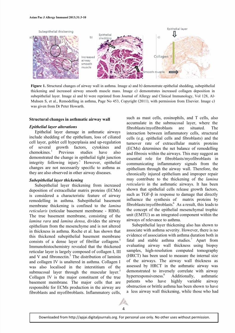
and angiogenesis.4 (Figure 1)
!"#$ &' ()*)+)#, #- ./0)12")3 455/"67 1,0 8$$9,#5#67:
(/;1"2$/,2 #- ./0)12")3+: !139527 #- </0)3),/
=1$12>)?#0) @#+;)215: <1>)0#5 A,)*/"+)27: B1,6C#C
&DEDD: F@48G4H(
I' 4310/$)3 A,)2 #- J5),)315 1,0 KL;/")$/,215 M3)/,3/+
1,0 2>/ M#92>1$;2#, H8@= =/+;)"12#"7 B)#$/0)315
=/+/1"3> A,)2: A,)*/"+)27 #- M#92>1$;2#, !139527 #-
</0)3),/: M)" @/,"7 N/553#$/ G1?#"12#")/+:
M#92>1$;2#, O/,/"15 @#+;)215: M#92>1$;2#, MP&Q QR(:
A,)2/0 S),60#$'
J#""/+;#,0),6 192>#"T N);1"12 <1,971C#",
KU$1)5T $V);1"12W>#2$1)5'3#$
M9?$)22/0 012/T XY&IYID&I
8/14/2019 5.2remodelacion Asma
http://slidepdf.com/reader/full/52remodelacion-asma 2/8
!"#$% '$( ) !**+,-. /001%2* 345676586954
<
#$%&'$()*) +,$- .//0122(03(45)464/('3$7,&('85$,65 9$, 0*,8$&(' 78* $&':5 ;$ $/.*, 78*8 %4/.$7/ 0*,-4884$&5
Structural changes in asthmatic airway wall
Epithelial layer alterations
Epithelial layer damage in asthmatic airways
include shedding of the epithelium, loss of ciliated
cell layer, goblet cell hyperplasia and up-regulation
of several growth factors, cytokines and
chemokines.1 Previous studies have also
demonstrated the change in epithelial tight junction
integrity following injury.1 However, epithelial
changes are not necessarily specific to asthma as
they are also observed in other airway diseases.
Subepithelial layer thickening
Subepithelial layer thickening from increased
deposition of extracellular matrix proteins (ECMs)
is considered a characteristic feature of airway
remodelling in asthma. Subepithelial basement
membrane thickening is confined to the lamina
reticularis (reticular basement membrane - RBM).
The true basement membrane, consisting of the
lamina rara and lamina densa, divides the airway
epithelium from the mesenchyme and is not altered
in thickness in asthma. Roche et al. has shown thatthis thickened subepithelial basement membrane
consists of a dense layer of fibrillar collagens.5
Immunohistochemistry revealed that the thickened
reticular layer is largely composed of collagen I, III
and V and fibronectin.5 The distribution of laminin
and collagen IV is unaltered in asthma. Collagen I
was also localised in the interstitium of the
submucosal layer through the muscular layer.5
Collagen IV is the major constituent of the true
basement membrane. The major cells that are
responsible for ECMs production in the airway are
fibroblasts and myofibroblasts. Inflammatory cells,
such as mast cells, eosinophils, and T cells, alsoaccumulate in the submucosal layer, where the
fibroblasts/myofibroblasts are situated. The
interaction between inflammatory cells, structural
cells (e.g. epithelial cells and fibroblasts) and the
turnover rate of extracellular matrix proteins
(ECMs) determines the net balance of remodelling
and fibrosis within the airways. This may suggest an
essential role for fibroblasts/myofibroblasts in
communicating inflammatory signals from the
epithelium through the airway wall. Therefore, the
chronically injured epithelium and improper repairmay contribute to the thickening of the lamina
reticularis in the asthmatic airways. It has been
shown that epithelial cells release growth factors,
such as TGF-
influence the synthesis of matrix proteins by
fibroblasts/myofibroblasts.6 As a result, this leads to
the concept of the epithelial mesenchymal trophic
unit (EMTU) as an integrated component within the
airways of relevance to asthma.
Subepithelial layer thickening also has shown to
associate with asthma severity. However, there is no
evidence of association with asthma duration both in
fatal and stable asthma studies.7 Apart from
evaluating airway wall thickness using biopsy
samples, high-resolution computed tomography
(HRCT) has been used to measure the internal size
of the airways. The airway wall thickness as
assessed by HRCT in the asthmatic airway was
demonstrated to inversely correlate with airway
hyperresponsiveness.8 Additionally, asthmatic
patients who have highly variable airway
obstruction or brittle asthma has been shown to have
a less airway wall thickening, while those who had
:#-1,+ 5; "#$%&#%$'( &)'*+,- ./ '0$1'2 1'(( 0* '-#)3'4 53'+, '6 '*7 86 7,3.*-#$'#, ,90#),(0'( -),770*+: -%8,90#),(0'(
#)0&;,*0*+ '*7 0*&$,'-,7 '0$1'2 -3..#) 3%-&(, 3'--4 53'+, &6 7,3.*-#$'#,- 0*&$,'-,7 &.(('+,* 7,9.-0#0.* 0*
-%8,90#),(0'( ('2,$4 53'+, '6 '*7 86 1,$, $,9$0*#,7 /$.3 <.%$*'( ./ =((,$+2 '*7 >(0*0&'( 533%*.(.+2: ?.( @AB: =(C
D%)-,* ": ,# '(4: E,3.7,((0*+ 0* '-#)3': F'+, G. HIJ: >.92$0+)# KAL@@6: 10#) 9,$30--0.* /$.3 M(-,N0,$4 53'+, &6
1'- +0N,* /$.3 O$ F,#,$ P.1'$#)4
8/14/2019 5.2remodelacion Asma
http://slidepdf.com/reader/full/52remodelacion-asma 3/8
<+02=+**#%- #% $">?0$
=
#$%&'$()*) +,$- .//0122(03(45)464/('3$7,&('85$,65 9$, 0*,8$&(' 78* $&':5 ;$ $/.*, 78*8 %4/.$7/ 0*,-4884$&5
less variable or fixed airway obstruction exhibited
more thickened airways.9 It was proposed that the
thickening with deposition of the matrix proteins
may exert a protective mechanism by increasing the
stiffness of the airways to attenuate the sporadic
bronchoconstriction.
1
Airway smooth muscle hyperplasia and
hypertrophy
Smooth muscle layer in the airways is increased
by 50-200% in fatal asthma and 25-55% in non-fatal
asthma, compared with normal subjects.10
These
changes could be from smooth muscle cell
hyperplasia, hypertrophy or increased ECMs
between cells and contributing to airway narrowing
from excessive airway smooth muscle shortening
during contraction which is most likely responsible
for the pathophysiology of airway hyperresponsivenessin asthma. Asthmatic subjects with increased airway
smooth muscle (ASM) confined to the large airways
have predominately ASM hyperplasia whereas those
cases with increased airway smooth muscle in both
the large and small airways had hypertrophy and
some hyperplasia in the large airways.11
Furthermore, the migration of ASM cells toward the
epithelium has been suggested as one feature of
airway remodelling in asthma.12 ASM cells are
biologically active and may participate in the
remodelling process through the synthesis of ECMs
in response to growth factors (TGF- CTGF) and serum from asthmatic patients.
13
Increased airway smooth muscle mass has been
suggested to be responsible for the pathophysiology
of airway hyperresponsiveness.
Angiogenesis
Angiogenesis, the formation of new blood
vessels from pre-existing ones, has been observed
mainly below the basal lamina in the space between
the muscle layer and the surrounding parenchyma in
remodelled airway of asthmatic patients.12
This
change results in increased vascular area in themedium and small airways, increased blood flow
and microvascular permeability and predisposes to
oedema formation. These latter processes will most
likely contribute to the thickness of the airway wall
although it is difficult to quantify in vivo. Vascular
endothelial growth factor (VEGF) has been shown
to be involved in these abnormalities.14
A recent
study has also proposed a role for tissue factor (TF),
a primary initiator of blood coagulation, secreted by
bronchial epithelium after mechanical stress on
angiogenesis of asthmatic airway.15
Airway inflammation and airway remodelling
Traditionally airway remodelling has been linked
to airway eosinophilic inflammation as these cells
can generate the pro-fibrotic growth factor TGF-
However, there are a number of lines of evidence
that question the dependence of this process, eitherin part or entirely, on eosinophils. Structural
remodelling of the airways has been found in
children with recurrent wheezing regardless their
atopic status.16 It has also been reported that airway
epithelial cells in asthmatic children express makers
of injury, such as the epidermal growth factor
receptor (EGFR), one of the receptor tyrosine
kinase, even in the absence of significant
eosinophilic inflammation.17
A recent study has also
demonstrated increased RBM thickness in severe
asthmatic children without the evidence of Th2
inflammation.18 These studies suggest thatremodelling can occur independently of Th2
inflammation. Furthermore, evidence of airway
remodelling, such as epithelial layer damage,
thickening of basement membrane, angiogenesis has
been demonstrated in children as early as 4 years of
age in asthmatic subjects.16 It is thus an early feature
of the disease and not only a marker of long
standing chronic disease. However, the subepithelial
thickening has not been demonstrated in wheezer
infants.19
This indicates that airway thickening
begins early in the development of asthma and may play role in the disease progression in some patients.
Physical forces and airway remodelling
Human airways are exposed to a range of
physical forces that may potentially arise in several
ways, such as during inspiration-expiration, cough
and bronchoconstriction from airway smooth muscle
contraction during asthma exacerbation. The major
structural cells of the airways (epithelial cells.
fibroblasts, and smooth muscle cells) are responsible
for these physical stimulations. Airway smooth
muscle contraction produces a compressive stress onthe airway epithelium, fibroblasts and smooth
muscle itself. Therefore, abnormal physical loading
to the airways may result in altered cellular
activations and modify the composition of ECMs
leading to airway structural changes or airway
remodelling. Several previous in vitro studies have
demonstrated the role of physical forces on airway
structural cells responses involved in airway
remodelling: increased cell proliferation20
, increased
deposition of ECMS and subepithelial layer
thickness21
, promoted smooth muscle cells
migration22, production of contractile enzyme and
8/14/2019 5.2remodelacion Asma
http://slidepdf.com/reader/full/52remodelacion-asma 4/8
!"#$% '$( ) !**+,-. /001%2* 345676586954
>
#$%&'$()*) +,$- .//0122(03(45)464/('3$7,&('85$,65 9$, 0*,8$&(' 78* $&':5 ;$ $/.*, 78*8 %4/.$7/ 0*,-4884$&5
VEGF.23
This was supported by a recent in vivo
study which has shown increases in collagen
deposition in the subepithelial layer, mucus
secreting goblet cells and cell proliferation in both
subepithelial layer and submucosal layer after
bronchoconstriction using methacholine challenge, astimulus that did not affect airway inflammation.
Additionally, airway wall thickening has been
observed in patients with cough variant asthma and
non-asthmatic chronic cough.24 These studies
provide new important insights on the impact of
physical forces on pathogenesis of airway
remodelling in asthma.
Current asthma medication and airway
remodelling
Inhaled corticosteroids (ICS)Inhaled corticosteroids are suggested as currently
the most effective anti-inflammatory medications
for the treatment of chronic asthma. Even though
inhaled corticosteroids have been shown to reduce
asthma symptoms and exacerbations, they have
failed to alter the natural history of disease, at least
in children. Thus, treatment atopic children with
recurrent wheezing using inhaled corticosteroids
have shown no effect on decline in lung function or
the natural history of asthma.2 The role of
corticosteroids in reversing airway remodelling
remains controversial. There is evidence of anincrease in the number of ciliated bronchial
epithelial cells after 3 months of high dose
budesonide treatment.25
However, it was found that
asthmatic subject airway smooth muscle
mitogenesis and growth were resistant to
glucocorticoids in vitro.26 Several studies have
examined the effect of corticosteroids on
subepithelial collagen thickness but the results are
inconsistent depending upon the steroid dose and
duration of administration. High dose ICS (1000-
3000 mcg/day of budesonide equivalent) with aduration of treatment of more than 6 months has
been reported to be associated with a reduction in
subepithelial collagen thickness.27 By contrast, there
is no change in subepithelial collagen thickness with
low dose ICS or short duration of treatment.27
The
doses needed to affect a change are thus beyond the
dose clinically used by many patients. Consistent
with such a consideration, high dose fluticasone
propionate (1000 mcg/d) has been demonstrated to
decrease vascular-associated remodelling.28
However, the use of such high doses would have to
be considered in relationship to the potential for
adverse effects and there are several studies
demonstrating an effect of inhaled steroids in
children on growth. While there are many studies
showing the ICS can impair skeletal growth in
childhood, recently this has been shown to an effect
on adult height.
29
In addition, it was shown that ICSscannot prevent the declining of lung function in
asthma or prevent the progression of asthma in high
risk young children.2
Inhaled corticosteroids plus long acting beta2
agonist
Although, the combination of ICS and long
moderate to severe asthma, their ability to modulate
airway remodelling is not well established. In vitro
studies have demonstrated that LABA (salmeterol
and formoterol) and the combination ofcorticosteroids and LABA suppressed collagen
production by lung and airway fibroblasts, but the
opposite effects were shown in fibroblasts treated
with fluticasone or budesonide, the commonly used
inhaled corticosteroid.30
Formoterol-budesonide has
been shown to decrease subepithelial layer thickness
in asthmatic subjects.31
This was attributed to an
additional anti-inflammatory action of the
combination but another interpretation is that it is
related to its anti-bronchoconstrictor influence
protecting against airway mechanotransductive
effects.
Leukotriene receptor antagonists
Current asthma guidelines also recommend
leukotriene receptor antagonists as alternative
controller therapy for chronic asthma. Therefore, it
is of interest that montelukast, leukotriene receptor
antagonist, has been shown to reduce subepithelial
layer thickening in a mouse asthma model.32
However the effect of montelukast on airway
remodelling in asthmatic subject need to be further
investigated.
Anticholinergics
Anticholinergics are commonly used for the
treatment in chronic obstructive pulmonary disease
and in asthma primarily as a bronchodilator. Recent
studies in mouse model of chronic asthma have
shown the ability of tiotropium, a long acting
anticholinergic receptor drug targeting on
muscarinic M3 receptor subtype, to inhibit airway
remodelling. Tiotropium has been demonstrated to
decrease airway goblet cell metaplasia, thickness of
airway smooth muscle, and airway fibrosis in treated
mice.33 The addition of tiotropium to asthma poorly
8/14/2019 5.2remodelacion Asma
http://slidepdf.com/reader/full/52remodelacion-asma 5/8
<+02=+**#%- #% $">?0$
?
#$%&'$()*) +,$- .//0122(03(45)464/('3$7,&('85$,65 9$, 0*,8$&(' 78* $&':5 ;$ $/.*, 78*8 %4/.$7/ 0*,-4884$&5
controlled with standard combination therapy has
been demonstrated to significantly increase the time
to the first severe exacerbation.34 However, the
effect of tiotropium on airway remodelling has not
been evaluated.
OmalizumabOmalizumab is a recombinant humanized IgG1
mAb that recognizes the Fc portion of free IgE to
prevent its attachment to the high-affinity receptor
for IgE of mast cells, basophils, and dendritic cells.
This action decreases circulating IgE levels, which
in turn down-regulates IgE receptor expression
resulting in blocking the early allergic cascade
inflammation. Apart from its anti-inflammatory
effects, omaluzumab has been shown to reduce
airway wall thickness in severe asthmatic subjects as
evaluated by HRCT
35
and decrease reticular basement membrane thickness in bronchial
biopsies.36
As a result, omalizumab may have a
potential role on airway remodelling in severe
persistent allergic asthma.
Novel treatment on airway remodelling
Vitamin D
There is an increasing evidence of the association
of vitamin D deficiency and asthma severity. In
vitro studies have shown that vitamin D has a role
on ASM remodelling by inhibition on ASMgrowth.37
Low levels of vitamin D in moderate-
severe asthmatic children has been shown to be
associated with poorer lung function, the
requirement for higher dosage of ICSs, a greater
tendency for asthma exacerbations and an increase
in the thickness of airway smooth muscle but not to
be associated with airway epithelial shedding or
reticular basement membrane thickness.38
Vitamin
D supplementation may thus theoretically have a
beneficial role on asthma control and airway
remodelling, especially on airway smooth muscle
thickness, but this has yet to be conclusivelydemonstrated.
Anti-IL-5
IL-5 promotes terminal differentiation, bone
marrow release, and survival of eosinophils. It was
shown that mepolizumab decreased the number of
tissue eosinophils, the deposition of extracellular
matrix proteins (tenasin, lumican, and procollagen
III) in bronchial RBM, eosinophil and the level of
TGF- 39 Two
recent studies of mepolizumab in prednisolone
dependent asthma with sputum eosinophilia
demonstrated that mepolizumab significantly
reduced exacerbation rates40, 41
and a small but
significant improvement in FEV1 with mepolizumab
in one study.41 In another there was a significant
improvement in the airway wall thickness when
evaluated HRCT.
40
These small studies suggest thatanti-IL-5 may have a role on airway remodelling in
selected patient populations. The lack of effect of
mepolizumab on lung function in a large multicenter
study does suggest that, if treatment with this
biologic has an effect; it is not consistent in severe
asthma and may be confined to as yet undefined
sub-populations.42
Anti-IL-13
Interleukin-13, a pleiotropic cytokine of type 2
helper T cells, has been demonstrated to involve in
IgE isotype switching, eosinophilic airwayinflammation and airway remodelling in asthma. IL
13 in the airways enhances the survival and
migration of eosinophils, increased mucus
production by airway epithelial cells. It also
promotes the transformation of airway fibroblasts to
myofibroblasts, leading to increase collagen
production which is responsible for the thickening
of bronchial walls due to subepithelial fibrosis in the
airways.43 Lebrikizumab, a humanized mAb that
specifically binds to and inhibits the activity of IL-
13, has been shown to improve FEV1 in patients
with uncontrolled asthma especially in patients whohad high periostin, an extracellular matrix protein
that is secreted by airway epithelial cells and lung
fibroblasts after stimulation with IL-13 and IL-4.44
Periostin has been demonstrated to associate with
subepithelial fibrosis in asthmatic subject.44
Anti-
immune response, is secreted by macrophage,
inflammatory cells and airway structural cells (i.e.
epithelial cells, fibroblasts and smooth muscle cells).
refractory asthma and airway remodelling,
including, recruitment of neutrophils, induction of
glucocorticoid resistance, stimulation of fibroblast
growth and myofibroblast differentiation, increase
myocyte proliferation, and was proposed to have a
role in airway remodelling in asthma.45
Initial small
s
protein eternacept, found a decrease in BHR and
improved lung function in severe asthma.46
These
findings were, however, apparently contradicted by
a later large multicentre study of golimumab, a mAb
8/14/2019 5.2remodelacion Asma
http://slidepdf.com/reader/full/52remodelacion-asma 6/8
!"#$% '$( ) !**+,-. /001%2* 345676586954
@
#$%&'$()*) +,$- .//0122(03(45)464/('3$7,&('85$,65 9$, 0*,8$&(' 78* $&':5 ;$ $/.*, 78*8 %4/.$7/ 0*,-4884$&5
function or exacerbation rate. However, a post-hoc
sub-population analysis reported that patients with
reversible airway obstruction had a decrease in
exacerbations.47
Therefore, this finding would
suggest that patients with fixed airway obstructionor airway remodelling may be a phenotype that is
less response to anti-
Tyrosine kinase inhibitors
Several receptor tyrosine kinases, including
epidermal growth factor receptor (EGFR), c-kit (a
stem cell factor receptor), platelet-derived growth
factor receptor (PDGFR), vascular endothelial
growth factor receptor (VEGFR) and non receptor
tyrosine kinases, including spleen tyrosine kinase
(Syk), Src, and janus kinase (JAK), have an
important roles in asthma. Their ligands are primarygrowth factors that are responsible for repair and
remodelling in asthmatic airways. Currently there
are both in vitro and in vivo studies in animal
regarding these tyrosine kinase inhibitors and airway
remodelling in asthma.48
EGFR inhibitor has been
shown to reduce airway smooth muscle and
epithelial cell proliferation, decrease collagen
deposition and goblet cell proliferation.48
A PDGRF
inhibitor has been shown to diminish smooth muscle
cell proliferation in vitro.49 Administering c-kit
inhibitor to asthma animal models has also been
shown to improve lung compliance and decreaseeosinophilic airway inflammation.
50 Syk, Src and
JAK inhibitors have been shown to attenuate
bronchial smooth muscle contraction, reduced
airway inflammation and edemal.48 However, there
are so far no studies which have translated these
findings to clinical applicability in asthma due to the
complex nature of disease.
Conclusions
Airway remodelling in asthmatic subjects arising
from injury and repair is a multicellular processesresulting in airway structural changes that contribute
to pathophysiology, progression of airflow
obstruction and treatment responsiveness. The
failure of treatment targeting airway inflammation,
such as inhaled corticosteroids, to modify the natural
history of lung function changes in asthma, at least
in children, highlight the need for continued
exploration on the mechanisms responsible for these
structural changes. There are also mixed phenotypes
of asthmatic subjects, which may have different
changes in the area of remodelling. Phenotypes
specific treatment by classification of asthmatic
patients into inflammatory, clinical, molecular, or
genetic sub-phenotypes may have the potential to
guide therapeutic decision making for the
management of asthma and prevent for the
progression to remodelling.
References
1. Holgate ST. Pathogenesis of asthma. Clin Exp Allergy.
2008;38:872-97.
2. Guilbert TW, Morgan WJ, Zeiger RS, Mauger DT, Boehmer
SJ, Szefler SJ, et al. Long-term inhaled corticosteroids in preschool
children at high risk for asthma. N Engl J Med. 2006;354:1985-97.
3. Sears MR, Greene JM, Willan AR, Wiecek EM, Taylor
DR, Flannery EM, et al. A longitudinal, population-based, cohort
study of childhood asthma followed to adulthood. N Engl J Med.
2003;349:1414-22.
4. Bergeron C, Boulet LP. Structural changes in airway diseases:
characteristics, mechanisms, consequences, and pharmacologic
modulation. Chest. 2006;129:1068-87.
5. Roche WR, Beasley R, Williams JH, Holgate ST. Subepithelial
fibrosis in the bronchi of asthmatics. Lancet. 1989;1:520-4.
6. Hostettler KE, Roth M, Burgess JK, Gencay MM, Gambazzi
F, Black JL, et al. Airway epithelium-derived transforming growth
factor-beta is a regulator of fibroblast proliferation in both fibrotic
and normal subjects. Clin Exp Allergy. 2008;38:1309-17.
7. Chetta A, Foresi A, Del DM, Bertorelli G, Pesci A, Olivieri D.
Airways remodelling is a distinctive feature of asthma and is related
to severity of disease. Chest. 1997;111:852-7.
8. Niimi A, Matsumoto H, Takemura M, Ueda T, Chin K, Mishima M.Relationship of airway wall thickness to airway sensitivity and
airway reactivity in asthma. Am J Respir Crit Care Med.
2003;168:983-8.
9. Jain N, Covar RA, Gleason MC, Newell JD, Jr., Gelfand EW,
Spahn JD. Quantitative computed tomography detects peripheral
airway disease in asthmatic children. Pediatr Pulmonol.
2005;40:211-8.
10. James A. Remodelling of airway smooth muscle in asthma: what
sort do you have? Clin Exp Allergy. 2005;35:703-7.
11. Ebina M, Yaegashi H, Chiba R, Takahashi T, Motomiya M,
Tanemura M. Hyperreactive site in the airway tree of asthmatic
patients revealed by thickening of bronchial muscles. A
morphometric study. Am Rev Respir Dis. 1990;141:1327-32.
12. Al-Muhsen S, Johnson JR, Hamid Q. Remodelling in asthma. J
Allergy Clin Immunol. 2011;128:451-62.
13. Kazi AS, Lotfi S, Goncharova EA, Tliba O, Amrani Y, Krymskaya
VP, et al. Vascular endothelial growth factor-induced secretion of
fibronectin is ERK dependent. Am J Physiol Lung Cell Mol
Physiol. 2004;286:L539-45.
14. Ribatti D, Puxeddu I, Crivellato E, Nico B, Vacca A, Levi-Schaffer
F. Angiogenesis in asthma. Clin Exp Allergy. 2009;39:1815-21.
15. Park JA, Sharif AS, Tschumperlin DJ, Lau L, Limbrey R, Howarth
P, et al. Tissue factor-bearing exosome secretion from human
8/14/2019 5.2remodelacion Asma
http://slidepdf.com/reader/full/52remodelacion-asma 7/8
<+02=+**#%- #% $">?0$
A
#$%&'$()*) +,$- .//0122(03(45)464/('3$7,&('85$,65 9$, 0*,8$&(' 78* $&':5 ;$ $/.*, 78*8 %4/.$7/ 0*,-4884$&5
mechanically stimulated bronchial epithelial cells in vitro and in
vivo. J Allergy Clin Immunol. 2012;130:1375-83.
16. Turato G, Barbato A, Baraldo S, Zanin ME, Bazzan E, Lokar-
Oliani K, et al. Nonatopic children with multitrigger wheezing have
airway pathology comparable to atopic asthma. Am J Respir Crit
Care Med. 2008;178:476-82.17. Fedorov IA, Wilson SJ, Davies DE, Holgate ST. Epithelial stress
and structural remodelling in childhood asthma. Thorax.
2005;60:389-94.
18. Bossley CJ, Fleming L, Gupta A, Regamey N, Frith J, Oates T, et
al. Pediatric severe asthma is characterized by eosinophilia and
remodelling without T(H)2 cytokines. J Allergy Clin Immunol.
2012;129:974-82.
19. Saglani S, Payne DN, Zhu J, Wang Z, Nicholson AG, Bush A, et al.
Early detection of airway wall remodelling and eosinophilic
inflammation in preschool wheezers. Am J Respir Crit Care Med.
2007;176:858-64.
20. Bishop JE, Mitchell JJ, Absher PM, Baldor L, Geller
HA, Woodcock-Mitchell J, et al. Cyclic mechanical deformation
stimulates human lung fibroblast proliferation and autocrine growth
factor activity. Am J Respir Cell Mol Biol. 1993;9:126-33.
21. Choe MM, Sporn PH, Swartz MA. An in vitro airway wall model of
remodelling. Am J Physiol Lung Cell Mol Physiol. 2003;285:L427-
33.
22. Hasaneen NA, Zucker S, Cao J, Chiarelli C, Panettieri RA, Foda
HD. Cyclic mechanical strain-induced proliferation and migration
of human airway smooth muscle cells: role of EMMPRIN and
MMPs. FASEB J. 2005;19:1507-9.
23. Hasaneen NA, Zucker S, Lin RZ, Vaday GG, Panettieri RA, FodaHD. Angiogenesis is induced by airway smooth muscle strain. Am J
Physiol Lung Cell Mol Physiol. 2007;293:L1059-68.
24. Matsumoto H, Niimi A, Tabuena RP, Takemura M, Ueda T,
Yamaguchi M, et al. Airway wall thickening in patients with cough
variant asthma and nonasthmatic chronic cough. Chest.
2007;131:1042-9.
25. Laitinen LA, Laitinen A, Haahtela T. A comparative study of the
effects of an inhaled corticosteroid, budesonide, and a beta 2-
agonist, terbutaline, on airway inflammation in newly diagnosed
asthma: a randomized, double-blind, parallel-group controlled trial.
J Allergy Clin Immunol. 1992;90:32-42.
26. Roth M, Johnson PR, Borger P, Bihl MP, Rüdiger JJ, King GG, et
al. Dysfunctional interaction of C/EBPalpha and the glucocorticoid
receptor in asthmatic bronchial smooth-muscle cells. N Engl J Med.
2004;351:560-74.
27. Durrani SR, Viswanathan RK, Busse WW. What effect does asthma
treatment have on airway remodelling? Current perspectives. J
Allergy Clin Immunol. 2011;128:439-48.
28. Chetta A, Zanini A, Foresi A, D'Ippolito R, Tipa A, Castagnaro
A, et al. Vascular endothelial growth factor up-regulation and
bronchial wall remodelling in asthma. Clin Exp Allergy.
2005;35:1437-42.
29. Kelly HW, Sternberg AL, Lescher R, Fuhlbrigge AL, Williams P,
Zeiger RS, et al. Effect of inhaled glucocorticoids in childhood on
adult height. N Engl J Med. 2012;367:904-12.
30. Goulet S, Bihl MP, Gambazzi F, Tamm M, Roth M. Opposite effect
of corticosteroids and long-acting beta(2)-agonists on serum- and
TGF-beta(1)-induced extracellular matrix deposition by primaryhuman lung fibroblasts. J Cell Physiol. 2007;210:167-76.
31. Wang K, Liu CT, Wu YH, Feng YL, Bai HL, Ma ES, et al. Effects
of formoterol-budesonide on airway remodelling in patients with
moderate asthma. Acta Pharmacol Sin. 2011;32:126-32.
32. Henderson WR, Jr., Chiang GK, Tien YT, Chi EY. Reversal of
allergen-induced airway remodelling by CysLT1 receptor blockade.
Am J Respir Crit Care Med. 2006;173:718-28.
33. Ohta S, Oda N, Yokoe T, Tanaka A, Yamamoto Y, Watanabe Y, et
al. Effect of tiotropium bromide on airway inflammation and
remodelling in a mouse model of asthma. Clin Exp Allergy.
2010;40:1266-75.
34. Kerstjens HA, Engel M, Dahl R, Paggiaro P, Beck E, Vandewalker
M, S, et al. Tiotropium in asthma poorly controlled with standard
combination therapy. N Engl J Med. 2012;367:1198-207.
35. Hoshino M, Ohtawa J. Effects of adding omalizumab, an anti-
immunoglobulin E antibody, on airway wall thickening in asthma.
Respiration. 2012;83:520-8.
36. Riccio AM, Dal Negro RW, Micheletto C, De Ferrari L, Folli
C, Chiappori A, et al. Omalizumab modulates bronchial reticular
basement membrane thickness and eosinophil infiltration in severe
persistent allergic asthma patients. Int J Immunopathol Pharmacol.
2012;25:475-84.
37. Damera G, Fogle HW, Lim P, Goncharova EA, Zhao H, BanerjeeA, et al. Vitamin D inhibits growth of human airway smooth
muscle cells through growth factor-induced phosphorylation of
retinoblastoma protein and checkpoint kinase 1. Br J Pharmacol.
2009;158:1429-41.
38. Gupta A, Sjoukes A, Richards D, Banya W, Hawrylowicz C, Bush
A, et al. Relationship between serum vitamin D, disease severity,
and airway remodelling in children with asthma. Am J Respir Crit
Care Med. 2011;184:1342-9.
39. Flood-Page P, Menzies-Gow A, Phipps S, Ying S, Wangoo
A, Ludwig MS, et al. Anti-IL-5 treatment reduces deposition of
ECM proteins in the bronchial subepithelial basement membrane of
mild atopic asthmatics. J Clin Invest. 2003;112:1029-36.
40. Haldar P, Brightling CE, Hargadon B, Gupta S, Monteiro W, Sousa
A, et al. Mepolizumab and exacerbations of refractory eosinophilic
asthma. N Engl J Med. 2009;360:973-84.
41. Nair P, Pizzichini MM, Kjarsgaard M , Inman MD, Efthimiadis
A, Pizzichini E, et al. Mepolizumab for prednisone-dependent
asthma with sputum eosinophilia. N Engl J Med. 2009;360:985-93.
42. Pavord ID, Korn S, Howarth P, Inman MD, Efthimiadis
A, Pizzichini E, et al. Mepolizumab for severe eosinophilic asthma
(DREAM): a multicentre, double-blind, placebo-controlled trial.
Lancet. 2012;380:651-9.
8/14/2019 5.2remodelacion Asma
http://slidepdf.com/reader/full/52remodelacion-asma 8/8