Embed Size (px)
Citation preview
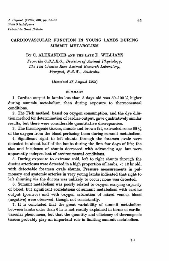
J. Phy8iol. (1970), 208, pp. 65-83 65With 5 text-flguresPrinted in Great Britain
CARDIOVASCULAR FUNCTION IN YOUNG LAMBS DURINGSUMMIT METABOLISM
By G. ALEXANDER AND THE LATE D. WILLIAMSFrom the C.S.I.R.O., Division of Animal Physiology,The Ian Clunies Ross Animal Research Laboratory,
Prospect, N.S.W., Australia
(Received 28 August 1969)
SUIMMARY
1. Cardiac output in lambs less than 3 days old was 50-100% higherduring summit metabolism than during exposure to thermoneutralconditions.
2. The Fick method, based on oxygen consumption, and the dye dilu-tion method for determination of cardiac output, gave qualitatively similarresults, but there were considerable quantitative discrepancies.
3. The thermogenic tissues, muscle and brown fat, extracted some 80%of the oxygen from the blood perfusing them during summit metabolism.
4. Significant right to left shunts through the foramen ovale weredetected in about half of the lambs during the first few days of life; thesize and incidence of shunts decreased with advancing age but wereapparently independent of environmental conditions.
5. During exposure to extreme cold, left to right shunts through theductus arteriosus were detected in a high proportion of lambs, < 12 hr old,with detectable foramen ovale shunts. Pressure measurements in pul-monary and systemic arteries in very young lambs indicated that right toleft shunting via the ductus was unlikely to occur; none was detected.
6. Summit metabolism was poorly related to oxygen carrying capacityof blood, but significant correlations of summit metabolism with cardiacoutput (positive) and with oxygen saturation of mixed venous blood(negative) were observed, though not consistently.
7. It is concluded that the great variability of summit metabolismbetween lambs older than 6 hr is not readily explained in terms of cardio-vascular phenomena, but that the quantity and efficiency of thermogenictissues probably play an important role in limiting summit metabolism.
3-2
66 G. ALEXANDER AND THE LATE D. WILLIAMS
INTRODUCTION
At birth, sheep (Ovis aries L.) have well developed mechanisms forgenerating heat. In the lamb exposed to cold within minutes of birth, heatproduction (oxygen consumption) can increase to several times the basalrate, by both shivering and non-shivering mechanisms, but the increase isvery variable, ranging from 2- to 5 times the basal metabolic rate of about11. oxygen/kg.hr (Alexander, 1962a; Alexander & Williams, 1968).Reasons for this wide variability in the maximum metabolic response tocold (sUmmit metabolism) are unknown and do not appear to be associatedwith birth weight, pre-natal nutritional history of the ewe or with littersize (Alexander, 1962 a). Also, there is no information to define the physio-logical or biochemical processes which are limiting factors in the intensedemands that are made upon the lamb during summit metabolism. Itseemed reasonable, therefore, to investigate whether the variability insummit metabolism could be explained in terms of cardiovascular func-tion. Considerably more is known about cardiovascular function duringintense exercise than during exposure to extreme cold, both of which resultin considerable metabolic effort (see review by Horvath & Howell, 1964).Very little is known about the effects of severe cold on the circulation ofthe new-born animal, although there are some data on cardiac output inthe rat (Thompson & Moore, 1968).
This paper presents a study of cardiovascular factors that might beexpected to limit summit metabolism in young lambs. The results sug-gested that other factors play the major limiting role.
METHODS
Source of data. The present paper is confined to the data obtained during the initialcontrol periods of three series of experiments on Merino lambs. The purpose andresults of two series are presented elsewhere (Alexander & Williams, 1970).
Animals. Ninety Merino lambs and sixteen Merino x Suffolk x Border Leicesterlambs, from well nourished ewes, were used. Many lambs were born indoors and allwere brought under cover within 12 hr of birth; none had been exposed to inclementweather before study. The time of birth was known to within approximately 6 hr.
Apparatus and procedure&. The closed circuit respiration apparatus and relatedprocedures for the mea8urement of 8ummit oxygen consumption have been describedin detail elsewhere (Alexander, 1961 a; Alexander & Williams, 1968). Wool wasclipped from the lambs to facilitate heat loss and a thermocouple was inserted 5 cminto the lamb's rectum. Catheters were inserted as described below, and in mostexperiments safety-pin electrodes were fastened to the skin for electrocardiographicand electromyographic recording. Each animal, supported in a standing position bya sling of cord netting, was placed in the chamber at an air temperature within therange -5 to - 150 C. After connexion of the electrical leads and placement ofcatheters through airtight glands, the lid was closed and the wind speed in the
CARDIOVASCULAR FUNCTION IN LAMBS 67
chamber was adjusted until rectal temperature began to fall at about 0 03-0.050 C/min. Oxygen consumption (summit metabolism) was then measured during I hr(Series 3) or 1 hr (Series 1 and 2) comprising consecutive periods of 10 min (Series 1)or 15 min (Series 2). Oxygen content of the air in the chamber remained at 20-21 %.A similar procedure was followed for the measurement of oxygen consumption underthermoneutral conditions, but the lamb was not clipped, the chamber temperature was29-30° C and air movement in the chamber was minimal.
Before the chamber was closed in Series 2 and 3, the lamb was heparinized (5 mg/kg i.v.) to facilitate handling of blood for dye dilution studies; heparin was withouteffect on summit metabolism in control experiments (unpublished observations).
Catheterization. Blood vessels were catheterized for blood sampling, injection ofindicator dye and drug infusion. Polyvinyl chloride catheters (Dural Plastics, Dural,N.S.W.) of i.d. 0-8 mm, o.d. 1P27 mm or occasionally of i.d. 1.0 mm, o.d. 1-5 mm wereused.
All catheters were inserted under local anaesthesia using diethylamino-dimethyl-acetanilide (Xylotox, Pharmaceutical Manufacturing Co. England). Catheters in thejugular vein and carotid artery were inserted by a modification of the method ofHerd & Barger (1964), and were cemented into position with isobutyl-2-cyano-acrylate (Ethicon, tissue adhesive, Ethnor Pty. Ltd). Catheters in the posteriorvena cava and aorta or femoral artery were introduced via the recurrent tarsal veinand saphenous artery. Carotid catheters were introduced via a thyroidal artery insome lambs of Series 1. Catheters were jacketed in electrically heated sheaths toprevent freezing.
In Series 1 catheters were placed so that blood was sampled from the posterior endof the posterior vena cava, from the femoral or posterior end of the aorta, as well asfrom the carotid artery in some animals.
In Series 2 the sampling site of the catheter introduced via the recurrent tarsalvein varied from caudal to the renal inflow to within the heart, but was usually atabout the level of the diaphragm and above the hepatic vein. Catheters in thejugular vein were inserted until the orifice lay about 2 cm above the heart, or, inabout half of the experiments, the catheter was successfully passed into the pul-monary artery with the orifice usually distal to the ductus arteriosus. Catheters inthe carotid artery were usually passed into the left ventricle. Successful placement ofarterial catheters was indicated by the blood pressure trace, and in many animalscatheter positions were checked at post mortem examination.
In Series 3, catheters were placed in the posterior vena cava, the femoral arteryor posterior end of the aorta and in the jugular vein as in Series 2.
Indicator dye dilution curves. Dye dilution curves were obtained during the latterhalf of each period of measurement of oxygen consumption, and the curves were usedfor the estimation of cardiac output and the size of shunts through the foramen ovale;procedures similar to that of Stahlman, Merrill & Le Quire (1962) were used.A known amount (0.51- mg) of indocyanine green dye (Cardiogreen, Hynson,
Westcott & Dunning Inc. Baltimore, U.S.A.) in 0-20 ml. distilled water was intro-duced into the jugular, posterior vena cava or carotid catheter, and sufficient saline(0.9 g sodium chloride per 100 ml. water) was injected to bring the dye solution towithin 0-3 ml. of the catheter orifice; 1.1 ml. saline was then injected into thecatheter over 1 sec to deliver the dye into the blood stream. Insignificant tracesof dye remained in the catheter after this procedure. Immediately before injectionof dye, withdrawal of blood from the femoral artery, through a densitometer cuvette(X-250, The Waters Co., Minn., U.S.A.) was commenced. Withdrawal was effectedby a Harvard pump and steady flows of approximately 20 ml./min could usually beachieved, although in a few lambs it was necessary to reduce withdrawal rates to 8,and very occasionally to 4 ml./min, to achieve a steady flow. The 'dye dilution curve'
68 G. ALEXANDER AND THE LATE D. WILLIAMSwas traced on a potentiometric recorder, connected to the cuvette through its asso-ciated control unit, the output of which was proportional to the concentration ofdye in the blood passing through the cuvette.The 5-10 ml. blood withdrawn during the completion of each curve was immedi-
ately returned to the lamb, and the whole process was then repeated, using the sameor another injection site. There was no evidence of haemolysis in plasma samplescollected after 12 or more injections in any of the lambs. During each i hr period ofmeasurement of oxygen consumption, curves were usually obtained in sets of twoor three, using injection into the posterior vena cava and jugular vein and sometimesinto the carotid artery. Replicate curves were sometimes obtained for one or moreinjection sites. The cuvette was flushed through with detergent (Pyroneg, Diversey,Sydney, Australia) and rinsed with saline after each set of injections.A similar procedure was used for the detection of left to right shunts through the
ductus arteriosus; dye was injected into the left ventricle and blood was withdrawnfrom the pulmonary artery at the rate of 12, or occasionally at 6 ml./min.At the start of each experiment a blood sample was withdrawn and divided into
two parts. The electrical output of the cuvette was then calibrated by the use of aknown solution of dye in one subsample of blood. The other subsample was used tozero the instrument for calibration.
Cardiac output was calculated by conventional methods from the area under thedye dilution curve, using a semi-logarithmic plot and straight line extrapolation (forexample see Stahlman et al. 1962). Right to left shunts were demonstrated by anearly appearance time (Fig. 2) and two peaks in the curve. The size of the shuntrelative to the total blood flow past the point of exit of the shunt was estimated byassuming that the extrapolated semi-logarithmic disappearance slope for the dyepassing through the shunt was parallel to that for the main disappearance phase ofthe curve, estimated as above; the area under the shunt curve was then expressed asa percentage of the total area (see Stahiman et al. 1962).
Left to right shunts were demonstrated by the early appearance of dye, in pul-monary artery blood (Fig. 2) superimposed on the normal appearance curve afterinjection into the left ventricle. The area under the curve, due to the shunt, wasestimated by superimposing the characteristic normal re-appearance curve, freehand,and the area was then expressed as a percentage of the mean area of the curvesobtained by femoral sampling during the same experimental period, and after in-jection of the same amount of dye.
E8timation of cardiac output by the Fick method. The mean cardiac output, duringi hr periods of measurement of metabolic rate, was estimated by dividing the rate ofoxygen consumption by the product of the oxygen capacity and the differencebetween the oxygen saturation percentage of arterial and mixed venous blood.Oxygen capacity and oxygen saturation were determined from single blood samplesdrawn during the first half of the relevant i hr period.
Blood 8ampling and analyticil procedures. Blood was collected anaerobically, viathe various catheters during the first half of each period of measurement of oxygenconsumption; samples were stored at 40 C until analysed at the end ofthe experiment.Arterial and venous oxygen eaturativno were determined by the spectrophotometricmethod of Verel, Saynor & Kesteven (1960), calibrated against the manometricmethod of Peters & Van Slyke (1932). Haemoglobin was measured as oxyhaemoglobinbyspectrophotometric absorption at 540m,u (Dacie,1956). Oxygen capacity (ml./l00 ml.blood) was calculated from the haemoglobin percentage by conventional methods.
Analysi8 of resuls. Mean values of the various parameters measured during the+ hr period of Series 1 and 2, and the j hr period of Series 3, were calculated for eachanimal, and these means were used in conventional statistical analyses. Data fromthe first i hr period of Series 2 pooled with that from Series 3, were also examined.
CARDIOVASCULAR FUNCTION IN LAMBS
RESULTS
Arteriovenous oxygen saturation differencesThe mean oxygen saturation in the blood in the descending aorta ranged
from 90 to 98% in the various groups of Series 1, 2 and 3 (Table 1); therewas no clear difference between lambs exposed to severe cold and lambsunder thermoneutrality (Series 3).
In determination of arteriovenous (a-v) oxygen saturation differences,it was assumed that blood in the right ventricle or pulmonary artery repre-sented mixed venous blood. In lambs from which right heart blood wasnot obtained, oxygen saturation of mixed venous blood was estimated asthe average saturation of blood in the posterior vena cava and anteriorvena cava (or jugular vein). These saturations were usually similar and ofthe same magnitude as in right heart blood from other lambs; in nineexperiments of Series 2 and seven experiments of Series 3, mean saturation( ± S.E. of mean) of posterior vena caval blood during summit metabolismwas 7 ± 3% and 6 ± 2% lower than saturation of the anterior vena cavalsample. Differences tended to be greater when posterior caval blood wasdrawn below the renal veins; saturations as low as 12% were sometimesencountered in such samples and the low venous saturations recorded inSeries 1 (Table 1) are no doubt reflexions of the sampling site.In young lambs under thermoneutral conditions (Series 3) the mean
saturation of 'mixed venous blood' was 70 %, and the mean a-v oxygensaturation difference was 28% (Table 1). However, during summit meta-bolism the mean percentage saturation ofmixed venous blood in the lambsless than 3 days old in Series 2 and 3 (Table 1) was 31 and 37 and mean a-vdifference was 60 and 58 respectively.The arterial and venous saturations and a-v differences appeared to be
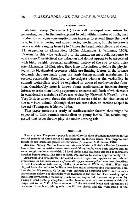
independent of age (Fig. 1, for example).The mixed venous oxygen saturation was significantly and negatively
correlated with summit metabolism in the young lambs in Series 2 and inSeries 2 and 3 pooled (Tables 3 and Fig. 1) but not in Series 3. There was noindication of such a relationship in the older lambs of Series 2 (Fig. 1).
Cardiac output
(i) Comparison of methods. Although changes in cardiac output followedthe same trends (see below) when determined by the Fick method or bydye dilution, numerical agreement between the two methods was not good.In Series 3 when data from the twenty-one experiments on cold exposureand exposure to thermoneutral conditions were pooled, the coefficient ofcorrelation between the values obtained by the two methods was 0-78(P < 0.001). However, in Series 2 in which the range of values of cardiac
69
70 G. ALEXANDER ANTD THE LATE D. WILLIAMS
output was more restricted, due to inclusion of cold conditions only, thecoefficient was a mere 0-24 (twenty-three experiments). In lambs less than12 hr old, the Fick method tended to give higher values than the dye dilu-tion method, whereas in lambs 12-60 hr old the direction of the differenceswas more random; in Series 2 the mean difference + s.E. of mean (Fickminus dye) of 12 + 2-4 ml./100 g . min in the younger group was significantlyhigher than the mean of - 1-2 + 3-7 in the 12-60 hr old lambs (P < 0-01 int test). Similar results were obtained from Series 2 and 3, pooled. In lambs
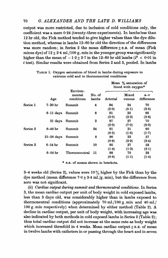
TABLE 1. Oxygen saturation of blood in lambs during exposure toextreme cold and to thermoneutral conditions
Mean % saturation ofblood with oxygen*
Environ-mental No. of Mixed a-v
Age conditions lambs Arterial venous difference
Series 1 7-30 hr Summit 4 94 24 70(2.0) (5.1) (3.5)
6-11 days Summit 8 91 25 66(3.0) (3.3) (2*8)
21 days Summit 2 97 27 70(2.0) (7.0) (8 0)
Series 2 6-48 hr Summit 24 91 31 60(0.5) (1-6) (1.7)
21-28 days Summit 8 90 33 57(0.9) (3.9) (3.4)
Series 3 6-54 hr Summit 10 95 37 58(1.4) (1.3) (2.1)
6-54 hr Thermoneutral 11 98 70 28(0-8) (1 1) (1.4)
* s.E. of means shown in brackets.
3-4 weeks old (Series 2), values were 10% higher by the Fick than by thedye method (mean difference 7 0 + 3 4 ml./g . min), but the difference fromzero was not significant.
(ii) Cardiac output during summit and thermoneutral conditions. In Series3, the mean cardiac output per unit of body weight in cold exposed lambs,less than 3 days old, was considerably higher than in lambs exposed tothermoneutral conditions (approximately 70 ml./100 g. min and 40 ml./100 g. min respectively) when determined by either method (Table 2). Adecline in cardiac output, per unit of body weight, with increasing age wasalso indicated by both methods in cold exposed lambs in Series 2 (Table 2);thus total cardiac output did not increase at the same rate as body weightwhich increased threefold in 4 weeks. Mean cardiac output + S.E. of meanin twelve lambs with catheters in or passing through the heart and in seven
CARDIOVASCULAR FUNCTION IN LAMBSlambs without cardiac catheters was 75 + 2*0 and 74 + 3*1 ml./100 g.minrespectively, by the Fick method and 63 + 2-2 and 68 + 5 0 by the dyemethod (Series 2). Thus there was no obvious effect of cardiac catheteri-zation on cardiac output, although blood pressure fell abruptly when extra-systoles occurred in most lambs with cardiac catheters (Fig. 5).
There was no apparent relation between summit metabolism and cardiacoutput, measured by either method, in the data from Series 2 (Fig. 1), norin Series 2 and 3 combined, although in Series 3 the coefficient of correla-tion was significant when the dye method was used, and approached signi-ficance when the Fick method was used (Fig. 1).
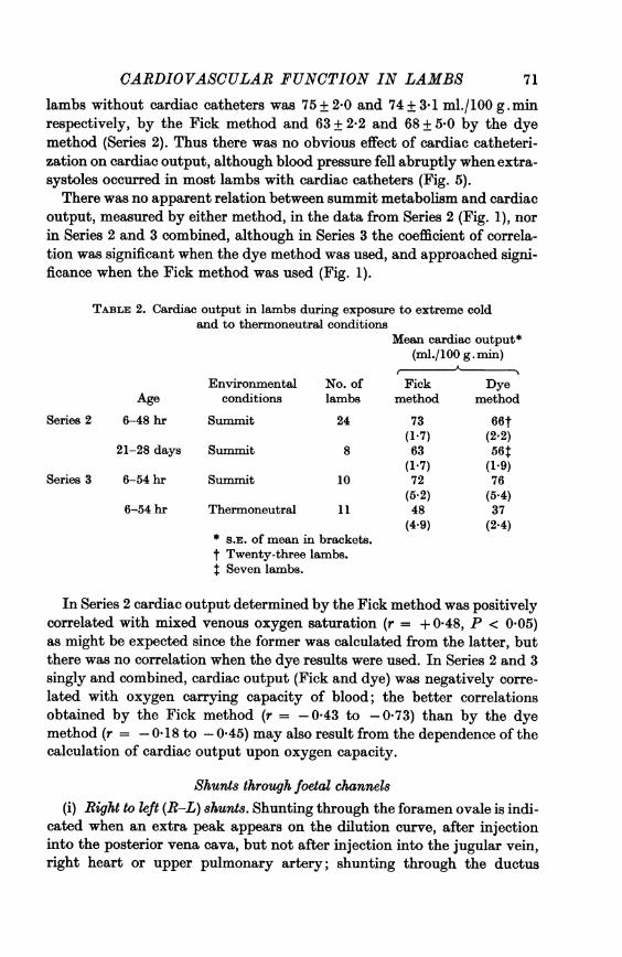
TABLE 2. Cardiac output in lambs during exposure to extreme coldand to thermoneutral conditions
Mean cardiac output*(ml./100 g.min)
A-
Environmental No. of Fick DyeAge conditions lambs method method
Series 2 6-48 hr Summit 24 73 66t(1.7) (2.2)
21-28 days Summit 8 63 56t(1.7) (1.9)
Series 3 6-54 hr Summit 10 72 76(5.2) (5.4)
6-54 hr Thermoneutral 11 48 37(4.9) (2.4)
* S.E. of mean in brackets.t Twenty-three lambs.t Seven lambs.
In Series 2 cardiac output determined by the Fick method was positivelycorrelated with mixed venous oxygen saturation (r = + 0-48, P < 0.05)as might be expected since the former was calculated from the latter, butthere was no correlation when the dye results were used. In Series 2 and 3singly and combined, cardiac output (Fick and dye) was negatively corre-lated with oxygen carrying capacity of blood; the better correlationsobtained by the Fick method (r = - 0 43 to - 0.73) than by the dyemethod (r = -0-18 to -0.45) may also result from the dependence of thecalculation of cardiac output upon oxygen capacity.
Shunts through foetal channels(i) Right to left (R-L) shunts. Shunting through the foramen ovale is indi-
cated when an extra peak appears on the dilution curve, after injectioninto the posterior vena cava, but not after injection into the jugular vein,right heart or upper pulmonary artery; shunting through the ductus
71
72 G. ALEXANDER AND THE LATE D. WILLIAMSarteriosus is indicated if extra peaks appear after both posterior vena cavaland jugular vein injections.Dye was injected into the posterior vena cava in thirty-four lambs, less
than 4 days old, during exposure to summit conditions (twenty-one lambsfrom Series 2, eight from Series 3, and five miscellaneous lambs). Evidenceof a significant R-L shunt (3-63 %, but usually < 25% of the blood flowin the posterior vena cava) was obtained in fifteen of the thirty-four; verysmall shunts (< 3 %) were detected in another nine (examples in Fig. 2).
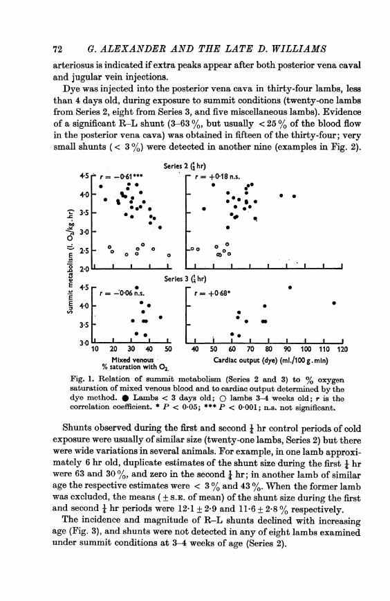
Series 2 (; hr)+5 r -0.61** r= +018 n.s.
0 0g0-* .00.
0e 0~~~~~~T' 3-5 _*bO
3-0~~~~~~~~~O 30 _00- 00 0 0 000E ~~~0 0 00
Series 3 G hr)EE 4-50r =-006n.s.
40_*0* _+ 8*:0
3.50
3010 20 30 40 50 40 50 60 70 80 90 100 110 120
Mixed venous Cardiac output (dye) (ml./100 g. min)% saturation with 02.
Fig. 1. Relation of summit metabolism (Series 2 and 3) to % oxygensaturation of mixed venous blood and to cardiac output determined by thedye method. * Lambs < 3 days old; 0 lambs 3-4 weeks old; r is thecorrelation coefficient. * P < 005; *** P < 0 001; n.s. not significant.
Shunts observed during the first and second j hr control periods of coldexposure were usually of similar size (twenty-one lambs, Series 2) but therewere wide variations in several animals. For example, in one lamb approxi-mately 6 hr old, duplicate estimates of the shunt size during the first i hrwere 63 and 30 %, and zero in the second i hr; in another lamb of similarage the respective estimates were < 3% and 43 %. When the former lambwas excluded, the means ( + S.E. of mean) of the shunt size during the firstand second I hr periods were 12-1 + 2-9 and 11-6 + 2-8% respectively.The incidence and magnitude of R-L shunts declined with increasing
age (Fig. 3), and shunts were not detected in any of eight lambs examinedunder summit conditions at 3-4 weeks of age (Series 2).
CARDIOVASCULAR FUNCTION IN LAMBS
bo
E
II
C,
IUI
6 hr old-flow 20 mt/mln
0 5 10 15
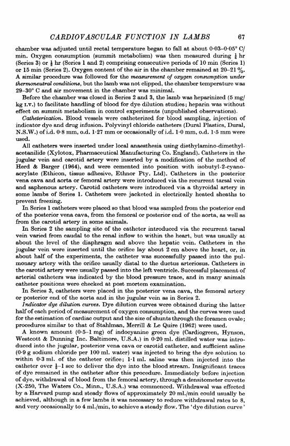
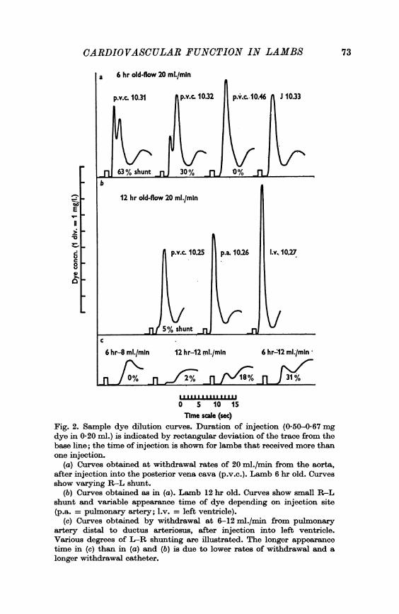
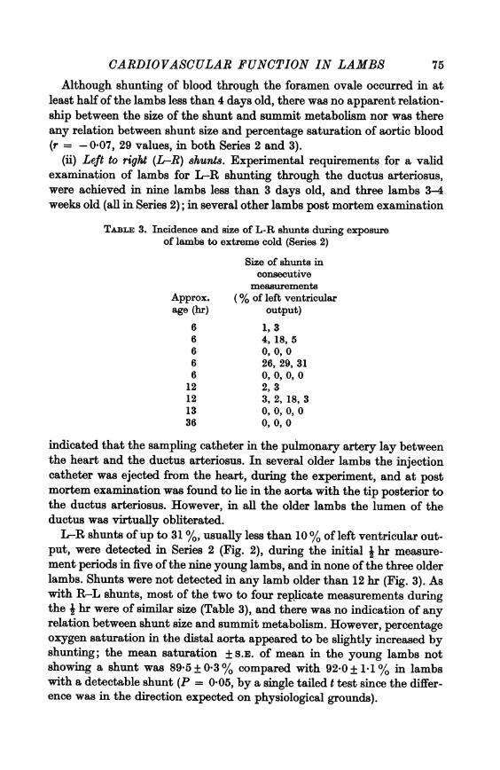
Time scale (sec)Fig. 2. Sample dye dilution curves. Duration of injection (0.50-0.67 mgdye in 0-20 ml.) is indicated by rectangular deviation of the trace from thebase line; the time of injection is shown for lambs that received more thanone injection.
(a) Curves obtained at withdrawal rates of 20 ml./min from the aorta,after injection into the posterior vena cava (p.v.c.). Lamb 6 hr old. Curvesshow varying R-L shunt.
(b) Curves obtained as in (a). Lamb 12 hr old. Curves show small R-Lshunt and variable appearance time of dye depending on injection site(p.a. = pulmonary artery; l.v. = left ventricle).
(c) Curves obtained by withdrawal at 6-12 ml./min from pulmonaryartery distal to ductus arteriosus, after injection into left ventricle.Various degrees of L-R shunting are illustrated. The longer appearance
time in (c) than in (a) and (b) is due to lower rates of withdrawal and a
longer withdrawal catheter.
73
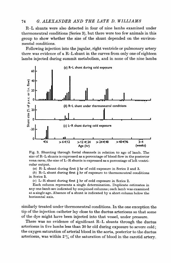
74 G. ALEXANDER AND THE LATE D. WILLIAMSR-L shunts were also detected in four of nine lambs examined under
thermoneutral conditions (Series 3), but there were too few animals in thisgroup to show whether the size of the shunt depended on the environ-mental conditions.
Following injection into the jugular, right ventricle or pulmonary arterythere was evidence of a R-L shunt in the curves from only one of eighteenlambs injected during summit metabolism, and in none of the nine lambs
(a) R-L shunt during cold exposure
40o
' 20o
%0-
0
-Z 204N
W O-
.20 -
0W
(b) R-L shunt under thermoneutral conditions
a ..,.I I I I
(c) L-R shunt during cold exposure
I 1Ii3 I
>6<12 >12<24 >24<48 >48<96 3-4-Age (hr) (weeks)
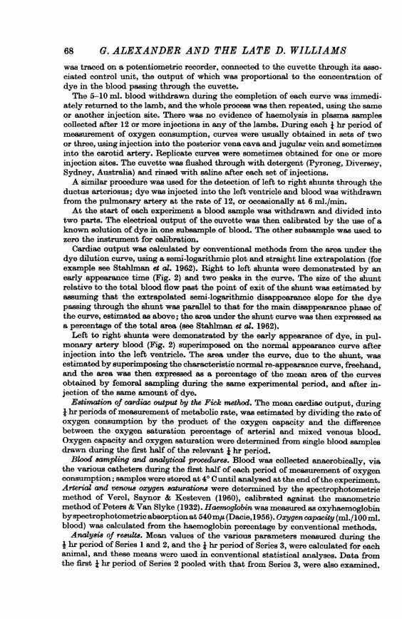
Fig. 3. Shunting through foetal channels in relation to age of lamb. Thesize of R-L shunts is expressed as a percentage of blood flow in the posteriorvena cava; the size of L-R shunts is expressed as a percentage of left ventri-cular output.
(a) R-L shunt during first i hr of cold exposure in Series 2 and 3.(b) R L shunt during first i hr of exposure to thermoneutral conditions
in Series 3.(c) L-R shunt during first X hr of cold exposure in Series 2.Each colunm represents a single determination. Duplicate estimates in
any one lamb are indicated by conjoined columns; each lamb was examinedat a single age. Absence of a shunt is indicated by a short column below thehorizontal axis.
similarly treated under thermoneutral conditions. In the one exception thetip of the injection catheter lay close to the ductus arteriosus so that someof the dye might have been injected into that vessel, under pressure.
There was no evidence of significant R-L shunts through the ductusarteriosus in five lambs less than 30 hr old during exposure to severe cold;the oxygen saturation of arterial blood in the aorta, posterior to the ductusarteriosus, was within 2 % of the saturation of blood in the carotid artery.
CARDIOVASCULAR FUNCTION IN LAMBSAlthough shunting of blood through the foramen ovale occurred in at
least half of the lambs less than 4 days old, there was no apparent relation-ship between the size of the shunt and summit metabolism nor was thereany relation between shunt size and percentage saturation of aortic blood(r = - 0*07, 29 values, in both Series 2 and 3).
(ii) Left to right (L-R) shunt8. Experimental requirements for a validexamination of lambs for L-R shunting through the ductus arteriosus,were achieved in nine lambs less than 3 days old, and three lambs 3-4weeks old (all in Series 2); in several other lambs post mortem examination
TABLE 3. Incidence and size of L-R shunts during exposureof lambs to extreme cold (Series 2)
Size of shunts inconsecutive
measurementsApprox. (% of left ventricularage (hr) output)
6 1,36 4,18,56 0,0,06 26, 29, 316 0,0,0,0
12 2, 312 3, 2, 18, 313 0, 0, 0, 036 0, 0, 0
indicated that the sampling catheter in the pulmonary artery lay betweenthe heart and the ductus arteriosus. In several older lambs the injectioncatheter was ejected from the heart, during the experiment, and at postmortem examination was found to lie in the aorta with the tip posterior tothe ductus arteriosus. However, in all the older lambs the lumen of theductus was virtually obliterated.L-R shunts of up to 31 %, usually less than 10% of left ventricular out-
put, were detected in Series 2 (Fig. 2), during the initial i hr measure-ment periods in five of the nine young lambs, and in none ofthe three olderlambs. Shunts were not detected in any lamb older than 12 hr (Fig. 3). Aswith R-L shunts, most of the two to four replicate measurements duringthe i hr were of similar size (Table 3), and there was no indication of anyrelation between shunt size and summit metabolism. However, percentageoxygen saturation in the distal aorta appeared to be slightly increased byshunting; the mean saturation + s.E. of mean in the young lambs notshowing a shunt was 89*5 + 0*3% compared with 92.0 + 1*1 % in lambswith a detectable shunt (P = 0 05, by a single tailed t test since the differ-ence was in the direction expected on physiological grounds).
75
76 G. ALEXANDER AND THE LATE D. WILLIAMS
Haemoglobin concentrationHaemoglobin concentration in the blood of lambs tended to decrease,
usually by 0.5-1 g per 100 ml. blood (i.e. by about 5 %) during the courseof experiments in Series 2, in which sampling usually entailed a blood lossof 10-15 ml.Haemoglobin concentration (or oxygen capacity) in lambs less than 3
days old was unrelated to summit metabolism, measured at the same timeand was also unrelated to a-v oxygen saturation differences (r = approxi-mately - 0x16 in each set of data).
Heart rateHeart rate was considerably higher during cold exposure (approximately
300/min) than during exposure to thermoneutral conditions (approxi-mately 200/min). Rates of 300/min were observed in lambs less than 3 daysold and in lambs 3-4 weeks old.
There was no apparent relation between heart rate and summit meta-bolism nor was there any significant relation between heart rate andcardiac output measured either by the Fick method or by dye dilution(r ranged from + 0 6 to -0 3 in the three sets).
Blood pressure(i) Systemic pressure. In Series 3, mean blood pressure (diastolic pres-
sure plus one third of pulse pressure) in lambs exposed to summit condi-tions (Table 4) was 8 mm Hg higher than that in lambs under thermo-neutral conditions (P < 0 05 by t test). Blood pressure during summitmetabolism increased with age (Table 4) but there was no apparent relation-ship with age in lambs less than 3 days old (Series 2).Blood pressure was significantly and positively correlated with summit
metabolism in Series 1, but the correlation was significant and negative inSeries 3.
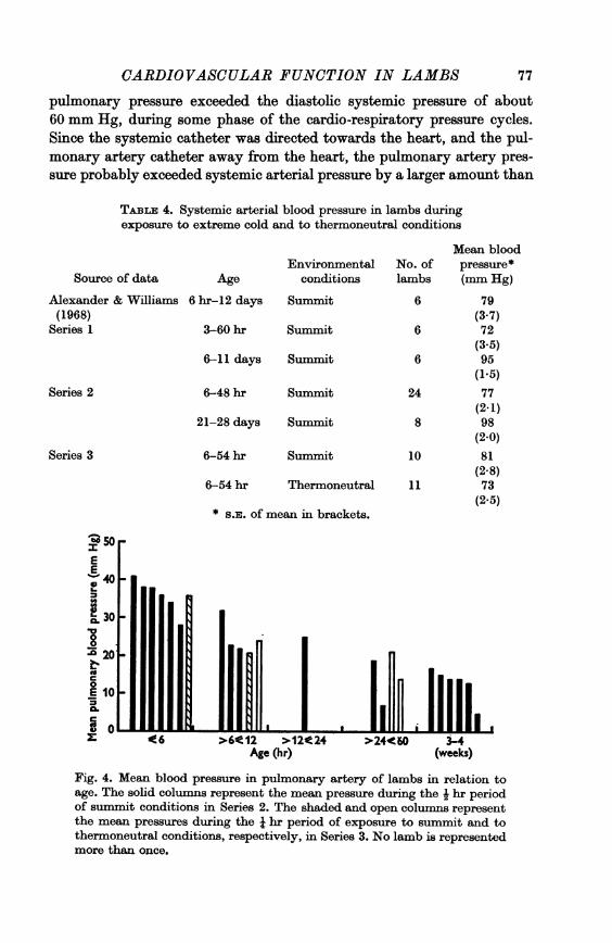
(ii) Pulmonary arterial pressure. In contrast to systemic pressure, themean pulmonary arterial pressure clearly declined with increasing agefrom about 35 mm Hg at about 6 hr of age to about 15 mm Hg before thelambs were 3 weeks old (Fig. 4).
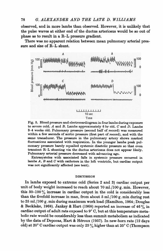
In each of fourteen lambs (twelve in Series 2 and two in Series 3)examined under summit conditions within 3 days of birth, the mean pul-monary pressure was considerably below the mean systemic pressure.However, there were considerable variations in pulmonary arterial pres-sure associated with respiration (Fig. 5) which increased the excursions ofpressure from an average pulse pressure of 29 mm Hg to an average ofabout 50 mm Hg; and in six of twelve animals, 13 hr old or less, the peak
CARDIOVASCULAR FUNCTION IN LAMBS
pulmonary pressure exceeded the diastolic systemic pressure of about60 mm Hg, during some phase of the cardio-respiratory pressure cycles.Since the systemic catheter was directed towards the heart, and the pul-monary artery catheter away from the heart, the pulmonary artery pres-sure probably exceeded systemic arterial pressure by a larger amount than
TABLE 4. Systemic arterial blood pressure in lambs duringexposure to extreme cold and to thermoneutral conditions
Source of data
Alexander & Williams(1968)
Series 1
Age
6 hr-12 days
Environmentalconditions
Summit
3-60 hr Summit
6-11 days Summit
Series 2 6-48 hr Summit
21-28 days Summit
Series 3 6-54 hr Summit
6-54 hr Thermoneutral
* S.E. of mean in brackets.
Mean bloodNo. of pressure*lambs (mm Hg)
6 79(3.7)
6 72(3.5)
6 95(1.5)
24 77(2.1)
8 98(2.0)
10 81(2 8)
11 73(2.5)
00
L
t-l
C.
.0
t.4C0E
CL01
>6e12 >12)24Age (hr)
tLAnI(-4
(weeks)
Fig. 4. Mean blood pressure in pulmonary artery of lambs in relation toage. The solid columns represent the mean pressure during the i hr periodof summit conditions in Series 2. The shaded and open columns representthe mean pressures during the i hr period of exposure to summit and tothermoneutral conditions, respectively, in Series 3. No lamb is representedmore than once.
77
78 G. ALEXANDER AND THE LATE D. WILLIAMS
observed, and in more lambs than observed. However, it is unlikely thatthe pulse waves at either end of the ductus arteriosus would be so out ofphase as to result in a R-L pressure gradient.There was no apparent relation between mean pulmonary arterial pres-
sure and size of R-L shunt.
A 83__ . - A"*L. t I. LkII
la tOoi *|
S O
I lID
0I Mf IiA'=
10 sac
~~~~~~m..
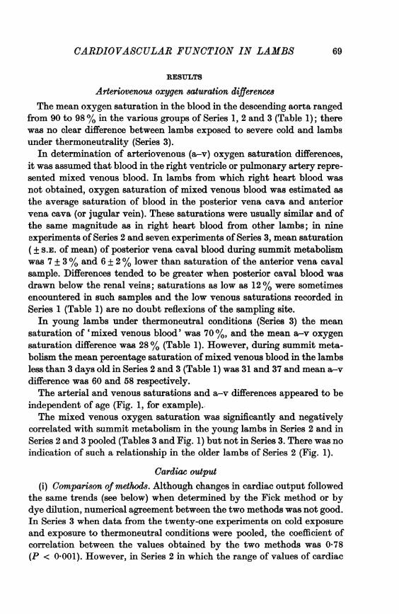
Fig. 5. Blood pressure and electrocardiogram in four lambs during exposureto severe cold. A and B. Lambs approximately 6 hr old. C and D. Lambs3-4 weeks old. Pulmonary pressure (second half of record) was measuredwithin a few seconds of aortic pressure (first part of record), and with thesame transducer. The pressure in the pulmonary artery shows markedfluctuations associated with respiration. In the younger lambs peak pul-monary pressure barely equalled systemic diastolic pressure so that eventransient R-L shunting via the ductus arteriosus does not appear likely.Pulmonary arterial pressure decreased with advancing age.
Extrasystoles with associated falls in systemic pressure occurred inlambs A, B and C with catheters in the left ventricle, but cardiac outputwas not significantly affected (see text).
DISCUSSION
In lambs exposed to extreme cold (Series 2 and 3) cardiac output perunit of body weight increased to reach about 70 ml./100 g . min. However,this 50-100% increase in cardiac output in the cold is considerably lessthan the fivefold increase in man, from about 5 ml./100 g.min during restto 25 ml./100 g . min during maximum work load (Hamilton, 1964; Douglas& Becklake, 1968). Janksy & Hart (1968) reported an increase of 46% incardiac output of adult rats exposed to 9QC, but at this temperature meta-bolic rate would be considerably less than summit metabolism as indicatedby the data of Depocas, Hart & Heroux (1957). In new-born rats (12 daysold) at 200 C cardiac output was only 25% higher than at 350 C (Thompson
LA 16L
wr

CARDIOVASCULAR FUNCTION IN LAMBS
& Moore, 1968), but metabolic rate at 200 C was probably close to summitin rats of this age.
Previous measurements of cardiac output in lambs by Cross, Dawes &Mott (1959) using the Fick method, and by Downing & Rocamora (1968)using the dye method were done with the animals under anaesthesia andin rather ill-defined thermal conditions; the mean values per unit of bodyweight (33 and 20 ml./100 g . min respectively) were somewhat lower thanthose recorded in the present experiments by the Fick and dye methods(58 and 37 ml./100 g . min) in resting conscious lambs under strict thermo-neutrality (Series 3). The higher values (up to 60 ml./100 g. min) in thedata of Cross et al. (1959) may have been due to rates of heat loss that weregreater than those authors suspected; room temperatures were some 100 Cbelow the critical temperature for lambs (Alexander, 1961 b), and if thecoat was still wet, heat loss would have been higher still (Alexander,1962 b).Heart rate in lambs exposed to extreme cold was about 50% higher than
the rate in lambs under thermoneutral conditions (300 and 200/minrespectively), but the effects of cold on stroke volume are not clear. On thebasis of the Fick measurements stroke volume was approximately 2-4 ml./kg under both thermoneutral and cold conditions; but on the basis of thedye method, stroke volume increased from 1-8 ml./kg, under thermo-neutrality, to 2-5 ml./kg during cold exposure (Series 3). An increase instroke volume accompanies strenuous exercise, at least in man (see reviewby Horvath & Howell, 1964).The poor agreement between the Fick and dye methods of estimating
cardiac output (Table 2) may have arisen from several factors. In the pre-sence of a shunt through the foramen ovale, blood in the right ventricle orpulmonary artery would not be truly representative of mixed venous blood;the use of the average saturation of anterior and posterior venous samplesas an estimate of mixed venous saturation may introduce errors; oxygensaturation of blood was not continuously monitored during the periods ofmeasurement of oxygen consumption; and the two methods could not beapplied simultaneously. Differences may also be partly due to patency offoetal shunts; for example, dye rapidly recirculated (L-R) through theductus arteriosus would reduce the slope of the disappearance curve, andlead to over-estimation of the area of the curve, and hence under-estima-tion of the cardiac output, which is in conformity with the results on lambsless than 12 hr old; problems of interpretation of estimations of cardiacoutput in the presence of foetal shunts have been stressed by Cross et al.(1959). A low order repeatability of the dye method (Hanson & Tabakin,1964) may also have contributed to the discrepancies.In addition to the considerable increase in cardiac output due to acute
79
80 G. ALEXANDER AND THE LATE D. WILLIAMScold exposure in new-born lambs, there was a marked increase in thedegree of extraction of oxygen from the blood; the mixed venous oxygensaturation was reduced from about 60 to 30 %. Similar changes have beenseen in new-born rats (Thompson & Moore, 1968) and in exercising men(Saltin, Blomqvist, Mitchell, Johnsson, Wildenthal & Chapman, 1968). Inthe present experiments the venous saturation distal to the renal veins wasusually about 20% and sometimes as low as 12 %, so that the thermogenictissues (muscle and brown adipose tissue) from which this blood largelydrains, were able to extract some 80% of oxygen from the blood flowingthrough them. A similar high extraction of oxygen from brown adiposetissue was reported by Heim & Hull (1966) when new-born rabbits werestimulated by infusion of noradrenaline.Oxygen saturation of arterial blood was not obviously affected by ex-
posure of neonatal lambs to extreme cold; the low mean figure of about93% is in conformity with figures of Cross et al. (1959). Surprisingly, therewas no marked depression in arterial saturation due to shunting via theforamen ovale; however, shunting and arterial saturation were not ex-amined simultaneously. Arterial saturation was slightly elevated by L-Rshunting via the ductus arteriosus; but it is doubtful whether this wouldrepresent a physiological advantage in lambs more than a few hours old(Dawes, Mott & Widdicombe, 1955).Shunts through foetal channels were detected in about half of the lambs
examined under summit conditions; and the combination of a L-R shuntthrough the ductus arteriosus and R-L shunt through the foramen ovale,considered by Cross et al. (1959) to be improbable, was encountered in fiveof the six lambs less than 12 hr old in which the relevant examinationswere made.
Results from the few animals examined under thermoneutral conditionssuggested that there was no major effect of cold exposure on the degree ofshunting via the foramen ovale. The measurements of foramen shuntingmade by Stahlman et al. (1962) in young lambs, apparently near thermalneutrality, were slightly higher than those in the present cold exposedlambs; available data are therefore not consistent with an increase due tocold exposure. Data were not obtained in L-R shunting through the ductusarteriosus under thermoneutral conditions, but L-R shunting appeared tobe unimportant in lambs older than 6 hr (Fig. 3).
Right to left shunts through the ductus arteriosus were not detected inthe present studies, nor in those of Stahlman et al. (1962), although peakpulmonary pressure transiently exceeded diastolic systemic pressure duringpart of the cardio-respiratory cycle in some of the youngest lambs (Fig. 5).The high initial pulmonary artery pressure and the rapid decline withadvancing age (Fig. 4) was also seen in the sheep by Polosa, Dagianti,

CARDIOVASCULAR FUNCTION IN LAMBS
Guiliano & Condorelli (1957), in dog and goat by Rudolph, Auld, Golinko& Paul (1961), in the calf by Reeves & Leathers (1964) and in man byEmmanouilides, Moss, Duffie & Adams (1964); presumably these obser-vations were made under conditions approximating thermoneutrality.The use of correlation coefficients, to indicate factors that restrict the
metabolic response to cold, is of somewhat limited value, for the magnitudeof the correlation coefficient depends largely on the range of values en-countered amongst the factors under examination. Examples are illu-strated in Fig. 1. Nor do correlations distinguish cause from either primaryor secondary effect. However, oxygen-carrying capacity of blood, heartrate and size of R-L shunt appeared to be of little or no importance, whilecardiac output, blood pressure, and mixed venous saturation assumedimportance under some circumstances; errors in the methods of measuringcardiac output, particularly in the presence of shunts, may have maskedthe full degree of dependence of summit metabolism upon this parameter.Interpretation of the correlations with blood pressure is difficult, sincethere were both significant positive and negative correlations with summitmetabolism.The significant negative correlation between summit metabolism and
mixed venous saturation focuses attention on the extraction of oxygenfrom the circulation by the thermogenic tissues. The relationship couldarise from differences between animals in the proportion of muscle andbrown fat in the body, or from differences in the efficiency with which thesetissues extract oxygen from the blood, but data are not available on thesepoints. The low oxygen content of the venous blood draining the thermo-genic tissues suggest that summit metabolism in lambs may be limited tosome extent by the supply of oxygen to the thermogenic tissues; if this isso a correlation between summit metabolism and oxygen-carrying capacityof blood might be expected, but the absence of correlation is at least partlyexplained by the tendency for lambs with low oxygen capacity to havehigh cardiac output. Summit metabolism may, on the other hand, belimited by cardiac output, and the circulation of metabolites other thanoxygen, but the correlations suggest that this dependence is not verystrong. In 12-day-old rats (Thompson & Moore, 1968) both cardiac outputand supply of oxygen to heat producing tissues appear to limit the meta-bolic response to cold, but elucidation of the relative importance of oxygensupply and cardiac output, at least in lambs, must await more specificexperimentation. However, it appears that the major source of individualvariations in summit metabolism does not lie in the circulation; thesevariations seem more likely to reside in the amount of thermogenic tissuepossessed by the individual or in the capacity per unit weight of this tissueto produce heat. It must be emphasized, however, that the present results
81
82 G. ALEXANDER AND THE LATE D. WILLIAMSapply to lambs mostly older than 6 hr; the limiting factors in youngerlambs may well include circulatory phenomena.The technical assistance of Miss L. Younger, Mr R. Edols and Mr M. J. Zweng is
gratefully acknowledged. Cardio-green dye was generously supplied gratis byHynson, Westcott & Dunning Inc., as was the 'Ethicon' tissue adhesive fromEthnor Pty. Ltd. The C.S.I.R.O. Division of Mathematical Statistics provided con-siderable assistance with the statistical analyses.
REFERENCES
ALEXANDER, G. (1961 a). Temperature regulation in the new-born lamb. 2. Aclimatic respiration chamber for the study of thermoregulation, with an appendixon a correction for leaks and imperfect measurement of temperature and pressurein closed circuit respiration chambers. Au8t. J. agric. Res. 12, 1139-1151.
ALEXANDER, G. (1961 b). Temperature regulation in the new-born lamb. 3. Effect ofenvironmental temperature on metabolic rate, body temperatures and respiratoryquotient. Aust. J. agric. Res. 12, 1152-1174.
ALEXANDER, G. (1962a). Temperature regulation in the new-born lamb. 5. Summitmetabolism. Au8t. J. agric. Res. 13, 100-121.
ALEXANDER, G. (1962 b). Temperature regulation in the new-born lamb. 4. The effectof wind and evaporation of water from the coat on metabolic rate and body tem-perature. Aust. J. agric. Res. 13, 82-99.
ALEXANDER, G. & WILLIAMS, D. (1968). Shivering and non-shivering thermogenesisduring summit metabolism in young lambs. J. Physiol. 198, 251-276.
ALEXANDER, G. & WILLIAMS, D. (1970). Sunmmit metabolism and cardiovascularfunction in young lambs during hyperoxia and hypoxia. J. Physiol. 208, 85-97.
CROSS, K. W., DAWES, G. S. & MoTT, JOAN C. (1959). Anoxia, oxygen consumptionand cardiac output in new-born lambs and adult sheep. J. Physiol. 146, 316-343.
DACIE, J. V. (1956). Practical Haematology, 2nd edn. London: Churchill.DAWES, G. S., MOTT, JOAN C. & WmDICOMrBE, J. G. (1955). The patency of the ductus
arteriosus in new-born lambs and its physiological consequences. J. Physiol. 128,361-383.
DEPOCAS, E., HART, J. S. & HEROUX, 0. (1957). Energy metabolism of the white ratafter acclimation to warm and cold environments. J. appl. Phy8iol. 10, 393-397.
DouGLAs, F. G. V. & BECKLAKE, J. R. (1968). Effect of seasonal training on maximalcardiac output. J. appl. Physiol. 25, 600-605.
DOWNING, S. E. & ROCAMORA, J. M. (1968). Cardiovascular responses to hypoxemiaand acidemia in the intact anaesthetized lamb. Yale J. Biol. Med. 40, 296-312.
EMMANOUILIDES, G. C., Moss, A. J., DUFFIE, E. R. & ADAMS, F. H. (1964). Pul-monary arterial pressure changes in human newborn infants from birth to 3 days ofage. J. Pediat. 65, 327-333.
HAMILTON, W. F. (1964). Measurement of cardiac output. In Hanmbook of Physiology,section 2, ed. HAMILTON, W. F., pp. 551-584. Washington, D. C.: AmericanPhysiological Society.
HANSON, J. S. & TABAKIN, B. S. (1964). Simultaneous and rapidly repeated cardiacoutput determinations by dye-dilution method. J. appl. Phy8iol. 19, 275-278.
HEIM, T. & HULL, D. (1966). The blood flow and oxygen consumption of brownadipose tissue in the new-born rabbit. J. Physiol. 186, 42-55.
HERD, J. A. & BARGER, A. C. (1964). Simplified technique for chronic catheteriza-tion of blood vessels. J. appl. Physiol. 19, 791-792.
CARDIOVASCULAR FUNICI'ION IN LAMBSHORVATH, S. M. & HOWELL, C. D. (1964). Organ systems in adaptation: the cardio-
vascular system. In Handbook of Physiology, section 4, ed. DILL, D. B., pp. 153-166.Washington, D.C.: American Physiological Society.
JANSKY. L. & HART, J. S. (1968). Cardiac output and organ blood flow in warm- andcold-acclimated rats exposed to cold. Can. J. Physiol. Pharmac. 46, 653-659.
PETERS, J. P. & VAN SLYKE, D. D. (1932). Quantitative Clinical Chemistry, vol. II.Baltimore: Williams & Wilkins.
POLOSA, C., DAGIANTI, A., GUILIANO, G. & CONDORELLI, M. (1957). Valori tensivi delgrande e del piccolo circolo in agnelli neonati. Boll. Soc. ital. Biol. sper. 33,1593-1596.
REEVES, J. T. & LEATHERS, J. E. (1964). Circulatory changes following birth of thecalf and the effect of hypoxia. Circulation Re8. 15, 343-354.
RUDOLPH, A. M., AULD, P. A. M., GOLnEiO, R. J. & PAUL, M. H. (1961). Pulmonaryvascular adjustments in the neonatal period. Pediatrics, Springfield 28, 28-34.
SATTIN, B., BLOMQVIST, B., MITCHELL, J. H., JOHNSSON, R., WILDENTHAL, K. &CHAPMAN, C. B. (1968). Response to submaximal and maximal exercise afterbed-rest and training. Circulation 38, suppl. 7.
STAHLEMAN, M. T., MERRILL, R. E. & LE QUIRE, V. S. (1962). Cardiovascular adjust-ments in normal newborn lambs. Am. J. Dis. Child. 104, 360-365.
THOMPSON, G. E. & MOORE, R. E. (1968). A study of newborn rats exposed to thecold. Can. J. Physiol. Pharmac. 46, 865-871.
VEREL, D., SAYNOR, R. & KESTEVEN, A. B. (1960). A spectrophotometric method ofestimating blood oxygen using the Univam SP 600. J. clin. Path. 13, 361-363.
83