Embed Size (px)
Citation preview
Skull-2
Norma Basalis Interna
Dr. Heba Kalbouneh
Assistant Professor of Anatomy and Histology
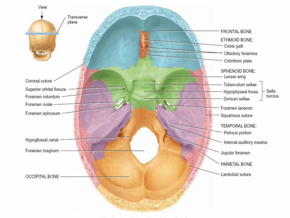
Anterior cranial fossa
Middle cranial fossa
Posterior cranial fossa
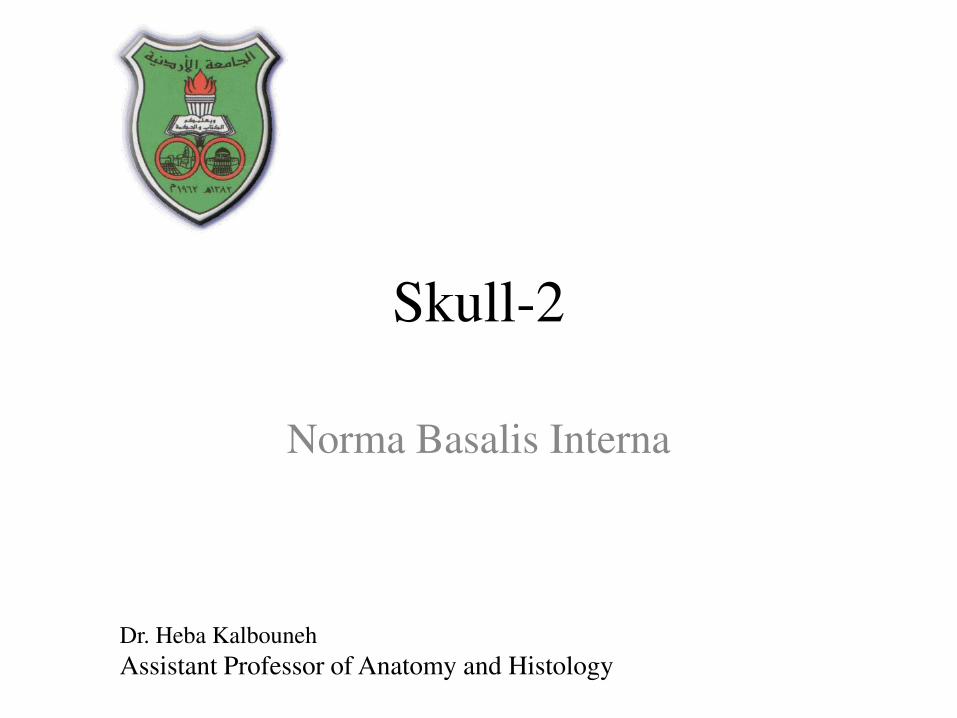
The interior of the base of the skull
is divided into three cranial fossae
Norma basalis interna
Base of the skull- superior view
5
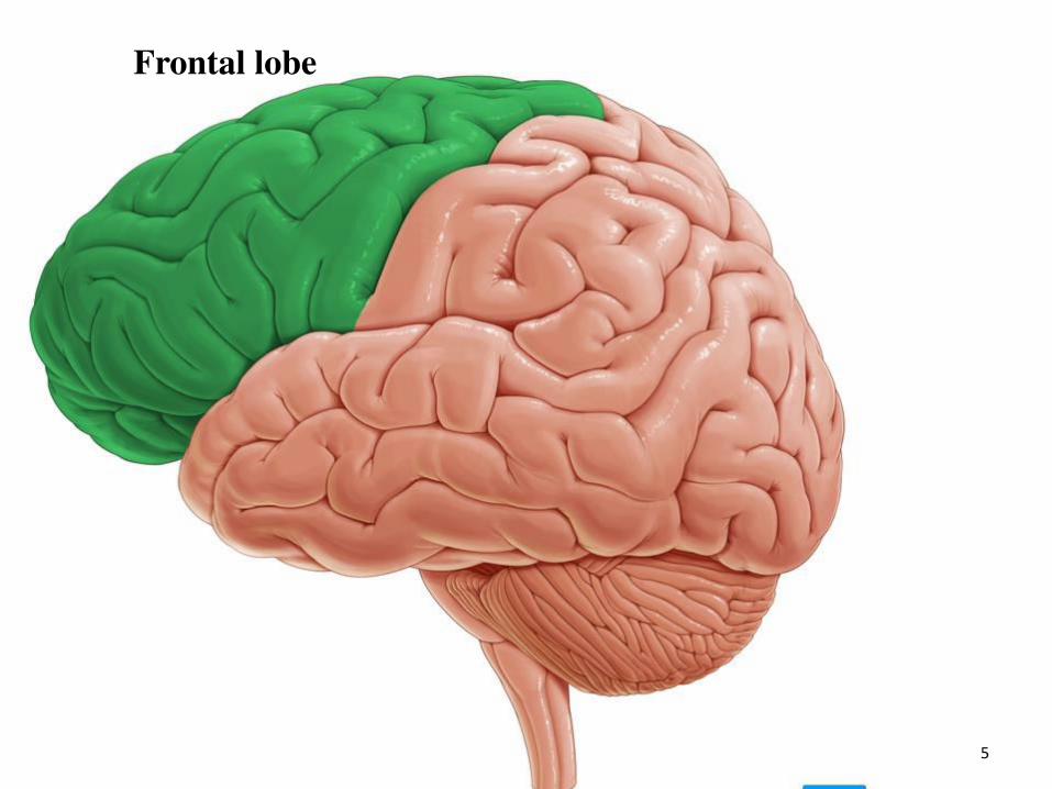
Frontal lobe
6
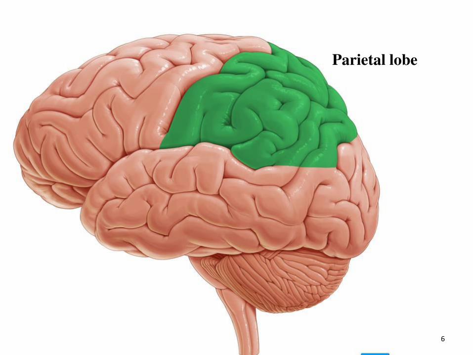
Parietal lobe
7
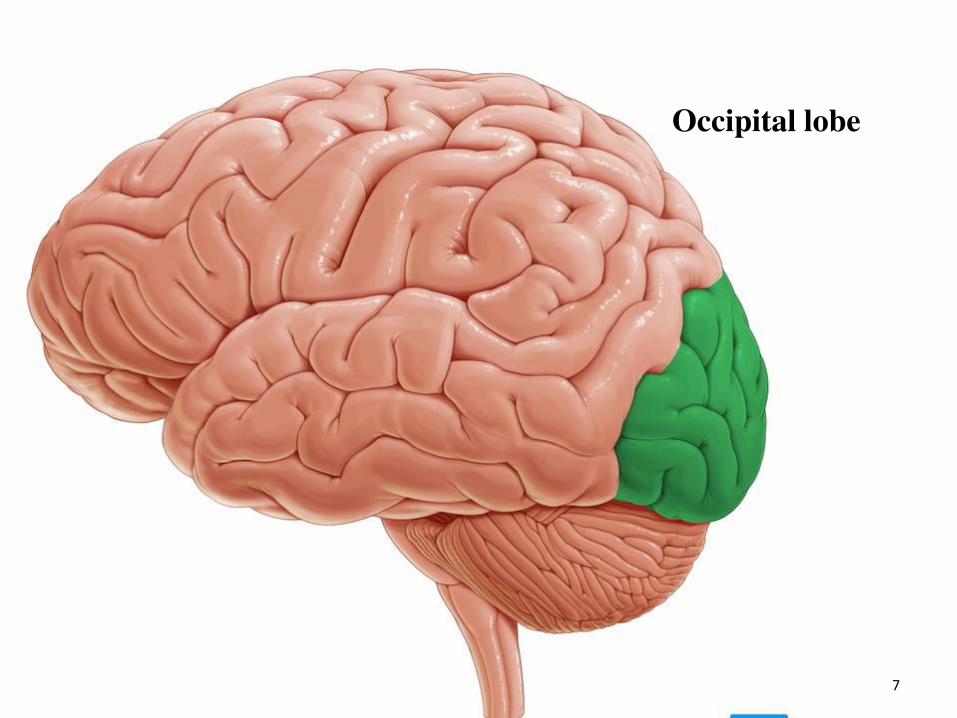
Occipital lobe

8
Temporal lobe
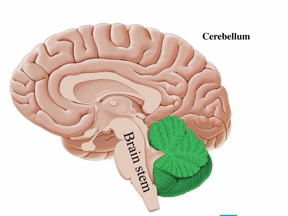
Cerebellum
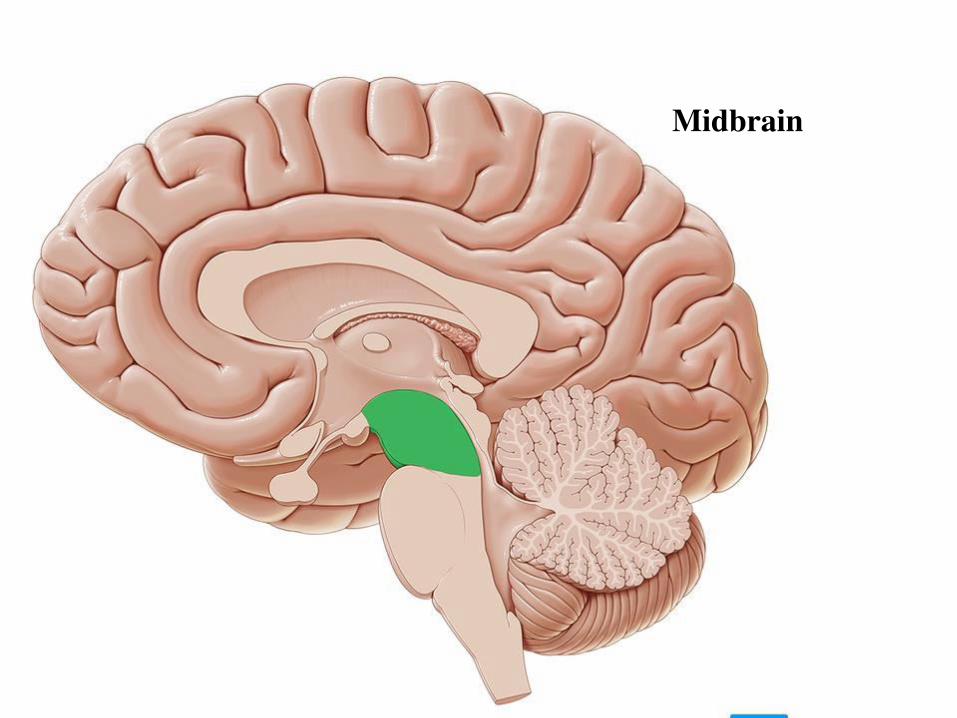
Midbrain
11
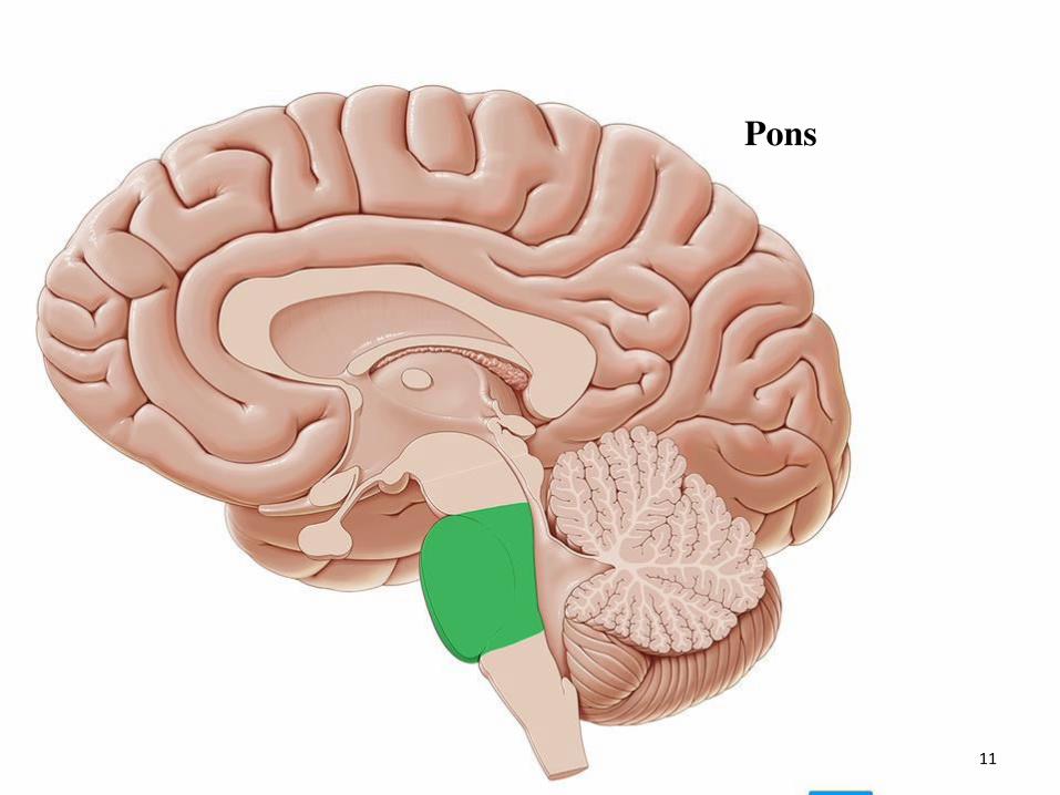
Pons
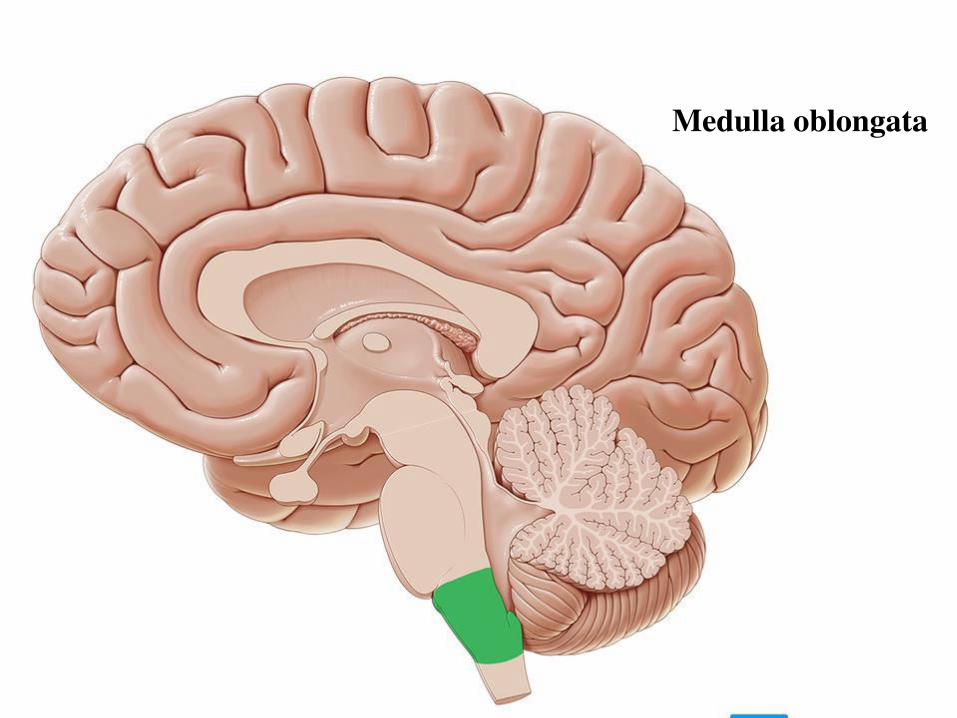
Medulla oblongata
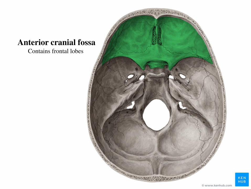
Anterior cranial fossa Contains frontal lobes
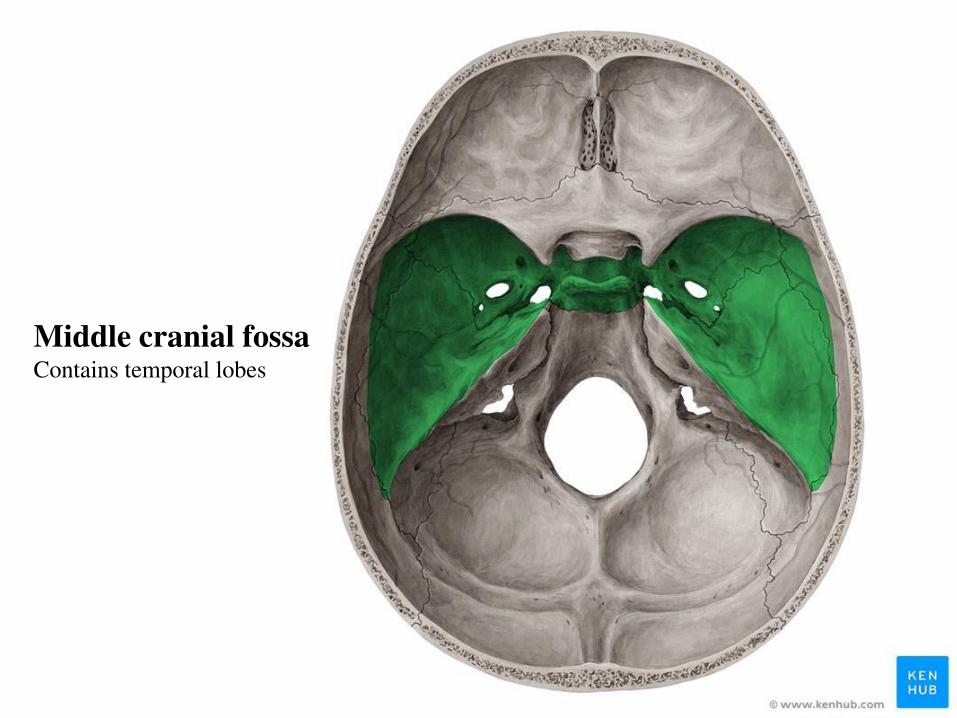
Middle cranial fossa Contains temporal lobes

Posterior cranial fossa Contains the brain stem (midbrain,
pons and medulla oblongata)
and cerebellum
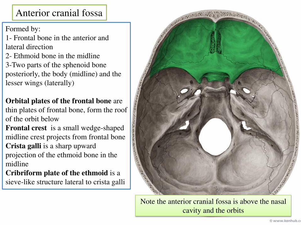
Anterior cranial fossa
Formed by:
1- Frontal bone in the anterior and
lateral direction
2- Ethmoid bone in the midline
3-Two parts of the sphenoid bone
posteriorly, the body (midline) and the
lesser wings (laterally)
Orbital plates of the frontal bone are
thin plates of frontal bone, form the roof
of the orbit below
Frontal crest is a small wedge-shaped
midline crest projects from frontal bone
Crista galli is a sharp upward
projection of the ethmoid bone in the
midline
Cribriform plate of the ethmoid is a
sieve-like structure lateral to crista galli
Note the anterior cranial fossa is above the nasal
cavity and the orbits
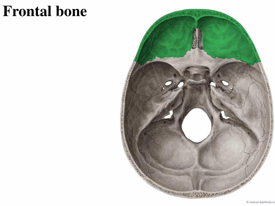
Frontal bone
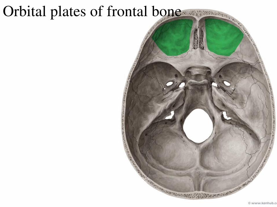
Orbital plates of frontal bone
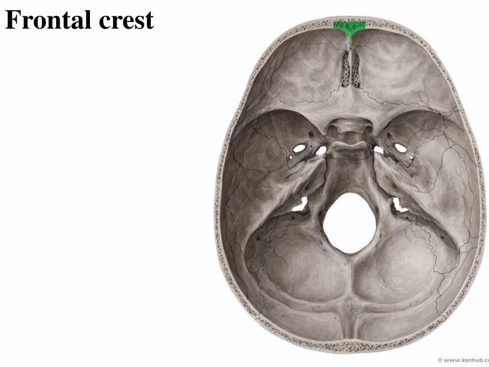
Frontal crest
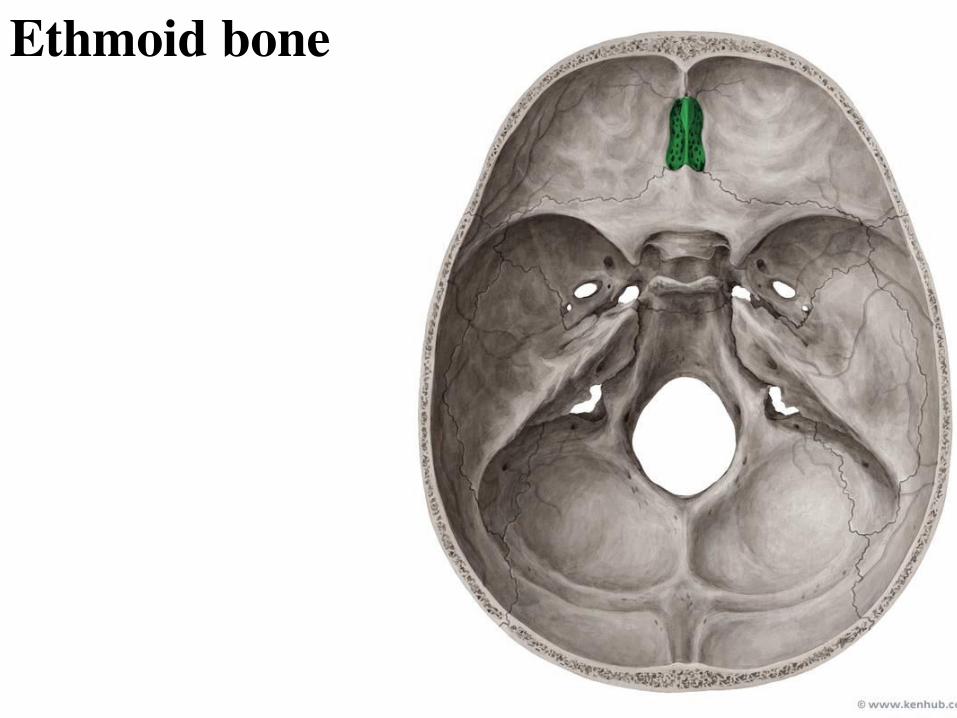
Ethmoid bone
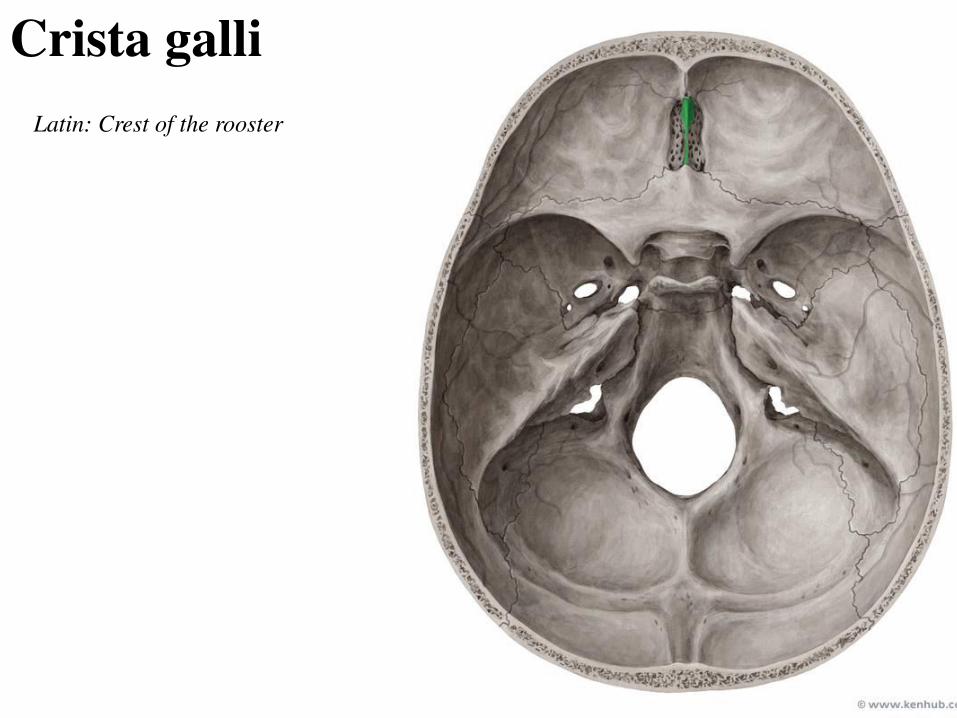
Crista galli
Latin: Crest of the rooster
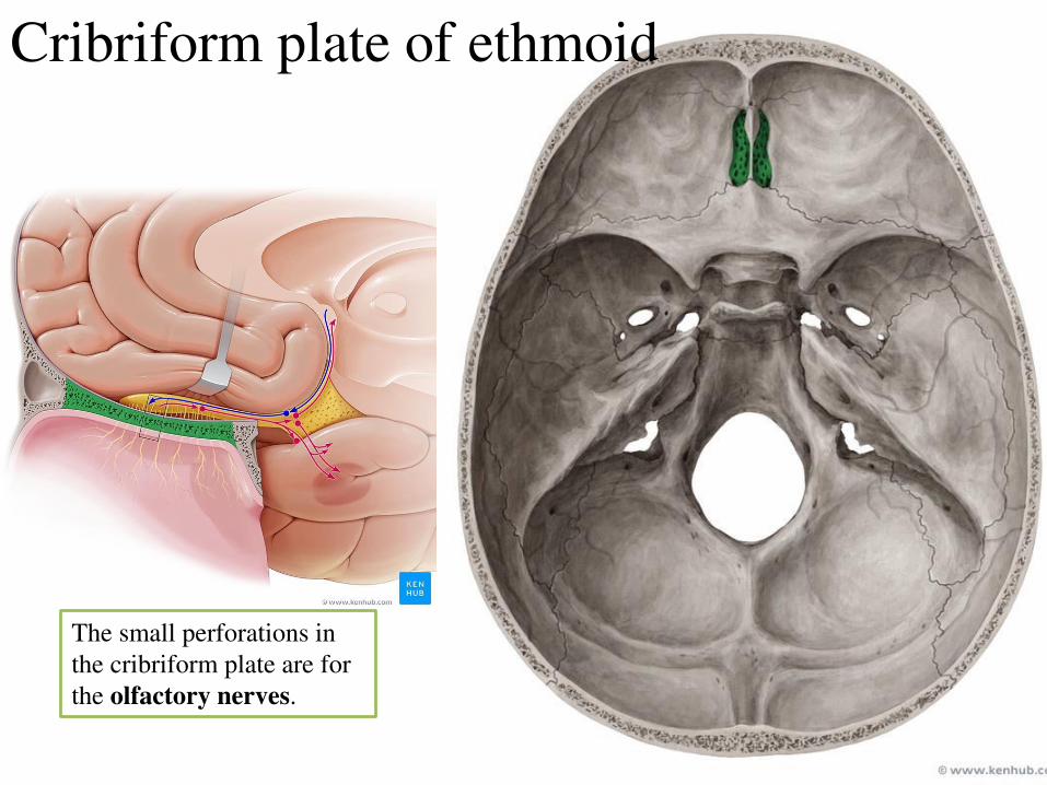
Cribriform plate of ethmoid
The small perforations in
the cribriform plate are for
the olfactory nerves.

The small perforations in
the cribriform plate are for
the olfactory nerves.
Foramen caecum
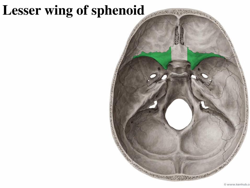
Lesser wing of sphenoid
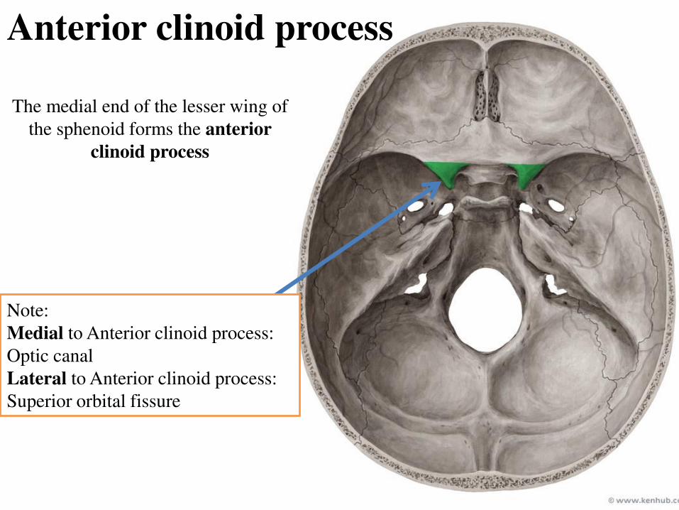
The medial end of the lesser wing of
the sphenoid forms the anterior
clinoid process
Anterior clinoid process
Note:
Medial to Anterior clinoid process:
Optic canal
Lateral to Anterior clinoid process:
Superior orbital fissure
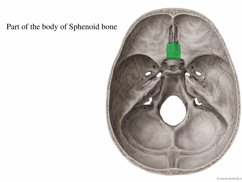
Part of the body of Sphenoid bone
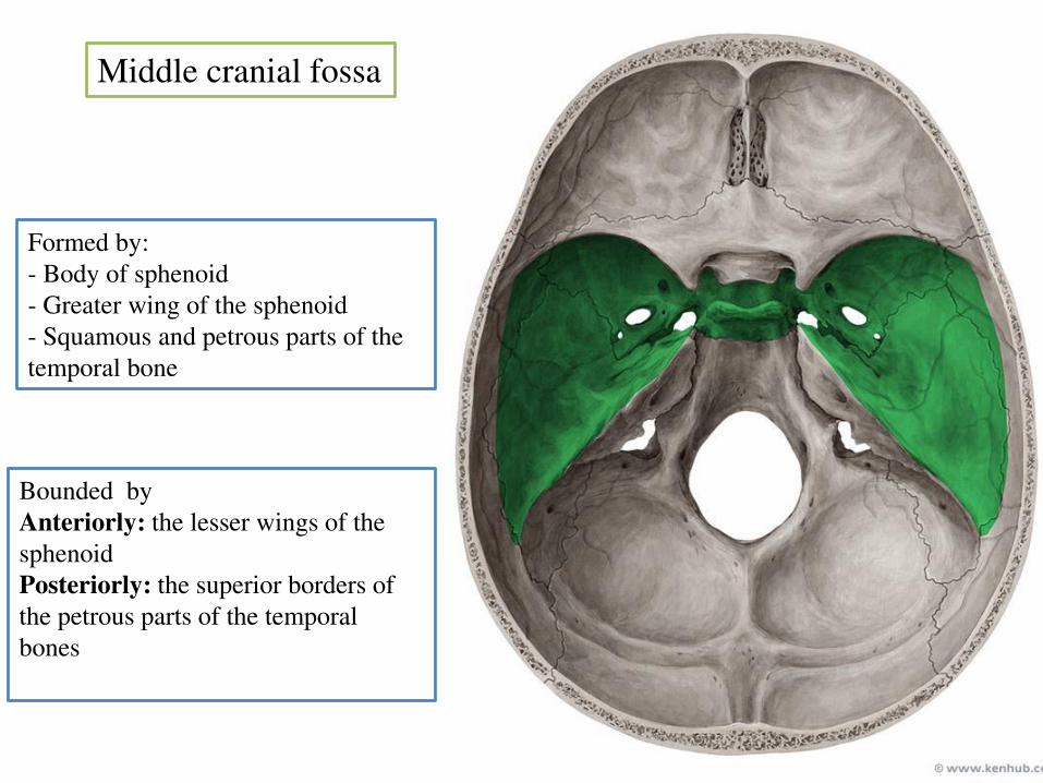
Middle cranial fossa
Bounded by
Anteriorly: the lesser wings of the
sphenoid
Posteriorly: the superior borders of
the petrous parts of the temporal
bones
Formed by:
- Body of sphenoid
- Greater wing of the sphenoid
- Squamous and petrous parts of the
temporal bone
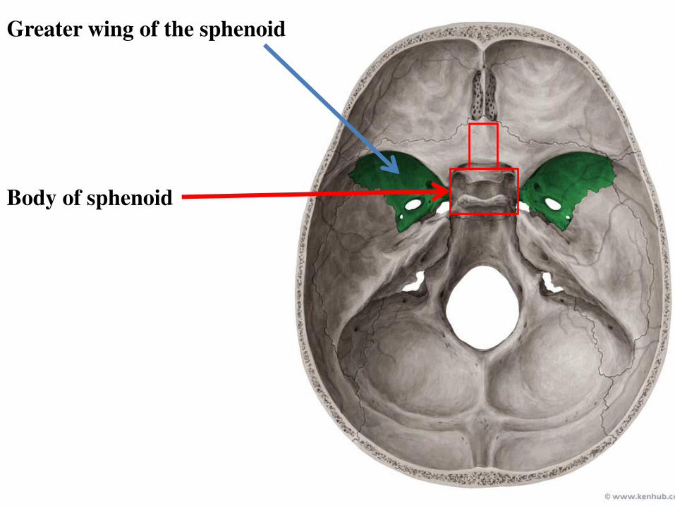
Greater wing of the sphenoid
Body of sphenoid
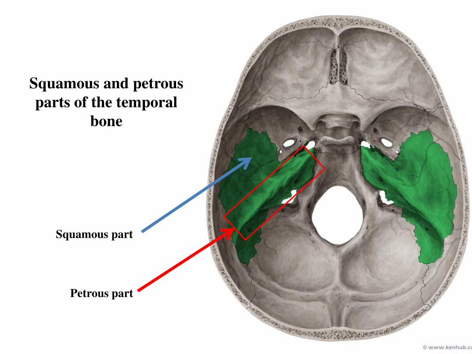
Squamous and petrous
parts of the temporal
bone
Squamous part
Petrous part
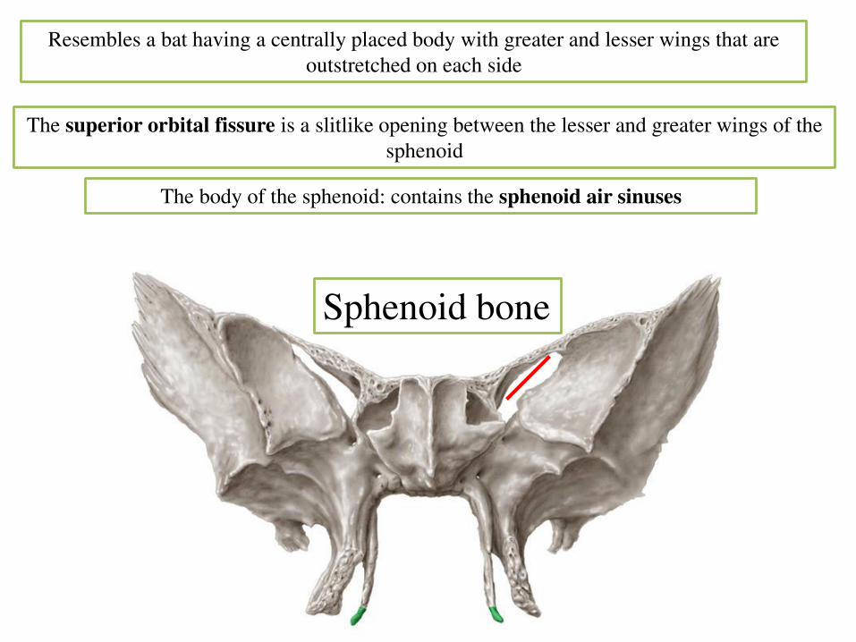
Resembles a bat having a centrally placed body with greater and lesser wings that are
outstretched on each side
Sphenoid bone
The superior orbital fissure is a slitlike opening between the lesser and greater wings of the
sphenoid
The body of the sphenoid: contains the sphenoid air sinuses

Body of Sphenoid bone
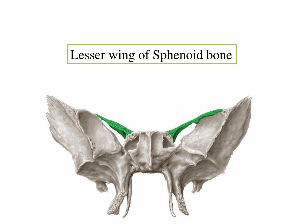
Lesser wing of Sphenoid bone
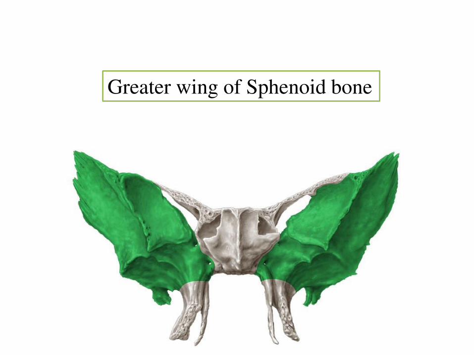
Greater wing of Sphenoid bone

Pterygoid plates of Sphenoid bone
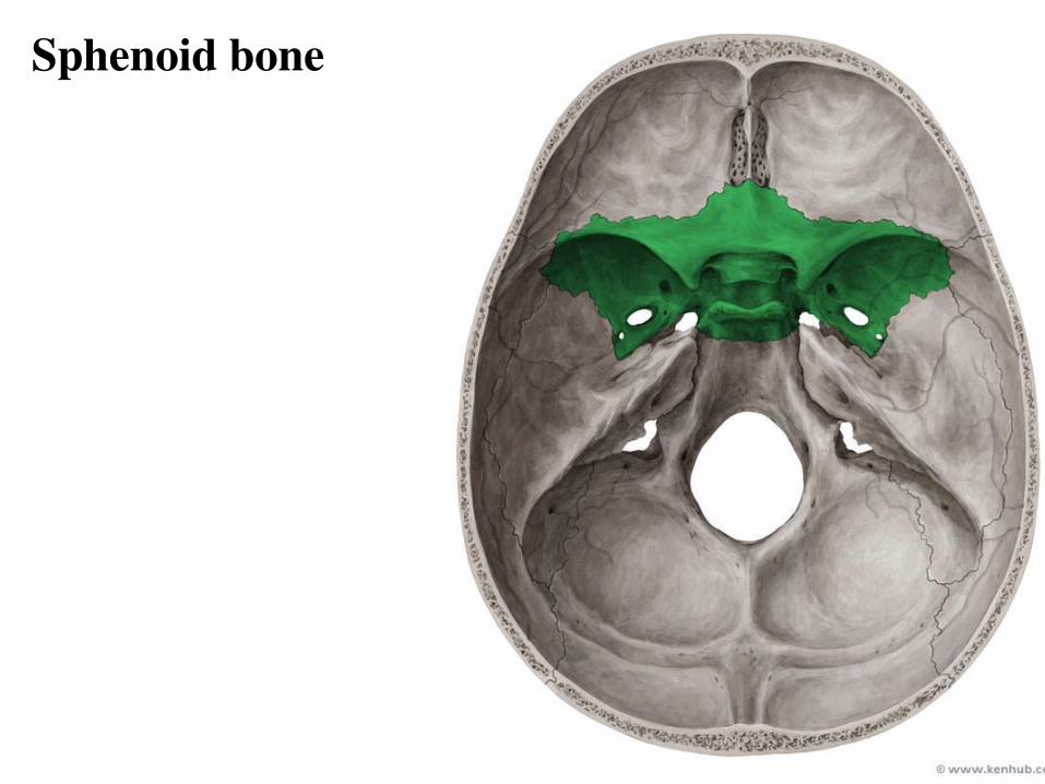
Sphenoid bone
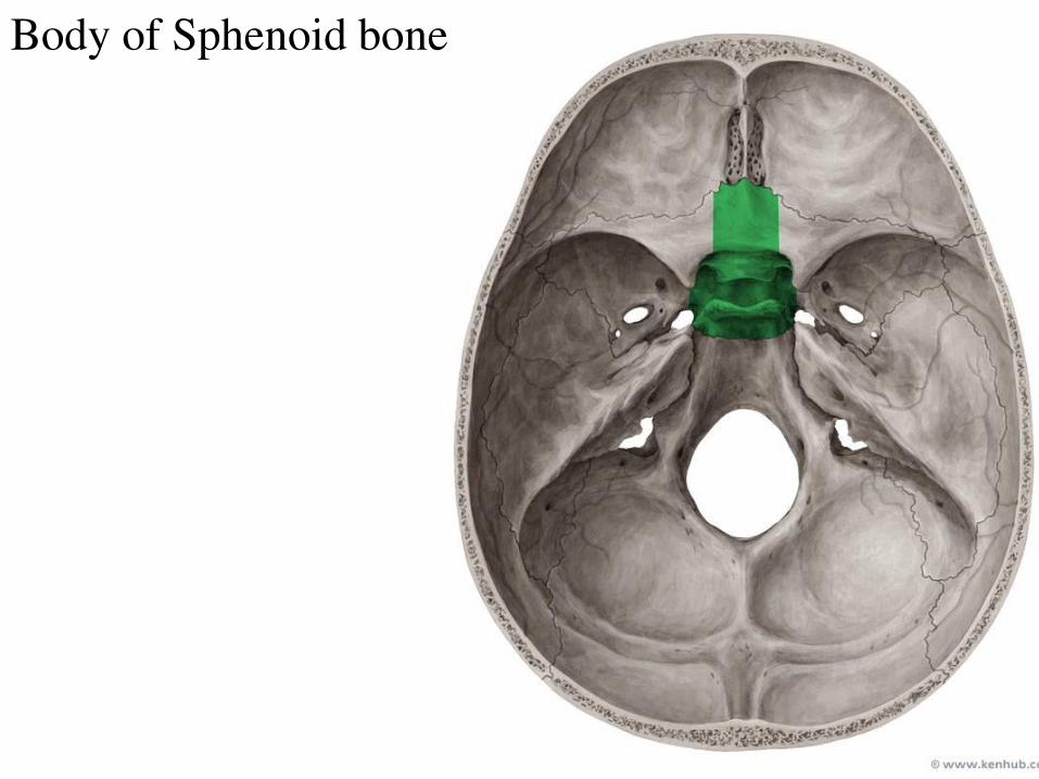
Body of Sphenoid bone
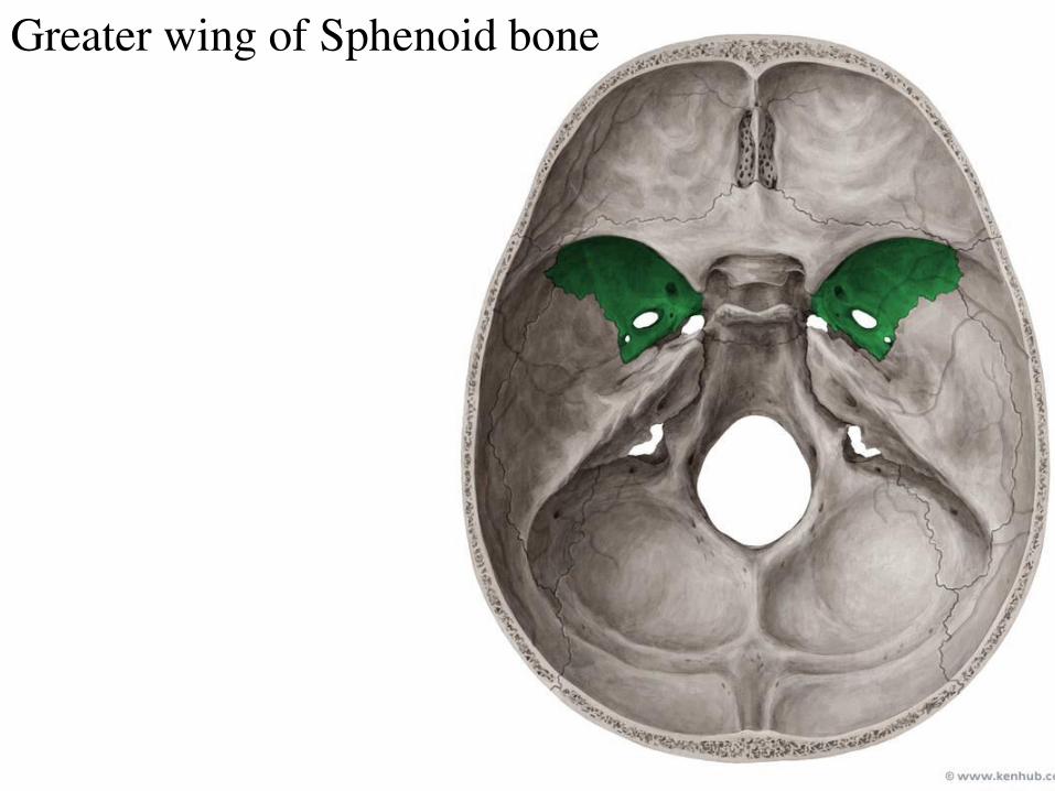
Greater wing of Sphenoid bone

Lesser wing of Sphenoid bone
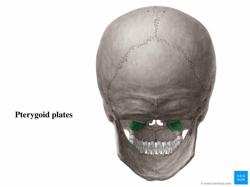
Pterygoid plates
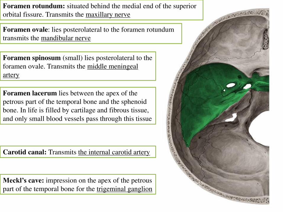
Foramen lacerum lies between the apex of the
petrous part of the temporal bone and the sphenoid
bone. In life is filled by cartilage and fibrous tissue,
and only small blood vessels pass through this tissue
Foramen ovale: lies posterolateral to the foramen rotundum
transmits the mandibular nerve
Foramen rotundum: situated behind the medial end of the superior
orbital fissure. Transmits the maxillary nerve
Foramen spinosum (small) lies posterolateral to the
foramen ovale. Transmits the middle meningeal
artery
Carotid canal: Transmits the internal carotid artery
Meckl’s cave: impression on the apex of the petrous
part of the temporal bone for the trigeminal ganglion
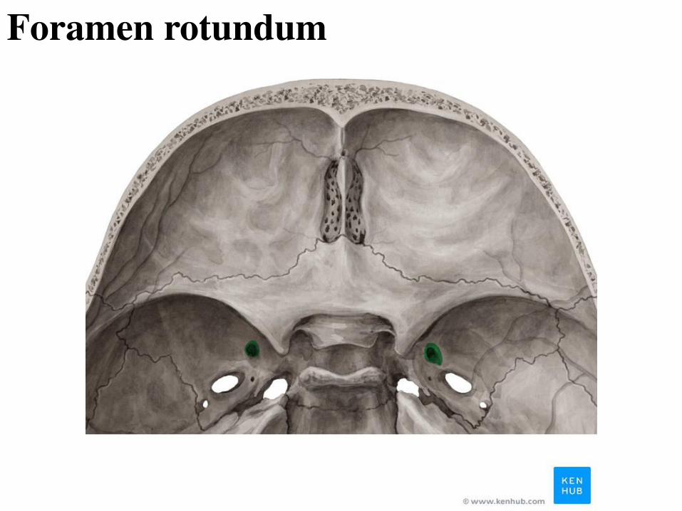
Foramen rotundum
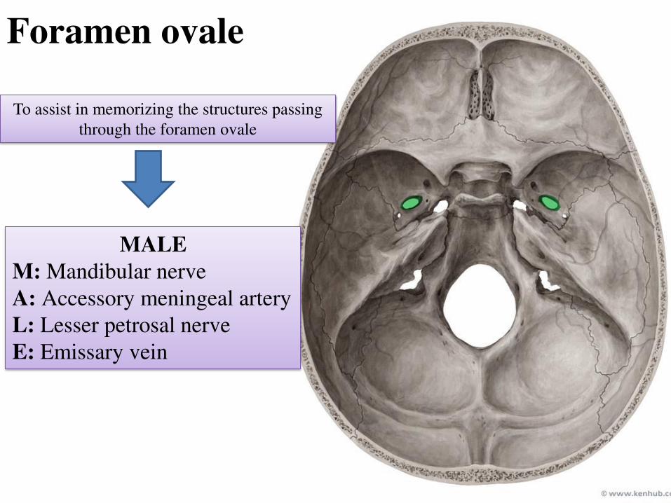
Foramen ovale
MALE
M: Mandibular nerve
A: Accessory meningeal artery
L: Lesser petrosal nerve
E: Emissary vein
To assist in memorizing the structures passing
through the foramen ovale
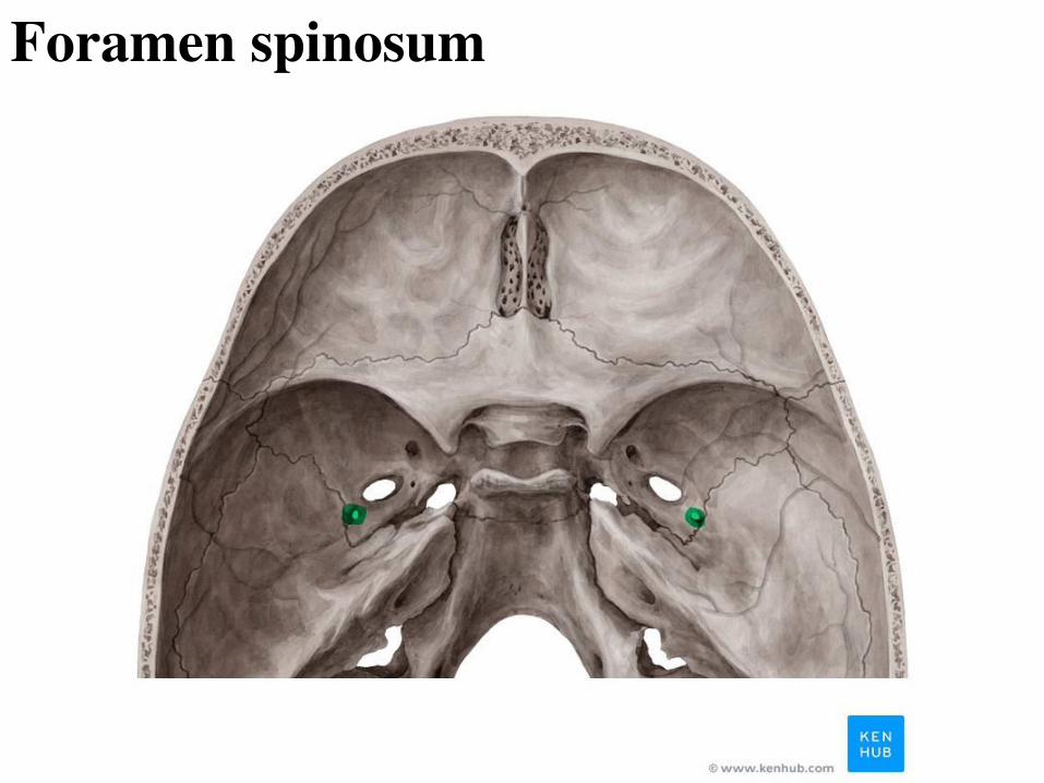
Foramen spinosum
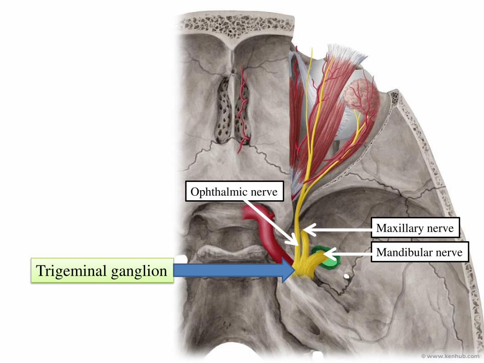
Ophthalmic nerve
Maxillary nerve
Mandibular nerve
Trigeminal ganglion
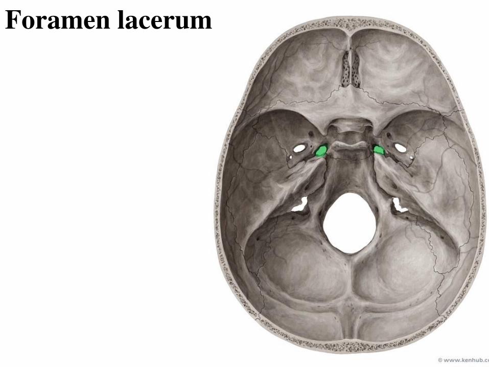
Foramen lacerum
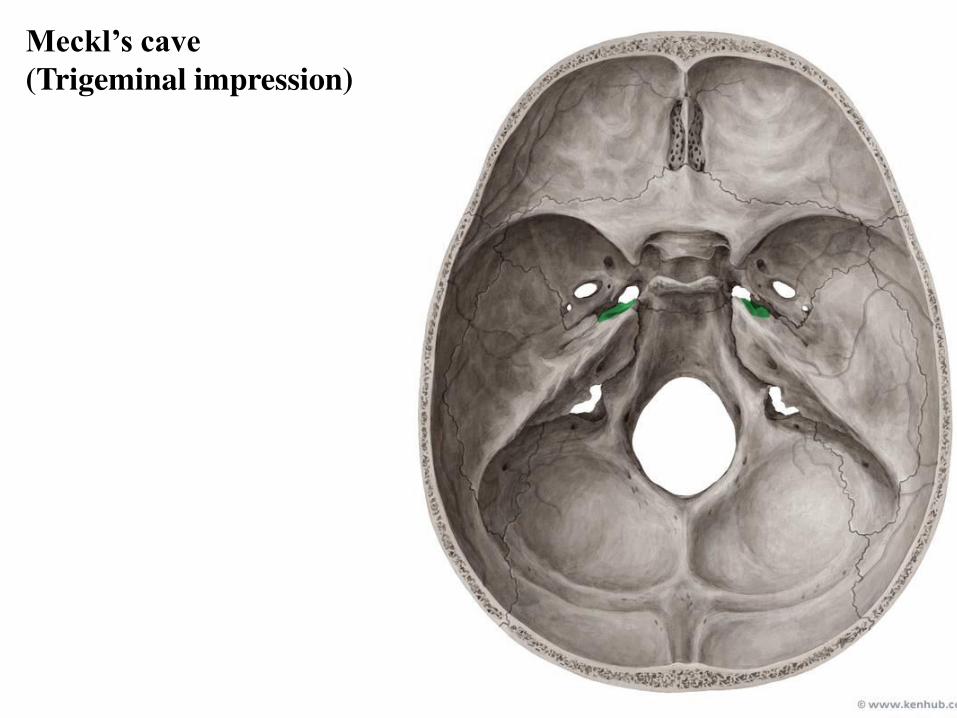
Meckl’s cave
(Trigeminal impression)
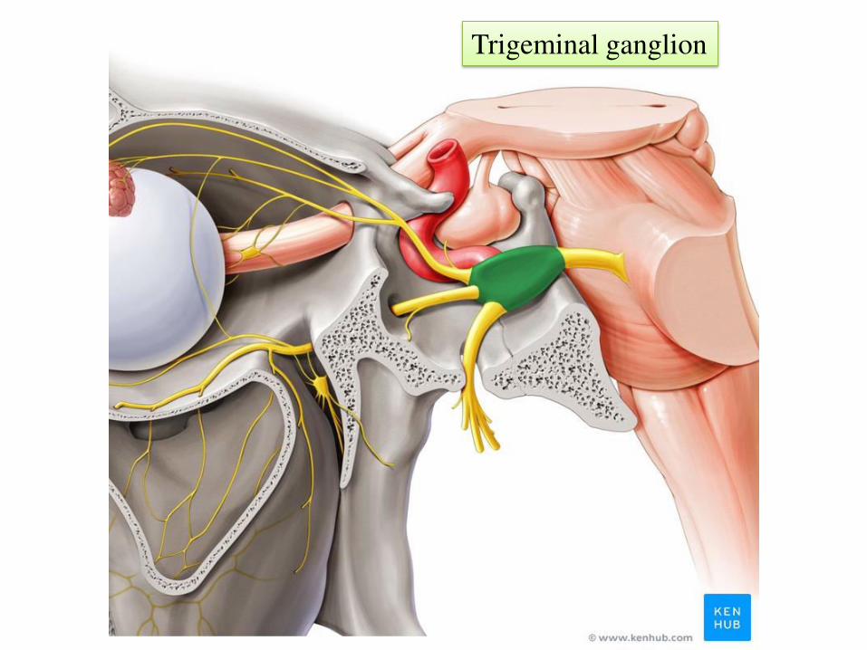
Trigeminal ganglion
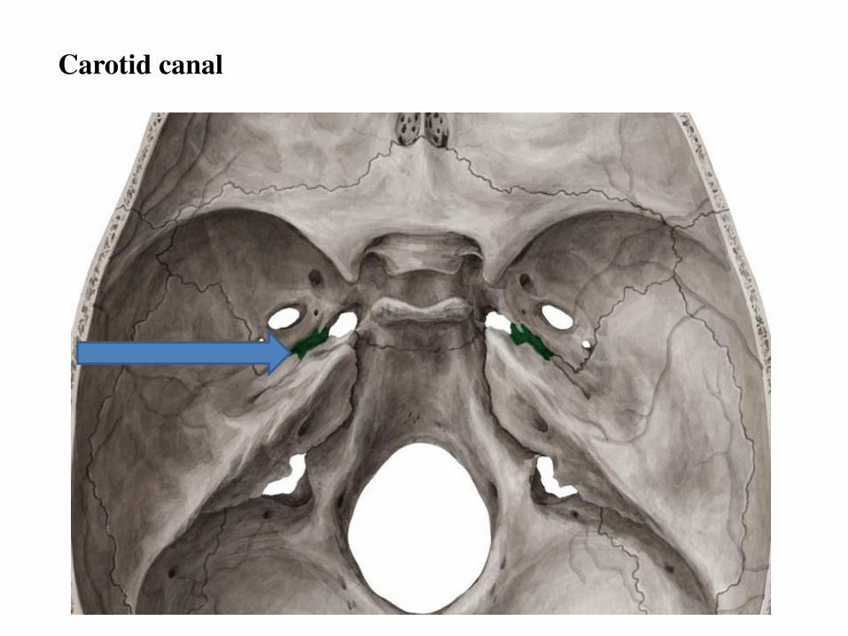
Carotid canal
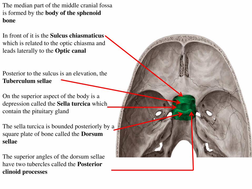
The median part of the middle cranial fossa
is formed by the body of the sphenoid
bone
In front of it is the Sulcus chiasmaticus
which is related to the optic chiasma and
leads laterally to the Optic canal
Posterior to the sulcus is an elevation, the
Tuberculum sellae
On the superior aspect of the body is a
depression called the Sella turcica which
contain the pituitary gland
The sella turcica is bounded posteriorly by a
square plate of bone called the Dorsum
sellae
The superior angles of the dorsum sellae
have two tubercles called the Posterior
clinoid processes
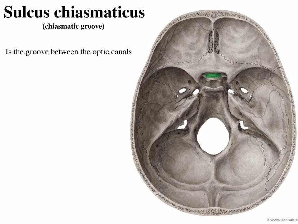
Sulcus chiasmaticus (chiasmatic groove)
Is the groove between the optic canals
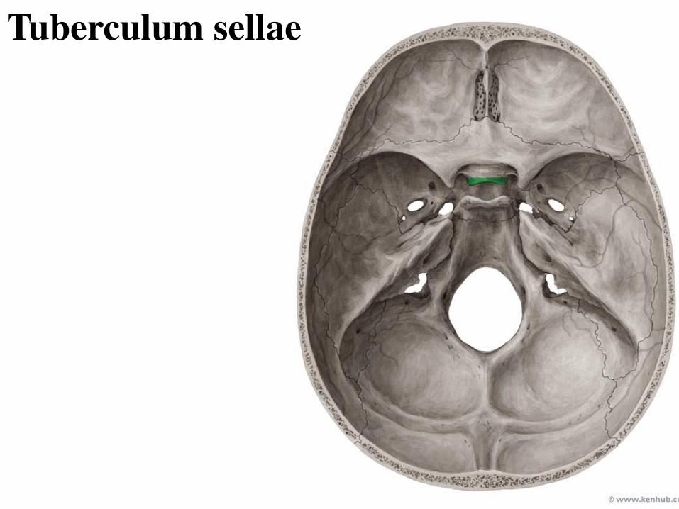
Tuberculum sellae
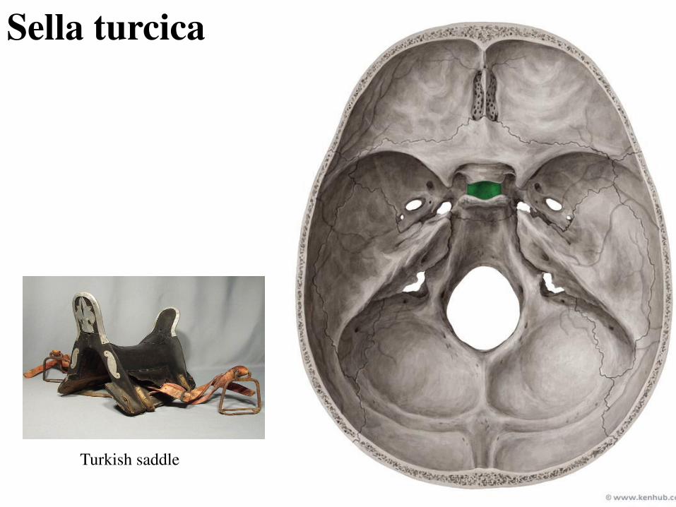
Sella turcica
Turkish saddle
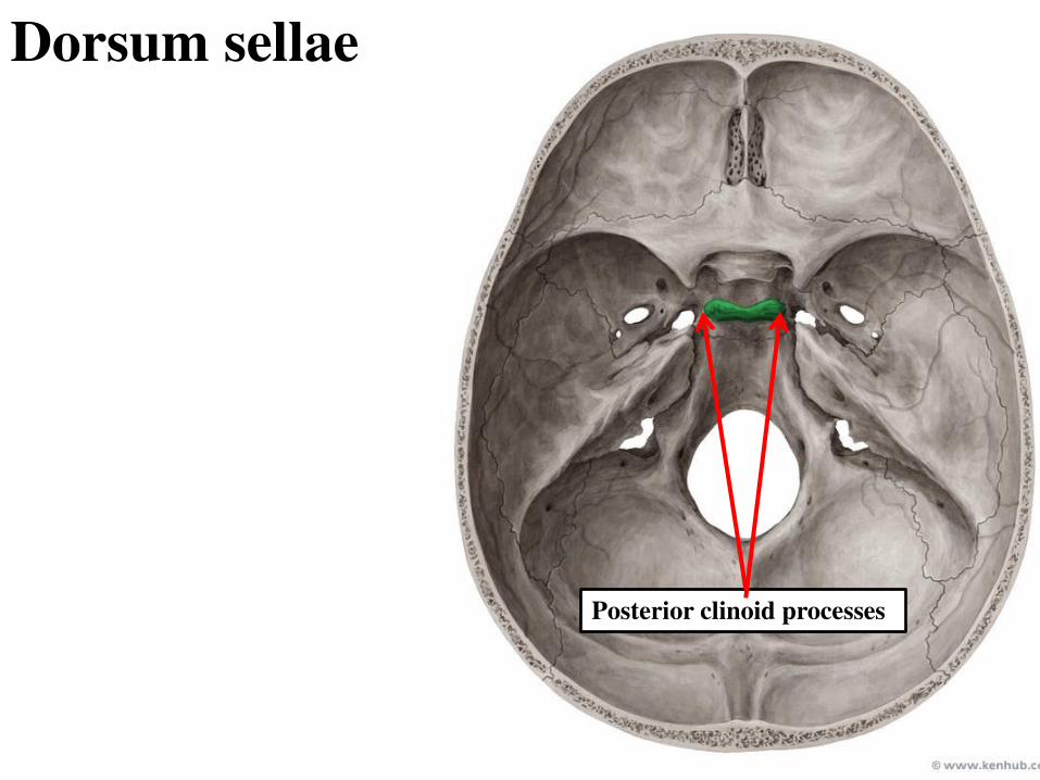
Dorsum sellae
Posterior clinoid processes
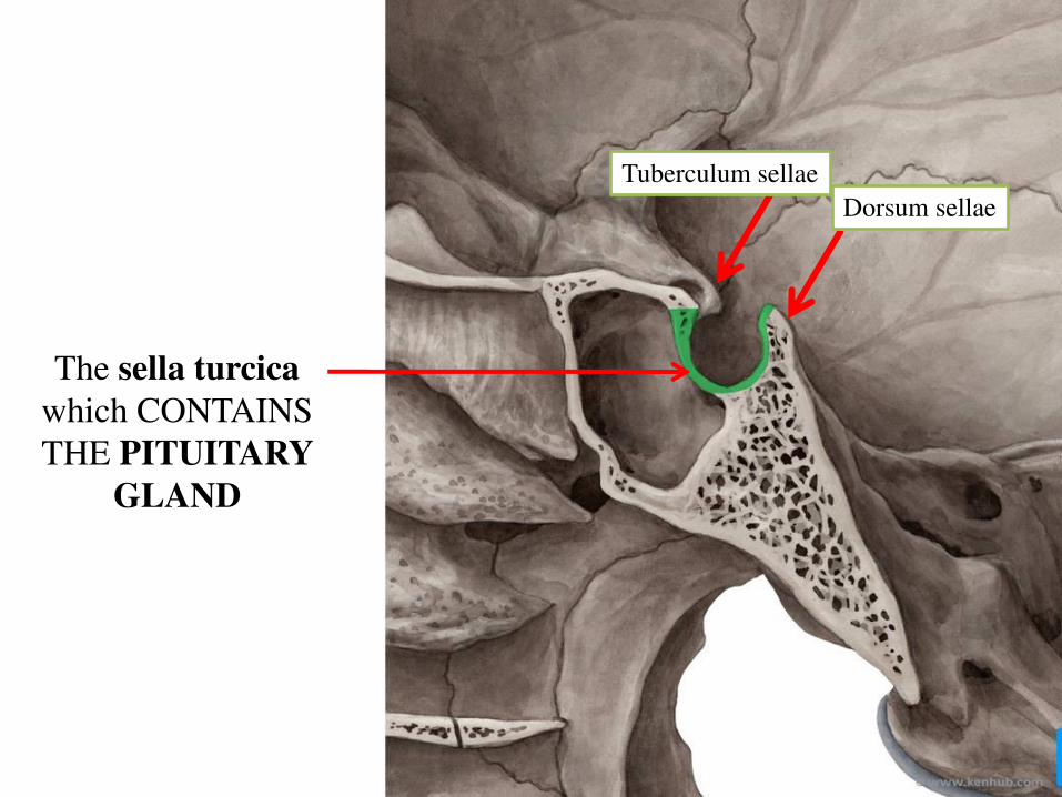
The sella turcica
which CONTAINS
THE PITUITARY
GLAND
Dorsum sellae
Tuberculum sellae
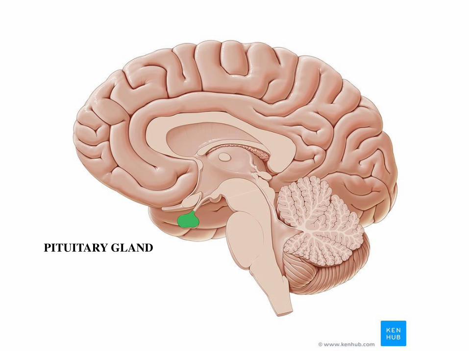
PITUITARY GLAND
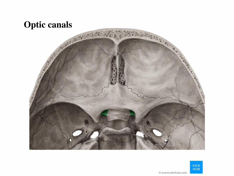
Optic canals
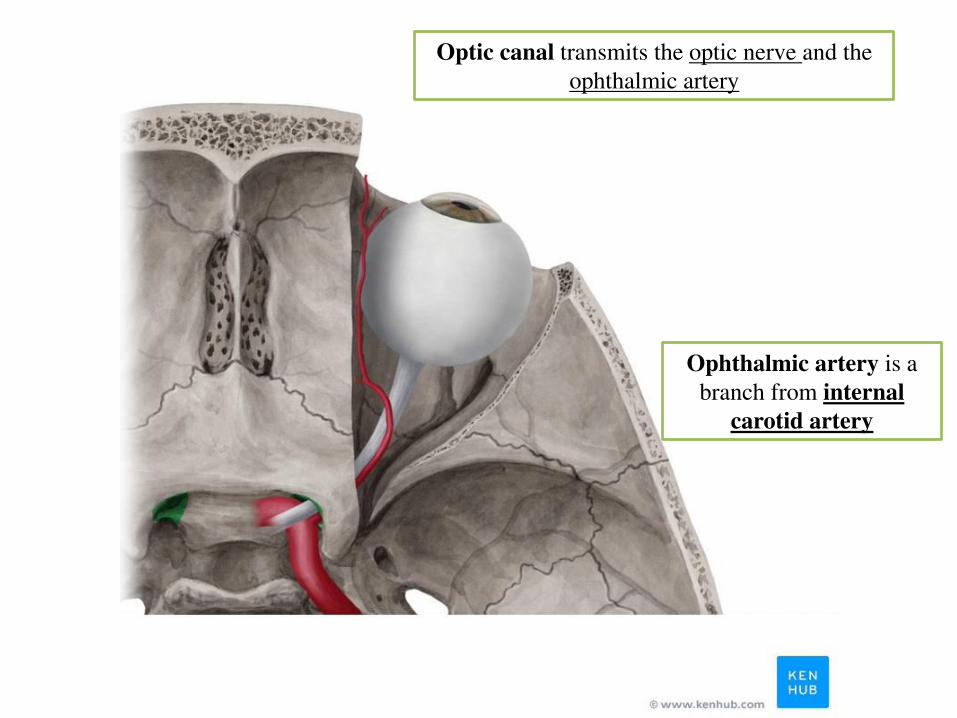
Optic canal transmits the optic nerve and the
ophthalmic artery
Ophthalmic artery is a
branch from internal
carotid artery
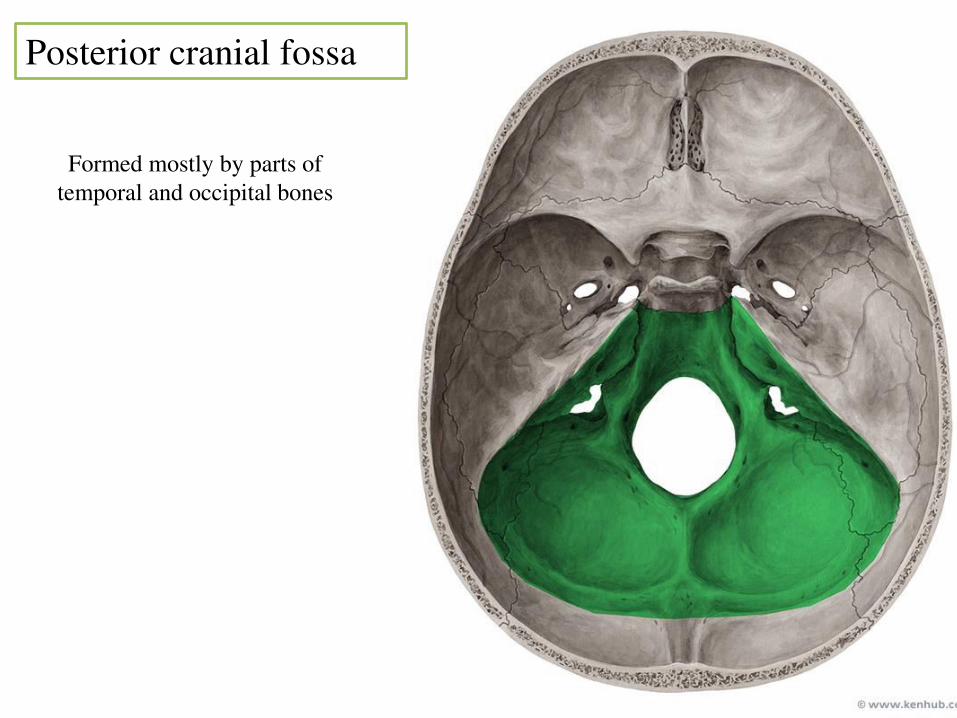
Posterior cranial fossa
Formed mostly by parts of
temporal and occipital bones
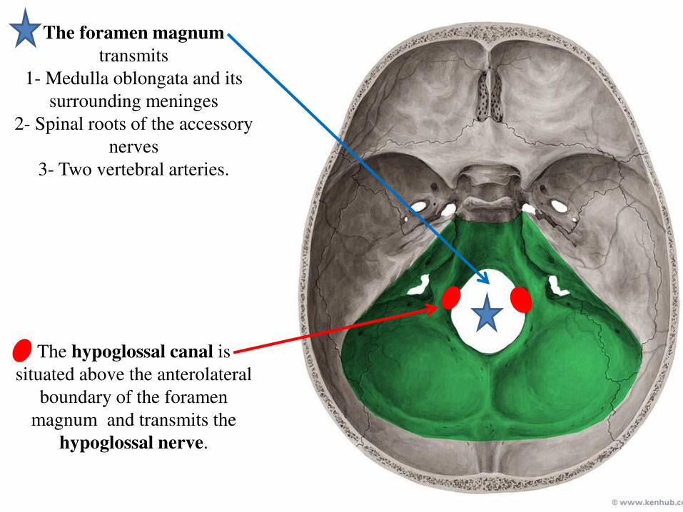
The foramen magnum
transmits
1- Medulla oblongata and its
surrounding meninges
2- Spinal roots of the accessory
nerves
3- Two vertebral arteries.
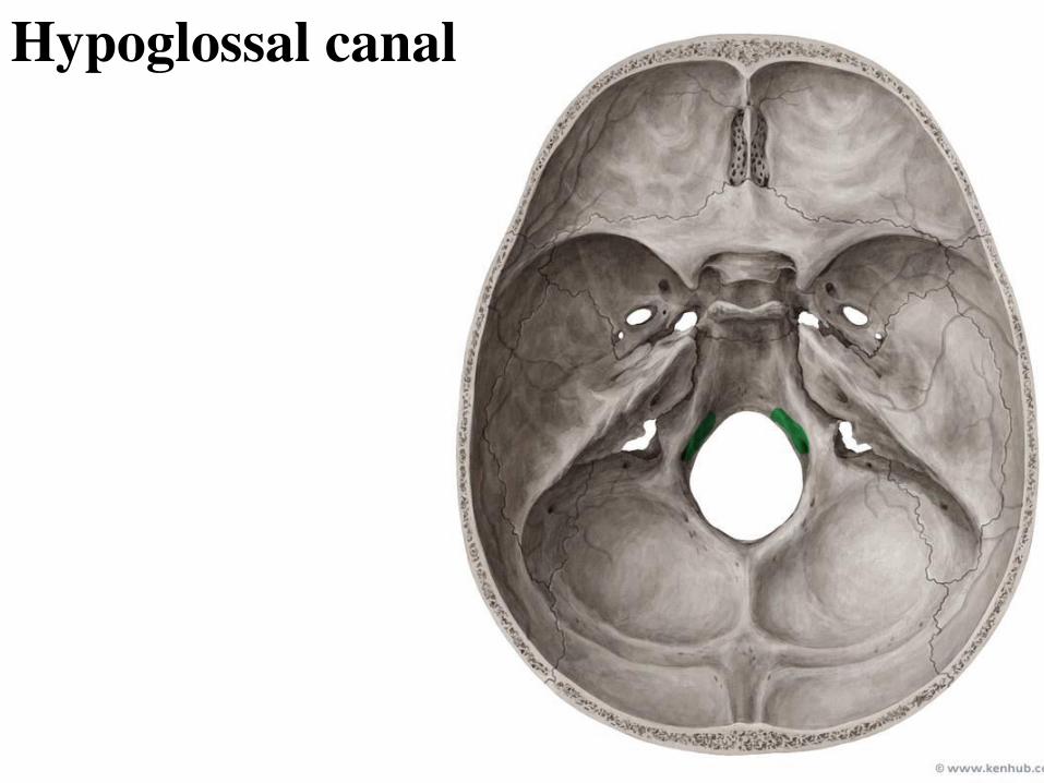
The hypoglossal canal is
situated above the anterolateral
boundary of the foramen
magnum and transmits the
hypoglossal nerve.
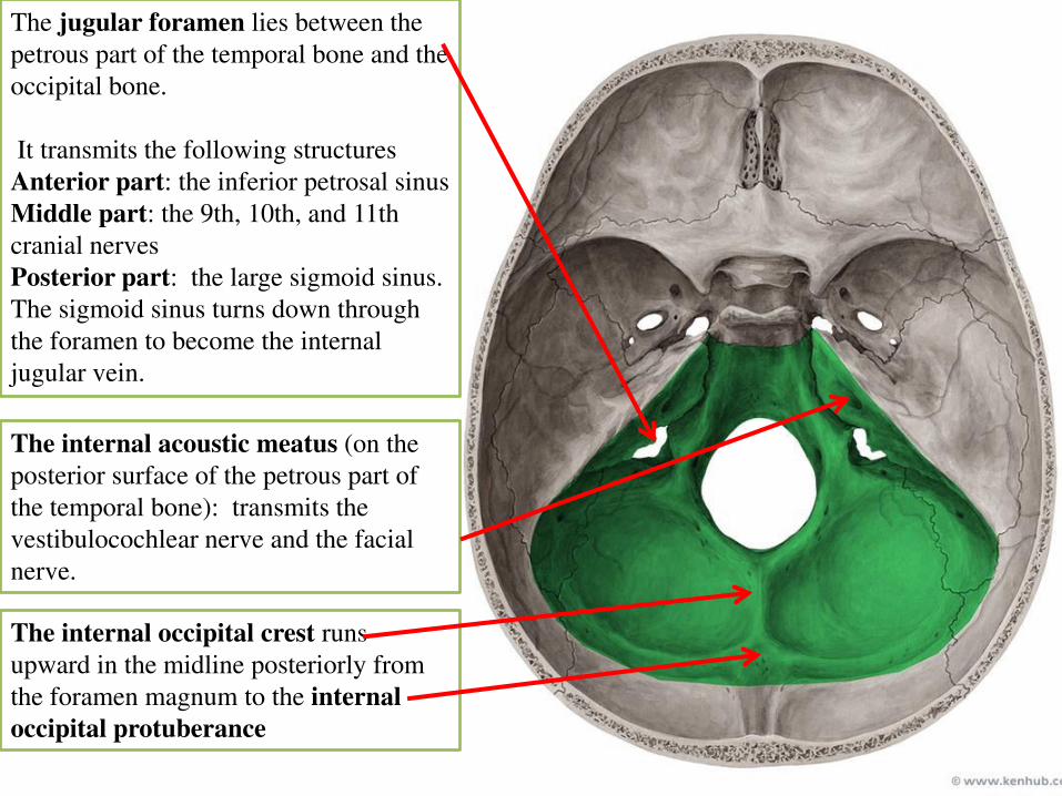
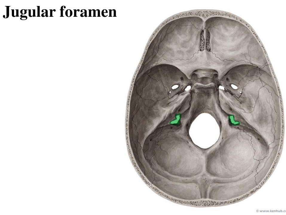
The jugular foramen lies between the
petrous part of the temporal bone and the
occipital bone.
It transmits the following structures
Anterior part: the inferior petrosal sinus
Middle part: the 9th, 10th, and 11th
cranial nerves
Posterior part: the large sigmoid sinus.
The sigmoid sinus turns down through
the foramen to become the internal
jugular vein.
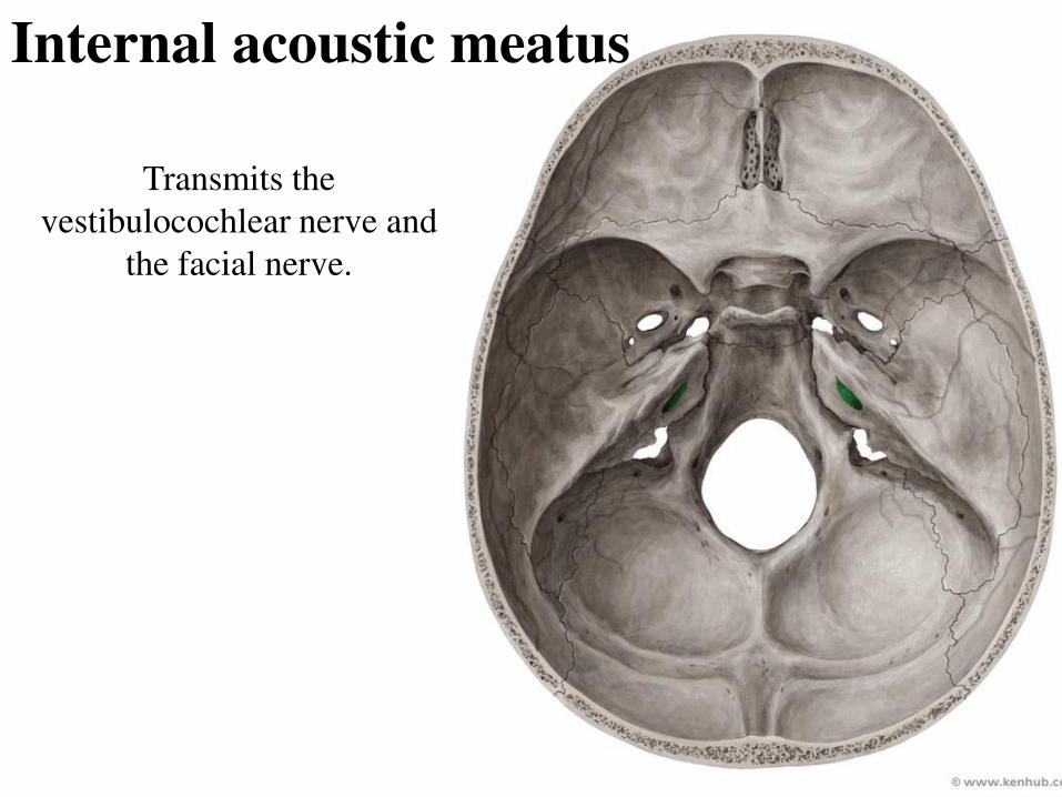
The internal acoustic meatus (on the
posterior surface of the petrous part of
the temporal bone): transmits the
vestibulocochlear nerve and the facial
nerve.
The internal occipital crest runs
upward in the midline posteriorly from
the foramen magnum to the internal
occipital protuberance
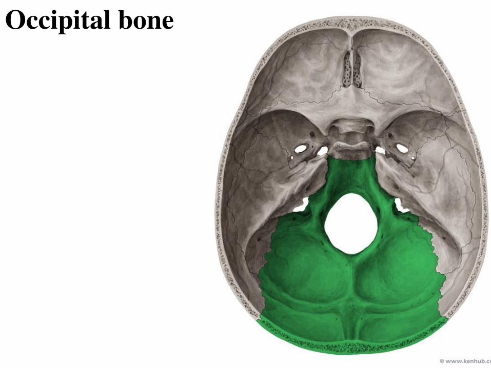
Occipital bone
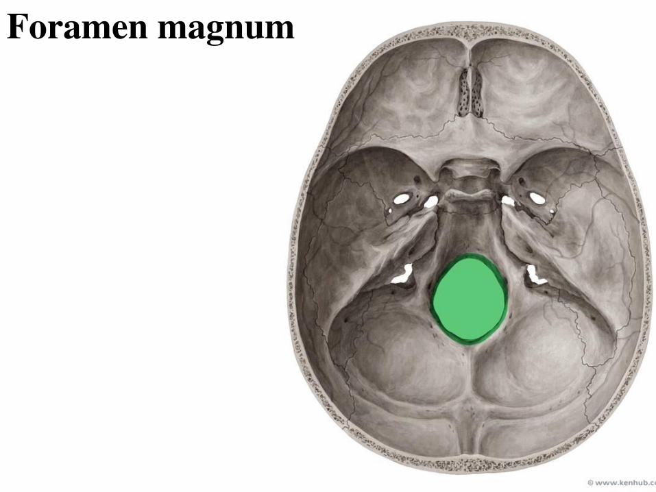
Foramen magnum
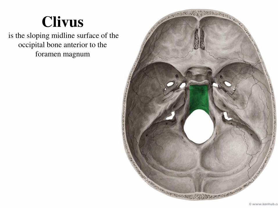
Clivus
is the sloping midline surface of the
occipital bone anterior to the
foramen magnum
Hypoglossal canal
Internal acoustic meatus
Transmits the
vestibulocochlear nerve and
the facial nerve.
Jugular foramen
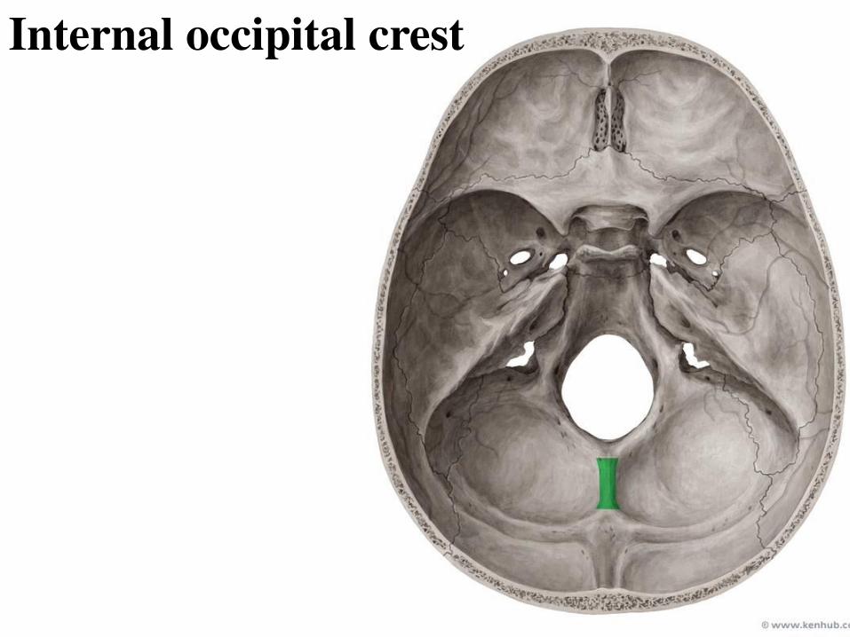
Internal occipital crest

Internal occipital protuberance
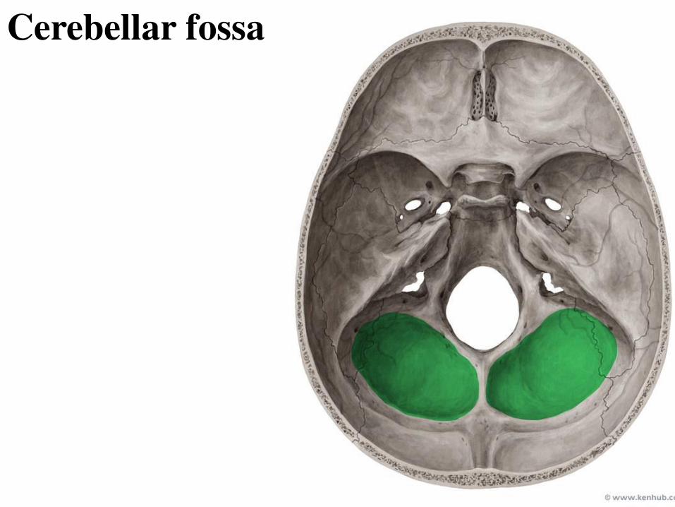
Cerebellar fossa

Ethmoid bone Delicate bone located between the two orbits

Ethmoid bone
Orbital plate
Crista galli Cribriform plate
Perpendicular plate