Embed Size (px)
Citation preview

�
3 Aktuelle und potenzielle Gefahren für die Gesundheit
Hazards by Enhanced UV-Iradiation: While UV-C is totally absorbed by the ozone layer, a part of the solar UV-B and total UV-A reach the Earth’s surface. In the skin UV irradiation induces a variety of acute effects like tanning, thickening of the skin or reddening and chronic effects like skin ageing. In the worst case UV-irradiation induces skin cancer, the most frequent cancer in the white population world-wide. Following the world-wide trend the incidence of skin cancer (malignant melanoma MM, basal cell carcinoma (BCC) and squamous cell carcinoma (SCC)) is continuously increasing in Germany due to a changed social behavior resulting in an enhanced UV-exposure. Nevertheless, thinning of the ozone layer, leading to enhanced UV-B irradiation on the Earth, may still play a role for the skin cancer incidence in future. UV-radiation can induce DNA damage that is repaired very effectively by the skin cells. Furthermore heavily damaged cells will be removed from the body by means of programmed cell death (apoptosis). However, in the case of misrepair mutations can occur that are characteristic for UV-damage (UV-signature mutations). There is increasing evidence that the occurrence of these mutations in epidermal stem cells of human skin characterizes an early step in the multi-step process of skin cancer development, involving tumor suppressor- or proto-oncogenes. Other effects like e.g. deregulation of epi-genetic control of cellular functions as well as immunosuppression by UV-radiation also play important roles in the induction and progression of skin cancer. Educational campaigns and balanced information of the public, teaching a reasonable behavior concerning solar UV- or artificial (sunbed) radiation and/or early detection of skin cancer are useful tools to decrease the skin cancer risk as well as morbidity and mortality of skin cancer in the population. However, it is also important to take ecological measures for the protection of the ozone layer to prevent a further enhancement of UV-B irradiation at ground-level.Gefährdung durch verstärkte UV-Strahlung - Während UV-C-Strahlung vollständig von einer intakten Ozon-Schicht absorbiert wird, erreichen Anteile von UV-B- Strahlung und die gesamte UV-A-Strahlung der Sonne die Erdoberfläche. In Haut kann UV-Strahlung akute Effekte wie Bräunung, Haut-Verdickung oder Rötung hervor-rufen, aber auch chronische Effekte, wie die Hautalterung. Im schlimmsten Fall kann UV-Strahlung zu Haut-krebs führen, der weltweit häufigsten Krebserkrankung in der weißen Bevölkerung. Wie auf der ganzen Welt, nimmt auch die Hautkrebs-Inzidenz (malignes Melanom, MM, Basalzellkarzinom, BCC und Plattenepithelkar-zinom, SCC) auch in Deutschland kontinuierlich zu. Dies hat seinen Grund, hauptsächlich, in einem geänderten Sozialverhalten, welches zu erhöhter UV-Exposition führt. Allerdings kann auch eine weitere Ausdünnung der Ozonschicht, welche zu erhöhter UV-B-Einstrahlung bewirkt, für die Hautkrebsinzidenz in der Zukunft eine Rol-le spielen. UV-Strahlung induziert DNA-Schäden, die von der Zelle effektiv repariert werden können. Darüber hinaus werden schwer geschädigte Zellen aus dem Körper durch programmierten Zelltod (Apoptose) entfernt. Im Falle von Fehlreparaturen kann es aber zur Bildung von Mutationen kommen, welche charakteristisch für UV-Schäden sind (»UV-signature mutations«). Es gibt zunehmende Evidenz, dass das Auftreten solcher Muta-tionen in epidermalen Stammzellen der Haut als frühes Ereignis im mehrstufigen Prozess der Hautkrebsent-stehung angesehen werden kann, wobei Tumor-Suppressorgene und Onkogene beteiligt sind. Andere Prozesse, wie z.B. die Fehlregulation der epigenetischen Kontrolle zellulärer Funktionen und die UV-bedingte Immun-suppression stellen ebenfalls wichtige Faktoren für die Induktion und Progression von Hautkrebs dar. Aufklä-rungskampagnen und ausgewogene Informierung der Bevölkerung, welche einen verantwortlichen Umgang mit solarer aber auch künstlicher (Solarien) UV-Strahlung vermitteln und/oder die Früherkennung von Haut-krebs zeigen sich als nützliche Maßnahmen, die das Hautkrebs-Risiko sowie die Erkrankung und Sterblichkeit an Hautkrebs reduzieren können.Trotzdem ist es immer noch wichtig, ökologische Maßnahmen zum Schutz der Ozonschicht zu ergreifen, um eine weitere Zunahme von UV-B-Strahlung an der Erdoberfläche zu verhindern.
3.1.10 HazardsbyEnhancedUVIrradiation RüdigeR gReineRt & Beate VolkmeR
�
3.1.10 Greinert & Volkmer
Based on recent estimates in �0�� more than �30,000 new cases of cutaneous malignant mela-
noma (MM), the induction of which is associated with UV exposure, occur globally each year (FeRlay �0�3). It has been furthermore estimated that, additionally, �-3 million non-melanocytic skin cancers (NMSC,) which are mainly basal cell carcinoma (BCC) and squamous cell carcinoma (SCC) are diagnosed each year world-wide (SuRdu et al. �0�3). Furthermore, �0% of ��–�5 million cases of blindness due to damage of the lens (cataract) are supposed to be UV induced (Repacholi �996). The increased incidence of these diseases is to be attributed in particular to excessive sun exposure due to a change in people‘s recreational and social be-havior. In addition, the decrease of the protective ozone layer in the atmosphere - and thus the increase in solar UV radiation on the ground - might also considered as a cause for an increase in skin cancer. However, new re-sults indicate a recovery of the ozone layer (WeatheR-head & andeRSen �006). Whether these changes have influences on skin cancer incidence has to be monitored in the future.
The investigation of effects caused from UV irradi-ation in the biosphere, and especially the estimation of the connected risks for man, require exact knowledge of the contributive quantities and the underlying inter-action processes. Accordingly, some basic mechanisms of UV induced skin damage will be described in this chapter and the resulting risk of long term effects (skin cancer) will be pointed out.
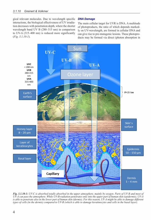
AcuteandChronicEffectsofUVIrradiationWhen describing biological effects of UV irradiation, the UV spectrum is frequently divided into three spec-tral bands, UV-A (3�5–400 nm), UV-B (�80–3�5 nm) and UV-C (�00–�80 nm).While UV-C is absorbed to-tally by the upper atmosphere mainly by oxygen and to some degree also by ozone, UV-B is absorbed by ozone (but not totally) and UV-A radiation is mainly scattered by air molecules and a large part reaches Earth surface (Fig. 3.1.10-1). In dependence of the wavelength and an associated depth of penetration into the skin, UV radiation induces a variety of effects, based on photo-physical and photochemical transformation processes of radiation energy. The most noticeable reaction of the skin to UV exposure is pigmentation. This tanning as well as a thickening of the skin‘s outermost layers are considered to be protection mechanisms. If UV expos-ure exceeds a certain, individually different, threshold, an acute damage occurs and several hours after irradia-tion a reddening of skin (erythema) appears. Additional UV exposure, further exceeding the threshold, leads to
the development of blisters and eventually necrosis of the skin surface.
Chronic effects, such as premature aging of the skin, result from cumulative damage induced by years of UV exposure and are associated with irreversible changes like damage of connective tissue and wrinkling (solar elastosis), permanent capillary dilation (teleangiecta-sia) and enlargement of pores and blackheads (come-dos). Furthermore, enhanced sun exposure can induce a disturbance of the keratinization (actinic keratosis), representing a precursor lesion of the squamous cell carcinoma. However, the induction of skin cancer is by far the most fatal result of UV exposure, associated with changes of the genetic material of the skin cells.
SkincancerThe incidence of skin cancer has been considerably increasing worldwide during the last decades. Besides malignant melanoma (MM), the main cause of skin cancer death, non-melanocytic skin cancers (NMSC) like basal cell carcinoma (BCC) and squamous cell carcinoma (SCC), which in total belong to man‘s most frequent malignant tumors, are of importance (diepgen & mahleR �00�). UV-exposure has been shown to be mainly responsible for these observations (IARC �0��). MM accounts for about 5–�0% of all skin cancer, whe-reas in terms of non-melanoma skin cancer (NMSC), BCC accounts for approximately 80–85% and SCC for about �5–�0%. CM derives from pigment (melanin) producing melanocytes, whereas NMSC derives from epidermal keratinocytes (IARC �0��).
UV-A and UV-B from the sun and from UV-emit-ting devices (e.g. sunbeds) are classified as known carcinogens to humans (IARC Group �) (IARC �0��). This classification was based on a large number of experimental data, epidemiological studies and meta-analyses thereof. It was concluded that there was suf-ficient evidence in humans for the carcinogenicity of solar radiation for MM, BCC and SCC. With regards to artificial sources of UVR, it was concluded that there was sufficient evidence for an increased risk of MM and of ocular melanoma, and a positive association was observed for sunbed use and SCC (IARC �0��).
Skin Cancer OccurrenceOver the last 50 years a steep increase in both MM and NMSC has been documented in Caucasian populations (eRdmann et al. �0�3; lomaS et al. �0��). Worldwi-de, highest incidence rates are by far those observed in Australia and New Zealand where fair-skinned po-pulations are exposed to intensive UVR (eRdmann et al. �0�3; lomaS et al. �0��). Based on estimations for �0��, more than �30,000 new cases of MM occurred

3
3 Aktuelle und potenzielle Gefahren für die Gesundheit
globally, �00,000 alone within Europe (FeRlay �0�3). The lifetime risk of getting MM is highest in New Zea-land and Australia with 3.6%. Lifetime risk for getting melanoma in Europe countries is ranging from 0.3% to �.6% (eRdmann et al. �0�3). In Europe particular-ly high MM incidences are found in Nordic countries, Switzerland, the Netherlands, the Czech Republic and Slovenia. Mediterranean countries tend to have lower rates as well as the Baltic and Eastern European coun-tries (eRdmann et al. �0�3; FeRlay �0�3).
Incidence rates NMSCs are difficult to estimate as they are often not or only incompletely registered in cancer registries (lomaS et al. �0��). However, based on data from few countries with population-based re-gistries (Denmark, Finland, Scotland, Malta, Germany and the Netherlands) age-standardized incidence rates (ASR) of primary BCC are estimated to be 77–�58 cases per �00,000 person-years in those regions (de VRieS et al. �0��). Data from the Cancer Registry of Schleswig-Holstein (Germany) for the year �0�� lead to an estimate of �5�.430 new skin cancer cases/year in Germany (MM: �9.450; BCC: �45.570 and SCC: 76.370).
Incidence rates of SCCs are much lower than those reported for BCCs (BiRch-JohanSen et al. �0�0; do-heRty et al. �0�0; lomaS et al. �0��), e.g. ��/�9 (f/m) cases per �00,000 person/years among women and �9.� cases among men in Denmark (world standard) (BiRch-JohanSen et al. �0�0), �3.8/36.9 (f/m) in Scotland (do-heRty et al. �0�0), and �0.5/35.4 (f/m) cases among in The Netherlands (per �00,000, European standard) (holleStein et al. �0��).
According to recent estimates, 55,500 deaths from melanoma occurred in �0�� worldwide, including ��,�00 in Europe (FeRlay �0�3). In comparison, mor-tality from NMSCs is low (< 0.�% of incident cases) (JenSen et al. �008; leWiS & WeinStock �007). Howe-ver, due to their high incidence NMSCs contribute to the rising morbidity as well as to a significant oecono-mic burden to health services.
The risk of additional induction of basal cell car-cinoma and squamous cell carcinoma can roughly be calculated. People with a yearly solar UV dose of �00 MED (MED = minimal erythemal dose, � MED cor-responds to the lowest UV dose to achieve a reddening of the skin) obtain a � fold risk to develop skin cancer if they achieve an additional dose of �00 MED at the age of 35 to 65 years. An additional dose at the age of �5 to 45 years enhances the risk to develop a skin cancer du-ring 75 years of life by a factor of 3.4 (SlapeR �998).
In accordance with a worldwide trend, the in-cidence of all skin cancers in Germany is increasing faster than that of all other cancers. This fact is to be
attributed in particular to excessive sun exposure due to a change in people‘s recreational and social behavior. Since sun tanned skin is in general considered beautiful and a sign of a healthy body, sun tanned skin is no lon-ger only an expression of people‘s outdoor activity and recreation but has become a purpose of itself. This has led to excessive recreational exposure to UV radiation by sunlight or UV tanning devices (solaria). In addition to this changed behavior of large parts of the popula-tion, the decrease of the protective ozone layer in the atmosphere - and thus an increase in solar UV radiation on the ground - may also become a cause for a future increase of skin cancer. Based on UV monitoring, data models have been developed correlating the ozone re-duction with UV-B increases at the earth‘s surface, re-sulting in an enhanced biological effectiveness of solar radiation. For a decrease of �% of the ozone layer an increase of UV induced skin cancer of �–3% has been calculated (SSK �996).
BiologicalEffectsofUVIrradiationUVR exposure has been shown to be the main cause of skin cancer, including cutaneous malignant melanoma (MM), basal cell carcinoma (BCC) and squamous cell carcinoma (SCC). In �009, within the IARC mono-graph program UVR as well as the use of tanning de-vices was classified as carcinogenic to humans (group �) (el ghiSSaSSi et al. �009; IARC �0��).
Although UV exposure has been recognized as a main risk factor for the incidence of the skin cancer, the underlying molecular mechanisms have further to be elucidated in more detail. Nevertheless, UV-induced mutations in specific genes, involved in the cell regu-lation, as well as chromosomal instability have been connected with the development of skin cancer.
UVR is that part of the electromagnetic spectrum emitted naturally from the sun or from artificial sour-ces (e.g. sunbeds) which covers the wavelength-region from �00–400 nm. Historically, this wavelength band has been sub-divided into 3 further wavelength regions, UV-C in the range of �00–�80 nm, UV-B in the range of �80–3�5 nm and UV-A in the range of 3�5-400 nm. The UV component reaching Earth’s surface comprises about 95% UV-A and only 5% UV-B (IARC �0��). So-lar UV-C is absorbed by (an intact) stratospheric ozone layer and hardly reaches the Earth’s surface (see Fig. 3.1.10-1).
UV irradiation can penetrate the skin, interacting with photo sensible components of the cells (DNA, proteins, melanin, and other photo sensible molecules). These interactions cause a series of photophysical and photochemical reactions affecting the nature of biolo-
4
3.1.10 Greinert & Volkmer
gical relevant molecules. Due to wavelength specific interactions, the biological effectiveness of UV irradia-tion decreases with penetration depth, where the shorter wavelength band UV B (�80–3�5 nm) in comparison to UV-A (3�5–400 nm) is reduced more significantly (Fig. 3.1.10-1).
DNA DamageThe main cellular target for UVR is DNA. A multitude of photoproducts, the ratio of which depends marked-ly on UV-wavelength, are formed in cellular DNA and can give rise to pre-mutagenic lesions. These photopro-ducts may be formed via direct (photon absorption in
Fig. 3.1.10-1: UV-C is absorbed totally absorbed in the upper atmosphere, mainly by oxygen. Parts of UV-B and most of UV-A can pass the atmosphere. While UV-B-radiation penetrates only into the upper part of human skin (epidermis), UV-A is able to penetrate also in the lower part of human skin (dermis). For this reason, UV-A might be able to damage different types of cells (in the dermis) compared to UV-B (which is able to damage keratinocytes and cells in the basal layer).

5
3 Aktuelle und potenzielle Gefahren für die Gesundheit
DNA) or indirect mechanisms. Unlike UV-B, UV-A is only weakly absorbed by DNA. DNA-damage induc-tion by UV-A occurs indirectly via absorption of UV-A-photons by endogenous (melanins, porphyrin, flavin groups) or exogenous photosensitizers (e.g. azathiopri-ne, an immunosuppressive drug) (Ridley et al. �009). These photosensitizers absorb in the UV-A range and release, in a complex reaction scheme, reactive oxygen species (ROS), giving rise, e.g. to guanine modifica-tions including 8-oxo-guaninine which is known to be an important pre-mutagenic lesion after UV-A-irradi-ation (Ridley et al. �009). UV-A is also able to pro-duce reactive nitrogen species like e.g. nitric acid and peroxynitrite which are able to introduce cellular and DNA-damage (didieR et al. �999). UV-A predominant-ly induces oxidized purines and relatively few oxidized pyrimidines and few (single) strand breaks in DNA (di-dieR et al. �00�; kielBaSSa et al. �997; pouget et al. �000). In vitro, UV-A is also able to introduce DNA double strand breaks in DNA of human keratinocytes and skin fibroblast (gReineRt et al. �0��; WiScheRmann et al. �008), rendering a UV-A irradiated genome prone to possible production of chromosomal aberrations. UV-A can also introduce epigenetic changes (gene promotor methylation, histone methylation) in human keratino-cytes, after chronic exposure. Via these modifications it can silence tumor suppressor p�6 expression (chen et al. �0��). UV-A is also able to introduce the main mu-tagenic lesion, cyclobutane-pyrimidine dimers (CPDs) in the genome of human skin cells; CPDs, not oxidative lesions, were the main type of DNA damage induced in human skin (mouRet et al. �006; mouRet et al. �0��).
UV-B is more than �,000 time more effective in producing CPDs (via direct photon absorption in DNA) than UV-A, and is therefore the main source of CPDs in human cells (mouRet et al. �0�0). In addition to CPDs, UV-B induces a second pyrimidine-dimer, the pyrimidine (6–4) pyrimidone photoproduct ((6–4)PP), in a ratio of 3:� (CPD:(6-4)PP) (mitchell et al. �990) . Irradiation of human keratinocytes, in-vitro, with UV-B induces hundreds of thousands CPDs in the genome (gReineRt et al. �000) .
If these CPDs are not repaired by cellular repair systems or repaired error-prone they give rise to C→T or CC→TT transition or tandem mutations which are considered “UV-signature mutations” (matSumuRa & ananthaSWamy �00�). These types of mutations have been found frequently in tumour suppressor genes and oncogenes (e.g. p53, PTCH, p�6, RAS) which play important roles in the aetiology of skin cancer. It has been demonstrated that for instance >90% of all SCC detected in the US carry UV-signature mutations in the p53 gene (oRtonne �00�).
Programmed Cell Death (Apoptosis)Under certain circumstances, e. g. the accumulation of UV induced lesions in the cell, it may be favorable for the organism to eliminate the damaged cell from the tissue by destroying it. This programmed cell death (apoptosis) can be induced by UV irradiation by a se-quence of complex genetic activation processes and via membrane mediated signal transduction, where UV induced DNA lesions (e.g. DNA strand breaks) as well as protein modifications are most important as initial events. In contrast to necrosis (non programmed cell death) apoptosis starts with a specific fragmentation of the DNA resulting in a fragmentation of the whole cell into membrane wrapped apoptotic bodies that are remo-ved from the tissue without inflammation processes.
Immune cells of the skin (e.g. T-cells) are very sen-sitive about UV exposure. In contrast to the cells mainly constructing the skin (keratinocytes, fibroblasts) T-cells undergo apoptosis at doses below the threshold for ery-thema induction. The anti-inflammatory effect of UV-irradiation has been attributed to this phenomenon.
Furthermore, UV overexposure of the skin can induce apoptotic keratinocytes, called »sunburn cel-ls« that are removed from the tissue. Supposing that UV induced mutations occur in genes involved in the programmed cell death (e.g. p53, see paragraph 5), the regulatory function of the apoptosis (removing of damaged cells) fails and subsequent cell divisions can lead to an accumulation of the genetic damage resulting in rising skin cancer risk (de gRuiJl and ReBel �008; ZiegleR et al., �994).
ImmunosuppressionUVR causes suppression of certain aspects of the im-mune system (FiSheR & kRipke �977). The develop-ment of skin cancer appears to be partly controlled by the immune system. In human skin all necessary cellu-lar requirements are present to induce and elicit anti-tu-mour immunity (SchRodeR et al. �006). It has been well documented that patients with organ transplants who receive immunotherapy are very prone to skin cancer (BoRdea et al. �004). Immunosuppression by solar-si-mulated UVR in men has been observed at doses three times lower than those required for immunosuppressi-on in women (damian et al. �008; IARC �0��). UV-B ultimately suppresses the immune system by inducing the production of immunosuppressive mediators, by damaging and triggering the premature migration of antigen-presenting cells required to stimulate antigen-specific immune responses, by inducing the generation of suppressor cells and by inhibiting the activation of effector and memory T cells (IARC �0��).
For UV-A-induced immunosuppression the pro-

6
3.1.10 Greinert & Volkmer
duction of reactive oxygen species and reactive nitro-gen species alter the redox equilibrium, target proteins, lipids and DNA. These might modulate immune com-petent cells resulting in aberrant behaviour and migra-tion of antigen-presenting cells, the inhibition of T-cell activation and generation of suppressor cells (noRVal et al. �008). In experimental systems and in human skin, UVR can induce immunosuppression locally and systemically (IARC �0��). Minimal sun exposure with doses of one MED was found to influence the immu-nosystem with probably far-reaching consequences. Immunosuppression can have an influence upon carci-nogenesis if the detection and elimination of transfor-med cells by the immunosystem is prohibited. Moreo-ver, immunosuppressive effects are of importance for health policy according to virus and bacteria mediated effects like herpes simplex infection, leprosy and tuber-culosis. In particular the progress of the acquired im-mune deficiency disorder [AIDS] may be aggravated by an additional UV-induced immunosuppression. Ac-cordingly, investigations focused on the mechanisms of UV-induced immunosuppression which in spite of many data are still unclear, are classified by the World Health Organization (WHO) as very important (kJell-StRöm �996).
Ocular DamageAs an acute effect of intensive sun exposure, inflam-mation of the cornea (photokeratitis) and the conjunc-tiva (photoconjunctvitis) can occur. These injuries are most frequently observed in an environment with high reflectance, eg. snow. The most serious sun affection on the eye is manifested as snow blindness. A WHO study indicates that �0% of cataracts (opacity of the lens), the most prevalent cause of blindness in the world (��–�5 million cases per year), are induced by UV irradiation (Repacholi �996). Investigations on the cellular and molecular level (e.g. in human lens epithelial cells) showed that UV-B as well as UV-A induced lesions in DNA and in membranes are responsible for the induc-tion of cataracts. Additionally, a degenerative modifica-tion of the conjunctiva (pinguecula) and a wing-shaped overgrowth of the conjunctiva (pterygium), as well as a non-inflammatory affection of the retina, caused by a photochemical injury of the pigmented epithelium and the photoreceptors, are attributed to an enhanced UV exposition.
MolecularMechanismsofSkinCancerInductionThere exists overwhelming evidence from epidemi-ological studies and basic science that the main envi-ronmental risk factor for the three main types of skin
cancers (MM, BCC, SCC) is UVR; other risk factors are related to sensitivity to UVR (IARC �0��).
For MM, one of the largest meta-analyses shows that most risk factors are associated with UVR, such as number of acquired nevi (which are UVR-induced), number of atypical nevi, sunburn, intermittent sun ex-posure, total cumulative sun exposure and presence of actinic tumours (all statistically significantly related with MM) and chronic sun exposure (not statistically significant) (gandini et al. �005a; gandini et al. �005b; gandini et al. �005c). The »intermittent sun exposure«-hypothesis stems from observations showing that certa-in sun-intensive activities, such as sunbathing, outdoor recreation activities and sun-seeking holidays, general-ly yielded moderate-to-strong positive associations with MM risk, particularly if exposure occurred at young ages (see below), but that more regular exposure as well as total cumulative sun exposure generally showed weaker, no or even inverse associations (aRmStRong & kRickeR �00�; IARC �0��). Recent studies show that MM of the head and neck are strongly associated with actinic keratoses (»chronic« UVR-exposure), and MM on the trunk are strongly associated with acquired nevi (»intermittent« UVR-exposure) (IARC �0��; puRdue et al. �005; Whiteman et al. �006). About 50–60% of all MM carry mutations in the BRAF-gene, leading to activation of the MAP-Kinase (MAPK-) pathway in-ducing proliferation of melanocytes and impairment of apoptotic response to metabolic stress. These BRAF-mutations occur more frequently in MM on intermit-tent UVR-exposed human skin areas compared to MM developing in more chronically exposed parts of human skin (maldonado et al. �003), indicating that UVR-ex-posure patterns are determinants of mutation induction. Although BRAF-mutations only include about �-3% UV-signature mutations (hockeR & tSao �007), results of a melanoma metastasis genome sequencing study showed that about 70% of all mutations had been C-T, CC-TT »UV-signature mutations« (pleaSance et al. �0�0).
Important risk factors for NMSC are closely related to individual UV sensitivity, such as skin type (UV-sen-sitive skin types are at higher risk for the development of BCC or SCC than those with less sun-sensitive skin (gallagheR et al. �995a; gallagheR et al. �995b), pre-sence of actinic keratosis (SalaSche �000), a personal history of NMSC (maRcil & SteRn �000), and immu-nosuppression (eSpana et al. �995; JenSen et al. �999; ong et al. �999).
There is increasing evidence that risk factors for BCC are comparable to CM, partly also dependent on intermittent UVR-exposure such as sunburns (kRickeR et al. �995; Zanetti et al. �006). Interestingly, UV-si-
7
3 Aktuelle und potenzielle Gefahren für die Gesundheit
gnature mutations have been found in the p53-, PTCH- and smoothened-gene (kim et al. �00�; RatneR et al. �00�), all involved in BCC development which has been taken as a further indication that UVR plays the important role in the aetiology of BCC.
SCC appear frequently on sun exposed areas of the human body (nose, forehead, ears) and depend to a high degree solely on total cumulative sun exposu-re (aRmStRong & kRickeR �00�). Therefore, SCC are often found in the occupational UVR-exposed popula-tions like in farmers, street workers, or seamen. A well-described p53-dependent model for SCC development exists since several years according to which specific p53 mutations lead to a pre-cancerous skin lesion, ac-tinic keratosis (AK), where one allele of the p53 gene is already mutated. This mutation disturbs the p53-depen-dent apoptosis of UVR-damaged cells (»sunburn cel-ls«) and favours clonal expansion of AK-cells (Zhang et al. �005). If AK-cells are further UVR exposed, this can induce mutation of the second p53 allele leading to a total loss of the »p53 checkpoint« responsible for cell cycle control of skin keratinocytes. This leads to un-controlled cell division and eventually to development of invasive SCC alongside additional gene mutations (e.g. RAS) (BRaSh 2006; BRaSh et al. �996). p53 muta-tions are found in more than 90% of SCC in-situ cases (Ortonne �00�). These mutations are predominantly of a »UV-signature« type and occur non-randomly in the p53 gene in so called »mutational hot spots« which are located in the gene in certain positions where nucleoti-de excision repair of pre-mutagenic lesions (CPDs) is hindered (Tornaletti �009). There is good evidence that SCC in mouse models as well as in human skin origi-nate from inter-follicular epidermal stem cells (Watt et al. �006) which might not be able to fully repair UVR-induced damage and therefore accumulate persistent DNA lesions (CPD retaining basal cells) (mitchell et al. �00�; niJhoF et al. �007).
ConcludingDiscussionUV radiation (UV-A and UV-B) reaching the earth‘s surface can cause cellular and genetic changes in the skin and in the eye, where the induction of skin can-cer is by far the most fatal result of UV exposure. The multi-step process of skin cancer induction is attributed to UV induced lesions in the desoxyribonucleic acid (carrying the genetic information), removed incom-pletely or incorrectly by the cellular repair processes. This error-prone repair can result in the mutation of cell regulating genes, subsequently leading to malignant transformation of the cells. The complex mechanisms of skin cancer induction still have to be elucidated.
In recent decades the incidence of skin cancer has
been increasing faster than that of all other cancers. Ac-cording to this world wide tendency, in Germany an increased incidence is observed for squamous cell and basal cell carcinoma as well as for malignant melano-ma. Above all, this fact is to be attributed in particular to excessive sun exposure due to a change in people‘s recreational and social behavior. Sun tanned skin is in general considered beautiful and a sign of a healthy body, holidays in southern countries have become more frequent. This has led to excessive recreational exposu-re to UV radiation by sunlight or solaria resulting in an enhanced skin cancer risk. Furthermore, other effects like the UV induced immunosuppression may have far-reaching consequences to health policy activities (e.g. vaccinations).
In addition to the changed recreational behavior of the population the reduction of the protecting ozone layer has to be mentioned as a reason for an enhanced skin cancer risk in future. As a result of the increasing UV-B irradiation on the earth‘s surface, calculated in models, a further increase of skin cancer incidence could be expected. If this really the case has to be con-tinuously monitored in international UV-networks and by population based (skin) cancer registries.
Nevertheless, the population has to be educated by means of instruction campaigns on reasonable behavior in the sun and on dangers when using solaria (BReit-BaRt et al. �006; gReineRt et al. �008). Additionally, the continued development of ecological measures pro-hibiting a further degradation of the protecting ozone layer is necessary to decrease the incidence of skin can-cer and other UV related diseases. Furthermore, early detection (skin cancer screening) of skin cancer has been shown to be effective to reduce the burden of this disease (choudhuRy et al. �0��).
ReferencesARMSTRONG, B. K. & A. KRICKER (�00�): The epidemiology
of UV induced skin cancer. J Photochem Photobiol B 63, 8-�8.
BIRCH-JOHANSEN, F., JENSEN, A., MORTENSEN, L., OLE-SEN, A. B. & S. K. KJAER (�0�0): Trends in the incidence of nonmelanoma skin cancer in Denmark �978-�007: Rapid incidence increase among young Danish women. Int J Cancer ��7, ��90-��98.
BORDEA, C., WOJNAROWSKA, F., MILLARD, P. R., DOLL, H., WELSH, K. & P. J. MORRIS (�004): Skin cancers in renal-transplant recipients occur more frequently than previ-ously recognized in a temperate climate. Transplantation 77, 574-579.
BRASH, D. E. (�006): Roles of the transcription factor p53 in keratinocyte carcinomas. Br J Dermatol �54 Suppl �, 8-�0.
BRASH, D. E., ZIEGLER, A., JONASON, A. S., SIMON, J. A., KUNALA, S. & D. J. LEFFELL (�996): Sunlight and sunburn in human skin cancer: p53, apoptosis, and tumor promotion. J Investig Dermatol Symp Proc �, �36-�4�.
BREITBART, E. W., GREINERT, R. & B. VOLKMER (�006): Effectiveness of information campaigns. Prog Biophys Mol Biol 9�, �67-�7�.
CHEN, I. P., HENNING, S., FAUST, A., BOUKAMP, P., VOLK-
8
3.1.10 Greinert & Volkmer
MER, B. & R. GREINERT (�0��): UV-A-induced epigenetic regulation of P�6(INK4a) in human epidermal keratinocytes and skin tumor derived cells. Photochem Photobiol Sci ��, �80-�90.
CHOUDHURy, K., VOLKMER, B., GREINERT, R., CHRIS-TOPHERS, E. & E. W. BREITBART (�0��): Effectiveness of skin cancer screening programmes. Br J Dermatol �67 Suppl �, 94-98.
DAMIAN, D. L., PATTERSON, C. R., STAPELBERG, M., PARK, J., BARNETSON, R. S. & G. M. HALLIDAy (�008): UV radiation-induced immunosuppression is greater in men and prevented by topical nicotinamide. J Invest Dermatol ��8, 447-454.
DE GRUIJL, F.R., AND REBEL, H. (�008): Early events in UV carcinogenesis--DNA damage, target cells and mutant p53 foci. Photochem Photobiol 84, 38�-387.
DE VRIES, E., MICALLEF, R., BREWSTER, D. H., GIBBS, J. H., FLOHIL, S. C., SAKSELA, O., SANKILA, R., FORREST, A. D., TRAKATELLI, M., COEBERGH, J. W. et al. (�0��): Population-based estimates of the occurrence of multiple vs first primary basal cell carcinomas in 4 European regions. Arch Dermatol �48, 347-354.
DIDIER, C., EMONET-PICCARDI, N., BEANI, J. C., CADET, J. & M. J. RICHARD (�999): L-arginine increases UV-A cy-totoxicity in irradiated human keratinocyte cell line: potential role of nitric oxide. FASEB J �3, �8�7-�8�4.
DIDIER, C., POUGET, J. P., CADET, J., FAVIER, A., BEANI, J. C. & M. J. RICHARD (�00�): Modulation of exogenous and endogenous levels of thioredoxin in human skin fibroblasts prevents DNA damaging effect of ultraviolet A radiation. Free Radic Biol Med 30, 537-546.
DIEPGEN, T. L. & V. MAHLER (�00�): The epidemiology of skin cancer. Br J Dermatol �46 Suppl 6�, �-6.
DOHERTy, V. R., BREWSTER, D. H., JENSEN, S. & D. GOR-MAN (�0�0): Trends in skin cancer incidence by socioeco-nomic position in Scotland, �978-�004. Br J Cancer �0�, �66�-�664.
EL GHISSASSI, F., BAAN, R., STRAIF, K., GROSSE, y., SECRETAN, B., BOUVARD, V., BENBRAHIM-TALLAA, L., GUHA, N., FREEMAN, C., GALICHET, L. et al. (�009): A review of human carcinogens--part D: radiation. Lancet On-col �0, 75�-75�.
ERDMANN, F., LORTET-TIEULENT, J., SCHUZ, J., ZEEB, H., GREINERT, R., BREITBART, E. W. & F. BRAy (�0�3): International trends in the incidence of malignant melanoma �953-�008--are recent generations at higher or lower risk? Int J Cancer �3�, 385-400.
ESPANA, A., REDONDO, P., FERNANDEZ, A. L., ZABALA, M., HERREROS, J., LLORENS, R. & E. QUINTANILLA (�995): Skin cancer in heart transplant recipients. J Am Acad Dermatol 3�, 458-465.
FERLAy, J., SOERJOMATARAM, I., ERVIK, M., DIKSHIT, R,, ESER, S., MATHERS, C., REBELO, M., PARKIN, D., FOR-MAN, D. & F. BRAy (�0�3): GLOBOCAN �0�� v�.0, Cancer Incidence and ortality Worldwide: IARC Cancer Base No.�� International Agency for Research on Cancer, Lyon, France.
FISHER, M. S. & M. L. KRIPKE (�977): Systemic alteration induced in mice by ultraviolet light irradiation and its relation-ship to ultraviolet carcinogenesis. Proc Natl Acad Sci U S A 74, �688-�69�.
GALLAGHER, R. P., HILL, G. B., BAJDIK, C. D., COLDMAN, A. J., FINCHAM, S., MCLEAN, D. I. & W. J. THRELFALL (�995a): Sunlight exposure, pigmentation factors, and risk of nonmelanocytic skin cancer. II. Squamous cell carcinoma. Arch Dermatol �3�, �64-�69.
GALLAGHER, R. P., HILL, G. B., BAJDIK, C. D., FINCHAM, S., COLDMAN, A. J., MCLEAN, D. I. & W. J. THRELFALL (�995b): Sunlight exposure, pigmentary factors, and risk of nonmelanocytic skin cancer. I. Basal cell carcinoma. Arch Dermatol �3�, �57-�63.
GANDINI, S., SERA, F., CATTARUZZA, M. S., PASQUINI, P., ABENI, D., BOyLE, P. & C. F. MELCHI (�005a): Meta-analysis of risk factors for cutaneous melanoma: I. Common and atypical naevi. Eur J Cancer 4�, �8-44.
GANDINI, S., SERA, F., CATTARUZZA, M. S., PASQUINI, P., PICCONI, O., BOyLE, P. & C. F. MELCHI (�005b): Meta-analysis of risk factors for cutaneous melanoma: II. Sun expo-sure. Eur J Cancer 4�, 45-60.
GANDINI, S., SERA, F., CATTARUZZA, M. S., PASQUINI, P., ZANETTI, R., MASINI, C., BOyLE, P. & C. F. MELCHI (�005c): Meta-analysis of risk factors for cutaneous melano-ma: III. Family history, actinic damage and phenotypic factors. Eur J Cancer 4�, �040-�059.
GREINERT, R., BOGUHN, O., HARDER, D., BREITBART, E. W., MITCHELL, D. L. & B. VOLKMER (�000): The dose dependence of cyclobutane dimer induction and repair in UV-B-irradiated human keratinocytes. Photochem Photobiol 7�, 70�-708.
GREINERT, R., BREITBART, E. W., MOHAR, P. & B. VOLK-MER (�008): Health initiatives for the prevention of skin can-cer. Adv Exp Med Biol 6�4, ��5-�36.
GREINERT, R., VOLKMER, B., HENNING, S., BREITBART, E. W., GREULICH, K. O., CARDOSO, M. C. & A. RAPP (�0��): UV-A-induced DNA double-strand breaks result from the repair of clustered oxidative DNA damages. Nucleic Acids Res 40, �0�63-�0�73.
HOCKER, T. & H. TSAO (�007): Ultraviolet radiation and mela-noma: a systematic review and analysis of reported sequence variants. Hum Mutat �8, 578-588.
HOLLESTEIN, L. M., DE VRIES, E. & T. NIJSTEN (�0��): Trends of cutaneous squamous cell carcinoma in the Nether-lands: increased incidence rates, but stable relative survival and mortality �989-�008. Eur J Cancer 48, �046-�053.
IARC (�0��): A Review of Human Carcinogens. D. Radiation. IARC Monographs on the Evaluation of Carcinogenic Risks to Humans. IARC Monographs, Volume �00 (D): Solar and UV Radiation. 35-�0�.
JENSEN, A. O., BAUTZ, A., OLESEN, A. B., KARAGAS, M. R., SORENSEN, H. T. & S. FRIIS (�008): Mortality in Da-nish patients with nonmelanoma skin cancer, �978-�00�. Br J Dermatol �59, 4�9-4�5.
JENSEN, P., HANSEN, S., MOLLER, B., LEIVESTAD, T., PFEFFER, P., GEIRAN, O., FAUCHALD, P. & S. SIMON-SEN (�999): Skin cancer in kidney and heart transplant re-cipients and different long-term immunosuppressive therapy regimens. J Am Acad Dermatol 40, �77-�86.
KIELBASSA, C., ROZA, L. & B. EPE (�997): Wavelength de-pendence of oxidative DNA damage induced by UV and visi-ble light. Carcinogenesis �8, 8��-8�6.
KIM, M. y., PARK, H. J., BAEK, S. C., ByUN, D. G. & D. HOUH (�00�): Mutations of the p53 and PTCH gene in basal cell carcinomas: UV mutation signature and strand bias. J Der-matol Sci �9, �-9.
KJELLSTRöM, T. A. R. M. H. (�996): The INTERSUN project of WHO. In: Environmental UV-Radiation, Risk of Skin Can-cer and Primary Prevention. Veröffentlichungen der Strahlen-schutzkommission G Fischer Verlag 34.
KRICKER, A., ARMSTRONG, B. K., ENGLISH, D .R. & P. J. HEENAN (�995): Does intermittent sun exposure cause basal cell carcinoma? a case-control study in Western Australia. Int J Cancer 60, 489-494.
LEWIS, K. G. & M. A. WEINSTOCK (�007): Trends in nonme-lanoma skin cancer mortality rates in the United States, �969 through �000. J Invest Dermatol ��7, �3�3-�3�7.
LOMAS, A., LEONARDI-BEE, J. & F. BATH-HExTALL (�0��): A systematic review of worldwide incidence of non-melanoma skin cancer. Br J Dermatol �66, �069-�080.
MALDONADO, J. L., FRIDLyAND, J., PATEL, H., JAIN, A. N., BUSAM, K., KAGESHITA, T., ONO, T., ALBERTSON, D. G., PINKEL, D. & B. C. BASTIAN (�003): Determinants of BRAF mutations in primary melanomas. J Natl Cancer Inst 95, �878-�890.
MARCIL, I. & R. S. STERN (�000): Risk of developing a sub-sequent nonmelanoma skin cancer in patients with a history of nonmelanoma skin cancer: a critical review of the literature and meta-analysis. Arch Dermatol �36, �5�4-�530.
MATSUMURA, y. & H. N. ANANTHASWAMy (�00�): Mo-lecular mechanisms of photocarcinogenesis. Front Biosci 7, d

9
3 Aktuelle und potenzielle Gefahren für die Gesundheit
765-783.MITCHELL, D. L., BRASH, D. E. & R. S. NAIRN (�990): Rapid
repair kinetics of pyrimidine(6-4)pyrimidone photoproducts in human cells are due to excision rather than conformational change. Nucleic Acids Res �8, 963-97�.
MITCHELL, D. L., VOLKMER, B., BREITBART, E. W., By-ROM, M., LOWERy, M. G. & R. GREINERT (�00�): Identi-fication of a non-dividing subpopulation of mouse and human epidermal cells exhibiting high levels of persistent ultraviolet photodamage. J Invest Dermatol ��7, 590-595.
MOURET, S., BAUDOUIN, C., CHARVERON, M., FAVIER, A., CADET, J. & T. DOUKI (�006): Cyclobutane pyrimidine dimers are predominant DNA lesions in whole human skin exposed to UV-A radiation. Proc Natl Acad Sci U S A �03, �3765-�3770.
MOURET, S., LECCIA, M. T., BOURRAIN, J. L., DOUKI, T. & J. C. BEANI (�0��): Individual photosensitivity of human skin and UV-A-induced pyrimidine dimers in DNA. J Invest Dermatol �3�, �539-�546.
MOURET, S., PHILIPPE, C., GRACIA-CHANTEGREL, J., BANyASZ, A., KARPATI, S., MARKOVITSI, D. & T. DOU-KI (�0�0): UV-A-induced cyclobutane pyrimidine dimers in DNA: a direct photochemical mechanism? Org Biomol Chem 8, �706-�7��.
NIJHOF, J. G., VAN PELT, C., MULDER, A. A., MITCHELL, D. L., MULLENDERS, L. H. & F. R. DE GRUIJL (�007): Epidermal stem and progenitor cells in murine epidermis accu-mulate UV damage despite NER proficiency. Carcinogenesis �8, 79�-800.
NORVAL, M., MCLOONE, P., LESIAK, A. & J. NARBUTT (�008): The effect of chronic ultraviolet radiation on the human immune system. Photochem Photobiol 84, �9-�8.
ONG, C. S., KEOGH, A. M., KOSSARD, S., MACDONALD, P. S. & P. M. SPRATT (�999): Skin cancer in Australian heart transplant recipients. J Am Acad Dermatol 40, �7-34.
ORTONNE, J. P. (�00�): From actinic keratosis to squamous cell carcinoma. Br J Dermatol �46 Suppl 6�, �0-�3.
PLEASANCE, E. D., CHEETHAM, R. K., STEPHENS, P. J., MCBRIDE, D. J., HUMPHRAy, S. J., GREENMAN, C. D., VARELA, I., LIN, M. L., ORDONEZ, G. R., BIGNELL, G. R. et al. (�0�0): A comprehensive catalogue of somatic mutations from a human cancer genome. Nature 463, �9�-�96.
POUGET, J. P., DOUKI, T., RICHARD, M. J. & J. CADET (�000): DNA damage induced in cells by gamma and UV-A radiation as measured by HPLC/GC-MS and HPLC-EC and Comet assay. Chem Res Toxicol �3, 54�-549.
PURDUE, M. P., FROM, L., ARMSTRONG, B. K., KRICKER, A., GALLAGHER, R. P., MCLAUGHLIN, J. R., KLAR, N. S. & L. D. MARRETT (�005): Etiologic and other factors predicting nevus-associated cutaneous malignant melanoma. Cancer Epidemiol Biomarkers Prev �4, �0�5-�0��.
RATNER, D., PEACOCKE, M., ZHANG, H., PING, x. L. & H. C. TSOU (�00�): UV-specific p53 and PTCH mutations in sporadic basal cell carcinoma of sun-exposed skin. J Am Acad Dermatol 44, �93-�97.
REPACHOLI, M. H. (�996): Strategies for public informa-tion (summary): In: Environmental UV-Radiation, Risk of Skin Cancer and Primary Prevention. Veröffentlichungen der Strahlenschutzkommission G Fischer Verlag 34, 3�9-3��.
RIDLEy, A. J., WHITESIDE, J. R., MCMILLAN, T. J. & S. L. ALLINSON (�009): Cellular and sub-cellular responses to UV-A in relation to carcinogenesis. Int J Radiat Biol 85, �77-�95.
SALASCHE, S.J. (�000): Epidemiology of actinic keratoses and squamous cell carcinoma. J Am Acad Dermatol 4�, 4-7.
SCHRODER, J. M., REICH, K., KABASHIMA, K., LIU, F. T., ROMANI, N., METZ, M., KERSTAN, A., LEE, P. H., LOSER, K., SCHON, M. P., et al. (�006): Who is really in control of skin immunity under physiological circumstances - lymphocytes, dendritic cells or keratinocytes? Exp Dermatol �5, 9�3-9�9.
SLAPER, H. A.V. D. L., J. C. (�998): Human exposure to ultravio-let radiation: quantitative modelling of skin cancer incidence. In: Human exposure to ultraviolet radaition : Risk and regula-tion. �55-�77.
SSK (�996): Environmental UV-Radiation, Risk of Skin Cancer and Primary Prevention (Bewertung): Veröffentlichungen der Strahlenschutzkommission G Fischer Verlag 34, 9-�0.
SURDU, S., FITZGERALD, E. F., BLOOM, M. S., BOSCOE, F. P., CARPENTER, D. O., HAASE, R. F., GURZAU, E., RUD-NAI, P., KOPPOVA, K., FEVOTTE, J. et al. (�0�3): Occupa-tional exposure to ultraviolet radiation and risk of non-mela-noma skin cancer in a multinational European study. PLoS One 8, e6�359.
TORNALETTI, S. (�009): DNA repair in mammalian cells: Trans-cription-coupled DNA repair: directing your effort where it‘s most needed. Cell Mol Life Sci 66, �0�0-�0�0.
WATT, F. M., LO CELSO, C. & V. SILVA-VARGAS (�006): Epidermal stem cells: an update. Curr Opin Genet Dev �6, 5�8-5�4.
WEATHERHEAD, E. C. & S. B. ANDERSEN (�006): The search for signs of recovery of the ozone layer. Nature 44�, 39-45.
WHITEMAN, D. C., STICKLEy, M., WATT, P., HUGHES, M. C., DAVIS, M. B. & A. C. GREEN (�006): Anatomic site, sun exposure, and risk of cutaneous melanoma. J Clin Oncol �4, 3�7�-3�77.
WISCHERMANN, K., POPP, S., MOSHIR, S., SCHARFET-TER-KOCHANEK, K., WLASCHEK, M., DE GRUIJL, F., HARTSCHUH, W., GREINERT, R., VOLKMER, B., FAUST, A. et al. (�008): UV-A radiation causes DNA strand breaks, chromosomal aberrations and tumorigenic transformation in HaCaT skin keratinocytes. Oncogene �7, 4�69-4�80.
ZANETTI, R., ROSSO, S., MARTINEZ, C., NIETO, A., MI-RANDA, A., MERCIER, M., LORIA, D. I., OSTERLIND, A., GREINERT, R., NAVARRO, C. et al. (�006): Comparison of risk patterns in carcinoma and melanoma of the skin in men: a multi-centre case-case-control study. Br J Cancer 94, 743-75�.
ZHANG, W., HANKS, A. N., BOUCHER, K., FLORELL, S. R., ALLEN, S. M., ALExANDER, A., BRASH, D. E. & D. GROSSMAN (�005): UV-B-induced apoptosis drives clonal expansion during skin tumor development. Carcinogenesis �6, �49-�57.
ZIEGLER, A., JONASON, A. S., LEFFELL, D. J., SIMON, J. A., SHARMA, H. W., KIMMELMAN, J., REMINGTON, L., JACKS, T. & D. E. BRASH (�994): Sunburn and p53 in the onset of skin cancer. Nature 37�, 773-776.
Contact:Dr. Rüdiger GreinertDr. Beate VolkmerSkin Cancer Center Buxtehude, Dept. Mol. Cellbiology, Elbekliniken Stade/Buxtehude, [email protected]
Greinert, R. & B. Volkmer (2014): Hazards by enhanced UV irradiation. In: Lozán, J.L., Grassl, H., Karbe, L. & G. Jendritzky (Eds.). Warnsignal Klima: Gesundheitsrisiken - Gefahren für Pflanzen, Tiere und Menschen. 2nd. Edition. Electron. Publication (Chapter 3.1.10) - www.klima-warnsignale.uni-hamburg.de.


![Diminishing Returns and Economic Sustainability - Erick Reinert[1]](https://img.pdfslide.us/doc/110x75/547ec794b4af9f6a688b4582/diminishing-returns-and-economic-sustainability-erick-reinert1.jpg)