Embed Size (px)
Citation preview
P ulmonary embolism (PE) is the thirdgreatest cause of mortality from car-diovascular disease, after myocar-
dial infarction and cerebrovascular stroke.From hospital epidemiological data it hasbeen calculated that the incidence of PE inthe USA is 1 per 1,000 annually.1 The realnumber is likely to be larger, since the con-dition goes unrecognised in many patients.Mortality due to PE has been estimated toexceed 15% in the first three months afterdiagnosis.1
PE is a dramatic and life-threateningcomplication of deep venous thrombosis(DVT). For this reason, the prevention, di-agnosis and treatment of DVT is of specialimportance, since symptomatic PE occursin 30% of those affected. If asymptomaticepisodes are also included, it is estimatedthat 50-60% of DVT patients develop PE.2
DVT and PE are manifestations of thesame entity, namely thromboembolic dis-ease.
If we extrapolate the epidemiologicaldata from the USA to Greece, which has apopulation of about ten million, 20,000 newcases of thromboembolic disease may be ex-pected annually. Of these patients, PE willoccur in 10,000, of which 6,000 will havesymptoms and 900 will die during the firsttrimester.
Pathophysiology of pulmonary embolism
The pathophysiology and clinical manifes-
tations of PE depend upon four main fac-tors: a) the extent of occlusion of the vascu-lar tree and the size of the emboli; b) thepatient’s pre-existing cardiopulmonary con-dition; c) chemical vasoconstriction due tothe release of serotonin and thromboxanefrom platelets that adhere to the embolus,as well as to fibropeptide B, which is a prod-uct of fibrinogen breakdown; and d) the re-flex vasoconstriction that is likely to occuras a consequence of pulmonary artery di-latation.3,4
Effect of PE on gas exchange
Arterial CO2 pressure (PCO2) depends onCO2 production (VCO2) in the organismand on minute alveolar ventilation (VC),via the equation PCO2 = k . VCO2 / VC.The sum of VC and the minute dead spaceventilation gives the minute ventilatory gasvolume (VE).
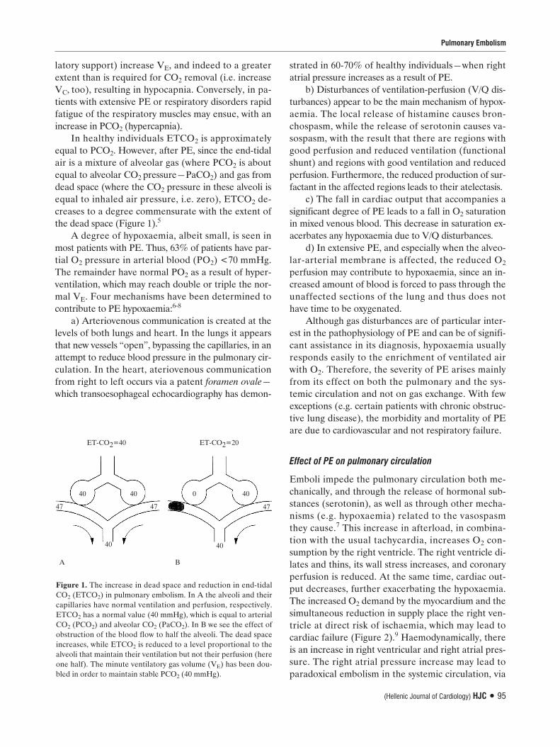
The obstruction of flow in embolisedarteries results in the creation of dead spacein the corresponding regions of the lung(Figure 1).5 An increase in dead space hasa direct effect on PCO2 and on end-tidalCO2 pressure (ETCO2). If VE does notchange—as is the case in patients in the in-tensive care unit (ICU) under mechanicalventilatory support and pharmaceuticalparalysis, where respiration is fully con-trolled—PCO2 will increase (provided thatCO2 production is unchanged). However,most patients (with non-mechanical venti-
94 ñ HJC (Hellenic Journal of Cardiology)
Pulmonary Embolism: Pathophysiology,Diagnosis, TreatmentELENI KOSTADIMA, EPAMINONDAS ZAKYNTHINOS
Intensive Care Unit, University Hospital of Larissa
Manuscript received:August 9, 2006;Accepted:November 24, 2006.
Address:
EpaminondasZakynthinos
3 Tampara St.,414 47 Larisa, Greecee-mail: [email protected]
Key words: Pulmonaryembolism risk factors,D-dimers, computedtomography,anticoagulant therapy,thrombolysis,embolectomy.
Hellenic J Cardiol 48: 94-107, 2007
Review ArticleReview Article
latory support) increase VE, and indeed to a greaterextent than is required for CO2 removal (i.e. increaseVC, too), resulting in hypocapnia. Conversely, in pa-tients with extensive PE or respiratory disorders rapidfatigue of the respiratory muscles may ensue, with anincrease in PCO2 (hypercapnia).
In healthy individuals ETCO2 is approximatelyequal to PCO2. However, after PE, since the end-tidalair is a mixture of alveolar gas (where PCO2 is aboutequal to alveolar CO2 pressure—PaCO2) and gas fromdead space (where the CO2 pressure in these alveoli isequal to inhaled air pressure, i.e. zero), ETCO2 de-creases to a degree commensurate with the extent ofthe dead space (Figure 1).5
A degree of hypoxaemia, albeit small, is seen inmost patients with PE. Thus, 63% of patients have par-tial O2 pressure in arterial blood (PO2) <70 mmHg.The remainder have normal PO2 as a result of hyper-ventilation, which may reach double or triple the nor-mal VE. Four mechanisms have been determined tocontribute to PE hypoxaemia:6-8
a) Arteriovenous communication is created at thelevels of both lungs and heart. In the lungs it appearsthat new vessels “open”, bypassing the capillaries, in anattempt to reduce blood pressure in the pulmonary cir-culation. In the heart, ateriovenous communicationfrom right to left occurs via a patent foramen ovale—which transoesophageal echocardiography has demon-
Pulmonary Embolism
(Hellenic Journal of Cardiology) HJC ñ 95
ET-CO2=40 ET-CO2=20
40 40 0 40
47
40 40
47 47
A B
Figure 1. The increase in dead space and reduction in end-tidalCO2 (ETCO2) in pulmonary embolism. In A the alveoli and theircapillaries have normal ventilation and perfusion, respectively.ETCO2 has a normal value (40 mmHg), which is equal to arterialCO2 (PCO2) and alveolar CO2 (PaCO2). In B we see the effect ofobstruction of the blood flow to half the alveoli. The dead spaceincreases, while ETCO2 is reduced to a level proportional to thealveoli that maintain their ventilation but not their perfusion (hereone half). The minute ventilatory gas volume (VE) has been dou-bled in order to maintain stable PCO2 (40 mmHg).
strated in 60-70% of healthy individuals—when rightatrial pressure increases as a result of PE.
b) Disturbances of ventilation-perfusion (V/Q dis-turbances) appear to be the main mechanism of hypox-aemia. The local release of histamine causes bron-chospasm, while the release of serotonin causes va-sospasm, with the result that there are regions withgood perfusion and reduced ventilation (functionalshunt) and regions with good ventilation and reducedperfusion. Furthermore, the reduced production of sur-factant in the affected regions leads to their atelectasis.
c) The fall in cardiac output that accompanies asignificant degree of PE leads to a fall in O2 saturationin mixed venous blood. This decrease in saturation ex-acerbates any hypoxaemia due to V/Q disturbances.
d) In extensive PE, and especially when the alveo-lar-arterial membrane is affected, the reduced O2
perfusion may contribute to hypoxaemia, since an in-creased amount of blood is forced to pass through theunaffected sections of the lung and thus does nothave time to be oxygenated.
Although gas disturbances are of particular inter-est in the pathophysiology of PE and can be of signifi-cant assistance in its diagnosis, hypoxaemia usuallyresponds easily to the enrichment of ventilated airwith O2. Therefore, the severity of PE arises mainlyfrom its effect on both the pulmonary and the sys-temic circulation and not on gas exchange. With fewexceptions (e.g. certain patients with chronic obstruc-tive lung disease), the morbidity and mortality of PEare due to cardiovascular and not respiratory failure.
Effect of PE on pulmonary circulation
Emboli impede the pulmonary circulation both me-chanically, and through the release of hormonal sub-stances (serotonin), as well as through other mecha-nisms (e.g. hypoxaemia) related to the vasospasmthey cause.7 This increase in afterload, in combina-tion with the usual tachycardia, increases O2 con-sumption by the right ventricle. The right ventricle di-lates and thins, its wall stress increases, and coronaryperfusion is reduced. At the same time, cardiac out-put decreases, further exacerbating the hypoxaemia.The increased O2 demand by the myocardium and thesimultaneous reduction in supply place the right ven-tricle at direct risk of ischaemia, which may lead tocardiac failure (Figure 2).9 Haemodynamically, thereis an increase in right ventricular and right atrial pres-sure. The right atrial pressure increase may lead toparadoxical embolism in the systemic circulation, via
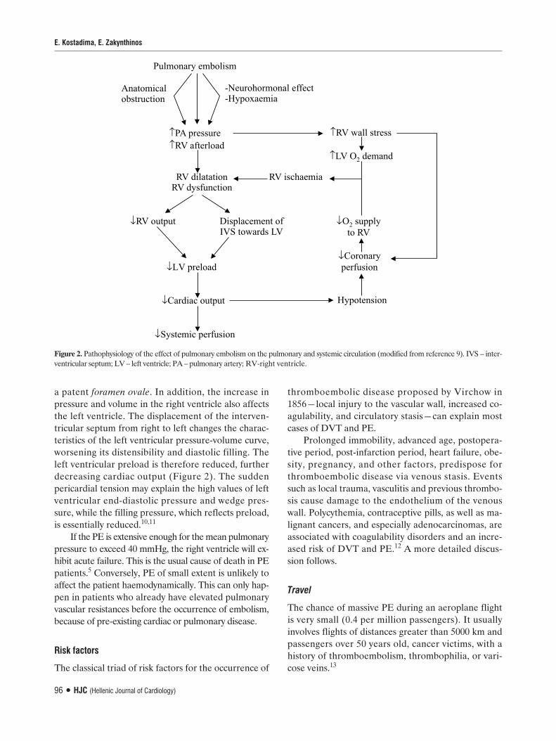
a patent foramen ovale. In addition, the increase inpressure and volume in the right ventricle also affectsthe left ventricle. The displacement of the interven-tricular septum from right to left changes the charac-teristics of the left ventricular pressure-volume curve,worsening its distensibility and diastolic filling. Theleft ventricular preload is therefore reduced, furtherdecreasing cardiac output (Figure 2). The suddenpericardial tension may explain the high values of leftventricular end-diastolic pressure and wedge pres-sure, while the filling pressure, which reflects preload,is essentially reduced.10,11
If the PE is extensive enough for the mean pulmonarypressure to exceed 40 mmHg, the right ventricle will ex-hibit acute failure. This is the usual cause of death in PEpatients.5 Conversely, PE of small extent is unlikely toaffect the patient haemodynamically. This can only hap-pen in patients who already have elevated pulmonaryvascular resistances before the occurrence of embolism,because of pre-existing cardiac or pulmonary disease.
Risk factors
The classical triad of risk factors for the occurrence of
thromboembolic disease proposed by Virchow in1856—local injury to the vascular wall, increased co-agulability, and circulatory stasis—can explain mostcases of DVT and PE.
Prolonged immobility, advanced age, postopera-tive period, post-infarction period, heart failure, obe-sity, pregnancy, and other factors, predispose forthromboembolic disease via venous stasis. Eventssuch as local trauma, vasculitis and previous thrombo-sis cause damage to the endothelium of the venouswall. Polycythemia, contraceptive pills, as well as ma-lignant cancers, and especially adenocarcinomas, areassociated with coagulability disorders and an incre-ased risk of DVT and PE.12 A more detailed discus-sion follows.
Travel
The chance of massive PE during an aeroplane flightis very small (0.4 per million passengers). It usuallyinvolves flights of distances greater than 5000 km andpassengers over 50 years old, cancer victims, with ahistory of thromboembolism, thrombophilia, or vari-cose veins.13
E. Kostadima, E. Zakynthinos
96 ñ HJC (Hellenic Journal of Cardiology)
Figure 2. Pathophysiology of the effect of pulmonary embolism on the pulmonary and systemic circulation (modified from reference 9). IVS – inter-ventricular septum; LV – left ventricle; PA – pulmonary artery; RV-right ventricle.
Obesity
The degree of correlation between obesity and theoccurrence of PE depends on the body mass index(BMI). The relative risk of PE has been found to be1.7 for BMI 25-28.9 kg/m2 and 3.2 for BMI 29 kg/m2
and above.14
Contraceptives
Third generation contraceptives, which contain newerprogesterones, appear to be free of side effects suchas acne and piliation. However, they have been impli-cated in actions related to the coagulation mecha-nism, such as resistance to activated protein C; thereis thus an increased risk of thromboembolism, indeedeven greater than for second generation contracep-tives.15 Advanced age and the smoking habit increasethe likelihood of complications among contraceptiveusers.16 Despite the increased risk of thromboem-bolism, the chance of a fatal episode of PE remainssmall.17
Pregnancy
During pregnancy, the risk of thromboembolism in-creases with the week of gestation. It often occurs dur-ing the gestation period, but more rarely after delivery.Advanced age and Caesarean section increase the like-lihood of thromboembolic disease.18
Hormone therapy
An important meta-analysis of 12 studies determinedthat the relative risk of thromboembolic disease inwomen under hormone replacement therapy in thepost-menopausal period is 2.1, with higher values (3.5)during the first year of treatment.19 An interesting ran-domised, placebo-controlled, prospective study foundthat the risk of PE in post-menopausal women taking acombination of oestrogen and progesterone was abouttwice that in the control group.20
The incidence of PE has also been investigatedin relation to the use of raloxifen and tamoxifen forthe prevention and treatment of breast cancer. Therate of occurrence of PE in recently published studieswas found to be 2.5 to 3 times higher than in controlgroups.21,22
Cancer
The chance of diagnosing a malignancy is increased
for around 2 years after an episode of thromboembolicdisease; usually, these are cancers of advanced stagewith a consequently poor prognosis.23,24 Especially inpatients with idiopathic thromboembolic disease, theexistence of cancer is very probable and should there-fore be checked for thoroughly.25
Trauma/surgery
Local trauma and orthopaedic operations, especiallyin the region of the pelvis, hips, thighs and knees,cause damage to the venous wall endothelium. It isbelieved that surgery predisposes to PE, for an inter-val of more than a month post operation. It has beenfound that 25% of cases of PE occur 15-30 days aftersurgery, and 15% after the 30th day. The 18th postop-erative day has the highest degree of risk.26
Thrombophilia
In one fifth of cases, genetic predisposition is the maincause of PE, although one of the classical risk factorsfrom Virchow’s triad may also be present. The doctorshould suspect genetic predisposition when there is:a) a strong family history of thromboembolic disease;b) thrombosis in unusual anatomical sites (upper bodyor upper limbs, when there is no central line catheter);c) repeated thrombosis with no known risk factors; d)thrombosis occurring at a young age; e) resistance tousual anticoagulant therapy.
It has been known for a long time that a lack ofprotein C, protein S, and antithrombin III is associat-ed with an extremely high risk of thromboembolic dis-ease. However, these genetic abnormalities are onlyidentified in 5% of patients with DVT.27 In an evensmaller percentage of these patients, insufficiency ofthe fibrinolytic system (hypoplasminogenaemia, ab-normal plasminogen, insufficiency of plasminogen-tPA activator) and insufficiency of factor XII may beencountered. Relatively recently, a mutation of factorV has been found (replacement of arginin with gluta-min in position 506 on factor V) which is known asfactor V Leiden. This hereditary abnormality is en-countered in a high proportion of the general popula-tion with heterozygous dominant form (3-4%) and isresponsible for 20% of cases of DVT.27 Factor V Lei-den increases coagulability, causing resistance to acti-vated protein C.28,29 Even though by itself factor VLeiden exerts only a mild thrombogenic effect, in-creasing coagulability by 2-3 times, the knowledge ofits existence is extremely important in circumstances
Pulmonary Embolism
(Hellenic Journal of Cardiology) HJC ñ 97
that increase resistance to activated protein C, suchas the use of contraceptive tablets or pregnancy. Theuse of contraceptives in combination with factor VLeiden increases the likelihood of thromboembolicdisease by 35 times.30 In conditions of increased pro-bability of thromboembolic disease—prolonged im-mobility, postoperative period, etc.—it is essential tointensify preventive treatment in patients who arecarriers of this factor.31,32
An elevated titre of antiphospholipid antibodies,especially lupus anticoagulant, is found in around 8.5%of cases of DVT, while being practically nonexistentin the general population.33 It should be noted that alarge number of patients with positive lupus anticoag-ulant do not suffer from systemic lupus erythemato-sus.
Hyperhomocysteinaemia is usually an acquiredabnormality, which increases the risk of thromboem-bolic disease by 2-3 times and is due to an insufficientintake of vitamins B1 and B6.
34
The investigation of hypercoagulant states in theacute phase of thromboembolic disease must neces-sarily include checks for: a) Factor V Leiden, becauseit is the most common anomaly responsible—checkedusing polymerase chain reaction; b) Hyperhomocys-teinaemia, because it may usually be treated com-pletely and quickly by the administration of vitaminsB1 and B6; c) Lupus anticoagulant, because if it is pre-sent it requires intensive and immediate therapy. Inthe acute phase it is not necessary to check proteinC, protein S, or antithrombin III, firstly because theyare rarely deficient, and secondly because their levelsin the blood decrease in acute thrombosis. In addi-tion, heparin lowers antithrombin III levels, whilecoumarin anticoagulants reduce the levels of pro-teins C and S.
Diagnosis
The clinical diagnosis of PE is particularly difficult,since it may easily be confused with other conditions; asa result it is often overlooked (Table 1). Venous throm-boembolic disease is often asymptomatic, which addsto the difficulty, while when symptoms of PE are pre-sent they tend to be non-specific. Tachycardia, chestpain, cough, unexplained loss of consciousness,and/or haemoptysis, raise the suspicion of PE, whilehypoxaemia, haemodynamic instability, syncopalepisode and/or cyanosis are characteristic of massivePE.36-38
Large series of patients have been studied with a
E. Kostadima, E. Zakynthinos
98 ñ HJC (Hellenic Journal of Cardiology)
view to evaluating the role of clinical signs, findingsand symptoms in the diagnosis of PE (Table 2).36,39
Pain of pleuritic type is usually associated withperipheral embolism, which causes irritation of thepleura and is associated with pulmonary infiltrationon X-ray. Histopathologically, it is associated withalveolar haemorrhage, often with haemoptysis as asymptom.39,40
Dyspnoea is mainly associated with central PE,which does not affect the pleura, although the haemo-dynamic consequences are more serious. It is the mostcommon symptom, while tachypnoea (>20 breaths/minute) is the most frequent sign of acute PE (70-80% of patients with angiographically proven PE ex-hibit dyspnoea).36
On clinical examination tachycardia is usuallyseen, while there may be signs of right heart failure,such as dilation of the jugulars with a V wave, leftparasternal cardiac pulsion, greatly increased pul-monary element of the second heart sound, and a sys-tolic murmur, low left parasternally, increasing duringinspiration on auscultation, probably with a thirdsound in the same region. These symptoms, of course,are often masked by tachypnoea, obesity, pithoidchest, etc.35
In arterial blood gases, the coexistence of hypox-aemia and hypocapnia help in the diagnosis of PE.However, these signs are not specific, since PO2 andPCO2 may be normal, especially in young people withno prior disease. In addition, PCO2 may be elevatedin patients with massive PE. ETCO2 is always re-duced, but lacks specificity.4-6,38
The presence or absence of risk factors for ve-nous thromboembolic disease is an essential piece ofknowledge for the evaluation of the likelihood of PE.We should be aware that the risk of PE increases withthe number of risk factors present, and that PE does
Table 1. Differential diagnosis of pulmonary embolism.
Pneumonia or bronchitisAsthmaExacerbation of chronic obstructive pulmonary diseaseMyocardial infarctionPulmonary oedemaAnxiety-hysteriaAortic dissectionLung cancerPrimary pulmonary hypertensionRib fracturesPneumothoraxMusculoskeletal pain
not usually occur in the absence of any risk factors.41
Isolated clinical signs and symptoms are not useful,since they have neither good sensitivity nor satisfacto-ry specificity (Table 2). For this reason, Wells et al42
proposed a prognostic rule (pretest probability) in-corporating 7 weighted variables for the diagnosisof PE: existence of clinical signs and symptoms ofthromboembolic disease (3 points), absence of alter-native diagnosis (3 points), heart rate above 100 (1.5points), immobility or surgery during the previous 4weeks (1.5 points), previous thromboembolic diseaseor PE, (1.5 points), haemoptysis (1 point), and malig-nancy (1 point). A total score less than 2 means lowprobability, 2-6 points medium probability, while ascore of over 6 points suggests a high probability ofPE.
Although the electrocardiogram and chest X-rayare of limited value, often being normal in patientswith PE, they should nevertheless be taken into ac-count.35 The most common electrocardiographic find-
Pulmonary Embolism
(Hellenic Journal of Cardiology) HJC ñ 99
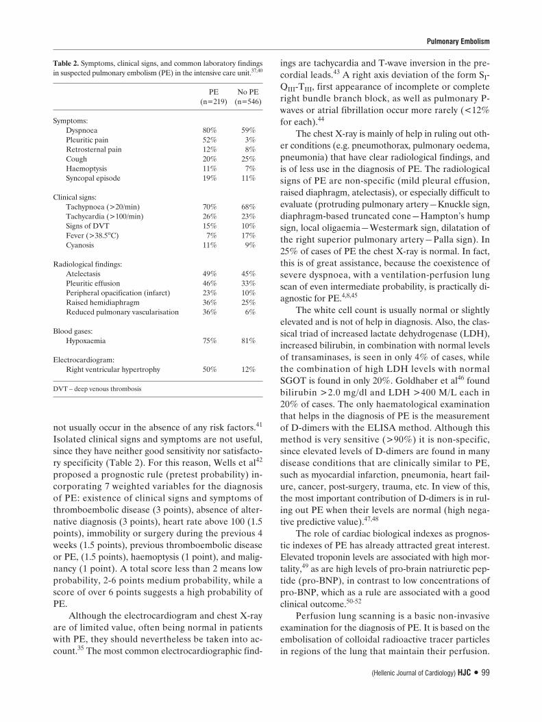
Table 2. Symptoms, clinical signs, and common laboratory findingsin suspected pulmonary embolism (PE) in the intensive care unit.37,40
PE No PE(n=219) (n=546)
Symptoms:Dyspnoea 80% 59%Pleuritic pain 52% 3%Retrosternal pain 12% 8%Cough 20% 25%Haemoptysis 11% 7%Syncopal episode 19% 11%
Clinical signs:Tachypnoea (>20/min) 70% 68%Tachycardia (>100/min) 26% 23%Signs of DVT 15% 10%Fever (>38.5oC) 7% 17%Cyanosis 11% 9%
Radiological findings:Atelectasis 49% 45%Pleuritic effusion 46% 33%Peripheral opacification (infarct) 23% 10%Raised hemidiaphragm 36% 25%Reduced pulmonary vascularisation 36% 6%
Blood gases:Hypoxaemia 75% 81%
Electrocardiogram:Right ventricular hypertrophy 50% 12%
DVT – deep venous thrombosis
ings are tachycardia and T-wave inversion in the pre-cordial leads.43 A right axis deviation of the form Sπ-Qπππ-Tπππ, first appearance of incomplete or completeright bundle branch block, as well as pulmonary P-waves or atrial fibrillation occur more rarely (<12%for each).44
The chest X-ray is mainly of help in ruling out oth-er conditions (e.g. pneumothorax, pulmonary oedema,pneumonia) that have clear radiological findings, andis of less use in the diagnosis of PE. The radiologicalsigns of PE are non-specific (mild pleural effusion,raised diaphragm, atelectasis), or especially difficult toevaluate (protruding pulmonary artery—Knuckle sign,diaphragm-based truncated cone—Hampton’s humpsign, local oligaemia—Westermark sign, dilatation ofthe right superior pulmonary artery—Palla sign). In25% of cases of PE the chest X-ray is normal. In fact,this is of great assistance, because the coexistence ofsevere dyspnoea, with a ventilation-perfusion lungscan of even intermediate probability, is practically di-agnostic for PE.4,8,45
The white cell count is usually normal or slightlyelevated and is not of help in diagnosis. Also, the clas-sical triad of increased lactate dehydrogenase (LDH),increased bilirubin, in combination with normal levelsof transaminases, is seen in only 4% of cases, whilethe combination of high LDH levels with normalSGOT is found in only 20%. Goldhaber et al46 foundbilirubin >2.0 mg/dl and LDH >400 M/L each in20% of cases. The only haematological examinationthat helps in the diagnosis of PE is the measurementof D-dimers with the ELISA method. Although thismethod is very sensitive (>90%) it is non-specific,since elevated levels of D-dimers are found in manydisease conditions that are clinically similar to PE,such as myocardial infarction, pneumonia, heart fail-ure, cancer, post-surgery, trauma, etc. In view of this,the most important contribution of D-dimers is in rul-ing out PE when their levels are normal (high nega-tive predictive value).47,48
The role of cardiac biological indexes as prognos-tic indexes of PE has already attracted great interest.Elevated troponin levels are associated with high mor-tality,49 as are high levels of pro-brain natriuretic pep-tide (pro-BNP), in contrast to low concentrations ofpro-BNP, which as a rule are associated with a goodclinical outcome.50-52
Perfusion lung scanning is a basic non-invasiveexamination for the diagnosis of PE. It is based on theembolisation of colloidal radioactive tracer particlesin regions of the lung that maintain their perfusion.

Thus, the normal regions opacify, while the regionsthat have undergone PE do not.46,53 Despite initialenthusiasm, this examination has not managed to re-place angiography, since its specificity is not satisfac-tory in the diagnosis of PE. Every cause of reducedperfusion in a region of the lung, such as hypoxic va-sospasm, or the presence of a West I zone, causes aperfusion defect indistinguishable from those of PE.False positive perfusion scans are seen in patients suf-fering from asthma, chronic obstructive pulmonarydisease, atelectasis, pneumonia, hypovolemia, etc., aswell as in patients who are on mechanical ventilationwith positive end-expiratory pressure.3 In contrast tothe pathological scan (which is non-specific), a nor-mal scan is particularly useful, essentially ruling outPE, since perfusion scintigraphy has high sensitivity.46
The specificity of the perfusion scan increases when itcan be combined with a ventilation scan. The pul-monary embolus shows a perfusion defect that is notassociated with a ventilation defect, or if one exists,the ventilation defect is small. Conversely, a perfusiondefect that is due to hypoxic vasospasm is usually ofsmaller size than the corresponding ventilation de-fect.4,46,53
For ease of evaluation, ventilation-perfusion (V/Q)scanning is classified as high probability, intermediateprobability and normal. In order to be classified ashigh probability for PE, a V/Q scan must have two ormore pulmonary regional perfusion defects, with noor very small changes in the ventilation scan and onthe chest X-ray. A high probability V/Q scan has 85%specificity in the diagnosis of PE, while for the inter-mediate probability scan the specificity is low. Unfor-tunately, however, high probability scans have lowsensitivity. In the PIOPED study it was found that on-ly 41% of patients with angiographically proven PEhad a high probability V/Q scan.53 This means thatmost patients with PE have an intermediate probabil-ity or a normal scan. Thus, the V/Q scan is consideredparticularly useful: a) when it is found to be normal,almost ruling out PE (especially when combined withnegative D-dimers), and b) when it is classified ashigh probability, in which case full treatment is rec-ommended, without the need for angiography. Ofcourse, the presence of another positive examination,or a high clinical suspicion of PE, reinforce the diag-nostic conclusion. In the case of an intermediateprobability scan further investigation is considered es-sential.54
Spiral computed tomography (CT) of the lung,with intravenous infusion of radiopaque medium, has
E. Kostadima, E. Zakynthinos
100 ñ HJC (Hellenic Journal of Cardiology)
been widely used in recent years for the diagnosis ofPE. Until recently, it had good sensitivity and speci-ficity in the identification of emboli in the large pul-monary arteries (80% and 90%, respectively), but itwas less successful in the peripheral vascular tree—beyond the third level of branching in the pulmonarycirculation.55 The new, multi-slice tomographic de-vices seem to be a great improvement in this respect.In a recent multi-centre study that used multi-sliceCT, the sensitivity and specificity were substantiallybetter (83% and 96%, respectively). Furthermore, ifCT was combined with imaging of the venous phase,the sensitivity increased significantly (90%) with noreduction in specificity. The positive prognostic valuewas evaluated as >90% if there was clinical suspicionof PE (96% with high clinical suspicion, 92% with in-termediate clinical suspicion).56 A combination ofmulti-slice CT and negative D-dimers essentially ruledout PE.57
Magnetic resonance pulmonary angiography withgadolinium may prove to be particularly safe and use-ful in the future,58 since apart from anatomical char-acteristics it provides information about right ventric-ular wall motion.59
Transthoracic echocardiography is considereduseful in the diagnosis of pressure overload in theright chambers. Thus, it is of particular use in identi-fying patients with a large PE, in whom the pressureof the pulmonary circulation is elevated (90% sensi-tivity).60 In these patients we can see dilatation ofthe right ventricle and hypokinesis of its free wall,while at the same time the shape of the left ventriclechanges, because of the displacement and flatteningof the interventricular septum, adopting a D shapeon its short axis during both systole and diastole.61 Acharacteristic sign is the maintenance of mobility ofthe apical region of the right ventricle—McConnellsign62—in contrast to the hypokinesis of the entirefree wall that is observed in chronic pulmonary hy-pertension.63 Measurement of systolic pulmonarypressure is feasible in 80-90% of cases with signifi-cant PE, via the accompanying tricuspid regurgita-tion, while diastolic pressure may be measured ifthere is also pulmonary valve insufficiency (Dopplermeasurements).63 However, in a recently publishedprospective study64 the echocardiogram was normaland failed to confirm the diagnosis in 50% of pa-tients with angiographically determined PE. Thetransoesophageal echocardiogram, a bedside exami-nation, is considered especially useful in ICU pa-tients with haemodynamic instability where moving
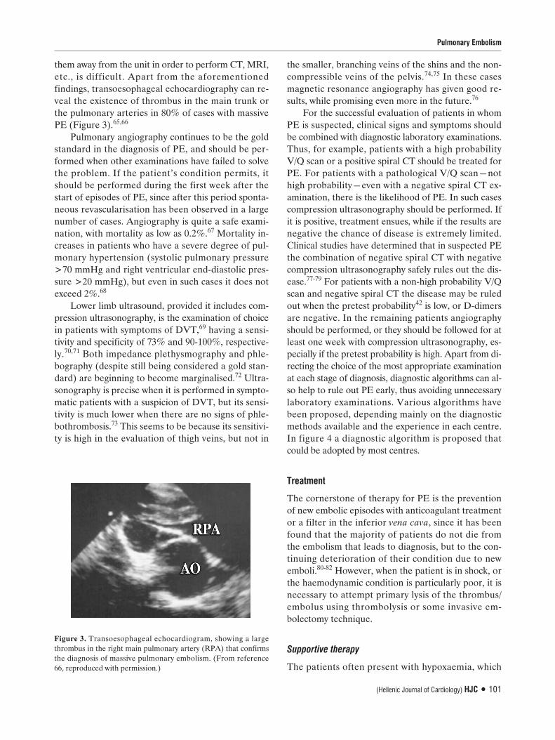
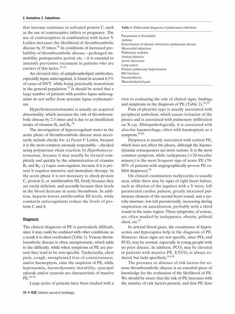
them away from the unit in order to perform CT, MRI,etc., is difficult. Apart from the aforementionedfindings, transoesophageal echocardiography can re-veal the existence of thrombus in the main trunk orthe pulmonary arteries in 80% of cases with massivePE (Figure 3).65,66
Pulmonary angiography continues to be the goldstandard in the diagnosis of PE, and should be per-formed when other examinations have failed to solvethe problem. If the patient’s condition permits, itshould be performed during the first week after thestart of episodes of PE, since after this period sponta-neous revascularisation has been observed in a largenumber of cases. Angiography is quite a safe exami-nation, with mortality as low as 0.2%.67 Mortality in-creases in patients who have a severe degree of pul-monary hypertension (systolic pulmonary pressure>70 mmHg and right ventricular end-diastolic pres-sure >20 mmHg), but even in such cases it does notexceed 2%.68
Lower limb ultrasound, provided it includes com-pression ultrasonography, is the examination of choicein patients with symptoms of DVT,69 having a sensi-tivity and specificity of 73% and 90-100%, respective-ly.70,71 Both impedance plethysmography and phle-bography (despite still being considered a gold stan-dard) are beginning to become marginalised.72 Ultra-sonography is precise when it is performed in sympto-matic patients with a suspicion of DVT, but its sensi-tivity is much lower when there are no signs of phle-bothrombosis.73 This seems to be because its sensitivi-ty is high in the evaluation of thigh veins, but not in
Pulmonary Embolism
(Hellenic Journal of Cardiology) HJC ñ 101
Figure 3. Transoesophageal echocardiogram, showing a largethrombus in the right main pulmonary artery (RPA) that confirmsthe diagnosis of massive pulmonary embolism. (From reference66, reproduced with permission.)
the smaller, branching veins of the shins and the non-compressible veins of the pelvis.74,75 In these casesmagnetic resonance angiography has given good re-sults, while promising even more in the future.76
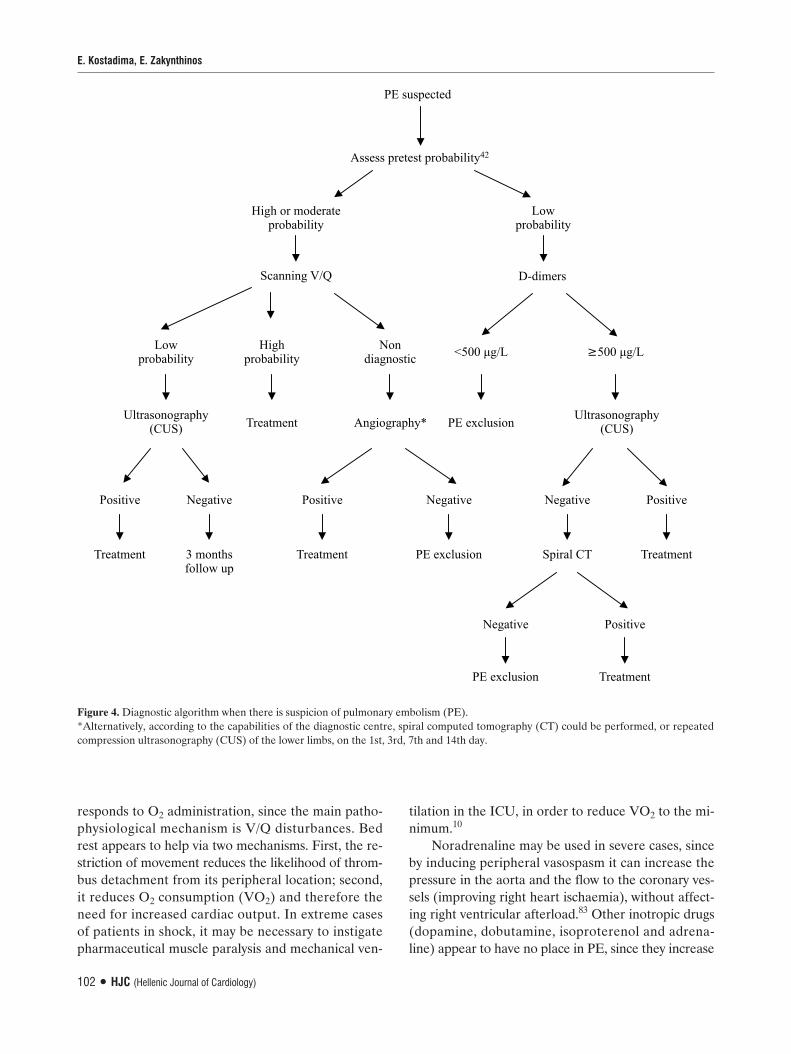
For the successful evaluation of patients in whomPE is suspected, clinical signs and symptoms shouldbe combined with diagnostic laboratory examinations.Thus, for example, patients with a high probabilityV/Q scan or a positive spiral CT should be treated forPE. For patients with a pathological V/Q scan—nothigh probability—even with a negative spiral CT ex-amination, there is the likelihood of PE. In such casescompression ultrasonography should be performed. Ifit is positive, treatment ensues, while if the results arenegative the chance of disease is extremely limited.Clinical studies have determined that in suspected PEthe combination of negative spiral CT with negativecompression ultrasonography safely rules out the dis-ease.77-79 For patients with a non-high probability V/Qscan and negative spiral CT the disease may be ruledout when the pretest probability42 is low, or D-dimersare negative. In the remaining patients angiographyshould be performed, or they should be followed for atleast one week with compression ultrasonography, es-pecially if the pretest probability is high. Apart from di-recting the choice of the most appropriate examinationat each stage of diagnosis, diagnostic algorithms can al-so help to rule out PE early, thus avoiding unnecessarylaboratory examinations. Various algorithms havebeen proposed, depending mainly on the diagnosticmethods available and the experience in each centre.In figure 4 a diagnostic algorithm is proposed thatcould be adopted by most centres.
Treatment
The cornerstone of therapy for PE is the preventionof new embolic episodes with anticoagulant treatmentor a filter in the inferior vena cava, since it has beenfound that the majority of patients do not die fromthe embolism that leads to diagnosis, but to the con-tinuing deterioration of their condition due to newemboli.80-82 However, when the patient is in shock, orthe haemodynamic condition is particularly poor, it isnecessary to attempt primary lysis of the thrombus/embolus using thrombolysis or some invasive em-bolectomy technique.
Supportive therapy
The patients often present with hypoxaemia, which
responds to O2 administration, since the main patho-physiological mechanism is V/Q disturbances. Bedrest appears to help via two mechanisms. First, the re-striction of movement reduces the likelihood of throm-bus detachment from its peripheral location; second,it reduces O2 consumption (VO2) and therefore theneed for increased cardiac output. In extreme casesof patients in shock, it may be necessary to instigatepharmaceutical muscle paralysis and mechanical ven-
tilation in the ICU, in order to reduce VO2 to the mi-nimum.10
Noradrenaline may be used in severe cases, sinceby inducing peripheral vasospasm it can increase thepressure in the aorta and the flow to the coronary ves-sels (improving right heart ischaemia), without affect-ing right ventricular afterload.83 Other inotropic drugs(dopamine, dobutamine, isoproterenol and adrena-line) appear to have no place in PE, since they increase
E. Kostadima, E. Zakynthinos
102 ñ HJC (Hellenic Journal of Cardiology)
Figure 4. Diagnostic algorithm when there is suspicion of pulmonary embolism (PE).*Alternatively, according to the capabilities of the diagnostic centre, spiral computed tomography (CT) could be performed, or repeatedcompression ultrasonography (CUS) of the lower limbs, on the 1st, 3rd, 7th and 14th day.

O2 consumption without a corresponding improve-ment in cardiac output.10
Fluid administration is contraindicated, as furtherdilatation of the right ventricle leads to an increase inmyocardial O2 consumption and greater restriction ofthe left ventricle, because of the displacement of theinterventricular septum, and therefore a reduction incardiac output.84
Anticoagulant therapy
Heparin is the basic treatment for PE, preventing theformation of new thrombi and giving time for the en-dogenous fibrinolysis to take effect, dissolving olderthrombi.80,85 Heparin administration should be startedimmediately, even before the diagnosis of PE is estab-lished, provided that anticoagulant therapy is not con-traindicated. The recommended regimen is rapid in-travenous administration of 5,000-10,000 IU (80IU/kg), followed by a continuous drip infusion of1,000-1,200 IU (18 IU/kg) per hour. The maintenancedose is determined by the activated partial thrombo-plastin time (aPTT), which should be between 60 and80 s (aPTT=1.5-2.5 times that before heparin admin-istration). If the aPTT is below the lower limit desired,the maintenance dose is increased by 200 IU, and arapid bolus infusion is given (for aPTT <35 s, 80 IU/kg; for aPTT 35-40 s, 40 IU/kg).86,87 In patients whoare resistant to heparin—defined as inability to achie-ve the desired aPTT with drip infusion above 50,000IU/24 hours—and in patients who exhibit thromboem-bolic disease with high aPTT prior to heparin adminis-tration (patients with lupus anticoagulant or other an-tibodies against anticardiolipin), the dosage is deter-mined on the basis of serum heparin levels (0.3-0.4IU/ml).88 Once the desired aPTT has been achieved,oral coumarin anticoagulants may be given. Coadmin-istration with heparin is required for 5 days, since thefull anticoagulant effect of coumarins is achieved with-in that time.89 The desired international normalisedratio (INR) in this case is 3, as heparin administrationprolongs it somewhat. An INR of around 2.5 is idealafter heparin is discontinued. The duration of antico-agulant therapy after PE has not yet been firmly es-tablished. The minimum interval seems to be 6months.90 In patients with repeated episodes of PE,lifelong treatment may be required,91 while in pa-tients deficient in antithrombin III, protein C or S,and those with factor V Leiden and PE, therapy islikely to be needed for many years, but not for therest of their lives.92,93
Pulmonary Embolism
(Hellenic Journal of Cardiology) HJC ñ 103
Relatively recently, low molecular weight heparinhas been used in the treatment of haemodynamicallystable PE, with similar efficacy and greater safetycompared to standard heparin.81,94,95 It should be not-ed, though, that treatment with low molecular weightheparin has not been used for massive PE and shouldnot be used until the necessary studies have been re-ported.
Filter placement in the inferior vena cava
Filter use is indicated in cases of PE where there arecontraindications for anticoagulant administration(active haemorrhage, endangered haemorrhage fol-lowing severe brain injury or craniotomy), or when re-peated episodes of PE occur despite full anticoagu-lant therapy.85 Filters do not appear to help in casesof proximal DVT with free-floating thrombus,96 whiletheir combination with anticoagulants does not im-prove survival in comparison with anticoagulant ther-apy alone.97
Thrombolysis
Thrombolysis should be performed immediately inpatients with circulatory shock, or obvious haemody-namic instability (massive PE). The outcome in thesepatients is clearly better in comparison with anticoagu-lant therapy alone.98-101 A recent meta-analysis found asignificant reduction in deaths and PE reoccurrencein studies that included haemodynamically unstablepatients (9.4% versus 19%).98
In patients with stable blood pressure, but signs ofright cardiac dysfunction on echocardiography (sub-massive PE), there is no consensus regarding the use ofthrombolysis.102-110 Konstantinides et al,108 in a recentlarge, randomised, blind study in which heparin was ad-ministered with either alteplase or placebo, observed abetter clinical course in the patients who were given al-teplase. Specifically, they found that in the alteplasegroup (rt-PA) fewer patients needed scaling of thera-py, i.e. a need for inotrope administration, secondarythrombolysis because of circulatory shock, intubationand mechanical ventilation, emergency surgical em-bolectomy, or cardiopulmonary resuscitation (24.6%versus 10.2%, p=0.004). However, the mortality didnot differ between the two groups.108
Supporters of thrombolysis in patients with sub-massive PE maintain that it directly improves cardiacfunction, while at the same time reducing the epi-sodes of reembolisation in these patients.104,108-110

Those who argue for heparin treatment alone in pa-tients with submassive PE, base their position on theincreased risk of haemorrhage as a result of throm-bolysis.105,106 The recent meta-analysis mentionedabove98 found that thrombolysis was associated with anon-significant increase in major haemorrhages (9.1%versus 6.1%; OR 1.42, 95% CI 0.81-2.46) but with asignificant increase in minor haemorrhages (22.7%versus 10.0%; OR 2.63, 95% CI 1.53-4.54).
From the above it appears that the benefit of throm-bolysis in this group of patients with submassive PEshould be assessed together with the risk of majorhaemorrhage, which increases with age.105,106 Review-ing a recent disagreement in the literature betweenKonstantinides109 and Dallen,106 concerning the roleof thrombolysis in submassive PE, Goldhaber107 con-cluded that thrombolysis would most probably have aplace in the subgroup of patients who are haemody-namically stable but at high risk. High risk patientscould be defined on the basis of echocardiographiccriteria and cardiac biological indexes (troponin, brainnatriuretic peptide). This would, of course, need to beevaluated in a randomised, prospective study.
In the case of thrombolysis, rt-PA in a dosage of100 mg, in continuous drip administration within twohours, seems to be superior to urokinase and strep-tokinase, which are also used. Thrombolysis is effec-tive (administration window) even 14 days after PE.111
Newer thrombolytic drugs that are used in cases ofinfarction (reteplase, tenecteplase, lanoteplase) havenot been tested in randomised studies of PE andshould not be given. For patients in whom aggressiveintervention is necessary, but the risk of haemor-rhage is high, the decision for embolectomy is con-sidered mandatory.112,113
Embolectomy
In patients with haemodynamic instability, in whomthrombolysis has failed or is contraindicated (intracra-nial haemorrhage, recent surgery or trauma), transve-nous catheter thrombectomy is performed.112 Variousdevices have been developed for the aspiration or pul-verisation of thrombus in the pulmonary circulation.114
If such a device is not available, or the procedure fails,surgical thrombus removal is indicated, with open tho-racotomy and extracorporeal circulation.115,116 Althoughemergency embolectomy has found wide acceptance,the results are not satisfactory, since the condition of pa-tients who are referred for surgery is extremely serious.Meyer et al112 reported 58% mortality among patients
E. Kostadima, E. Zakynthinos
104 ñ HJC (Hellenic Journal of Cardiology)
undergoing emergency embolectomy who suffered car-diac arrest. Cardiac arrest and cardiogenic shock are in-dependent additional risk factors.117,118 Rapid diagnosisand haemodynamic stabilisation play a determining rolein the improvement of results.117,118
Pulmonary endarterectomy in patients with pul-monary hypertension and evidence of thromboembol-ic disease on pulmonary angiography has been well-documented in the literature.119-123 Although the inci-dence of the disease after an acute episode of PE ishard to determine—many cases remain undiagnosed—it is estimated to be around 3.8% during the two firstyears after PE.124 These patients, if left without surgicaltreatment, have an extremely poor prognosis. The re-sults from centres with experience in these proceduresare of great interest. They report a significant reduc-tion in mean pulmonary pressure and pulmonary resis-tances, and mainly a significant improvement in the pa-tients’ World Health Organisation functional class,with high 5-year survival (74-86%).122,123 The perioper-ative mortality depends entirely on the experience ofthe centre. It seems that with greater experience mor-tality is limited to 4.4-9%.121-123 Independent factorsaffecting in-hospital mortality are advanced age (>60years) and the existence of mainly peripheral, and notcentral, thrombi in the pulmonary circulation.123 Per-sistent pulmonary hypertension and haemorrhage arethe usual causes of perioperative death.122,123
References
1. Goldhaber SZ, Visani L, De Rosa M: Acute pulmonary em-bolism: clinical outcomes in the International CooperativePulmonary Embolism Registry (ICOPER). Lancet 1999; 353:1386-1389.
2. Moser KM, Fedullo PF, LitteJohn JK, et al: Frequent asymp-tomatic pulmonary embolism in patients with deep venousthrombosis. JAMA 1994; 271: 223-225.
3. Smulders YM: Contribution of pulmonary vasoconstrictionto hemodynamic instability after acute pulmonary em-bolism. Implications for treatment? Neth J Med 2001; 58:241-247.
4. Bobadilla RA, Garcia-Juarez JA, Hong E, et al: Serotonergicreceptors involved in the hemodynamic changes observedduring pulmonary embolism. Proc West Pharmacol Soc 1991;34: 439-442.
5. Schmidt GA: Pulmonic embolic disorders: Thrombus, Airand Fat, in Hall JB, Schmidt GA, Wood LDH (eds): Princi-ples of Critical Care. McGraw-Hill 1992; pp 1476-1492.
6. Crapo RO, Jensen RL, Wanger JS: Single-breath carbonmonoxide diffusing capacity. Clin Chest Med 2001; 22: 637-649.
7. Elliott CG: Pulmonary physiology during pulmonary em-bolism. Chest 1992; 101(Suppl): 163S-171S.
8. Itti E, Nguyen S, Robin F, et al: Distribution of ventilation/
perfusion ratios in pulmonary embolism: an adjunct to the in-terpretation of ventilation/perfusion lung scans. J Nucl Med2002; 43: 1596-1602.
9. Wood KE: Major pulmonary embolism: review of a patho-physiologic approach to the golden hour of hemodynamical-ly significant pulmonary embolism. Chest 2002; 121: 877-905.
10. Belenkie I, Dani R, Smith ER, et al: Ventricular interactionduring experimental acute pulmonary embolism. Circulation1988; 78: 761-768.
11. Goldhaber SZ: Echocardiography in the management of pul-monary embolism. Ann Intern Med 2002; 136: 691-700.
12. Anderson FA Jr, Spencer FA: Risk factors for venous throm-boembolism. Circulation 2003; 107: 109-116.
13. Lapostolle F, Surget V, Borron SW, et al: Severe pulmonaryembolism associated with air travel. N Engl J Med 2001; 345:779-783.
14. Goldhaber SZ, Grodstein F, Stampfer MJ, et al: A prospec-tive study of risk factors for pulmonary embolism in women.JAMA 1997; 277: 642-645.
15. Rosing J, Middeldorp S, Curvers J, et al: Low dose oral con-traceptives and acquired resistance to activated protein C: arandomised cross-over study. Lancet 1999; 354: 2036-2040.
16. Vandenbroucke JP, Rosing J, Bloemenkamp KW, et al: Oralcontraceptives and the risk of venous thrombosis. N Engl JMed 2001; 344: 1527-1535.
17. Parkin L, Skegg DC, Wilson M, et al: Oral contraceptives andfatal pulmonary embolism. Lancet 2000; 355: 2133-2134.
18. Greer IA: Thrombosis in pregnancy: maternal and fetal is-sues. Lancet 1999; 353: 1258-1265.
19. Nelson HD, Humphrey LL, Nygren P, et al: Postmenopausalhormone replacement therapy: scientific review. JAMA 2002;288: 872-881.
20. Women’s Health Initiative Investigators: Risks and benefitsof estrogen plus progestin in healthy postmenopausal wo-men: principal results from the Women’s Health Initiativerandomized controlled trial. JAMA 2002; 288: 321-333.
21. Cummings SR, Eckert S, Krueger KA, et al: The effect ofraloxifene on risk of breast cancer in postmenopausal wo-men: results from the MORE randomized trial. JAMA 1999;281: 2189-2197.
22. Cuzick J, Forbes J, Edwards R, et al: First results from theInternational Breast Cancer Intervention Study (IBIS-I): arandomized prevention trial. Lancet 2003; 360: 817-824.
23. Schulman S, Lindmarker P: Incidence of cancer after prophy-laxis with warfarin against recurrent venous thromboem-bolism: Duration of Anticoagulation Trial. N Engl J Med2000; 342: 1953-1958.
24. Sorensen HT, Mellemkjaer L, Olsen JH, et al: Prognosis ofcancers associated with venous thromboembolism. N Engl JMed 2000; 343: 1846-1850.
25. Prandoni P, Lensing AW, Buller HR, et al: Deep-vein throm-bosis and the incidence of subsequent symptomatic cancer. NEngl J Med 1992; 327: 1128-1133.
26. Huber O, Bounameaux H, Borst F, et al: Postoperative pul-monary embolism after hospital discharge: an underestimat-ed risk. Arch Surg 1992; 127: 310-313.
27. Hyers TH: Venous thromboembolism. Am J Respir Crit CareMed 1999; 159: 1-14.
28. Bertina RM, Koeleman BPC, Koster T, et al: Mutation in bloodcoagulation factor V associated with resistance to activatedprotein C. Nature 1994; 369: 64-67.
29. Price DT, Ridker PM: Factor V Leiden mutation and the
Pulmonary Embolism
(Hellenic Journal of Cardiology) HJC ñ 105
risks for thromboembolic disease: a clinical perspective. AnnIntern Med 1997; 127: 895-903.
30. Bloemenkamp KWM, Rosendaal FR, Helmerhorst FM, et al:Enhancement by factor V Leiden mutation of risk of deep-vein thrombosis associated with oral contraceptives contain-ing a third-generation progestagen. Lancet 1995; 346: 1593-1596.
31. Ryan DH, Crowther MA, Ginsberg JS, et al: Relation of fac-tor V Leiden genotype to risk for acute deep venous throm-bosis after joint replacement therapy. Ann Intern Med 1998;128: 270-276.
32. Zakynthinos E, Pantazopoulos K: Acute mechanical mitralvalve thrombosis early after neurosurgery: is factor V Leidenmutation contributing? Int J Cardiol 2004; 96: 487-488.
33. Levine JS, Branch DW, Rauch J: The antiphospholipid syn-drome. N Engl J Med 2002; 346: 752-763.
34. Hankey GJ, Eikelboom JW: Homocysteine and vascular dis-ease. Lancet 1999; 354: 752-763.
35. Goldhaber SZ: Pulmonary embolism. N Engl J Med 1998;339: 93-104.
36. Stein PD, Terrin ML, Hales CA, et al: Clinical, laboratory,roentgenographic and electrocardiographic findings in pa-tients with acute pulmonary embolism and no pre-existingcardiac or pulmonary disease. Chest 1991; 100: 598-603.
37. Hirsh J, Hoak J: Management of deep vein thrombosis andpulmonary embolism. A statement for healthcare profession-als. Council on Thrombosis (in consultation with the Councilon Cardiovascular Radiology), American Heart Association.Circulation 1996; 93: 2212-2245.
38. Stein PD, Goldhaber SZ, Henry JW, et al: Arterial blood gasanalysis in the assessment of suspected acute pulmonary em-bolism. Chest 1996; 109: 78-81.
39. Miniati M, Prediletto R, Formichi B, et al: Accuracy of clini-cal assessment in the diagnosis of pulmonary embolism. Am JRespir Crit Care Med 1999; 159: 864-871.
40. Palla A, Petruzzelli S, Donnamaria V, et al: The role of suspi-cion in the diagnosis of pulmonary embolism. Chest 1995;107: 21-24.
41. Ferrari E, Baudouy M, Cerboni P, et al: Clinical epidemiolo-gy of venous thrombo-embolic disease. Results of a Frenchmulticentre registry. Eur Heart J 1997; 18: 685-691.
42. Wells PS, Anderson DR, Rodger M, et al: Derivation of asimple clinical model to categorize patients’ probability ofpulmonary embolism: increasing the model’s utility with theSimpliRED D-dimer. Thrombosis and Haemostasis 2000; 83:416-420.
43. Ferrari E, Imbert A, Chevalier T, et al: The ECG in pulmo-nary embolism: predictive value of negative T waves in pre-cordial leads—80 case reports. Chest 1997; 111: 537-543.
44. Daniel KR, Courtney DM, Kline JA: Assessment of cardiacstress from massive pulmonary embolism with 12-lead ECG.Chest 2001; 120: 474-481.
45. Pistolesti M, Miniati M: Imaging techniques in treatment al-gorithms of pulmonary embolism. Eur Respir J 2002; 19: 28-39.
46. Goldhaber SZ, Dricker E, Buring JE, et al: Clinical suspicionof autopsy-proven thrombotic and tumor pulmonary em-bolism in cancer patients. Am Heart J 1987; 114: 1432-1435.
47. Kelly J, Hunt BJ: Role of D-dimers in diagnosis of venousthromboembolism. Lancet 2002; 359: 456-458.
48. Wells PS, Anderson DR, Rodger M, et al: Excluding pul-monary embolism at the bedside without diagnostic imaging:management of patients with suspected pulmonary embolism
presenting to the emergency department by using a simple clini-cal model and d-dimer. Ann Intern Med 2001; 135: 98-107.
49. Konstantinides S, Geibel A, Olschewski M, et al: Importance ofcardiac troponins I and T in risk stratification of patients withacute pulmonary embolism. Circulation 2002; 106: 1263-1268.
50. Kucher N, Printzen G, Doernhoefer T, et al: Low pro-brainnatriuretic peptide levels predict benign clinical outcome inacute pulmonary embolism. Circulation 2003; 107: 1576-1578.
51. Wolde M, Tulevski II, Mulder JW, et al: Brain natriuretic pep-tide as a predictor of adverse outcome in patients with pul-monary embolism. Circulation 2003; 107: 2082-2084.
52. Kucher N, Printzen G, Goldhaber SZ: Prognostic role ofbrain natriuretic peptide in acute pulmonary embolism. Cir-culation 2003; 107: 2545-2556.
53. A Collaborative Study by the PIOPED Investigators: Valueof the ventilation/perfusion scan in acute pulmonary em-bolism-results of the prospective investigation of pulmonaryembolism diagnosis (PIOPED). JAMA 1990; 263: 2753-2759.
54. Bone RC: The low-probability lung scan: a potentially lethalreading. Arch Intern Med 1993; 153: 2621-2622.
55. Perrier A, Howarth N, Didier D, et al: Performance of helicalcomputed tomography in unselected outpatients with suspect-ed pulmonary embolism. Ann Intern Med 2001; 135: 88-97.
56. Stein PD, Fowler SE, Goodman LR, et al: PIOPED II Inves-tigators: Multidetector computed tomography for acute pul-monary embolism. N Engl J Med 2006; 354: 2317-2327.
57. Perrier A, Roy PM, Sanchez O, et al: Multidetector-row com-puted tomography in suspected pulmonary embolism. N EnglJ Med 2005; 352: 1760-1768.
58. Oudkerk M, van Beek EJ, Wielopolski P, et al: Comparisonof contrast-enhanced magnetic resonance angiography andconventional pulmonary angiography for the diagnosis of pul-monary embolism: a prospective study. Lancet 2002; 359:1643-1647.
59. Meaney JFM, Weg JG, Chenevert TL, et al: Diagnosis of pul-monary embolism with magnetic resonance angiography. NEngl J Med 1997; 336: 1422-1427.
60. Kasper W, Geibel A, Tiede N, et al: Distinguishing betweenacute and subacute massive pulmonary embolism by conven-tional and Doppler echocardiography. Br Heart J 1993; 70:352.
61. Jardin F, Dubourg O, Bourdarias JP: Echocardiographic pat-tern of acute cor pulmonale. Chest 1997; 111: 219-1217.
62. McConnell MV, Solomon SD, Rayan ME, et al: Regionalright ventricular dysfunction detected by echocardiography inacute pulmonary embolism. Am J Cardiol 1996; 78: 469-473.
63. Zakynthinos E, Zakynthinos S: Contemporary diagnosis andtherapy of pulmonary hypertension. Hellenic J Cardiol 1991;32: 111-123.
64. Miniati M, Monti S, Pratali L, et al: Value of transthoracicechocardiography in the diagnosis of pulmonary embolism: re-sults of a prospective study in unselected patients. Am J Med2001; 110: 528-535.
65. Pruszczyk P, Torbicki A, Pacho R, et al: Noninvasive diagno-sis of suspected severe pulmonary embolism. Transesophagealechocardiography vs spiral CT. Chest 1997; 112: 722-728.
66. Zakynthinos E, Douka E, Daniil Z, Konstantinidis K, Marka-ki V, Zakynthinos S: Anuria due to acute bilateral renal veinocclusion after thrombolysis for pulmonary embolism. Int JCardiol 2005; 101: 163-166.
67. Stein PD, Athanasoulis C, Alavi A, et al: Complications andvalidity of pulmonary angiography in acute pulmonary em-bolism. Circulation 1992; 85: 462-468.
E. Kostadima, E. Zakynthinos
106 ñ HJC (Hellenic Journal of Cardiology)
68. Permlutt LM, Braun SD, Newman GE, et al: Pulmonary an-giography in the high-risk patient. Radiology 1987; 162: 187.
69. Fraser J, Anderson R: Venous protocols, techniques, and in-terpretation of the upper and lower extremities. Radiol ClinN Am 2004; 42: 279-296.
70. Kearon C, Julian JA, Newman TE, et al: Noninvasive diagno-sis of deep venous thrombosis: McMaster diagnostic imagingpractice guidelines initiative. Ann Intern Med 1998; 128: 663-671.
71. Rose SC, Zwiebel WJ, Nelson BD, et al: Symptomatic lowerextremity deep venous thrombosis: accuracy limitations androle of color duplex flow imaging in diagnosis. Radiology1990; 175: 639-644.
72. Wells PS, Hirsh J, Anderson DR, et al: Comparison of the ac-curacy of impedance plethysmography and compression ul-trasonography in outpatients with clinically suspected deepvenous thrombosis: a two centre paired-design clinical trial.Thromb Haemost 1995; 74: 1423-1427.
73. Heijboer H, Buller HR, Lensing AWA, et al: A comparison ofreal-time compression ultrasonography with impedanceplethysmography for the diagnosis of deep-vein thrombosis insymptomatic outpatients. N Engl J Med 1993; 329: 1365-1369.
74. Pedersen OM, Aslaksen MA, Vik-Mo A, et al: Compressionultrasonography in hospitalized patients with suspected deepvenous thrombosis. Arch Intern Med 1991; 151: 2217-2220.
75. Davidson BL, Elliott CG, Lensing AWA, for the RD HeparinArthroplasty Group: Low accuracy of color Doppler ultra-sound in the detection of proximal leg thrombosis in asymp-tomatic high-risk patients. Ann Intern Med 1992; 117: 735-738.
76. Carpenter JP, Holland GA, Baum RA, et al: Magnetic reso-nance venography for the detection of deep venous thrombo-sis: comparison with contrast venography and duplex Dopplerultrasonography. J Vasc Surg 1993; 18: 734-744.
77. Kearon C: Excluding pulmonary embolism with helical (spi-ral) computed tomography: evidence is catching up with en-thusiasm. CMAJ 2003; 168: 1430-1431.
78. van Strijen MJ, de Monye W, Schiereck J, et al: Single-detec-tor helical computed tomography as the primary diagnostictest in suspected pulmonary embolism: a multicenter clinicalmanagement study of 510 patients. Ann Intern Med 2003;138: 307-714.
79. Musset D, Parent F, Meyer G, et al: Diagnostic strategy forpatients with suspected pulmonary embolism: a prospectivemulticentre outcome study. Lancet 2002; 360: 1914-1920.
80. Samama MM: Treatment of deep vein thrombosis and pul-monary embolism. Archives of Hellenic Medicine 2000; 17:75-77.
81. Mirianthefs M, Zampartas K: Pulmonary embolism – Case re-port. Advances in treatment. Cyprus Medicine 1994; 12: 24-29.
82. Kouraklis G: Pulmonary embolism as a clinical problem; itstreatment. Iatriki 1987; 52: 395-404.
83. Molloy WD, Lee KY, Girling L, et al: Treatment of shock ina canine model of pulmonary embolism. Am Rev Respir Dis1984; 130: 870.
84. Belenkie I, Dani R, Smith ER, et al: Effects of volume load-ing during experimental acute pulmonary embolism. Circula-tion 1989; 80: 178.
85. Tsapogas M: Contemporary aspects in vein thrombosis. Hel-lenic Surgery 1987; 59: 411-418.
86. Cruickshank MK, Levine MN, Hirsh J, et al: A standard he-parin nomogram for the management of heparin therapy.Arch Intern Med 1991; 151: 333.

87. Raschke RA, Reilly BM, Guidry JR, et al: The weight-basedheparin dosing nomogram compared with a “standard care”nomogram: A randomized controlled trial. Ann Intern Med1993; 119: 874-881.
88. Levine MN, Hirsh J, Gent M, et al: A randomized trial com-paring activated thromboplastin time with heparin assay inpatients with acute venous thromboembolism requiringlarge daily doses of heparin. Arch Intern Med 1994; 154: 49-56.
89. Pearson SD, Lee TH, McCabe-Hassan S, et al: A criticalpathway to treat proximal lower extremity deep vein throm-bosis. Am J Med 1996; 100: 283.
90. Schulman S, Rhedin A-S, Lindmarker P, et al: A comparisonof 6 weeks with 6 months of oral anticoagulant therapy after afirst episode of venous thromboembolism. N Engl J Med1995; 332: 1661-1665.
91. Schulman S, Granqvist S, Holmstrom M, et al: The durationof oral anticoagulant therapy after a second episode of ve-nous thromboembolism. N Engl J Med 1997; 336: 393-398.
92. Van den Belt AGM, Sanson B-J, Simioni P, et al: Recurrenceof venous thromboembolism in patients with familial throm-bophilia. Arch Intern Med 1997; 157: 2227-2232.
93. Hirsh J, Kearon C, Ginsberg J: Duration of oral anticoagu-lant therapy after first episode of venous thromboembolismin patients with inherited thrombophilia. Arch Intern Med1997; 157: 2174-2177.
94. The Columbus Investigators: Low-molecular-weight heparinin the treatment of patients with venous thromboembolism.N Engl J Med 1997; 337: 657-662.
95. Simonneau G, Sors H, Charbonnier B, et al: A comparison oflow-molecular-weight heparin with unfractionated heparin foracute pulmonary embolism. N Engl J Med 1997; 337: 663-669.
96. Pacouret G, Alison D, Pottier J-M, et al: Free-floating throm-bus and embolic risk in patients with angiographically con-firmed proximal deep venous thrombosis: a prospective study.Arch Intern Med 1997; 157: 305-308.
97. Decousus H, Leizorovicz A, Parent F, et al: A clinical trial ofvena caval filters in the prevention of pulmonary embolism inpatients with proximal deep-vein thrombosis. N Engl J Med1998; 338: 409-415.
98. Wan S, Quinlan DJ, Agnelli G, et al: Thrombolysis comparedwith heparin for the initial treatment of pulmonary embolism:a meta-analysis of the randomized controlled trials. Circula-tion 2004; 110: 744-749.
99. Jerjes-Sanchez C, Ramirez-Rivera A, Garcia ML, et al: Strep-tokinase and heparin versus heparin alone in massive pulmo-nary embolism: a randomized controlled trial. J ThrombThrombolysis 1995; 2: 227-229.
100. Arcasoy SM, Kreit JW: Thrombolytic therapy of pulmonaryembolism: a comprehensive review of current evidence. Ch-est 1999; 115: 1695-1707.
101. Goldhaber SZ, Haire WD, Feldstein ML, et al: Alteplase ver-sus heparin in acute pulmonary embolism: randomized trialassessing right-ventricular function and pulmonary perfusion.Lancet 1993; 341: 507-511.
102. Buller HR, Agnelli G, Hull RD, et al: Antithrombotic thera-py for venous thromboembolic disease: the Seventh ACCPConference on Antithrombotic and Thrombolytic Therapy.Chest 2004; 126(Suppl 3): 401-428.
103. Goldhaber SZ: Thrombolysis for pulmonary embolism. NEngl J Med 2002; 347: 1131-1132.
104. Konstantinides S: Should thrombolytic therapy be used in pa-tients with pulmonary embolism? Am J Cardiovasc Drugs2004; 4: 69-74.
105. Dong B, Jirong Y, Liu G, et al: Thrombolytic therapy for pul-monary embolism. Cochrane Database Syst Rev 2006; 19:CD004437.
106. Dalen JE: Thrombolysis in submassive pulmonary embolism?No. J Thromb Haemost 2003; 1: 1130-1132.
107. Goldhaber SZ: Thrombolysis in submassive pulmonary em-bolism. J Thromb Haemost 2004; 2: 1473-1474.
108. Konstantinides S, Geibel A, Heusel G, Heinrich F, Kasper W;Management Strategies and Prognosis of Pulmonary Embo-lism-3 Trial Investigators: Heparin plus alteplase comparedwith heparin alone in patients with submassive pulmonary em-bolism. N Engl J Med 2002; 347: 1143-1150.
109. Konstantinides S: Thrombolysis in submassive pulmonaryembolism? Yes. J Thromb Haemost 2003; 1: 1127-1129.
110. Konstantinides S: Diagnosis and therapy of pulmonary em-bolism. Vasa 2006; 35: 135-146.
111. Daniels LB, Parker JA, Patel SR, et al: Relation of durationof symptoms with response to thrombolytic therapy in pul-monary embolism. Am J Cardiol 1997; 80: 184-188.
112. Meyer G, Koning R, Sors H: Transvenous catheter embolec-tomy. Semin Vasc Med 2001; 1: 247-252.
113. Aklog L, Williams CS, Byrne JG, et al: Acute pulmonary em-bolectomy: a contemporary approach. Circulation 2002; 105:1416-1419.
114. Koning R, Cribier A, Gerber L, et al: A new treatment for se-vere pulmonary embolism: percutaneous rheolytic thrombec-tomy. Circulation 1997; 96: 2498-2500.
115. Gulba DC, Schmid C, Borst H-G, et al: Medical comparedwith surgical treatment for massive pulmonary embolism.Lancet 1994; 343: 576-577.
116. Meyer G, Tamisier D, Sors H, et al: Pulmonary embolectomy:a 20 year experience at one center. Ann Thorac Surg 1991; 51:232-236.
117. Stulz P, Schlapfer R, Feer R, et al: Decision making in thesurgical treatment of massive pulmonary embolism. Eur JCardiothorac Surg 1994; 8: 188-193.
118. Hartz RS: Surgery for chronic thromboembolic pulmonaryhypertension. World J Surg 1999; 23: 1137-1147.
119. Archibald CJ, Auger WR, Fedullo PF, et al: Long-term out-come after pulmonary thromboendartectomy. Am J RespirCrit Care Med 1999; 160: 523-528.
120. Jamieson S, Kapelanski D, Sakakibara N, et al: Pulmonaryendarterectomy: experience and lessons learned in 1,500 cas-es. Ann Thorac Surg 2003; 76: 1457-1464.
121. Mellemkjaer S, Ilkjaer LB, Klaaborg KE, et al: Pulmonaryendarterecomy for chronic thromboembolic pulmonary hy-pertension. Ten years experience in Denmark. Scand Cardio-vasc J 2006; 40: 49-53.
122. Ogino H, Ando M, Matsuda H, et al: Japanese single-centerexperience of surgery for chronic thromboembolic pulmonaryhypertension. Ann Thorac Surg 2006; 82: 630-636.
123. Pengo V, Lensing AW, Prins MH, et al: Incidence of chronicthromboembolic pulmonary hypertension after pulmonaryembolism. N Engl J Med 2004; 350: 2257-2264.
Pulmonary Embolism
(Hellenic Journal of Cardiology) HJC ñ 107