Embed Size (px)
Citation preview

609
609
28 The eye
28.1 Introduction
There are c. 285 million visually impaired people in the
world, of whom >39 million are blind. WHO estimates
that 43% of the visually impaired are so because of a lack
of spectacles while 30% have cataract. Major causes of
blindness in the world are cataract (50%), corneal
infections (particularly trachoma, 25%), glaucoma,
vitamin A deficiency, and onchocerciasis.
Washing the face regularly and the use of azithromycin
every 3 months in children under 12yrs reduces the
incidence of trachoma. In the industrial world 0·2% are
blind, but in low-income countries blindness is ten times
more common. You can treat cataracts, arrest glaucoma
and prevent trachoma and vitamin A deficiency.
It is unfortunate therefore that ophthalmology scares most
doctors, who imagine that treating the eye must be
impossibly difficult. This is not true: you can diagnose
90% of eye diseases with a torch and an ophthalmoscope.
Nonetheless, the eye may be difficult to examine,
particularly in a child, and if the eyelids are swollen or the
eye painful, the patient may forcefully resist examination.
Do not give up, because the signs of serious trouble may
be subtle. Danger signs are: haziness of the cornea,
inequality of pupil size (especially if associated with
reduced visual acuity), or circumcorneal redness.
Ideally someone in your district should be able to perform
cataract removals. Surgery inside the eye, however,
is difficult, so try to learn these operations by
apprenticeship from an expert; they are not described
here.
ANATOMY
The eye lies within the orbit, a V shaped enclosure,
designed to protect the eye from trauma. Its blood supply
comes from the ophthalmic artery, a branch of the carotid
artery. Six muscles are attached to the eye and wall of the
orbit; the IIIrd, IVth and VIth cranial nerves pass through a
fissure in the superior part of the orbit to supply the
muscles and the Vth cranial nerve gives sensation to the
eye.
A septum is attached to the rim of the orbit and the
eyelids are attached to this. The eyelids protect the
corneal surface are made from skin, muscle, tarsal plate
(28-18) and conjunctiva.
The eye itself can be divided into:
(a) the external eye: lids, conjunctiva, sclera,
(b) the anterior segment: cornea, iris and lens,
(c) the posterior segment: vitreous & retina, optic nerve.
Fig. 28-1 BASIC EYE ANATOMY.
A,B, flow of aqueous from the ciliary body (15) into the posterior
chamber (6), through the pupil into the anterior chamber (3), then
through the trabecular meshwork (19) via Schlemm’s canal (18) into
the scleral sinus (17). C, The visual axis passes through the middle of
the pupil (made by the iris (4) and through the centre of the lens
(6), and the vitreous (7) to the fovea (8) which is at the centre of the
macula (9). The optic nerve (11) enters the globe at the ‘blind spot’
and makes the optic disc (10). It is contiguous with the light-sensitive
retina (14), bounded by the choroid (13), and sclera (12), which joins
the cornea (2) anteriorly at the limbus (20), where the conjunctiva
makes a groove known as the fornix. The globe rotates within a
fascial layer, Tenon’s capsule, which covers the sclera and forms the
sheaths of the extraocular muscles, the outer layer joining the
conjunctiva at the limbus.
HISTORY
Always take a careful history; it may be critically
important. Focus on how vision has changed and whether
there is discomfort in the eye.
Vision can be divided into distance, near, peripheral,
stereo double or single), colour, or night vision.
Ask which type has been most affected.
Has vision been lost rapidly (specifically ask about
trauma, resulting in retinal detachment, haemorrhage,
or optic nerve damage), or slowly (cataract 28.4,
presbyopia 28.8, diabetic retinopathy 28.9)?
Is central vision lost (macular disease from diabetes,
macular degeneration, cataract) or peripheral vision
(glaucoma, retinal detachment, inherited eye disease)?
Remember ‘double vision’ may actually be blurred vision.
Ask about ‘floaters’ and ‘flashes’ in the vision.
Ask about ocular discomfort: conjunctival pain tends to
feel like sand or hair in the eye, while very high pressure
or inflammation of the eye can feel more like a deep ache
or throbbing pain. Note any watering of the eye.
Optic nerve disease can present with pain on eye
movements and loss of vision. Light sensitivity can be
due to inflammation of the eye, or sometimes cataract and
post trauma or surgery problems.
Ask about a family history of eye disease such as
glaucoma, cataract, or night blindness.

610
610
Fig. 28-2 TEST THE VISUAL ACUITY before you do anything
else. Stand the patient 6m from the test chart and ask her to tell you
if the 'three legs go up, down, right, or left'. After: www.talcuk.org.
EXAMINING AN EYE The standard examination of an
eye is time-consuming to do well, so train a nurse or
medical assistant to test the visual acuity and examine the
eyes. Your consulting room must be at least 6m long and
you should be able to darken it. You must have a good
pen light. Most examinations can be done while a patient
sits in front of you.
ALWAYS test the visual acuity. Explain that you want to
test the eyes. Begin by testing them separately
(with distance glasses if worn); test them again on each
subsequent visit; and record your results, so that you will
know if vision is deteriorating or not.
If the patient can read, test each eye separately either with
Snellen's or LogMAR charts. Stand him 6m from the
well-lit chart (28-2), and close the left eye with a piece of
paper or your left hand. Ask him to start at the top until he
cannot read any more. If he is a young child or cannot
recognize letters, use the tumbling E chart. It may be
helpful to get him to point with fingers in the direction of
the letters on the chart. Values are written with the top
figure as the distance in metres to the test chart, the
bottom one the distance at which a person with normal
vision can read that line. The standard chart is calibrated:
6/6, 6/9, 6/12, 6/18, 6/60, and 3/60: these represent
steadily deteriorating vision measurements. A value of
6/12 is normally required for driving a car. Counting
fingers (CF) at 3m is equivalent to 3/60. If CF<1m, try
hand movements (HM), and then test for the perception of
light (PL). Get children to point at pictures of objects.
Visual acuity can be usefully divided into four groups:
(1) good vision 6/6-6/18,
(2) poor vision 6/24-6/60,
(3) partially blind CF5m to PL,
(4) totally blind to NPL.
The LogMAR charts have 5 standard shape letters in each
row, varying logarithmically in size and spacing.
The score is based on the number of smallest letters read.
If you shine a torch into each of the 4 quadrants of the
visual field, can he tell you where it is coming from?
Blindness is ‘a loss of vision which results in the patient
being unable to continue with normal life, and to walk
unassisted’. It is usually equivalent to binocular vision of
<3/60, which is the same as CF<3m. Before you decide
that there is complete blindness, test with a very strong
light. If an eye cannot see any light, and its pupil does not
react to light, it is sure to be beyond help, so there is no
point advising otherwise. If the vision is normal and
remains normal and the eye is not inflamed, pathology is
unlikely.
Here is some basic eye equipment: it does not include
equipment for operating inside the eye. Many of the
instruments are very fine: look after them with the
greatest care:
CHARTS, visual acuity, (a) Snellen and (b) illiterate E charts, both for use at 6m. These are essential, and can usually be produced locally.
They have patterns of 'Es' of different sizes in different positions,
and can be used by patients who cannot read. TEST TYPE, reading pattern, Use this for examining older patients with
presbyopia (28.8) who need glasses. If necessary, you can also use a
book or newspaper. TORCH, for focal illumination, local pattern, preferably pen type,
with 'lens bulb'. A locally available torch is adequate: it can be easily
replaced, as can its bulb and batteries. LOUPE (x2-8 magnifying spectacles), binocular, surgical, headband
type, Some simple form of magnification is useful for examining the
front of the eye, for removing superficial foreign bodies, and for other kinds of fine work, such as suturing nerves.
TONOMETER, Schiötz, (28-3). You must be able to measure the
intraocular pressure (IOP) if you are going to diagnose glaucoma. Digital measurement is simple but unreliable. This instrument is not
much seen nowadays, but is still very useful.
OPHTHALMOSCOPE, simple pattern, Keeler type, battery handle. An ophthalmoscope is very useful, but you can do much good eye work
without one.
SLIT LAMP MICROSCOPE, on stand, simple pattern. You will find a slit lamp useful, although you can diagnose uveitis without one. If you
need to do a lot of eye work, this is very useful: spend some time,
though, with an experienced operator before you purchase one of these delicate instruments.
SPECULUM ophthalmic lid, solid blades, hinged with screw
adjustment. SCISSORS, ophthalmic lid, blunt points. If necessary, you can use any
fine scissors.
FORCEPS (clamp), tarsal cyst (chalazion, 28.12), 8mm ring, Lambert pattern. This has two blades, one with a ring and the other with a plate.
Use it to hold an eyelid while you incise a tarsal cyst. CURETTE chalazion (tarsal cyst).
CAUTERY simple type, ball pattern. Heat this on a spirit lamp, or get a
battery-operated type.. CLAMP, eyelid, entropion, Desmarre's or Snellen's, (a) medium &
(b) large. Use this to hold the eyelid when you operate for entropion.
SCISSORS, ophthalmic, spring pattern, Westcott's or Castroviejo's. These are very delicate instruments which need treating with special
care.
BLADE, Crescent type FORCEPS, fine, toothed, St Martin's.
RETRACTOR, eye, Desmarre's. Use this for examining children.
NEEDLE HOLDER, ophthalmic, curved with lock, Castroviejo pattern, LENS LOOP for cataract extraction
INTRAOCULAR LENS: standard PMMMA 21 dioptre are sufficient.
CHEAP SPECTACLES can be made from malleable wire and polycarbonate instead of glass for the lens (www.onedollarglasses.org)
Otherwise there are self-adjustable fluid-filled glasses available (28.8)
GLASSES, simple frames, second-hand if necessary, spherical lenses +1 to +3.50 - the most commonly needed glasses are +2 and +2·50. Collect
unwanted glasses and allow patients to try for themselves: this way you
can deal simply and effectively with the reading difficulties of many. Do not operate with the large instruments of a basic set. Use special fine
instruments listed above. For operations on the globe an operating
loupe and a bright focal beam are almost essential, using preferably a 12V spotlight or LED source.

611
611
THE PIN-HOLE TEST is a useful way of screening for
refractive errors. If there is poor vision, place a card with several
1mm holes (punched 5mm apart) in front of the eye.
For repeated use, attach the card to the insides of each lens of a
pair of spectacles. Cover each eye in turn for testing. If there is
an uncorrected refractive error, the vision will be significantly
improved to 6/9 or 6/12.
CHECK THE VISUAL FIELDS, in all 4 quadrants, while
sitting face-to-face with the patient, covering his right eye
and your left eye and vice versa, comparing them.
EXAMINING THE OUTER EYE
Start by looking at the whole face. Note any asymmetry
of the position of the eyes, or any protrusion (best judged
from above and behind the patient). Palpate for a lump,
and for high intra-ocular pressure.
Ask the patient to look down and keep looking down,
but not to actively close the eyes. Put the tips of both your
index fingers on one of the globes, so as to feel the sclera
through the upper lid above the upper border of the tarsal
plate.
Gently press with alternate finger tips towards the centre
of the globe:
(1) Gently fluctuate it from one finger to another.
(2) Indent it with one finger and estimate the sense of
fluctuation imparted to your stationary finger.
(3) Estimate the indentation of the sclera as you relax
your indenting finger. You can judge the eye to be
‘soft’ (<10mmHg), ‘normal’ (10-40mmHg), or ‘hard’
(>40mmHg). This is a crude test, and there must be a
significant rise of pressure (>40mm Hg) before you can
detect a raised intra-ocular pressure.
Test the movements of both the eyes together, and then
test each eye separately, in all directions, including
convergence. Note any squint (28.9).
If there is much pain, and the eyelids are in spasm,
a drop of sterile LA will make examination tolerable.
This will allow also you to insert a speculum to examine
the eye more easily. Whilst the patient is looking, grasp
the top lid with your finger, and slip the top blade of the
speculum under it. Then ask the patient to look up,
grasp the bottom lid, and slip the lower blade of the
speculum under that. Adjust the arm of the speculum until
the eye is exposed, and then tighten the locking nut.
CAUTION! Beware that the speculum does not press
on the eye or damage the cornea.
Note abnormalities of the lids, lacrimal apparatus,
puncta and canaliculi, the lacrimal glands and sacs,
and also any epiphora (tears running down the cheeks).
Do the eyelids open and close normally? You can see this
best on blinking. Check the lids for swellings, and check
that the lashes are in their normal position.
Look at the conjunctiva. Note particularly the distribution
of any redness. If it is maximal near the corneoscleral
junction, this occurs in iritis and corneal ulcer. If it is
maximal at the periphery but often extending all over, it is
likely to be conjunctivitis.
To examine the conjunctiva of the upper lid, evert it
(28-8H-K). This is necessary to exclude a foreign body.
Look for pus or mucopus in the inferior fornix.
This is present in all cases of bacterial conjunctivitis, and
in some cases of viral conjunctivitis. Look also for signs
of vitamin A deficiency: dry-looking conjunctivae, or
Bitot's spots (white patches on the temporal side of the
conjunctiva, produced by keratin mixed with gas-forming
bacteria).
Look at the cornea of each eye. Is it shining and clear,
reflecting the light of a torch, or its surface irregular?
(corneal ulcer). Is there clouding superiorly (trachoma,
28.13), or a general haziness? (oedema from trauma,
keratitis, or glaucoma). A bright light and a loupe can
detect keratic precipitates and adhesions (synechiae,
28-9B, of iritis).
If you suspect the surface is injured or ulcerated,
instil 1 drop of 2% fluorescein, or dip the end of a
fluorescein impregnated filter paper inside the lower lid
for a few seconds. Mop out the excess fluorescein with
tissue paper. Shine a light on the eye at an angle.
Gaps in the corneal epithelium (ulcers, abrasions) stain
bright yellow-green.
Look at the anterior chamber and note its depth.
Is there any blood (hyphaema), or pus (hypopyon, 28-9C)
at the bottom of the anterior chamber?
Look at the pupils. Do they look black and do they react
to bright light? Are the pupils grey or white? (opacities in
the lens, cataract). Note their size, shape, and if their
outline is irregular (synechiae, due to iritis, 28.5).
If a pupil constricts incompletely when light is shone into
that eye, and then constricts further when it is shone into
the good eye, and when the light is shone back into the
abnormal eye, both pupils enlarge, this means there is
optic nerve damage, commonly caused by glaucoma,
but you should exclude a stroke or brain injury.
This is known as a relative afferent pupillary defect.
FUNDOSCOPY (OPHTHALMOSCOPY) examines the
fundus and media of the eye. You must, either, dilate the
pupils with a mydriatic such as cyclopentolate 1%,
or do your examination in a dark room.
This is however ineffective where the vitreous or cornea is
opaque, or very unevenly curved (extreme astigmatism).
(1).Get the patient to keep both eyes open and look
straight ahead.
(2).Start with the '0' lens in the ophthalmoscope
(unless you have a refractive error and are not wearing
glasses; if so select the appropriate correcting lens and
remember this as your starting point.
(3).Use your right hand and your right eye for the
patient’s right eye and your left hand and your left eye for
the patient’s left eye.
(4).Hold the sight hole of the ophthalmoscope close to
your eye, resting it against your nose and orbit, and move
it with you as if it was attached to your head. To find this
position, look through the sight hole at some distant
object.

612
612
(5).With your thumb on the patient’s forehead,
gently raise the upper lid clear of the pupil.
(6).Start with the ophthalmoscope 20cm from the eye,
and shine the light into the pupil; it should glow
uniformly red (the red reflex). This indicates the absence
of a cataract.
(7).Move closer and watch for any opacities in the media
silhouetted against the red reflex. Corneal opacities
appear to move in the opposite direction to the
ophthalmoscope; vitreous and posterior lens opacities
appear to move with the ophthalmoscope. If you see a
shadow, use the + lenses (+5 to +12) to see it more
clearly.
(8).Ask the patient to look straight ahead, and move as
close as you can to the eye without touching the eyelashes
or cornea.
(9).Find and look at the optic disc: it is 15º to the nasal
side of the optical axis of the eye.
(10) Turn the lens wheel in the ophthalmoscope with your
forefinger from +6 down to zero to get the best view of
the disc. Examine:
(a) the vertical cup/disc ratio (a ratio of >0·7 suggests
glaucoma, 28.6, 28-11),
(b) the disc margins; if these are blurred all round (360º)
it suggests papilloedema,
(c) the blood vessels, look for nasal displacement of the
central retinal vessels and for haemorrhages and exudates
suggestive of diabetic retinopathy,
(d) the macula (28-1C), by asking the patient to look
directly into the light source, for black and white
pigmentation which may suggest choroiditis (28.5)
involving the macula (maculopathy).
Fig. 28-3 SCHIÖTZ TONOMETRY.
The scale is merely an example; use the scale which is supplied with
your instrument. 3 weights (5·5, 7·5, 10G) are usually supplied with
each instrument.
SCHIÖTZ TONOMETRY You may well never see this old-fashioned, but simple, instrument.
If you happen to find one, clean the instrument with a pipe cleaner and
ether. You’ll find it quite useful to diagnose glaucoma. It is however, a delicate instrument, so keep it carefully!
Using the standard 5·5g weight and the metal footpad, make sure the instrument is calibrated to zero.
Explain what you are going to do, lay the patient flat and instil LA into the conjunctiva. Ask him to open both eyes, and look straight up at a
target placed on the ceiling.
With the 5·5g weight in place, put the tonometer plunger gently on the centre of the cornea with the eye open, and read the scale. If in doubt,
repeat the reading 3 times. Use the tables provided with the instrument
to calculate the IOP from the scale reading. The normal IOP is 7-25mmHg. In practice, using the 5·5g weight, a
scale reading of ≤2 (>28mmHg) indicates a raised IOP. A reading of ≥3
(<25mmHg) is 'normal'. If the IOP is >40mmHg, the cornea is likely to become oedematous (the characteristic 'hazy cornea' of glaucoma), and
you can see this with a torch. This is usually a late sign of glaucoma.
BINOCULAR INDIRECT OPHTHALMOSCOPE
Fig. 28-4 BINOCULAR INDIRECT OPHTHALMOSCOPE.
This allows good examination of the anterior & posterior segments
at much less cost than a slit lamp: It is also portable (28-5). After Chaudhury A, Chugh A, Role of Indirect Ophthalmoscopy in Rural
Settings, Rural Surgery 2009;5(3):19-20.
(www.mercoframes.net/product/binocular-indirect-ophthalmoscope)
INDIRECT OPHTHALMOSCOPY
A binocular indirect ophthalmoscope (28-4) provides
stereoscopic, wide angle, high resolution views of the
entire ocular fundus. It is not hindered, as is the standard
ophthalmoscope by a hazy media or scleral or central
opacification. With the addition of a +20dioptre
condensing lens, by varying the illumination and viewing
angle, you can readily look at both the anterior and
posterior segments.
N.B. Examine layer by layer: lid margin → conjunctiva
→ cornea → anterior chamber → lens → vitreous.
Lid margin: plugged orifices, lice, erosions?
Conjunctiva: foreign body?
Cornea: foreign body embedded in the cornea? Ulcer?
Note its size and shape after instilling fluorescein and
using the blue light. On the back of the cornea look for
keratic precipitates (KP, these are clumps of white cells),
indicating uveitis.
Anterior chamber: look for cells and flare, pus and blood;
estimate its depth.
Lens: diffuse opacity, discolouration? Posterior synechiae
from the iris? Focal opacities?
Vitreous: Particles from a recent posterior uveitis,
or bleeding?

613
613
BINOCULAR INDIRECT OPHTHALMOSCOPY
Fig. 28-5 PRACTICE OF INDIRECT OPHTHALMOSCOPY.
Use a +20D magnifying lens held close to the patient as shown.
SLIT LAMP MICROSCOPY
Fig. 28-6 SLIT LAMP MICROSCOPY (A) shines a narrow pencil of
light illuminating the eye from an angle while it is examined with a
low-power microscope. B, layers of the cornea and lens
demonstrated. Particles in the aqueous and vitreous reflect light,
like dust particles illuminated by a sunbeam in a darkened room.
C, you may be able to see keratic particles directly with bright light.
A-B After Parr J. Introduction to Ophthalmology, OUP 2nd ed 1982,
with kind permission, C After Trevor Roper PD. Lecture Notes on
Ophthalmology, Blackwell 6th ed 1980, p.8.
SLIT LAMP MICROSCOPY. Use this for accurate
visualization of the anterior part of the eye and its
contents (28-6).
Position the head by placing the forehead and chin on the
rest. Vary the angle of the light as convenient.
Fig. 28-7 EXAMINING A BABY'S EYES.
Sit him on the mother's lap and hold the head between your knees.
BASIC EYE MEDICATION.
Drugs for use on the eye differ from other preparations:
for topical use they come either as ointments or drops.
The former are for longer-lasting effect, the latter for
immediate and, usually, short-lasting effect.
Some drugs are toxic to the eye through systemic use:
these include chloroquine and ethambutol.
Others are locally toxic, such as penicillin, or dangerous if
used for the wrong condition, such as steroids if used
when there is a herpetic corneal ulcer present.
N.B. Many antibiotic eye preparations also contain
steroids: avoid these!
Certain drugs are specifically used to help examination:
cyclopentolate 1%, or phenylephrine 10%, will dilate the
pupil for some hours only, so use these when you
want a temporary effect for example when using an
ophthalmoscope. Useful LA agents are: lidocaine
hydrochloride 4% or amethocaine hydrochloride 1%.
Remember that an anaesthetized eye is in great ganger if
an unnoticed foreign body gets into it, or that an abrasion
injury is not felt; so shield it (28-8B) after appropriate
examination.
To diagnose corneal injury, fluorescein papers are better
than fluorescein drops, because you can more readily
keep them sterile.
To use eye drops, pull the lower eyelid down so that you
can see the conjunctiva. Ask the patient to look up.
Put drops or ointment into the outer third of the
conjunctiva.
Close the eye for 2mins to allow the drug to enter the
eye. Do not let the dropper touch the eye, and do not put
the dropper down on a surface, as it may become
contaminated. If possible, each patient should keep his
own drops, because of the danger of cross-infection.

614
614
TO MAKE YOUR OWN CHLORAMPHENICOL EYE DROPS dissolve two 250mg capsules in 100ml of water. Filter the solution into
sterile 10ml dropper bottles. Screw the caps on loosely, and sterilize
them in a hot water bath or autoclave at 100ºC for 30mins, without letting water splash over the necks of the bottles. Refrigerate them; their
shelf-life is 2months at 2-8ºC. The shelf-life of commercial drops is only
4months, so this is a useful method.
Subconjunctival antibiotics are indicated if there is a
severe corneal infection or ulceration, especially with
hypopyon (28-9A,C).
N.B. If you use 0·2ml of 2% lidocaine, the injection will
be almost painless, but be careful mixing antibiotics and
LA in the same syringe to maintain sterility.
Fig. 28-8 SOME BASIC EYE METHODS.
A, eyepad. B, eye shield. C, insert the upper blade of a lid speculum
while the patient is looking down. D, insert the lower blade while he
is looking up. E, subconjunctival injection is an effective way of
getting a high concentration of an antibiotic inside the eye. F, insert
the first lid suture. G, stay sutures in place: 2 for the upper and
1 for the lower lid. H-K, steps in everting the upper lid.
Otherwise anaesthetize the eye with a few drops of LA
solution. Ask the patient to look up. Pull down the lower
lid, with your finger on the cheek. Use a sharp 0·4mm
needle on a 2ml syringe (28-8E).
Rest the needle flat on the conjunctival surface of the
globe, with the bevel facing away from it. Push the needle
under the conjunctiva, parallel to the surface of the globe,
rotating it gently as you do so.
If it is in the right layer, you will see its point under the
conjunctiva. Then inject 0·5mL (max 1mL).
If the infection is getting worse, repeat the injection the
next day. A severely infected eye is likely to improve,
or be lost within 6hrs, so use a subconjunctival injection
usually only once. If you fear the development of an
endophthalmitis, use IV antibiotics.
28.2 Operating on the eye
Try to get special training, especially for cataracts.
Learn how to do the more important procedures:
entropion (28.13), tarsal cysts (chalazion: 28.12),
tarsorrhaphy (28.10), and evisceration, enucleation,
and perhaps exenteration of the eye (28.14).
PREPARATION.
Prepare the face from the hairline to the chin and from ear
to ear, using iodine 10% in a non-alcoholic lotion which
will not harm the eyes, if it spills on them accidentally.
Make a special drape with a slit from the middle of one
end to the centre. Place this under the chin, and up each
side of the face. Fold it over the head and keep it there
with a towel clip. Place another drape across the forehead
over the eyebrows, and clip this to the first one.
If the patient is intubated, place a third drape over the
nose and the mount, which connects the patient’s
endotracheal tube.
If you are using LA, do not cover the nose or mouth.
ANAESTHESIA. You can usually use LA, using a
retrobulbar block with a 15mm 27G needle; otherwise use
ketamine or GA for a perforation (if LA is complicated by
retrobulbar haemorrhage, it may aggravate loss of eye
contents).
POSITION the table so that you can sit comfortably with
your knees underneath it. If necessary, put the head at the
foot end, or rest it on a board, or sheet of wood, pushed
under the mattress, and projecting beyond the table.
Sit your assistant on your left for a right eye, and on your
right for a left eye. Keep your own eyes on the patient.
This means you need to have trained your assistant to
hand you the right instruments properly.
Use a speculum, or lid stay sutures, to hold an eye open
while you operate on it (28-8G). These sutures serve
simply to hold the lids away from the eye while you
operate on it. They avoid the risk of a speculum
which may press on the eye, and perhaps scratch the
cornea.
In the upper lid insert two 3/0 silk or monofilament
sutures, just above the lash line and down to the tarsal
plate. In the lower lid insert one suture just below the lash
line. Do not penetrate the conjunctiva of either lid.
Hold these sutures with haemostats.

615
615
BLEEDING. The cornea is avascular and cannot bleed.
If the conjunctiva or sclera bleed, apply a pad and very
gentle pressure. Or irrigate the wound with saline from a
syringe and an irrigating needle. The blood will stream in
the clear saline, so that you can see the exact point where
it is coming from, and control it with cautery.
Heat a squint hook or a small cautery in the flame of a
spirit lamp, until it is hot, but not red hot.
Touch the bleeding point with this, through the stream of
saline. This will cool its tip enough to prevent burning,
but will leave it hot enough to seal the bleeding vessel.
Do not use diathermy.
PAD THE EYE if there is had a minor injury with no
suspicion of perforation (28-8A). An eyepad, with gentle
firm pressure, will reduce discomfort, and promote
healing by preventing the lids moving over the injured
area.
Close the eye, put a pad of gauze over it; place 2 pieces of
adhesive strapping diagonally across the pad, from the
forehead to the cheek, to hold the pad in place.
Change the pad daily, and look for signs of ulceration or
infection.
CAUTION! The great danger of an eyepad is that it
may rub against an anaesthetized eye, and cause an
abrasion. A layer of vaseline gauze on the pad will help to
avoid this. So shut the eye when you apply the pad.
SHIELD THE EYE:
(1).after any severe injury, especially if there is a
perforation.
(2) after any eye operation.
Shielding it (28-8B) allows it to open and close, without
anything extraneous touching the cornea, and perhaps
scratching it. A shield is the safest way to protect an
anaesthetized eye, and is very helpful for a painful
inflamed eye with photophobia.
Cut an 8cm diameter circle from cardboard, or an old
X-ray film. Cut a radius in this, fold it into a cone,
and maintain the cone with a piece of strapping.
Hold the cone in place with two pieces of adhesive
strapping, or plastic tape from the forehead to the cheek.
CAUTION! Never occlude the eye of a child <7yrs for
>7days, because this may cause amblyopia (28.9).
28.3 The painful red eye
Acute red painful eyes are due to:
(1) conjunctivitis (much the most common cause at any
age). In the newborn this is often due to gonococcus,
in children between 6months and 6yrs secondary to
measles, and in adults in endemic areas, chlamydia.
(2) a corneal ulcer.
(3) acute iritis.
(4) acute glaucoma.
(5) trauma.
The problem in a busy clinic is that conjunctivitis is so
much more common that these other causes are easily
missed. So your first task in managing red eyes is to make
sure that these rarer causes are recognized. The history,
the visual acuity, and the examination of the eye with a
torch should enable you to distinguish between
conjunctivitis and something more serious.
Conjunctivitis can be infectious, allergic, or chemical.
Bacterial conjunctivitis is common (especially from
neisseria, listeria and corynebacterium) in the developing
world, and may be mild, or so severe that the conjunctiva
extrudes pus, and the lids swell so much that the eyes
remain closed.
Bacterial conjunctivitis needs an antibiotic.
Viral conjunctivitis usually resolves spontaneously
without, if the cornea is not involved.
Allergic conjunctivitis rarely needs steroid treatment.
Besides infecting the conjunctiva, bacteria can infect the
lids (blepharitis), or the cornea, where they can cause
changes in the stroma (keratitis and sometimes a corneal
abscess), which may result in corneal ulceration, through
which infection may spread inside the eye as an
endophthalmitis, which may end in blindness.
A corneal ulcer may be due to:
(1) Bacteria.
(2) Herpes simplex virus.
(3) Fungi.
(4),Other conditions such as leprosy, causing incomplete
eyelid closure (lagophthalmos) and exposing the cornea to
trauma.
Demonstrate a corneal ulcer with fluorescein.
Bacterial infection can follow even a minor injury which
damages the epithelium, or it can be spontaneous.
Bacteria enter the eye through the anterior chamber.
If pus gathers there, you will see a fluid level (hypopyon:
28-9C) when the patient stands upright.
Endophthalmitis may be the result of:
(1) a corneal ulcer, especially bacterial.
(2) a perforating injury of the cornea or sclera, especially
if a foreign body has been left in situ, or if a wound is
neglected, or after recent eye surgery. Once bacteria have
entered the eye, the chance of total blindness is high.
If presenting early, when the infection is fairly localized,
some useful vision may remain. If you cannot control the
infection, an evisceration is necessary (28.14).
DIAGNOSIS.
If there is conjunctivitis, the discomfort is of a gritty
nature caused by rubbing of the conjunctivae on the
cornea; pain varies from mild to severe:
(1) Both the eyes are usually involved.
(2) The visual acuity is normally good.
(3) There usually is a purulent discharge.
(4) Red conjunctivae, especially in the fornices (28-6C).
(5) The cornea is clear and does not stain with fluorescein
(unless the conjunctivitis has produced a corneal ulcer).
(6) The pupils are normal.
(7) The tension in the globe is normal.

616
616
DIFFERENTIAL DIAGNOSIS.
Distinguish particularly between the redness of
conjunctivitis, which is typically bilateral and maximal at
the periphery, but is often uniform everywhere
(very common), with redness which is most marked at the
corneoscleral junction (less common).
CAUTION! Look for mucopus in the inferior fornix
(28-6C): it is always present in bacterial conjunctivitis;
hesitate to diagnose conjunctivitis if you do not find any.
Suggesting acute iritis (28.5): one (sometimes two)
moderately painful red eye(s) with no discharge.
Pain is often only mild. Reduction in visual acuity is
usually mild. A clear cornea is surrounded by redness at
the corneoscleral junction. A small constricted pupil
which becomes irregular on dilation, due to posterior
synechiae (adhesions) is typical. An inflammatory
exudate in the anterior chamber is visible most easily with
a slit lamp: the aqueous is not as clear as it should be.
The beam from the lamp shows a flare, like a beam of
light shining across a dusty room. You may also see little
lumps of cells (keratic precipitates or KP) sticking to the
back of the cornea, and posterior synechiae between the
iris and the front of the lens. The inflammatory cells in the
anterior chamber may form a sterile hypopyon.
The intraocular pressure (IOP) may be increased due to
secondary glaucoma (28.6).
Suggesting acute angle closure glaucoma (28.6):
one (seldom 2) very painful red eye(s) with severe
unilateral headache, and slight watering. There is severely
impaired visual acuity, often down to hand movements or
perception of light only, with haloes, and sometimes even
blindness. Circumcorneal hyperaemia is mild in the early
stages. A hazy cornea (due to raised IOP) without its
normal shine is associated with a shallow anterior
chamber; this is best seen by shining a torch from the
side. A vertically oval dilated pupil which does not react
to light is classical. IOP is raised (28.1).
Suggesting a corneal ulcer: one severely painful red eye
with reduced visual acuity (if the ulcer is central), scleral
redness most marked round where the ulcer is situated,
photophobia, swollen eyelids, and watering.
Look for a grey-white spot (the ulcer) on the cornea,
which stains with fluorescein. If it is not obvious,
look for a defect in the smooth surface of the cornea in the
reflection from a focused light. If the infection is severe,
pus cells sediment at the bottom of the anterior chamber,
with a fluid level (hypopyon). The pupil is usually
regular.
Suggesting a foreign body: The signs of an abrasion, and
a foreign body, are similar to those of a corneal ulcer:
unilateral pain, photophobia, a watery discharge,
sometimes impaired vision, and hyperaemia, which is
marked near the lesion. Ask if there is a history
suggesting trauma, and do not forget that contact lenses
are foreign bodies and easily become infected if not kept
scrupulously clean. Check underneath the upper eyelid!
ACUTE INFECTIVE CONJUNCTIVITIS
TREATMENT. Clean the eyes with a cotton swab and
saline. Instil chloramphenicol or ciprofloxacin ointment
hrly in severe infections, and 3hrly if less severe.
Continue for 2days after symptoms have resolved.
Allow the exudate to escape, clean the eyes with a clean
cloth and water, add an ointment at night to prevent the
eyelids sticking together, and do not put a pad on the
eyes.
If the conjunctivitis is severe, use subconjunctival
antibiotics. Watch carefully for a corneal ulcer, and if
necessary examine the cornea repeatedly with fluorescein.
If the conjunctivitis is very severe, and especially if there
is a corneal ulcer, instil chloramphenicol eye drops every
min for 1hr, every hour for 1day, and then 3hrly.
If the cornea is not clear and the visual acuity is poor,
there is a corneal ulcer and the eyesight is in danger.
If a neonate has severe conjunctivitis after birth
(ophthalmia neonatorum), this may be gonococcal or
chlamydial. This is an acute emergency, which may cause
blindness. Treat with chloramphenicol drops as above,
and add either ceftriaxone or gentamicin IV,
and oral erythromycin.
If you are treating a child between 6months and 6yrs,
check for a combination of malnutrition, vitamin A
deficiency, and recent measles. Look for:
(1) Night blindness (inability to see in dim light).
(2).Bitot's spots (white foamy spots on the lateral
conjunctiva).
(3).Xerosis (dryness of the conjunctiva with inability to
produce tears, or a dry hazy cornea).
(4).Keratomalacia (corneal ulceration, softening of the
cornea). Treat with vitamin A 200,000IU by mouth
immediately, again after 24hrs, and again after 1wk.
Also, use a topical antibiotic such as ciprofloxacin.
Improve the nutrition especially with plenty of dark green
leafy vegetables.
CHRONIC LOW-GRADE CONJUNCTIVITIS,
characterised by yellow-grey dots (follicles) under the
upper eyelid, in someone from an endemic area, is almost
certainly TRACHOMA caused by chlamydia trachomatis.
N.B. Different strains of this bacteria cause 3 distinct
diseases: urethritis & PID; lymphogranuloma venereum;
and trachoma.
Trachoma passes through 4 stages (28.13).
During the acute stage, make sure the patient actually puts
tetracycline eye ointment 1% into the eyes bd for 6wks.
Add a single dose of 1g azithromycin orally.
Advise thorough washing of the face and hands several
times daily, avoiding rubbing of the eyes. Explain that the
disease is due to the entry of dirt, often from flies,
but also from sharing face towels with an infected person.

617
617
ALLERGIC CONJUNCTIVITIS
Suspect this if large gelatinous vegetations have formed
on the upper tarsal conjunctiva, and look like
cobblestones, or on the bulbar conjunctiva surrounding
the limbus. It is common in children and young adults.
Their eyes may or may not be itchy but typically there is
extreme watering. Suppress the inflammation with
antihistamine drops or a very weak steroid.
Beware of steroid glaucoma (28.6, 28.12D),
because steroids, once started, may be needed for many
years. Inject triamcinolone 1ml (40mg) IM into the upper
eyelid for severe symptoms.
CORNEAL ULCER
This is an emergency needing admission. Start aggressive
treatment with antibiotics urgently. A shield (28.2, 28-8B)
or sunglasses will make life more comfortable.
Do not use an eyepad or patch.
If the ulcer is severe, and particularly if there is a
hypopyon, inject subconjunctival (28.1) gentamicin
20mg, or chloramphenicol 100mg and apply hourly
chloramphenicol 1%, or ciprofloxacin 0·3% eye ointment.
If the ulcer is not so severe, and there is no hypopyon,
treat as conjunctivitis.
Also, with any corneal ulcer, provided it has not already
perforated, use atropine eye ointment bd or tid to keep the
pupil dilated. This will prevent adhesions forming
between the iris and the lens (posterior synechiae, 28-9A).
Advise warm soaks: their use is effective for soothing a
painful eye. Wrap a cloth round a spoon, dip this into very
hot water, and let it cool till you can hold it as close to the
eye as is bearable. Soaks are also useful for a stye
(infected eyelash follicle, hordeolum).
Use vitamin A supplements if there is any suspicion that
it may be deficient.
COMPLICATIONS of corneal ulceration include:
(1) Diffuse scarring of the cornea (28.4).
(2) A dense white scar (leucoma: 28.4).
(3) Perforation of the cornea, with adherence of the iris,
and perhaps staphyloma (an opaque protrusion of the
cornea, not related to staphylococci).
(4) Endophthalmitis.
If there is pain and watering without a history of a
foreign body, look for a DENDRITIC ULCER (28-9E).
Stain the cornea with fluorescein and look for a branching
irregular pattern. This is due to infection by herpes
simplex. Dendritic ulcers occur especially after fevers,
particularly measles, malaria, and meningitis.
If possible use an antiviral agent: idoxuridine ointment
(x5 daily), trifluorothymidine drops (hourly), or aciclovir
ointment (x5 daily).
If the lesion is severe, combine this with mechanical
removal of the epithelium containing the virus.
Apply a topical anaesthetic, and stain the cornea with
fluorescein.
Using a loupe, a good light, and a ball of cotton wool on
the end of an applicator, gently scrub the surface of the
cornea in the region of the ulcer to remove its epithelium.
A chronic stromal keratitis with corneal scarring and
blindness can complicate herpetic eye disease.
CAUTION! Never apply steroids, because these may
spread the infection to the stroma of the cornea, and make
the condition worse.
ENDOPHTHALMITIS (PANOPHTHALMITIS)
The anterior chamber is full of pus.
If the endophthalmitis is early, with some hope of
vision, try to control infection and minimize pain.
Use subconjunctival chloramphenicol and IV gentamicin
for 7days. The infection may settle.
If the endophthalmitis is due to a foreign body in the
eye, remove it. It is usually superficial, so that it is
possible to remove it through the wound by which it
entered, which is usually in the cornea, even if this has to
be enlarged. Remove any prolapsing iris, and leave the
cornea unsutured. Use subconjunctival chloramphenicol
and IV gentamicin.
If presentation is late, with no hope of vision and an
anterior chamber full of pus, and the corneal ulcer has
weakened, softened, and distorted the globe
(phthisis bulbi, 28.4), especially with no improvement
after 48hrs antibiotic treatment, eviscerate the eye (28.14).
Be sure that the patient understands the necessity of
removing the eye because of the mortal danger of orbital
cellulitis and meningitis.
DIFFICULTIES WITH RED PAINFUL EYES
If a chemical has got into the eye, the conjunctiva is
intensely red (more so than in infective conjunctivitis),
the cornea may be opaque (from keratitis or an ulcer),
and the vision impaired. Unlike infective conjunctivitis,
mucopus is absent. Traditional medicine may have been
inserted for a painful eye, which has made it worse.
If the chemical is still present, wash it out with much
water, making sure it does not spill over the other eye.
Remember that if it is acid or alkali, a 1ml cavity needs 1l
water to alter the pH by a level of 3. Use an analgesic, and
shield the eye. Instil an antibiotic ointment; its vaseline
base will be soothing, and the antibiotic may prevent
secondary infection.
If there is an acutely inflamed and oedematous lid or
face, with a black slough, and surrounding thick
oedema, this may be ANTHRAX, especially if there has
been contact with animal carcasses or hides.
The eyelid may be completely destroyed, but the eye is
normal. Use high doses of IV penicillin and
sulphonamides. Anthrax responds rapidly to penicillin.
Later, if necessary, toilet the slough and graft the raw
area. If you leave raw lids ungrafted, severe scarring and
a scar-induced ectropion (lid eversion) will follow.

618
618
Fig. 28-9 THE IRIS AND THE CORNEA.
A, vertical section of the eye showing keratic precipitates floating in
the aqueous (1), posterior synechiae (2), and a hypopyon (3) B, iris
bombé: the iris is adherent to the lens all round and is bulging
forwards. C, acute bacterial corneal ulcer with a hypopyon.
D, acute iridocyclitis. The pupil is small and irregular, because
posterior synechiae have formed. E, dendritic ulcer of the cornea,
the result of herpes simplex infection.
After Parr J. Introduction to Ophthalmology, OUP 2nd ed 1982 with kind
permission.
28.4 Loss of vision in a white eye
This is one of the common presentations of eye disease.
Loss of vision in a white eye can be slow or fast.
If there is slow loss of vision over months or years,
there may be:
(1) A corneal scar.
(2) Cataracts.
(3) Glaucoma.
(4) A refractive error.
(5) Disease of the retina due to:
(a) Senile macular degeneration.
(b) Retinitis pigmentosa (congenital photoreceptor
deficiency)
(c) Chloroquine maculopathy.
(d) Old macular scars.
(6) Optic atrophy.
If there is sudden loss of vision over minutes or days,
the cause is usually inflammatory or vascular.
If the complaint is simply that reading is difficult,
especially in poor light, this is usually presbyopia (28.8).
Corneal scars cause 70% of blindness in children and
25% in adults in the developing world. They can be:
(1) Diffuse.
(2) A circumscribed white patch (leucoma).
(3).A staphyloma, which is a bulging of the cornea
forwards between the lids, due to its thinning, caused by
previous ulceration (not staphylococci).
(4).Phthisis bulbi, which is disorganization of the entire
eye, leaving it small and shrunken. Bilateral scarring
follows neonatal conjunctivitis (ophthalmia
neonatorum), vitamin A deficiency, traditional eye
medicine & trachoma (28.3,13). Unilateral scars are
more likely to be caused by corneal ulceration due to
bacteria, herpes simplex, fungi or trauma.
Cataracts cause about half the blindness in the world,
where the incidence is c.1:200. A large majority (85%)
of cataracts occur in the elderly, and the rest are either
congenital or familial, or due to trauma, iritis, or
diabetes.
Vitamin D deficiency causes lamellar (flaky) cataracts in
infants. Cataract presents with gradual loss of vision,
in one or both the eyes. The corneas are clear, and there
is an opacity in the pupil(s). A cataract can be immature
(making the pupil grey), or mature, or hypermature
(making it white). Sometimes a cataract swells, pushes the
iris forwards, occludes the angle of the eye, and causes
secondary glaucoma.
Removing cataracts is a standardized and repetitive task;
it is also a skilled one but is rarely urgent. To learn this it
is best to apprentice yourself to an expert for several
months, and try to remove at least 50 under supervision.
Or, better, send a motivated assistant to learn this skill.
Cataracts can often be removed on a mass scale in special
'eye camps'.
In good hands the chance of success is >90%.
If you operate on a patient for the right indications, even
moderate success in one eye only will provide much
sought-for independence.
Aim to insert an intra-ocular lens (IOL), which provides
much better vision. The IOL can now be obtained for a
reasonable price and is made in Eritrea & Nepal for
example, so this is no longer an impossible dream in the
developing world. It should be standard, as refractive
errors are better corrected and waiting for maturity of the
cataract is no longer necessary.
Manual small incision cataract surgery (MSICS) has
become the preferred extracapsular method for removal of
cataracts in low-income settings. It does not require
sutures, can be done inexpensively, and produces high
quality results. High cost cataract removal alternatives
are not necessary. An operating microscope is especially
valuable for reducing the incidence of complications;
the less experience the surgeon has, the more important
the microscope quality becomes. If you are more
experienced you can use loupes, but if you are
less experienced, you may cause problems which you
may not even see. A cheaper special MSICS microscope
is currently being developed.

619
619
Bad outcomes are related to:
(1) poor case selection, i.e. operating on patients who
actually have a corneal scar or glaucoma,
(2) complications such as vitreous loss or infection,
(3) uncorrected refractive error,
(4) postoperative posterior capsule opacification.
COMMON CAUSES OF GRADUAL LOSS OF VISION IN
A WHITE EYE
CORNEAL SCARS
If the cause of the scar is still present, and it is getting
worse, remove the cause. This may include scratching of
the cornea by the inwardly turned eyelashes of trachoma
(trichiasis, 28.13). Vitamin A deficiency causes an acute
ulcer in young children, and does not cause progressive
scarring.
If there is still adequate vision in the other eye and disability is not severe, no treatment is indicated.
If there is no light visible at all, explain that nothing can
be done.
If there is blindness, and a central leucoma which
obscures the pupil, with an area of clear peripheral
cornea, a peripheral iridectomy is necessary.
This will provide an artificial pupil peripherally, behind
the area of clear cornea, and should give enough vision
for independent mobility. It is contraindicated if there is
already enough vision for mobility, or if the peripheral
cornea is opaque.
If there is blindness due to diffuse corneal scarring
which has not made the eye perforate, a corneal graft is
the only solution.
If the eye is blind and painful, consider evisceration or
enucleation (28.14).
CATARACTS
(1);Measure the visual acuity accurately in both eyes.
The pupils should react briskly to light. If they do not,
suspect that there is also some other condition, such as
optic nerve disease.
(2) Measure the IOP to make sure that the loss of vision is
not due to glaucoma (28.6).
(3),Dilate the pupil and examine the red reflex with an
ophthalmoscope to assess how dense the cataract is,
especially if it is immature. If you can easily see the optic
discs, the cataract may not yet be dense enough to be
worth extracting.
CATARACT EXTRACTION (GRADE 2.5)
INDICATIONS.
(1) To improve sight.
(2).To treat complications, specially secondary glaucoma.
If there are bilateral cataracts, operate when the acuity
in both eyes has fallen to worse than 6/60 (CF at 6m).
If there is a unilateral cataract, surgery is only indicated
to treat or prevent secondary glaucoma, or uveitis. It will
not improve sight significantly.
If there is already loss of sight in the other eye for any
reason, and there is now a cataract in the remaining eye
(cataract in an only eye), delay surgery until there is
difficulty getting around independently and near blindness
(CF <3m), because any complication will cause total
blindness.
If the one cataract has already been successfully
removed, you can schedule the second cataract at any
time. But, this case now will be a lower priority.
If the cataract extraction is not possible,
atropine ointment weekly, or minus (concave) glasses
may improve eyesight.
CONTRAINDICATIONS.
(1);Unilateral cataracts with adequate sight in the other
eye.
(2);Bilateral small immature cataracts with acuity above
6/60 in both eyes together; review the progress in
3-6months.
(3) Active uveitis: do not perform a cataract extraction at
the same time as an iridectomy
METHOD
The principle is to make a self-sealing tunnel to extract
the cataract and insert the new lens. It may be combined
with a trabeculectomy (28.6).
Dilate the pupil with cyclopentolate 1%.

620
620
Fig. 28-10 MANUAL SMALL INCISION CATARACT SURGERY
A, superior rectus stay suture. B, fornix-based conjunctival flap.
C, scleral groove: a 6-7·5mm shelved incision posterior to the limbus
on the superior side of the eye.

621
621
D, make the scleral tunnel trapezoid-shaped with a crescent blade.
E, enlarge the tunnel. F, paracentesis: make a side port and inject
dye to stain the lens capsule and air to protect the underside of the
cornea.
G, capsulotomy: pierce the anterior capsule in a complete ring
c.7·5mm in diameter with a keratome.

622
622
H, capsulorhexis: tear off the anterior capsule with a special hook or
one made from a 27gauge needle. I, irrigate and dislodge the lens
capsule. Avoid touching the inner surface of the cornea! J, introduce
the lens loop within the capsular bag under the lens nucleus, and
slowly ease both loop and lens out, whilst irrigating and pressing on
the posterior lip of the tunnel. K, lens removed. Do not irrigate
>30sec. Do not aspirate the posterior capsular surface!

623
623
L, instil 0·3ml viscoelastic (a cohesive substance usually of sodium
hyaluronate 1·4%) and air to re-create the anterior chamber and
seal the incision. M, introduce the intraocular lens into the capsular
bag, holding onto the trailing haptic, which should curve to the
right. Make sure the lens is the correct way up! Do not hold onto the
lens itself!
Insert the eyelid retractor, or place stay sutures on the
eyelids. Grasp the superior rectus at its insertion, about
7·5mm behind the limbus (28-1C) and rotate the eye
inferiorly; insert a 4/0 or 5/0 stay suture through the
conjunctiva and beneath the muscle (28-10A).
Raise a flap by picking up the conjunctiva at the superior
limbus (junction of sclera and conjunctiva) and
buttonhole the conjunctiva with fine scissors.
Extend this in the sub-Tenon’s space and lift the
conjunctiva and Tenon’s capsule (28-1C) off the sclera
for 1cm (28-10B). Ensure haemostasis with cautery and
that the field is dry before you proceed to the next step.
Make a 6-7·5mm long curvilinear scleral partial thickness
(0·3mm deep) shelved incision 3mm posterior to the
limbus on the superior side of the eye (28-10C).
Deepen this incision by advancing the crescent blade into
the sclera and slowly cutting on either side, thus making
further room for the blade. Judge the correct depth by
making sure the crescent remains visible through the
sclera (28-10D).
Keep the crescent flat on the globe during dissection,
so that the tunnel depth remains uniform. Once you reach
the limbus, extend the tunnel by forward and backward
motion, cutting as you come out; this way, you create
scleral pockets on either side of the tunnel which becomes
trapezoid in shape with its inner margin 7-8·5mm,
i.e. larger than the outer margin 6-7·5mm long, adapted to
the size of the nucleus (28-10E).
Pierce the cornea just above the limbus to enter the
anterior chamber with the keratome. At this point you can
make the lens capsule more visible, by injecting 0·2ml
trypan blue dye through a sideport, using a 25gauge
cannula and adding a little air which protects the
underside of the cornea. Wait a full 30sec but no more,
otherwise you will dye all the tissues blue. Then wash out
the dye with balanced saline (0·9% saline made up to pH
7·3 with bicarbonate). Then deepen the chamber by
injecting 0·3-0·5ml air (28-10F).
Now open the tunnel into the anterior chamber by
advancing a keratome through the tunnel, tilting it
downwards, and advancing into the anterior chamber
(28-10G). Move the keratome medially and laterally the
full length of the tunnel while keeping the tip oft he blade
in the anterior chamber. Insert a 27G needle with the tip
bent slightly downwards like a hook.

624
624
Press the hook into the anterior capsular lens surface to
create a circular 360° opening about 7·5-8.0mm cutting
parallel to the limbus (28-10H).
Irrigate this space to help free the nucleus (28-10I);
rocking it side to side, or turning it round may help free it.
Then introduce a lens loop into the tunnel, and pass this
under the cataract in the capsular bag and slowly ease it
and the nucleus out of the anterior chamber (28-10J),
at the same time pressing gently on the posterior lip of the
tunnel to help expel it. Avoid touching the inner corneal
surface.
Continue irrigating as you manipulate the lens nucleus
out, and once it is out, pick up the sclera edge and aspirate
any cataract fragments, leaving a clear anterior chamber.
Keep pressing gently on the posterior lip of the tunnel to
allow débris to flow easily out. Do not irrigate for longer
than 30sec. Do not aspirate the posterior capsule.
Inject 0.3ml viscoelastic (28-10L) and insert a 21dioptre
polymethylmethacrylate (PMMA) intraocular lens into
the capsular bag (28-10M). This is the standard size and
is suitable for 80% or more of patients. Make sure the
lens is the correct way (flat or concave surface) up!
Hold the lens with long smooth (e.g. McPherson
long-angled) forceps with the leading haptic (curved hook
attached to the lens) sweeping to your left and the trailing
haptic curving to the right. Advance the lens by holding
the haptic with the forceps, but don’t hold the lens itself
with the forceps.
Recreate the anterior chamber by injecting 0·3-0·5ml air
through the sideport without applying any pressure on the
tunnel (28-10N). No sutures are required since the
properly formed tunnel acts as a one-way valve to prevent
leaks. The conjunctival flap becomes covered by
the eyelid, and needs no suture. Apply topical
chloramphenicol or ciprofloxacin, and dexamethasone
0·1% 3hrly for 1wk and then 6hrly for 2wks, with or
without LA.
N.B. Don’t forget to remove the stay suture in the superior
rectus muscle!
POSTOPERATIVELY, watch for a leaking wound
(with or without iris prolapse), infection, bleeding,
and a raised IOP. Gently open the lids, and examine the
eye with a torch.
If the patient is restless and expels the air bubble in
the anterior chamber causing it to flatten, take him
back to theatre and re-inject air through the wound.
If this keeps leaking out, suture the tunnel with 10/0
nylon.
If the remaining lens matter is swollen and fluffy,
keep the pupil dilated with atropine drops 1% bd.
If there is any iris prolapse, return to theatre, reduce the
prolapsed, make sure the anterior chamber is filled with
balanced saline, and suture the wound.
If the cornea is hazy with a striate pattern (striate keratitis): it will probably settle.
If there is blood in the anterior chamber (hyphaema),
pad the eye and insist on bed rest.
If the anterior chamber is shallow and the pupil not
round, the wound may be leaking. (You may prove this
with a fluorescin test.) Return to theatre and wipe the
wound with cellulose swabs, fill the anterior chamber
with 0·3-0·5ml air, and close the wound properly.
If there is pus in the anterior chamber (hypopyon),
there is infection (endophthalmitis). The eye is likely to
be painful and the visual acuity very low.
Use subconjunctival gentamicin or cefuroxime (28.1),
and topical chloramphenicol or ciprofloxacin hrly.
The eye may be lost in any case.
If the red reflex is absent after several months,
there is some opacity in the media. This may be from
re-growth of new fibres in the posterior capsule; perform
a CAPSULOTOMY by holding the medial rectus tendon
in forceps and pass a keratome through the cornea
laterally backwards to cut the posterior capsule.
Then keep the pupil dilated with 2 drops atropine 1% bd
and add 2 drops chloramphenicol 0∙5% 4hrly for 3days.
If there is much pain and the cornea is hazy, the IOP is
probably raised (aphakic glaucoma) so measure it.
The vitreous jelly may be blocking the pupil. Immediately
dilate the pupil with cyclopentolate and phenylephrine
drops, followed by atropine ointment for 6wks.
If visual acuity is not improved and there is no evidence
of endophthalmitis, increase topical steroid to 2hrly and
check for improvement in 1wk.
RARER CAUSES OF GRADUAL LOSS OF VISION IN A
WHITE EYE
Examine the macula and the optic cup with particular
care. For many of the following, there is no remedy.
If an old person has gradual loss of central vision,
atrophy, and irregular pigment at the maculae, suspect
senile macular degeneration.
If there are pale, white, flat optic discs (distinguish
these from the pale cupped discs of glaucoma, 28-11C),
and normal maculae, there is optic atrophy. Try to find
the cause (there are many, including a space-occupying
lesion around the optic chiasma).
If there is gradual loss of vision at any age, often
starting with night blindness, a family history,
and dark pigmentation which follows the retinal
vessels and takes the form of 'bone spicules',
suspect retinitis pigmentosa, a congenital disease of
photoreceptor loss.
625
625
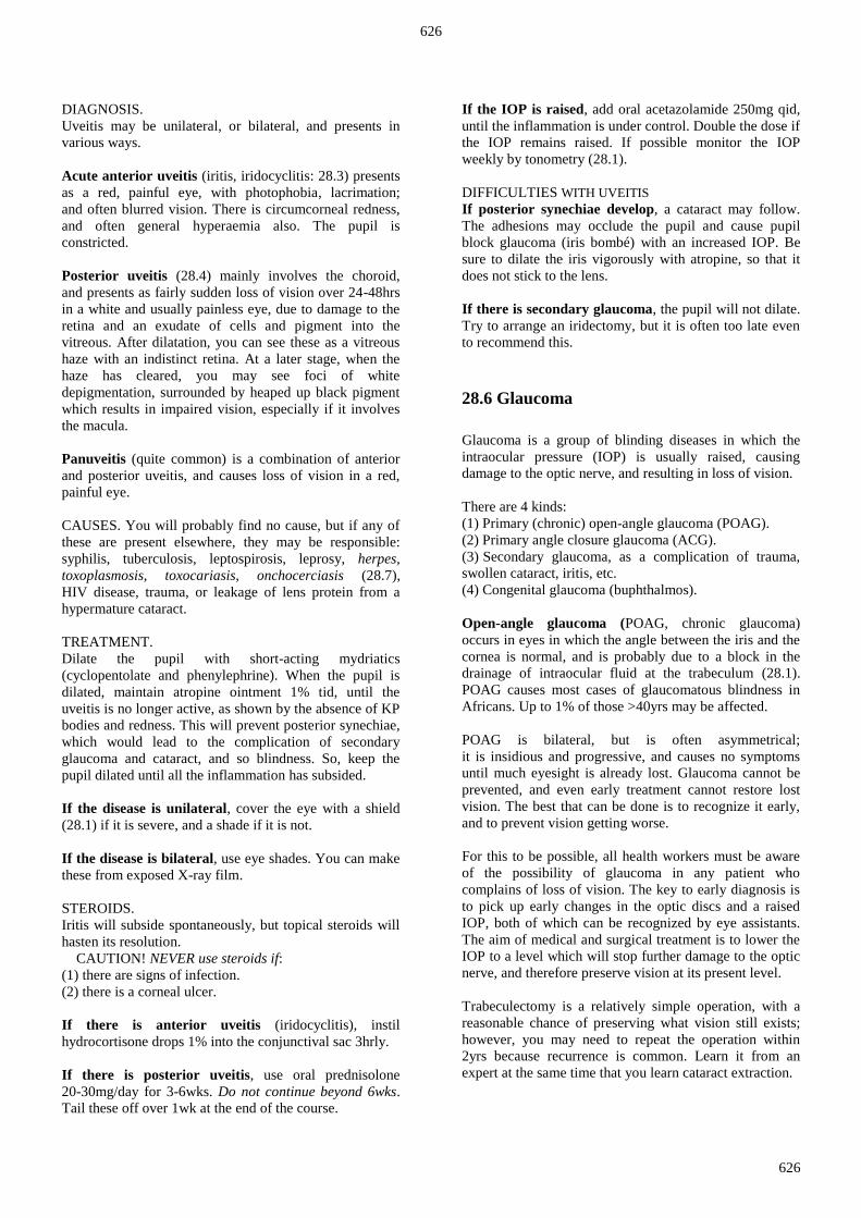
If there is gradual loss of central vision from excessive
doses of chloroquine (>1·5g weekly for >1yr),
or ethambutol, suspect maculopathy. The macula has a
typical 'bull's eye' pattern with a dark centre and a paler
surrounding ring. Stop the drugs.
If there are old macular scars (large white areas with
black edges, often around the optic disc and the
macula), they may be due to previous toxoplasmosis
(treat with pyrimethamine 25mg od & cotrimoxazole
40mg/kg od for 3wks) or toxocariasis (treat with
albendazole 5mg/kg bd for 5days)
SUDDEN LOSS OF VISION IN A WHITE EYE
Loss of vision can occur over minutes, hours or days,
in one or both eyes, which are white.
If at any age there is steady loss of vision over 24hrs,
in one eye or occasionally both eyes, suspect posterior
choroiditis (28.5) due to toxoplasmosis or other causes.
The important sign is inability to see the retinal vessels
due to hazy vitreous caused by inflammatory cells.
Treat as above.
If symptoms started with a flash of light followed by
black objects floating in the field of vision, and then a
curtain or cobweb across it, suspect retinal detachment.
Part of the retina may look grey-green. Dilate the pupil
and examine the fundus. You will see an abnormal red
reflex in one part of the fundus, with elevation of part of
the retina, and tortuosity of its vessels, which are difficult
to focus on. Expert surgery may save eyesight.
If there is instantaneous loss of vision, suspect
occlusion of the central retinal vein (a swollen disc with
many haemorrhages all over the retina), or artery
(a swollen disc, oedema of the retina, and often a
cherry-red spot at the macula). Or, suspect a stroke
(cerebrovascular accident).
Check the blood pressure. If it is not elevated, and visual
loss is less than 6hrs old, start anticoagulants. There is
otherwise no definitive treatment. If there is central retinal
vein thrombosis, follow up to check for secondary
glaucoma, which needs treatment.
If there is loss of central vision with an abnormal pupil
response to light, suspect optic neuritis (any age, usually
in the 3rd and 4th decades, and usually unilateral).
The vitreous and optic disc are usually normal. This will
usually improve over about 8wks. Bilateral optic neuritis
following methyl alcohol or quinine is permanent.
There is no specific treatment.
28.5 Anterior uveitis: iritis & iridocyclitis &
posterior uveitis: choroiditis
Any part of the uveal tract can become inflamed: the iris
(iritis), the ciliary body (cyclitis), or the choroid
(choroiditis). More than one part may be involved at the
same time (iridocyclitis).
Although iridocyclitis may be caused by bacteria invading
the eye through a corneal ulcer (28.3), it and other forms
of uveitis are more often due to a sterile inflammation,
usually from an unknown cause.
Uveitis of several kinds is common.
Iritis (more strictly iridocyclitis) has several
consequences:
(1) The inflamed iris may stick to the lens by posterior
synechiae (adhesions) or less often to the back of the
cornea by anterior synechiae.
(2) If the entire margin of the pupil sticks to the lens,
the iris balloons forwards (iris bombé: 28-9B), and causes
secondary glaucoma (28.6).
(3) Abnormal proteins enter the aqueous, and cause an
aqueous flare, which you can see with a slit lamp.
You can also see leucocytes as tiny particles floating in
the aqueous.
(4) These particles may stick to the back of the cornea as
keratic precipitates (KP), and they may be numerous
enough to gather at the bottom of the anterior chamber
similarly to a hypopyon. Unlike the hypopyon that results
from entry of bacteria through a corneal ulcer, the fluid in
iridocyclitis is usually sterile. Untreated iridocyclitis
eventually subsides spontaneously, typically in c.6wks,
leaving the eye severely damaged. It may relapse,
or it may be insidious and chronic, with few symptoms
except progressive loss of vision.
Uveitis presents in 2 ways (or when in both ways together
as panuveitis), as anterior uveitis (iritis) or posterior
uveitis (choroiditis); the former presents as an 'acute red
eye', so being one of the important differential diagnoses
of conjunctivitis (28.3), whilst the latter presents as
progressive loss of vision in a white eye (28.4).
Iritis is usually a sterile reaction to one of the infections
listed below. If onchocerciasis (28.7) is endemic, it will
certainly be the most common cause. Usually, no cause is
found, and iritis is presumed to be due to an autoimmune
disease. Atropine will keep the pupils well dilated, and
help break down synechiae. Steroid use is controversial:
it probably hastens resolution, but do not use it if there is
any sign of infection, especially a corneal ulcer.
Remember also that steroids:
(1) will make a red eye white, regardless of the cause,
without necessarily curing it;
(2) will suppress the normal inflammatory response,
without killing the causative agent;
(3) may raise intraocular pressure, and may rarely cause a
secondary glaucoma that could produce blindness;
(4) may cause a cataract if used long-term, but this will
not happen in the short time needed to treat acute iritis.
626
626
DIAGNOSIS.
Uveitis may be unilateral, or bilateral, and presents in
various ways.
Acute anterior uveitis (iritis, iridocyclitis: 28.3) presents
as a red, painful eye, with photophobia, lacrimation;
and often blurred vision. There is circumcorneal redness,
and often general hyperaemia also. The pupil is
constricted.
Posterior uveitis (28.4) mainly involves the choroid,
and presents as fairly sudden loss of vision over 24-48hrs
in a white and usually painless eye, due to damage to the
retina and an exudate of cells and pigment into the
vitreous. After dilatation, you can see these as a vitreous
haze with an indistinct retina. At a later stage, when the
haze has cleared, you may see foci of white
depigmentation, surrounded by heaped up black pigment
which results in impaired vision, especially if it involves
the macula.
Panuveitis (quite common) is a combination of anterior
and posterior uveitis, and causes loss of vision in a red,
painful eye.
CAUSES. You will probably find no cause, but if any of
these are present elsewhere, they may be responsible:
syphilis, tuberculosis, leptospirosis, leprosy, herpes,
toxoplasmosis, toxocariasis, onchocerciasis (28.7),
HIV disease, trauma, or leakage of lens protein from a
hypermature cataract.
TREATMENT.
Dilate the pupil with short-acting mydriatics
(cyclopentolate and phenylephrine). When the pupil is
dilated, maintain atropine ointment 1% tid, until the
uveitis is no longer active, as shown by the absence of KP
bodies and redness. This will prevent posterior synechiae,
which would lead to the complication of secondary
glaucoma and cataract, and so blindness. So, keep the
pupil dilated until all the inflammation has subsided.
If the disease is unilateral, cover the eye with a shield
(28.1) if it is severe, and a shade if it is not.
If the disease is bilateral, use eye shades. You can make
these from exposed X-ray film.
STEROIDS.
Iritis will subside spontaneously, but topical steroids will
hasten its resolution.
CAUTION! NEVER use steroids if:
(1) there are signs of infection.
(2) there is a corneal ulcer.
If there is anterior uveitis (iridocyclitis), instil
hydrocortisone drops 1% into the conjunctival sac 3hrly.
If there is posterior uveitis, use oral prednisolone
20-30mg/day for 3-6wks. Do not continue beyond 6wks.
Tail these off over 1wk at the end of the course.
If the IOP is raised, add oral acetazolamide 250mg qid,
until the inflammation is under control. Double the dose if
the IOP remains raised. If possible monitor the IOP
weekly by tonometry (28.1).
DIFFICULTIES WITH UVEITIS
If posterior synechiae develop, a cataract may follow.
The adhesions may occlude the pupil and cause pupil
block glaucoma (iris bombé) with an increased IOP. Be
sure to dilate the iris vigorously with atropine, so that it
does not stick to the lens.
If there is secondary glaucoma, the pupil will not dilate.
Try to arrange an iridectomy, but it is often too late even
to recommend this.
28.6 Glaucoma
Glaucoma is a group of blinding diseases in which the
intraocular pressure (IOP) is usually raised, causing
damage to the optic nerve, and resulting in loss of vision.
There are 4 kinds:
(1) Primary (chronic) open-angle glaucoma (POAG).
(2) Primary angle closure glaucoma (ACG).
(3);Secondary glaucoma, as a complication of trauma,
swollen cataract, iritis, etc.
(4) Congenital glaucoma (buphthalmos).
Open-angle glaucoma (POAG, chronic glaucoma)
occurs in eyes in which the angle between the iris and the
cornea is normal, and is probably due to a block in the
drainage of intraocular fluid at the trabeculum (28.1).
POAG causes most cases of glaucomatous blindness in
Africans. Up to 1% of those >40yrs may be affected.
POAG is bilateral, but is often asymmetrical;
it is insidious and progressive, and causes no symptoms
until much eyesight is already lost. Glaucoma cannot be
prevented, and even early treatment cannot restore lost
vision. The best that can be done is to recognize it early,
and to prevent vision getting worse.
For this to be possible, all health workers must be aware
of the possibility of glaucoma in any patient who
complains of loss of vision. The key to early diagnosis is
to pick up early changes in the optic discs and a raised
IOP, both of which can be recognized by eye assistants.
The aim of medical and surgical treatment is to lower the
IOP to a level which will stop further damage to the optic
nerve, and therefore preserve vision at its present level.
Trabeculectomy is a relatively simple operation, with a
reasonable chance of preserving what vision still exists;
however, you may need to repeat the operation within
2yrs because recurrence is common. Learn it from an
expert at the same time that you learn cataract extraction.

627
627
The symptoms of POAG are non-specific. There is slow
loss of vision in one or both the eyes over months or years
(28.4). Sometimes, there is marked loss of vision in one
eye, while the other eye is normal, or nearly so.
Occasionally, there is pain and headache, but this is late.
Glaucoma is often familial.
Angle closure glaucoma (ACG, acute glaucoma) usually
occurs >35yrs, in women more often than men, with an
abnormally narrow angle between the iris and the cornea.
If this angle should happen to close a little more than
usual, it causes an abrupt rise in the IOP with resulting
unilateral episodic attacks of pain, misty vision, and
rainbow-coloured haloes round lights. Between attacks
the eye is normal. Sooner or later, an episode of
raised IOP does not resolve, causing classical acute
congestive glaucoma (28.3). Acute glaucoma is
relatively uncommon, and is rare in Africa.
Its incidence is highest in Inuits and Mongolian
peoples, in Burma, and in South East Asia.
The dangers of atropine in glaucoma result from its
effect in dilating the pupil:
(1).The iris is kept away from the lens, and
prevents adhesions (synechiae) forming between
them, which is valuable in iritis.
(2).The iris is crowded into the angle of the anterior
chamber, where it impedes the drainage of
aqueous. This is never a desirable effect, but it does
not matter in a normal eye or in iritis;
it can however turn an eye blind if drainage is
already impaired by glaucoma!
So, use atropine in iritis, but not in glaucoma!
IF AN OLDER PERSON COMPLAINS OF POOR
VISION, CHECK FOR GLAUCOMA.
PRIMARY OPEN-ANGLE GLAUCOMA, (POAG, CHRONIC GLAUCOMA)
DIAGNOSIS. Measure the visual acuity of any patient
who presents with loss of vision. A hazy cornea or a pupil
which does not respond normally to light should make
you suspect glaucoma. If the IOP is >28, or the cup/disc
ratio is >½, there may be glaucoma (28-12G,H,I).
The end stage of glaucoma is a patient with a blind,
or nearly blind eye, with a large pupil that does not react
to light. Aim to diagnose it long before this with the
following three tests.
Loss of visual field is an early sign, but is not easy to test
with simple equipment.
CAUTION!
(1);The level of IOP is useful in confirming glaucoma,
but is not absolute, but is not always related to optic nerve
loss.
(2);Glaucoma can occur with a normal IOP (30% of
patients with glaucoma have an IOP <22mmHg).
(3);The IOP fluctuates, so if you are in doubt, repeat the
measurements over a few days.
(4) LOSS OF VISUAL ACUITY is not an early sign in
POAG.
(5);CUPPING OF THE OPTIC DISCS is the important
sign. Chronic glaucoma causes the discs to become
deeper and wider, and the remaining rims of disc tissue to
atrophy (28-11). One eye is commonly affected more than
the other, so that a definite difference between the eyes is
probably abnormal. Enlargement starts at the upper or
lower margins, so that a vertically ovoid cup with a
cup/disc diameter ratio of >½ is probably abnormal.
Eventually, the margin of the cup approaches the margin
of the disc, so that only a narrow rim of tissue remains.
Its wall becomes steep, so that vessels bend abruptly as
they reach the level of the surrounding disc. If the edge of
the disc overhangs the cup, you may lose sight of the
vessels until they appear over the edge of the cup.
Fig. 28-11 SOME OPTIC DISCS OF THE LEFT EYE.
A, normal optic disc with a.moderately sized cup. The lamina
cribrosa is a mesh-like structure of collagen in the sclera wall which
maintains the pressure gradient. B, another normal optic disc.
This has a large physiological cup and a temporal scleral crescent.
C, optic disc of a patient with gross 'chronic' open-angle
glaucomatous cupping. After Parr J. Introduction to Ophthalmology. OUP 2nd ed 1982, with kind permission
A large physiological cup can, however, be difficult to
distinguish from that in early glaucoma. With practice,
eye assistants should be able to distinguish 'normal discs',
'suspicious discs' and 'advanced glaucomatous cupping'.
This is the most important sign to look out for in
glaucoma: you usually need to dilate the pupils.
Normal discs:
(a) The discs are the same in both eyes.
(b) The ratio of optic cup to optic disc is ≤½ (28-12G).
(c) The cup is circular and the periphery of the disc
(the optic nerve rim) is pink.
(d) The appearance of the disc remains constant over
time.
Signs suggestive of glaucoma:
(a) A cup/disc ratio >½.
(b) A vertically oval cup, perhaps with notching at the
upper or lower poles.
(c) An area of pallor >30% of the disc area.
(d) Asymmetry of the cup/disc ratio of the 2 eyes.

628
628
(6) ABNORMAL PUPIL RESPONSES are a useful way
of testing for glaucoma, and only need a torch.
Initially, one pupil does not react as briskly as the other.
Finally, there is no response at all.
If you shine a light into a normal eye in a semi-dark room,
its pupil will constrict (direct response), and so will also
the other pupil automatically (consensual response).
If the optic nerve is completely destroyed, there will be
no direct or consensual response (total afferent pupil
defect).
If the optic nerve is partly destroyed (for example 90%),
the pupil will constrict slowly when the light shines in it
(partial afferent pupil defect), and the consensual response
will be present.
The swinging torch test, is a useful test for early
asymmetrical optic nerve damage, and does not need an
ophthalmoscope. It is theoretically difficult, but is easy in
practice. In a semi-dark room shine a light into the good
eye, and then swing it across into the bad eye
(the eye with reduced vision).
As the light shines in the good eye, the pupil of the bad
eye will constrict. As you swing the light quickly across
to the bad eye, its pupil, which was previously
constricted, will now dilate. This indicates a relative
afferent pupil defect, early optic nerve damage,
and a difference in function between the two optic nerves.
The practical test is to swing the torch from one pupil to
the other and back again in a semi-dark room. If one pupil
consistently dilates as light shines on it, that eye has a
reduced pupil response, relative to the better eye.
You should investigate this for optic nerve disease,
perhaps POAG.
RISK FACTORS FOR POAG:
(1) Age >40yrs.
(2) A +ve family history in first-degree relatives.
(3) A vertical cup/disc ratio >½.
(4) An IOP of >28mmHg (<2 with a 5·5g weight).
MANAGEMENT will preserve what sight exists,
but will not improve it.
If there is any sight left, perform a trabeculectomy.
Even when sight is as poor as CF 1m (28.1),
there may still be some benefit from treatment.
Medical treatment can lower the IOP, but it has to be
constant, consistent and continue for life, which is usually
impracticable, so that immediate surgery is better.
Start with pilocarpine 4% qid or timolol 0·5% bd
(expensive), and if this fails to maintain the IOP
<20mmHg, add acetazolamide 250mg qid orally for short
periods.
N.B. Several side-effects may occur with continued use.
To treat glaucoma effectively, measure the intraocular
pressure, and monitor the visual fields regularly.
Do this in a specialist clinic.
Fig. 28-12 GLAUCOMA.
A, normal visual field with its normal blind spot (28-1C).
B, paracentral scotoma (blind area) in early glaucoma. C, arcuate
scotoma in early glaucoma. D, ring scotoma in early glaucoma.
E, complete loss of visual field apart from a small central island, and
a larger temporal island, in advanced glaucoma. F, schematic
representation of the cup/disc ratio. G, normal optic disc with its
physiological cup. H, cup/disc ratio of 0∙5, the borderline of
normality. I, pathological cupping of the optic disc due to glaucoma
with a cup/disc ratio of 0·7. Kindly contributed by Allen Foster.
Surgical treatment is a trabeculectomy which removes a
piece of the filter (the trabecular meshwork), and so
allows the intraocular fluid to drain under the conjunctiva;
this increases drainage and reduces the IOP.
The operation has an immediate success rate of >80%,
and is the recommended treatment for most patients with
POAG, but it does not have a permanent effect.
You should follow up these patients long-term; they may
need repeat surgery.

629
629
ANGLE-CLOSURE GLAUCOMA, (ACG, ACUTE GLAUCOMA)
Most acute glaucoma is usually this type, but secondary
glaucoma (see below) may occasionally present acutely.
DIAGNOSIS.
Presentation is with an 'acute red eye' at any age
(one of its rarer causes, 28.3), severe unilateral headache
in and around the eye, and sudden profound loss of vision.
Vision is reduced, the eye is red, the cornea is hazy from
oedema, the anterior chamber is shallow, the pupil
is usually dilated, and IOP usually >40mmHg.
CAUTION! Blindness is inevitable unless you treat this
quickly.
TREATMENT.
Admit the patient as an emergency. If referral is delayed,
start treatment before operating. Use analgesic to ease the
pain, and treat both the eyes. Aim to:
(1).Lower the IOP by increasing the drainage of aqueous.
Use acetazolamide 500mg orally followed by 250mg qid,
as soon as the nausea has subsided. Apply timolol 0·5%
immediately and repeat this twice.
(2).Keep the pupils constricted. This will keep the
periphery of the iris away from the angle of the eye,
where the aqueous flows out, and so help it to drain.
Treat both eyes with drops of pilocarpine 1% every
15mins, for 2hrs. If this makes the pupil constrict,
the angle will be opened.
(3).You can also lower the IOP by increasing the tonicity
of the blood. Use 50ml of flavoured glycerine by mouth if
nausea has settled. Alternatively, use 200ml of 20%
mannitol IV over 20mins.
When medical treatment has reduced the IOP to normal,
arrange a peripheral iridectomy soon, to prevent a future
attack; recommend a prophylactic iridectomy on the other
side also.
CAUTION!
(1).Blindness can occur in 12hrs, so treatment is urgent.
(2).Rainbow-coloured haloes round lights, and misty
vision, are important prodromal signs, and need urgent
investigation and treatment.
(3) Atropine can precipitate an attack in a patient with a
shallow angle!
TRABECULECTOMY & PERIPHERAL IRIDECTOMY
(GRADE 2.4)
It helps to have a microscope to do this operation.
Insert the eyelid retractor. Secure the eye by means of a
traction suture of 4/0 silk through the belly of the superior
rectus muscle. Select the site of the incision cranially
(usually at the 10-11 o’clock position) by injecting 0∙2ml
saline subconjunctivally. Incise the conjunctiva at the
limbus (28-19B) for 5mm and undermine it to free it from
Tenon’s capsule (28-1C). Control bleeding with a fine
cautery. Mark a rectangular sclera flap 5x5mm of half the
scleral thickness hinged at the limbus, dissecting this
forward till you see the transparent cornea.
Make an incision through the cornea into the anterior
chamber behind the hinge of the sclera flap and cut out a
4x2mm block of tissue containing the trabeculum and
Schlemm’s canal (28-1B). Then grasp the peripheral iris
with forceps, prolapsed it and excise it to make a
peripheral iridectomy.
N.B. If you incise too much sclera, you may reach the
ciliary body and cause a choroidal detachment.
Return the sclera flap on its hinge to its bed, and close
wound, and then close the conjunctiva at each corner both
with with 9/0 or 10/0 nylon.
SECONDARY GLAUCOMA.
This complicates:
(1).trauma, including hyphaema (blood in the anterior
chamber).
(2) swollen cataract (see below).
(3).some cases of iritis. Treat the primary condition,
and try to arrange referral: in the meantime, treat with
acetazolamide, mannitol, or glycerol as above.
DIFFICULTIES WITH GLAUCOMA
If there is an acute red painful eye, a fixed dilated
white pupil, a shallow anterior chamber and a hazy
cornea, this is a SWOLLEN CATARACT, causing
secondary glaucoma. Use acetazolamide 500mg
immediately followed by 250mg qid, and arrange cataract
extraction.
If there is loss of vision from chronic use of steroid
drops, suspect STEROID GLAUCOMA. Topical steroids
cause a genetically determined rise in IOP in 30% of
people. This is sometimes severe enough to cause
glaucoma, exactly like POAG. Stop the steroids.
If a child has big eyes, which may be associated with
photophobia, blepharospasm, and tears, suspect
CONGENITAL GLAUCOMA (buphthalmos, ox-eye),
due to maldevelopment of the angle of the anterior
chamber. The sclera of a child are soft, so that the eyes
enlarge when the IOP rises.
Other signs are: an increased IOP, a corneal haze
(variable), sluggish reaction of the pupils to light,
and enlargement of the cornea (>12mm), or of the whole
globe. Try to arrange an urgent incision of the trabecular
meshwork to allow flow of aqueous.
28.7 Onchocerciasis (River blindness)
Onchocerciasis is a parasitic infection of the skin and eyes
caused by Onchocerca volvulus, which used to be
endemic in parts of West Africa, with foci in East Africa
and Latin America. WHO-led programmes have virtually
eliminated this disease except in West & Central Africa.
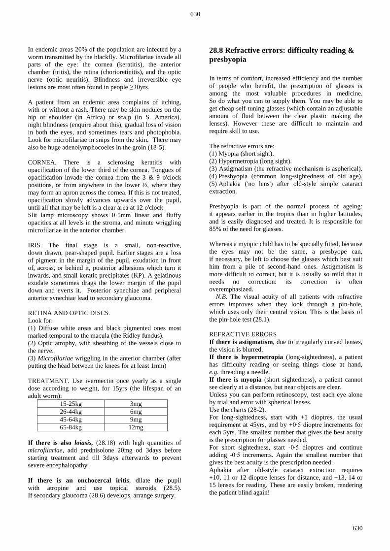
630
630
In endemic areas 20% of the population are infected by a
worm transmitted by the blackfly. Microfilariae invade all
parts of the eye: the cornea (keratitis), the anterior
chamber (iritis), the retina (chorioretinitis), and the optic
nerve (optic neuritis). Blindness and irreversible eye
lesions are most often found in people ≥30yrs.
A patient from an endemic area complains of itching,
with or without a rash. There may be skin nodules on the
hip or shoulder (in Africa) or scalp (in S. America),
night blindness (enquire about this), gradual loss of vision
in both the eyes, and sometimes tears and photophobia.
Look for microfilariae in snips from the skin. There may
also be huge adenolymphocoeles in the groin (18-5).
CORNEA. There is a sclerosing keratitis with
opacification of the lower third of the cornea. Tongues of
opacification invade the cornea from the 3 & 9 o'clock
positions, or from anywhere in the lower ½, where they
may form an apron across the cornea. If this is not treated,
opacification slowly advances upwards over the pupil,
until all that may be left is a clear area at 12 o'clock.
Slit lamp microscopy shows 0·5mm linear and fluffy
opacities at all levels in the stroma, and minute wriggling
microfilariae in the anterior chamber.
IRIS. The final stage is a small, non-reactive,
down drawn, pear-shaped pupil. Earlier stages are a loss
of pigment in the margin of the pupil, exudation in front
of, across, or behind it, posterior adhesions which turn it
inwards, and small keratic precipitates (KP). A gelatinous
exudate sometimes drags the lower margin of the pupil
down and everts it. Posterior synechiae and peripheral
anterior synechiae lead to secondary glaucoma.
RETINA AND OPTIC DISCS.
Look for:
(1) Diffuse white areas and black pigmented ones most
marked temporal to the macula (the Ridley fundus).
(2) Optic atrophy, with sheathing of the vessels close to
the nerve.
(3) Microfilariae wriggling in the anterior chamber (after
putting the head between the knees for at least 1min)
TREATMENT. Use ivermectin once yearly as a single
dose according to weight, for 15yrs (the lifespan of an
adult worm):
15-25kg 3mg
26-44kg 6mg
45-64kg 9mg
65-84kg 12mg
If there is also loiasis, (28.18) with high quantities of
microfilariae, add prednisolone 20mg od 3days before
starting treatment and till 3days afterwards to prevent
severe encephalopathy.
If there is an onchocercal iritis, dilate the pupil
with atropine and use topical steroids (28.5).
If secondary glaucoma (28.6) develops, arrange surgery.
28.8 Refractive errors: difficulty reading &
presbyopia
In terms of comfort, increased efficiency and the number
of people who benefit, the prescription of glasses is
among the most valuable procedures in medicine.
So do what you can to supply them. You may be able to
get cheap self-tuning glasses (which contain an adjustable
amount of fluid between the clear plastic making the
lenses). However these are difficult to maintain and
require skill to use.
The refractive errors are:
(1) Myopia (short sight).
(2) Hypermetropia (long sight).
(3) Astigmatism (the refractive mechanism is aspherical).
(4),Presbyopia (common long-sightedness of old age).
(5).Aphakia ('no lens') after old-style simple cataract
extraction.
Presbyopia is part of the normal process of ageing:
it appears earlier in the tropics than in higher latitudes,
and is easily diagnosed and treated. It is responsible for
85% of the need for glasses.
Whereas a myopic child has to be specially fitted, because
the eyes may not be the same, a presbyope can,
if necessary, be left to choose the glasses which best suit
him from a pile of second-hand ones. Astigmatism is
more difficult to correct, but it is usually so mild that it
needs no correction: its correction is often
overemphasized.
N.B. The visual acuity of all patients with refractive
errors improves when they look through a pin-hole,
which uses only their central vision. This is the basis of
the pin-hole test (28.1).
REFRACTIVE ERRORS
If there is astigmatism, due to irregularly curved lenses,
the vision is blurred.
If there is hypermetropia (long-sightedness), a patient
has difficulty reading or seeing things close at hand,
e.g. threading a needle.
If there is myopia (short sightedness), a patient cannot
see clearly at a distance, but near objects are clear.
Unless you can perform retinoscopy, test each eye alone
by trial and error with spherical lenses.
Use the charts (28-2).
For long-sightedness, start with +1 dioptres, the usual
requirement at 45yrs, and by +0∙5 dioptre increments for
each 5yrs. The smallest number that gives the best acuity
is the prescription for glasses needed.
For short sightedness, start -0∙5 dioptres and continue
adding -0∙5 increments. Again the smallest number that
gives the best acuity is the prescription needed.
Aphakia after old-style cataract extraction requires
+10, 11 or 12 dioptre lenses for distance, and +13, 14 or
15 lenses for reading. These are easily broken, rendering
the patient blind again!

631
631
Check that the eye is not red, and that there are no other
serious eye problems.
A child with reduced visual acuity due to refractive errors
needs glasses, whereas an adult probably does not.
A child <7yrs with a squint needs surgery (28.9)
Remember glasses for reading and other close work,
are not useful for seeing at a distance.
28.9 Disease of the neuromuscular system:
squints, amblyopia, and diplopia
If the eyes do not look in the same direction,
this is known as a squint (strabismus). Squints are
common, and are usually accepted with resignation.
Although treating a squint needs relatively simple
technology, it is time-consuming and needs skill.
If the eyes do not look in the same direction, two images
are generated. This causes confusion, and to avoid this,
the brain suppresses one image. If this suppression
continues for long enough, vision disappears in that eye.
This is called amblyopia, which is a reduction in vision,
due to lack of use of an apparently normal eye.
If amblyopia is uncorrected in a child by 7yrs, it becomes
permanent. So try to diagnose a squint and arrange
surgery before this age.
CAUTION! Never occlude the eye of a child <7yrs for
several days, because this may cause amblyopia.
SQUINTS AND AMBLYOPIA
DIAGNOSIS.
The corneal light reflex: Shine a pen torch directly in
front of you, and ask the patient to look at it. If each of the
eyes is properly fixing the torch, its reflection from the
corneal mirrors will be the same, and more or less central
on each cornea. Are the reflections from your torch
equally centred on the pupils?
The cover test: Ask the patient to look straight ahead at
some target in the distance. Cover the left eye with a piece
of paper. If the right eye moves, in or out, to fix on the
distant target, it was previously squinting. If it does not
move, it was looking straight at the target.
Now put the paper in front of the right eye. If the left eye
moves as you remove the paper, it was previously
squinting in or out. If it does not move, there is no
manifest squint, and both eyes look straight.
MANAGEMENT is limited.
If an adult has a squint and double vision, this suggests
a serious recent disease of the extraocular muscles or their
nerves, such as diabetic neuropathy, myasthenia gravis,
or raised intracranial pressure. He needs a full medical
and neurological examination.
If an adult has a squint and no double vision it may be
the cause of reduced vision in the squinting eye
(amblyopia). There is no treatment at this age.
If a child <7yrs presents with a squint:
(1) Dilate both the pupils, and use an ophthalmoscope to
make sure that the squint is not due to a retinoblastoma in
one of the eyes (an uncommon cause, 28.16: look for a
yellowish mass on the retina).
(2) If the retinae are normal, try to assess the visual
acuity in both eyes. This is relatively easy at >3yrs,
but is difficult at younger ages (28.1).
If there is reduced vision in either eye, or a definite
squint at >7yrs, there may be need for glasses,
and perhaps surgery on the extraocular muscles.
Correct any refractive error and occlude the eye that is
most used. Occlude it for ½-2hrs/day during close work,
reading, or drawing. The duration of treatment depends on
the duration of the amblyopia. If treatment is prompt,
6-8wks of intermittent occlusion treatment may be
enough. If it is delayed you may need to continue for 1yr.
AFTER THE AGE OF 6 MONTHS
DON’T IGNORE A SQUINT!
28.10 Diseases of the lids & nasolacrimal
apparatus
Diseases of the eyelids and nasolacrimal system include
tumours, deformities of the lids, and watering (epiphora).
Globally, the most important disease of the lids is
trachoma, which scars the lids, and causes them to turn
inwards (entropion, 28.13).
The commonest and usually the most harmless disease of
the lid is a stye. The lid is involved in herpes zoster
ophthalmicus, where the virus affects the VIIth cranial
nerve, often associated with HIV infection. Cutaneous
leishmaniasis causes the lid to scar and produce an
everted eyelid (ectropion). Loiasis is found in equatorial
rain forests, where the vigorously mobile loa loa worm is
found under the conjunctiva, causing an acute
inflammatory swelling.
THE LIDS
If a patient has a red swelling on the lid margin,
with an eyelash coming out of it, this is a stye
(hordeolum). It is a staphylococcal infection of an eyelash
follicle. Pull the eyelash out of the swelling,
using an analgesic. Only use antibiotics in recurrent styes,
or if infection is spreading beyond the lid
(cavernous sinus thrombosis is a rare complication, 6.6).
Warm soaks (28.3) are useful.

632
632
If there is a swelling in either lid, some distance from
its margin, pointing towards its conjunctival surface,
it is probably a tarsal cyst, (chalazion, Meibomian cyst).
Avoid an external scar by incising the conjunctiva
wherever the cyst is about to burst (28.12).
If a few lashes turn in on the eye (trichiasis),
remove them with electrolysis.
If most of the lashes or the margin of the lid are
turned in (entropion), perform surgery (28.13).
If the upper eyelid, eyebrow and forehead are involved
in a vesicular eruption with an abrupt demarcation in
the midline, this is herpes zoster. Acyclovir given early
will help. Later complications can result in ectropion,
keratitis, uveitis and secondary glaucoma.
If there is an acute attack of localized oedema of the
orbit in an endemic area, consider loiasis.
If you can see the worm under the conjunctiva,
instil lidocaine drops, make a small incision and remove
the worm with forceps. Ivermectin annually is useful as a
prophylactic.
If the lids do not close properly, as a result of a
VIIth cranial nerve palsy (from herpes, iatrogenic injury in
parotid surgery, leprosy, or parotid malignancy),
the cornea will remain exposed, especially at night, and
exposure keratitis will result. To avoid this,
do a tarsorrhaphy unless you can supply artificial tears.
If the margin of either lid is everted (ectropion), usually
as the result of scarring, or long-standing palsy, release of
lid contracture with skin grafting of the defect is
necessary.
TARSORRHAPHY (GRADE 2.2)
INDICATIONS
Eyelids which permanently fail to cover the cornea
properly, especially when the cornea is anaesthetic.
N.B. This is usually a temporary measure.
METHOD.
Stretch the lid by grasping one end of the lid margin and
make an incision 2mm deep along the middle of the upper
and lower margins, just beyond the eyelash follicles
(28-13A). Then pass a 4/0 silk suture through the skin
above the upper eyelash line and out through the incision,
and similarly in through the lower lid incision and out
below the lower eyelash line (28-13B).
Turn the suture back, through 3mm rubber rings to take
the tension, through the skin (28-13C) and tie it so that
the lids are well approximated (28-13D).
You need as many sutures as required to get adequate
eyelid closure; this may only be necessary on the lateral
aspect, but do not make the tarsorrhaphy too small.
Put chloramphenicol eye ointment or drops qid and make
sure no eyelashes project back inside the suture line
TARSORRHAPHY
Fig. 28-13 TARSORRPHAPHY. A, lid margin incision on upper
lid. Make the same incision on the lower lid. B, the suture passes
from above the eyelash through the two incisions and out below the
lower eyelash. C, pass the suture through a rubber ring, and then
back through the incision, not on the undersurface. D, lock the
suture on 2 protecting rings. E, completed tarsorrhaphy.
F, cross-section of position of suture. After Galbraith JEK. Basic Eye Surgery Churchill Livingstone 1979 p.44 Figs 5.29-33.
RELEASE OF LID CONTRACTURES (GRADE 2.2)
INDICATIONS
Contractures starting to expose the eyelids and expose the
cornea. You should have done a tarsorrhaphy already.
METHOD
Put stay sutures through the lids (28-8F), so that you can
move them up or down as necessary. Make the relaxation
incisions (28-14A).
When you have prepared a satisfactory bed for the graft,
and controlled bleeding, stretch it, apply a piece of split
skin graft, and hold it in place with tie-over sutures using
4/0 monofilament (28-14C). If the graft contracts after
12wks, repeat the release and apply another graft.
Do not worry too much about what the patient's eye looks
like at this stage. What matters is that the cornea should
not be exposed.
DO NOT DELAY GRAFTING THE LIDS
CAUTION! Stretch the lid first so that there will be
some slack tissue when it contracts later. The thinner the
graft, the more the shrinkage. If you are skilled, apply a
full thickness graft. Primary skin grafting will not prevent
ectropion, and you may need 2 or 3 operations to insert
enough skin.
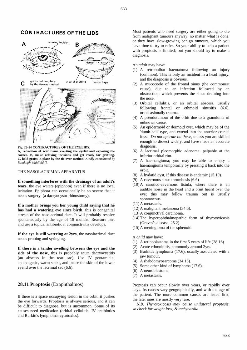
633
633
Fig. 28-14 CONTRACTURES OF THE EYELIDS.
A, retraction of scar tissue everting the eyelid and exposing the
cornea. B, make relaxing incisions and get ready for grafting.
C, hold grafts in place by the tie-over method. Kindly contributed by
Randolph Whitfield II.
THE NASOLACRIMAL APPARATUS
If something interferes with the drainage of an adult's
tears, the eye waters (epiphora) even if there is no local
irritation. Epiphora can occasionally be so severe that it
needs surgery (a dacryocysto-rhinostomy).
If a mother brings you her young child saying that he
has had a watering eye since birth, this is congenital
atresia of the nasolacrimal duct. It will probably resolve
spontaneously by the age of 18 months. Reassure her,
and use a topical antibiotic if conjunctivitis develops.
If the eye is still watering at 2yrs, the nasolacrimal duct
needs probing and syringing.
If there is a tender swelling between the eye and the
side of the nose, this is probably acute dacryocystitis
(an abscess in the tear sac). Use IV gentamicin,
an analgesic, warm soaks, and incise the skin of the lower
eyelid over the lacrimal sac (6.6).
28.11 Proptosis (Exophthalmos)
If there is a space occupying lesion in the orbit, it pushes
the eye forwards. Proptosis is always serious, and it can
be difficult to diagnose, but is uncommon. Some of its
causes need medication (orbital cellulitis: IV antibiotics
and Burkitt's lymphoma: cytotoxics).
Most patients who need surgery are either going to die
from malignant tumours anyway, no matter what is done,
or they have slow-growing benign tumours, which you
have time to try to refer. So your ability to help a patient
with proptosis is limited; but you should try to make a
diagnosis.
An adult may have:
(1) A retrobulbar haematoma following an injury
(common). This is only an incident in a head injury,
and the diagnosis is obvious.
(2) A mucocoele of the frontal sinus (the commonest
cause), due to an infection followed by an
obstruction, which prevents the sinus draining into
the nose.
(3) Orbital cellulitis, or an orbital abscess, usually
following frontal or ethmoid sinusitis (6.6),
or occasionally trauma.
(4) A pseudotumour of the orbit due to a granuloma of
unknown cause.
(5) An epidermoid or dermoid cyst, which may be of the
'dumb-bell' type, and extend into the anterior cranial
fossa. Do not operate on these, unless you are skilled
enough to dissect widely, and have made an accurate
diagnosis.
(6) A lacrimal pleomorphic adenoma, palpable at the
inferior orbital rim.
(7) A haemangioma; you may be able to empty a
haemangioma temporarily by pressing it back into the
orbit.
(8) A hydatid cyst, if this disease is endemic (15.10).
(9) A cavernous sinus thrombosis (6.6)
(10) A carotico-cavernous fistula, where there is an
audible noise in the head and a bruit heard over the
eye; this may follow trauma but is usually
spontaneous.
(11) A metastasis.
(12) A malignant melanoma (34.6).
(13) A conjunctival carcinoma.
(14) The hyperophthalmopathic form of thyrotoxicosis
(Graves's disease, 25.2).
(15) A meningioma of the sphenoid.
A child may have:
(1) A retinoblastoma in the first 5 years of life (28.16).
(2) Acute ethmoiditis, commonly around 2yrs.
(3) Burkitt's lymphoma (17.6), usually associated with a
jaw tumour.
(4) A rhabdomyosarcoma (34.15).
(5) Some other kind of lymphoma (17.6).
(6) A neuroblastoma.
(7) A metastasis.
Proptosis can occur slowly over years, or rapidly over
days. Its causes vary geographically, and with the age of
the patient. The more common causes are listed first;
the later ones are mostly very rare.
N.B. Thyrotoxicosis may cause unilateral proptosis,
so check for weight loss, & tachycardia.
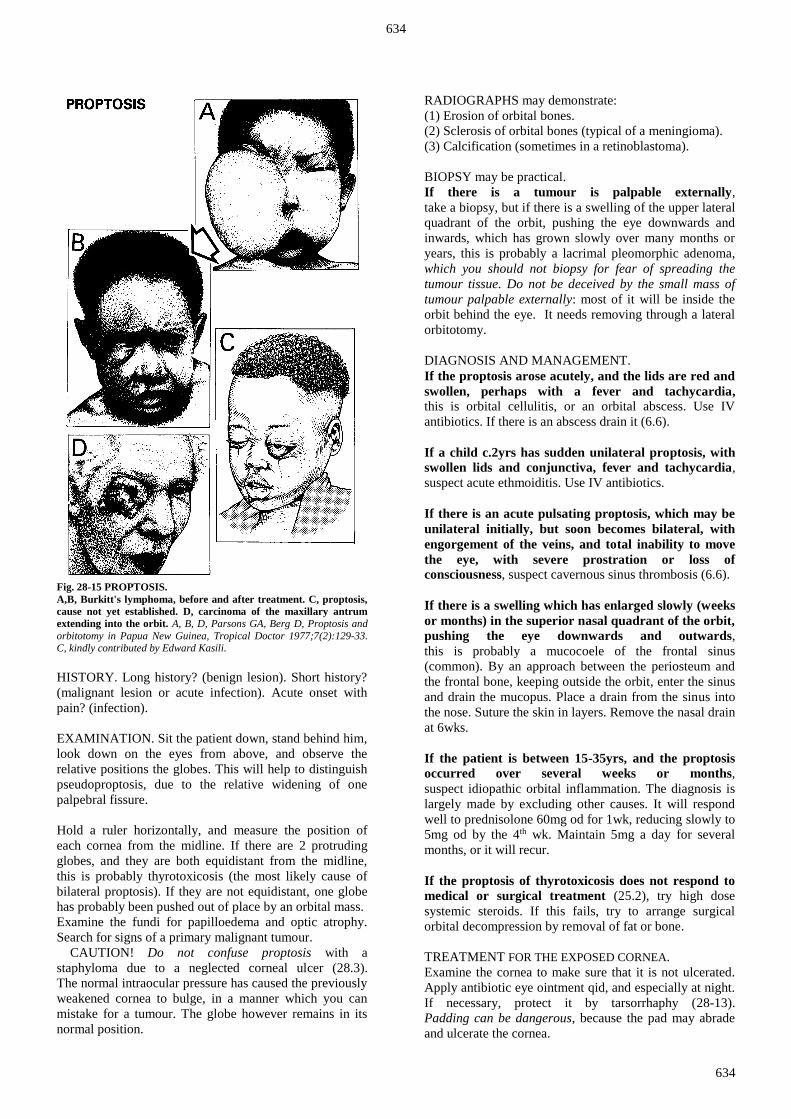
634
634
Fig. 28-15 PROPTOSIS.
A,B, Burkitt's lymphoma, before and after treatment. C, proptosis,
cause not yet established. D, carcinoma of the maxillary antrum
extending into the orbit. A, B, D, Parsons GA, Berg D, Proptosis and
orbitotomy in Papua New Guinea, Tropical Doctor 1977;7(2):129-33. C, kindly contributed by Edward Kasili.
HISTORY. Long history? (benign lesion). Short history?
(malignant lesion or acute infection). Acute onset with
pain? (infection).
EXAMINATION. Sit the patient down, stand behind him,
look down on the eyes from above, and observe the
relative positions the globes. This will help to distinguish
pseudoproptosis, due to the relative widening of one
palpebral fissure.
Hold a ruler horizontally, and measure the position of
each cornea from the midline. If there are 2 protruding
globes, and they are both equidistant from the midline,
this is probably thyrotoxicosis (the most likely cause of
bilateral proptosis). If they are not equidistant, one globe
has probably been pushed out of place by an orbital mass.
Examine the fundi for papilloedema and optic atrophy.
Search for signs of a primary malignant tumour.
CAUTION! Do not confuse proptosis with a
staphyloma due to a neglected corneal ulcer (28.3).
The normal intraocular pressure has caused the previously
weakened cornea to bulge, in a manner which you can
mistake for a tumour. The globe however remains in its
normal position.
RADIOGRAPHS may demonstrate:
(1) Erosion of orbital bones.
(2) Sclerosis of orbital bones (typical of a meningioma).
(3) Calcification (sometimes in a retinoblastoma).
BIOPSY may be practical.
If there is a tumour is palpable externally,
take a biopsy, but if there is a swelling of the upper lateral
quadrant of the orbit, pushing the eye downwards and
inwards, which has grown slowly over many months or
years, this is probably a lacrimal pleomorphic adenoma,
which you should not biopsy for fear of spreading the
tumour tissue. Do not be deceived by the small mass of
tumour palpable externally: most of it will be inside the
orbit behind the eye. It needs removing through a lateral
orbitotomy.
DIAGNOSIS AND MANAGEMENT.
If the proptosis arose acutely, and the lids are red and
swollen, perhaps with a fever and tachycardia, this is orbital cellulitis, or an orbital abscess. Use IV
antibiotics. If there is an abscess drain it (6.6).
If a child c.2yrs has sudden unilateral proptosis, with
swollen lids and conjunctiva, fever and tachycardia,
suspect acute ethmoiditis. Use IV antibiotics.
If there is an acute pulsating proptosis, which may be
unilateral initially, but soon becomes bilateral, with
engorgement of the veins, and total inability to move
the eye, with severe prostration or loss of
consciousness, suspect cavernous sinus thrombosis (6.6).
If there is a swelling which has enlarged slowly (weeks
or months) in the superior nasal quadrant of the orbit,
pushing the eye downwards and outwards,
this is probably a mucocoele of the frontal sinus
(common). By an approach between the periosteum and
the frontal bone, keeping outside the orbit, enter the sinus
and drain the mucopus. Place a drain from the sinus into
the nose. Suture the skin in layers. Remove the nasal drain
at 6wks.
If the patient is between 15-35yrs, and the proptosis
occurred over several weeks or months,
suspect idiopathic orbital inflammation. The diagnosis is
largely made by excluding other causes. It will respond
well to prednisolone 60mg od for 1wk, reducing slowly to
5mg od by the 4th wk. Maintain 5mg a day for several
months, or it will recur.
If the proptosis of thyrotoxicosis does not respond to
medical or surgical treatment (25.2), try high dose
systemic steroids. If this fails, try to arrange surgical
orbital decompression by removal of fat or bone.
TREATMENT FOR THE EXPOSED CORNEA.
Examine the cornea to make sure that it is not ulcerated.
Apply antibiotic eye ointment qid, and especially at night.
If necessary, protect it by tarsorrhaphy (28-13).
Padding can be dangerous, because the pad may abrade
and ulcerate the cornea.

635
635
28.12 Tarsal (Meibomian) cysts (Chalazions)
Meibomian glands secrete oil at each blinking of the
eyelid; this prevents evaporation of the fluid film on the
eye. Cysts may form in these glands on the conjunctival
side of the tarsus (or eyelid). They present as a swelling in
either lid, which may become chronically or acutely
infected. Small asymptomatic ones need no treatment,
and may resolve spontaneously. Incise an acute infection
and curette a chronic one. These cysts are common
everywhere, so that treating them is a common outpatient
eye operation. Sometimes, they present as granulomas.
Fig. 28-16 CURETTING A TARSAL CYST.
A, chalazion close to the medial canthus. B, chalazion forceps.
C, introducing LA. D, chalazion clamp in place ready to incise a
chalazion in the centre of the lower lid. E, curetting the chalazion
ANAESTHESIA.
Anaesthetize the conjunctiva with drops of lidocaine 4%,
or amethocaine hydrochloride 1%. Infiltrate the lid with
lidocaine and adrenaline around the chalazion. Insert the
needle at the upper margin of the upper tarsus,
and the lower margin of the lower tarsus. Carry it
forwards to the lid margin, on either side of the chalazion.
EQUIPMENT. Chalazion clamp, # 11 scalpel blade and curette.
CURETTAGE. (GRADE 1.2)
Evert the lid slightly. Put the chalazion clamp over the
cyst, so that the solid blade lies on the skin of the eyelid,
and the ring lies on the conjunctiva over the cyst.
Close it so that it holds the lid and the cyst. Insert the tip
of a #11 blade, so that it cuts away from the eye.
Always make the incision perpendicular to the lid margin
so as to avoid cutting the levator muscle. Swab its
contents clean.
CAUTION! Take care to curette away any pockets of
granulation tissue, which may be hidden by a flap of
conjunctiva, or have herniated themselves through the
tarsal plate into the orbicularis muscle. If you do not do
this, it may recur.
Remove the clamp and pinch the lid until it stops
bleeding. If this is troublesome, wash it with warm saline.
Place chloramphenicol ointment in the conjunctival sac
tid for 1wk.
.
If the material you incise is hard, and not gelatinous,
suspect a carcinoma.
Send it for histological
examination.
28.13 Entropion
Trachoma is the commonest eye infection in the tropics
and also amongst Aborigines in Australia, and in its
blinding hyperendemic form is worldwide the
2nd commonest single cause of blindness and impaired
vision (cataract is the first). It is a chlamydial infection,
which spreads from the eyes of one person to another,
especially among children, in the poorest and most
disadvantaged communities, particularly those in the
Middle East and Africa.
Trachoma is a chronic follicular conjunctivitis; it scars the
conjunctiva of the eyelids and the cornea,
and goes through 4 stages (28.3). A single dose of
20mg/kg azithromycin (or 500mg bd erythromycin for
1wk in pregnancy) orally is effective in arresting the
disease:

636
636
STAGE I. There is a mildly red watery, eye due to
bilateral conjunctivitis, especially of the upper lids,
but without any distinguishing features.
STAGE II. Under the upper lid there are dilated blood
vessels and hyperaemic, oedematous epithelial tissue
(papillae). There are also yellow-grey swellings
(follicles). Look at the corneoscleral junction with a
loupe. If the edge of the cornea looks mildly grey,
owing to an arcuate (crescent-shaped) grey infiltration,
and blood vessels go beyond the grey area into the cornea,
there is pannus (meaning a curtain). This starts at the
12 o'clock position, and extends to 9 & 3 o'clock.
Follicles and pannus indicate stage II trachoma.
Follicles are not diagnostic, but pannus is.
STAGE III. The follicles in the lids become coarser and
pannus spreads, sometimes across the pupillary area of
the cornea. Scarring makes the margin of the upper lid
irregular, and turns the upper tarsus inwards (entropion),
taking the lashes with it, so that they scratch
the cornea during every blink (trichiasis, 28-18C).
This causes recurrent attacks of keratitis, which
eventually results with a corneal opacity causing
blindness.
STAGE IV. Fibrous tissue replaces the follicles in the
lids. This is the stage of scarred, or healed trachoma.
The cornea is grey and scarred, the vision severely
impaired, and the lids are deformed.
If you work in an endemic area, you are likely to have
many patients with entropion, so learn how to correct
their eyelids yourself, and if necessary train an assistant to
do this. The operation is always worth doing, even if the
lids are severely scarred: sight may recover surprisingly.
Several operations are possible:
(1) Splitting the eyelid margin
(2) Radical eyelash excision
(3) Tarsal eversion
If only a few lashes are turning in, try removing these
with an electrolysis needle under LA. Epilation (pulling
out the lashes) is ineffective as they will grow back.
Do not cut the eyelashes short because this makes them
sharp and even more irritant.
ENTROPION CORRECTION
INDICATIONS.
Trachoma which has distorted the upper tarsus, so that it
has curled inwards and made the lashes scratch the globe.
Operate as soon as possible after entropion occurs.
You can operate on both the eyes at the same time,
but this will require admission for 3days, to allow the
oedema of the eyelids to subside. Absorbable sutures
allow discharge without the patient needing to return.
EQUIPMENT. An eye set, a scalpel with #15 blade, 4/0 absorbable
suture. A cautery will help to control bleeding.
ANAESTHESIA.
Anaesthetize the upper lids through the skin with 1ml of
2% lidocaine with adrenaline. Anaesthetize the
conjunctiva with 2 drops of amethocaine, or lidocaine.
EYELID MARGIN SPLITTING FOR TRICHIASIS
(GRADE 1.3)
Split the eyelid into two parts along a ‘grey’ line defined
by the orbicularis oculi muscle: just inside are the
openings of the Meibomian glands and just outside are the
eyelash roots. The divided eyelid will then have an inner
part containing the conjunctiva and tarsal plate, and an
outer part containing the orbicularis oculi muscle,
the inverted eyelashes and their roots. Rotate this outer
part outwards and fix it by an everting suture over a gauze
swab.
Fig. 28-17 RADICAL EYELASH EXCISION.
A, direction of the incision. B, operation complete.
RADICAL EYELASH EXCISION FOR ENTROPION
(Malcolm Phillips) (GRADE 2.3)
Removing the eyelashes completely will mean they
cannot re-grow and scratch the cornea. This results in
little cosmetic disability, especially with darker skin,
and the relief that follows is dramatic.
If you are a right-handed operator, start with the right eye.
Use a scalpel to incise the margin of the upper lid, at the
lateral end of the lashes, to a depth of 3mm (28-17A).
Using small sharp scissors, remove the margin of the lid
bearing the roots of the lashes. Cut towards the medial
end and preserve the punctum. Evert the lid as you do
this, by pressing it with a swab. Then repeat the procedure
on the left eye.
Control the considerable bleeding that will result by
suturing the conjunctiva to the skin of the eyelid with 3/0
absorbable sutures on a cutting needle. Insert about
5 sutures, 5mm apart, knotting them, and use the same
suture to hold little rolls of gauze. Apply an eyepad for
24hrs. Remove the part of the sutures holding the gauze
rolls after 3days.

637
637
Fig. 28-18 EVERTING THE TARSUS FOR LATE TRACHOMA.
A, inwardly turned upper lid, with the lashes scratching the cornea.
B, normal eyelid. C, lid scarred with trachoma, with its lashes
rubbing against the cornea. D, the incision, extended through the lid
margin at both ends (arrows). E, evert the lid with tension sutures.
Make the incision, and undermine the superficial surface of the
tarsus gently in both directions. F, rotate the margin of the lid and
suture it in place. G, completed lid eversion.
Kindly contributed by Roy Pfaltzgraff.
TARSAL EVERSION FOR ENTROPION (GRADE 2.4)
For this method, the tarsal plate must be stiff enough to
take sutures. Place 3 stay sutures of black braided silk in
the upper lid, evert it over a roll of gauze, and clamp the
sutures to a drape.
Using a #15 scalpel, make an incision about 3mm from
the inner margin of the lid, and parallel to it. Cut through
the conjunctiva and the full length of the tarsal plate,
at 90º to its surface, so as to free a strip from its edge
(28-18E). Curve each end of the incision towards the free
edge of the lid, so that you can evert the strip of lid that
bears the lashes.
Use skin hooks to retract the
free edge of the lid. Use the
tip of your scalpel to free the
tissues from the anterior
surface of the strip of tarsal
plate for about 2mm, until
you see the follicles of the
lashes in the base of the
wound.
Now undermine the anterior
surface of the main part of
the plate to a depth of about
4mm. A little undermining
like this will help you to
mobilize the free edge of the
tarsus. Do this in the plane
between the orbicularis
muscle and the insertion of
the levator palpebrae
superioris tendon.
CAUTION! Take care
not to buttonhole the skin.
You will now be able to
rotate the distal fragment
through 180º (28-18F).
If you cannot, undermine the
conjunctiva more widely.
Insert 3 small mattress
sutures of 4/0 absorbable,
so that the knots are buried
(28-18G). Put tetracycline
eye drops into the
conjunctiva tid for 1wk.
This operation gives the upper lid a new edge, and makes
it c. 3mm shallower; but it will still meet the lower lid on
shutting the eye.
Alternatively, if the tarsal plate is shrunken and
degenerated, it will not take sutures, so remove it entirely.
A more complicated procedure is a tarsal plate rotation
with a mucosal graft from the mouth, but this is for an
expert.
If the eye is painful and blind, it may be better removed.
This is one of the occasions on which the indications are
more critical than the operation.

638
638
28.14 Destructive methods for the eye
Evisceration is the least radical procedure;
scrape out the contents of the globe and leave
the sclera intact. This is the only safe procedure
if the eye is infected, because a sleeve of dura
containing CSF surrounds the optic nerve.
Other operations require that you cut it, and so
open up a potential path of infection to the
meninges. You may need to eviscerate the eye:
(1);When antibiotic therapy fails to control
a severe infection causing suppurative
endophthalmitis, leading to orbital cellulitis, and
oedema of the lids. If you do not eviscerate the
eye and drain the pus from it, the infection may
spread and cause cavernous sinus thrombosis
and meningitis, and death.
(2);When there is a chronic less urgent infection
in a blind painful useless eye.
Try to find a prosthesis to insert when the sepsis
has settled: it vastly improves the patient’s
appearance.
Enucleation (excision) removes the globe by
dividing the conjunctiva, the extrinsic muscles
of the eye, and the optic nerve. Do this only
where there is no active infection; it is contraindicated
when there is.
Exenteration is a bloody, mutilating operation.
It removes the entire contents of the orbit, together with
its periosteum, the globe, and all its extrinsic muscles.
Consider doing this when there is a fungating malignant
tumour of the eye or orbit. It will not prolong life, but the
last days might at least be more comfortable. An empty
orbital cavity will remain, which you can line with split
skin, or allow to granulate.
Before you start any destructive operation:
(a) Get signed informed consent from a child's parent or
guardian or from an adult himself
(b) Make sure you operate on the correct eye!
Do not rush in to perform these procedures:
(a);The main indication for enucleation is persistent
severe pain in a blind eye.
(b);If it has been injured, always try to repair it first,
no matter how hopelessly injured it is. In some cases the
eye may scar but remain stable for years.
(c) Prostheses may be difficult to find, and the best one
is the natural eye, even if it is blind.
CAUTION!
(1);Unless you are operating for malignancy or acute
infection to save life, the eye must be totally blind.
Test this with a strong light.
(2);When pain is the main indication, it must be
considerable. Pain is subjective, so make sure, if you can,
that it is genuine. Review this on several occasions.
If there is any sensation of light, do not do a destructive
procedure. That little eyesight may be useful later.
Fig. 28-19 EVISCERATING AN EYE.
A, conjunctiva already incised and undermined. Stab a #11 scalpel
blade through the cornea into the anterior chamber. B, continue the
corneal incision with scissors. C, excise the cornea completely.
D, scoop out the contents of the eye with a curette. E, cut a
triangular section out of each side of the hole in the sclera. F, inspect
the inside of the eye to make sure that no choroid remains.
After Galbraith JEK. Basic Eye Surgery, Churchill Livingstone 1979 p.84 Figs 9.17-24 with kind permission.
EVISCERATION OF AN EYE (GRADE 3.2)
INDICATIONS.
(1);The failure of antibiotics to control a suppurative
endophthalmitis.
(2) A blind, painful eye, especially if it is infected.
Do not take the eye out unless you are left with no other
option.
ANAESTHESIA. If there is no significant infection, you
can use the combination of a facial and a retrobulbar
block. Otherwise use ketamine or GA.
METHOD.
Incise the conjunctiva all round 3600 at its junction with
the cornea, using fine-toothed forceps and fine scissors
(28-19A). Separate Tenon’s capsule (28-1C) bluntly from
the underlying sclera in 4 quadrants. Cut through the
corneoscleral junctional at the limbus with scissors
(28-19B). Excise the entire cornea (28-19C).
Scoop out all uveal tissue contents of the eye with a
evisceration spoon, curette or a periosteal elevator
(28-19D). Pack the sclera for a few minutes to control
bleeding. Excise a 5mm triangle of sclera from each
side (28-19E), to help make the globe collapse.
Rinse the inside of the globe with hydrogen peroxide.

639
639
Make sure that no black choroid remains to avoid the risk
of sympathetic ophthalmitis. Close Tenon’s fascia and
conjunctiva with 6/0 nylon.
If you are operating for acute infection, leave it open to
drain.
If there is a chance of getting an artificial eye,
insert a plastic conformer shell.
POSTOPERATIVELY, control bleeding by bandaging
2 eyepads firmly over the socket. Leave the dressing on
for 2 days. Clean the lids and lashes bd, and put 2 drops
of chloramphenicol 0·5% into the socket. You can insert
an artificial eye after 4-6wks.
ENUCLEATION OF AN EYE (GRADE 3.3)
INDICATIONS.
(1);A malignant intraocular tumour (retinoblastoma or
melanoma) is an absolute indication.
(2) A blind, persistently painful eye, which is not infected
(evisceration is an alternative).
(3) A penetrating wound, especially in the circumcorneal
region, complicated by iridocyclitis, and entanglement of
the iris, lens capsule, and vitreous. If you leave an eye like
this, sympathetic ophthalmitis may follow in the other eye.
ANAESTHESIA.
A retrobulbar block using ≥6ml of lidocaine, combined
with a VIIth cranial nerve block. Otherwise use ketamine
or GA.
METHOD.
Incise the conjunctiva at its junction with the cornea,
using fine-toothed forceps and fine scissors (28-20A).
Cut around it, and undermine it back
to the insertion of the extraocular
muscles, about 8mm from the edge of
the cornea (28-20B).
Push closed scissors through the
conjunctiva to open up the plane
between the conjunctiva and the
globe. Open them to expose the sclera,
anterior to the insertion of the rectus
muscles.
Snip Tenon's capsule (28-1C) between
the insertions of these muscles.
Pass scissors through the incision,
until you have defined the muscle
insertions. Slip a squint hook under
the medial rectus muscle (28-20C),
and pull it into view.
If you are going to put in an implant,
lift the muscle and insert a mattress
suture of chromic catgut through its
belly, about 3mm from its insertion.
Clamp its insertion, remove the squint
hook, and cut the muscle with
scissors.
Separate each rectus muscle in the
same way. Leave the stump of the
medial rectus tendon a little longer,
so that you have something with
which to hold the globe.
Fig. 28-20 ENUCLEATING AN EYE.
A, incise the conjunctiva. B, undermine the
conjunctiva for about 8 mm. C, slip a muscle
hook under each rectus muscle, bring it
forwards into the wound, and cut it.
D, draw the eye forwards by pulling on the
insertion of the medial rectus muscle.
E, cut the optic nerve from the medial side.
F, cut any remaining adherent tissue.
G, suture the conjunctiva with absorbable
suture. After Galbraith JEK Basic Eye Surgery,
Churchill Livingstone 1979 p.81-2 Figs. 9.9-15
with kind permission.

640
640
Make the globe prolapse forwards out of the orbit by
closing the arms of the speculum behind it, and pushing
them backwards. If the eye is so enlarged, that it will not
fit between the blades of the speculum, pull it forwards by
holding the stump of the medial rectus muscle with a
haemostat (28-20D). Pass a pair of curved scissors,
with their blades closed, down the medial side of the
orbit, feel for the optic nerve behind the eye, open the
scissors, and cut it (28-20E).
If you are excising it for a malignant tumour,
cut it as far posteriorly as you can, because it may have
been infiltrated by tumour. Pull the eye forwards, and cut
any tissue that remains attached to it (28-20F).
Put a hot wet pack into the orbit, and press on it until
bleeding stops.
If you are going to put in an implant, it will probably be
a simple glass globe. Place it in the muscle cone,
and Tenon's capsule, and suture the conjunctiva over it.
If there is no possibility of an implant,
close the conjunctiva and Tenon's capsule separately with
5/0 absorbable (28-20G). Irrigate the socket with 0·5%
chloramphenicol.
EXENTERATION OF THE ORBIT (GRADE 3.4)
INDICATIONS.
A malignant tumour of the orbit, often an advanced
conjunctival carcinoma (28.15) or a retinoblastoma,
which has penetrated the globe and caused proptosis.
ANAESTHESIA.
Use ketamine or GA. Have blood for transfusion
available.
METHOD.
If the lids have been involved by tumour, suture their
margins together with 3/0 silk. Use a #15 scalpel blade to
cut round the margins of the orbit. If the lids are not
involved, incise closer to the scleral margins, so as to save
all, or some, of the skin from the lids to line the empty
orbit.
Control bleeding from the upper inner margin of the orbit
with diathermy, adrenaline solution or hydrogen peroxide.
Incise the periosteum round the margin of the orbit,
and reflect it as far posteriorly as you can. It is firmly
adherent at the bony suture lines of the skull.
CAUTION!
(1) The bone on the medial wall of the orbit is very thin,
so elevate the periosteum here with special care.
(2) The tumour may have eaten through the wall of the
orbit, into the brain. If so, you may find it difficult to be
sure of the anatomy.
The periosteum should strip easily until you reach the
orbital fissures, and the nasolacrimal duct (6-4). Cut this.
Separate the palpebral ligaments, the trochlea (the pulley
structure for the tendon of the superior oblique), and the
inferior oblique muscles from the bone with the
periosteum. Use curved scissors to cut the structures
entering through the orbital fissures. Pull the contents of
the orbit forwards, and cut the tissues at its apex with
strong scissors as far back as you can.
Bleeding may be profuse. Remove the contents of the
orbit quickly, and then control bleeding.
Turn the skin at the edges of the orbit back into it.
Graft its raw surfaces with split skin, either now or as a
secondary procedure in 10-14days. Gently pack the orbit.
If you are not grafting it, dress it with disinfectant such as
acriflavine wool, and apply a firm bandage.
DIFFICULTIES WITH DESTRUCTIVE METHODS FOR
THE EYE
If the patient refuses to have a painful eye enucleated
or eviscerated, consider injecting absolute alcohol behind
it to destroy its sensory nerves. You can use any strength
of alcohol, provided it is more than 50%, but you may
need to repeat the injection if pain returns. Permanent
relief is uncertain.
Retrobulbar alcohol is very painful for about 30secs,
so use a retrobulbar block of lidocaine 1ml. Remove the
syringe and needle. When the block is effective,
put another syringe on the needle and inject 2mL of
alcohol. The orbit will become severely oedematous for
10days. Add chloramphenicol eye drops qid for 1wk.
29.15 Conjunctival carcinoma
The conjunctiva is a thin transparent mucous membrane
which covers the inner aspects of the eyelids and is
reflected over the anterior part of the eyeball and ends at
the limbus. Conjunctival epithelium is non-keratinized.
It is continuous with the cornea at the limbus and the skin
at the lid margins.
Squamous carcinoma of the conjunctiva used to occur
mostly in the elderly but now is commoner among young
HIV+ve patients, in whom it acts much more
aggressively. It may arise on its own or from a
pre-existing lesion like a pterygium (28.18). When small,
it appears as a whitish lesion surrounded by a hyperaemic
area, whose surface is irregular with small nodular parts.
Initially it is mobile but as it invades, it becomes fixed.
Advanced cases are easy to diagnose, and may be
associated with metastases to pre-auricular and
submandibular lymph nodes.

641
641
MANAGEMENT
Small lesions you can completely excise; this is advisable
as it can then provide histological confirmation as well as
information on the invasiveness of the tumour.
Large lesions need an exenteration (28.14) with removal
of regional lymph nodes. Radiotherapy is useful, but
chemotherapy needs the help of an expert, especially if
HIV disease is present.
28.16 Retinoblastoma
This uncommon, malignant, radiosensitive tumour of the
embryonic cells of the retina usually presents in children
<2yrs. It is bilateral in 25% of cases, and occurs in
2 forms, familial and sporadic. At first, the tumour
enlarges within the eye; later it grows through the sclera,
or chambers of the eye, to perforate the cornea. It can also
spread through the optic nerve (where it may cause
glaucoma) to the subarachnoid space. Familial cases are
inherited as autosomal dominants. Healthy patients with
one affected child have only a 5% risk of producing a
second one. If a child survives, there is a high risk of
transmitting the disease to his offspring.
The earliest sign is a squint or a fixed dilated pupil with a
white reflection ('cat's eye'), and greyish white tumour
visible with an ophthalmoscope. Presentation is often late,
with a large globe, and tumour penetrating the sclera or
cornea.
DIFFERENTIAL DIAGNOSIS varies with the stage of
presentation, and is easy after glaucoma and intraocular
extension have occurred. Before this, the diagnosis can be
difficult, when all you can see is a white mass in an
infant's vitreous. Dilate the pupils, and examine both the
fundi under LA. Enquire for a family history.
Early:
(1) Traumatic perforation of the globe.
(2) Traditional medicine in the eye.
(3);Retrolental fibroplasia, (history of prematurity at
birth).
Late:
(1),A corneal ulcer leading to perforation, an anterior
staphyloma (due to bulging of a weak cornea, 28.3).
(2) Panophthalmitis.
(3) Congenital glaucoma (28.6).
MANAGEMENT depends on the stage of presentation.
If early with the tumour confined to the globe,
try to refer for radiotherapy, which cures 85% of cases.
Otherwise, enucleate the globe (28.14).
If the tumour has extended through the globe, but not
through the wall of the orbit, you should remove the
globe and refer for radiotherapy, but the prognosis is so
poor that a long journey to a referral hospital is not
worthwhile. Consider chemotherapy.
If there is proptosis and a fungating mass, exenteration
of the orbit (28.14) will remove the mass, but is unlikely
to prolong life.
CHEMOTHERAPY.
In 35% of cases the following regime causes a partial
response, and in 65% it prevents progression for a while.
Use vincristine 1·4mg/m², doxorubicin 50mg/m² and
cyclophosphamide 1g/m². Administer all drugs IV once
every 21days.
28.17 The eyes in leprosy
Leprosy causes:
(1) Paralysis of the facial nerve, affecting the orbicularis
muscle so that the eye does not close (lagophthalmos).
(2);Loss of sensation in the ophthalmic division of the Vth
cranial nerve, which makes the cornea anaesthetic.
(3);An acute iritis (uncommon), which is usually
associated with a type II reaction and is less common if
the patient is taking clofazimine.
(4);A chronic iritis (common) causing atrophy of the
dilator pupillae and a small unreactive pupil.
The combination of these lesions can have a devastating
effect on the sight. An anaesthetic cornea prevents a
patient noticing that there is something in the eye, or that
it is dry. The blink reflex disappears, so that, even if there
is still enough power in the VIIth cranial nerve to blink,
it does not wash and wet the conjunctiva automatically.
As a result, the cornea may be unprotected, especially
during sleep, so that it may develop exposure keratitis,
and ulcerate. If the centre of the cornea becomes opaque,
sight is damaged. So warn of the danger of an anaesthetic
cornea, and examine the eyes regularly.
To decide if the cornea has been damaged, look for
superficial scars, and use fluorescein drops, or papers,
to search for central staining. If the cornea is anaesthetic,
the eye is at great risk. If there is lagophthalmos,
but the cornea is not anaesthetic, he may have enough
sensation to complain of discomfort or burning.
To find out if there is significant lagophthalmos, examine
the closed eyes. If the cornea is completely covered, all is
well. But if any part of it remains exposed, you should do
something to protect eyesight. Several operations are
possible. Tarsorrhaphy aims to reduce, or remove entirely,
the gap between the closed eyelids. It has cosmetic
disadvantages, but it does save sight, and it is not
difficult, so you should be able to do it if you care for
leprosy patients. A more effective procedure is transfer of
the temporalis muscle.

642
642
28.18 Other eye problems
Here are some of the other eye problems you may meet.
If opening the eye is impossible, this is PTOSIS.
This may be congenital, traumatic, acute as the result of
an oculomotor palsy, HIV disease, or the result of
myasthenia gravis.
If there is a small yellowish-white lump adjacent to the
cornea in the region exposed by the palpebral opening,
this is a PINGUECULA. It is harmless so give
reassurance, though it may develop into a pterygium.
If there is a wing-shaped vascular thickening of the
conjunctiva which grows on to the cornea, usually
from the medial side, this is a PTERYGIUM.
It is a wedge-shaped benign fleshy growth on the scleral
conjunctiva that may grow onto the cornea; this is more
frequent in increased UV exposure from outdoor
activities). It is seldom serious. If vision is good, leave it.
If it is advancing over the centre of the cornea and
impairing vision, excise the pterygium carefully under LA
off the conjunctiva, and dissect it off the cornea. Leave
the sclera bare, and gently cauterize it. Up to 50% recur.
Scarring can result in a squint.
If there is a worm under the conjunctiva
(e.g. in the rainforest belt of West Africa), this is loiasis.
Use albendazole 5mg/kg bd for 3wks.
If an old person has a complete or incomplete white
ring encircling the cornea about 1mm within the
limbus, this is ARCUS SENILIS. It is a sign of high
blood cholesterol levels.
If the sclera are deep yellow, this is jaundice.
There may be surgical correction possible if the cause is
cholestasis (15.9).