Embed Size (px)
DESCRIPTION
Glucose utilization and the PI3-K pathway: mechanisms for cell survival in preimplantation embryos Department of Obstetrics and Gynecology and the 2 Department of Cell Biology and Physiology, Washington University School of Medicine, 4911 Barnes-Jewish Hospital Plaza, St Louis, Missouri 63110, USA Presence and function of PI3-K/Akt in mammalian preimplantation embryos
Citation preview
REPRODUCTIONREVIEW
Glucose utilization and the PI3-K pathway: mechanisms forcell survival in preimplantation embryos
Joan K Riley1 and Kelle H Moley1,2
1Department of Obstetrics and Gynecology and the 2Department of Cell Biology and Physiology, WashingtonUniversity School of Medicine, 4911 Barnes-Jewish Hospital Plaza, St Louis, Missouri 63110, USA
Correspondence should be addressed to KH Moley; Email: [email protected]
Abstract
The maintenance of optimal glucose utilization during the preimplantation period is critical for embryo survival. A decrease
in glucose transport during preimplantation development has been linked to the early steps of programmed cell death in these
embryos. Decreased glucose transport is not thought to be simply a consequence of cell death, rather it is thought to be a trig-
ger that can initiate the apoptotic cascade. Extensive apoptosis during the preimplantation period may manifest later in preg-
nancy as a malformation – or miscarriage, if cell loss is excessive. Phosphatidylinositol 3-kinase (PI3-K) is a known regulator
of a number of physiologic responses including cellular proliferation, growth, and survival as well as glucose metabolism.
Studies performed in other cell systems have demonstrated that the PI3-K pathway plays a critical role in maintaining glucose
transport and metabolism. This review will present the current evidence that suggests that PI3-K is vital for preimplantation
embryo survival and development. In addition, data demonstrating that PI3-K activity is important for glucose metabolism
during this early developmental period will be discussed.
Reproduction (2006) 131 823–835
Introduction
Glucose uptake and utilization is vital for embryo survivaland development during the preimplantation period.A decrease in glucose uptake during this stage can com-promise the developing embryo. The preimplantationperiod extends from the time of fertilization through the1-cell, 2-cell and 4-cell stages to the morula and finally tothe blastocyst stage. Prior to the blastocyst stage of preim-plantation development, murine embryos are unable tometabolize glucose via glycolysis. During the early blasto-cyst stage, the embryo differentiates from totipotent cellsinto two cell lineages: the trophectoderm (TE) that devel-ops into the placenta and the inner cell mass (ICM) whichgives rise to the embryo proper. It is during this time thatembryonic metabolism switches from the oxidation of lac-tate and pyruvate via the Krebs cycle and oxidative phos-phorylation to the anaerobic metabolism of glucosethrough glycolysis (Leese & Barton 1984, Wales 1986).This change in substrates is thought to be due to the bio-synthetic and developmental demands placed on theembryo as the blastocyst creates the fluid-filled blastocoeland prepares for implantation.
In eucaryotes, glucose enters a cell by one of two mech-anisms. Glucose transport may be an active processin which glucose uptake occurs via sodium coupledglucose transporters (SGLT). The presence of SGLTs during
preimplantation embryo development is equivocal and thisreview will focus on the family of facilitative glucose trans-porters known as GLUTs. Glucose transport across cellmembranes via GLUT proteins is an energy independentprocess in which glucose is transported down its concen-tration gradient. Currently, there are thirteen members ofthe facilitative glucose transporter family, GLUT1–12 andthe Hþ coupled myo-inositol-transporter (HMIT) (Joost et al.2002, Wood & Trayhurn 2003). The GLUT family of pro-teins has been subdivided into three classes: class I consistsof GLUT1–4; class II contains GLUT5,7,9,11; and class IIIconsists of GLUT6,8,10,12 and HMIT (see Joost & Thorens2001, Joost et al. 2002). GLUTs exhibit a high degree ofsequence homology, however they differ in their substratespecificity, kinetic characteristics, tissue and subcellulardistribution as well as their response to extracellular stim-uli. Members of the GLUT family contain an intracellularamino- and carboxy-terminus, 12 membrane spanningdomains, a glycosylated extracellular loop and an intra-cellular loop (Mueckler et al. 1985, Cope et al. 1994).
Facilitative glucose transporters in preimplantationdevelopment
Glucose transport in murine preimplantation embryos haspreviously been attributed to the known facilitative
q 2006 Society for Reproduction and Fertility DOI: 10.1530/rep.1.00645
ISSN 1470–1626 (paper) 1741–7899 (online) Online version via www.reproduction-online.org

glucose transporters, GLUT1, GLUT2, and GLUT3 (Hoganet al. 1991, Aghayan et al. 1992, Pantaleon et al. 1997,Moley et al. 1998b) however more recently the expressionof a growing number of GLUTs has been detected inpreimplantation embryos. The function of these newlyidentified transporters during the preimplantation period islargely unknown. One of the first transporters to becharacterized in preimplantation embryos was GLUT1.GLUT1 mRNA is expressed throughout preimplantationdevelopment, namely from the 1-cell through the blasto-cyst stage in the mouse (Hogan et al. 1991, Aghayan et al.1992, Morita et al. 1992), rabbit (Robinson et al. 1990),cow (Lequarre et al. 1997, Wrenzycki et al. 1998, Navar-rete Santos et al. 2000, Augustin et al. 2001) and human(Dan-Goor et al. 1997). Interestingly, GLUT1 was shownby immunofluorescent confocal microscopy to beexpressed in the pronuclei of oocytes and in the nucleusof cleavage stage embryos (Pantaleon et al. 2001). Thisstudy suggests an additional yet undefined role for GLUT1which the authors propose may be related to the physio-logical state of the cell. While GLUT1 is expressedthroughout preimplantation development it is predomi-nantly cytoplasmic until compaction (Pantaleon et al.2001). In rabbit blastocysts GLUT1 is expressed predomi-nantly at the basolateral surface of the polarized TE cells(Robinson et al. 1990). In contrast, the cellular distributionof GLUT1 was found to be different in murine blastocystswhere it was expressed on the apical and basolateralsurfaces of the TE cells as well as in intercellularmembranes (Aghayan et al. 1992). In addition, it wasexpressed on the membranes of the ICM. A separate studyrevealed a more restricted expression pattern for GLUT1in murine blastocysts namely on the basolateral surface ofTE cells and on the plasma membrane of the ICM(Pantaleon et al. 1997). It is hypothesized that the functionof GLUT1 in the developing embryo given its cell surfaceexpression in the ICM is to transport glucose into thesecells from the embryonic extracellular space (Pantaleonet al. 1997).
GLUT2 expression in preimplantation embryos iscontroversial. GLUT2 transcripts were detected at the8-cell/compacted morula stage (Hogan et al. 1991,Schultz et al. 1992) and the protein was detected at theblastocyst stage in mouse embryos (Aghayan et al. 1992,Schultz et al. 1992). In addition bovine embryos wereshown to express GLUT2 transcripts during blastocystelongation at d14 and d16 (Augustin et al. 2001). GLUT2is reportedly expressed on the basal membrane of TEcells, intracellular vesicles, and on the plasma membraneof the ICM (Aghayan et al. 1992). The function of GLUT2at these locations in the blastocysts is not yet defined.However it has been speculated that GLUT2 may beresponsible for glucose transport into the blastocoel cavity(Pantaleon et al. 1997). Other studies also conducted inmice did not demonstrate GLUT2 expression (Morita et al.1992, Tonack et al. 2004). In addition, studies conductedin other species namely rabbit and cow did not find
GLUT2 to be expressed during the preimplantation period(Augustin et al. 2001, Navarrete Santos et al. 2004a).
GLUT3 transcripts are detected from the 4-cell throughthe blastocysts stage of murine development (Pantaleonet al. 1997). GLUT3 mRNA has also been detected fromthe 2/4-cell stage through the blastocyst stage in bovineembryos (Augustin et al. 2001). In mice, GLUT3 protein isdetected starting at the late 4-cell stage, where the immu-noreactivity is weak and the protein is present in cyto-plasmic vesicles (Pantaleon et al. 1997). It remains invesicles through the 6- and 8-cell stages. In the uncom-pacted morula this protein is present at the plasma mem-brane. As the embryo develops from the compactedmorula to the blastocyst stage, GLUT3 expression isdetected on the apical surface of the polarized TE cells. Itis thought that the function of GLUT3 in blastocysts is tofacilitate the uptake of maternal glucose. In addition,down-regulation of GLUT3 using an antisense oligonu-cleotide in pooled blastocysts demonstrated a lower per-centage of embryos progressing to a blastocyst stage,suggesting that this protein may facilitate blastocyst for-mation by its ability to transport glucose. Since this treat-ment did not fully knock down GLUT3 proteinexpression, it is difficult to conclude that GLUT3 is essen-tial for preimplantation embryo development.
The facilitative fructose transporter, GLUT5, is expressedin but not limited to tissues which are insulin sensitive inboth humans and rodents (Shepherd et al. 1992, Kristian-sen et al. 1997, Darakhshan et al. 1998, Hajduch et al.1998) where it transports dietary fructose into cells. Fruc-tose is present in human uterine fluid and thus at the blas-tocysts stage of development, the early embryo is exposedto this hexose sugar (Casslen & Nilsson 1984). GLUT5transcripts were not detected in rabbit blastocysts (Navar-rete Santos et al. 2004a). However, GLUT5 transcripts canbe detected at the 8/16-cell stage in bovine embryos thepoint at which embryonic genome activation occurs(Augustin et al. 2001). The authors suggest that fructoseuptake through GLUT5 in preimplantation embryos maycorrespond with the shift from the pentose-phosphatepathway towards the production of ribose-5-phosphatewhich is necessary for nucleotide synthesis.
Most recently, two additional facilitative glucose trans-porters have been identified in the preimplantationembryo, GLUT9 and GLUT12. Three different isoforms ofGLUT9 have been identified in the mouse embryo(Carayannopoulos et al. 2004). The full-length isoform(GLUT9a) contains 12 transmembrane-spanning domains.The two additional isoforms, GLUT9a(D209–316) andGLUT9b(NH2bD209–316), are short forms of GLUT9 thatcontain 10 transmembrane-spanning domains. These iso-forms have deleted transmembrane domains 6 and 7and appear to be splice variants of the same gene.GLUT9b(NH2bD209–316) contains an alternate amino-terminus but the remainder of the protein is identical toGLUT9a(D209–316). The short isoforms of GLUT9 have notyet been identified in humans. Two of the GLUT9
824 J K Riley and K H Moley
Reproduction (2006) 131 823–835 www.reproduction-online.org

isoforms, GLUT9a and GLUT9a(D209–316), have beenshown to transport glucose. However, of these two onlyGLUT9a(D209–316), is present in murine blastocysts.GLUT9a(D209–316), is expressed at the plasma membranein 1-cell and 2-cell zygotes and in an intracellularcompartment in TE cells at a blastocyst stage. The down-regulation of GLUT9a(D209–316) expression using antisenseoligonucleotides did not result in decreased glucose uptakein blastocysts nor in the induction of apoptosis. However,when antisense treated embryos were transferred intopseudo-pregnant female mice an increase in pregnancyloss occurred. Thus GLUT9a(D209–316), expression isimportant during early preimplantation development.
GLUT12 is potentially another insulin-sensitive glucosetransporter (Rogers et al. 2002). The presence of GLUT12transcripts was examined at the 2-cell, morula and blasto-cyst stages of development in murine embryos. GLUT12expression was strongest at the 2-cell stage and declinedthereafter such that the presence of GLUT12 transcripts atthe morula and blastocyst stage was very low. Preliminaryexperiments using an antiserum raised against humanGLUT12 (Rogers et al. 2002) showed immunoreactivity in2-cell embryos but not in blastocysts. The promoter regionof murine GLUT12 contains sequences that are homolo-gous to known insulin response elements (Zhou et al.2004). However, whether GLUT12 is an insulin responsiveglucose transporter expressed at a protein level in murineembryos remains to be determined.
In murine blastocysts, insulin and IGF-I stimulate glu-cose uptake through the IGF-I receptor (Gardner & Leese1988, Harvey & Kaye 1991, Pantaleon & Kaye 1996,Carayannopoulos et al. 2000). Two insulin responsiveGLUTs have been identified in preimplantation embryos,namely GLUT8 and GLUT4. In a non-insulin-stimulatedstate GLUT8 is predominantly located in the cytoplasm ofboth the ICM and TE in murine blastocysts (Carayanno-poulos et al. 2000). Upon insulin stimulation the proteintranslocates to the plasma membrane of the TE cells. Theinhibition of GLUT8 expression via antisense oligonucleo-tides results in the abrogation of insulin-stimulatedglucose uptake at the blastocyst stage. Thus GLUT8 playsa role in insulin-stimulated glucose uptake in murine blas-tocysts. GLUT8 expression has also been documented inboth rabbit and bovine embryos. GLUT8 was onlyexpressed at the blastocyst stage in rabbit embryos(Navarrete Santos et al. 2004a) whereas it was expressedfrom the 2-cell through the blastocyst stage in bovineembryos (Augustin et al. 2001). The presence of GLUT4 inmammalian preimplantation embryos is controversial.GLUT4 was not detected in either human or murine pre-implantation embryos (Hogan et al. 1991, Aghayan et al.1992, Schultz et al. 1992, Dan-Goor et al. 1997). In con-trast, GLUT4 expression has been reported in otherspecies namely bovine, rabbit, rat and C57/BL6 murineblastocysts (Navarrete Santos et al. 2000, 2004a, Augustinet al. 2001, Korgun et al. 2001, Tonack et al. 2004). Inmurine embryos, GLUT4 is expressed in the cytoplasm of
both the ICM and TE where it maintains a perinuclearstaining pattern (Tonack et al. 2004). Similar to murineblastocysts, rabbit blastocysts displayed cytoplasmic andperinuclear GLUT4 staining. In addition, GLUT4expression was detected at the plasma membrane of TEcells and in association with nuclear membranes in therabbit embryos (Navarrete Santos et al. 2004a). To datehowever, GLUT4 has not been shown to translocate to theplasma membrane in response to either insulin or IGF-1.Thus it remains to be seen whether GLUT4 is involved ininsulin-stimulated glucose uptake in preimplantationembryos.
Presence and function of PI3-K/Akt in mammalianpreimplantation embryos
Mammalian preimplantation embryos express a number ofgrowth factors and growth factor receptors that are criticalduring embryo development (for review see Hardy &Spanos 2002). Growth factors have been shown to affectpreimplantation embryo gene expression, metabolism andcell death and thus they may have profound effects onembryo development. Insulin and IGF-I are important reg-ulators of cell growth and differentiation and have beenshown to have both mitogenic and anti-apoptotic effectson mammalian preimplantation embryos. Studies havedemonstrated that the addition of physiologic levels ofinsulin or IGF-I during in vitro culture results in decreasedapoptosis (Herrler et al. 1998, Spanos et al. 2000, Byrneet al. 2002, Makarevich & Markkula 2002, Augustin et al.2003, Sirisathien & Brackett 2003, Fabian et al. 2004)and or increased cellular proliferation in human, mouse,rabbit and bovine blastocysts (Matsui et al. 1995, Herrleret al. 1998, Byrne et al. 2002, Makarevich & Markkula2002, Augustin et al. 2003). Insulin treatment of culturedmurine and bovine embryos results in increased cell num-ber specifically in the ICM of the developing blastocyst(Harvey & Kaye 1990, Gardner & Kaye 1991, Smith et al.1993, Sirisathien et al. 2003). Thus insulin and insulin-likegrowth factors support embryo growth and survival.
Given the anti-apoptotic nature of insulin and IGF-I,inhibition of the IGF-I receptor should lead to increasedapoptosis during development. Indeed studies havedemonstrated that down-regulation of the IGF-I receptorvia exposure to high IGF-I and insulin concentrationsresults in the induction of apoptosis in murine blastocysts(Chi et al. 2000b). The transfer of the high IGF-I treatedblastocysts into pseudo-pregnant female recipient micerevealed increased pregnancy loss (Pinto et al. 2002b).Thus the activation of growth factor receptors, such asinsulin and IGF-I, during the preimplantation period inmammals is vital for embryo survival and development.The signal transduction mechanisms by which growthfactors mediate their effects in preimplantation embryosare beginning to be elucidated.
A number of growth factor receptors activate phospha-tidylinositol 3-kinase (PI3-K). Growth factor activation of
PI-3K and cell survival in preimplantation embryos 825
www.reproduction-online.org Reproduction (2006) 131 823–835

the PI3-K pathway has been reviewed elsewhere (Brazil &Hemmings 2001, Cantley 2002, Thompson & Thompson2004, Woodgett 2005). PI3-Ks are a family of enzymesthat phosphorylate phosphoinositides (Chan et al. 1999).These kinases are divided into three classes, growth fac-tors such as insulin and IGF-I activate class I PI3-Ks. Onceactivated by cell surface receptors class I PI3-Ks phosphor-ylate plasma membrane phosphoinositides thus generatingdocking sites for pleckstrin homology domain containingproteins such as the serine-threonine kinase Akt. It is gen-erally believed that Akt is the primary mediator of theanti-apoptotic signal generated via the PI3-K pathway(Dudek et al. 1997, Khwaja et al. 1997, Philpott et al.1997, Songyang et al. 1997). PI3-K activity is known toregulate a number of physiologic responses includingcellular proliferation, growth, and survival as well as glu-cose metabolism. Recently, studies have described theexpression of PI3-K and its downstream target the serine-threonine kinase Akt in preimplantation embryos.
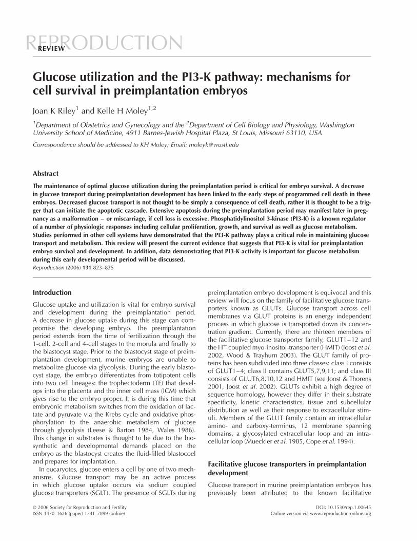
PI3-K is a heterodimeric enzyme that consists of a p85regulatory subunit and a p110 catalytic subunit. Murine2-cell embryos were shown to express the mRNA of mul-tiple PI3-K isoforms including p85a and b and p110 a, b,g, d (Lu et al. 2004). In addition, Kawamura et al. (2005)demonstrated the presence of p110 transcripts from theoocyte through the hatched blastocysts stage of murinepreimplantation development. PI3-K activity results in therecruitment of Akt to the plasma membrane. NavarreteSantos et al. (2004b) demonstrated the presence of Aktprotein in rabbit blastocysts. The Akt protein present in therabbit blastocysts was phosphorylated indicating that atthis stage of development, Akt is likely to be activated bygrowth factors present in its milieu. A final study exam-ined the expression of both PI3-K and Akt protein through-out murine preimplantation development (Fig. 1). Usingpan p85, p110 and Akt antibodies it was determined thatboth PI3-K subunits and Akt are expressed from the 1-cellthrough the blastocyst stage of murine preimplantationdevelopment (Riley et al. 2005b). These proteins are loca-lized predominantly at the cell surface from the 1-cellthrough the morula stage. At the blastocyst stage, bothPI3-K and Akt exhibited an apical staining pattern in theTE cells. Similar to what Navarrete Santos et al. (2004b)reported in rabbit, Akt was phosphorylated throughoutmurine preimplantation embryo development and its pre-sence at the plasma membrane is a reflection of its acti-vation status. Most recently, the presence of the PI3-Kpathway has also been detected in human blastocystsusing a cDNA microarray approach (Adjaye et al. 2005).These studies establish the presence of the PI3-K/Akt path-way in preimplantation embryos and the phosphorylationstatus of Akt suggests the pathway is active during thisdevelopmental period.
Targeted gene deletion experiments initially revealed theimportance of the PI3-K pathway during embryo develop-ment. The physiological roles of different classes and iso-forms of PI3-K are complex and are beginning to become
more well defined. Deletion of certain PI3-K subunits leadsto embryonic lethality. Mice deficient in the p110a cataly-tic subunit of PI3-K die in utero between E9.5 and E10.5(Bi et al. 1999). The null mice have a proliferation defect(Bi et al. 1999) and multiple vascular defects (Lelievre et al.2005). Deletion of the p110b subunit leads to earlyembryonic lethality suggesting a more critical role for thisprotein during development (Bi et al. 2002). A few p110bdeficient embryos were detected at E3.5 (blastocyst stage)however there is a deficit in the number of homozygousknockout embryos detected during this period of develop-ment according to Mendelian ratios. Thus both p110a andp110b are vital during embryonic development. Thedeletion of some PI3-K isoforms does not result in embryo-nic lethality implying either that these isoforms are notimportant during development or that functionalredundancy exists among these proteins.
The physiologic importance of the PI3-K pathway hasrecently been documented during the preimplantationperiod. Lu et al. (2004) demonstrated the importance ofthe PI3-K pathway in mammalian preimplantation embryodevelopment by showing that the activation of PI3-K byan embryonic trophic factor, platelet-activating factor(PAF), is critical for embryo development and survival.PAF treatment of 2-cell embryos results in a transientincrease in calcium that is inhibited by both LY-294002and wortmannin implying that PI3-K activity is requiredfor this PAF-induced biological response. In addition, aseparate global gene expression study demonstrated thatgenes involved in inositol phosphate and calcium signal-ing such as Pik3c2a (a class II PI3-K a polypeptide) areincreased in activated as compared with dormant blasto-cysts (Hamatani et al. 2004). The importance of the PI3-Kpathway during the preimplantation period was high-lighted by a study that demonstrated that inhibition ofPI3-K results in decreased numbers of embryos thatdevelop to the morula and blastocyst stage in vitro whencultured from a zygote stage, similar to Pafr deficientembryos (Lu et al. 2004). The blastocysts that did developcontained fewer cells and a larger number of fragmentednuclei. The effects induced by the PI3-K inhibitors weredose-dependent. Interestingly, if the embryos were onlyexposed to the PI3-K inhibitors from the zygote throughthe 2-cell stage and then cultured to the blastocyst stagethere was again a decrease in cell number and an increasein the number of fragmented nuclei. However, the effectof this limited treatment was not as great as when theembryos were cultured with the inhibitors throughout pre-implantation development. Thus activation of the PI3-Kpathway, in this case via PAF, is critical for the survivaland development of the preimplantation embryo.
Insulin has been shown in other cell systems to activatethe PI3-K pathway and thus stimulate glucose uptake bycausing the translocation of insulin responsive GLUTs,such as GLUT4, to the plasma membrane (for reviewsee Welsh et al. 2005). It is controversial whetherinsulin-stimulation results in the activation of this pathway
826 J K Riley and K H Moley
Reproduction (2006) 131 823–835 www.reproduction-online.org
in preimplantation embryos. Navarrete Santos et al.
(2004b) found that insulin-stimulation results in the acti-
vation of the mitogen-activated protein kinase (MAPK)
pathway but not the PI3-K/Akt pathway in rabbit blasto-
cysts. In contrast, Riley et al. (2005b) demonstrated insulin
treatment of murine blastocysts resulted in a 2-fold
increase in Akt phosphorylation in comparision with con-
trols. Moreover, LY-294002 and wortmannin were shown
to completely inhibit insulin-stimulated glucose uptake at
the blastocyst stage (Fig. 2). Thus whether insulin triggers
the activation of the PI3-K/Akt pathway and thereby
regulates glucose homeostasis in mammalian blastocystsremains to be determined.
Other experiments highlighting the functional import-ance of the PI3-K pathway during the preimplantationperiod include those demonstrating that inhibition of thePI3-K pathway results in decreased blastocyst hatching(Riley et al. 2005b). Blastocyst hatching from the zonapellucida is required for successful implantation. Althoughhatching is delayed in these embryos it does occur as blas-tocysts that are cultured in vitro for 24 h in the presence ofLY-294002 and then transferred back into pseudo-pregnantfemale recipient mice, resulted in approximately the same
Figure 1 Detection of PI3-K subunits, p85 and p110, in preimplantation embryos. Reproduced from Riley et al. 2005b. Preimplantation mouseembryos were retrieved at different stages of development and stained with either preimmune antisera as a negative control or with antibodiesspecific for the p85 or p110 subunit of PI3-K. The embryos were then incubated with a secondary antibody, Alexa Fluor 488 goat anti-rabbit ordonkey anti-goat IgG (green fluorescence). Embryos were counterstained with TO-PRO-3 iodide, which stains nuclei (blue channel).
PI-3K and cell survival in preimplantation embryos 827
www.reproduction-online.org Reproduction (2006) 131 823–835

number of implantation sites but a dramatic increase in
the fetal resorption rate as compared with controls
(Fig. 3B) (Riley et al. 2005a).Inhibition of the PI3-K pathway is known to result in the
induction of apoptosis in many cell systems. A recent
report has shown that inhibition of PI3-K induces
apoptosis in murine blastocysts (Gross et al. 2005).Gross et al. (2005) treated in vivo derived blastocysts withLY-294002 for 24 h and showed a five-fold increase inTUNEL-positive nuclei as compared with controls.Apoptotic cells were detected in both the TE as well asthe ICM. Blastocysts cultured for 48 h completely degener-ated and displayed rampant apoptosis. We have alsocultured in vivo derived blastocyst for 30 h in vitro, in thepresence of increasing concentrations of LY-294002 anddemonstrated a dose-dependent increase in the number ofTUNEL-positive nuclei per embryo as compared with con-trols (Fig. 3A) (Riley et al. 2005a). A final study conductedby Kawamura et al. (2005) demonstrated that TGF-a hasanti-apoptotic effects on murine preimplantation embryosexposed to suboptimal culture conditions. The mechanismof the anti-apoptotic effect involves the up-regulation ofsurvivin, a member of the inhibitor of apoptosis family.Inhibition of PI3-K using either LY-294002 or wortmanninresulted in the abrogation of TGF-a-induced up-regulationof survivin expression in blastocysts. PI3-K activity istherefore required for the anti-apoptotic effects of TGF-a,as mediated by survivin, in murine preimplantationembryos. Taken together the aforementioned studiesdemonstrate that PI3-K activity is critical for embryodevelopment and survival during the preimplantationperiod and that inhibition of this pathway even for discreteperiods during preimplantation development has longlasting detrimental effects on embryo development,survival and pregnancy outcome.
Figure 2 Insulin-stimulated glucose uptake is inhibited by LY-294002and wortmannin. Reproduced from Riley et al. 2005b. Insulin-stimu-lated glucose uptake was measured in blastocysts exposed to mediaalone (no treatment), 250mM LY-294002 and 100 nM wortmannin.The percentage of insulin-stimulated glucose uptake over basalglucose uptake is plotted.
Figure 3 Inhibition of the PI3-K pathway at the blastocysts stage results in increased apoptosis and an increased frequency of fetal resorptions.Reproduced from Riley et al. 2005a. (A) Blastocysts were recovered and cultured in vitro in the presence of DMSO (vehicle control shown isfor the highest concentration of LY-294002 used) or increasing concentrations of the PI3-K inhibitor LY-294002. The TUNEL assay was per-formed and the apoptotic nuclei are depicted in red. Embryos were counterstained with the nuclear dye TO-PRO-3 iodide as shown in blue.(B) Blastocysts were recovered and then cultured in vitro for 24 h in media containing either DMSO or 250mM LY-294002. Unhatched blasto-cysts were then transferred back into the uterine horn of pseudopregnant female recipient mice at 2.5 dpc. This panel shows representativeuterine horns derived from mice into which blastocysts cultured in either DMSO or LY-294002 were transferred.
828 J K Riley and K H Moley
Reproduction (2006) 131 823–835 www.reproduction-online.org

Glucose metabolism and apoptosis
Significant progress has been made in recent years linkingglucose transport, cell metabolism, the PI3-K pathway,and apoptosis both in preimplantation embryos as well asin other cell systems (for reviews see Moley & Mueckler2000, Moley 2001, Plas et al. 2002, Plas & Thompson2002). Apoptosis or programmed cell death in preimplan-tation embryos is a routine process by which the embryoeliminates abnormal or extraneous cells (for reviews seeHardy 1997, 1999, Jurisicova & Acton 2004, Fabian et al.2005). Studies demonstrated that in vivo-derived murineand porcine preimplantation embryos commonly do notshow TUNEL-positive staining before the blastocyst stage(Long et al. 1998, Kamjoo et al. 2002). Jurisicova et al.(1996) demonstrated in vitro that of 200 cleavage stagehuman-arrested embryos, the majority showed signs ofapoptosis including TUNEL-labeling of the nuclei. Inaddition, agents such as staurosporine do induce apopto-sis very early during development (Weil et al. 1996, Mat-wee et al. 2000). In mammalian development, apoptosisnormally occurs at the blastocyst stage and arises in boththe ICM and TE (Mohr & Trounson 1982, Handyside &Hunter 1986, Brison & Schultz 1997, Jurisicova et al.1998). During this stage of development, one purpose ofprogrammed cell death may be to eliminate cells with TEpotential from the ICM (Pierce et al. 1989). There aremany insults that induce apoptosis in the preimplantationembryo, one of which is glucose deprivation. Previously itwas shown that the down-regulation of certain glucosetransporters during preimplantation development results inincreased apoptosis at the blastocyst stage. Specificallythe down-regulation of either GLUT1 via high glucoseconcentrations in vitro or GLUT1 and GLUT8 utilizingantisense oligonucleotides results in increased apoptosisat the blastocyst stage (Chi et al. 2000a, Pinto et al.2002a). In the case of GLUT8, down-regulation via anti-sense oligonucleotides leads to increased fetal resorptionrates when the blastocysts were transferred into pseudo-pregnant recipient mice. Thus down-regulation of glucosetransporter expression resulting in altered intraembryonicglucose concentrations leads to the induction of apoptosisin blastocysts.
Women with diabetes mellitus are at a higher risk forspontaneous abortions and fetal malformations (Greeneet al. 1989, Casson et al. 1997, Hawthorne et al. 1997).Studies have established that an insult to the preimplanta-tion embryo, triggered by maternal hyperglycemia, haslong lasting detrimental effects on development. Thehypothesis is that the decrease in glucose transport result-ing from the hyperglycemia-induced down-regulation ofthe GLUTs, results in apoptosis of key progenitor cells orthat this apoptosis and metabolic changes adversely affectthe differentiation of the remaining cells. Either or both ofthese events then lead to either increased pregnancyresorptions or malformations. This hypothesis has beensubstantiated by two recent studies. First, Heilig et al.
(2003) developed a transgenic mouse over expressingGLUT1 antisense. The homozygote GLUT1AS fetuses didnot survive, and demonstrated a 7-fold higher stillbornrate than controls. Embryonic GLUT1 deficiency wasassociated with growth retardation (31.1%) and majormalformations (35.3%) consistent with those seen ininfants of diabetic women including caudal regression,anencephaly, microphthalmia, and micrognathia. Examin-ation of homozygote embryos at the blastocyst stagerevealed increased apoptosis and decreased glucose trans-port, consistent with the embryos derived from diabeticmice, suggesting that the decrease in glucose transportand resulting apoptosis may be responsible for the devel-opmental abnormalities seen in the GLUT1AS model.Second, in recent studies fetuses that developed from dia-betic 1-cell embryos, which were transferred into normalpseudo-pregnant recipient female mice were consistentlyand significantly smaller than controls (K Moley, unpub-lished observations). Importantly, these fetuses haveapproximately a 10% malformation rate, whereas no mal-formations were detected in control fetuses. The malfor-mations detected in the diabetic embryos consisted ofskeletal anomalies and they demonstrated delayed neuraltissue development. Similarly, blastocysts recovered fromdiabetic mice and then transferred into normal recipientfemales develop malformations, whereas control blasto-cysts do not. The control blastocysts gave rise to fetusesthat had 0% malformations, were on average 1.2 cm inlength, and had an overall resorption rate of 21%. In con-trast, blastocysts derived from diabetic mice gave rise tofetuses that had a 33% malformation rate, were on aver-age 0.9 cm in length, and had an overall resorption rate of52%. These data establish that the preimplantation periodin mammalian development is a critical stage and that ahyperglycemic insult incurred during this period alonecan have long lasting detrimental effects on embryo survi-val and development (K Moley, unpublished observations).Other studies have also demonstrated that apoptosisinduced at this early stage leads to abnormal developmentand poor pregnancy outcome (Chi et al. 2000a, Pinto et al.2002b, Heilig et al. 2003).
PI3-K and glucose metabolism
One hypothesis for the etiology of diabetes-associatedmalformations is hyperglycemia-induced apoptosis (Pamp-fer et al. 1997, Phelan et al. 1997, Moley et al. 1998a).Exaggerated apoptosis during the preimplantation periodmay result in fetal resorption or malformation due to theloss of key progenitor cells. Using a mouse diabetesmodel it was shown that hyperglycemia results indecreased glucose transport at the blastocysts stage, thatcorresponds with decreased GLUT1 protein expression(Moley et al. 1998b). As a consequence of glucose trans-porter down-regulation, these blastocysts have lowerintraembryonic glucose concentrations. This decrease in
PI-3K and cell survival in preimplantation embryos 829
www.reproduction-online.org Reproduction (2006) 131 823–835

glucose utilization by the blastocyst occurs concurrentlywith increased apoptosis in the embryo (Chi et al. 2000a).Similar results have been found in other cell systems,including retinal pericytes (Mandarino et al. 1994, Li et al.1998) where hyperglycemia-induced apoptosis of thesecells is thought to play a role in the development of dia-betic retinopathy. The cell death that occurs in the blasto-cysts requires both p53 and Bax (Moley et al. 1998a, Chiet al. 2000a, Keim et al. 2001). Embryos derived from dia-betic Bax-deficient mice demonstrated decreased glucoseuptake but no apoptosis (Chi et al. 2000a). In addition,the diabetic Bax2/2 embryos exhibit a decreased resorp-tion and malformation rate as compared with controls.This data suggests that hyperglycemia results in decreasedglucose transport and that this event may trigger apoptosisin the murine blastocyst leading to poor pregnancy out-come. In vitro studies in which 2-cell embryos were cul-tured to the blastocysts stage in the presence of highglucose concentrations also resulted in decreased glucosetransporter expression, decreased glucose uptake, andincreased apoptosis (Moley et al. 1998a, Chi et al.2000a). Therefore, inhibition of glucose uptake and thusmetabolism via the down-regulation of specific glucosetransporters is linked to the induction of apoptosis in mur-ine blastocysts. These data imply that glucose transportand the maintenance of optimal intraembryonic glucoseconcentrations are crucial for preimplantation embryodevelopment and survival.
Some of the first direct in vivo evidence demonstratingthe importance of specific PI3-K subunits in the regulationof insulin sensitivity and glucose homeostasis came fromgenetic deletion studies. Mice deficient in the p85asubunit of PI3-K show increased insulin sensitivity andhypoglycemia (Terauchi et al. 1999). Insulin-stimulation ofmuscle and adipocytes derived from the null mice resultsin increased GLUT4 translocation to the plasma mem-brane with a corresponding increase in glucose transport.In addition, heterozygous deletion of p85a increasesinsulin sensitivity and glucose homeostasis in mice thatare insulin-resistant due to heterozygous deletion of theinsulin receptor or insulin receptor substrate-1 (IRS-1)(Mauvais-Jarvis et al. 2002). These data are surprising andseveral hypotheses have been put forward to explain thisresult. It has been suggested that the p85 subunit hasnegative regulatory effects on the p110 subunit eitherthrough competing with the p85/p110 heterodimer forreceptor binding sites (Yu et al. 1998) or by inhibitingp110 activation by Ras (Chan et al. 2002, Jimenez et al.2002). It is thought that insulin sensitivity is controlled bya equilibrium between the p85 and p110 subunits(Brachmann et al. 2005). Other studies have shown thatp85b null mice have enhanced insulin sensitivity and areboth hypoinsulinemic and hypoglycemic (Ueki et al.2002) while the p50a/p55a double knockout mice alsodisplay increased insulin sensitivity and insulin-stimulatedglucose uptake in muscle and adipocytes (Chen et al.2004). Finally, mice lacking p85a/p50a/p55a die a
few days after birth and are hypoglycemic with decreasedinsulin levels and improved glucose tolerance (Frumanet al. 2000). Taken together these data suggest that theregulatory subunits of PI3-K play an essential role in insu-lin signaling and glucose metabolism.
A recent study conducted in murine blastocysts hassuggested that PI3-K activity is critical for glucose uptakeand metabolism during the preimplantation period. Simi-lar to what was previously found in lymphocytes deprivedof exogenous growth factors (Whetton et al. 1984, Kanet al. 1994, Rathmell et al. 2000, Vander Heiden et al.2001) inhibition of the PI3-K/Akt pathway in murine blas-tocysts using LY-294002, resulted in decreased cell surfaceexpression of GLUT1 with a corresponding decrease in2-deoxyglucose uptake (Fig. 4) (Riley et al. 2005a). Alongwith the decrease in glucose utilization, the blastocystsdisplayed increased levels of apoptosis. Thus one mechan-ism by which PI3-K may promote embryo survival isthrough the maintenance of glucose uptake by the regu-lation of glucose transporter expression at the cell surface.A second mechanism by which PI3-K may regulate gly-colysis is through its effects on the activity of glycolyticenzymes. Inhibition of the PI3-K pathway using LY-294002resulted in decreased hexokinase activity at the blastocyststage. Hexokinase is the first enzyme involved inthe glycolytic pathway. It converts intracellular glucose toglucose-6-phosphate and thus a decrease in hexokinaseactivity may have a large effect on the overall rate of glu-cose utilization. It was previously shown in lymphocytesthat the ability of activated Akt to inhibit apoptosisrequires the presence of glucose and is linked to itsmetabolism (Plas et al. 2001, Rathmell et al. 2003). A sep-arate study demonstrated the anti-apoptotic activity of Aktrequires the first committed step of glucose metabolismthat is catalyzed by hexokinase (Gottlob et al. 2001).Finally, Akt was shown to increase hexokinase activity(Gottlob et al. 2001, Rathmell et al. 2003). Taken togetherthese data suggest that the PI3-K/Akt pathway is criticalfor glucose metabolism in the preimplantation embryo viaits ability to regulate GLUT1 expression at the plasmamembrane and thus glucose uptake by blastocysts as wellas the activity of a key glycolytic enzyme.
The inhibition of glucose metabolism is in large partsufficient to explain the physiologic outcomes of inhibitingthe PI3-K pathway in preimplantation embryos. We havedemonstrated that iodoacetate, a glyceraldehyde 3-phos-phate dehydrogenase (GAPDH) inhibitor, induces apopto-sis in both blastocysts and TS cells (Fig. 5A) (Riley et al.2005a). GAPDH is a glycolytic enzyme which catalyzesthe conversion of glyceraldehyde 3-phosphate to1,3 bisphosphoglycerate resulting in the production ofNADH. These findings suggest that the maintenance ofglycolysis is important for embryo survival as the inhi-bition of this pathway results in the induction of celldeath. Importantly, a significant increase in the number offetal resorptions was seen in embryos derived from iodoa-cetate-treated blastocysts as compared with controls
830 J K Riley and K H Moley
Reproduction (2006) 131 823–835 www.reproduction-online.org

(Fig. 5B) (Riley et al. 2005a). Therefore, the maintenanceof glycolysis at the blastocyst stage is crucial forsubsequent developmental success. Importantly, onemechanism by which PI3-K promotes cell survival and
reproductive success in preimplantation embryos isthrough the maintenance of glucose metabolism. A pre-vious study showed that the inhibition of glycolysis laterin development also has deleterious effects on the fetus.
Figure 4 Inhibition of PI3-K results in decreased GLUT1 expression at the plasma membrane. Reproduced from Riley et al. 2005a. Blastocystswere recovered and cultured in vitro in the presence of DMSO (vehicle control) or 250mM LY-294002. The embryos were subjected to theTUNEL assay (red), immunofluorescent staining for GLUT1 or GLUT3 (green), and nuclear staining (blue).
Figure 5 Inhibition of GAPDH results in the induction of apoptosis in blastocysts and an increased frequency of fetal resorptions. Reproducedfrom Riley et al. 2005a. (A) Blastocysts were recovered and cultured in vitro in the presence of HTF media alone or increasing concentrations ofthe GAPDH inhibitor iodoacetate. The TUNEL assay was performed and the apoptotic nuclei are depicted in red. Embryos were counterstainedwith the nuclear dye TO-PRO-3 iodide as shown in blue. (B) Blastocysts were recovered and then cultured in vitro for 24 h in media containingeither HTF alone or 2.5mM iodoacetate. Unhatched blastocysts were then transferred back into the uterine horn of pseudopregnant female reci-pient mice at 2.5 dpc. This panel shows representative uterine horns derived from mice into which blastocysts cultured in either control mediaor iodoacetate were transferred.
PI-3K and cell survival in preimplantation embryos 831
www.reproduction-online.org Reproduction (2006) 131 823–835

Wentzel et al. (2003) demonstrated that the inhibition ofGAPDH activity by iodoacetate in gestational day 11 ratembryos, results in increased malformation rates as wellas decreased size, somite number, and DNA and proteincontent. Thus the inhibition of glycolysis has harmfuleffects on both pre- and post-implantation embryodevelopment.
Conclusion
A growing body of evidence suggests that the PI3-K path-way plays a critical role in embryo survival. In additionPI3-K activity regulates glucose utilization in the preim-plantation blastocyst. The ability of PI3-K to promoteembryo survival and metabolism depends on its ability tomaintain glucose uptake and the activity of at least oneglycolytic enzyme (Fig. 6). It will be important to deter-mine whether this key survival pathway is inhibited duringthe preimplantation period in maternal disease states suchas diabetes and insulin resistance that are known to affectglucose utilization in the preimplantation embryo andhave adverse effects on pregnancy outcome.
Acknowledgements
This work is supported by a Juvenile Diabetes Research Foun-dation Fellowship to J K R and by HD40390 and DK0070351from the National Institutes of Health to K H M. The authorsdeclare that there is no conflict of interest that would preju-dice the impartiality of this scientific work.
References
Adjaye J, Huntriss J, Herwig R, Benkahla A, Brink T, Wierling C,Hultschig C, Groth D, Yaspo ML, Picton H, Gosden R & LehrachH 2005 Primary differentiation in the human blastocyst: Compara-tive molecular portraits of ICM and TE cells. Stem Cells 231514–1525.
Aghayan M, Rao LV, Smith RM, Jarett L, Charron MJ, Thorens B &Heyner S 1992 Developmental expression and cellular localiz-ation of glucose transporter molecules during mouse preimplanta-tion development. Development 115 305–312.
Augustin R, Pocar P, Navarrete Santos A, Wrenzycki C, Gandolfi F,Niemann H & Fischer B 2001 Glucose transporter expression isdevelopmentally regulated in in vitro derived bovine preimplanta-tion embryos. Molecular Reproduction and Development 60370–376.
Augustin R, Pocar P, Wrenzycki C, Niemann H & Fischer B 2003Mitogenic and anti-apoptotic activity of insulin on bovine embryosproduced in vitro. Reproduction 126 91–99.
Bi L, Okabe I, Bernard DJ, Wynshaw-Boris A & Nussbaum RL 1999Proliferative defect and embryonic lethality in mice homozygousfor a deletion in the p110alpha subunit of phosphoinositide3-kinase. Journal of Biological Chemistry 274 10963–10968.
Bi L, Okabe I, Bernard DJ & Nussbaum RL 2002 Early embryoniclethality in mice deficient in the p110beta catalytic subunit of PI3-kinase. Mammalian Genome 13 169–172.
Brachmann SM, Ueki K, Engelman JA, Kahn RC & Cantley LC 2005Phosphoinositide 3-kinase catalytic subunit deletion and regulat-ory subunit deletion have opposite effects on insulin sensitivity inmice. Molecular and Cellular Biology 25 1596–1607.
Brazil DP & Hemmings BA 2001 Ten years of protein kinase B sig-nalling: a hard Akt to follow. Trends in Biochemical Sciences 26657–664.
Brison DR & Schultz RM 1997 Apoptosis during mouse blastocystformation: evidence for a role for survival factors including trans-forming growth factor alpha. Biology of Reproduction 561088–1096.
Byrne AT, Southgate J, Brison DR & Leese HJ 2002 Regulation ofapoptosis in the bovine blastocyst by insulin and the insulin-likegrowth factor (IGF) superfamily. Molecular Reproduction andDevelopment 62 489–495.
Cantley LC 2002 The phosphoinositide 3-kinase pathway. Science296 1655–1657.
Carayannopoulos M, Chi M, Cui Y, Pingsterhaus J & Moley K 2000GLUT8, a glucose transporter responsible for insulin-stimulateduptake in the blastocyst. PNAS 97 7313–7318.
Carayannopoulos MO, Schlein A, Wyman A, Chi M, KeembiyehettyC & Moley KH 2004 GLUT9 is differentially expressed andtargeted in the preimplantation embryo. Endocrinology 1451435–1443.
Casslen B & Nilsson B 1984 Human uterine fluid, examined in un-diluted samples for osmolarity and the concentrations of inorganicions, albumin, glucose, and urea. American Journal of Obsteticsand Gynecology 150 877–881.
Casson IF, Clarke CA, Howard CV, McKendrick O, Pennycook S,Pharoah POD, Platt MJ, Stanisstreet M, van Velszen D &Walkinshaw S 1997 Outcomes of pregnancy in insulin dependentdiabetic women: results of a five year population cohort study.British Medical Journal 315 275–278.
Figure 6 PI3-K activity regulates glucose utilization in the preimplan-tation blastocyst. The ability of PI3-K to promote embryo survival andmetabolism depends in part on its ability to maintain glucose uptake.PI3-K activity is required to retain the facilitative glucose transporterGLUT1 at the plasma membrane. It is thought that Akt is involved inthe mechanism by which PI3-K promotes GLUT1 cell surfaceexpression. In addition the PI3-K pathway is required for the optimalactivity of at least one glycolytic enzyme in blastocysts.
832 J K Riley and K H Moley
Reproduction (2006) 131 823–835 www.reproduction-online.org

Chan TO, Rittenhouse SE & Tsichlis PN 1999 AKT/PKB and other D3phosphoinositide-regulated kinases: kinase activation by phosphoi-nositide-dependent phosphorylation. Annual Review of Biochem-istry 68 965–1014.
Chan TO, Rodeck U, Chan AM, Kimmelman AC, Rittenhouse SE,Panayotou G & Tsichlis PN 2002 Small GTPases and tyrosinekinases coregulate a molecular switch in the phosphoinositide3-kinase regulatory subunit. Cancer Cell 1 181–191.
Chen D, Mauvais-Jarvis F, Bluher M, Fisher SJ, Jozsi A, Goodyear LJ,Ueki K & Kahn CR 2004 p50alpha/p55alpha phosphoinositide3-kinase knockout mice exhibit enhanced insulin sensitivity.Molecular and Cellular Biology 24 320–329.
Chi MM, Pingsterhaus J, Carayannopoulos M & Moley KH 2000aDecreased glucose transporter expression triggers BAX-dependentapoptosis in the murine blastocyst. Journal of Biological Chemistry275 40252–40257.
Chi MM, Schlein AL & Moley KH 2000b High insulin-like growthfactor 1 (IGF-1) and insulin concentrations trigger apoptosis in themouse blastocyst via down-regulation of the IGF-1 receptor. Endo-crinology 141 4784–4792.
Cope DL, Holman GD, Baldwin SA & Wolstenholme AJ 1994Domain assembly of the GLUT1 glucose transporter. BiochemicalJournal 300 (Pt 2) 291–294.
Dan-Goor M, Sasson S, Davarashvili A & Almagor M 1997Expression of glucose transporter and glucose uptake in humanoocytes and preimplantation embryos. Human Reproduction 122508–2510.
Darakhshan F, Hajduch E, Kristiansen S, Richter EA & Hundal HS1998 Biochemical and functional characterization of the GLUT5fructose transporter in rat skeletal muscle. Biochemical Journal336 (Pt 2) 361–366.
Dudek H, Datta SR, Franke TF, Birnbaum MJ, Yao R, Cooper GM,Segal RA, Kaplan DR & Greenberg ME 1997 Regulation of neur-onal survival by the serine-threonine protein kinase Akt. Science275 661–665.
Fabian D, Il’kova G, Rehak P, Czikkova S, Baran V & Koppel J 2004Inhibitory effect of IGF-I on induced apoptosis in mouse pre-implantation embryos cultured in vitro. Theriogenology 61745–755.
Fabian D, Koppel J & Maddox-Hyttel P 2005 Apoptotic processesduring mammalian preimplantation development. Theriogenology64 221–231.
Fruman DA, Mauvais-Jarvis F, Pollard DA, Yballe CM, Brazil D,Bronson RT, Kahn CR & Cantley LC 2000 Hypoglycaemia, livernecrosis and perinatal death in mice lacking all isoforms of phos-phoinositide 3-kinase p85 alpha. Nature Genetics 26 379–382.
Gardner DK & Leese HJ 1988 The role of glucose and pyruvate trans-port in regulating nutrient utilization by preimplantation mouseembryos. Development 104 423–429.
Gardner HG & Kaye PL 1991 Insulin increases cell numbers andmorphological development in mouse pre-implantation embryosin vitro. Reproduction, Fertility and Development 3 79–91.
Gottlob K, Majewski N, Kennedy S, Kandel E, Robey RB & Hay N2001 Inhibition of early apoptotic events by Akt/PKB is dependenton the first committed step of glycolysis and mitochondrial hex-okinase. Genes Development 15 1406–1418.
Greene MF, Hare JW, Cloherty JP, Benacerraf BR & Soeldner JS1989 First-trimester hemoglobin A1 and risk for major malfor-mation and spontaneous abortion in diabetic pregnancy. Teratol-ogy 39 225–231.
Gross VS, Hess M & Cooper GM 2005 Mouse embryonic stem cellsand preimplantation embryos require signaling through the phos-phatidylinositol 3-kinase pathway to suppress apoptosis. MolecularReproduction and Development 70 324–332.
Hajduch E, Darakhshan F & Hundal HS 1998 Fructose uptake in ratadipocytes: GLUT5 expression and the effects of streptozotocin-induced diabetes. Diabetologia 41 821–828.
Hamatani T, Daikoku T, Wang H, Matsumoto H, Carter MG, Ko MS &Dey SK 2004 Global gene expression analysis identifies molecular
pathways distinguishing blastocyst dormancy and activation. PNAS101 10326–10331.
Handyside A & Hunter S 1986 Cell division and death in the mouseblastocyst before implantation. Roux’s Archives of DevelopmentalBiology 195 519–525.
Hardy K 1997 Cell death in the mammalian blastocyst. MolecularHuman Reproduction 3 919–925.
Hardy K 1999 Apoptosis in the human embryo. Reviews in Repro-duction 4 125–134.
Hardy K & Spanos S 2002 Growth factor expression and function inthe human and mouse preimplantation embryo. Journal of Endo-crinology 172 221–236.
Harvey MB & Kaye PL 1990 Insulin increases the cell number of theICM and stimulates morphological development of mouse blas-tocysts in vitro. Development 110 963–967.
Harvey MB & Kaye PL 1991 Mouse blastocysts respond metaboli-cally to short-term stimulation by insulin and IGF-1 through theinsulin receptor. Molecular Reproduction and Development 29253–258.
Hawthorne G, Robson S, Ryall EA, Sen D, Roberts SH & Ward PlattMP 1997 Prospective population based survey of outcome of preg-nancy in diabetic women: results of the Northern Diabetic Preg-nancy Audit, 1994 [see comments]. British Medical Journal 315279–281.
Heilig CW, Saunders T, Brosius FC 3rd, Moley K, Heilig K, Baggs R,Guo L & Conner D 2003 Glucose transporter-1-deficient miceexhibit impaired development and deformities that are similar todiabetic embryopathy. PNAS 100 15613–15618.
Herrler A, Krusche CA & Beier HM 1998 Insulin and insulin-likegrowth factor-I promote rabbit blastocyst development and preventapoptosis. Biology of Reproduction 59 1302–1310.
Hogan A, Heyner S, Charron MJ, Copeland NG, Gilbert DJ, JenkinsMA, Thorens B & Schultz GA 1991 Glucose transporter geneexpression in early mouse embryos. Development 113 363–372.
Jimenez C, Hernandez C, Pimentel B & Carrera AC 2002 The p85regulatory subunit controls sequential activation of phosphoinosi-tide 3-kinase by Tyr kinases and Ras. Journal of Biological Chem-istry 277 41556–41562.
Joost HG & Thorens B 2001 The extended GLUT-family of sugar/polyol transport facilitators: nomenclature, sequence character-istics, and potential function of its novel members. MolecularMembrane Biology 18 247–256.
Joost HG, Bell GI, Best JD, Birnbaum MJ, Charron MJ, Chen YT,Doege H, James DE, Lodish HF, Moley KH, Moley JF, Mueckler M,Rogers S, Schurmann A, Seino S & Thorens B 2002 Nomenclatureof the GLUT/SLC2A family of sugar/polyol transport facilitators.American Journal of Physiology, Endocrinology and Metabolism282 E974–E976.
Jurisicova A & Acton BM 2004 Deadly decisions: the role of genesregulating programmed cell death in human preimplantationembryo development. Reproduction 128 281–291.
Jurisicova A, Varmuza S & Casper RF 1996 Programmed cell deathand human embryo fragmentation. Molecular Human Reproduci-ton 2 93–98.
Jurisicova A, Rogers I, Fasciani A, Casper RF & Varmuza S 1998Effect of maternal age and conditions of fertilization on pro-grammed cell death during murine preimplantation embryo devel-opment. Molecular Human Reproduction 4 139–145.
Kamjoo M, Brison DR & Kimber SJ 2002 Apoptosis in the pre-implantation mouse embryo: effect of strain difference and in vitroculture. Molecular Reproduction and Development 61 67–77.
Kan O, Baldwin SA & Whetton AD 1994 Apoptosis is regulated bythe rate of glucose transport in an IL-3-dependent haemopoieticcell line. Biochemical Society Transactions 22 275S.
Kawamura K, Fukuda J, Shimizu Y, Kodama H & Tanaka T 2005Survivin Contributes to the Anti-Apoptotic Activities of Transform-ing Growth Factor alpha in Mouse Blastocysts Through Phospha-tidylinositol 30-Kinase Pathway. Biology of Reproduction 731094–1101.
PI-3K and cell survival in preimplantation embryos 833
www.reproduction-online.org Reproduction (2006) 131 823–835

Keim AL, Chi MM & Moley KH 2001 Hyperglycemia-induced apop-totic cell death in the mouse blastocyst is dependent on expressionof p53. Molecular Reproduction and Development 60 214–224.
Khwaja A, Rodriguez-Viciana P, Wennstrom S, Warne PH & Down-ward J 1997 Matrix adhesion and Ras transformation both activatea phosphoinositide 3-OH kinase and protein kinase B/Akt cellularsurvival pathway. EMBO Journal 16 2783–2793.
Korgun ET, Demir R, Hammer A, Dohr G, Desoye G, Skofitsch G &Hahn T 2001 Glucose transporter expression in rat embryo anduterus during decidualization, implantation, and early post-implantation. Biology of Reproduction 65 1364–1370.
Kristiansen S, Darakhshan F, Richter EA & Hundal HS 1997 Fructosetransport and GLUT-5 protein in human sarcolemmal vesicles.American Journal of Physiology 273 E543–E548.
Leese HJ & Barton AM 1984 Pyruvate and glucose uptake by mouseova and preimplantation embryos. Journal of Reproduction andFertility 72 9–13.
Lelievre E, Bourbon PM, Duan LJ, Nussbaum RL & Fong GH 2005Deficiency in the p110alpha subunit of PI3K results in diminishedTie2 expression and Tie2(2 /2 )-like vascular defects in mice.Blood 105 3935–3938.
Lequarre AS, Grisart B, Moreau B, Schuurbiers N, Massip A & Dessy F1997 Glucose metabolism during bovine preimplantation develop-ment: analysis of gene expression in single oocytes and embryos.Molecular Reproduction and Development 48 216–226.
Li W, Liu X, He Z, Yanoff M, Jian B & Ye X 1998 Expression of apop-tosis regulatory genes by retinal pericytes after rapid glucosereduction. Investagative Opthalmology and Visual Science 391535–1543.
Long CR, Dobrinsky JR, Garrett WM & Johnson LA 1998 Dual label-ing of the cytoskeleton and DNA strand breaks in porcine embryosproduced in vivo and in vitro. Molecular Reproduction and Devel-opment 51 59–65.
Lu DP, Chandrakanthan V, Cahana A, Ishii S & O’Neill C 2004Trophic signals acting via phosphatidylinositol-3 kinase arerequired for normal pre-implantation mouse embryo development.Journal of Cell Science 117 1567–1576.
Makarevich AV & Markkula M 2002 Apoptosis and cell proliferationpotential of bovine embryos stimulated with insulin-like growthfactor I during in vitro maturation and culture. Biology of Repro-duction 66 386–392.
Mandarino LJ, Finlayson J & Hassell JR 1994 High glucose down-regulates glucose transport activity in retinal capillary pericytes butnot endothelial cells. Investigations in Ophthalmology and VisualScience 35 964–972.
Matsui M, Takahashi Y, Hishinuma M & Kanagawa H 1995 Insulinand insulin-like growth factor-I (IGF-I) stimulate the developmentof bovine embryos fertilized in vitro. Journal of Veterinary MedicalScience 57 1109–1111.
Matwee C, Betts DH & King WA 2000 Apoptosis in the early bovineembryo. Zygote 8 57–68.
Mauvais-Jarvis F, Ueki K, Fruman DA, Hirshman MF, Sakamoto K,Goodyear LJ, Iannacone M, Accili D, Cantley LC & Kahn CR 2002Reduced expression of the murine p85alpha subunit of phosphoi-nositide 3-kinase improves insulin signaling and amelioratesdiabetes. Journal of Clinical Investigations 109 141–149.
Mohr LR & Trounson AO 1982 Comparative ultrastructure of hatchedhuman, mouse and bovine blastocysts. Journal of Reproductionand Fertility 66 499–504.
Moley KH 2001 Hyperglycemia and apoptosis: mechanisms for con-genital malformations and pregnancy loss in diabetic women.Trends in Endocrinological Metabolism 12 78–82.
Moley KH & Mueckler MM 2000 Glucose transport and apoptosis.Apoptosis 5 99–105.
Moley KH, Chi M & Mueckler M 1998a Maternal hyperglycemiaalters glucose transport and utilization in mouse preimplantationembryos. American Journal of Physiology 275 E38–E47.
Moley KH, Chi MM, Knudson CM, Korsmeyer SJ & Mueckler MM1998b Hyperglycemia induces apoptosis in preimplantation
embryos via cell death effector pathways. Nature Medicine 121421–1424.
Morita Y, Tsutsumi O, Hosoya I, Taketani Y, Oka Y & Kato T 1992Expression and possible function of glucose transporter proteinGLUT1 during preimplantation mouse development from oocytesto blastocysts. Biochemical and Biophysical Research Communi-cations 188 8–15.
Mueckler M, Caruso C, Baldwin SA, Panico M, Blench I, Morris HR,Allard WJ, Lienhard GE & Lodish HF 1985 Sequence and structureof a human glucose transporter. Science 229 941–945.
Navarrete Santos A, Augustin R, Lazzari G, Galli C, Sreenan JM &Fischer B 2000 The insulin-dependent glucose transporter isoform4 is expressed in bovine blastocysts. Biochemical and BiophysicalResearch Communications 271 753–760.
Navarrete Santos A, Tonack S, Kirstein M, Kietz S & Fischer B 2004aTwo insulin-responsive glucose transporter isoforms and the insulinreceptor are developmentally expressed in rabbit preimplantationembryos. Reproduction 128 503–516.
Navarrete Santos A, Tonack S, Kirstein M, Pantaleon M, Kaye P &Fischer B 2004b Insulin acts via mitogen-activated protein kinasephosphorylation in rabbit blastocysts. Reproduction 128 517–526.
Pampfer S, Vanderheyden I, McCracken JE, Vesela J & De Hertogh R1997 Increased cell death in rat blastocysts exposed to maternaldiabetes in utero and to high glucose or tumor necrosis factor-alpha in vitro. Development 124 4827–4836.
Pantaleon M, Harvey MB, Pascoe WS, James DE & Kaye PL 1997Glucose transporter GLUT3: Ontogeny, targeting and role in themouse blastocyst. PNAS 94 3795–3800.
Pantaleon M, Ryan JP, Gil M & Kaye PL 2001 An unusual subcellularlocalization of GLUT1 and link with metabolism in oocytes andpreimplantation mouse embryos. Biology of Reproduction 641247–1254.
Pantaleon M & Kaye PL 1996 IGF-I and insulin regulate glucosetransport in mouse blastocysts via IGF-I receptor. Molecular Repro-duction and Development 44 71–76.
Phelan SA, Ito M & Loeken MR 1997 Neural tube defects in embryosof diabetic mice: role of the Pax-3 gene and apoptosis. Diabetes46 1189–1197.
Philpott KL, McCarthy MJ, Klippel A & Rubin LL 1997 Activatedphosphatidylinositol 3-kinase and Akt kinase promote survival ofsuperior cervical neurons. Journal of Cell Biology 139 809–815.
Pierce GB, Lewellyn AL & Parchment RE 1989 Mechanism of pro-grammed cell death in the blastocyst. PNAS 86 3654–3658.
Pinto A, Carayannopoulos M, Hoehn A, Dowd L & Moley K 2002aGLUT8 expression and translocation are critical for murine blas-tocyst survival. Biology of Reproduction 66 1729–1733.
Pinto AB, Schlein AL & Moley KH 2002b Preimplantation exposureto high insulin-like growth factor I concentrations results inincreased resorption rates in vivo. Human Reproduction 17457–462.
Plas DR, Talapatra S, Edinger AL, Rathmell JC & Thompson CB 2001Akt and Bcl-xL promote growth factor-independent survivalthrough distinct effects on mitochondrial physiology. Journal ofBiological Chemistry 276 12041–12048.
Plas DR, Rathmell JC & Thompson CB 2002 Homeostatic control oflymphocyte survival: potential origins and implications. NatureImmunology 3 515–521.
Plas DR & Thompson CB 2002 Cell metabolism in the regulation ofprogrammed cell death. Trends in Endocrinology and Metabolism13 75–78.
Rathmell JC, Vander Heiden MG, Harris MH, Frauwirth KA &Thompson CB 2000 In the absence of extrinsic signals, nutrientutilization by lymphocytes is insufficient to maintain either cellsize or viability. Molecular Cell 6 683–692.
Rathmell JC, Fox CJ, Plas DR, Hammerman PS, Cinalli RM &Thompson CB 2003 Akt-directed glucose metabolism can preventBax conformation change and promote growth factor-independentsurvival. Molecular and Cellular Biology 23 7315–7328.
834 J K Riley and K H Moley
Reproduction (2006) 131 823–835 www.reproduction-online.org
Riley JK, Carayannopoulos MO, Wyman AH, Chi M & Moley KH2005a PI3-kinase activity is critical for glucose metabolism andembryo survival in murine blastocysts. Journal of BiologicalChemistry (In Press).
Riley JK, Carayannopoulos MO, Wyman AH, Chi M, Ratajczak CK &Moley KH 2005b The PI3K/Akt pathway is present and functionalin the preimplantation mouse embryo. Developmental Biology284 377–386.
Robinson DH, Smith PR & Benos DJ 1990 Hexose transport in pre-implantation rabbit blastocysts. Journal of Reproduction andFertility 89 1–11.
Rogers S, Macheda ML, Docherty SE, Carty MD, Henderson MA,Soeller WC, Gibbs EM, James DE & Best JD 2002 Identification ofa novel glucose transporter-like protein-GLUT-12. American Jour-nal of Physiology, Endocrinology and Metabolism 282 E733–E738.
Schultz GA, Hogan A, Watson AJ, Smith RM & Heyner S 1992 Insu-lin, insulin-like growth factors and glucose transporters: temporalpatterns of gene expression in early murine and bovine embryos.Reproduction, Fertility and Development 4 361–371.
Shepherd PR, Gibbs EM, Wesslau C, Gould GW & Kahn BB 1992Human small intestine facilitative fructose/glucose transporter(GLUT5) is also present in insulin-responsive tissues and brain.Investigation of biochemical characteristics and translocation.Diabetes 41 1360–1365.
Sirisathien S & Brackett BG 2003 TUNEL analyses of bovine blas-tocysts after culture with EGF and IGF-I. Molecular Reproductionand Development 65 51–56.
Sirisathien S, Hernandez-Fonseca HJ & Brackett BG 2003 Influencesof epidermal growth factor and insulin-like growth factor-I onbovine blastocyst development in vitro. Animal ReproductionScience 77 21–32.
Smith RM, Garside WT, Aghayan M, Shi CZ, Shah N, Jarett L &Heyner S 1993 Mouse preimplantation embryos exhibit receptor-mediated binding and transcytosis of maternal insulin-like growthfactor I. Biology of Reproduction 49 1–12.
Songyang Z, Baltimore D, Cantley LC, Kaplan DR & Franke TF 1997Interleukin 3-dependent survival by the Akt protein kinase. PNAS94 11345–11350.
Spanos S, Becker DL, Winston RM & Hardy K 2000 Anti-apoptoticaction of insulin-like growth factor-I during human preimplanta-tion embryo development. Biology of Reproduction 631413–1420.
Terauchi Y, Tsuji Y, Satoh S, Minoura H, Murakami K, Okuno A,Inukai K, Asano T, Kaburagi Y, Ueki K, Nakajima H, Hanafusa T,Matsuzawa Y, Sekihara H, Yin Y, Barrett JC, Oda H, Ishikawa T,Akanuma Y & Komuro I 1999 Increased insulin sensitivity andhypoglycaemia in mice lacking the p85 alpha subunit of phosphoi-nositide 3-kinase. Nature Genetics 21 230–235.
Thompson JE & Thompson CB 2004 Putting the rap on Akt. Journal ofClinical Oncology 22 4217–4226.
Tonack S, Fischer B & Navarrete Santos A 2004 Expression of theinsulin-responsive glucose transporter isoform 4 in blastocysts ofC57/BL6 mice. Anatomy and Embryology 208 225–230.
Ueki K, Yballe CM, Brachmann SM, Vicent D, Watt JM, Kahn CR &Cantley LC 2002 Increased insulin sensitivity in mice lackingp85beta subunit of phosphoinositide 3-kinase. PNAS 99 419–424.
Vander Heiden MG, Plas DR, Rathmell JC, Fox CJ, Harris MH &Thompson CB 2001 Growth factors can influence cell growth andsurvival through effects on glucose metabolism. Molecular andCellular Biology 21 5899–5912.
Wales RG 1986 Measurement of metabolic turnover in single mouseembryos. Journal of Reproduction and Fertility 76 717–725.
Weil M, Jacobson MD, Coles HS, Davies TJ, Gardner RL, Raff KD &Raff MC 1996 Constitutive expression of the machinery for pro-grammed cell death. Journal of Cell Biology 133 1053–1059.
Welsh GI, Hers I, Berwick DC, Dell G, Wherlock M, Birkin R, LeneyS & Tavare JM 2005 Role of protein kinase B in insulin-regulatedglucose uptake. Biochemical Society Transactions 33 346–349.
Wentzel P, Ejdesjo A & Eriksson UJ 2003 Maternal diabetes in vivoand high glucose in vitro diminish GAPDH activity in rat embryos.Diabetes 52 1222–1228.
Whetton AD, Bazill GW & Dexter TM 1984 Haemopoietic cellgrowth factor mediates cell survival via its action on glucose trans-port. EMBO Journal 3 409–413.
Wood IS & Trayhurn P 2003 Glucose transporters (GLUT and SGLT):expanded families of sugar transport proteins. British Journal ofNutrition 89 3–9.
Woodgett JR 2005 Recent advances in the protein kinase B signalingpathway. Current Opinion in Cell Biology 17 150–157.
Wrenzycki C, Herrmann D, Carnwath JW & Niemann H 1998Expression of RNA from developmentally important genes inpreimplantation bovine embryos produced in TCM supplementedwith BSA. Journal of Reproduciton and Fertility 112 387–398.
Yu J, Zhang Y, McIlroy J, Rordorf-Nikolic T, Orr GA & Backer JM1998 Regulation of the p85/p110 phosphatidylinositol 30-kinase:stabilization and inhibition of the p110alpha catalytic subunit bythe p85 regulatory subunit. Molecular and Cellular Biology 181379–1387.
Zhou Y, Kaye PL & Pantaleon M 2004 Identification of the facilitativeglucose transporter 12 gene Glut12 in mouse preimplantationembryos. Gene Expression Patterns 4 621–631.
Received 3 November 2005First decision 17 November 2005Revised manuscript received 9 January 2006Accepted 19 January 2006
PI-3K and cell survival in preimplantation embryos 835
www.reproduction-online.org Reproduction (2006) 131 823–835