Embed Size (px)
Citation preview

©2013 MFMER | 3332228-1
Advances in Laboratory TB Diagnosis
Nancy L. Wengenack, PhD, D(ABMM)Rochester, MN
February 12, 2014

©2013 MFMER | 3332228-2
Disclosures
• Trek diagnostics – grant/research support
©2013 MFMER | 3332228-3
Overview
• Stains for Mycobacteria
• Culture of Mycobacteria
• Molecular methods for identification of M. tuberculosis
• From culture• Directly from specimen
• M. tuberculosis drug resistance testing• Rapid broth-based methods• Molecular markers of resistance

©2013 MFMER | 3332228-4
Stains for Mycobacteria

©2013 MFMER | 3332228-5
Mycobacterium Tuberculosis does not Stain well With the Gram Stain
M. tuberculosis ghosting on Gram stain

©2013 MFMER | 3332228-6
Lipoarabinomannan
Mycolic acid
Arabinogalactan
Peptidoglycan
Cytoplasmicmembrane
M. Tuberculosis cell wall
Mycobacteria Cell Wall
• Contain >60% lipid• Mycolic acids
(C60-C90 fatty acids)• Waxes• Gram positive organism
contains ~5% lipid• Gram negative organism
contains ~20% lipid
• Mycolic acid make the cell surface extremely hydrophobic and resistant to staining with basic aniline dyes or penetration by drugs
©2013 MFMER | 3332228-7
Mycobacterial Stains
• Mycobacteria are “acid-fast” bacilli (AFB)
• A complex is formed between mycolic acid and dye (carbol-fuchsin or auramine O)
• The complex is resistant to destaining by mineral acids (ie, acid-fast)
• So mycobacteria retain the carbol-fuchsin or auramine O stain and other bacteria do not

©2013 MFMER | 3332228-8
Ziehl-Neelsen StainUses heat to help drive fuchsin stain into waxy cell wall; phenol as mordant to fix stain; (Kinyoun stain method – no heat, instead uses higher concentration of phenol and fuchsin dye to aid uptake; less effective as direct stain)
AFB’s stain in red; non-AFB’s stain in blue
©2013 MFMER | 3332228-9
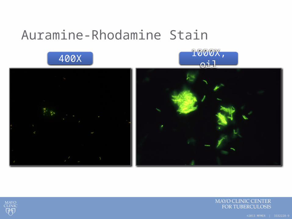
Auramine-Rhodamine Stain
400X 1000X, oil
©2013 MFMER | 3332228-10
Other Tidbits about AFB Smears/Stains• Fite stain
• Modification of ZN; often used in pathology • Uses a more mild decolorizing agent that supposedly works better for
“delicate” mycobacteria like M. leprae• Tissue processing in pathology can damage the mycolic acid, sometimes
making it difficult to find the AFB regardless of the stain used
• LED microscopy • WHO study indicated it was superior to ZN and equivalent to fluorescence
microscopy and recommended replacement of fluorescence and ZN with LED microscopy
• Gaining traction in developing countries where fluorescent microscopes scarce/expensive; can run on batteries
• Cannot reliably speciate using microscopy – Mtb looks like MAC which looks like M. abscessus, etc
• Positive smear suggests higher likelihood of infectivity if the patient has pulmonary Mtb

©2013 MFMER | 3332228-11
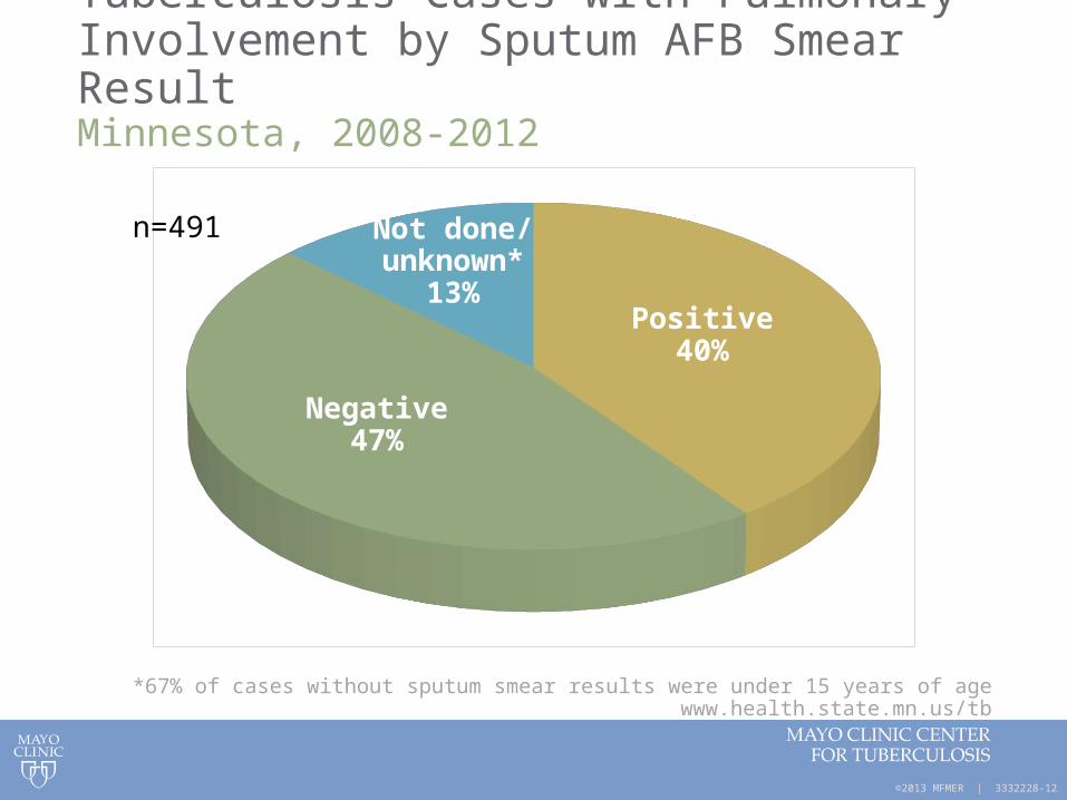
What % of Culture-Confirmed TB Cases have Positive Smears?
©2013 MFMER | 3332228-12
Tuberculosis Cases with Pulmonary Involvement by Sputum AFB Smear ResultMinnesota, 2008-2012
*67% of cases without sputum smear results were under 15 years of agewww.health.state.mn.us/tb
Not done/unknown*
13%Positive
40%
Negative47%
n=491

©2013 MFMER | 3332228-13
Are Two AFB Smears Better than One?Yield of Serial AFB Smears
Total positives detected by (%)
Study 1st smear 2nd smear 3rd smear
Walker et al: Int J Tuberc Lung Dis, 4:246, 2000
77.1 15.0 7.9
Ipuge et al: Trans R Soc Trop Med Hyg, 90:258, 1996
83.4 12.2 4.4
Saleem et al: Pak J Med Res, 46:94, 2007
66.2 24.0 9.8
Mathew et al: J Clin Microbiol, 40:3482 (low prevalence pop), 2002
89.4 5.3 5.3

©2013 MFMER | 3332228-14
Are Early Morning Sputum Specimens Still Preferred?
Study
Spot (random) specimen positive
(%)
Early morning specimen positive
(%)
Ssengooba et al: 2012, Tuberc Res Treat: 1-6 (MGIT culture positive for MTB), 2012
12/21 (57) 21/21 (100)
Abraham et al: Indian J Med Res, 135:249-51 (smear is positive), 2012
21/49 (43) 32/49 (65)

©2013 MFMER | 3332228-15
Culture of M. tuberculosis Complex
Sensitivity of culture is much better than smear; only 10-100 viable organisms/mL required for positive culture
Culture
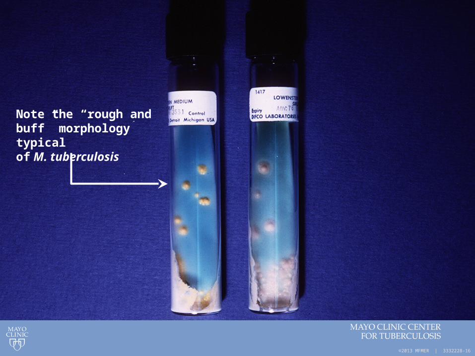
• Solid Medium• Egg-based – Lowenstein-Jensen (LJ); TTP ~30 days• Agar-based – Middlebrook
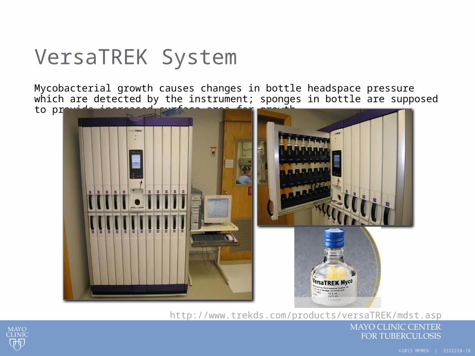
• Rapid Broth (Liquid) Medium (FDA-cleared systems)• Reduces TTP to ~ 10 days• BACTEC MGIT (fluorimetric, BD)• VersaTREK (pressure, TREK)
©2013 MFMER | 3332228-16
Note the “rough and buff” morphology typical of M. tuberculosis

©2013 MFMER | 3332228-17
BACTEC MGIT 960 Culture System
MGIT - Mycobacterial Growth Indicator Tubes (Becton Dickinson)• fluorescent indicator in bottom of tube quenched by O2 • mycobacterial growth = O2 and fluorescence
©2013 MFMER | 3332228-18
VersaTREK SystemMycobacterial growth causes changes in bottle headspace pressure which are detected by the instrument; sponges in bottle are supposed to provide increased surface area for growth
http://www.trekds.com/products/versaTREK/mdst.asp

©2013 MFMER | 3332228-19
www.health.state.mn.us/tb
Tuberculosis Cases by Mycobacterial Culture ResultMinnesota, 2008-2012
Not done/unknown*4%
Positive75%
Negative21%
n=806

©2013 MFMER | 3332228-20
Identification of M. tuberculosis Complex from Culture

©2013 MFMER | 3332228-21
Traditional Methods of Identification
• Historically, positive mycobacterial cultures were identified on the basis of
• Colonial morphology• Growth characteristics• Biochemical testing
(niacin, nitrate, pyraziniamidase)• Slow process taking up to 8 weeks
• Sometimes, HPLC or GLC for cell wall constituents – generally at CDC or State Public Health Labs
©2013 MFMER | 3332228-22
Molecular Methods Allow for Rapid IdentificationIdentification Methods for Culture Isolates

©2013 MFMER | 3332228-23
1. Nucleic Acid Hybridization Probes
From culture only • No amplification step
• Need lots of target nucleic acid!
• Add probe with unique, complementary sequence to known species; chemiluminescent detection
• Identification within 2-3 hours after growth in culture
Hologic Gen-Probe AccuProbes® (nucleic acid hybridization probes) available for• M. tuberculosis complex
• M. avium complex
• M. gordonae
• M. kansasii

©2013 MFMER | 3332228-24
Hybridization Probes
Step 1 Step 2 Step 3 Step 4
Microbiology culture plate Sonicator for
15 minutes
Heat at 95oc for 10 minutes
Lysingreagent
Add DNAprobe
reagent
DNA probe
DNA-rRNA hybrids detected with
chemiluminescent reads
©2013 MFMER | 3332228-25
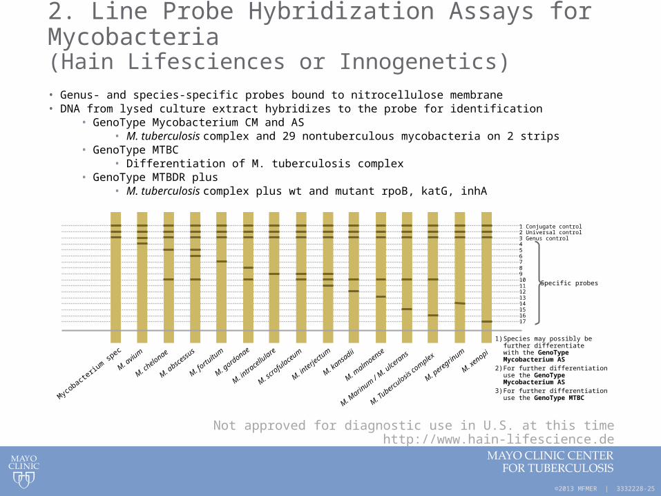
2. Line Probe Hybridization Assays for Mycobacteria(Hain Lifesciences or Innogenetics)• Genus- and species-specific probes bound to nitrocellulose membrane• DNA from lysed culture extract hybridizes to the probe for identification
• GenoType Mycobacterium CM and AS• M. tuberculosis complex and 29 nontuberculous mycobacteria on 2 strips
• GenoType MTBC• Differentiation of M. tuberculosis complex
• GenoType MTBDR plus• M. tuberculosis complex plus wt and mutant rpoB, katG, inhA
Not approved for diagnostic use in U.S. at this timehttp://www.hain-lifescience.de
1 Conjugate control2 Universal control3 Genus control4567891011121314151617
Specific probes
1) Species may possibly befurther differentiatewith the GenoTypeMycobacterium AS
2) For further differentiationuse the GenoTypeMycobacterium AS
3) For further differentiationuse the GenoType MTBC
Mycobacte
rium sp
ec
M. aviu
m
M. chelonae
M. absc
essus
M. fortu
itum
M. gordonae
M. intra
cellu
lare
M. scro
fulaceum
M. interje
ctum
M. kansa
dii
M. malm
oense
M. Marin
um / M. u
lcerans
M. Tubercu
losis co
mplex
M. peregrin
um
M. xenopi
©2013 MFMER | 3332228-26
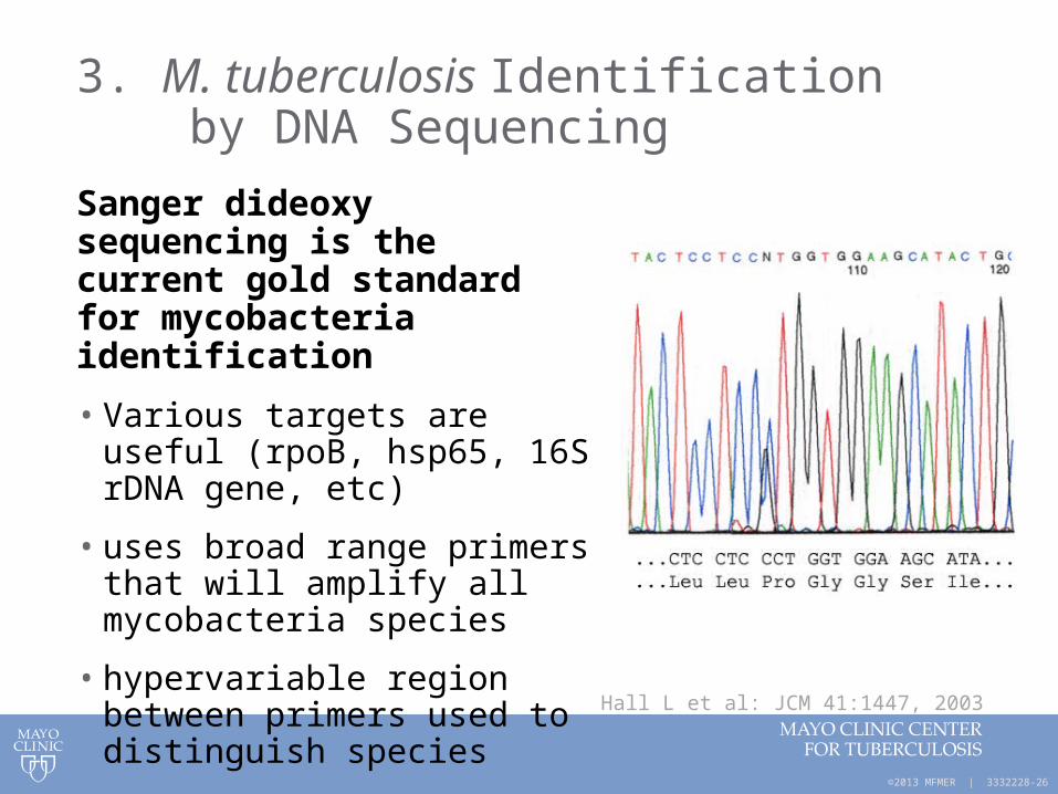
3. M. tuberculosis Identification by DNA Sequencing
Sanger dideoxy sequencing is the current gold standard for mycobacteria identification
• Various targets are useful (rpoB, hsp65, 16S rDNA gene, etc)
• uses broad range primers that will amplify all mycobacteria species
• hypervariable region between primers used to distinguish species
Hall L et al: JCM 41:1447, 2003
©2013 MFMER | 3332228-27
Sequence Analysis
• Compare the isolate sequence to known mycobacterial sequence libraries
• Microseq library (AB)• Lab-specific custom library• Genbank BLAST (NCBI)• Curated, web-based database tools
• Smartgene or isentio
• TAT can be as fast as 8hrs after growth of the organism in culture; in our lab we run in batches of ~96 isolates
• Select colonies to be sequenced in am• Pcrs in afternoon• Electrophorese overnight• Read/report next am

©2013 MFMER | 3332228-28
Advantages and Limitations of Sequencing for Identifcation of Mycobacteria
Advantages
• Allows for objective identification of a wide variety of mycobacteria
• Next day identification after growth in culture
Limitations
• Labor-intensive, requires skilled, trained (dedicated) technologists
• Equipment and reagent costs drive total test cost up
• Results are highly dependent upon the quality of your sequence library database

©2013 MFMER | 3332228-29
4. MALDI-TOF MS – a Paradigm Shift in Microbiology
• Matrix-assisted laser desorption ionization – time of flight (MALDI-TOF) mass spectrometry is changing the way we identify microbes
• Already becoming the main technique used in many laboratories for bacterial and yeast identification
• Mycobacteria and mold identification by MALDI-TOF MS is not far behind

©2013 MFMER | 3332228-30
Two examples of MALDI-TOF MS Instruments
for Identification of Microorganisms
Bruker Biotyper bioMérieux Vitek MS

©2013 MFMER | 3332228-31
Bruker Biotyper MALDI-TOF MS
©2013 MFMER | 3332228-32
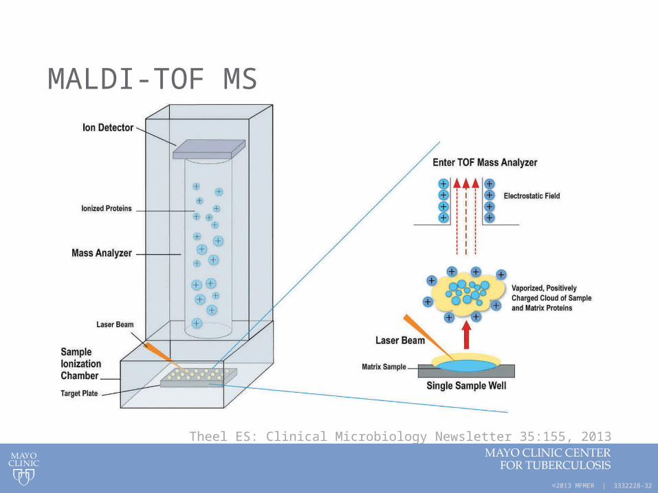
MALDI-TOF MS
Theel ES: Clinical Microbiology Newsletter 35:155, 2013

©2013 MFMER | 3332228-33
Laboratory Workflow for MALDI-TOF MS ID of M. tuberculosis Complex after Growth in Culture
Beads+500µl70% Ethanol
Bead Beat 2 minutes
BSL3Activities
10 ul loop-ful of organism
Incubate roomtemp 10 min
Speed ac10 min
Centrifuge 5 min
Decant supernatant
70% Formic Acid &Acetonitrile
Spot 1ul sample + 2ul of Matrix
BSL2Activities
MALDI-TOF
Start to finish takes ~2 hrs for 24 samples
©2013 MFMER | 3332228-34
Advantages of MALDI-TOF MS for Mycobacteria Identification
• Similar work-flow regardless of organism (bacteria, yeast, mycobacteria, mold)
• Cost effective and “Green” – low consumable costs
• Rapid turn around time, high throughput
• Automated, robust, interlaboratory reproducibility
• Single colony requirement
• Small footprint
• Low exposure risk – sample inactivation
• Adaptable – can be an open system w/ databases expandable by user
©2013 MFMER | 3332228-35
Limitations of MALDI-TOF MS for Identification of Mycobacteria
• Need growth in culture• Requires pure isolate • Phase of growth, media, timing all factors
• Best performance, your spectral library needs to be composed of spectra produced under comparable conditions to your everyday working practices
• Databases need expansion for less common organisms• Instrument maintenance downtime
(if using a single instrument)• Regulatory issues• May not be a bit slower than sequencing for
slowly growing mycobacteria
©2013 MFMER | 3332228-36
Mass Spectrometry Equipment Costs
• Purchase cost: ~$200,000
• Steel plates (10): ~$5,000
• Service contract (year): ~20,000
• Maintenance cost (year): ~$5,000
Remember – Mass spectrometry can also be use for identification of bacteria, mycobacteria, moulds on the same platform; next generation instruments will likely
be linked with susceptibility platforms too

©2013 MFMER | 3332228-37
New Workflow for Mycobacteria ID
Culture to media; wait for growth
MALDI-TOF MS(same day ID)
Sequencing(next day ID)
If no ID

©2013 MFMER | 3332228-38
Direct Identification of M. tuberculosis Complex Without Waiting for Growth in Culture

©2013 MFMER | 3332228-39
Nucleic Acid Amplification-Based (NAA) Tests
CDC recommends
• NAA testing be performed on at least one (preferrably the first) respiratory specimen from each patient with suspected pulmonary TB
• If it would alter case management• If it would alter TB control activities
• NAA testing does not replace the need for culture

©2013 MFMER | 3332228-40
1. Mycobacterium tuberculosis Direct Test (MTD) from Hologic Gen-Probe
• People frequently refer to this as the “TB probe” assay but that is not correct; this is a PCR-like amplification method
• Transcription-mediated amplification of M. tuberculosis complex rRNA directly from respiratory specimens
• Clinical specificity: 99-100%
• Clinical sensitivity• Smear positive: 91-95%• Smear negative: 83-100%
©2013 MFMER | 3332228-41
Limitations of MTD test
• Technically “fussy” test• Inhibition from specimen components a concern• Open PCR system so false positives due to
contamination are possible
• Negative does not rule out M. tuberculosis infection (still need to do a culture
• Detects presence of nucleic acid but doesn’t indicate if the organism is still viable
• Cross-reactions occur w/ some rare mycobacteria: M. celatum, M. terrae-like organisms, M. holsiaticum
• Can be costly

©2013 MFMER | 3332228-42
2. Laboratory-Developed PCR Tests (LDTs)
• Closed PCR system – reduced opportunity for false-positives
• Good sensitivity and specificity but it can vary since each test developed/verified independently
• Often less expensive than MTD
• Some can be used on a wider variety of specimen types included smear negative specimens and formalin-fixed, paraffin-embedded tissue blocks

©2013 MFMER | 3332228-43
Example of Real-time PCR Workflow in our Laboratory
Approximate turn-around time = 4 hr
Specimen or culture lysis, inactivation and
processing
DNA extraction PCR amplification and detection

©2013 MFMER | 3332228-44
Direct Comparison of Mayo LDT PCR Assay With the GenProbe MTD Test
Assay
MTD
+ - Agreement (%)
Kappacoefficient
LightCyclerPCR
+ 49 1 538/542 (99.3%) 0.96
- 3 489

©2013 MFMER | 3332228-45
3. Cepheid Xpert MTB/RIF Test
• WHO-endorsed
• Runs on the Cepheid GeneXpert system
• recently FDA-approved for respiratory specimens
• Detects M. tuberculosis complex and provides information about RIF resistance
www.finddiagnostics.org
©2013 MFMER | 3332228-46
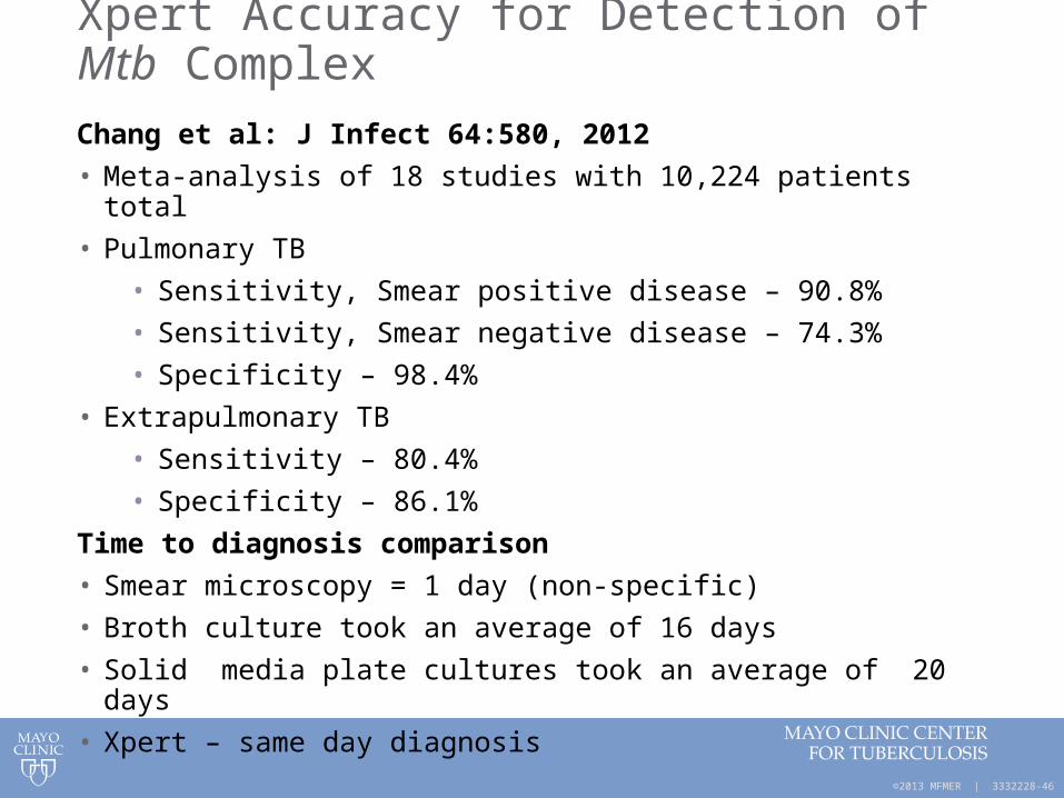
Xpert Accuracy for Detection of Mtb Complex
Chang et al: J Infect 64:580, 2012
• Meta-analysis of 18 studies with 10,224 patients total
• Pulmonary TB
• Sensitivity, Smear positive disease – 90.8%
• Sensitivity, Smear negative disease – 74.3%
• Specificity – 98.4%
• Extrapulmonary TB
• Sensitivity – 80.4%
• Specificity – 86.1%
Time to diagnosis comparison
• Smear microscopy = 1 day (non-specific)
• Broth culture took an average of 16 days
• Solid media plate cultures took an average of 20 days
• Xpert – same day diagnosis

©2013 MFMER | 3332228-47
Xpert MTB/RIF and Rifampin Resistance
• rpoB: gene encoding beta subunit of bacterial RNA polymerase
• Mutations in an 81bp region of the rpoB gene are responsible for ~96% of RIF resistance in Mtb; also predicts MDR TB since the majority of RIF-resistant isolates will also be INH-resistant
• Some false positive RIF resistance with Xpert• PPV is lower in low prevalence settings• CDC recommends reporting Xpert RIF-R as a
preliminary result pending confirmation with sequencing; growth-base DST is still required

©2013 MFMER | 3332228-48
Strengths of Xpert MTB/RIF Assay
• Good sensitivity and specificity for respiratory specimens
• Rapid 2 hr TAT
• Detect MTB and RIF resistance
• Closed PCR system with low risk of cross-contamination
• GeneXpert platform is multi-functional and can be used for other tests (eg, C. difficile, HIV viral load)
• Simple for operators to perform
• No advanced biosafety equipment needed

©2013 MFMER | 3332228-49
Weaknesses of Xpert MTB/RIF Assay
• Xpert has better sensitivity than smear with respiratory specimens but a culture is still necessary
• False-positive RIF resistance is possible; need to confirm RIF-resistance with sequencing
• Not as sensitive or specific for extrapulmonary specimens
• Expensive – need to purchase GeneXpert platform; cartridges are $65 each in E.U. and U.S.; $10 discounted price for high burden and developing countries
• Need continuous electrical power and air conditioning (challenge in developing countries)
• Sample storage limited to 3 days at RT, 7 days at refrigerated temps
• Can’t differentiate between live and dead M. tuberculosis (can’t use to monitor treatment)
©2013 MFMER | 3332228-50
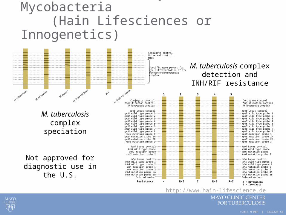
4. Line Probe Assays for Mycobacteria (Hain Lifesciences or Innogenetics)
M. Bovis
ssp bovis
M. Bovis
ssp ca
prae
M. tubercu
losis
Conjugate controlUniversal controlMTBC
Specific gene probes forthe differentiation of theMycobacterium tuberculosiscomplex
M. afric
anum
M. micr
otiBCG
1 2 3 4 5
Conjugate controlAmplification control
M. Tuberculosis complex
- R+I I R+I R+I
rpoB Locus controlrpoB wild type probe 1rpoB wild type probe 2rpoB wild type probe 3rpoB wild type probe 4rpoB wild type probe 5rpoB wild type probe 6rpoB wild type probe 7rpoB wild type probe 8rpoB mutation probe 1
rpoB mutation probe 2ArpoB mutation probe 2B
rpoB mutation probe 3
katG Locus controlkatG wild type probekatG mutation probe
katG mutation probe 2
inhA Locus controlinhA wild type probe 1inhA wild type probe 2inhA mutation probe 1inhA mutation probe 2
inhA mutation probe 3AInhA mutation probe 3B
Colored marker
Resistance
Conjugate controlAmplification controlM. Tuberculosis complex
rpoB Locus controlrpoB wild type probe 1rpoB wild type probe 2rpoB wild type probe 3rpoB wild type probe 4rpoB wild type probe 5rpoB wild type probe 6rpoB wild type probe 7rpoB wild type probe 8rpoB mutation probe 1rpoB mutation probe 2ArpoB mutation probe 2BrpoB mutation probe 3
katG Locus controlkatG wild type probekatG mutation probekatG mutation probe 2
inhA Locus controlinhA wild type probe 1inhA wild type probe 2inhA mutation probe 1inhA mutation probe 2inhA mutation probe 3AInhA mutation probe 3BColored marker
R = RifampicinI = Isoniazid
http://www.hain-lifescience.de
M. tuberculosis complex speciation
M. tuberculosis complex detection and INH/RIF
resistance
Not approved for diagnostic use in the U.S.

©2013 MFMER | 3332228-51
Drug Susceptibility Testing of M. tuberculosis Complex

©2013 MFMER | 3332228-52
Susceptibility Testing of Mycobacteria, Nocardiae, and Other Aerobic Actinomycetes; Approved Standard – Second Edition
Clinical and Laboratory Standards Institute (CLSI)
CLSI Document M24-A2, published 2011Provides guidance for resistance testing of
• M. tuberculosis complex
• M. avium complex (clarithromycin)
• Other slowly growing mycobacteria (limited guidelines)
• Rapidly growing mycobacteria
• Nocardia spp. and other aerobic actinomycetes

©2013 MFMER | 3332228-53
M. tuberculosis Complex DST
• Agar proportion is the current gold standard for all drugs except pyrazinamide
• Not rapid (14-21 days)• Labor-intensive, technically complex• No FDA-cleared, commercially-available kit
• Broth method is recommended for rapid TAT• CDC goal is results for first-line drugs
reported within 15-30 days after receipt of the specimen
©2013 MFMER | 3332228-54
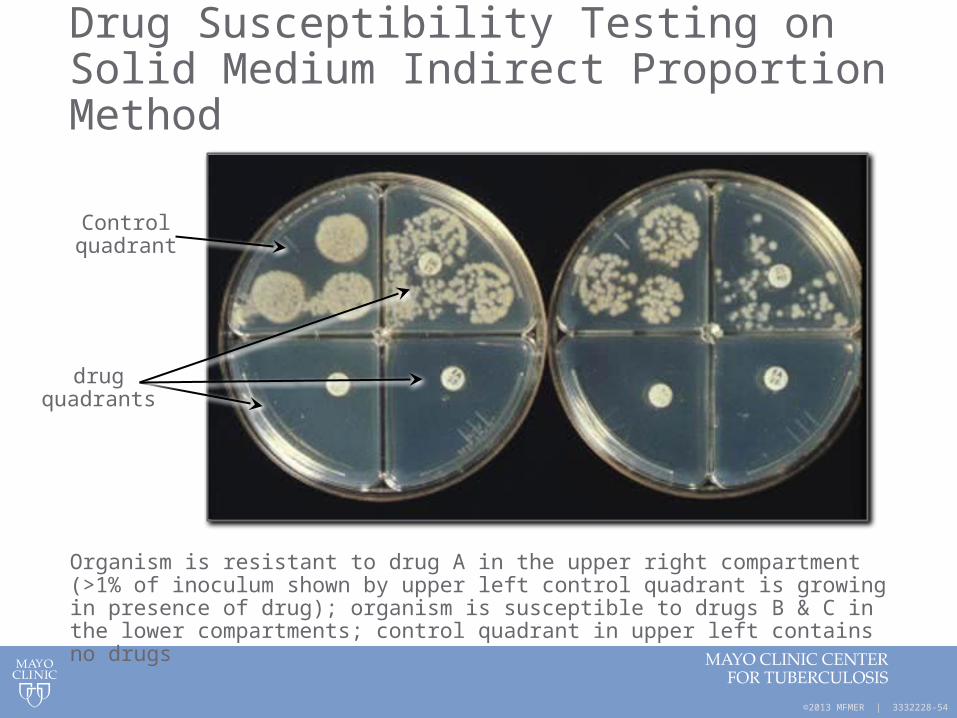
Drug Susceptibility Testing on Solid Medium Indirect Proportion Method
Organism is resistant to drug A in the upper right compartment (>1% of inoculum shown by upper left control quadrant is growing in presence of drug); organism is susceptible to drugs B & C in the lower compartments; control quadrant in upper left contains no drugs
Control quadrant
drug quadrants
©2013 MFMER | 3332228-55
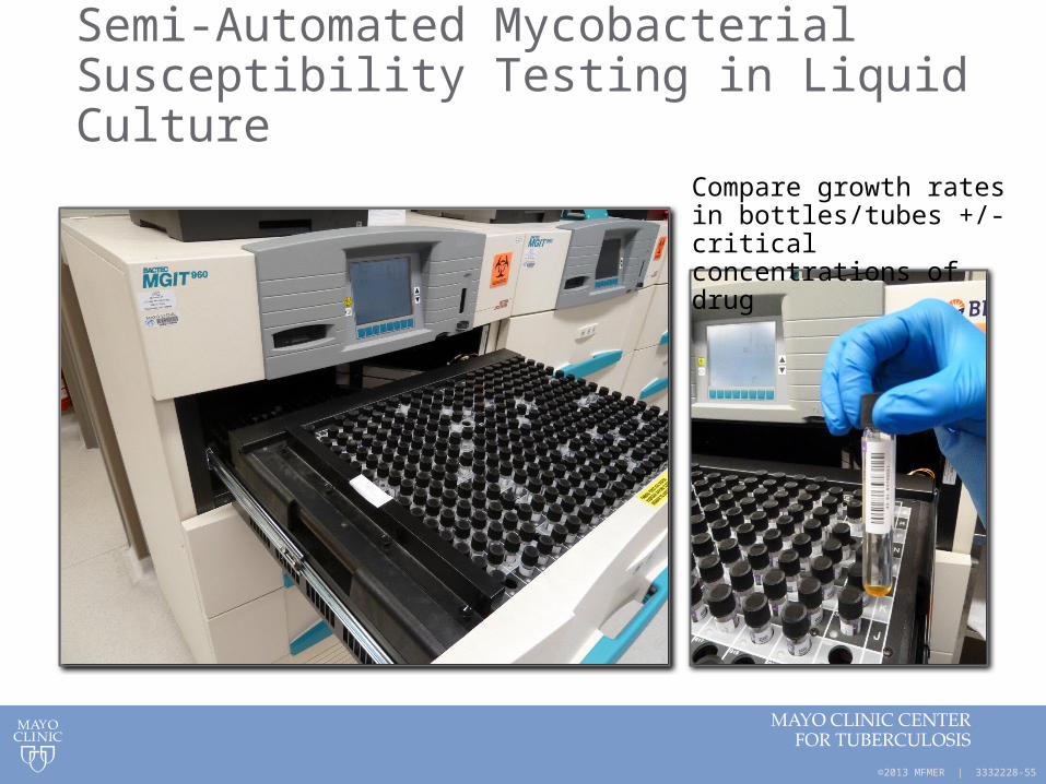
Semi-Automated Mycobacterial Susceptibility Testing in Liquid Culture
Compare growth rates in bottles/tubes +/- critical concentrations of drug

©2013 MFMER | 3332228-56
M. tuberculosis Complex Resistant Isolates
If the isolate is resistant to any agent
• Preliminary report issued
• Consider confirming resistance by 2nd method or 2nd lab
• Consider initiating testing of secondary agents to avoid delays
If the isolate is resistant to only PZA consider
• Speciation• M. bovis is mono-PZA-resistant• Most isolates of M. tuberculosis are PZA-susceptible

©2013 MFMER | 3332228-57
New Method for Mtb DST – MIC Plate
Hall et al: J Clin Microbiol 50:3732, 2012
• Broth microdilution method• Multi-center studies supporting
FDA-submission completed• Rapid (14 days)• Contains INH, RIF, EMB and
9 second-line drugs• Test 1st and 2nd line drugs
simultaneously with same inoculum
• provides MIC endpoint – helpful for isolates with MIC near critical concentration (CC) breakpoint that give fluctuating results w/CC method
©2013 MFMER | 3332228-58
Molecular Detection of Mtb Drug Resistance Markers

©2013 MFMER | 3332228-59
Direct Detection of INH Resistance Using Real-Time PCR
50 52 54 56 58 60 62 64 66 68 70 72 74 76-0.030
-0.015
0.000
0.015
0.030
0.045
Temperature (°C)
Flu
ores
cenc
e -d
(F2/
F1)
/dT
S315TWT
©2013 MFMER | 3332228-60
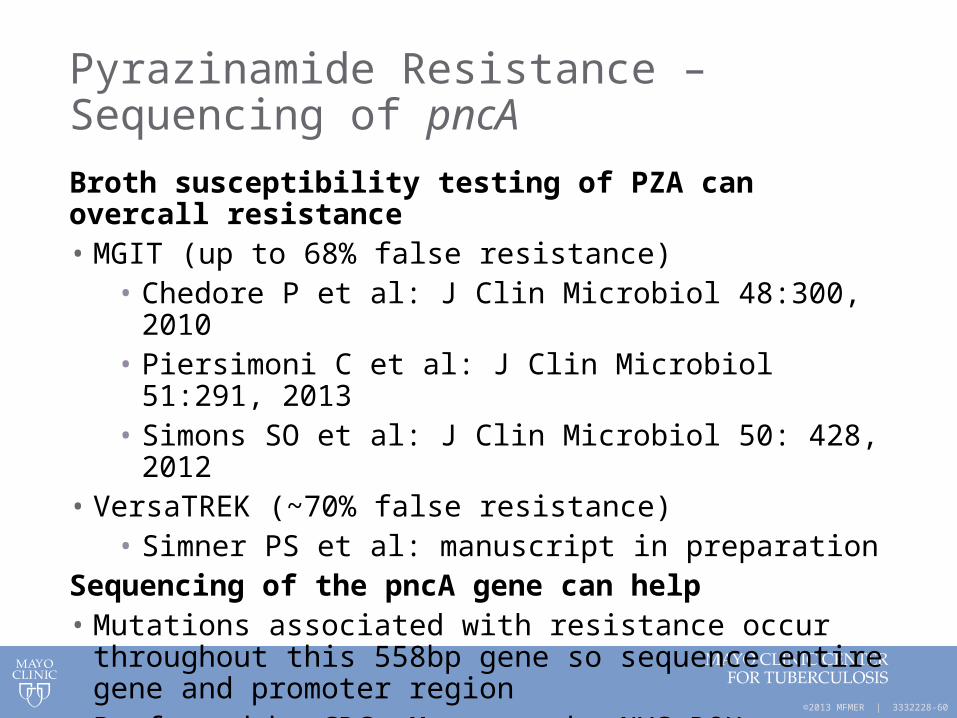
Pyrazinamide Resistance – Sequencing of pncA
Broth susceptibility testing of PZA can overcall resistance• MGIT (up to 68% false resistance)
• Chedore P et al: J Clin Microbiol 48:300, 2010• Piersimoni C et al: J Clin Microbiol 51:291, 2013• Simons SO et al: J Clin Microbiol 50: 428, 2012
• VersaTREK (~70% false resistance)• Simner PS et al: manuscript in preparation
Sequencing of the pncA gene can help• Mutations associated with resistance occur throughout this
558bp gene so sequence entire gene and promoter region • Performed by CDC, Mayo or the NYS DOH Wadsworth Center

©2013 MFMER | 3332228-61
RIF Resistance using Cepheid GeneXpert MTB/RIF
• Amplifies an 81bp region of the rpoB gene
• Contains 96% of known mutations conferring rifampin resistance
• Also predicts MDR TB since most isolates resistant to rifampin are also isoniazid resistant
http://www.cepheid.com

©2013 MFMER | 3332228-62
Molecular Detection of Drug Resistance at the CDC
• Offered for M. tuberculosis complex isolates and nucleic-acid amplification-positive (NAAT+) sputum sediments
• Perform pyrosequencing and conventional sequencing• Provides rapid identification of mutations associated
with resistance to many TB drugs• Limitations include
• Insufficient data to definitively associate all mutations detected with resistance;
• Not all mechanisms of resistance are known • Not all resistance loci are sequenced
• Use in conjunction with conventional DST results

©2013 MFMER | 3332228-63
Molecular Resistance Testing at the CDC
DrugLocus/
Loci examined Sensitivity Specificity
Rifampin rpoB 97.1 97.4
Isoniazid inhA & katG 86.0 99.1
Fluoroquinolones gyrA 79.0 99.6
Kanamycin rrs & eis 86.7 99.6
Amikacin rrs 90.0 98.4
Capreomycin rrs & tlyA 55.2 91.0
Ethambutol embB 78.8 94.3
Pyrazinamide pncA 86.0 95.9
http://www.cdc.gov/tb/topic/laboratory/MDDRUsersGuide.pdf
©2013 MFMER | 3332228-64
Summary
• AFB stains are rapid but insensitive and nonspecific
• Culture should always be ordered together with AFB stain
• Identification after growth in culture is rapid using molecular methods
• Direct identification using molecular methods most often uses smear-positive respiratory specimens; certain methods allow for other specimens
• Detection of drug resistance markers is available for culture isolates and directly for smear-positive respiratory specimens

©2013 MFMER | 3332228-65
Questions & Discussion