Embed Size (px)
Citation preview

©2013 Delmar, Cengage Learning. All Rights Reserved. May not be scanned, copied, duplicated, or posted to a publicly accessible website, in whole or in part.
Extraoral and Digital Radiography
Chapter 23

©2013 Delmar, Cengage Learning. All Rights Reserved. May not be scanned, copied, duplicated, or posted to a publicly accessible website, in whole or in part.
Extraoral Radiographs
• Panoramic– Common in general and specialty offices
• Cephalometric– Common with orthodontists
• Digital– Becoming standard– Easier for staff

©2013 Delmar, Cengage Learning. All Rights Reserved. May not be scanned, copied, duplicated, or posted to a publicly accessible website, in whole or in part.
Panoramic Radiography
• Tomography– One layer shown
• Rotational centers– Tubehead– Cassette
• Focal trough– Image layer or sharpness
©2013 Delmar, Cengage Learning. All Rights Reserved. May not be scanned, copied, duplicated, or posted to a publicly accessible website, in whole or in part.
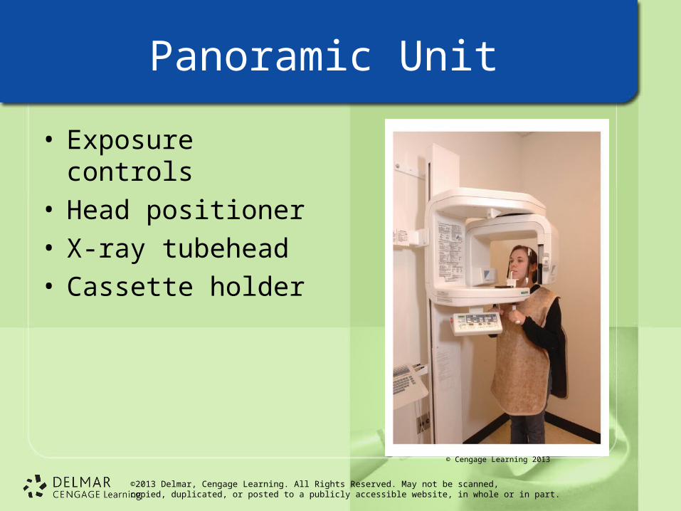
Panoramic Unit
• Exposure controls• Head positioner• X-ray tubehead• Cassette holder
© Cengage Learning 2013

©2013 Delmar, Cengage Learning. All Rights Reserved. May not be scanned, copied, duplicated, or posted to a publicly accessible website, in whole or in part.
Panoramic Techniques
• Lead apron without thyroid collar
• Bite block barrier
• Cassette preparation in dark room
• Explanation of procedure– Patient must be still

©2013 Delmar, Cengage Learning. All Rights Reserved. May not be scanned, copied, duplicated, or posted to a publicly accessible website, in whole or in part.
Common Errors
• Preparation– Ghost image– Lead apron artifact
• Positioning– Too far forward or too
far back– Patient’s head tilted
down or up– Patient’s tongue not
against roof of mouth– Patient not standing
up straight

©2013 Delmar, Cengage Learning. All Rights Reserved. May not be scanned, copied, duplicated, or posted to a publicly accessible website, in whole or in part.
Dental Check
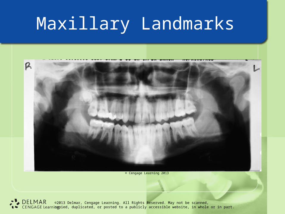
• What can be visualized on a panoramic radiograph?– Entire dentition, nasal and orbital areas,
alveolar bone, carious lesions, fractures, cysts, tumors, malocclusion, maxilla, mandible, sinuses, unerupted teeth, dental appliances, restorations, periodontal disease, and TMJ

©2013 Delmar, Cengage Learning. All Rights Reserved. May not be scanned, copied, duplicated, or posted to a publicly accessible website, in whole or in part.
Cephalometric Radiographs
• Patient’s skeletal structure and profile
• Orthodontists mainly use
• Lateral jaw radiograph• Transcranial TMJ
radiograph
© Cengage Learning 2013

©2013 Delmar, Cengage Learning. All Rights Reserved. May not be scanned, copied, duplicated, or posted to a publicly accessible website, in whole or in part.
Radiographic Interpretation
• Terminology– Anatomical landmarks– Radiopaque– Radiolucent– Diagnosis– Interpretation– Superimposition

©2013 Delmar, Cengage Learning. All Rights Reserved. May not be scanned, copied, duplicated, or posted to a publicly accessible website, in whole or in part.
Tooth and Surrounding Tissues
© Cengage Learning 2013

©2013 Delmar, Cengage Learning. All Rights Reserved. May not be scanned, copied, duplicated, or posted to a publicly accessible website, in whole or in part.
Mandibular Landmarks
© Cengage Learning 2013
©2013 Delmar, Cengage Learning. All Rights Reserved. May not be scanned, copied, duplicated, or posted to a publicly accessible website, in whole or in part.
Maxillary Landmarks
© Cengage Learning 2013

©2013 Delmar, Cengage Learning. All Rights Reserved. May not be scanned, copied, duplicated, or posted to a publicly accessible website, in whole or in part.
Imaging Systems and Digital Radiology
• Computed tomography (CT)
• Magnetic resonance imaging (MRI)
• Digital radiography– New technology becoming common– Dental film not needed

©2013 Delmar, Cengage Learning. All Rights Reserved. May not be scanned, copied, duplicated, or posted to a publicly accessible website, in whole or in part.
Digital Radiography
• Analog image– No limits
• Intraoral and extraoral possible
• Pixels and gray scale

©2013 Delmar, Cengage Learning. All Rights Reserved. May not be scanned, copied, duplicated, or posted to a publicly accessible website, in whole or in part.
Digital Radiography
• Direct digital imaging– X-ray machine– Sensor– Computer monitor and software

©2013 Delmar, Cengage Learning. All Rights Reserved. May not be scanned, copied, duplicated, or posted to a publicly accessible website, in whole or in part.
Digital Radiology
• Indirect digital imaging– Converts traditional X-rays to images– Scanner– Computer monitor and software
• Storage phosphor imaging

©2013 Delmar, Cengage Learning. All Rights Reserved. May not be scanned, copied, duplicated, or posted to a publicly accessible website, in whole or in part.
Advantages of Digital Radiology
• Less radiation to patient
• Quick results
• Image alterations possible
• Storage
• Darkroom, equipment, and solutions eliminated
• Data files can be attached

©2013 Delmar, Cengage Learning. All Rights Reserved. May not be scanned, copied, duplicated, or posted to a publicly accessible website, in whole or in part.
Disadvantages of Digital Radiology
• Initial expense
• Proficiency training
• Uncomfortable sensors
• System failures

©2013 Delmar, Cengage Learning. All Rights Reserved. May not be scanned, copied, duplicated, or posted to a publicly accessible website, in whole or in part.
3-Dimensional (3-D) Imaging in Dentistry
• Shows immediate 3D reconstruction of patient’s mouth, face, and jaw areas
• Lowest possible radiation levels
• “Cone beam” shaped to cover image in single scan

©2013 Delmar, Cengage Learning. All Rights Reserved. May not be scanned, copied, duplicated, or posted to a publicly accessible website, in whole or in part.
Uses and Benefits of 3-D Imaging
• Locate detailed anatomy
• Evaluate deformities• Locate pathology
– E.g., cysts, bone lesions
• Show miniscule detail• Determine bone
quality• Evaluate
temporomandibular disorders (TMD) and TMJ diagnosis

©2013 Delmar, Cengage Learning. All Rights Reserved. May not be scanned, copied, duplicated, or posted to a publicly accessible website, in whole or in part.
Patient Preparation
• Patient may stand or sit comfortably in open scanner
• Scans can be done in dental office
• Scans are quick– 8 to 20 seconds
• Information is processed in less than one minute and ready to be examined by dentist to discuss diagnosis with patient

©2013 Delmar, Cengage Learning. All Rights Reserved. May not be scanned, copied, duplicated, or posted to a publicly accessible website, in whole or in part.
Hand-Held Intraoral Radiography
• Battery-operated and portable
• Low radiation dose
• Digital control panel– Adult-Child– Anterior, posterior, and bitewings– Expose film, digital sensors, or phosphor
plates

©2013 Delmar, Cengage Learning. All Rights Reserved. May not be scanned, copied, duplicated, or posted to a publicly accessible website, in whole or in part.
Dental Check
• Discuss what makes digital imaging more efficient.– Is immediate – no processing involved – Patient can view at the same time as the
dentist– Can easily be e-mailed to third parties when
necessary