Embed Size (px)
Citation preview

X ray chest Dr. Mamdouh
P a g e | 1
X ray chest
32013
X ray of the chest
basis in the clinical practice
chest X ray
is very simple examination low cost very sensitive excellent resolution
chest
X ray tomography
CT isotopes MRI
angiography conventional CT MRI
interventional techniques biopsies
Primitive
chest X ray
chest quality
chest X ray

X ray chest Dr. Mamdouh
P a g e | 2
1 patient well positioned centralized
2 chest
6
3 full inspiration
4 adequate penetration
X ray
postro-anterior view
PA view
is facing the tube
chest AP view
PA view
heart is near to the film
Heart
magnification AP view
PA AP
heart
relatively
increased transverse cardiac diameterleft ventricular hypertrophy
consider
AP view
AP view

X ray chest Dr. Mamdouh
P a g e | 3
intensive care
you consider alsosupine view heart erect view
intensive care
heart size
comment heart lateral view
lateral view heart
X ray
cardiomegaly for echocardiography
if you sayheart
echo signs Left ventricle
signs right ventricle
significance ventricle atrium
please
to acquire this knowledgeknowledge
daily practice
cardiomegaly for echocardiogrpahy
and the patient paysEchocardiography
X ray
conclusion CT
300

X ray chest Dr. Mamdouh
P a g e | 4
conclusion appreciatedclinician
Not to the level
examples
clinicians
lateral view
this is left lateral view
right lateral view
chest PA normal
left lateral right lateral
copula
Chest PA
102013
Chest PA
Lateral
lesion << left
lateral Lesion <<right
lateral
lesion
lateral decubitis

X ray chest Dr. Mamdouh
P a g e | 5
lateral decubitis
right diaphragm
apex of the diaphragmatic
copula Is laterally shifted
diaphragm elevated
sub pulmonary
effusion
lateral decubitis
effusion
costo-phrenic angle
ultra sound
This is typical for sub pulmonary
effusion
elevation of the copula
And shift of the apex of the copula to the lateral aspect
chest chest hard Over exposed
dist spaces
vertebra
well and good
chest
Then you look carefully here And you can see the details of the
lung

X ray chest Dr. Mamdouh
P a g e | 6
Over exposed You cannot see the details of the lung
details
optimal exposure
Over exposure
copula
Heart transverse diameter
Heart normal size
Increased transverse diameterechocardiography
copula rib 10<<Posteriorly
ribs
rib
rib
copula
copula
Lateral view
ABCs
Lateral view
Upper dorsal vertebrae Lower dorsal
vertebrae
shoulders are superimposed on the spine
X ray chest Dr. Mamdouh
P a g e | 7
And if you looked to this lateral view
upper dorsal vertebrae
Lower dorsal vertebrae
Then You diagnose
lower lobe pathology
Lateral view
chest heart
Lung free
If you look to the AP
Then you got a large opacity in the left lower lobe
Over exposed film Under
exposed film
details Lung basis
under exposed
Haziness
cardiac border << hazy
Over exposure
You don’t see the details of the lung
Then
Then you dealwell exposed film
centralization of the patient
You identify the spinous process of the dorsal vertebrae<<

X ray chest Dr. Mamdouh
P a g e | 8
Then you measure the distance between the spinous process and the medial end of the clavicle
And this distance
centralized well centralized
chest
site of the spinous process
Medial edge of the clavicle
medial edge of the clavicle
And you got this lung<< hyper translucent
lung << opacity
And you should consider this
emphysematous opacities
centralized not
And this is well centralized patient
This the spinous process And measure the distance
And the distance
Left lung two lobes
Divided by the major fissure
right lung three lobes
Divided by the major and minor fissures

X ray chest Dr. Mamdouh
P a g e | 9
lateral view You should specify the site of the lesion
lesion upper lobe
Middle lobe
Lower lobe
Lingula lateral view
lateral view
Then you mention the zone
chest PA
Chest PA Lesion
Then you say lesion Upper zone
Lower zone
Middle zone
zones
upper zone first
second rib Anteriorly
anterior aspect of the first rib
anterior aspect of the second rib
this is the upper zone
The middle zoneanteriorly
This is the area of the middle zone

X ray chest Dr. Mamdouh
P a g e | 10
Lower zone
edge 4th rib anteriorly
Chest PA
lesion
This is upper lung zone
in the middle lung zone
in the lower lung zone
Lateral view
You should mention the lobe
And this the case of right sided pleural effusion
And this effusion extending into the fissure
And you can see the upper lobe, Middle lobe and the lower lobe
And the major fissure as well as the minor fissure
Pathology
spine disk bulge protrusion relaxation

X ray chest Dr. Mamdouh
P a g e | 11
well documented
signs
Silhouette sign Air bronchogram
expressions
Pulmonary nodules Mass cavity infiltration opacity
chest
Silhouette sign
Silhouette
definitions
chest Opacity
cardiac borders
cardiac border
Opacity cardiac border
Opacity cardiac border
This is known as Silhouette sign
Sign
Opacity heart
heart << is anterior
opacity << is anterior

X ray chest Dr. Mamdouh
P a g e | 12
anterior
Middle lobe
Lateral view
this opacity should be present in the middle lobe
chest
opacity
cardiac border
opacity
cardiac border
Opacity is posterior
Then this opacity is located in the lower lobe and not in the middle lobe
sign Silhouette sign
Air bronchogram
air bronchogram
air in the patent bronchi
background of alveolar consolidation Lung
opacity
bronchi patent
air bronchogram
And this air bronchogram can be seen in the X ray, can be seen in the CT
Can be seen
air bronchogram
showing air bronchogram inside
air bronchogram
air bronchogram 3

X ray chest Dr. Mamdouh
P a g e | 13
1 this is pulmonary parenchymal disease
lesion air bronchogram
Lung parenchyma
2 this lesion arising in the alveoli
this is alveolar pathology
pulmonary parenchymal diseasebronchi
nerves
vessels
this is parenchymal disease air bronchogram
alveolar pathology
3 this is consolidation
consolidation
consolidationalveoli
pulmonary edema pneumonia blood pulmonary hemorrhagic disorders tumor alveolar cell carcinoma protein alveolar proteinosis
consolidation
consolidation pneumonia
consolidationPneumonia
opacity nodule Mass cavity abscess
X ray chest Dr. Mamdouh
P a g e | 14
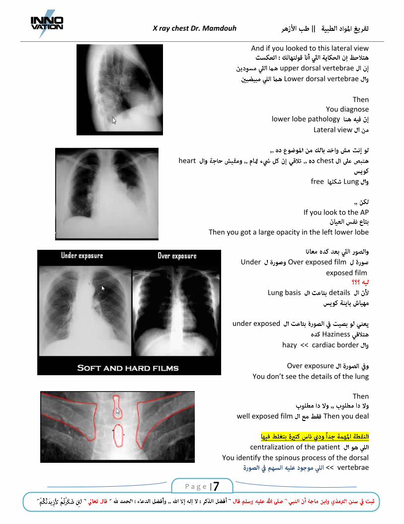
pulmonary nodule Pulmonary nodule
The pulmonary nodule is well defined lesion that measure less than 3 cm
This is the definition of the nodule
chest
This is the well defined lesion measuring
pulmonary nodules
pulmonary mass
well defined lesion
3
Nodule
mass
Pulmonary opacitychest Opacity
Opacity
patchy opacity
This is ill defined lesion containing air bronchogramOpacity
cavity
Cavitary lesionabscess
cavity
This is known as cavitary lesion
cavity
cavity

X ray chest Dr. Mamdouh
P a g e | 15
cavity
fluid mass
differential diagnosis
pulmonary infiltration
strand
nodular retriculo nodular
fibro nodular
in the upper lobe
this is tuberculous
lesion ill defined strand nodular opacities
Upper lobe
Then you mention
this is most likely of tuberculous nature
chest X ray
chest X ray lesion normal
Normal chest
Normal chest
Clear both lung fields, costo phrenic angle, normal cardiac size and shape normal cardio-thorax ratio
cardio-thorax ratio Normal
Heart
Heart
normal cardiac size and shape
Normal cardiac size and configuration
chest
Prominent vascular markings

X ray chest Dr. Mamdouh
P a g e | 16
Prominent vascular marking
prominent
prominent aortic knuckle
what do you mean by prominent aortic knuckle
plain X ray
aorta
aorta << dilated and folded
cardiology
Prominent aortic knuckleaortic knob
calcification atheromatous calcification of the aortic knuckle or knob
arterio sclerotic disease
Lesion
1 definition Lesion well defined ill defined well defined
relatively well defined
relatively ill defined ill defined lesion
2 lesion Type Lesion Lung Nodule
Mass

X ray chest Dr. Mamdouh
P a g e | 17
cavity
patch Opacity infiltration
retriculo-nodular infiltration
focal lung disease
diffuse lung disease
focal lung pathology
Nodule Mass cavity patch
3 Size and shape 2 X 3 4 X 5 Oval rounded lobulated
4 Multiple Multiplicity
5 costophrenic angle
6 heart Normal cardiac size shape Cardiac size and shape
chest
Chest X ray
Chest X ray
chest 3 Lung mediastinum pleura and chest wall
Lung
focal lung pathology
diffuse lung disease
Lung
Mediastinum pleura
chest X ray
You have to discriminate
lesion focal lung disease diffuse lung pathology

X ray chest Dr. Mamdouh
P a g e | 18
diffuse lung disease affect
both lungs lung
focal lung lesion lesion
Lung
diffuse lung disease
Focal lung pathology
focal lung pathology
: focal lung Nodule mass patch cavity
Nodule
Nodules
Lung 6
Nodule
frequently seen in the clinical
practice
frequency 3
4
Provided
nodule
nodule
Tuberculoma Hamartoma Bronchogenic carcinoma Metastases Pulmonary AVM

X ray chest Dr. Mamdouh
P a g e | 19
Hydatid cyst nodules
chest
nodule
rheumatoid nodules
frequently seen in the clinical practice
rheumatoid nodule
rheumatoid nodule
rheumatoid rheumatoid nodule
Nodule
Nodule frequently seen
chest X ray
pulmonary nodule well defined lesion << 3
at ease
Nodule
Nodules tuberculoma Hamartoma
bronchogenic carcinoma metastasis AVM hydatid cyst
History
this is metastatic deposits

X ray chest Dr. Mamdouh
P a g e | 20
by multiplicity
Nodules Multiple
hydatid hydatid Multiple
AVM pulmonary vascular malformation Multiple
rare
tuberculoma Hamartoma
Extremely rareMultiple
chest
metastasis Hydatid
Multiple
metastasis hydatid CT
Hydatid
Metastasis soft tissue
you can discriminate by the history
is a clinician
adequate clinical data
web
This is not sciencehistory
on the spot
nodules
Tuberculoma Hamartoma
They are benign lesions
And show

X ray chest Dr. Mamdouh
P a g e | 21
edge is very smooth
chest X ray
lesion
Meaningcontaining calcium
Nodule edge smooth
Tuberculoma Hamartoma
tuberculoma Hamartoma
tuberculoma Upper lobe
Hamartoma
Tuberculoma calcification
Hamartoma Pop corn calcification
Tuberculoma fat
Hamartoma fat
in the clinical practice
Tuberculoma Hamartoma
No
clinician
No
never turn malignant
tuberculoma Hamartoma malignancy
chest X ray
Nodules calcium
tuberculoma Hamartoma
CT

X ray chest Dr. Mamdouh
P a g e | 22
CT
Nodule edge
speculated edge
Lesion is invading the adjacent lung parenchyma
<< this is malignant
Nodule
edge is speculated
this is bronchogenic carcinoma
Nodule
nodule to the hilum
plain X ray
CT
feeding artery and the draining vein
Nodule
Is pulmonary vascular malformation
Nodule content
exceptions
Lung hydatid cyst
Lung
Hydatid cyst
encysted pleural effusion
lung
fissure
abscess totally

X ray chest Dr. Mamdouh
P a g e | 23
support metastasis
plain X ray
CT
you are dealing with Hydatid cyst
And this is the difference between the
hydatid cyst and the abscess
abscess
CT
Stillwall abscess
Is very thickwall
And the wall of the abscess
Hydatid cyst wall
hydatid cyst
wall
abscess
plain X ray
CT Nodule
nodule edge is smooth
And is containing calcium
you are dealing with benign lesion Either Tuberculoma or
Hamartoma
Well defined, small pulmonary nodule,Measuring

X ray chest Dr. Mamdouh
P a g e | 24
Seen
showing <<central multiple Pop corn calcification parenchymal
Lung is clear no other nodules detected
costophrenic angles are free Normal cardiac size and shape
Conclusion conclusion
conclusion
this is true
clinician
3clinician
chest X ray
pulmonary nodule
Nodule benign
Nodule Hilum AVM
Nodule hydatid
Nodules Multiple metastasis nodules edge speculated
Bronchogenic carcinoma
plain X ray You can never judge
Nodule
malignant
CT
CT
edge speculated

X ray chest Dr. Mamdouh
P a g e | 25
Plain X ray
Then you are dealing with a bronchogenic carcinoma
edge is speculated
…
Then if you look at this lesion This is typical appearance of peripheral bronchogenic carcinoma
showing speculated margin
…
And this is the typical appearance of pulmonary vascular malformation
showing the feeding artery and draining vein
…
And then you look this chest X ray You can see a nodule
chest quality
You can see two cord like structures Connecting the lesion to the hilum Then you suspect the possibility of
AV malformation
biopsy plain X ray
Is very important and very crucialNodule screen
AVM femoral vein
inferior vena cava

X ray chest Dr. Mamdouh
P a g e | 26
right atrium
right ventricle
Pulmonary artery
feeding artery AVM
And the draining veinleft atrium
Multi detector CT
Then you inject small amount of intra venous contrast Then you adjust CT
Pulmonary vasculature
Then you can see the vascular malformation And the feeding artery and draining vein
AV malformation biopsy
chest X ray Pulmonary nodule
well defined lesion3
bronchial adenoma bronchial carcinoid
Foreign body granuloma
differential diagnosis
Nodule 99 %
biopsy
biopsy bronchial adenoma
the common lesions seen daily in the clinical practice

X ray chest Dr. Mamdouh
P a g e | 27
rare rare
rare
rare
common
common
200
Then you know the commonclinical practice
mass lung
mass
mass Lesion well defined 3
Possibilities
masses lung adult
1 Bronchogenic carcinoma
2 Hydatid cyst
3 Metastatic deposit
Mass Lung differential diagnosis
masses lung
bronchogenic carcinoma Hydatid cyst metastasis
Mass Mass
hydatid cyst
hydatid
extreme sharp margin

X ray chest Dr. Mamdouh
P a g e | 28
are malignant
metastasis bronchogenic carcinoma
Margin sharp
hydatid
margin
Is extremely well defined
Hydatid
bronchogenic
needs further verification
verified
further evaluation
lesion Mass
Hydatid
two possibilities
Solitary deposits Bronchogenic carcinoma
deposits
Primary malignancy
bronchogenic carcinoma until proved otherwise
Proved otherwise
investigations primary malignancy
you should consider Mass lung adult
As bronchogenic carcinoma until proved otherwise

X ray chest Dr. Mamdouh
P a g e | 29
Mass CT
CT mass
mass
benign
benign Nodule
benign nodule benign mass
expressions
mass Lung
this is bronchogenic carcinoma
break down break down edge smooth edge
speculated pleural tail tail
mass lung adult Is a bronchogenic carcinoma until proved otherwise
adult
neurobalstoma
adult
Mass in an adult should be considered as bronchogenic carcinoma
Until proved otherwise
CT Mass
osteosarcoma
Mass metastasis
osteosarcoma
chest X ray
Masses Nodules
Masses

X ray chest Dr. Mamdouh
P a g e | 30
differential diagnosis chest
Mass
possibilities Mass
bronchogenic carcinoma Metastasis hydatid cyst
Masses Lung
Multiple
hydatid Metastasis
bronchogenic carcinoma Multiple
Hydatid cyst Metastasis
Can I know
Possibility hydatid cyst CT
1 extreme sharp margin
2 By the presence of air in the wall of the lesion
and this never exist in the metastasis lesion
hydatid cyst
air fluid level
hydatid rupture

X ray chest Dr. Mamdouh
P a g e | 31
Hydatid rupture
metastasis
Hydatid
Plain X ray
signs Hydatid
1 Extreme sharp margin 2 Lesion which is know as
3 Hydatid air fluid level
CT
CT
Lung
hydatid cyst
Pulmonary consolidation patch
patch
opacity
air bronchogram
Opacity CT
air bronchogram
opacity CT
air broncho gram
What are the possibilities
Opacity
pulmonary edema pneumonia pulmonary hemorrhage alveolar cell carcinoma alveolar proteinosis
Pneumonia << are diffuse lung disease
Then the only possibility

X ray chest Dr. Mamdouh
P a g e | 32
patch air bronchogram
pneumonia
infraction
History
pneumonia
sputum
pulmonary infarction
acute chest pain
basis
patch in the lung
It is easyconsolidation
infarction
patch
lung base
pneumonic patch
infraction
How can you discriminate
History
history
CT CT << you can see the embolus inside the pulmonary artery
and this the only possibility
CT
main pulmonary artery
right main branch
And you can see the embolus inside the artery

X ray chest Dr. Mamdouh
P a g e | 33
Then you settle down the diagnosispleural effusion
patch lung
History
Pneumonia infarction
pneumonia
By the common
infarction
plain X ray CT
pneumonia
Then you diagnose by the commonrare
You need supportclinical CT
38 fever
expectoration
And can you see a lesionX ray
There is ill defined opacity
History pneumonic consolidation
CT
it happened
CT is recommended
CT
You can see ill defined lesion

X ray chest Dr. Mamdouh
P a g e | 34
air bronchogram
Diagnostic of pneumonic consolidation
antibiotic
follow up
Is clear
…
Then you see a lesion like thatinfarction
base
apex
lesion
air bronchogram
pneumonia
infarction
support history Infarction
Well and good
CT
Pulmonary artery
embolus
pulmonary artery
opacity
infarctions Lung pleural effusion

X ray chest Dr. Mamdouh
P a g e | 35
possibility
cavity
Nodule
masses
focal patches
cavity
cavity
cavity lesion Lung
Well defined
lesion
This is a cavitary lesion containing air
Cavitary lesion containing air and fluid level
Cavitary lesion containing mass
surface of the fluid level
mass cavity
differential diagnosis cavities

X ray chest Dr. Mamdouh
P a g e | 36
1 cavity
2 soft tissue
differential diagnosis
X ray cavity
cavity
differential diagnosis
wall of the cavity
wall
cavity thick margin Containing air
cavitary lesion in the lung
margin
Possibilities
chronic abscess break down in a tumor
abscess Lung
area of pneumonic consolidation
break down
fluid level
cavity
cavity
And this is known as chronic lung abscess
abscess break down
This is crucial point
chronic lung abscess
antibiotics
metastasis

X ray chest Dr. Mamdouh
P a g e | 37
And you should knowchronic abscess
break down in a tumor
inner margin of the lesion
Please
The inner margin of the lesion is smooth abscess
Is irregularbreak down in a tumor
Lung abscess
It is very smooth extremely smooth
chronic lung abscess
tumor smooth
necrosis lesion
hazards
cavity cavity
wall
Inner margin of the lesion
If it is smooth<< you are dealing with a chronic lung abscess If it is irregular << <<you are dealing with a cavitary neoplasm
This different stages of the abscess
abscess

X ray chest Dr. Mamdouh
P a g e | 38
air fluid level
acute abscess
Sub-acute abscess
Chronic abscess
cavity
Containing
wall thin
cavity
cavity
thin
possibility
pneumatocele emphysematous bulla
lesion
lesion lung
lung << this is pneumatocele
peripherally located sub pleural
this is emphysematous bulla

X ray chest Dr. Mamdouh
P a g e | 39
This is an example of multiple Pneumatocele
staphylococcus pneumonia
Pneumatocele
rupture
Pneumothorax
This is an example of emphysemawhich is known as paraseptal emphysema
This is very common typeCan see multiple cavities containing air
And the cavities have thin wall And the cavities are sub pleural located
Emphysmatous bulla
…..
cavity
fluid level
cavity fluid level
cavity fluid level
fluid level straight
fluid level
the water lily sign

X ray chest Dr. Mamdouh
P a g e | 40
water lily sign
This is ruptured hydatid cyst
cavity
air fluid level << straight
abscess
cavity
air fluid level
ruptured hydatid cyst
cavity
cavity
fungal ball Mycetoma ruptured hydatid cyst cyst endocyst
cavity necrosis tumor tumor necrosis necrotic material
cavity
cavity thick wall margin irregular
shreds tumor cavity cavity
cavity
mass cavity
What is the diagnosis
Fungal ballThis is the most common intra cavitary soft tissue mass
blood clot

X ray chest Dr. Mamdouh
P a g e | 41
Lesion
This lesion consolidation
cavity
cavity soft tissue density
to evaluate
cavity
Then you have toCT
This is the CTAnd you can separate the consolidation from the cavity
and the cavity contain mass
What is the diagnosis
This is fungal ballapex left lung
infiltration infection TB
fungus chronic cavities
Lung
Lesion
This is a cavity
cavity
Inner margin << is irregular
cavity
inner margin << irregular
tumor
tumor
the shreds of the tumor
cavity
cavity mass

X ray chest Dr. Mamdouh
P a g e | 42
fungal ball Mycetoma
cavity Neoplasm
thick wall
Inner margin << is irregular
shreds of the tumor
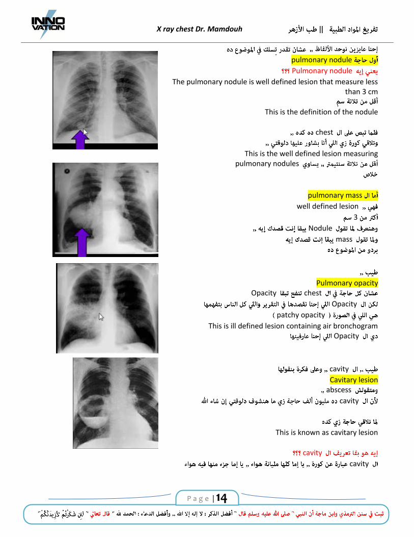
Then, how to deal with a cavitary lesion in the lung
You look to the contents ….
If the cavity contains air fluid level Look to the surface of the fluid level
If the surface is straight
You are dealing with an abscess
If the surface if irregular You are dealing with ruptured hydatid cyst
…. If the cavity contains air only
X ray chest Dr. Mamdouh
P a g e | 43
Look to the wall thickness
If the wall is thick Look to the inner margin
If the inner margin is regular
You are dealing with chronic abscess
If the inner margin is irregular You are dealing with a cavitating neoplasm
..
If the wall is thin and the lesion contain air only Look the site of the lesion
If it is centrally located in the lung
This is pneumatocele
If it is peripherally located This is an emphysematous bulla
Then you look to this lesion This is a cavitary lesion in the lung
And the wall is thick
What about the inner marging of this lesion
Is irregular Then this is not an abscess It is a cavitating neoplasm
….
this lesion
This is a pneumatocelethin wall
Containing airLung

X ray chest Dr. Mamdouh
P a g e | 44
Then based
I will show you some cases
Then you need to answer
data
interpret chest X ray
will start by this case chest X ray
interpret chest X ray
category
focal lung disease
category nodule Mass
cavitypatch DD
category
Nodules
nodules are multiple
possibilities
Metastasis
13Ewing’s sarcoma
differential diagnosis
And you don’t needCT metastasis
metastasis

X ray chest Dr. Mamdouh
P a g e | 45
surgically removed
34
Plain X ray
you don’t need CT
The Ewing’s sarcoma of the femur
Then, this is another caseand what the category we are dealing with
mass
What are the possibilities of this mass
hydatid
Basedhydatid
sharp border
chest
well defined soft tissue mass
Seen in the right lower lung lobelateral view
And the mass shows smooth border with no matrix calcification And the surrounding lung is clear The costophrenic angles are free
The left lung is free The heart is normal
Then you give me your conclusionThis is a soft tissue mass in the lung
And diagnostic possibility
X ray chest Dr. Mamdouh
P a g e | 46
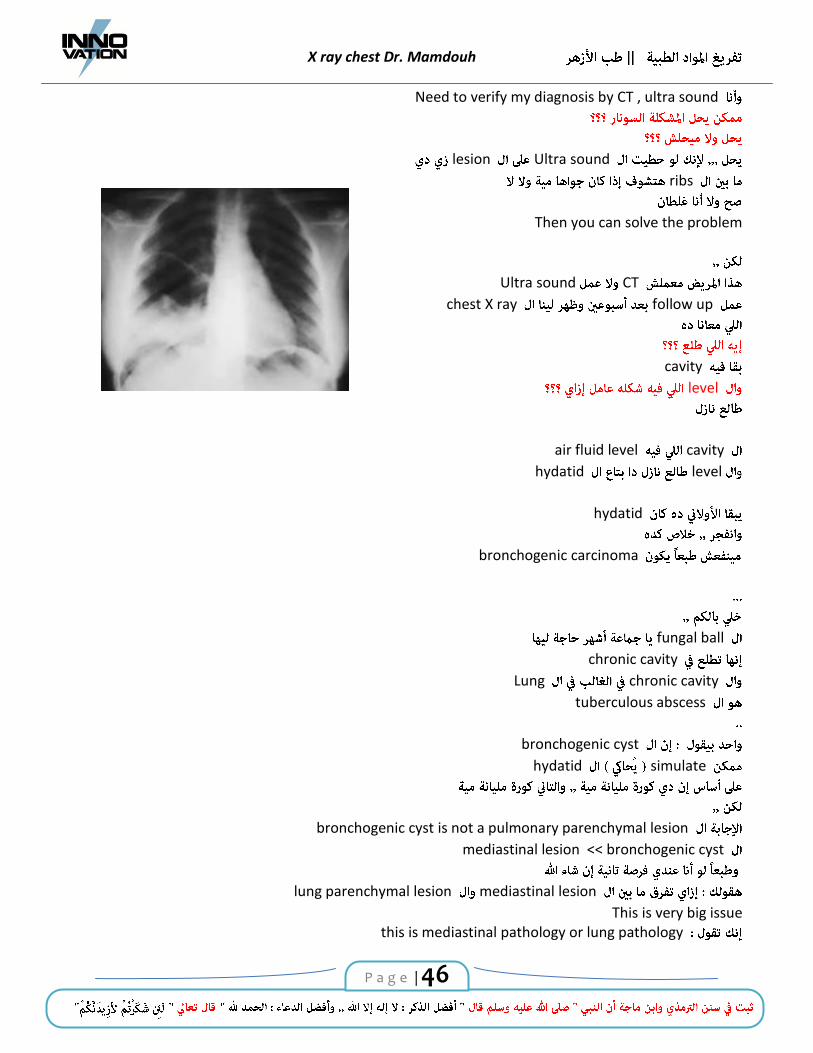
Need to verify my diagnosis by CT , ultra sound
Ultra sound lesion
ribs
Then you can solve the problem
CT Ultra sound
follow up chest X ray
cavity
level
cavity air fluid level
level hydatid
hydatid
bronchogenic carcinoma
fungal ball
chronic cavity
chronic cavity Lung
tuberculous abscess
bronchogenic cyst
simulate hydatid
bronchogenic cyst is not a pulmonary parenchymal lesion
bronchogenic cyst << mediastinal lesion
mediastinal lesion lung parenchymal lesion
This is very big issuethis is mediastinal pathology or lung pathology

X ray chest Dr. Mamdouh
P a g e | 47
cyst mediastinum
cyst Lung
hydatid cyst
mediastinum
Pericardial cyst Bronchogenic cyst Esophageal duplication cyst
Mediastinum cyst
pleuro-pericardial cyst anterior
anterior mediastinum
bronchogenic cyst
middle mediastinum
esophageal duplication cyst
posterior mediastinum
What are the category are dealing with
cavity mass nodule
patch
Yes , this is a cavity
cavity air fluid
level mass
Mass cavity
Fungal ballAspergilloma Mycetoma

X ray chest Dr. Mamdouh
P a g e | 48
Aspergilloma within a cavity in a 63 year old man. Frontal and lateral chest radiographs showed a mass of soft tissue opacity with air cresent sign in the right upper lobe. Strand opacities are seen around the
lesion.
category
differential diagnosis
mass
hydatid cyst metastasis bronchogenic carcinoma
sharp border
Hydatid
CT
CT
This is typical of hydatid cyst
category Patch opacity
patch opacity
Infarctionpneumonia
Pneumonia
Pneumoniainfarction

X ray chest Dr. Mamdouh
P a g e | 49
You need to know
Pneumonic consolidation
middle lobe pneumonia
Silhouette sign heart
And you can see the lesion is located in the middle lobe
category
Nodule
Nodule
edge regular
Nodule
smooth outline
upper lobe
tuberculoma
nodules
CT
CT
Bronchogenic carcinoma
edge is speculated

X ray chest Dr. Mamdouh
P a g e | 50
you can see in the reconstracted image
reconstructed images
nodules fissure
Once nodule lobe lobe
approach surgical
Jugde
stage
This is smooth noduleupper lobe
This is tuberculoma
chest
anti tuberculous treatment

X ray chest Dr. Mamdouh
P a g e | 51
cavity
Upper lobe
follow up cavity mass
And you know this is a cavitary lesionWith intra cavitary mass
And the most common intra cavitary mass is mycetoma Aspergilloma fungal ball
mycetoma
Formation of an aspergilloma within a cavity in a 46-year- old man With no symptoms and no changes in clinical condition
patchy opacity
air bronchogram patchy opacity

X ray chest Dr. Mamdouh
P a g e | 52
patchy opacity
air bronchogram
patchy opacity air bronchogram
homogenous
mass nodule
lesion
Lesion
Lesion
Can you see the diaphragm
Can you see patch containing air bronchogram
patch
It is not a patch containing air bronchogram
Lateral
copula
The diaphragmatic copula is elevated
apex
Shifted laterally

X ray chest Dr. Mamdouh
P a g e | 53
apex diaphragm
laterally
Elevation of the diaphragm with shift of the apex laterlly
Sub pulmonary effusion
sub-pulmonary effusion
two lesions
upper abdominal pain and
dyspnea
lesion category Nodule
Lesion category mass
Nodules Masses
Multiple nodules

X ray chest Dr. Mamdouh
P a g e | 54
Metastasis hydatid
Multiple masses
metastasis Hydatid
metastasis
hydatid
CT
You are dealing with hydatid cyst
Upper abdomen Showed multiple hydatid cyst
In the liver
This is lateral decubitus film
With recurrent hemoptysis
You can see
Cavitary lesion
And inside the cavity there is soft tissue mass Diagnostic of mycetoma
Yes , this is fungal ball
The most common complaintmycetoma hemoptysis

X ray chest Dr. Mamdouh
P a g e | 55
fungal ball
Hemopytsis
category
patchy opacity air bronchogram
pneumonia
pneumonia
chest
You can seeair bronchogram
What about the rest of the lesion
The rest of the lesionair bronchogram
mass
mass Lung lung aerated
air bronchogram
this totally consolidation
further verification
verification should be judged by the clinical
mass symptoms
Hemopytsis
consolidation fever

X ray chest Dr. Mamdouh
P a g e | 56
CT This is the CT
And you can see The very big mass containing dense areas of calcium
Any mass in the lung in an adult should be considered as bronchogenic carcinoma until proved otherwise
Proved otherwise
biopsy
plain X ray
CT
this is bronchogenic carcinoma
biopsy
carcinosarcoma
mixture of sarcomatous and carcinomatous cells
clinician this is malignant
tuberculous
In the clinical practice
plain X ray tumor
Lesion upper right
lesions
Nodule nodule cavity cavity
mass Patch
patch << is ill defined lesion containing air bronchogram
Mass << is a well defined lesion air bronchogram

X ray chest Dr. Mamdouh
P a g e | 57
Lesion
lesion
air bronchogram
Lesion air bronchogram
consolidation
chest x ray
this is not an ordinary consolidation
I should verify by CT
CT
huge mass
arch of the aorta
dense areas of calcification
areas of break down
malignant
biopsy which malignancy
bronchogenic carcinoma
16
11
bronchogenic carcinoma

X ray chest Dr. Mamdouh
P a g e | 58
Which category
Yes, it is a cavityair fluid level
Ruptured hydatid cyst
This is a cavitary lesion in the left lower lung zone and the surface of the fluid level is
wavey >> this ruptured hydatid cys
Left side
This is the breast
pleural effusion
lamellar effusion
Yes, may be
CT
CT
Nodule
CT edge
cavity
air fluid level
CT
This is patientfever cough
Pneumonia
Pneumonia
Left middle lobe
This is the lingula

X ray chest Dr. Mamdouh
P a g e | 59
lateral You should specify the site of the lesion
The lesion in the middle lung zone
lateral
sub segmental lingular pneumonic consolidation
Male patient, 64 yrs, presented with right sided chest pain and hemoptysis
lesion
Cavity
This is the lesion
lesion
Cavitary lesion
cavitary lesion
the cavitary lesion
thick thin
X ray chest Dr. Mamdouh
P a g e | 60
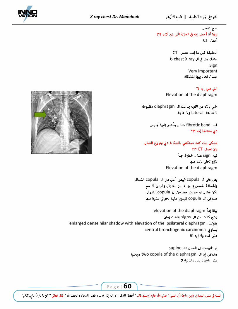
CT
CT chest X ray
SignVery important
Elevation of the diaphragm
diaphragm
lateral
fibrotic band
CT
sign
Elevation of the diaphragm
copula copula
4
copula
copula
elevation of the diaphragm
signs
enlarged dense hilar shadow with elevation of the ipsilateral diaphragm central bronchogenic carcinoma
supine
two copula of the diaphragm

X ray chest Dr. Mamdouh
P a g e | 61
CT
This is a cavity
cavity
thick wall
inner margin of the cavity
Is irregular
Then you are dealing with A neoplasm
copula is markedly elevated
This is not a consolidation
Malignant lesion invading the adjacent lung parenchyma
vessels
Lung
parenchyma clear bronchogenic carcinoma
air fluid level
wall thick inner margin is irregular
malignant

X ray chest Dr. Mamdouh
P a g e | 62
category
NoduleMass cavity
Cavity
cavity mass
air fluid level
air
Thick
differential diagnosis
cavity possibilities
Chronic abscess break down in a tumor
cavity
septum
chronic abscess septum
chronic abscess septum
lesion
break down
break
break down in the tumor
And this is not an abscess
CT
CT

X ray chest Dr. Mamdouh
P a g e | 63
conclusion
Recommended CTmediastinum lymph nodes
metastasis supra renal
spot diagnosis
Well defined cavitary lesion
cavitary
lesion
superior segment of the
right lower lobe
site of the lesion
This lesion located in the superior segment of the right lower lobe
Apical segment of the right lower lobe
lesion<< measuring
It shows thick margin with fluid level inside And the surface of the fluid level is straight
The adjacent lung parenchyma is almost free The left lung is clear
Clear both costophrenic angles Normal cardiac size and shadow
Conclusion
The cavitary lesion in the right lower lobe highly suggestive of lung abscess

X ray chest Dr. Mamdouh
P a g e | 64
category
Nodule
Nodule
nodule
CT
Can you see calcium inside the nodule
Yes
foci of calcification
Within the nodule
Nodule
Which represent the strandy nodular opacitiesT.B.
lesion That is smooth outlines, with central calcification
tuberculous infiltration
diagnosis Tuberculoma
CT
Nodule
Is totally calcified
Lung
reticulation
T.B. bold

X ray chest Dr. Mamdouh
P a g e | 65
67chest
MR of the brain
MR of the brain
Multiple lesions
Posterior fossa
Lesions
Representing metastatic deposits And this is the frequent finding in the clinical
practice
His first presentation by metastasis in the brain
Chest X ray
The primary is in the lung
frequent scenario
metastasis in the brain
chest X ray
CT of the chest You discover the lesion
Chest X ray
pulmonary nodule
CT
You can see the nodule You can see the speculated margin

X ray chest Dr. Mamdouh
P a g e | 66
peripheral bronchogenic carcinoma with brain deposits
clinical practice
Masses Lung adult
This is bronchogenic carcinoma
biopsy
biopsy
cell type
small cell
non small cell
Non small cell
Adenocarcinoma Squamous cell carcinoma Large cell Undifferentiated
irradiated
irradiation chemotherapy
This is a bronchogenic carcinoma
Then biopsy
clinician
biopsy
biopsy
well known
bronchogenic carcinoma
to know the cell type

X ray chest Dr. Mamdouh
P a g e | 67
15
What is the category of this
Massnodule cavity
Mass
investigation
mass in the lung mass in the lung
bronchogenic carcinoma metastasis hydatid cyst
bronchogenic
chest bronchogenic carcinoma
history
15known to have Ewing’s sarcoma
metastasis
80
fever cough
hemoptysis Loss of weight
chest X ray
category
Cavity

X ray chest Dr. Mamdouh
P a g e | 68
cavity wall thin thick
Thin
wall Pneumatocele
Can not seen
wall thin you can not seen in the plain X ray
wall thin
cavity thin lung differential diagnosis
Pneumatocele and emphysematous bulla
cavity thin
You can not see the wall
lesion
wall thin
You can not see the wall especially in the plain X ray
wall
cavity This is considered thick wall
Thick wall cavityContaining air only
Abscess Breakdown in a bronchogenic
Multiple

X ray chest Dr. Mamdouh
P a g e | 69
1 lesion lesion
In the upper lobe
2 the surrounding lung parenchyma is infiltrated
And this the typical appearance of cavitating T.B.
active T.B.
Active pulmonary tuberculosis
Mass
possibilities
bronchogenic Hydatid metastasis
You need further evaluation
CT
air bronchogram inside the lesion
X ray
X ray mass right side
pulmonary mass lesion in the upper lobe CT for further evaluation to exclude the possibility of malignancy
CT
Lesion

X ray chest Dr. Mamdouh
P a g e | 70
air bronchogram
Air bronchogram
Pulmonary parenchymal disease Alveolar pathology Consolidation
nodule Mass
differential diagnosis
in the clinical practice Unless
differential diagnosis
Pulmonary nodule Mass
air bronchogram
Broncho-alveolar carcinoma Lymphoma Round pneumonia
round pneumonia
Is a pneumonic consolidation
Pneumonia Peripherally located
pneumonia Lung
round pneumonia
round pneumonia, round pneumonia, round pneumonia round pneumonia
It is not for you
round pneumonia
PneumoniaLung

X ray chest Dr. Mamdouh
P a g e | 71
two other possibilities
Lymphomatous deposits
alveolar cell carcinoma
alveolar cell carcinoma
diffuse lung disease
It happensNodule Mass
In the lung containing air bronchogramThen one of the differential diagnosis
Should be alveolar cell carcinomaAnd the other diagnosis is lymphoma
round pneumonia
biopsy
round pneumonia
Cavitary lesion
rib
Middle lung zone
left middle lung zone
cavity air fluid level
fluid level
Straight
Lung abscess

X ray chest Dr. Mamdouh
P a g e | 72
CT
CT CT
Lung abscess : the cavity has a relatively thin wall With a smooth outer and inner edges.
It shows a straight fluid level.
….
Cavitary lesion Emphysematous bulla
emphysematous
bulla
And you can see
separation
Then you can see the wall Sometime
And you can see the lesion with peripherally located sub-pleural
this is an emphysematous bulla
bulla << air fluid level
Infected bullaabscess

X ray chest Dr. Mamdouh
P a g e | 73
Cavitary lesion
in the left middle
Middle Lower lung zone
cavitary lesion air fluid
level
fluid level straight
Lung abscess
This middle aged male low grade fever, one month duration, productive cough, loss of weight
And the diagnosis is acute lung abscess
pulmonary vessels
lung marking
18 hemoptysis
symptoms
category
Nodule
Nodule
inferior part of the
lingual

X ray chest Dr. Mamdouh
P a g e | 74
medial segment of the middle lobe
AVM
AVM
connection to the hilum
lateral view
well demarcated vessel
lesion
Pulmonary vascular malformation
dyspnea
Plain X ray
lesion
encysted pleural
efuusion sample
effusion
effusion
hydropneumo thorax
lesion
Opaque Pleural effusion
X ray chest Dr. Mamdouh
P a g e | 75
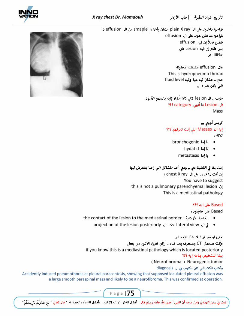
plain X ray smaple effusion
effusion
effusion Lesion
effusion
This is hydropneumo thoraxfluid level
lesion
Lesion category
Mass
Masses
bronchogenic hydatid metastasis
chest X ray
You have to suggestthis is not a pulmonary parenchyemal lesion
This is a mediastinal pathology
Based
Based
the contact of the lesion to the mediastinal border Lateral view << projection of the lesion posteriorly
CT if you know this is a mediastinal pathology which is located posteriorly
Neurogenic tumorNeurofibroma
diagnosis
Accidently induced pneumothoras at pleural paracentesis, showing that supposed loculated pleural effusion was a large smooth paraspinal mass and likely to be a neurofibroma. This was confirmed at operation.

X ray chest Dr. Mamdouh
P a g e | 76
Massive hemoptysisintubation
history of T.B.
chest
data
T.B.
Massive hemoptysis
Hemoptysis Mycetoma Then you are dealing with a cavitary lesion and intra cavitary mass
Mycetoma
fungal ball
metastatic deposits
history
carcinoma of the colon
This is a metastatic deposits

X ray chest Dr. Mamdouh
P a g e | 77
chest
cancer colon
metastasis
immune compermized
fungus Lung
fungus
68 epistaxis
AVM
chest X ray
CT
CT CT
clinician
CT MR isotope
file
clinical sense
totally dependant

X ray chest Dr. Mamdouh
P a g e | 78
History
chest X ray
conclusion
suggest suspect
recommend
CT is recommended
patch opacity in the right lung base for CT
clinician
Opacity
Opacity
Well definedright lower lobe Connected to the hilum by
then you suspect pulmonary vascular malformation

X ray chest Dr. Mamdouh
P a g e | 79
Then if you do CT
www.facebook.com/dr.tafreegh
![NRPavs SOLD items 2013 archive 2 · 2015-03-09 · NRPavs SOLD items 2013 archive 2 Archive 2 of 2013 SOLD items pdf download [0.60MB] ... Rega Planar 2 belt drive turntable black](https://img.pdfslide.us/doc/110x75/5e6f161ce3df0f46131db379/nrpavs-sold-items-2013-archive-2-2015-03-09-nrpavs-sold-items-2013-archive-2-archive.jpg)