Embed Size (px)
Citation preview
Proceedings
This publication is published by Meat & Livestock Australia Limited ABN 39 081 678 364(MLA). Care is taken to ensure the accuracy of information in the publication. Reproductionin whole or in part of this publication is prohibited without the prior written consent of MLA.
Co-sponsored by
PUBLISHED BYMeat & Livestock Australia LimitedLocked Bag 991NORTH SYDNEY NSW 2059
ISBN 1 74036 738 3
MLA OJD HARVEST YEARCONFERENCEDecember 8-9, 2005
MLA OJD Harvest Year Conference, December 2005
Page 2 of 200
Contents
MLA Harvest Year Conference Programme.....................4National and International Perspectives..........................6History of OJD and research priorities in the NOJDP....................................7A producer perspective – what has the research meant to me? ................11An advisor’s view of OJD research ...............................................................15
Diagnosis – Existing Projects.........................................20Hybridisation capture PCR and direct PCR on pooled faecal samples......21Validation of the gamma-interferon test for diagnosis of ovine Johne’sdisease………………………………………………………………………………...28Manipulation of the Interferon - gamma assay to maximise responses toM. ptb antigen in sheep ..................................................................................32Evaluation of a Pourquier ELISA kit in relation to agar gel immunodiffusion(AGID) test for assessment of the humoral immune response in sheepand goats with and without Mycobacterium paratuberculosis infection ...34
Diagnosis – New Approaches.........................................39Early cellular responses in ovine Johne’s disease......................................40Apoptotic responses during the pathogenesis of ovine Johne’s disease.44ELISPOT – new methods for detecting IFN-γ ................................................48Detection of Mycobacteria from blood..........................................................51Proteomic analysis of sheep serum by SELDI TOF-MS: Identification ofputative biomarkers of ovine Johne’s disease.............................................55Gene expression signals from the host ........................................................60Molecular approaches to studying the host-parasite interaction ...............62Review of Immunology, Pathology and Pathogenesis from 8th International Colloquium on Paratuberculosis............................................65Review of Molecular Biology, Microbiology, andCulture from 8th International Colloquium on Paratuberculosis................70
Vaccination .......................................................................77Overview of OJD vaccination – international perspective...........................79
MLA OJD Harvest Year Conference, December 2005
Page 3 of 200
Efficacy of a killed Mycobacterium paratuberculosis vaccine for thecontrol of OJD in Australian sheep flocks ....................................................86On-farm impacts of vaccination with GudairTM...........................................98
Public Health and National Programs ..........................105Review of National Level Prevention and Control from 8th InternationalColloquium on Paratuberculosis .................................................................106Review of Prevention and Control at a Herd Level from 8th InternationalColloquium on Paratuberculosis .................................................................112Review of Implications for Public Health 8th International Colloquium onParatuberculosis ...........................................................................................117Crohn’s Treatment Trial................................................................................120
Epidemiology, Economics ............................................121Epidemiology of ovine Johne’s disease cross species transmissionand survival of the organism in the environment.......................................122Development of grazing management strategies for the control of ovineJohne’s disease.............................................................................................128Within flock spread of OJD - a case study from a sheep flock located insoutheastern New South Wales...................................................................135Risk factors for OJD prevalence in infected flocks....................................144Economic modelling of the impact of OJD .................................................152Genomic and proteomic comparative study of the sheep and cattlestrains of Mycobacterium avium subsp. paratuberculosis .......................159A survey of potential wildlife reservoirs for Mycobacteriumparatuberculosis ………………………………………………………………… 174Review of Epidemiology from 8th International Colloquium onParatuberculosis ...........................................................................................183Epidemiology of Johne’s disease: Recent developments and futuretrends………………………………………………………………………………...189

MLA OJD Harvest Year Conference, December 2005
Page 4 of 200
MLA Harvest Year Conference Programme
Theme Topic Time SpeakerDay 1 - Thursday 8 December
Introduction & Chair 9:00 Lorna CiterWelcome Welcome Reuben Rose
Neil BuchananNational &Internationalperspectives
History of OJD and research priorities inthe NOJDP
David Kennedy
A Producer perspective -What has the research meant to me?
Steve Burgun
An advisor’s view of OJD research David Sackett Discussion/Questions Morning tea 10:30 - 11:00Theme 1 Introduction & Chair Evan SergeantDiagnosis – Specificity of molecular diagnosis Martin McLoonexisting projects HC and Direct PCR on pooled samples Ian Marsh
Evaluation of gamma-interferon assay David StewartImprovements to the IFNG-g assay Richard WhittingtonEvaluation of Pouquier ELISA Sanjeev Gumber
Discussion/Questions Lunch 12:30 - 1:30Theme 2 Introduction & Chair Richard WhittingtonDiagnosis – Early cellular responses in OJD Kumi De Silvanewapproaches
Apoptotic responses during thepathogenesis of OJD
Sally Browne
ELISPOT – new methods for detecting G-IFN
Doug Begg
Detection of Mptb in blood Kate GoldsmithProteomic analysis of sheep serum Ling ZhongGene expression signals from the host Lyrissa Di FioreMolecular approaches to studying the host-pathogen interaction
Deb Taylor
Afternoon Tea 3:00 - 3:30 Theme 2continued
Review of Immunology, pathology andpathogenesis stream from 8ICP
Mark Lanigan
Review of Molecular biology, Microbiologyand Culture stream from 8ICP
Ian Marsh
Discussion/Questions Introduction & Chair Joan LloydTheme 4 Overview – international picture Peter WindsorVaccination Gudair field evaluation Leslie Reddacliff
On-farm impact of vaccination Jeff EpplestonVaccine safety Dominic Dell'Osa, PfizerDiscussion and questions
Finish 5:30 Dinner 7:00 for 7:30 pm
MLA OJD Harvest Year Conference, December 2005
Page 5 of 200
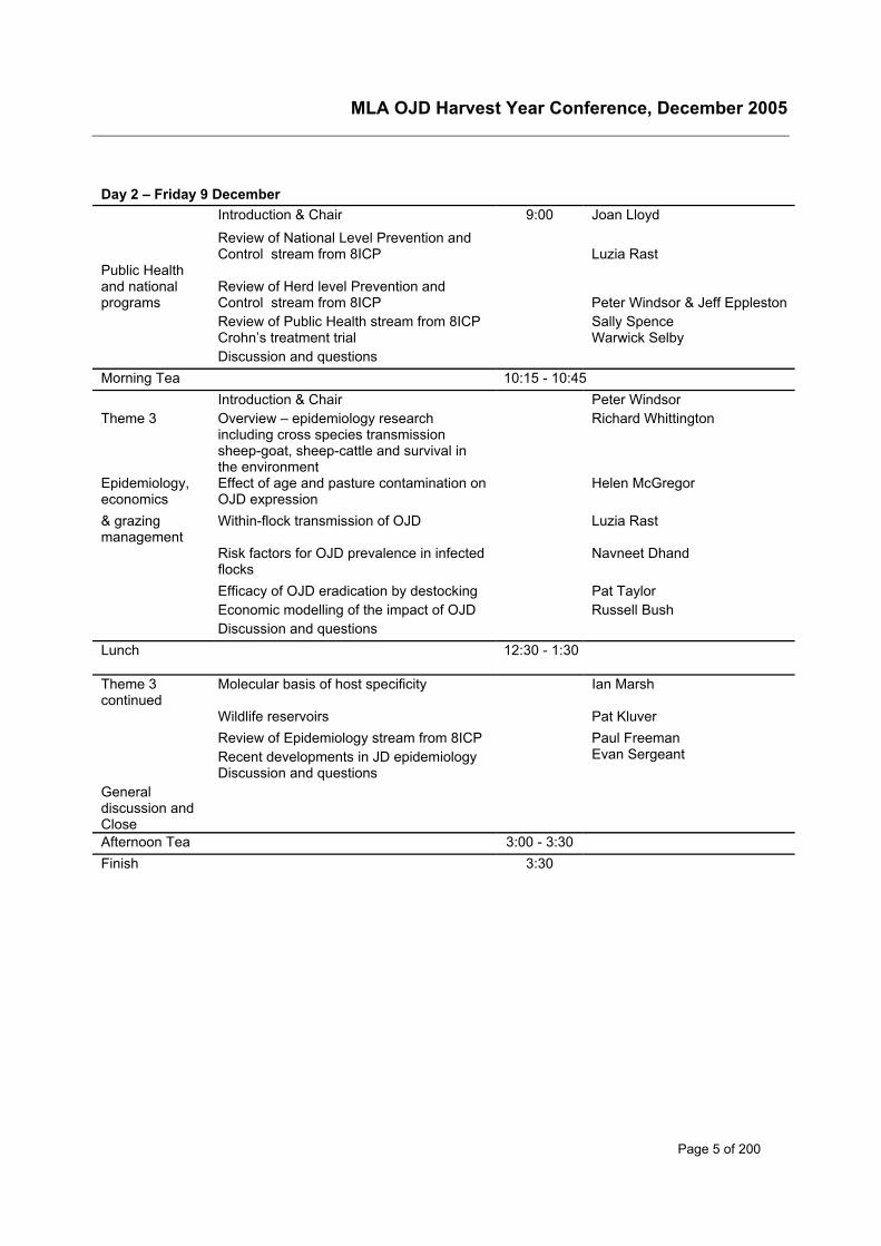
Day 2 – Friday 9 December Introduction & Chair 9:00 Joan Lloyd
Review of National Level Prevention andControl stream from 8ICP Luzia Rast
Public Healthand nationalprograms
Review of Herd level Prevention andControl stream from 8ICP Peter Windsor & Jeff EpplestonReview of Public Health stream from 8ICP Sally SpenceCrohn’s treatment trial Warwick SelbyDiscussion and questions
Morning Tea 10:15 - 10:45 Introduction & Chair Peter WindsorTheme 3 Overview – epidemiology research
including cross species transmissionsheep-goat, sheep-cattle and survival inthe environment
Richard Whittington
Epidemiology,economics
Effect of age and pasture contamination onOJD expression
Helen McGregor
& grazingmanagement
Within-flock transmission of OJD Luzia Rast
Risk factors for OJD prevalence in infectedflocks
Navneet Dhand
Efficacy of OJD eradication by destocking Pat TaylorEconomic modelling of the impact of OJD Russell BushDiscussion and questions
Lunch 12:30 - 1:30
Theme 3continued
Molecular basis of host specificity Ian Marsh
Wildlife reservoirs Pat KluverReview of Epidemiology stream from 8ICP Paul FreemanRecent developments in JD epidemiology Evan SergeantDiscussion and questions
Generaldiscussion andCloseAfternoon Tea 3:00 - 3:30 Finish 3:30

MLA OJD Harvest Year Conference, December 2005
Page 6 of 200
National and International Perspectives
MLA OJD Harvest Year Conference, December 2005
Page 7 of 200
History of OJD and research priorities in the NOJDP
David KennedyTechnical AdviserAnimal Health Australia’s National Johne’s Disease ProgramsPO Box 2321 Orange NSW 2800 AustraliaPhone: 02 6365 6016; Fax 02 6365 6088E-mail: [email protected]
The detection of ovine Johne’s disease (OJD) in the central tablelands of New South Wales in1980 did not precipitate a coordinated response for over 15 years. In the mid-1990s not onlywas it realised that the infection was spreading more widely and causing heavy losses in someflocks in NSW, but OJD was also confirmed in other south-eastern States. During 1997, AnimalHealth Australia unsuccessfully sought support from the national sheep industry organisationsto assist owners of infected flocks to control the disease. At the end of that year, theCommonwealth government commissioned an independent review of options available to dealwith OJD. The Hussey-Morris Report recommended an approach that was translated into thesix year $40.1 million dollar National OJD Control and Evaluation Program (NOJDP) in 1998.
It is pertinent that these developments took place shortly after the national cattle industries hadinitiated the first nationally coordinated approach to JD in 1995. The National FarmersFederation’s National JD Coordinating Committee aimed to restrict further spread of both formsof JD. Bovine Johne’s disease (BJD) had been largely confined to south-eastern Australia forover 70 years and regulatory control of movements and known infected herds had contributed tothis situation. Much more was known about BJD than OJD and diagnostic tools and capabilitieswere more advanced. In comparison, it appeared that an opportunity existed to have an impacton OJD, as its distribution appeared to still be largely restricted. However there was still a lot tolearn if it was going to be successfully controlled and possibly eradicated.
The NOJDP was managed under a Deed of Agreement1 with the purposes of:
• Providing, during a research and evaluation period, by 31 July 2004 sufficient information toallow an informed decision to be made on the national management of OJD, andespecially on the feasibility and cost-effectiveness of eradication; and
• Controlling OJD during the research and evaluation period.
Research and Development was one of four sub-programs within the NOJDP, which alsoincluded Management, Operations and Communications sub-programs. Surveillance andFinancial Assistance sub-programs were included subsequently.
Early research on OJDSince 1995 the three main livestock industry Research and Development Corporations, Meat &Livestock Australia (MLA, formerly Meat Research Council), the Dairy Research andDevelopment Corporation (DRDC) & Woolmark (latterly Australian Wool Innovation) hadmanaged a collaborative approach to JD research in Australia. The McGarvie Smith Institute inNSW and the Rural Industries Research and Development Corporation (RIRDC) also fundedcomplementary JD research. Research priorities were frequently reviewed and modified
1 National Ovine Johne’s disease Control and Evaluation Program (NOJDP) Deed of Agreement signed in March1999.
MLA OJD Harvest Year Conference, December 2005
Page 8 of 200
through extensive consultation with people involved in research and disease control and withindustry representatives.
Between 1995 and 1998, about $1.4 million had been invested on OJD research in the areas of,
• Diagnostic tests – including culture methods, an ELISA test, a gamma interferon assayand direct polymerase chain reaction (PCR) tests on faeces.
• Molecular and applied epidemiology – including an initial assessment of survival of theorganism, DNA typing of isolates from varying locations and host species and role ofwildlife in transmission of the disease.
The NOJDP initially allocated $10.5 million to the R&D sub-program. Half of the researchfunding was provided by the national sheep industry and half from the Commonwealth. MLAmanaged the NOJDP research sub-programs on behalf of Animal Health Australia (then theAustralian Animal Health Council, AAHC).
It is important to appreciate the context in which the NOJDP and its research sub-program weredeveloped in 1998. As Australia was faced with controlling a spreading epidemic, possibly witha view to eradicating it, the initial research program focused on addressing questions relevant tothe immediate challenge of controlling OJD. Hence, research projects were largely of an appliednature, as is evident from the objectives of the projects below that were initiated or proposedduring the first year of the NOJDP 2. Some of these projects were not formally part of theNOJDP but were complementary projects that were funded by MLA and/or AWI.
• Evaluation of Eradication Strategies
To determine if destocking and decontamination over a 15 month period could eradicateOJD from infected properties and if it was an economic option.
• Control Strategy Evaluation
To evaluate strategies to control and limit the impact of OJD in infected flocks
a) by management
b) by vaccination.
• Genetic Preservation
To identify means of ‘safely’ retaining valued genetics from sheep flocks or goat herds thatwere infected with JD.
• Cross Species Infection
To determine whether OJD can be controlled or eradicated from sheep in the presence ofother
a) farmed species such as goats and cattle; and
b) wildlife such as kangaroos, rabbits, wallabies and possums.
• Survival of M. paratuberculosis in the Environment
To determine how long M. ptb survives in the environment and to assess the influence on M.ptb survival times of factors such as soil pH, UV light, temperature, moisture and organicmatter.
2 Summary of Current (& Proposed) R&D For OJD, WE Sykes, August 1999.
MLA OJD Harvest Year Conference, December 2005
Page 9 of 200
a) laboratory tray trials
b) field plots
• Tracer Weaner Model
To demonstrate that Australian ovine M. ptb isolates are capable of colonising the intestinesof newly weaned merino lambs after oral dosing at levels likely to be representative of thosein the field, and that M. ptb can be detected in these lambs four to six weeks after dosing.
• Hybridisation Capture PCR (HC-PCR)
To develop a rapid, cost effective test for the diagnosis of OJD utilising pooled faecalsamples.
• Faecal Culture
To complete validation of the Pooled Faecal Culture Test.
• Sensitivity of Abattoir Surveillance
To evaluate the sensitivity of abattoir surveillance in detecting infected flocks.
During the course of the NOJDP, extensive consultation was maintained with people involved inresearch and disease control and with industry representatives and as a result researchpriorities were reviewed and modified. About $5.7 million was committed to projects in the firstthree years. After the Mid-Term Review of the NOJDP, another $2.7 million was earmarked forresearch, leaving approximately $2 million of the original budget unallocated3.
Later research focus The research focus for the second three years was on diagnosis, vaccination, control andepidemiology with an increased emphasis also on more fundamental research on pathogenesisand immunity. The sub-program aimed to:
• Add value to the current knowledge where there was a clear and direct benefit to thedevelopment of control strategies (knowledge gaps).
• Assist providers of advice and those that develop policy to apply the outcomes ofresearch through a better understanding of the disease.
• Seek understanding of the cellular pathogenesis in early stages of the disease, leadingto the development of improved diagnostic tests and potentially detecting other points ofintervention to limit the expression of the disease.
• Application of new and relevant technology applications to diagnose the disease at anearly stage in the infection process and possibly define vaccine targets3.
Impact of OJD researchThis Harvest Year Conference highlights the consolidated outcomes of the research sub-program but, very importantly, the outcomes were also communicated during the NOJDPthrough a range of advisory media. Primarily MLA’s excellent series of Tips and Tools andOvine Johne’s Disease R&D Updates kept industry, farmers and policy makers abreast ofdevelopments so that improvements in knowledge and tools could be incorporated into controlprograms.
3 National Ovine Johne’s Disease Control and Evaluation Program, Program Plan, 2001/02 – 2003/04.
MLA OJD Harvest Year Conference, December 2005
Page 10 of 200
Moreover the application and implementation of many of these research outcomes in the currentNational Approach to OJD demonstrates the significance and long-term contribution of theresearch to managing OJD in Australia. The Assurance Based Credit (ABC) Scheme is basedon a quantitative risk assessment that was commissioned by AWI to bring together much of theknowledge gained through research. For instance, it incorporates area prevalence based onabattoir surveillance, various levels of flock vaccination and flock testing utilising pooled faecalculture and abattoir surveillance.
ConclusionThe applied R&D sub-program of the NOJDP and the complementary OJD research that wasundertaken from 1995 to 2004 dramatically increased Australia’s (and the world’s)understanding of OJD and its capacity to manage the disease. The Australian sheep industryand those involved in managing OJD in Australia owe a great deal to the research scientists andtheir institutions. Many of them are participating in this Harvest Year Conference. Some of thekey people who are not participating but whose enthusiasm and commitment was also critical tothe success of the research program, especially in the early years, were Drs David Skerman,Bill Sykes and Bruce Allworth.
MLA OJD Harvest Year Conference, December 2005
Page 11 of 200
A producer perspective – what has theresearch meant to me?
Stephen BurgunArthursleigh FarmMarulan, NSW, 2579Phone: 02 4884 1514; Fax: 02 4884 1514Email: [email protected]
This can definitely not be considered as a paper but rather a short story about my take onMLA’s research into OJD, what I needed to learn and what I believe was delivered.
Firstly to introduce myself. I am a third generation ‘Farm Manager’ and while I am uniquelyemployed in that field by the University of Sydney I am definitely not an academic, scientist,researcher or technical assistant just a ‘Working Farm Manager’ employed to run theUniversity’s commercial grazing property ‘Arthursleigh Farm’ at Marulan in the SouthernTablelands of NSW.
I was literally born into large commercial merino sheep operations. I have Jackerooed,Overseen, been a Livestock Selling Agent and am an accredited sheep and lamb assessor. Ihave seen and handled enough sheep to consider myself a genuine sheep man but when wediscovered OJD on the property I was managing I knew ‘absolutely bloody nothing’ about it,had hardly known of its existence and could not (and actually still have trouble) pronouncingMycobacterium paratuberculosis .
Now despite everyone’s perception of what some one with reasonably extensive knowledge insheep husbandry should have known about such a disease at least I wasn’t on my own, myneighbours knew nothing, my ram suppliers new nothing and almost everyone I came acrossnew nothing of the disease. Speculation on the topic had become a hobby.
Anyone who has ever run sheep (in particular Merino sheep) will know that there are alwaysthose ‘poor doing bastards’, you know ‘the bastards like nothing better than to die’. Youcould drench them, draft them off and feed them but no matter what you did they were alwaysthere. Did we finally have the answer to our ever present dilemma? We assumed so and untilbetter informed myself and other sheep producers were proclaiming ‘it was bloody OJD all thetime’.
But we had to learn something about this disease and very quickly as it had (or was about to)drastically effect our viability, not to mention relationships with neighbours, clients and ramsuppliers. And what did we need to learn about it? Well frankly everything.
I remember going to the Ag-Facts (or such) looking for available information and at the timegathered these two (& sorry unsourced) paragraphs which I had included in a report to ourbusiness’s superiors (who are essentially accountants) to explain our dilemma.
“Johne’s disease is a disease of older sheep, characterised by progressive loss of bodycondition, leading to emaciation. Once symptoms have developed, animals will not respond toany treatment and eventually die.Mortality is rarely seen as an acute outbreak. However, up to5% may die in older mobs of sheep 5 years and older and in more severely affected flocks,younger sheep die. Deaths of 15% in mobs of 2 year old sheep have been reported.”

MLA OJD Harvest Year Conference, December 2005
Page 12 of 200
“Apart from trade restrictions, most economic loss from OJD results from the death of infectedsheep. Although difficult to measure, the production of infected animals is likely to be reducedbefore symptoms become obvious.”
O.K, we know what it is and we know the bastards die, but?
• How did we get it?
• How do we diagnose it?
• Who else had it and why were we so badly disadvantaged by regulations?
• Could we get rid of it?
• What was it costing us?
• If we de-stocked could we?
a. In fact clean country after the magic one, two or who knows how many summers?Did the ‘roos’ carry it? Wombats? or Ducks? Car tyres and boots?.
b. Could we use cattle as an alternative species to graze on pastures during that periodof de-stocking to remain commercially viable? Would the cattle get OJD?
c. Could we find clean sheep after country was deemed clean?
d. So on and so on……….
As I had mentioned my bosses are essentially accountants and when they were presented withthe initial ramifications of our status i.e.
• It was no longer possible to sell sheep other than for slaughter.
• While it was possible to continue to operate a commercial wool producing enterprisedespite having OJD, the disease introduced additional costs from mortalities andrestricted selling options would reduce overall income.
• The real estate market (at the time) had demonstrated a reduction in capital value ofland infected with OJD and by association there was a negative social stigma attachedto the status of the flock and its owners - OJD flocks were decidedly ‘second class’.
At that stage, weighing up the negative impacts an interim plan to eradicate OJD by de-stockingparts of the property serially over six years was commenced in 1998. At the time it was believedthat two summers (a period of 15 months at least) was necessary for contaminated land tobecome safe following the removal of sheep.
Land de-stocked of sheep during the eradication program was to be grazed by cattle becausethey are considered unlikely to contract and spread OJD. Alternative grazing of cattle wasaimed at maintaining an income from infected country (straight de-stocking was not considereda financially viable option). Following the two year de-stocking period, if the eradication programwas deemed effective, new sheep from a clean source were to be purchased to re-stock.
So where were we heading? – down an unknown track, with unknown outcomes and no scienceto back it up.
What did the research (science) do for us and how has it impacted on what we were doing?
Well firstly the stigma of being OJD infected started to wain as the surveillance researchexposed just how wide spread (certainly in our area) the disease was. The Southern Tablelandswas rife with the disease. Pooled faecal cultures are now the main test for OJD detection in

MLA OJD Harvest Year Conference, December 2005
Page 13 of 200
flocks and more sensitive than blood tests (MLA, 2002) but when you are infected you areinfected and testing becomes of little importance.
Neighbours started talking to each other again, as we tend to forget just how emotive that earlyperiod was, with sheep farmers looking for someone to blame, either for being the source oftheir infection, being a threat to them becoming infected or as the minority group that hadresulted in harsh trade restrictions being put on them, an innocent third party.
Research told us that it was unlikely that we contracted OJD from kangaroos, wombats or woodducks, although ‘Marsupials may be infected in unique and unusual situations, they were notconsidered to pose a threat to OJD on most farms” (MLA, 2002). O.K throw out the way outtheories, we most probably contracted OJD from our ram suppliers, from strays or contaminatedrun-off from neighbouring infected farms.
“Evaluations of Eradication Strategies for OJD (Project code: OJD.001)” results raised severedoubts about de-stocking as those that attempted had failed. In short why would we thencontinue a de-stocking policy in a high prevalence area and attempt to purchase in (veryexpensive) clean sheep when it was most likely deemed to fail?
An easy decision - we gave up on our de-stocking program, threw in the rams, vaccinated thenew crop of lambs as recommended (“vaccination at weaning reduces subsequent deaths fromOJD and delays the onset of bacterial shedding in dung” (MLA, 2002)) and it was back tobusiness as usual.
The cumulative research (and it is unclear to me which projects or combinations of projects)delivered us a best practice management for the disease, that with vaccination was extremelycompatible to best practice internal parasite management and weaning practices that we reallyshould be employing regardless of OJD.
You see, MLA (2002) research suggests that infection status of ewes and lambing paddockshas been found to be critical in the transmission of OJD and recommends that we prepare ‘lowrisk’ lambing paddocks to reduce transmission within flocks. Again, what would be a low riskpasture? MLA (2002) research suggests that “most OJD bacteria die within six weeks, with asmall number surviving over a year in shaded areas”.
This practice would fit perfectly (at least in good seasons) for sensible pasture and ewenutritional management. Paddocks spelled this way would build up a residual feed source forewes to be put onto just prior to the commencement of lambing so as to better match theirnutritional requirements.
As lambs are susceptible to infection before and after weaning (MLA OJD Update, 2004) thepractice of cleaning pastures of internal parasites by alternate grazing by cattle for 6 monthsprior to weaning is also compatible to providing ‘low risk’ OJD pastures as well.
Research (MLA OJD Update, 2004) suggests that cattle can very rarely be infected with thesheep strain of Johne’s disease, however we should avoid grazing young calves on pasturesgrazed by infected sheep. Therefore when we are preparing ‘low risk’ paddocks for lambing andweaning with cattle we should use dry cattle if they are available, or at worst cows with biggercalves at foot.
So, in short for me its ‘happy days’. While I haven’t had enough time to gauge the effectivenessof the vaccine on our hoggets, I can be a sheep farmer again. It appears the disease ismanageable using sensible management strategies that are not radical but actually complementor mirror existing sheep management strategies employed for internal parasite control andpasture allocation.

MLA OJD Harvest Year Conference, December 2005
Page 14 of 200
All I want now is for the price of wool to go up and we are back in business.
On completion I would just like to present to you all of my cards. As Manager of The SydneyUniversity Farm ‘Arthursleigh’ we were host to two major MLA funded epidemiological studiesand while I was not involved in the project other than outsourcing staff from time to time for siteconstruction, feed supplementation, shearing and crutching etc. I was able to observe first handsome of the findings as they unfolded.
As a University Farm manager please don’t get the impression that I have a vested interest inendorsing the research conducted, as I am a born sceptic who is not that fond of ‘some’academics impractical and unworkable ideas (believe me I have had some beauties). Howeveras an observer, the projects run on our farm were exemplary and should be complemented.They were well set up and thought out, and well (sometimes painful in detail and intensity)managed by extremely dedicated people.
If research generated from these projects is part of what we are now using as ‘best practice’ andof a standard of the other OJD research then I am a believer until better science tells ussomething different.
And by the way those poor doing sheep at the back of the mob that we grew up seeing andwere always there, well they’re ‘poor doing bastards’ or ‘wormy’. By the time you notice a sheepsick with clinical signs of OJD that sheep doesn’t come back into the yards he’s as good asdead, I know MLA research taught me that.
MLA OJD Harvest Year Conference, December 2005
Page 15 of 200
An advisor’s view of OJD researchDavid SackettHolmes Sackett & Associates Pty Ltd112 Fitzmaurice StreetWagga Wagga NSW 2650Phone: 02 6931 7110; Fax: 02 6931 7113Email: [email protected]
Considerable effort has gone into research on OJD since it emerged as an important issue inthe mid 1990’s. The value of the findings of that research are highlighted by the differences inapproach to managing OJD both at an industry and at the farm level.
Industry IssuesIt is now 10 years since NSW embarked upon the development of the Strategic Plan for thecontrol and possible eradication of OJD. This was closely followed by Victoria with its ownequally flawed version and other states adopted various strategies depending on their degree ofnaivety and regulatory zeal.
It is always a challenge to try to manage a disease when one has less than ideal information onwhich to develop a strategy. It is quite apparent that one cannot always stand back and donothing while waiting for all the key issues to be addressed by research. In many instances, todo so, would be wrong and not in the interests of the industries affected or at risk.
There were two key pieces of research that were missing in the early days of policydevelopment for OJD.
The first was to seriously review the probability of success of any control or eradication programagainst accepted criteria for successful disease and control programs. Had such a review beenundertaken in the early days of policy development, it is quite likely that programs aimed atcontrol or eradication would have taken quite different directions.
The second was an assessment of the economic impact of OJD on both individual businessesand for the broader sheep industry. There is no doubt that such an assessment would have hadto be done with less than perfect knowledge about the impact of the disease at the flock levelbut this could have been taken into account with sensitivity analyses. It was not until the study ofHassall & Associates (2003) that it began to be widely accepted that the majority of the cost ofthe disease related to the regulatory strategies (quarantine, zoning) rather than the costsassociated with the direct effects of OJD on flock productivity.
Instead of good research, we had well meaning but naïve people attempting to produce goodpolicy in what would be best described as a vacuum of good quality research on which to basedecisions. The objective was the eradication of disease rather than the improvement of industryproductivity and profitability. As mentioned above, it is not realistic that all questions should beanswered before a program can be commenced, because it would inevitably lead to inactionwhich in many cases would be an undesirable outcome. However there are some key issuesthat should have been addressed with greater rigour from the start.
MLA OJD Harvest Year Conference, December 2005
Page 16 of 200
Flock IssuesDuring most of the 1990’s advice to producers on the management of OJD was, at best, basedon experience in the other industries or other countries and could, at best, be described as a‘best guess’. Field observations of differences in estimated mortality rates could not beadequately explained and hence there were no sound recommendations on strategies thatcould be adopted to minimise the impact of the disease at the flock level.
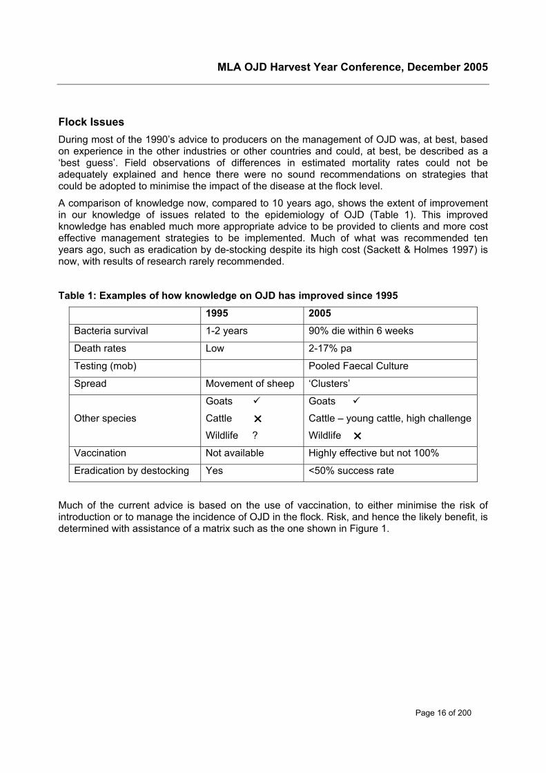
A comparison of knowledge now, compared to 10 years ago, shows the extent of improvementin our knowledge of issues related to the epidemiology of OJD (Table 1). This improvedknowledge has enabled much more appropriate advice to be provided to clients and more costeffective management strategies to be implemented. Much of what was recommended tenyears ago, such as eradication by de-stocking despite its high cost (Sackett & Holmes 1997) isnow, with results of research rarely recommended.
Table 1: Examples of how knowledge on OJD has improved since 1995
1995 2005
Bacteria survival 1-2 years 90% die within 6 weeks
Death rates Low 2-17% pa
Testing (mob) Pooled Faecal Culture
Spread Movement of sheep ‘Clusters’
Other species
Goats
Cattle
Wildlife ?
Goats
Cattle – young cattle, high challenge
Wildlife
Vaccination Not available Highly effective but not 100%
Eradication by destocking Yes <50% success rate
Much of the current advice is based on the use of vaccination, to either minimise the risk ofintroduction or to manage the incidence of OJD in the flock. Risk, and hence the likely benefit, isdetermined with assistance of a matrix such as the one shown in Figure 1.
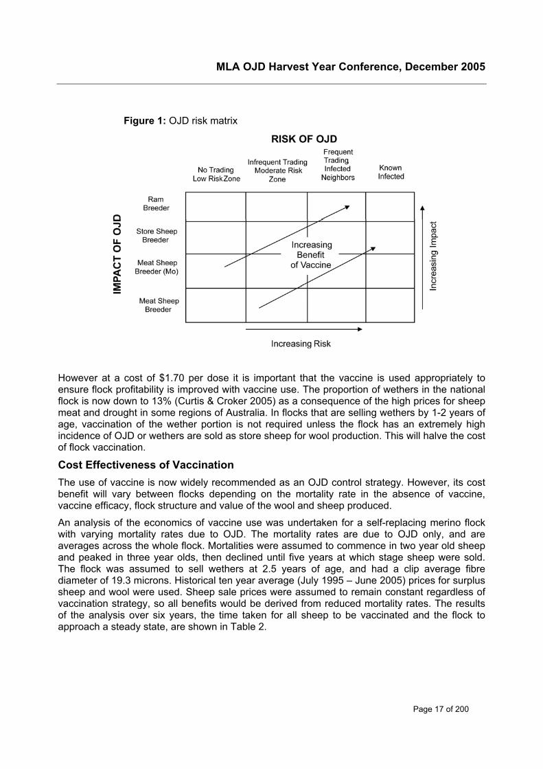
MLA OJD Harvest Year Conference, December 2005
Page 17 of 200
Figure 1: OJD risk matrix
However at a cost of $1.70 per dose it is important that the vaccine is used appropriately toensure flock profitability is improved with vaccine use. The proportion of wethers in the nationalflock is now down to 13% (Curtis & Croker 2005) as a consequence of the high prices for sheepmeat and drought in some regions of Australia. In flocks that are selling wethers by 1-2 years ofage, vaccination of the wether portion is not required unless the flock has an extremely highincidence of OJD or wethers are sold as store sheep for wool production. This will halve the costof flock vaccination.
Cost Effectiveness of VaccinationThe use of vaccine is now widely recommended as an OJD control strategy. However, its costbenefit will vary between flocks depending on the mortality rate in the absence of vaccine,vaccine efficacy, flock structure and value of the wool and sheep produced.
An analysis of the economics of vaccine use was undertaken for a self-replacing merino flockwith varying mortality rates due to OJD. The mortality rates are due to OJD only, and areaverages across the whole flock. Mortalities were assumed to commence in two year old sheepand peaked in three year olds, then declined until five years at which stage sheep were sold.The flock was assumed to sell wethers at 2.5 years of age, and had a clip average fibrediameter of 19.3 microns. Historical ten year average (July 1995 – June 2005) prices for surplussheep and wool were used. Sheep sale prices were assumed to remain constant regardless ofvaccination strategy, so all benefits would be derived from reduced mortality rates. The resultsof the analysis over six years, the time taken for all sheep to be vaccinated and the flock toapproach a steady state, are shown in Table 2.
MLA OJD Harvest Year Conference, December 2005
Page 18 of 200
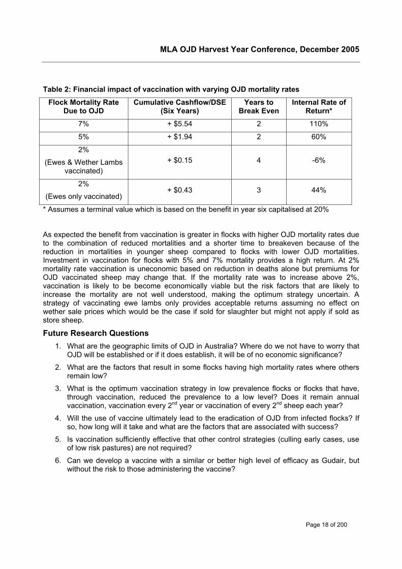
Table 2: Financial impact of vaccination with varying OJD mortality rates
Flock Mortality RateDue to OJD
Cumulative Cashflow/DSE(Six Years)
Years toBreak Even
Internal Rate ofReturn*
7% + $5.54 2 110%
5% + $1.94 2 60%
2%
(Ewes & Wether Lambsvaccinated)
+ $0.15 4 -6%
2%
(Ewes only vaccinated)+ $0.43 3 44%
* Assumes a terminal value which is based on the benefit in year six capitalised at 20%
As expected the benefit from vaccination is greater in flocks with higher OJD mortality rates dueto the combination of reduced mortalities and a shorter time to breakeven because of thereduction in mortalities in younger sheep compared to flocks with lower OJD mortalities.Investment in vaccination for flocks with 5% and 7% mortality provides a high return. At 2%mortality rate vaccination is uneconomic based on reduction in deaths alone but premiums forOJD vaccinated sheep may change that. If the mortality rate was to increase above 2%,vaccination is likely to be become economically viable but the risk factors that are likely toincrease the mortality are not well understood, making the optimum strategy uncertain. Astrategy of vaccinating ewe lambs only provides acceptable returns assuming no effect onwether sale prices which would be the case if sold for slaughter but might not apply if sold asstore sheep.
Future Research Questions1. What are the geographic limits of OJD in Australia? Where do we not have to worry that
OJD will be established or if it does establish, it will be of no economic significance?
2. What are the factors that result in some flocks having high mortality rates where othersremain low?
3. What is the optimum vaccination strategy in low prevalence flocks or flocks that have,through vaccination, reduced the prevalence to a low level? Does it remain annualvaccination, vaccination every 2nd year or vaccination of every 2nd sheep each year?
4. Will the use of vaccine ultimately lead to the eradication of OJD from infected flocks? Ifso, how long will it take and what are the factors that are associated with success?
5. Is vaccination sufficiently effective that other control strategies (culling early cases, useof low risk pastures) are not required?
6. Can we develop a vaccine with a similar or better high level of efficacy as Gudair, butwithout the risk to those administering the vaccine?

MLA OJD Harvest Year Conference, December 2005
Page 19 of 200
ReferencesCurtis K & Croker K (2005) Wool Desk Report – September 2005. WA Department ofAgriculture
Hassall & Associates (2003) Costs of OJD for Australian Sheep Producers and Options forAssistance
Sackett DM & Holmes PR (1997) Economic Assessment of Options for Eradication of OvineJohne’s Disease; Proceedings of the Fourth International Congress for Sheep Veterinarians,Armidale, February 1997 (p. 314). Australian Sheep Veterinary Society

MLA OJD Harvest Year Conference, December 2005
Page 20 of 200
Diagnosis – Existing Projects
MLA OJD Harvest Year Conference, December 2005
Page 21 of 200
Hybridisation capture PCR and direct PCRon pooled faecal samples
Ian Marsha, Richard Whittington, Leslie Reddacliff and Graeme Eamens
aElizabeth Macarthur Agricultural InstitutePMB 8 Camden, NSW 2570, Australia,Phone: 02 4640 6502; Fax: 02 4640 6384Email: [email protected]
SummaryHybridisation capture-PCR (HC-PCR) was first reported as a method for the detection ofMycobacterium avium subsp. paratuberculosis (M. ptb) in 1995 and was successfully trialled ona small number of faecal samples from cattle with Johne’s disease. In our laboratory HC-PCRwas found to be capable of detecting M. ptb in pellets from infected sheep diluted at rates of upto 1 in 100 in normal faeces, suggesting that the technique should be evaluated further as apotential low-cost diagnostic technique for flocks/herds using pooled samples. A locallyoptimised HC-PCR method was evaluated on faeces from infected and non-infected sheepusing faecal samples pooled from 50 sheep and individual faecal samples. The status of each ofthe faecal samples was determined by radiometric culture. A simpler direct-PCR (D-PCR)technique was evaluated on the same samples and was found to be more sensitive than HC-PCR. Initial results indicated that D-PCR from faeces can be used as a rapid means ofscreening pooled faecal samples for flock diagnosis of Johne’s disease in sheep, improvingdetection of M. ptb infection in the early stages of disease, or where prevalence is low, which iscritical for the management of ovine Johne’s disease (OJD). An improved D-PCR test, withsimilar or better sensitivity than current culture techniques and which would be acceptable forregulatory purposes, would be of immense benefit to the industry. This is a potentiallyachievable short term outcome, building on years of previous and on-going research in theEMAI Microbiology Laboratory.
Introduction and backgroundThe difficulty of diagnosis of M. ptb infection in the early stages of disease, or where prevalenceis low, is critical for the management of OJD [15]. The two most useful diagnostic tools currentlyavailable to the industry are abattoir surveillance and pooled faecal culture (PFC). Abattoirinspection is an extremely efficient and economical method of regional surveillance [1] but it isnot applicable to live animals, and can only ever detect animals that have progressed to havegross lesions. Sheep without gross lesions can shed M. ptb in their faeces and are a risk toother livestock. PFC is the most sensitive practical current test to detect OJD infection in aliving mob of sheep [13] [14]. However, PFC is expensive, and may take many months fordefinitive results.
The use of molecular techniques to directly detect the DNA of M. ptb in faeces, without havingto first grow the organism in culture would theoretically allow the rapid (several days) andsensitive diagnosis of OJD in living sheep. Hybridisation capture-PCR (HC-PCR) was firstreported as a method for the detection of M. ptb in 1995 and was successfully trialled on a smallnumber of faecal samples from cattle with Johne’s disease [12]. However, sample to samplecross contamination during the DNA purification step highlighted that the original format of thetest was unsuitable for routine diagnostic use. A modified HC-PCR was reported with analyticalsensitivity of 5000 organisms per 200 mg faecal sample and capable of detecting M. ptb inpellets from infected sheep diluted at rates of up to 1 in 100 in normal faeces, suggesting that
MLA OJD Harvest Year Conference, December 2005
Page 22 of 200
the technique should be evaluated further as a low-cost diagnostic technique for flocks/herdsusing pooled samples [9]. HC-PCR was evaluated on faecal samples pooled from 50 sheepand individual faecal samples as a rapid diagnostic test for M. ptb. The status of each of thefaecal samples was determined by radiometric culture. A simpler direct-PCR (D-PCR) techniquewas evaluated on the same samples and was found to be more sensitive than HC-PCR [10].Direct-PCR using primers from the 5’ region of IS900 was evaluated in a blind trial on 502pooled faecal samples which were concurrently examined by culture. Twenty one (64%) of the33 culture positive pools were detected by direct PCR, representing 11 (79%) of the 14 farmswith infected sheep. Direct-PCR was also more sensitive than the immunomagnetic beadcapture-PCR designed for the same purpose [11]. Using individual faecal samples, 74% ofculture positive samples were detected with direct-PCR compared to 44% with immunomagneticbead capture-PCR. Direct-PCR from faeces can be used as a rapid means of screening pooledfaecal samples for flock diagnosis of Johne’s disease in sheep.
The D-PCR test developed at EMAI under an MLA-funded project was completed in 2000. Thetest was further evaluated at EMAI in 2004 under routine diagnostic conditions in our laboratoryand at VIAS in Victoria, and was also included in several on-going OJD research projects.
The sensitivity of the current test is less than faecal culture. At the level of the pooled sample,the sensitivity for D-PCR compared to PFC varied from 64% in the early trials to about 40% inthe most recent investigations. At the farm level (more than one pool is normally examined),sensitivity was 79% in the early trials and 66% in the recent work. It is, however, alwaysmisleading to quote sensitivity estimates in isolation – they depend on the population beingtested, and this has varied over the years. For example, the early trials probably included moreflocks with a higher prevalence of infection, and D-PCR detected all the high prevalence flocksin the recent trials. When looked at from the view of number of M. ptb organisms in the sampleexamined, D-PCR appears to require about ten times as many to yield a positive result – about104 for D-PCR compared to about 103 for culture. In practical terms, this all means that D-PCRwill readily detect pools with large numbers of M. ptb organisms. This translates to sheep withsevere multibacillary disease, and these are the animals of greatest immediate risk oftransmitting infection.
The cost of D-PCR is similar to culture, but its great advantage is the short time for results. Thecurrent D-PCR test is now approved for use in NSW and is proving of use to some producers inthe management of infected flocks, by providing a rapid assessment of whether sheep areshedding large numbers of M. a. paratuberculosis. However, because of the lower sensitivitycompared to culture, negative results cannot be used for ABC points or to remove suspicion ofinfection.
There is opportunity to improve the current D-PCR test, and some of the avenues for this werealready suggested in the final report from the above project and in the peer reviewedpublications generated from the project [9] [10]. Several papers on M. ptb -specific D-PCR fromfaeces or tissues (often from humans) have been published, and their techniques can also beinvestigated for adaptation to D-PCR from sheep faeces [2] [3] [4] [5] [6] [7] [8]. Many of thetechniques as published, while apparently sensitive, are too time consuming, costly or at risk ofcross-contamination, for use as routine diagnostic tests. MLA has provided further funding toinvestigate improvements to the D-PCR procedure.
Use of the existing D-PCR test in diagnostic investigationsIn current diagnostic procedures at EMAI, pooled faeces are subjected to DNA extraction basedon a commercial resin and then tested in the PCR. The test can be completed within a weekand relies on two phases. The initial phase involves a ‘conventional’ PCR assay on the DNA
MLA OJD Harvest Year Conference, December 2005
Page 23 of 200
extract based on the IS900 gene sequence of M. ptb, using a special combination of IS900primers, and two sample dilution rates to reduce the risk of PCR inhibition. The resultant PCRproduct is assessed on an agarose gel for molecular size to ensure it conforms to the specificmolecular size expected for M. ptb. The second (confirmatory) phase then involves a restrictionendonuclease assay (REA), but requires a sufficiently strong DNA gel band from the first phasebefore it can be run. If there is a product of the correct size but too weak to run in the REA, theresult of the D-PCR testing is classified as a trace PCR reaction and is considered inconclusive.A positive D-PCR result requires both an IS900 PCR product of the correct size and an REAresult to confirm that M. ptb is present.
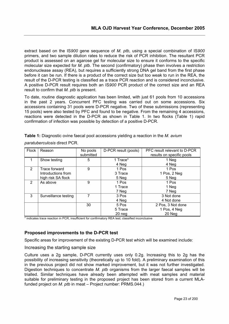
To date, routine diagnostic application has been limited, with just 61 pools from 10 accessionsin the past 2 years. Concurrent PFC testing was carried out on some accessions. Sixaccessions containing 31 pools were D-PCR negative. Two of these submissions (representing15 pools) were also tested by PFC and found to be negative. From the remaining 4 accessions,reactions were detected in the D-PCR as shown in Table 1. In two flocks (Table 1) rapidconfirmation of infection was possible by detection of a positive D-PCR.
Table 1: Diagnostic ovine faecal pool accessions yielding a reaction in the M. avium
paratuberculosis direct PCR.
Flock Reason No poolssubmitted
D-PCR result (pools) PFC result relevant to D-PCRresults on specific pools
1 Show testing 5 1 Trace*4 Neg
1 Neg4 Neg
2 Trace forwardIntroductions fromhigh risk SA flock
9 1 Pos3 Trace5 Neg
1 Pos1 Pos, 2 Neg
5 Neg2 As above 9 1 Pos
1 Trace7 Neg
1 Pos1 Neg7 Neg
3 Surveillance testing 7 3 Pos4 Neg
3 Not done4 Not done
30 5 Pos5 Trace20 neg
2 Pos, 3 Not done1 Pos, 4 Neg
20 Neg* indicates trace reaction in PCR, insufficient for confirmatory REA test; classified inconclusive
Proposed improvements to the D-PCR testSpecific areas for improvement of the existing D-PCR test which will be examined include:
Increasing the starting sample sizeCulture uses a 2g sample, D-PCR currently uses only 0.2g. Increasing this to 2g has thepossibility of increasing sensitivity (theoretically up to 10 fold). A preliminary examination of thisin the previous project did not show marked improvement, but it was not further investigated.Digestion techniques to concentrate M. ptb organisms from the larger faecal samples will betrialled. Similar techniques have already been attempted with meat samples and materialsuitable for preliminary testing in the proposed project has been stored from a current MLA-funded project on M. ptb in meat – Project number: PRMS.044.)
MLA OJD Harvest Year Conference, December 2005
Page 24 of 200
Removing competing DNA from the reactionBecause of the very resistant cell wall of mycobacteria, a pre-treatment targeted at more labilemicro-organisms may be effective. This seems not to have been used in any of the molecularwork published to date. Restriction enzyme digests prior to PCR could also be helpful to avoidnon-specific bands. Digestion techniques as above may also be helpful.
Improving the lysis of the resistant mycobacteriaIn the current protocol it is likely that many M. ptb cells remain intact, and thus their DNA isunavailable to the subsequent PCR. Alternate techniques to the simple boiling used in thecurrent protocol will be trialled to improve DNA extraction. We have experience with severalpotentially useful procedures through the proteomics work done by Ian Marsh during his MLA-funded PhD studies.
Removal of DNA inhibitorsOne of the difficulties with performing PCR on materials such as faeces is the loss of sensitivitydue to inhibition of the PCR reaction by many normal faecal components. At present, the D-PCR attempts to overcome this problem using a commercial product to separate the targetorganisms from such inhibitory components. However, 4 years in molecular biology is a longtime, and there are now many newer commercial kits or materials for DNA extraction fromfaecal, soil and other contaminated samples which might be trialled. Such kits are not targetedat mycobacteria, and will not give optimum results with ‘off-the-shelf’ use.
Developing an internal control targetThis would be used in each test to identify PCR inhibition. Currently, when we get a negativeresult in D-PCR, we have no way of knowing whether the negative result is because no M. ptbDNA was present (i.e a true negative) or whether inhibitors in that particular sample preventedthe PCR from amplifying any specific DNA that was present. A control target that can be addedto each reaction, and which gives a distinct band of different size to specific bands, allowsassessment of possible inhibition. Such a modification would allow more confidence in anegative D-PCR result for M. ptb. This confidence is important if D-PCR is to be acceptable for‘negative’ regulatory purposes.
Examination of DNA lossesSeveral stages of the extraction process may be susceptible to loss of target DNA thus resultingin reduced sensitivity. Minimising the amount of loss of this DNA will ultimately lead to a moresensitive diagnostic test.
Use of a second M. a. paratuberculosis-specific PCR targetCurrently, even using PCR/REA as confirmation of growth in Bactec medium, the identificationof M. ptb DNA is not considered to be fully confirmatory of infection unless sub-culture to solidmedia/mycobactin dependence is also positive. We are at present investigating the use of othertargets in multiplex PCR with IS900, that would satisfy regulators as definitive diagnostic criteriafor M. ptb. These techniques would be applicable to D-PCR.
The use of real-time PCRThis can further reduce turn-around time by a day, and would facilitate the quantification of apositive result. We are at present conducting preliminary investigations into the use of thistechnology for confirmation of growth from Bactec cultures. If successful, we could also applythis to D-PCR.
MLA OJD Harvest Year Conference, December 2005
Page 25 of 200
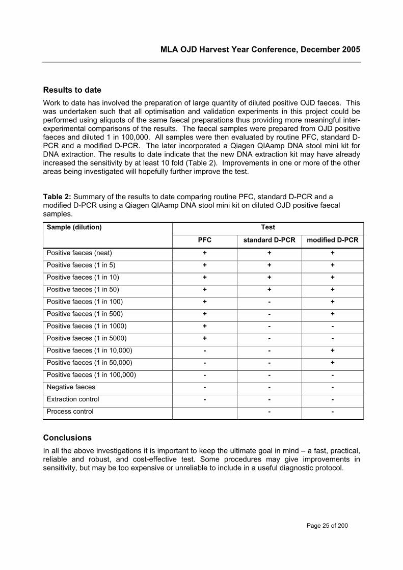
Results to dateWork to date has involved the preparation of large quantity of diluted positive OJD faeces. Thiswas undertaken such that all optimisation and validation experiments in this project could beperformed using aliquots of the same faecal preparations thus providing more meaningful inter-experimental comparisons of the results. The faecal samples were prepared from OJD positivefaeces and diluted 1 in 100,000. All samples were then evaluated by routine PFC, standard D-PCR and a modified D-PCR. The later incorporated a Qiagen QIAamp DNA stool mini kit forDNA extraction. The results to date indicate that the new DNA extraction kit may have alreadyincreased the sensitivity by at least 10 fold (Table 2). Improvements in one or more of the otherareas being investigated will hopefully further improve the test.
Table 2: Summary of the results to date comparing routine PFC, standard D-PCR and amodified D-PCR using a Qiagen QIAamp DNA stool mini kit on diluted OJD positive faecalsamples.
TestSample (dilution)
PFC standard D-PCR modified D-PCR
Positive faeces (neat) + + +
Positive faeces (1 in 5) + + +
Positive faeces (1 in 10) + + +
Positive faeces (1 in 50) + + +
Positive faeces (1 in 100) + - +
Positive faeces (1 in 500) + - +
Positive faeces (1 in 1000) + - -
Positive faeces (1 in 5000) + - -
Positive faeces (1 in 10,000) - - +
Positive faeces (1 in 50,000) - - +
Positive faeces (1 in 100,000) - - -
Negative faeces - - -
Extraction control - - -
Process control - -
ConclusionsIn all the above investigations it is important to keep the ultimate goal in mind – a fast, practical,reliable and robust, and cost-effective test. Some procedures may give improvements insensitivity, but may be too expensive or unreliable to include in a useful diagnostic protocol.
MLA OJD Harvest Year Conference, December 2005
Page 26 of 200
References[1] T. Bradley, R. Cannon. Determination of individual animal-level sensitivity of abattoir
surveillance for ovine Johne's disease. Proceedings of the Australian Sheep VeterinarySociety, Cairns Conference , 61-2. 2003.
[2] T.J. Bull, E.J. McMinn, K. Sidi-Boumedine, A. Skull, D. Durkin, P. Neild, G. Rhodes, R.Pickup, J. Hermon-Taylor. Detection and verification of Mycobacterium avium subsp.paratuberculosis in fresh ileocolonic mucosal biopsy specimens from individuals with andwithout Crohn's disease, J Clin Microbiol, 41, (2003) 2915-23.
[3] J. Christopher-Hennings, M.A. Dammen, S.R. Weeks, W.B. Epperson, S.N. Singh, G.L.Steinlicht, Y. Fang, J.L. Skaare, J.L. Larsen, J.B. Payeur, E.A. Nelson. Comparison of twoDNA extractions and nested PCR, real-time PCR, a new commercial PCR assay, andbacterial culture for detection of Mycobacterium avium subsp. paratuberculosis in bovinefaeces, J Vet Diagn Invest, 15, (2003) 87-93.
[4] D.M. Collins, D.M. Stephens, G.W. de Lisle. Comparison of polymerase chain reaction testsand faecal culture for detecting Mycobacterium paratuberculosis in bovine faeces, VetMicrobiol, 36, (1993) 289-99.
[5] J. Erume, J. Spergser, R. Rosengarten. Rapid detection of Mycobacterium avium subsp.paratuberculosis from cattle and zoo animals by nested PCR, Afr Health Sci, 1, (2001) 83-9.
[6] Y. Fang, W.H. Wu, J.L. Pepper, J.L. Larsen, S.A. Marras, E.A. Nelson, W.B. Epperson, J.Christopher-Hennings. Comparison of real-time, quantitative PCR with molecular beaconsto nested PCR and culture methods for detection of Mycobacterium avium subsp.paratuberculosis in bovine fecal samples, J Clin Microbiol, 40, (2002) 287-91.
[7] S. Halldorsdottir, S. Englund, S.F. Nilsen, I. Olsaker. Detection of Mycobacterium aviumsubsp. paratuberculosis by buoyant density centrifugation, sequence capture PCR and dotblot hybridisation, Vet Microbiol, 87, (2002) 327-40.
[8] S. Khare, T.A. Ficht, R.L. Santos, J. Romano, A.R. Ficht, S. Zhang, I.R. Grant, M. Libal, D.Hunter , L.G. Adams. Rapid and sensitive detection of Mycobacterium avium subsp.paratuberculosis in bovine milk and feces by a combination of immunomagnetic beadseparation-conventional PCR and real-time PCR, J Clin Microbiol, 42, (2004) 1075-81.
[9] I. Marsh, R. Whittington, D. Millar. Quality control and optimized procedure of hybridizationcapture-PCR for the identification of Mycobacterium avium subsp. paratuberculosis infaeces, Mol Cell Probes, 14, (2000) 219-32.
[10] I.B. Marsh, R.J. Whittington. Progress towards a rapid polymerase chain reactiondiagnostic test for the identification of Mycobacterium avium subsp. paratuberculosis infaeces, Mol Cell Probes, 15, (2001) 105-18.
[11] O. Mason, I.B. Marsh, R.J. Whittington. Comparison of immunomagnetic bead separation-polymerase chain reaction and faecal culture for the detection of Mycobacterium aviumsubsp paratuberculosis in sheep faeces, Aust Vet J, 79, (2001) 497-500.
[12] D.S. Millar , S.J. Withey, M.L. Tizard, J.G. Ford, J. Hermon-Taylor. Solid-phasehybridization capture of low-abundance target DNA sequences: application to thepolymerase chain reaction detection of Mycobacterium paratuberculosis andMycobacterium avium subsp. silvaticum, Anal Biochem, 226, (1995) 325-30.
[13] E.S. Sergeant, R.J. Whittington, S.J. More. Sensitivity and specificity of pooled faecal

MLA OJD Harvest Year Conference, December 2005
Page 27 of 200
culture and serology as flock-screening tests for detection of ovine paratuberculosis inAustralia, Prev Vet Med, 52, (2002) 199-211.
[14] R.J. Whittington, S. Fell, D. Walker, S. McAllister, I. Marsh, E. Sergeant, C.A. Taragel, D.J.Marshall, I.J. Links. Use of pooled fecal culture for sensitive and economic detection ofMycobacterium avium subsp. paratuberculosis infection in flocks of sheep, J ClinMicrobiol, 38, (2000) 2550-6.
[15] R.J. Whittington, E.S. Sergeant. Progress towards understanding the spread, detectionand control of Mycobacterium avium subsp paratuberculosis in animal populations, AustVet J, 79, (2001) 267-78.
MLA OJD Harvest Year Conference, December 2005
Page 28 of 200
Validation of the gamma-interferon test for diagnosis ofovine Johne’s disease
DJ Stewarta, PL Stiles, B Horvatic, KL Bruce, PG McWaters, JA Vaughan, GJBeddome, WP Michalski, RJ Whittington, C Lambeth, PA Windsor, LA Reddacliff,H McGregor, OP Dhungyel, DV Cousins, BR Francis, R Butler, DD Salmon, CFRoberts, J MacFarlane, L Gavey, R Badman, ESG Sergeant, I Jasenko and SLJonesaCSIRO Livestock Industries, Australian Animal Health LaboratoryPrivate Bag 24, Geelong, 3220, VictoriaPhone 03 5227 5749; Fax: 03 5227 5000E-mail: [email protected]
AbstractIn theory, the interferon-γ (IFN) test has potential for improved control of ovine Johne’s disease(OJD) by early detection before contamination of pasture and transmission of infection. Tovalidate the IFN test, a project has been completed for determining specificity and sensitivity.Because of non-specific IFN responses, raised cut-points were required to achieve highspecificity (≥98%). This resulted in reduction of sensitivity to below 50%, limiting its applicationfor early detection or certification from disease freedom. A major limitation for adoption is thewidespread use of vaccination precluding the use of immunological tests for diagnosis. Otherlimitations, apart from relatively low sensitivity, include cost of the test as well as a narrow timewindow for laboratory initiation of the assay so that test result validity is not compromised. Theassay may have application in a test and cull program as a surrogate test for faecal sheddingand the removal of sheep with severe disease but this approach to control of OJD will requirefurther confirmation.
BackgroundFor an effective OJD control program, it is important that diagnostic tests can identify infectedsheep prior to commencement of bacterial shedding, pasture contamination and exposure ofother livestock to infection. In theory, the interferon-γ (IFN) test has the potential to fill this rolesince the assay detects a cell-mediated immune (CMI) reaction, which occurs earlier than thehumoral antibody response in mycobacterial infections. Antibody assays have low sensitivity inOJD and seroconversion usually only occurs after the commencement of shedding.
Objectives1. Complete IFN test development by standardising potency of PPD antigens and
determine the criteria defining a cut-point for a positive reaction.
2. Determine the specificity and sensitivity of the IFN test in sheep flocks.
Specificity trialThe specificity of the IFN test was evaluated on 6 different unexposed flocks. Blood sampleswere collected from individual sheep on each of the properties in 3 NSW Rural Lands ProtectionBoards (Riverina, Dubbo and Armidale) and one property each in WA, SA and Vic. Theinterpretation criteria for the IFN test included: the mean optical densities (OD) values for theAvian PPD stimulation minus the nil antigen mean ODs with a difference of 0.05 or 0.10 beingrequired for a positive result (Avian PPD-Nil antigen ≥ 0.05 or A-N ≥ 0.10); the mean OD values
MLA OJD Harvest Year Conference, December 2005
Page 29 of 200
for the Johnin stimulation minus the nil antigen mean ODs with a difference of 0.05 or 0.10being required for a positive result (Johnin-N ≥ 0.05 or J-N ≥ 0.10) and the OD values for J-Nminus A-N with a difference of 0.05 or 0.10 being required for a positive result (J-A ≥ 0.05 or J-A≥ 0.10). Specificity varied according to the geographical location of the property and the age ofthe sheep and IFN reactors were most evident in the Armidale flock. It is probable that thesenon-specific IFN responses were due to false positive reactions induced by environmentalbacteria that shared antigens with Mycobacterium avium subspecies paratuberculosis (M. ptb).The location of the flocks, their history and diagnostic testing (pooled faecal culture andserology) were not indicative of OJD. For lambs, aggregate specificity was high (>99% CI ≥97.7%) irrespective of the scale (A-N, J-N, J-A) or the cut-point (≥ 0.05, ≥ 0.010). In the currentstudy, specificities for yearlings and adult ewes using the A-N ≥ 0.05 and J-N ≥ 0.05 diagnosticcriteria were 94-95% and 92-94%, respectively. Specificities improved when an OD difference of0.10 is exploited for A-N and J-N, with the aggregate specificity for yearlings increasing to 99%(CI 97.0-99.9, CI 96.4-99.7) and for adult ewes to 98% (CI 95.8-99.5) and 99% (CI 96.4-99.7),respectively. Alternatively, if the IFN cross-reactive responses to M. avium PPD were subtractedfrom the Johnin PPD, specificity was >99% (CI ≥ 97.7%). Increasing the specificity reduces thesensitivity of the test but the apparent false positive rate is also decreased. The Johnin PPDs,used for this trial and provided by Pfizer Animal Health, were from source A (JA) and source B(JB).
Sensitivity trialsFlock AThe flock, consisting of 145 adult pregnant Merino ewes, was part of another NSW Departmentof Primary Industries project (OJD.024) run by Dr Richard Whittington. In flock A, of 145 sheep46 (31.7%) were tissue culture positive, 36 (24.8%) were histopathology test positive and 54(37.2%) were positive to either reference test. The sensitivity of the IFN test was 67% (CI 52.5-78.9) and 52% (CI 37.8-65.7) for the cut-points, J-N ≥ 0.05 and J-A ≥ 0.05, respectively, incomparison to the combined reference standards, histopathology and tissue culture (interpretedin parallel). Estimated sensitivity for J-N ≥ 0.10 and J-A ≥ 0.10, was 46% and 32%, respectively.For J-N ≥ 0.05, sensitivity was 65% for all culture positives and 81% for all histopathologypositives. JA Johnin PPD was used for this trial.
Flock BIn flock B (part of University of Sydney project OJD.002 run by Professor Richard Whittington)there were 48 (22.9%) culture positive sheep out of 210 and 8 (3.8%) histopathology positivesheep. For both A-N and J-N ≥ 0.05, sensitivity was 8.3% (4 reactors out of 48) for thecombined reference standards. For culture alone, A-N and J-N sensitivity were both 8.3% (4reactors) and for histopathology alone, A-N and J-N sensitivity were both 25% (2 reactors). It isprobable that the poor sensitivity of the IFN test was mainly due to its low analytical sensitivity ina flock with a relatively high proportion of culture positive sheep (23%) and low proportion ofhistopathology positive sheep (4%). Despite the 9 h period between commencement of bloodcollection and the start of antigen stimulation, the mitogen response rate of 72% indicated thatblood cells were still viable. JB Johnin PPD was used for this trial.
Flock CThe IFN-γ test was evaluated in a 3-year longitudinal experiment as part of another study(University of Sydney project OJD.028 run by the Professor Richard Whittington and HelenMcGregor). In the trial, of 385 sheep 19 (4.9%) were tissue culture positive, 17 (4.4%) werehistopathology test positive and 23 (6.0%) were positive to either reference test. There were 13
MLA OJD Harvest Year Conference, December 2005
Page 30 of 200
(3.4%) sheep that were positive to both tissue culture and histopathology. The sensitivityestimates of the IFN test were approximately 57% (CI 34.5-76.8) and 44% (CI 23.2-65.5) for thecut-points of J-N ≥ 0.05 and J-A ≥ 0.05, respectively, in comparison to the combined referencestandards. The estimates were lower than those for flock A and much higher than those for flockB. In flock C, the IFN test (J-N ≥ 0.10) detected 6 of 9 faecal shedders (sensitivity 67%).
With flock A, there was a higher proportion of IFN test positive sheep with severehistopathological lesions (3a-3c) than mild (score 1 and 2) suggesting a higher test sensitivity inanimals with more severe lesions. This difference was not statistically significant. However, thissame trend was confirmed in the longitudinal trial (flock C) where there was a much higherproportion (80%) of IFN positive sheep with severe lesions compared to mild lesions (25%).Furthermore, in flock C, an IFN test sensitivity of 77% (reference standards interpreted in series)for J-N ≥ 0.10 was obtained in 10 of 13 sheep that had predominantly severe lesions and wereculture positive. Thus some sheep, that have cured their infection, and therefore not culturepositive but still have low grade lesions, may no longer be sensitised and consequently do notgenerate an IFN response. Sheep with more severe, active infections with tissue invasion,granuloma formation and subsequent faecal shedding (early lepromatous form) probablygenerate a more aggressive CMI response with release of high amounts of IFN from sensitisedlymphocytes.
In the flock A sensitivity trial, using the J-N ≥ 0.05 and J-N ≥ 0.10 criteria, there were a relativelyhigh number of apparent false positives (33% and 13%, respectively).The prevalence ofinfection in this flock was relatively high (37% culture or histopathology positive). For flock C, theprevalence of infection was relatively low (6% by culture or histopathology) and the falsepositive rate for J-N ≥ 0.05 was 10%. Raising the J-N cut-point to ≥ 0.10 reduced the sensitivityof the test (48%) but did not reduce the number of false positives (11%). Subtracting the aviumbackground response from the Johnin PPD (J-A ≥ 0.05) reduced both the sensitivity (44%) andthe number of false positives (2%). There are several explanations for the high false positiverate: (i) Infected, IFN positive sheep, were not detected by either tissue culture orhistopathology. (ii) The presence of IFN positive sheep that have cured their infections wouldalso increase the false positive rate. (iii) Exposure to environmental mycobacteria, induced non-specific IFN responses. All of these factors may have contributed, in an unknown proportion, tothe positive IFN responses in the sheep that were not detected as infected. The IFN test doesnot discriminate between true positives and false positives.
The Johnin PPDs (JA and JB sourced by Pfizer Animal Health) used for the specificity trial andsensitivity trials in flock A and B were not available in sufficient quantities for some of the bleedsin the longitudinal trial (flock C). As Johnin PPD could not be sourced globally from any othermanufacturer, Johnin (JI, in-house Johnin PPD) was prepared at CLI, Geelong from a standardbovine M. avium subsp. paratuberculosis strain (CLIJ623) isolated at the CLI laboratory.Comparisons of potency were undertaken during the longitudinal trial. For JA and JB there wasmoderate to substantial agreement in potency (bleed 5) and for JI in comparison to JA (bleed 6),substantial agreement. For the JI comparison, there were a larger number of positive reactorsand OJD infected sheep. JI PPD has been stored for standardising the potency of futurebatches.
MLA OJD Harvest Year Conference, December 2005
Page 31 of 200
Conclusions
• To achieve high specificity (>98%), raised cut-points were required and this reduced theoverall mean sensitivity of the IFN test to below 50% thus limiting its application for earlydetection of infection or disease freedom certification.
• Other major limitations for the IFN test are: (a) vaccination precludes its use and (b)there is the requirement for prompt initiation of laboratory testing following blood samplecollection so that test result validity is not compromised.
• In small high value stud flocks, the IFN assay may useful as a surrogate test for faecalshedding in unvaccinated OJD infected sheep and consequently for a test and cullprogram to reduce pasture contamination and exposure of uninfected sheep. This willneed further confirmation.
Recommendations1. A consistent supply of quality controlled, potency tested Johnin PPD from one source is
required.
2. Confirm the hypothesis that the IFN test has acceptable sensitivity (>60%) in sheep fordetecting OJD faecal shedders using the high cut-point criteria to increase specificity(>98%). If confirmed, test hypothesis in small flocks that a sheep culling strategy, usingrepeat IFN and absorbed ELISA assays can eradicate OJD from these flocks.
MLA OJD Harvest Year Conference, December 2005
Page 32 of 200
Manipulation of the Interferon - gamma assay to maximiseresponses to M. ptb antigen in sheep
AcknowledgementsThe assistance of the following is greatly appreciated. Staff from NSW Department of PrimaryIndustries, the NSW Rural Lands Protection Boards (Riverina, Dubbo and Armidale),Department of Agriculture Western Australia, Primary Industries SA and Department of PrimaryIndustries Victoria selected the sheep properties, collected faecal samples and assisted in thebleeding of sheep for the specificity trial. Peter Morcombe coordinated the sample collection inWestern Australia. Pooled faecal cultures were performed by the Elizabeth MacArthurAgricultural Institute (EMAI) Menangle, the Regional Veterinary Laboratory (RVL), Orange, theWestern Australia Department of Agriculture, South Perth, IDEXX Laboratories, Adelaide andthe Department of Primary Industries, Attwood. Graham Bailey and Leslie Reddacliff providedlaboratory space at the RVL, Orange (Dubbo specificity trial) and EMAI, respectively(longitudinal trial bleed 6). NSW Department of Primary Industries and EMAI performedautopsies, histopathology and culture of tissues for sensitivity trial flock A (part of NSWDepartment of Primary Industries project OJD.024). The University of Sydney performedautopsies, histopathology and culture of tissues for sensitivity trial flock B (part of University ofSydney project OJD.002) and the longitudinal trial flock C (part of University of Sydney projectOJD.028). Pfizer Animal Health provided the BOVIGAMTM, avian PPD and JA and JB JohninPPD antigens for the IFN tests and the PARACHEKTM kits for the absorbed ELISA assays. ColinTrengove (South Australia), Geoff Green (Armidale, NSW), Chris Hourigan (Victoria) and AidenMansfield and Chris Darcy, CSIRO Livestock Industries, assisted with sampling sheep in thespecificity trial. Funding for the project (OJD.025), validation of the interferon-γ test in sheep fordiagnosis of ovine Johne’s disease, was provided by Meat and Livestock Australia.
K.L. Boswarda, D. Begg, K. de Silva, L. Di Fiore, D. Taylor, R. WhittingtonaB14, McMaster Building,Faculty of Veterinary Science, University of Sydney, NSW Australia 2006Phone: 02 9351 7170; Fax: 02 9351 7421Email: [email protected]
IntroductionThe Bovigam® assay (CSL, Australia) is designed to detect IFN-γ released in response to M.paratuberculosis (M. ptb) antigen in a whole blood culture system by a sandwich enzymeimmunoassay. While there is published data on the optimisation of the assay conditions for usein cattle1, there is little published information on the optimal conditions for use in sheep.Investigations were therefore made into the effect of duration of incubation, anticoagulant used,blood storage temperature and time delay to incubation in order to improve detection of M. ptbinfected sheep.
Materials and MethodsThe experiments were performed on blood collected into tubes containing differentanticoagulants from vaccinated (Gudair®) Merino sheep. The blood was incubated at 37 ºC for24, 48 or 72 hours in either media alone or containing M. ptb antigen. To examine the effects ofstorage temperature and delay to incubation, the plates containing the blood and media ± M. ptb
MLA OJD Harvest Year Conference, December 2005
Page 33 of 200
antigen (added either at plate setup or just prior to placing the plate in the incubator) were heldfor 4, 8 or 24 hours at both room temperature and 4ºC.
ResultsMaximal IFN-γ responses were obtained when blood was collected into Lithium heparin tubesand culture was allowed to proceed for at least 48 hours. When the blood was stored for 24hours prior to incubation, storage at 4ºC resulted in lower IFN-γ responses than storage at roomtemperature. In addition, when incubation was delayed for 24 hours, adding the antigen at thetime of blood collection resulted in greater IFN-γ responses.
Conclusions and Future WorkWhile the findings of maximal IFN-γ response with Lithium heparin anticoagulant and storage ofthe blood at room temperature are consistent with previous studies in cattle1, in this study,maximal IFN-γ responses were obtained by harvesting at 48 hours rather than 16 to 24 hours asis recommended by the manufacturer. The results of this study also suggest that if incubation isto be delayed, collection tubes containing antigen and media supplementation may improveIFN-γ responses.
Experiments are currently being conducted on blood collected in a field setting from largernumbers of vaccinated sheep to determine if adding M. ptb antigen immediately after bloodcollection and commencing incubation at 37°C by means of a portable incubator throughout thetransportation phase to the laboratory will result in increased IFN-γ responses. Opportunitiesalso exist to improve specificity of the test by identifying then removing cell populations in bloodthat are responsible for false positive reactions.
AcknowledgementsThis work is funded by Meat and Livestock Australia (MLA). We thank Natalie Schiller andAnna Waldron for their excellent technical assistance and perseverance. The portable incubatorused in experiments currently underway was supplied by Cellestis Ltd. The Bovigam® assaytest kit was kindly supplied by Pfizer.
Reference1. Rothel, J. S., S. L. Jones, et al. (1992). "The gamma-inteferon assay for diagnosis of bovinetuberculosis in cattle: conditions affecting the production of gamma-interferon in whole culture."Australian Veterinary Journal 69(1): 1-4.
MLA OJD Harvest Year Conference, December 2005
Page 34 of 200
Evaluation of a Pourquier ELISA kit in relation to agar gelimmunodiffusion (AGID) test for assessment of the humoral
immune response in sheep and goats with and withoutMycobacterium paratuberculosis infection
Gumber Sa, Eamens G and Whittington RJaFaculty of Veterinary Science, The University of SydneyPrivate Bag 3, Camden 2570, NSW AustraliaPhone: 02 9351 1610; Fax: 02 9351 1693Email: [email protected]
Background and objectivesJohne’s disease in sheep was first reported in Australia in 1980 from an experimental farm inVictoria, while in goats it was first reported in 1977 1,2. Diagnosis of paratuberculosis is difficultbecause of the prolonged incubation period and the slow progression of the disease in theanimal 3. The main serological test used for flock screening for ovine Johne’s disease (OJD) inAustralia is the agar gel immunodiffusion (AGID) test. Serological tests are less expensive,faster and easier to perform than faecal culture but are also influenced by the age of the animaland the level of shedding of organisms 4. An ELISA for sheep was developed in the 1990’s bythe NSW Department of Primary Industries (DPI) but it has not been used except in researchbecause of inadequate specificity. There is considerable concern among the goat industries inefficient diagnosis of paratuberculosis as well as in tests for use in the market assuranceprogramme to provide confidence that herds are free of the infection 3. Currently there are anAGID test and two ELISA assays, the Elizabeth Macarthur Agricultural Institute (EMAI) assayand the CSL Parachek EIA approved for use in goats in Australia 5. As an alternative to theAGID already used, and several other ELISA assays, the development of a single, simpleELISA for use in sheep and goats would allow greater automation of testing procedures. Thepresent study was designed to evaluate a commercial ELISA kit (Institut Pourquier) underAustralian conditions and to compare its accuracy with the existing AGID test for the diagnosisof ovine and caprine paratuberculosis.
MethodsTo estimate diagnostic specificity of the tests for sheep, a total of 2200 serum samples weretested both by ELISA and AGID. Samples were comprised of 1,002 sera from Western Australiacollected in 2004 and 1,198 samples from different low prevalence locations in NSW, collectedin 1991. For goats, 945 serum samples were tested from Western Australia collected in 1997.Individual infection status was assumed to be negative. To estimate diagnostic sensitivity serumsamples were tested from 152 sheep with histopathological lesions. Serum samples from 36goats in which M. paratuberculosis (M. ptb) infection had been confirmed by histopathologicalexamination and tissue culture along with 88 serum samples from goats from infected propertiesbut without individual confirmatory tests were included in the study to calculate the diagnosticsensitivity of the tests. The samples were collected from different locations in NSW. Some of thegoats were likely to have been selected originally based on their reaction in the EMAI absorbedELISA for Johne’s disease 5, while others were reactors in this test but were not examined in theconfirmatory tests.
MLA OJD Harvest Year Conference, December 2005
Page 35 of 200
The ELISA was conducted according to the manufacturer’s instructions except that all controlsand tests were conducted in paired wells. The method used was very similar to that describedby OIE 6.
Results were expressed as signal of the test sample as a proportion of the positive control,corrected for the negative control (S/P) according to the formula:
OD450 value of the sample - OD450 value of the negative control X 100
OD450 value of the positive control - OD450 value of the negative control
Any sample with S/P equal to or greater than 70% was considered to be positive. Results forplates were accepted or all samples on the plate were retested according to decision-limitcriteria (mean ± 2 standard deviation) for positive and negative control sera, where the meanOD and standard deviation of positive and negative controls from 193 ELISA plates werecalculated to define the decision limit criteria. The results of the retest were used for analysis. Atotal of 8,187 serum samples were included in the study. Individual samples with a coefficient ofvariation (CV) for paired replicates of more than 15.0% were also retested. Reproducibility wasestimated as the coefficient of variation (CV) of the positive (P) and the negative (N) control andthe ratio N/P over all plates tested. In addition, 100 serum samples were tested in duplicate ondifferent days using independent dilutions of the sera and a different plate washer each day.
The AGID test was performed as described by Whittington et al 5.
ResultsEvaluation for sheepThe sensitivity of ELISA was estimated at one cut point S/P value (70%) according to themanufacturer’s recommendations. The overall sensitivity of ELISA was 34.9% (95% confidencelimits, 27.3 to 43.0) from 152 infected sheep and it varied from 19.3% to 50.0% depending onthe extent of histopathological lesions. The sensitivity of ELISA was greater for animals havingPerez histopathological lesion types 2 and type 3a than for animals with other lesion categories.The sensitivity on the basis of tissue culture results was 32.8%. The overall sensitivity of AGIDwas 13.8% (95% confidence limits, 8.8 to 20.3) and like ELISA also varied (from 6.5% to 36.8%)depending on histopathological lesion grade. The maximum sensitivity of AGID was recordedfor Perez type 3b multibacillary lesions. Only 11.2% of tissue culture positive animals weredetected by AGID. The sensitivity of the ELISA was significantly higher than that of the AGID(McNemar χ2 = 19.22, d.f. = 1, p < 0.001). The kappa value of 0.158 (95% confidence limits,0.024 to 0.292) suggested a slight agreement between the two tests.
Specificity was estimated individually for the two states of Australia. The specificity of AGID was100.0% for both states. The specificity of ELISA for sheep from Western Australia and NSWwas 99.4% and 98.3%, respectively. The overall specificity of ELISA irrespective of states was98.8%. Specificity of ELISA was estimated at different S/P cut points; it was increased to 99.0%at a cut point of 80, but sensitivity was then decreased from 34.8% to 31.5%.
Evaluation for goatsThe sensitivity of ELISA was estimated at one cut point S/P value (70%) according to themanufacturer’s recommendations. The overall sensitivities of ELISA and AGID for 124 likelyinfected goats were 56.4 % and 39.5 %, respectively. The sensitivity of ELISA and AGID fromthe 36 known-infected population was 77.8% (95% confidence limits, 60.8 to 89.9) and 58.3%(95% confidence limits, 40.8 to 74.5), respectively. The sensitivity of the ELISA was significantly
S/P =
MLA OJD Harvest Year Conference, December 2005
Page 36 of 200
higher than that of the AGID (McNemar χ2 = 9.25, d.f. = 1, p < 0.005). The kappa value was0.448 (95% confidence limits, 0.280 to 0.616), which suggested a moderate agreement betweenthe two tests.
The specificity for both ELISA and AGID was 100.0% based on testing 945 uninfected referencegoat samples. Specificity of ELISA was also calculated at different cut points. There was aconsiderable increase in overall sensitivity of ELISA (66.9%) after decreasing the cut point from70 to 30 without a decrease in specificity.
Quality assurance and reproducibility of the ELISAA total of 8,187 sera were tested on a total of 193 ELISA plates with the same kit lot and batchof reagents, over a 10-month period, by one person. The mean OD value of the positive controlwas 0.804 (spread 0.624 to 0.984) (mean ± 2 standard deviations). According to the decisionlimit criterion, 6 plates with values lying outside this range were rejected and retested. The datafor the positive control were normally distributed (Anderson Darling normality test, A-squared =0.028, p = 1.000). The mean OD of the negative control was 0.058, with a spread of 0.048 to0.068 (mean ± 2 standard deviations) and 6 plates with values outside this range were rejectedand retested. The negative control data were also normally distributed (Anderson Darlingnormality test, A-squared = 0.421, p = 0.320). The CV for the mean of the paired replicates ofthe positive and the negative controls were 11.2% and 8.6%, respectively. The CV for the ratioof mean OD of negative control and positive control (N/P) was 7.3%. Thirty percent of the totalplates tested showed a CV > 15.0% for the paired replicates of the positive control, but only3.0% of the plates showed a CV > 15.0% for paired replicates of the negative control.Approximately 12.3% of the 8,187 samples showed a CV > 15.0% for the paired well data. Onlythose samples whose infection status was influenced by paired well variation (n = 14) wereretested, that is those samples where the S/P value was close to 70%. The assay had a highlevel of reproducibility under different washing conditions using three plate washers. There werelinear relationships between the data for plate washer 1 and plate washer 2 (p = 0.000, R2 =92.0%), plate washer 1 and plate washer 3 (p = 0.000, R2 = 98.3%) and plate washer 2 andplate washer 3 (p = 0.000, R2 = 92.3%).
DiscussionThe specificity of the ELISA for sheep from Western Australia (99.4%) was higher than that ofNSW sheep (98.3%). Although the samples tested were from very low prevalence areas inNSW 7, the disease is endemic in NSW and it might be possible that the disease was presentbut not identified in 1991, which might have confounded the results. OJD was detected inWestern Australia in 2003 and 2004 and may have been present for more than 7 years 8.Consequently, the true specificity of ELISA for sheep is likely to be greater than the figuresreported, because it was not possible to be certain that all animals in the specificity series werefree of paratuberculosis. The specificity of the AGID was higher than that of the ELISA, butsensitivity was lower. An unexpected finding was that the ELISA detected more animals withPerez type 2 lesions along with the Perez type 3a and 3b lesions. However, the results of AGIDrevealed higher sensitivity in multibacillary lesions. The sensitivity of the ‘Institut Pourquier’ELISA for goats was comparable to other studies 9-13 but the specificity was higher. However,there was a bias in selection of samples from infected goats, as the samples were previouslyscreened by an alternate ELISA. Therefore, the true sensitivity for goats would be less thanreported here and most likely would be similar to the values for sheep. The ELISA in the presentstudy provided a high level of reproducibility under different washing conditions suggestingrobustness of the assay. The low CV for both the negative and positive control suggested highprecision of the ‘Institut Pourquier’ ELISA.
MLA OJD Harvest Year Conference, December 2005
Page 37 of 200
ConclusionThe results presented in this study suggest that the ‘Institut Pourquier’ ELISA can be used fornational disease control programmes for goats, as it would be unlikely to produce false positivereactions. Specificity of 98.8% suggests the need for confirmatory tests in national diseasecontrol programmes in sheep. Sample sizes for flock diagnosis can be selected to meet specificlevels of confidence using the data provided.
AcknowledgementsThe authors are thankful to Lab Diagnostics Australia for the supply of ELISA kits and Meat andLivestock Australia for funding the field trials. Laboratory management was provided by AnnaWaldron and serum samples were collected with the assistance of Helen McGregor, OmDhungyel, Craig Kristo and staff of Agriculture Western Australia.
References1. Seaman, J. T., Gardner, I. A. & Dent, C. H. Johne's disease in sheep. Aust Vet J 57, 102-3.
(1981).
2. Lenghaus, C., Badman, R. T. & Gillick, J. C. Johne's disease in goats. Aust Vet J 53, 460(1977).
3. Whittington, R. J. & Sergeant, E. S. Progress towards understanding the spread, detectionand control of Mycobacterium avium subsp paratuberculosis in animal populations. AustVet J 79, 267-78 (2001).
4. Kalis, C. H., Barkema, H. W., Hesselink, J. W., van Maanen, C. & Collins, M. T. Evaluationof two absorbed enzyme-linked immunosorbent assays and a complement fixation test asreplacements for fecal culture in the detection of cows shedding Mycobacterium aviumsubspecies paratuberculosis. J Vet Diagn Invest 14, 219-24. (2002).
5. Whittington, R. J., Eamens, G. J. & Cousins, D. V. Specificity of absorbed ELISA and agargel immuno-diffusion tests for paratuberculosis in goats with observations about use ofthese tests in infected goats. Aust Vet J 81, 71-75 (2003).
6. Anon. in Manual of diagnostic tests and vaccines for terrestrial animals. (Office Internationaldes Epizooties, Paris, France, 2005).
7. Anon. (Australian Animal Health Council, 2002).
8. Sunderman, F. in Ovine Johne's disease. An update of Australian and InternationalResearch. 87-88 (Meat and Livestock Australia Limited, Sydney, 2004).
9. Rajukumar, K., Tripathi, B. N., Kurade, N. P. & Parihar, N. S. An enzyme-linkedimmunosorbent assay using immonoaffinity-purified antigen in the diagnosis of caprineparatuberculosis and its comparison with conventional ELISAs. Vet Res Commun 25, 539-53. (2001).
10. Molina, A., Morera, L. & Llanes, D. Enzyme-linked immunosorbent assay for detection ofantibodies against Mycobacterium paratuberculosis in goats. Am J Vet Res 52, 863-8.(1991).
11. Burnside, D. M. & Rowley, B. O. Evaluation of an enzyme-linked immunosorbent assay fordiagnosis of paratuberculosis in goats. Am J Vet Res 55, 465-6 (1994).
12. Milner, A. R., Mack, W. N. & Coates, K. J. A modified ELISA for the detection of goatsinfected with Mycobacterium paratuberculosis. Aust Vet J 66, 305-7 (1989).

MLA OJD Harvest Year Conference, December 2005
Page 38 of 200
13. Dimareli-Malli, Z. et al. Statistical evaluation of ELISA methods for testing caprineparatuberculosis. J App Res Vet Med 2, 10-16 (2004).

MLA OJD Harvest Year Conference, December 2005
Page 39 of 200
Diagnosis – New Approaches
MLA OJD Harvest Year Conference, December 2005
Page 40 of 200
Early cellular responses in ovine Johne’s diseaseKumudika de Silvaa, Douglas Begg, Lyrissa Di Fiore, Deborah Taylor, DavidEmery and Richard WhittingtonaFaculty of Veterinary Science, University of Sydney425 Werombi Road, Camden NSW 2570Phone: 02 9036 7731; Fax: 02 9351 1693Email: [email protected]
Background and objectivesProtection against mycobacterial disease is dependent on T-cell dependent immune responses.In Johne’s disease, cell-mediated immune responses predominate in subclinical disease. Asdisease progresses these responses diminish and can be undetectable while there is aconcurrent strong humoral response, demonstrated by elevated serum anti-M.ptb(Mycobacterium paratuberculosis) antibodies.
If the infection is not eliminated by the initial T-cell response then M. ptb are able to persist andproliferate resulting in lesion formation and chronic infection. M. ptb are obligate intracellularorganisms and antibodies do not play an important role in eliminating disease.
Much work has been published on cellular responses throughout the course of bovine Johne’sdisease but knowledge of similar responses in sheep is lacking. The aim of this on-going studyis to identify changes in the distribution of lymphocyte subsets and antigen-driven proliferation oflymphocyte subsets during the course of early OJD.
MethodsAnimalsIn this on-going study, Merino lambs have been orally challenged with M. ptb; two groups oflambs will be experimentally infected annually. Peripheral blood and faecal samples were takenbimonthly while tissues, including ileal, jejunal and prescapular lymph nodes were collected atthe end of the study when animals were sacrificed. Faecal and tissue samples were used todetect the presence of M. ptb by culture in a radiometric system (Bactec).
Cell phenotypingPeripheral blood mononuclear cells (PBMC) and lymph node cells were isolated by densitygradient centrifugation. Expression of the following cell surface markers was determined by flowcytometric analysis: CD4, CD8αα, CD8αβ, 86D, WC1, CD5, CD6, CD14, CD11a, CD45R andWC4.
Proliferation assayCells were labelled with CFDA-SE (carboxyfluoroscein diacetate-succinimidyl ester) and thencultured for 4 days in the presence of culture medium alone, M. ptb antigen (316v; 10 µg/ml) orconcanavalin A (10 µg/ml). Cell proliferation was detected by the decrease in CFDA-SEfluorescence by flow cytometric analysis. Results are expressed as either percentage of CFDA-SEdim cells in the PBMC population or as a Stimulation Index (SI):
SI = % CFDA-SEdim cells in stimulated culture% CFDA-SEdim cells in control culture
An SI of greater than 2 is considered to indicate proliferation.
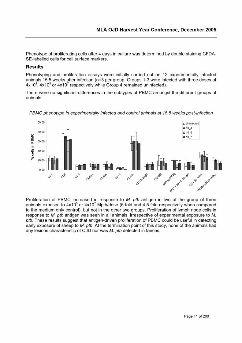
MLA OJD Harvest Year Conference, December 2005
Page 41 of 200
Phenotype of proliferating cells after 4 days in culture was determined by double staining CFDA-SE-labelled cells for cell surface markers.
ResultsPhenotyping and proliferation assays were initially carried out on 12 experimentally infectedanimals 15.5 weeks after infection (n=3 per group, Groups 1-3 were infected with three doses of4x104, 4x10⁵ or 4x10⁷ respectively while Group 4 remained uninfected).
There were no significant differences in the subtypes of PBMC amongst the different groups ofanimals.
PBMC phenotype in experimentally infected and control animals at 15.5 weeks post-infection
0.00
20.00
40.00
60.00
80.00
100.00
CD4CD5
CD6
CD8aa
CD8ab
CD14
CD11a
CD11abri
ght
CD45R
86D (g
dTCR)
WC1 (CD4-C
D8-/gd
)
WC4 (B ce
lls)
WC4brig
ht (B
cells
)
% c
ells
in P
BM
C
Uninfected10_410_510_7
Proliferation of PBMC increased in response to M. ptb antigen in two of the group of threeanimals exposed to 4x10⁵ or 4x10⁷ Mptb/dose (6 fold and 4.5 fold respectively when comparedto the medium only control), but not in the other two groups. Proliferation of lymph node cells inresponse to M. ptb antigen was seen in all animals, irrespective of experimental exposure to M.ptb. These results suggest that antigen-driven proliferation of PBMC could be useful in detectingearly exposure of sheep to M. ptb. At the termination point of this study, none of the animals hadany lesions characteristic of OJD nor was M. ptb detected in faeces.
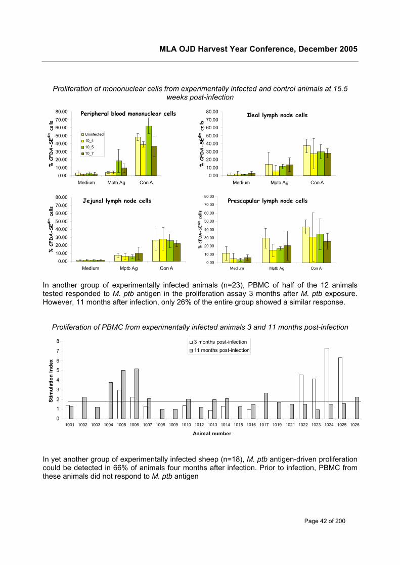
MLA OJD Harvest Year Conference, December 2005
Page 42 of 200
Proliferation of mononuclear cells from experimentally infected and control animals at 15.5weeks post-infection
Peripheral blood mononuclear cells
0.0010.00
20.0030.00
40.0050.00
60.0070.00
80.00
Medium Mptb Ag Con A
% C
FDA-S
Edim c
ells
Uninfected10_410_510_7
Ileal lymph node cells
0.0010.00
20.0030.00
40.0050.00
60.0070.00
80.00
Medium Mptb Ag Con A
% C
FDA-S
Edim c
ells
Jejunal lymph node cells
0.0010.00
20.0030.00
40.0050.00
60.0070.00
80.00
Medium Mptb Ag Con A
% C
FDA-S
Edim c
ells
Prescapular lymph node cells
0.00
10.00
20.00
30.00
40.00
50.00
60.00
70.00
80.00
Medium Mptb Ag Con A
% C
FDA-S
Edim c
ells
In another group of experimentally infected animals (n=23), PBMC of half of the 12 animalstested responded to M. ptb antigen in the proliferation assay 3 months after M. ptb exposure.However, 11 months after infection, only 26% of the entire group showed a similar response.
Proliferation of PBMC from experimentally infected animals 3 and 11 months post-infection
In yet another group of experimentally infected sheep (n=18), M. ptb antigen-driven proliferationcould be detected in 66% of animals four months after infection. Prior to infection, PBMC fromthese animals did not respond to M. ptb antigen
0
1
2
3
4
5
6
7
8
1001 1002 1003 1004 1005 1006 1007 1008 1009 1010 1012 1013 1014 1015 1016 1017 1019 1021 1022 1023 1024 1025 1026
Animal number
Stim
ulat
ion
Inde
x
3 months post-infection11 months post-infection
MLA OJD Harvest Year Conference, December 2005
Page 43 of 200
Proliferation of PBMC from experimentally infected animals 4 months post-infection
Disease status of both these groups of animals is being monitored.
Conclusions to dateThe M. ptb antigen-driven proliferation assay using PBMC detects exposure of most sheep toM. ptb as early as 3-4 months after experimental infection. However, whether these animals willlater succumb to infection is yet unknown. Therefore these experimentally infected animals arestill being monitored. Knowledge of their infection status will enable calculation of test sensitivityand specificity at each time point. The lymphocyte proliferation assay is currently a research toolbut acts as a defacto for other types of assay which might be applicable for practical diagnosticapplication. Results to date do not show an alteration in PBMC subsets as a result of exposureof sheep to M. ptb.
AcknowledgementsThis project is being funded by Meat & Livestock Australia. We are grateful to Nicole Carter,Reena Mehta and Anna Waldron for their excellent technical support.
0
1
2
3
4
5
6
7
1028 1029 1030 1031 1033 1034 1035 1038 1039 1040 1042 1043 1044 1045 1046 1047 1048 1049
Animal number
Stim
ulat
ion
Inde
x
MLA OJD Harvest Year Conference, December 2005
Page 44 of 200
Apoptotic responses during the pathogenesis of ovineJohne’s disease
Sally Brownea, Kumudika de Silva, Richard Whittington, David EmeryaFarm Animal Health GroupFaculty of Veterinary Science, University of Sydney425 Werombi Rd, Camden NSW 2570Phone: 02 9351 1610; Fax: 02 9351 1693E-mail: [email protected]
Background and objectivesThe immunological mechanisms underlying the progression of Johne’s disease from asubclinical to a clinical disease state remain unclear. More knowledge regarding the interactionsbetween host immune cells and Mycobacterium avium subsp. paratuberculosis (M. ptb),including apoptotic responses, may allow further advance in the area of early detection andcontrol of the disease and identification of resistant animals.
Apoptosis or programmed cell death is a tightly controlled form of cell death that typically leadsto DNA degradation and sequestering of cell contents into apoptotic bodies, without inducing aninflammatory response (Thornberry and Lazebnik 1998). Macrophages infected withmycobacteria will undergo apoptosis, in which TNF-α plays a key role, both in vitro and intissues (Gao and Abu Kwaik 2000).
Host macrophage apoptosis is an innate defense mechanism which is linked to the killing ofintracellular mycobacteria (Fratazzi, Arbeit et al. 1999). This process appears to require viablemycobacteria as apoptosis has been observed in bovine monocytes when infected with live butnot when infected with heat-killed M. ptb using Hoechst 33342 staining (Allen, Sotos et al.2001).
The ability of virulent mycobacteria to evade macrophage apoptosis has been documentedwidely: some mechanisms used to inhibit apoptosis include increasing sTNFR2 production,neutralising pro-apoptotic activity of TNF-alpha (Balcewicz-Sablinska, Keane et al. 1998) andinactivating pro- and anti-apoptotic proteins (Maiti, Bhattacharyya et al. 2001; Sly, Hingley-Wilson et al. 2003). Recently, specific genes associated with apoptosis and cell survival (anti-apoptotic) have been identified at significantly different levels in the blood of M. ptb infectedcattle compared to uninfected controls (Coussens, Pudrith et al. 2005).
Furthermore, lymphocytes can induce apoptosis of infected macrophages as part of the hostimmune response. Lymphocytes have also been shown to undergo apoptosis in response tomycobacterial infections both in vitro and in tissues (Roger and Bermudez 2001; Zhong,Gilbertson et al. 2003). Lymphocyte apoptosis seems to require direct contact withmycobacteria-infected macrophages (Roger and Bermudez 2001). Recently, antigen inducedapoptosis has been observed at significantly higher levels in the peripheral blood lymphocytesof M. ptb infected cattle than in control animals (Susanne Grell, personal communication).
The focus of this project is to investigate the apoptotic responses of lymphocytes andmacrophages throughout the course of Ovine Johnes Disease (OJD) with particular focus onevaluating T cell apoptosis as a diagnostic indicator of early subclinically infected animals. In thepresent study we report significant differences in antigen-induced apoptosis in vitro within agroup of clinically infected sheep when compared to uninfected control animals.
MLA OJD Harvest Year Conference, December 2005
Page 45 of 200
MethodMerino sheep aged 3 years were purchased from a NSW property with a high prevalence statusfor OJD. Infection was confirmed by AGID and faecal culture tests. Out of 100 sheep that weretested, 7 with moderate to advanced subclinical disease and 4 uninfected animals were chosenfor this study.
Mononuclear cells were isolated from peripheral blood (PBMCs) prior to sacrifice using densitygradient centrifugation. Samples were then taken from the ileal (ILN), jejunal (JLN) andprescapular (PLN) lymph nodes. Isolated cells were incubated with medium alone, Mptb antigen(10 µg/ml) or Con A (10 µg/ml) for up to 6 days.
Apoptosis was measured using a fluorescent analog of the pan caspase inhibitor Z-VAD-FMK(Promega), which irreversibly binds to activated caspases, a central component of mostapoptotic pathways. Fluorescence was detected using flow cytometry on days 0 and 6 ofculture.
ResultsThere were no significant differences in apoptosis in mononuclear cells, regardless of infectionstatus or sample site. PBMC apoptosis was < 10%. Apoptosis in ILN and JLN cells wasgenerally higher in all sheep ranging from 4-14% and 5-25% respectively with the exception ofone infected animal where apoptosis in the JLN was almost 60%. Generally apoptosis washigher in the JLN of infected animals than in uninfected animals (data not shown).
Antigen-induced apoptosis of PBMCs on day 6 was not significantly different from media alonein all animals except one infected sheep in which antigen induced apoptosis was significantlyless than media alone.
Antigen-induced apoptosis in ILN mononuclear cells was significantly increased (P <0.05) in 3of 4 infected sheep tested and significantly reduced in 1 of 4 of the tested animals compared tomedia alone. Importantly, M. ptb antigen did not significantly alter apoptosis in ILN mononuclearcells from all three uninfected sheep tested.
M. ptb antigen induced apoptosis in 4 of 7 infected animals to a significantly greater extent thanmedium control during culture of JLN mononuclear cells. A similar significant increase was seenin 1 of the 3 uninfected animals tested.
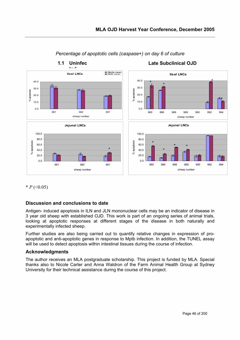
MLA OJD Harvest Year Conference, December 2005
Page 46 of 200
Percentage of apoptotic cells (caspase+) on day 6 of culture
* P (<0.05)
Discussion and conclusions to dateAntigen- induced apoptosis in ILN and JLN mononuclear cells may be an indicator of disease in3 year old sheep with established OJD. This work is part of an ongoing series of animal trials,looking at apoptotic responses at different stages of the disease in both naturally andexperimentally infected sheep.
Further studies are also being carried out to quantify relative changes in expression of pro-apoptotic and anti-apoptotic genes in response to Mptb infection. In addition, the TUNEL assaywill be used to detect apoptosis within intestinal tissues during the course of infection.
AcknowledgmentsThe author receives an MLA postgraduate scholarship. This project is funded by MLA. Specialthanks also to Nicole Carter and Anna Waldron of the Farm Animal Health Group at SydneyUniversity for their technical assistance during the course of this project.
Jejunal LNCs
0.0
20.0
40.0
60.0
80.0
100.0
985 986 988 989 990 992 994
sheep number
% a
popt
osis
Ileal LNCs
0.0
10.0
20.0
30.0
40.0
985 986 988 989 990 992 994
sheep number
% a
popt
osis
Ileal LNCs
0.0
10.0
20.0
30.0
40.0
981 982 991
sheep number
% a
popt
osis
Media meanMptb mean
Jejunal LNCs
0.0
20.0
40.0
60.0
80.0
100.0
981 982 991
sheep number
% a
popt
osis
* * *
*
**
* **
Late Subclinical OJD1.1 Uninfected
MLA OJD Harvest Year Conference, December 2005
Page 47 of 200
ReferencesAllen, S., J. Sotos, et al. (2001). "Use of Hoechst 33342 staining to detect apoptotic changes in
bovine mononuclear phagocytes infected with Mycobacterium avium subsp.paratuberculosis." Clin Diagn Lab Immunol 8(2): 460-4.
Coussens, P. M., C. B. Pudrith, et al. (2005). "Johne's disease in cattle is associated withenhanced expression of genes encoding IL-5, GATA-3, tissue inhibitors of matrixmetalloproteinases 1 and 2, and factors promoting apoptosis in peripheral bloodmononuclear cells." Vet Immunol Immunopathol 105(3-4): 221-34.
Fratazzi, C., R. D. Arbeit, et al. (1999). "Macrophage apoptosis in mycobacterial infections." JLeukoc Biol 66(5): 763-4.
Gao, L. and Y. Abu Kwaik (2000). "Hijacking of apoptotic pathwaysby bacterial pathogens."Microbes Infect 2(14): 1705-19.
Maiti, D., A. Bhattacharyya, et al. (2001). "Lipoarabinomannan from Mycobacterium tuberculosispromotes macrophage survival by phosphorylating Bad through a phosphatidylinositol 3-kinase/Akt pathway." J Biol Chem 276(1): 329-33.
Roger, P. M. and L. E. Bermudez (2001). "Infection of mice with Mycobacterium avium primesCD8+ lymphocytes for apoptosis upon exposure to macrophages." Clin Immunol 99(3):378-86.
Sly, L. M., S. M. Hingley-Wilson, et al. (2003). "Survival of Mycobacterium tuberculosis in hostmacrophages involves resistance to apoptosis dependent upon induction ofantiapoptotic Bcl-2 family member Mcl-1." J Immunol 170(1): 430-7.
Thornberry, N. A. and Y. Lazebnik (1998). "Caspases: enemies within." Science 281(5381):1312-6.
Zhong, J., B. Gilbertson, et al. (2003). "Apoptosis of CD4+ and CD8+ T cells duringexperimental infection with Mycobacterium avium is controlled by Fas/FasL and Bcl-2-sensitive pathways, respectively." Immunol Cell Biol 81(6): 480-6.
MLA OJD Harvest Year Conference, December 2005
Page 48 of 200
ELISPOT – new methods for detecting IFN-γ
Douglas Begga, Katrina Bosward, Kumudika De Silva, Lyrissa Di Fiore, DeborahTaylor and Richard Whittington.aFaculty of Veterinary ScienceUniversity of Sydney,425 Werombi Rd, Camden, NSW, 2570Phone: 02 90367737; Fax: 02 93511693Email: [email protected]
AbstractIFN-γ is a cytokine produced as part of the early cell mediated immune response to M.paratuberculosis. Detection of IFN-γ production is being used as a diagnostic test to detectmycobacterial infected ruminants. However in sheep the IFN-γ assay has not proved assuccessful as initially expected, especially for the detection of subclinically M. ptb infectedanimals. Therefore new strategies are being investigated to improve the sensitivity of IFN-γdetection methods. These include the use of ELISPOT and Cell ELISA where the IFN-γ isdetected directly from the culture of the animal’s leucocytes.
Background and objectivesEarly immune responses against mycobacterial infections such as M. paratuberculosis (M. ptb)are thought to be cell mediated, resulting in production of immune cytokines such as IFN-gamma (IFN-γ). Thus, detection of IFN-γ was initially thought to improve immunodiagnostictesting for mycobacterial infections in ruminants, providing early detection of infected animals.Unfortunately in the diagnosis of Johne’s disease, the use of commercially available IFN-γdetection methods has not resulted in the anticipated levels of detection. The IFN-γ assay fordetection of M. ptb infection in ruminants is based on IFN-γ production in blood samplesstimulated with mycobacterial antigens. The plasma from the stimulated blood sample is thenremoved from the culture and tested for IFN-γ using a sandwich ELISA (Enzyme Linked ImmunoSorbent Assay). The sensitivity of the IFN-γ assay can be directly correlated to the stage ofdisease. One study found that 70-90% of subclinically infected and 100% of clinically infectedcattle could be detected with the IFN-γ assay (Billman-Jacobe et al., 1992). Another study, alsoin cattle, reported a sensitivity of 50-75% with a specificity of 98% for the IFN-γ assay (Stabeland Whitlock, 2001). In sheep, similar sensitivity values of 50-75% were found using theBovigam (Pfizer) assay (Stewart et al., 2002). The ELISPOT assay differs from the ELISA inthat the cells are cultured with the antigen in the well used to detect IFN-γ and this can allowenumeration of the IFN-γ producing cells, with each individual IFN-γ producing cell shown as aspot on the bottom of the membrane. The ELISPOT (Enzyme Linked Immuno Spot) assay hasbeen shown to be 10-200 times more sensitive in the detection of cytokines than the traditionalELISA (Tanguay & Killion, 1994). The objectives of this study are to examine alternativemethods of IFN-γ detection other than the typical ELISA currently available. These methodshave included ELISPOT and its cousin, the Cell-ELISA assay, which yields a colour changerather than spots.
Methods and resultsThe discovery of several cross reactive monoclonal antibodies that detect ovine IFN-γ led to thedevelopment of a functional ELISPOT and Cell-ELISA that was then optimised in terms ofantibody dilutions, incubation times and changes in cell numbers using blood from vaccinated
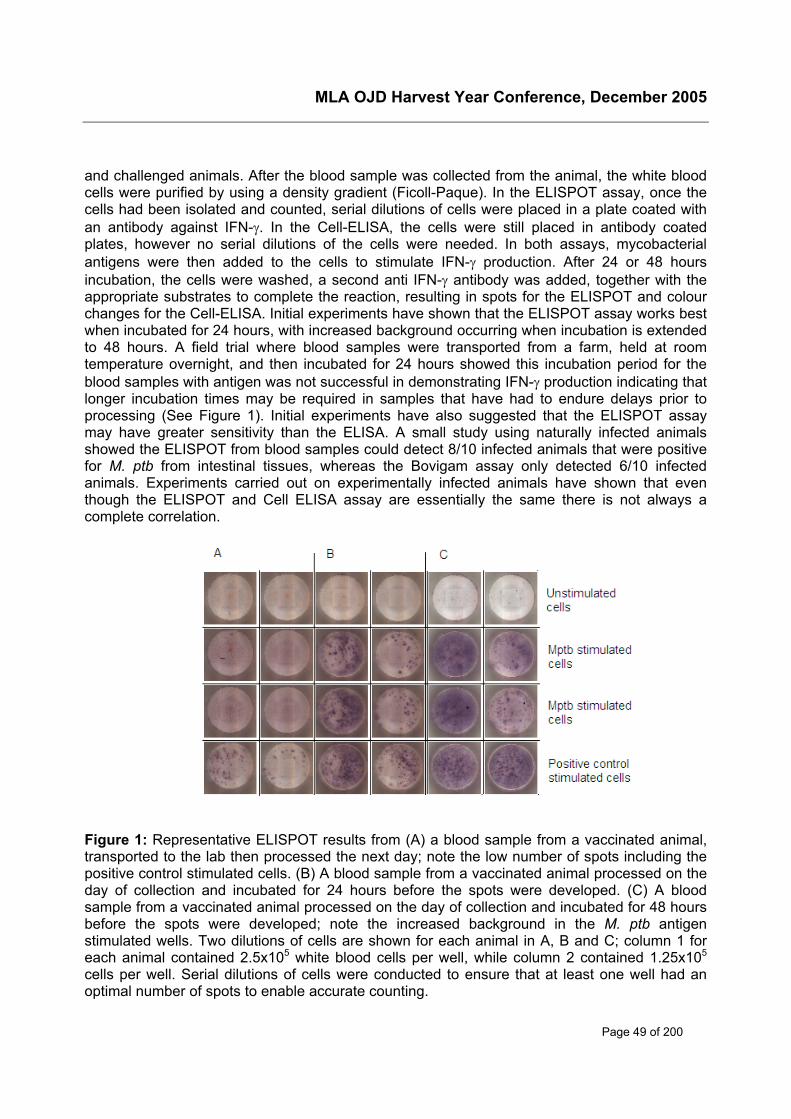
MLA OJD Harvest Year Conference, December 2005
Page 49 of 200
and challenged animals. After the blood sample was collected from the animal, the white bloodcells were purified by using a density gradient (Ficoll-Paque). In the ELISPOT assay, once thecells had been isolated and counted, serial dilutions of cells were placed in a plate coated withan antibody against IFN-γ. In the Cell-ELISA, the cells were still placed in antibody coatedplates, however no serial dilutions of the cells were needed. In both assays, mycobacterialantigens were then added to the cells to stimulate IFN-γ production. After 24 or 48 hoursincubation, the cells were washed, a second anti IFN-γ antibody was added, together with theappropriate substrates to complete the reaction, resulting in spots for the ELISPOT and colourchanges for the Cell-ELISA. Initial experiments have shown that the ELISPOT assay works bestwhen incubated for 24 hours, with increased background occurring when incubation is extendedto 48 hours. A field trial where blood samples were transported from a farm, held at roomtemperature overnight, and then incubated for 24 hours showed this incubation period for theblood samples with antigen was not successful in demonstrating IFN-γ production indicating thatlonger incubation times may be required in samples that have had to endure delays prior toprocessing (See Figure 1). Initial experiments have also suggested that the ELISPOT assaymay have greater sensitivity than the ELISA. A small study using naturally infected animalsshowed the ELISPOT from blood samples could detect 8/10 infected animals that were positivefor M. ptb from intestinal tissues, whereas the Bovigam assay only detected 6/10 infectedanimals. Experiments carried out on experimentally infected animals have shown that eventhough the ELISPOT and Cell ELISA assay are essentially the same there is not always acomplete correlation.
Figure 1: Representative ELISPOT results from (A) a blood sample from a vaccinated animal,transported to the lab then processed the next day; note the low number of spots including thepositive control stimulated cells. (B) A blood sample from a vaccinated animal processed on theday of collection and incubated for 24 hours before the spots were developed. (C) A bloodsample from a vaccinated animal processed on the day of collection and incubated for 48 hoursbefore the spots were developed; note the increased background in the M. ptb antigenstimulated wells. Two dilutions of cells are shown for each animal in A, B and C; column 1 foreach animal contained 2.5x10⁵ white blood cells per well, while column 2 contained 1.25x10⁵
cells per well. Serial dilutions of cells were conducted to ensure that at least one well had anoptimal number of spots to enable accurate counting.
MLA OJD Harvest Year Conference, December 2005
Page 50 of 200
Conclusions to dateWhile the ELISPOT appeared to be more sensitive in initial small group studies, cut off pointsstill need to be defined to distinguish between infected and non-infected animals and this can beexplored more after testing additional groups of known infected animals. In addition, work isneeded to improve the reliability of the assay and its overall robustness. At the present time, theBovigam assay is a very simple assay to complete due to the minimal blood sample processingrequirements. Simplification of the ELISPOT and Cell-ELISA assay technique, for example bysimplifying the way the white blood cells are purified from a blood sample, will allow these teststo become more cost effective and enable high throughput of blood samples. Typically, cellsused in the ELISPOT assay are subjected to complex Ficoll separation techniques to enhancepurity, however, initial ELISPOT studies have shown that white blood cells with reduced puritywill still show IFN-γ production. Further studies are needed to determine how reduced purity willaffect antigen specific production of IFN-γ. The ELISPOT and Cell-ELISA are technically moredifficult to perform than the traditional ELISA method but there is room to explore methods toenable automation of the processes involved. Unfortunately, unlike the Bovigam assay, repeattesting requires additional blood samples.
The use of the ELISPOT and Cell-ELISA tests in groups of naturally infected animals will assistin determining sensitivity and specificity and to define the cut-off points that will allowidentification of infected and non infected animals. Testing of greater numbers of naturallyinfected animals will provide more information on the effectiveness of these assays as adiagnostic test.
As has been noted in the Bovigam assay, it appears that transportation and storage of bloodsamples will be critical in how the assays work as diagnostic tests. There is a need to over-come problems caused by transport and longer term sample storage. This may involve the useof additives such as the cytokine, IL-12, (Jungersen et al., 2005) which has been shown toextend the storage time of cattle blood to 24 hours while still allowing IFN-γ detection.
AcknowledgementsThis project is funded by MLA. The authors would also like to acknowledge the help of AnnaWaldron, Craig Kirsto, Nicole Carter and Natelie Schiller with this work.
ReferencesBillman-Jacobe, H., M. Carrigan, F. Cockram, L. A. Corner, I. J. Gill, J. F. Hill, T. Jessep, A. R.Milner and P. R. Wood (1992). A comparison of the interferon gamma assay with the absorbedELISA for the diagnosis of Johne's disease in cattle. Aust Vet J 69(2): 25-8.
Jungersen, G., S. N. Grell, A. Clemensen and C. J. Howard (2005). Interleukin-12 potentiationof the interferon-gamma test rescues day old blood samples for diagnosis of paratuberculosisPPD specific cellular mediated immune responses. 8th International Colloquium onParatuberculosis, Copenhagen, Denmark.
Stabel, J. R. and R. H. Whitlock (2001). An evaluation of a modified interferon-gamma assay forthe detection of paratuberculosis in dairy herds. Vet Immunol Immunopathol 79(1-2): 69-81.
Stewart, D. J., P. L. Stiles, R. J. Whittington, C. Lambeth, P. A. Windsor, L. Reddacliff, H.McGregor, O. P. Dhungyel, D. Cousins, B. R. Francis, P. W. Morcombe, R. Butler, D. D.Salmon, C. F. Roberts, E. Sergeant, I. Jasenko and S. L. Jones (2002). Validation of theinterferon-g for diagnosis of ovine Johne's disease: sensitivity and specificity field trials. SeventhInternational Colloquium on Paratuberculosis, Bilbao, Spain.
MLA OJD Harvest Year Conference, December 2005
Page 51 of 200
Detection of Mycobacteria from bloodKate Goldsmitha, Doug Begg, Lyrissa Di Fiore and Richard WhittingtonaUniversity of Sydney425 Werombi Rd, Camden, 2570Phone: 02 9351 1610Email: [email protected]
AbstractJohne’s disease, or paratuberculosis, is a chronic wasting disease causing intestinal thickeningdue to granulomatous inflammation in the ileum and jejunum of ruminants. Transmission ofparatuberculosis is primarily by oral exposure to Mycobacterium avium subspeciesParatuberculosis (M. ptb) in young animals, with clinical signs of disease developing manyyears later. Between exposure and clinical disease there is a long subclinical phase in whichshedding of the organism in faeces occurs intermittently.
Demonstration of the organism in blood from infected sheep and cattle by PCR has sparkedrecent interest in developing a diagnostic test based on culture or molecular detection of theorganism from blood. Detection of M. ptb DNA in blood, culture of the organism from milk andextra intestinal sites, and demonstration of transmammary and intrauterine transmission,indicate that at some stage in the disease, bacteraemia occurs.
BackgroundIn paratuberculosis infected animals there is considerable evidence that the bacteria are presentin the blood at some stage in the disease process. M. ptb was isolated from peripheral bloodmononuclear cells by bacteriologic culture on Herrold’s egg yolk medium from 1 of 7 clinicallydiseased cattle [1]. Direct PCR of DNA extracted from the blood of clinically affected sheepusing IS900 primers in one study returned positive results in 9 out of 12 histologically positivesheep [2]. A follow-up study to examine the sensitivity of detection of M. ptb by PCR from bloodin experimentally-inoculated, subclinically-infected sheep returned unfavourable results withonly 2 positive blood samples after repeated serial testing of 7 sheep with subclinical infectionthat were confirmed by histology [3]. A study using nested PCR on milk and blood of cattledemonstrated positive results from both with 72% of clinically infected animals testing positive.PCR on blood was less sensitive than PCR on milk in subclinically infected cows withsensitivities of 33% and 45%, respectively [4]. Another study using PCR on blood, faeces, milkand liver in advanced subclinically infected cows demonstrated a sensitivity of 40% for PCR onblood samples [5]. Compared with ELISA testing, PCR on blood appeared to detect differentforms or stages of the disease [6].
In addition to demonstrating M. ptb DNA in blood of Johne’s infected animals, systemic spreadhas been observed in tissues such as the liver [2, 3, 5], mammary tissue [7], milk [4, 5, 7],spleen [8], foetal tissues [1, 7] reproductive tissues [8-10], and extra intestinal lymph nodes [1,8].
Culture of pathogenic mycobacteria from blood is of increasing importance to human medicinedue to the tendency for these pathogens to cause disease in HIV positive patients. While thegrowth requirements of M. tuberculosis and M. avium complex differ from M. ptb, sampleprocessing and culture techniques for these pathogens may be adaptable for culture of M. ptb.Concentrate from Isolator tubes (Wampole Laboratories, Cranbury, N.J., previously
MLA OJD Harvest Year Conference, December 2005
Page 52 of 200
manufactured by Du Pont Co., Wilmington, Del.), which contains saponin, a cell lysis agent, andSPS which neutralizes the bactericidal properties of blood, have been successfully inoculatedonto a variety of solid and liquid media to isolate mycobacteria [11, 12].
Concentration of mycobacteria from clinical samples has been shown to be improved by usingcarboxypropylbetaine (CB-18) to counter the buoyancy and clumping associated withmycobacteria. CB -18 has been used to concentrate M. ptb from cattle faeces and tissuesamples[13, 14], M. avium from tissue, bone marrow, blood and faeces[15], and M. tuberculosisfrom respiratory samples[16]. This method may be useful in concentrating organisms in bloodsamples from infected animals.
The Bactec TB 460 system, which measures radioactive CO2 generation to detect growth, hassuccessfully been used to culture ovine strains of M. ptb from faeces, tissues and milk usingBactec 12B media [7, 17]. The limitation of using Bactec 12B media for culture from blood is thesmall volume of sample used for inoculation; 0.2mls was used in one study [11]. Bactec 12Bmedia has been successfully used for culture of isolator concentrate [11], however some studieshave shown inhibition of growth due to the polypropylene glycol which is eliminated with a washstep prior to inoculation [18, 19].
The Bactec 13A media, which also uses the Bactec TB 460 radiometric detection system, isformulated specifically for the culture of mycobacteria directly from blood allowing largervolumes to be inoculated. This method when used to detect mycobacteraemia in HIV patients,returned a comparable number of positives to Isolator tubes [12]. Supplements required forgrowth of M. ptb, such as mycobactin J and egg yolk for sheep strains must be added.
ObjectivesThe objective of this project is to optimise detection methods for culture and molecular detectionfrom blood. To do this different processing methods using spiked blood and samples fromclinically and subclinically infected animals will be compared. This will be used to determinefeasibility of detection of the organism from blood as a diagnostic test. Currently methods underevaluation are lysis centrifugation and buffy coat preparations inoculated into the Bactec 12Bmedium, modified as previously described [17], whole blood culture using a modified Bactec13A medium, and conventional and real time IS900 PCR on buffy coat preparations and lysispellets.
Furthermore, this project aims to develop a better understanding of the behaviour of theorganism during the bacteraemic phase. Tracking M. ptb and infected monocytes followingexperimentally induced bacteraemia in unchallenged animals may allow us to better describethe spread of the organism following bacteraemia. Correlation of lesion types and stage ofdisease in experimentally induced and naturally infected animals may allow us to determine thestages of disease where bacteraemia occurs.
AcknowledgmentsThis project was funded by Meat and Livestock Australia who also provided a scholarship forthe senior author. Thanks are also due to Anna Waldron, Craig Kristo and Angela Reeves fortheir technical assistance.
MLA OJD Harvest Year Conference, December 2005
Page 53 of 200
References1. Koenig, G.J., et al., Isolation of Mycobacterium paratuberculosis from mononuclear cells in
tissues, blood, and mammary glands of cows with advanced paratuberculosis. Am J VetRes, 1993. 54(9): p. 1441-5.
2. Gwozdz, J.M., et al., Detection of Mycobacterium avium subsp. paratuberculosis in ovinetissues and blood by the polymerase chain reaction. Vet Microbiol, 1997. 57(2-3): p. 233-44.
3. Gwozdz, J.M., et al., Use of the polymerase chain reaction assay for the detection ofMycobacterium avium subspecies paratuberculosis in blood and liver biopsies fromexperimentally infected sheep. Aust Vet J, 2000. 78(9): p. 622-4.
4. Buergelt, C.D. and J.E. Williams, Nested PCR on blood and milk for the detection ofMycobacterium avium subsp paratuberculosis DNA in clinical and subclinical bovineparatuberculosis. Aust Vet J, 2004. 82(8): p. 497-503.
5. Barrington, G.M., et al., Temporal patterns of diagnostic results in serial samples from cattlewith advanced paratuberculosis infections. J Vet Diagn Invest, 2003. 15(2): p. 195-200.
6. Juste, R., et al. Comparison of blood PCR and ELISA for detection of Mycobacterium aviumsubsp. paratuberculosis infection in sheep and cattle. in Seventh International Colloquiumon Paratuberculosis. 2002. Bilbao, Spain.
7. Lambeth, C., et al., Intrauterine and transmammary transmission of Mycobacterium aviumsubsp paratuberculosis in sheep. Aust Vet J, 2004. 82(8): p. 504-8.
8. Ayele, W.Y., et al., Distribution of Mycobacterium avium subsp. paratuberculosis in organsof naturally infected bull-calves and breeding bulls. Vet Microbiol, 2004. 103(3-4): p. 209-17.
9. Eppleston, J. and R.J. Whittington, Isolation of Mycobacterium avium subspparatuberculosis from the semen of rams with clinical Johne's disease. Aust Vet J, 2001.79(11): p. 776-7.
10. Buergelt, C., G.A. Donovan, and J.E. Williams, Identification of Mycobacterium aviumsubspecies paratuberculosis by Polymerase Chain Reaction in Blood and Semen of a Bullwith Clinical Paratuberculosis. Intern J Appl Res Vet Med, 2004. 2(2): p. 130-134.
11. Gill, V.J., et al., Use of lysis-centrifugation (isolator) and radiometric (BACTEC) blood culturesystems for the detection of mycobacteremia. J Clin Microbiol, 1985. 22(4): p. 543-6.
12. Witebsky, F.G., et al., Comparison of BACTEC 13A medium and Du Pont isolator fordetection of mycobacteremia. J Clin Microbiol, 1988. 26(8): p. 1501-5.
13. Thornton, C.G., et al., Application of the C(18)-carboxypropylbetaine specimen processingmethod to recovery of Mycobacterium avium subsp. paratuberculosis from ruminant tissuespecimens. J Clin Microbiol, 2002. 40(5): p. 1783-90.
14. Ozbek, A., et al., Evaluation of two recovery methods for detection of Mycobacterium aviumsubsp. paratuberculosis by PCR: direct-dilution--centrifugation and C(18)-carboxypropylbetaine processing. FEMS Microbiol Lett, 2003. 229(2): p. 145-51.
15. Thornton, C.G., et al., Processing postmortem specimens with C18-carboxypropylbetaineand analysis by PCR to develop an antemortem test for Mycobacterium avium infections inducks. J Zoo Wildl Med, 1999. 30(1): p. 11-24.

MLA OJD Harvest Year Conference, December 2005
Page 54 of 200
16. Thornton, C.G., et al., Novel method for processing respiratory specimens for detection ofmycobacteria by using C18-carboxypropylbetaine: blinded study. J Clin Microbiol, 1998.36(7): p. 1996-2003.
17. Whittington, R.J., et al., Evaluation of Modified BACTEC 12 B Radiometric Medium andSolid Media for Culture of Mycobacterium avium subsp. paratuberculosis from Sheep. J ClinMicrobiol, 1999. 37(4): p. 1077-1083.
18. Wasilauskas, B.L. and R.M. Morrell, Inhibitory Effect of the Isolator Blood Culture System onGrowth of Mycobacterium avium - M. intracellulare in BACTEC 12B Bottles. J Clin Microbiol,1994. 32(3): p. 654-657.
19. Wasilauskas, B.L. and R.M. Morrell, Isolator Component Resopnsible for Inhibition ofMycobacterium avium - M. intracellulare in BACTEC 12 B Medium. J Clin Microbiol, 1997.35(3): p. 588-590.
MLA OJD Harvest Year Conference, December 2005
Page 55 of 200
Proteomic analysis of sheep serum by SELDI TOF-MS:Identification of putative biomarkers of ovine Johne’s
disease
Ling Zhonga, Deborah L. Taylor and Richard J. WhittingtonaFaculty of Veterinary Science, University of Sydney425 Werombi Road, Camden, NSW 2570, Australia.Phone: 02 9351 1610; Fax: 02 9351 1693Email: [email protected]
IntroductionM. paratuberculosis (M. ptb) is the causative agent of paratuberculosis or Ovine Johne’sDisease (OJD), a slow-developing, infectious disease characterised by chronic granulomatousenteritis, thickening of the intestinal wall and progressive weight loss resulting ultimately in thedeath of the animal. The current diagnostic tests for Johne’s disease require detection of themicroorganism by culture or PCR, or detection of specific host reactions to infection. Most ofthese tests are better able to detect animals in later stages of infection rather than in earlierstages and with moderate sensitivity and specificity. Therefore, there is a current need for newtests to be developed due to the deficiencies in their sensitivity and specificity.
The development of new diagnostic tests has been hindered largely by our limited knowledge ofthe host factors involved in the immune response to the organism, and the lack of appropriatemolecular tools to dissect the host-pathogen interaction. Recent developments in tools formolecular biology has meant that global gene expression of both the host and pathogen inresponse to infection can now be analysed for many species. However, the availability of thesegenomic resources for ruminants is still a limiting factor for advancement of research in thesespecies. A number of proteomic approaches are now available that require no pre-existingknowledge of host factors.
Surface enhanced laser desorption ionization time-of-flight mass spectrometry (SELDI TOF –MS) combines classical retention chromatography with TOF-MS detection, and recent studieshave shown that proteomic profiling of serum using this technique can be used as a diagnostictool and a platform for biomarker discovery of various pathologies (Petricoin et al., 2002,Petricoin et al., 2004, Conrads et al., 2004, Li et al., 2004, Wadsworth et al., 2004, Wulfkuhle etal., 2001, Koopmann et al., 2004).
Hypothetically, various physiological and pathological processes could modify the proteome,generating a unique, disease-specific signature, known as proteomic signature, or diseasesignature (Rocken et al., 2004). Proteomic profiling of any protein resource, such as serum,could facilitate the discovery of different patterns of protein expression between uninfected orinfected animals. This could lead to a new diagnostic test capable for detecting infected animalsat an earlier stage of disease.
MethodsSELDI TOF-MS based proteomic profiling of serum can generally be divided into four stages:optimisation of SELDI trace acquisition, pilot scale biomarker discovery, putative biomarkersverification and key protein identification and characterisation.
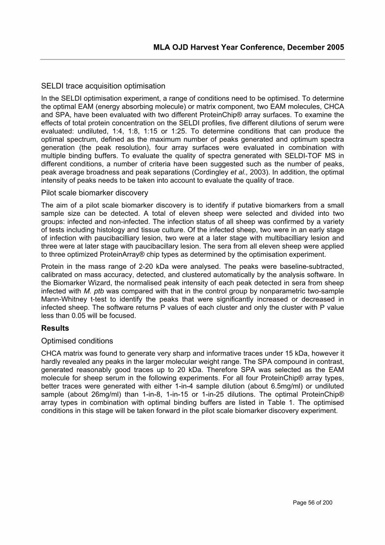
MLA OJD Harvest Year Conference, December 2005
Page 56 of 200
SELDI trace acquisition optimisationIn the SELDI optimisation experiment, a range of conditions need to be optimised. To determinethe optimal EAM (energy absorbing molecule) or matrix component, two EAM molecules, CHCAand SPA, have been evaluated with two different ProteinChip® array surfaces. To examine theeffects of total protein concentration on the SELDI profiles, five different dilutions of serum wereevaluated: undiluted, 1:4, 1:8, 1:15 or 1:25. To determine conditions that can produce theoptimal spectrum, defined as the maximum number of peaks generated and optimum spectrageneration (the peak resolution), four array surfaces were evaluated in combination withmultiple binding buffers. To evaluate the quality of spectra generated with SELDI-TOF MS indifferent conditions, a number of criteria have been suggested such as the number of peaks,peak average broadness and peak separations (Cordingley et al., 2003). In addition, the optimalintensity of peaks needs to be taken into account to evaluate the quality of trace.
Pilot scale biomarker discoveryThe aim of a pilot scale biomarker discovery is to identify if putative biomarkers from a smallsample size can be detected. A total of eleven sheep were selected and divided into twogroups: infected and non-infected. The infection status of all sheep was confirmed by a varietyof tests including histology and tissue culture. Of the infected sheep, two were in an early stageof infection with paucibacilliary lesion, two were at a later stage with multibacilliary lesion andthree were at later stage with paucibacillary lesion. The sera from all eleven sheep were appliedto three optimized ProteinArray® chip types as determined by the optimisation experiment.
Protein in the mass range of 2-20 kDa were analysed. The peaks were baseline-subtracted,calibrated on mass accuracy, detected, and clustered automatically by the analysis software. Inthe Biomarker Wizard, the normalised peak intensity of each peak detected in sera from sheepinfected with M. ptb was compared with that in the control group by nonparametric two-sampleMann-Whitney t-test to identify the peaks that were significantly increased or decreased ininfected sheep. The software returns P values of each cluster and only the cluster with P valueless than 0.05 will be focused.
ResultsOptimised conditionsCHCA matrix was found to generate very sharp and informative traces under 15 kDa, however ithardly revealed any peaks in the larger molecular weight range. The SPA compound in contrast,generated reasonably good traces up to 20 kDa. Therefore SPA was selected as the EAMmolecule for sheep serum in the following experiments. For all four ProteinChip® array types,better traces were generated with either 1-in-4 sample dilution (about 6.5mg/ml) or undilutedsample (about 26mg/ml) than 1-in-8, 1-in-15 or 1-in-25 dilutions. The optimal ProteinChip®array types in combination with optimal binding buffers are listed in Table 1. The optimisedconditions in this stage will be taken forward in the pilot scale biomarker discovery experiment.

MLA OJD Harvest Year Conference, December 2005
Page 57 of 200
Table1. Optimal ProteinArray® chip types in combination with optimal binding buffers.
ProteinChip®Array
General description(Ciphergen)
Washing / Binding buffer
Q10 Strong anion exchange Tris. HCL pH7.0,
CM10 Weak cation exchange Tris.HCL pH7.0
Low stringency buffer (Ciphergen)
High stringency buffer (50mM HEPES, pH7.0)
IMAC 30
Cu / Ni
Immobilized metal affinitycapture
PBS (Ciphergen)
Putative biomarker identificationAlthough all three different ProteinChip® array types were tested, the IMAC 30 ProteinChip®array surface loaded with copper sulphate generated the maximum number of significantlydifferent clusters (Figure 1). Five potential serum biomarker clusters (arrows in Figure 1 panelA) with differential expression in M.ptb infected sheep vs. the uninfected sheep were observed,and they were down-regulated in infected sheep in comparison with the control group.
MLA OJD Harvest Year Conference, December 2005
Page 58 of 200
A.
Fig 1. Example of significantly different clustersfrom two groups.
A. Clusters generated by Biomarker Wizard by usingIMAC 30 ProteinChip® array surface loaded withcopper metal. The square represents the uninfectedgroup and the circle represents the infected group.Only the clusters with P values less than 0.05 areshown. The five clusters with arrow were differentiallyexpressed in infected group vs. control sheep.
B. SELDI traces from the two groups correspond to adifferent cluster in Biomarker Wizard view in panel A.Top 4 traces belong to the un-infected group and thebottom seven traces are from the infected group. Thex-axis represents the mass/charge ration and y-axisrepresents the relative intensity.
B.
Conclusions to dateSurface enhanced laser desorption ionisation (SELDI) time of flight mass spectrometry hasfacilitated the discovery of a disease specific protein profile from many different biologicalsamples, including serum and tissues. Our pilot scale biomarker discovery results have raisedthe possibility that protein profiles may become a powerful diagnostic tool for OJD and need tobe further verified with an increased sample size and continued efforts on methodologicalstandardisation.
AcknowledgementThe author receives an MLA post graduate scholarship. This project is funded by MLA.
MLA OJD Harvest Year Conference, December 2005
Page 59 of 200
ReferencesConrads, T. P., Fusaro, V. A., Ross, S., Johann, D., Rajapakse, V., Hitt, B. A., Steinberg, S. M.,
Kohn, E. C., Fishman, D. A., Whitely, G., Barrett, J. C., Liotta, L. A., Petricoin, E. F., 3rdand Veenstra, T. D. (2004) Endocr Relat Cancer, 11, 163-78.
Cordingley, H. C., Roberts, S. L., Tooke, P., Armitage, J. R., Lane, P. W., Wu, W. andWildsmith, S. E. (2003) Biotechniques, 34, 364-5, 368-73.
Koopmann, J., Zhang, Z., White, N., Rosenzweig, J., Fedarko, N., Jagannath, S., Canto, M. I.,Yeo, C. J., Chan, D. W. and Goggins, M. (2004) Clin Cancer Res, 10, 860-8.
Li, J., White, N., Zhang, Z., Rosenzweig, J., Mangold, L. A., Partin, A. W. and Chan, D. W.(2004) J Urol, 171, 1782-7.
Petricoin, E. F., Ardekani, A. M., Hitt, B. A., Levine, P. J., Fusaro, V. A., Steinberg, S. M., Mills,G. B., Simone, C., Fishman, D. A., Kohn, E. C. and Liotta, L. A. (2002) Lancet, 359, 572-7.
Petricoin, E. F., Ornstein, D. K. and Liotta, L. A. (2004) Urol Oncol, 22, 322-8.
Rocken, C., Ebert, M. P. and Roessner, A. (2004) Pathol Res Pract, 200, 69-82.
Wadsworth, J. T., Somers, K. D., Cazares, L. H., Malik, G., Adam, B. L., Stack, B. C., Jr.,Wright, G. L., Jr. and Semmes, O. J. (2004) Clin Cancer Res, 10, 1625-32.
Wulfkuhle, J. D., McLean, K. C., Paweletz, C. P., Sgroi, D. C., Trock, B. J., Steeg, P. S. andPetricoin, E. F., 3rd (2001) Proteomics, 1, 1205-15.
MLA OJD Harvest Year Conference, December 2005
Page 60 of 200
Gene expression signals from the hostLyrissa Di Fiorea, Deborah Taylor, Kumudika de Silva, Kate Bosward, HelenMcGregor and Richard WhittingtonaUniversity of Sydney425 Werombi Road, Camden NSW 2570Phone: 02 9036 7737; Fax: 02 9351 1693Email: [email protected]
Background and objectivesMycobacterium paratuberculosis (M. ptb) causes a chronic progressive wasting disease insheep known as ovine Johne’s disease (OJD). A large number of organisms are typically foundwithin the gut in chronically infected animals. Lesions develop in the gut wall and these containepitheloid macrophages and giant cells and there is a general thickening of the mucosa. Thechanges occurring following infection at the molecular level have not been well characterised insheep. Most of the data on the molecular pathogenesis of the disease has come from studies inother species such as cattle in the advanced stages of the disease, or from macrophageinfection models. Molecular techniques used in these studies include microarray analysis,selective capture of transcribed sequences (SCOTS) and RNA arbitrarily primed-PCR (RAP-PCR). Few studies have looked at OJD using DD-PCR, and those have primarily focused onbacterial gene expression.
There are needs for effective ways of identifying all genes that are differentially expressed indifferent cell types or under altered or diseased conditions. Tools that allow us to determinewhen and where genes are turned on or off as these processes occur have becomeincreasingly important. Differential display PCR (DD-PCR), representational difference analysis(RDA), serial analysis of gene expression (SAGE) and cDNA microarray analysis are wellestablished tools for studying differentially expressed genes under normal and altered/diseasedconditions. These approaches all stem from the same origin; differential screening anddifferential hybridisation.
The objective of this study is to examine host gene expression during the initial stages of M. ptbinfection and during the clinical stage of infection to identify known or novel genes regulating theresponse to infection, and to define genes that can be used to identify M. ptb infected animalsearlier than currently possible.
Methods and resultsLong distance differential display PCR (DD-PCR) is a technique used to identify differences ingene expression patterns between two or more biological samples. It was employed to identifydifferentially expressed genes in lymph node and ileal tissues collected from uninfected, earlystage infected or clinically infected Merino sheep.
Animals - Female Merino sheep of three age groups (lambs, hoggets and adults) were co-grazed with infected sheep for 14.5 weeks after which they were removed. Animals were killed2.5 years after initial exposure and samples of terminal ileum and mesenteric lymph node werecollected. Histological data was obtained and animals were classified according to the Perezscale.
Long Distance DD-PCR – The Delta Differential Display kit (Clontech) was used. RNA wasextracted from tissues using the Qiagen RNeasy mini kit. DNAase treated RNA was reversetranscribed with oligo (dT) primers and AMV-RT to produce single stranded cDNA. PCR was
MLA OJD Harvest Year Conference, December 2005
Page 61 of 200
performed using anchored 3’ primers (T1-T9), arbitrary 5’ primers (P1 – P10) and α 35S-dATPto label cDNA products. PCR products were run on an 8% acrylamide gel, which was then driedand exposed to X-RAY film. Differentially expressed bands were excised and DNA extractedfrom the gel slice. Bands of interest were re-amplified using the primers in the original PCR.Amplified products were analysed on an agarose gel and cloned into a TOPO cloning vector.
Eleven bands appear to be differentially expressed between uninfected and clinically infectedanimals. Re-amplification of these bands produced products with sizes ranging from 400-1500bp. Two genes were found to be differentially expressed in lymph nodes and nine genes inthe terminal ileum. Most of the differences observed occurred between control and Perez 3binfected animals. One gene was found to be preferentially expressed in Perez 2 and 3a animalsand not in 3b or control animals. Expression of these genes will be confirmed by qPCR. Otherprimer combinations are being assessed on these control and infected animal samples.
Gene identification – Cloned products are currently being sequenced and the data from this willbe presented.
Conclusions to datePreliminary findings have shown 11 cDNA bands to potentially be differentially expressedbetween control and infected lymph nodes or ileum. The changes observed in the ileumprimarily occurred in Perez 3b animals (multibacillary disease). The identification of the clonedproducts will provide further insight into the pathways being regulated at the host level as aconsequence of M. ptb infection.
Long distance DD-PCR is an effective albeit expensive and labour intensive tool for identifyingdifferentially expressed genes between control and infected animals. It allows the opportunity todiscover genes that are up or down regulated and also allows the comparison of more than twosamples unlike microarrays, where only two samples can be compared per experiment.
This technique has the potential to identify unique or known genes that could be used to identifyinfected animals early in the disease process.
AcknowledgementsThis work is funded by Meat and Livestock Australia (MLA).
Thanks also to the technical staff in the Farm Animal group at Sydney University – AnnaWaldron, Reena Mehta, Nicole Carter, Natalie Schiller.
MLA OJD Harvest Year Conference, December 2005
Page 62 of 200
Molecular approaches to studying the host-parasiteinteraction
Deborah Taylora, Sanjeev Gumber, Kumudika de Silva, Kate Goldsmith, Lyrissa DiFiore, Doug Begg & Richard WhittingtonaUniversity of Sydney425 Werombi Road, Camden NSW 2570Phone: 02 9036 7731; Fax: 02 9351 1693Email: [email protected]
Background and objectivesSuccessful colonisation of the host by Mycobacterium avium subspecies paratuberculosis (M.ptb), the etiologic agent of Johne’s disease, involves a highly complex series of interactionsbetween the parasite and host. In order to facilitate their survival and growth within the host,successful pathogens have evolved a variety of specific gene products, as well as mechanismsto regulate expression of these factors in response to particular stimuli. The response to thesecues is the coordinately regulated expression of multiple virulence-associated genes thatfacilitate the organisms’ survival in an environment that can be hostile. In response, the host hasevolved a variety of defense mechanisms that aim to eliminate invading pathogens. The result isthe highly coordinated regulation in both cell types that ensures that genes required at specificstages of the infection process are expressed at the appropriate time. The identification of thegenetic elements that encode these virulence-associated factors and the alteration in hostimmune response is a major objective of molecular microbiology, as it allows betterunderstanding of how these factors contribute to pathogenesis in the susceptible host.
Following ingestion of M. ptb, the bacteria initially translocate through the mucosa, mostprobably via the M-cells overlaying the ileal peyer’s patches. From here they are internalisedand persist in the underlying epithelial macrophages. A major obstacle that M. ptb mustovercome is killing by the macrophage. The bacteria are able to survive and replicate within themacrophage phagosome, possibly by preventing the production of reactive oxygenintermediates (ROI) and reactive nitrogen species (RNS) or by preventing phagosomematuration and/or lysosome fusion as is the case with Mycobacterium tuberculosis. This is ahostile environment that presents a number of situations which the bacteria must overcome.These stresses include host sequestration of essential nutrients, the production of reactivespecies and the depletion in available oxygen resulting in a hypoxic environment.
Our current understanding of the biology of M. ptb infection has been hindered largely by ourlimited knowledge of the host factors involved in immune responses to the organism, and thelack of appropriate molecular tools to dissect the host-pathogen interaction. Nonetheless, thereis a clear need for better diagnosis, control and understanding of the pathogenesis of theorganism. Recent development in tools for molecular biology has meant that global geneexpression of both the host and pathogen in response to cell internalisation can now beanalysed for many species. However, the availability of these technologies and genomicsequences for livestock is still a limiting factor for advancement in these species.
We are using a variety of molecular techniques to study the host and pathogen responses undervarious conditions including natural infection, experimental infection and dormancy induced bynutrient starvation and hypoxia. In addition, novel assays for the development of new, molecularbased diagnostic tests involving detection of the organism are underway. This presentation willoutline some of our current and proposed work on the organism.
MLA OJD Harvest Year Conference, December 2005
Page 63 of 200
Genomic approachesAnalysis of the host response to infectionTo analyse the host response to experimental infection with M. ptb we are using a murinemacrophage model of infection in preliminary investigations to characterise alterations in hostgene expression, protein secretion, induction of apoptosis, macrophage survival and bacterialpersistence in response to long term infection with M. ptb. Cellular parameters assessed werecell number, apoptosis (caspase activity determined by flow cytometry), number of infected cellsand number of M. ptb per cell. RNA was isolated from the macrophages at each time point anddifferential display PCR (DD-PCR) is being used to identify novel changes in gene expression inresponse to infection over time. Differences in expression of known immuno-responsive geneshas also been determined using quantitative PCR. Our results demonstrate that the presence ofM. ptb reduces proliferation of RAW264.7 cells, and significantly affects the expression of anumber of gene products.
Novel assays for M.ptb detectionQuantitative polymerase chain reaction (qPCR) has become the most sensitive technique forthe detection of DNA and mRNA molecules, particularly if present in low copy number, and it isnow widely used to validate microarray data, quantify differential gene expression and diagnoseand quantitate microbial infection. Recently there has been an increasing application of qPCR tothe study of mycobacterial pathogenesis.
Quantitative PCR assays using Taqman based chemistries have been developed to improve thesensitivity and specificity of diagnosis of M. ptb infection from animal samples and will beemployed during an investigation of systemic bacterial infection.
In addition a number of gene expression assays have been designed to examine differentialgene expression in M. ptb during growth in the host and in experimentally infected cell lines.
Proteomic approachesTwo-dimensional polyacrylamide gel electrophoresis (2-D PAGE) has traditionally been used todiscriminate between differentially expressed proteins and peptides. However, its low resolutionof low-abundance and low mass proteins and requirement for large amounts of starting materiallimit its application. The development of protein chip mass spectrometry has complemented 2-DPAGE. Surface enhanced laser desorption ionisation time-of-flight mass spectrometry (SELDI-TOF MS) combines retention chromatography with TOF-MS detection. Its sensitivity allowsdetection of low abundance proteins and those in the low mass range with incredible resolutionbeyond that of 2D-PAGE. In addition, it requires very little starting material. SELDI-TOF MS isbeing used to analyse differential protein expression in M. ptb in various projects including:
• Comparison of Sheep and Cattle strains (see paper by Ian Marsh in this volume)
• Comparison of strains isolated from clinically infected ileum and those cultured in thelaboratory
• Analysis of dormancy associated factors induced by nutrient starvation and hypoxicstress

MLA OJD Harvest Year Conference, December 2005
Page 64 of 200
AcknowledgementsThis project is funded by Meat & Livestock Australia. We are grateful to Anna Waldron, CraigKristo, Reena Mehta and Nicole Carter for their excellent technical support.
MLA OJD Harvest Year Conference, December 2005
Page 65 of 200
Review of Immunology, Pathology and Pathogenesis from 8th International Colloquium on Paratuberculosis
Mark LaniganCSIRO Livestock Industries, Australian Animal Health Laboratory,5 Portarlington Road, Geelong VIC 3220Phone: 03 5227 5737; Fax: 03 5227 5555Email: [email protected]
SummaryThe colloquium was opened by Prof. Rod Chiodini who gave his perspectives on the history ofparatuberculosis. His central message was that while there was a period in the early part of lastcentury when elimination of paratuberculosis was feasible, that time has now well and trulypassed. Given that, with the exception of the introduction of IS900 PCR, there have been nodramatic improvements to testing technologies in the last 50 years, he emphasised that thefocus should instead be on control of the disease and that we have all the tools at our disposalto achieve this. Indeed, in silico modelling data presented during Theme 1 sessions showpromise for the use of programmes that encompass farm-to-farm trade restrictions inconjunction with differential milk pricing as a means of effectively controlling and reducingdisease prevalence. It is, therefore, as important as ever to continue to investigate the causativeagent, Mycobacterium avium subspecies paratuberculosis (M. ptb) at a molecular level so as toallow better detection-, control- and even treatment-strategies to be developed.
Oral presentationsTheme 2 sessions were preceded by a presentation by Dr Kingston Mills who set the scene forconsidering the interplay of various cytokines and immune cells in the progression oftuberculosis. Dr Mills gave a comprehensive review of the role of regulatory T cells (Tr) inmediating the pathogen-induced immunological response and suggested that some viruses,bacteria and parasites, by inducing IL-10 and in turn the Tr response, could be protectiveagainst auto-immune disease.
Theme 2 was opened with a keynote address by Dr Ingrid Olsen from the National VeterinaryInstitute, Oslo (Key note 3). Dr Olsen considered the limitations of using the well known IFN-γtest to detect M. ptb in cattle. This test is notoriously unreliable when used with young animals,either because of the exposure of young cattle to environmental mycobacteria or the activity ofso-called ‘non-specific responders’. Dr Olsen investigated the role of Natural Killer (NK) cells aspotential non-specific responders in the IFN-γ test using a monoclonal antibody (MAb) against areceptor protein found on NK cells.
Neutralising NK cells in peripheral blood mononuclear cells with this MAb resulted in the IFN-γresponse to secreted M. ptb antigens MPP14 and ESAT-6 being eliminated. Neutralising IL-12has some slight effect on IFN-γ in these experiments suggesting that other cytokines are likelyto be involved. Dr Olsen concluded that NK cells are able to produce IFN-γ in response tospecific antigens and that by paying more attention to these cells the efficacy of the IFN-γ test inyoung animals could be improved.
The importance of analysing cytokine response in M. ptb pathogenesis was reiterated with apresentation by Dr Paul Coussens (Short Oral 8) on the role of IL-1α and TRAF1 production by
MLA OJD Harvest Year Conference, December 2005
Page 66 of 200
macrophages. The mRNA of both these cytokines is highly expressed in infected cattle and bothare produced by M. ptb -infected macrophages. Dr Coussens hypothesised a role for TRAF1 inM. ptb survival; TRAF1 is produced, via the action of IL-1α on NF�B cells, by M. ptb -infectedmacrophages as a means by which the life of the macrophage is prolonged. Future work will befocussed on investigating the role that TRAF1 has on macrophage recruitment during diseaseprogression.
The second keynote address in Theme 2 was delivered by Dr Ayala Livneh (Key note 4). DrLivneh’s group is attempting to apply Koch’s postulates to their study of M. ptb; i.e. firstidentifying virulence genes by generating genetic knock-out mutants, then reintroducing thegene and thereby reintroducing virulence. Bacteriophage was used to randomly deliver atransposable element into the genome of the virulent M. ptb strain, K-10 and in so doingknocking-out the gene at the site of mutation. In this fashion approximately 13,500 mutants havebeen generated which theoretically represent approximately 95% of the M. ptb genome. Threemutants were chosen for further in vivo and in vitro study and results to date indicate that thesemutants showed no reduction in their ability to colonise the guts of baby goats or mice but thatin vitro, intracellular survival (in a transformed bovine macrophage cell-line) was reducedcompared with K-10. The production of NO via inducible NO synthase (iNOS) was alsoinvestigated; although NO production by macrophages was observed in the presence of M. ptbmutants and the use of a specific iNOS inhibitor confirmed the role of iNOS, the abrogation ofNO production in macrophages did not appear to affect the survival of M. ptb mutants.
The first proteomics paper for this colloquium was delivered by Dr Valerie Hughes (Short Oral 6)and focussed on a comparison of in vivo and in vitro M. ptb proteomes. Dr Hughes gave an aptsummary of the inherent difficulties in comparing such proteomes: in vitro proteomes are onesderived from pure cultures that are grown on defined medium under controlled conditions,whereas in vivo proteomes are derived from bacteria from a complex environment, incoexistence with other microflora under varying conditions. An additional challenge inconsidering the proteome of M. ptb taken from field isolates is the indeterminate length ofinfection and how this can be referred back to a laboratory reference. These obstacles notwithstanding, comparing the proteomes of in vitro and in vivo grown M. ptb should yield clues toproteins responsible for viability in the host over ‘house-keeping’ proteins responsible for growthand division of the bacilli under laboratory conditions. M. ptb was extracted from JD-infectedovine gut tissue and either homogenised and analysed on large-format 2D gel electrophoresis(2DE) or grown first in stirred cultures to provide an in vitro 2DE control. Using this method DrHughes’ group was able to identify several proteins from gels of in vivo M. ptb that were notpresent when the same isolate was grown in laboratory culture. These included proteinsinvolved in: heat-shock and hypoxic response, metabolic shifts (eg. activation of propionatemetabolism in nutrient-starved environment), responses to Fe- and arginine-restriction andseveral proteins with undefined roles in virulence.
During question time Dr Hughes was asked whether faecal M. ptb would have been a bettercontrol in this study. It would indeed be useful to compare the proteomes of resident M. ptb withthat shed into faeces as a way of understanding not only M. ptb survival in the host but also themolecular mechanisms of disease dissemination. This presentation by Dr Hughes provided acomplement to work presented later in the colloquium by Egan et al. (Merkal award 1) on anovel method for isolating M. ptb from gut tissue in a manner directly compatible withsubsequent proteomic analysis. As a co-author on the paper of Egan et al. I took the opportunityto discuss Dr Hughes’ work with her in greater detail. Whereas our approach has been to usean inert density gradient to remove any non- M. ptb (proteinaceous) material from extracts, DrHughes’ group uses a method that was originally developed specifically for the extraction ofnucleic acid and one that carries the risk of contamination with protein from bovine (host) tissue.
MLA OJD Harvest Year Conference, December 2005
Page 67 of 200
Dr Hughes explained that this obstacle is overcome by running parallel 2D gels of bovine tissueas controls, as well as ensuring during bioinformatic analysis (when identifying proteins from gelspots) that proteins are confirmed as bacterial, rather than bovine, in origin. For both bodies ofwork it remains to be established whether differences observed are real differences between thetwo experimental conditions studied and not simply artefacts of comparing bacteria at differentstages of growth.
An alternative method for undertaking comparative analyses of M. ptb proteins was presentedby Dr John Bannantine (Short Oral 7) with his group’s work on a M. ptb partial-protein array.The partial sequences of over 60 proteins, representing hypothetical proteins, cell-surface- andM. ptb -specific proteins and previously characterised M. ptb antigens, were expressed andproteins purified. The protein array was used to screen sera from cows with clinical JD,experimentally-infected JD cows as well as sera from rabbits and mice infected with M. ptb. Thearray was also used to monitor the response during the course of infection: from pre-challenge,70-days and 321 days post challenge. A range of responses were observed to several of thepartial protein sequences in the array and the strongest of these show promise as potentialvaccine candidates. In the best spirit of collegiality engendered by the colloquium, DrBannantine was keen to stress that the array is available for use by other labs for collaborativeactivities.
The final two oral presentations in this session concentrated on the investigation of specificvaccine candidates: Ag85, presented by Dr Kris Huygen, standing in for Dr Valerie Rosseels(Short Oral 9) and HSP70, presented by Dr Ad Koets (Short Oral 10). Ag85 and its homologuesare mycolyl transferases, responsible for strong, impermeable cell walls and are present in allmycobacteria. While Ag85 is cross-reactive with other species of mycobacteria, when used inconjunction with a good diagnostic tool, a subunit vaccine of Ag85 could be effective againstmultiple species. Dr Koets then showed work on the use of HSP70 as a paratuberculosiscandidate. HSPs are immunogenic despite conservation across several species and induce Band T cell activation. It was found that vaccination with HSP70 effected a significant reduction iffaecal shedding (approximately 60% relative to control) in M. ptb -infected female calves. Duringquestion time it emerged that necropsy was still in the process of being performed on theseanimals. Because orally-challenged animals do not always become infected it could not, at thatstage, be ruled out that those animals which did not display shedding were not actually infected.Follow-up experiments are underway to assess whether HSP70 can be used as a therapeuticvaccine for already infected animals.
Poster presentationsAs espoused in the oral presentations for not only theme 2 but throughout the colloquium, animportant aspect of understanding JD pathogenesis is understanding the interaction between M.ptb and the host cells it invades and resides in, at the molecular level. Studies from twoseparate laboratories covered this subject from the perspective of examining the effect of M. ptbon apoptosis. Browne et al. (Poster 14) monitored the level of the apoptotic marker enzyme,caspase, in Merino sheep orally-infected with M. ptb. Using flow cytometry, caspase activity wasdetermined in lymph-node cells sampled from illeal-, jejunal- and prescapular lymph nodes, butdid not appear to change with the level of disease status. While this approach may not be usefulin studying early exposure of sheep to M. ptb, further studies using peripheral bloodmononuclear cells (PBMC) may hold more promise.
Grell et al. (poster 19) also used flow cytometry to determine caspase activity in PBMC fromparatuberculosis-infected cattle. Their results support the suggestion that the development ofclinical paratuberculosis may involve the progressive loss of T cell-mediated immunologicalresponse by antigen-induced apoptosis.

MLA OJD Harvest Year Conference, December 2005
Page 68 of 200
This year the colloquium featured its first concurrent sessions, which were titled “Prevention andControl - Herd Level” (Theme 3a) and “Implications for Public Health” (Theme 3b). Theme 3bfeatured presentations that focused not only on the prevalence of M. ptb in the water- and food-supply chains but also the potential impact (or not) this organism might have on public health. Itis therefore important to consider the ability of animal species other than cattle, sheep and goatsto harbour M. ptb not only from a human health perspective, but also one of infection (and re-infection) of paratuberculosis-susceptible herds.
It was fitting that the poster session for Theme 2 contained several studies concerning the effectof M. ptb on animal species other than sheep, cattle or goats. Beran et al. (poster 13) usedgolden hamsters (Mesocricetus auratus) to investigate the virulence of 5 isolates of M. ptbsourced from syrphid fly larvae, infected cattle herds or human patients with Crohn’s Disease.Although no chronic diarrhoea or emaciation was recorded during the course of infection thiswork illustrates the potential for developing a new small-animal model for studyingparatuberculosis.
Griffin et al. (poster 20) investigated disease severity and differences in pathology betweensheep and red deer (Cervus elaphus) experimentally infected with M. ptb. A secondary aim oftheir study was to investigate histopathological differences between experimentally- andnaturally-infected animals. While no such differences were observed between experimentally-and naturally-infected animals, the histopathology of samples from infected deer showedcharacteristics not seen in sheep. Paratuberculosis in red deer was also the focus of work byMackintosh et al. (poster 29). They have developed a young-animal model of infection (3 monthold animals) that is comparable with natural infection. The strength of the model is illustrated bythe fact that it has subsequently been used in vaccine and epidemiology studies.
Similarly, Sivakumar et al. (poster 37) studied the efficacy of different diagnostic methods forparatuberculosis in water buffaloes (Bubalus bubalis). Gross and histological lesions wereexamined in intestinal tissues from over 400 animals; IS900 PCR was found to be moresensitive in detecting paratuberculosis than bacterial culture or examination of smears.
I had the fortune of being able to present two posters at the colloquium: two under this theme(Lanigan et al., poster 27 and McWaters et al., poster 31) and one by Wilkins et al. (poster 121)in Theme 4. Poster 27 summarised our laboratory’s efforts to compare the proteomes of M. ptbwith the closely related organism M. avium. In particular we are focussing on analysing thesubset of each organism’s proteome that contains only envelope, or surface-exposed proteins.The selective extraction of these proteins (which we refer to as the ‘membranome’) isundertaken using a non-ionic detergent, Triton X-114, that undergoes a phase separation onheating allowing the partitioning of hydrophobic and hydrophilic proteins. Over three hundredproteins have been identified from extracts of these two organisms and their usefulness asdisease biomarkers is being evaluated.
The second poster from our laboratory, by McWaters et al., summarised our efforts inunderstanding the interaction between M. ptb and bovine primary blood macrophages. Giventhat M. ptb is able to survive macrophage engulfment and persist in long-term infection, clues tothe pathogenicity of this organism should be found in understanding its survival mechanisms atthe molecular level. To this end, engulfment assays have been developed to evaluate uptakeand survival of M. ptb in bovine primary blood macrophages.
The molecular sequelae of M. ptb /macrophage interaction was also covered by other groupswithin this theme. Sommer et al. (poster 38) used a M. ptb -infected murine macrophage cell-line to examine cytokine production, gene expression and protein-DNA interactions and foundthat M. ptb infection is associated with reduced expression of the cytokine IL-6.
MLA OJD Harvest Year Conference, December 2005
Page 69 of 200
A murine macrophage model of infection was also used by Taylor et al. (poster 41) to examinehost gene expression, protein secretion, apoptotic events (described in poster 14), macrophagesurvival and bacterial persistence. In addition to observing significant changes to the expressionof a number of gene products it was noted that M. ptb infection reduces the proliferation of thiscell line.
ConclusionThe closing session of the meeting was delivered by the president of the InternationalAssociation for Paratuberculosis, Dr Michael Collins. He used this opportunity to summarisevarious issues raised during the week, paying particular attention to the putative link between M.ptb and Crohn’s Disease. Dr Collins stressed that if a link between the two is ever establishedthe implications would be global and would utterly dwarf those currently being dealt withregarding paratuberculosis and the agricultural sector. In his words “We must not dodge theissue or allow politics to influence objectivity”.
The 8ICP was the first of these colloquia that I have attended and, I trust, not the last. My lastingimpression of the meeting was that of diversity. The work presented at the colloquium, either asseminars or at poster sessions was of a very high quality and was a testament to the effortbeing undertaken worldwide to control this persistent disease. The depth of analysis wasmatched by the breadth of subjects covered over the course of the meeting and underlined theimportance of the colloquium as a means of bringing together experts from a variety ofdisciplines for the common goal of controlling paratuberculosis.
MLA OJD Harvest Year Conference, December 2005
Page 70 of 200
Review of Molecular Biology, Microbiology, and Culturefrom 8th International Colloquium on Paratuberculosis
Ian MarshElizabeth Macarthur Agricultural InstitutePMB 8 Camden, NSW 2570, AustraliaPhone: 02 4640 6502; Fax: 02 4640 6384Email: [email protected]
SummaryResearch presented at the 8th International Colloquium on Paratuberculosis (8ICP) was once againheavily influenced by current and emerging techniques in the field of molecular biology. As with theprevious ICPs, the molecular biology section continues to grow in the number of studies beingpresented (Table 1). At the 8ICP the number of molecular biology based presentations was equal inthe number of studies presented, to each of the other sections (Table 2).
Oral studies presented in the molecular biology section of the 8ICP covered a range of topicsincluding: genomic and proteomic comparative studies of the strains of Mycobacterium avium subsp.paratuberculosis (M. ptb) and between M. ptb and the closely related Mycobacterium avium subsp.avium (M. a. avium), in vivo proteomic analysis of M. ptb, development of luminescent markers used toaid in vivo studies of M. ptb and for the first time in M. ptb or Johne’s disease research the introductionof lipidomics which is the large-scale study of non-water-soluble metabolites (lipids).
A large number of posters were also presented at the 8ICP covering topics such as: production ofmonoclonal antibodies, new strain typing tests, potential vaccine candidates, genomic and proteomicstudies, DNA extraction, host specificity and molecular diagnosis techniques.
The aim of this paper is to clearly and concisely review the key studies presented in the molecularbiology section of the 8ICP. Results from these studies should contribute to our understanding of M.ptb and Johne’s disease.
Table 1: Trends in the molecular biology based studies presented at the 6th to the 8th
International Colloquium on Paratuberculosis
Colloquium Oral Poster Total
6 12 10 22
7 15 14 29
8 7 33 40
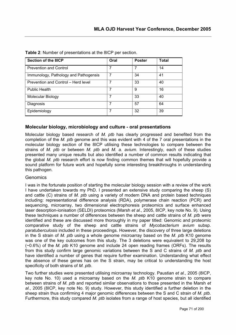
MLA OJD Harvest Year Conference, December 2005
Page 71 of 200
Table 2: Number of presentations at the 8ICP per section.
Section of the 8ICP Oral Poster Total
Prevention and Control 7 7 14
Immunology, Pathology and Pathogensis 7 34 41
Prevention and Control – Herd level 7 33 40
Public Health 7 9 16
Molecular Biology 7 33 40
Diagnosis 7 57 64
Epidemiology 7 32 39
Molecular biology, microbiology and culture - oral presentationsMolecular biology based research of M. ptb has clearly progressed and benefited from thecompletion of the M. ptb genome and this was evident with 4 of the 7 oral presentations in themolecular biology section of the 8ICP utilising these technologies to compare between thestrains of M. ptb or between M. ptb and M. a. avium. Interestingly, each of these studiespresented many unique results but also identified a number of common results indicating thatthe global M. ptb research effort is now finding common themes that will hopefully provide asound platform for future work and hopefully some interesting breakthroughs in understandingthis pathogen.
GenomicsI was in the fortunate position of starting the molecular biology session with a review of the workI have undertaken towards my PhD. I presented an extensive study comparing the sheep (S)and cattle (C) strains of M. ptb using a variety of modern DNA and protein based techniquesincluding: representational difference analysis (RDA), polymerase chain reaction (PCR) andsequencing, microarray, two dimensional electrophoresis proteomics and surface enhancedlaser desorption-ionisation (SELDI) proteomics (Marsh et al., 2005, 8ICP, key note No. 9). Usingthese techniques a number of differences between the sheep and cattle strains of M. ptb wereidentified and these are discussed more thoroughly in my paper titled: Genomic and proteomiccomparative study of the sheep and cattle strains of Mycobacterium avium subsp.paratuberculosis included in these proceedings. However, the discovery of three large deletionsin the S strain of M. ptb using a whole genome microarray based on the M. ptb K10 genomewas one of the key outcomes from this study. The 3 deletions were equivalent to 29,208 bp(~0.6%) of the M. ptb K10 genome and include 24 open reading frames (ORFs). The resultsfrom this study confirm large genomic variations between the S and C strains of M. ptb andhave identified a number of genes that require further examination. Understanding what effectthe absence of these genes has on the S strain, may be critical to understanding the hostspecificity of both strains of M. ptb.
Two further studies were presented utilising microarray technology. Paustian et al., 2005 (8ICP,key note No. 10) used a microarray based on the M. ptb K10 genome strain to comparebetween strains of M. ptb and reported similar observations to those presented in the Marsh etal., 2005 (8ICP, key note No. 9) study. However, this study identified a further deletion in thesheep strain thus confirming 4 major genomic differences between the S and C strain of M. ptb.Furthermore, this study compared M. ptb isolates from a range of host species, but all identified
MLA OJD Harvest Year Conference, December 2005
Page 72 of 200
as C strains and found to be identical to the M. ptb K10 genome strain. Interestingly, the copynumber of a new and unique M. ptb insertion sequence, ISMAP04, was found to vary acrossdifferent isolates and may represent a new target for molecular based diagnostic tests.
Semret et al., 2005 (8ICP, short oral No. 24) used a microarray based on a combination of M.ptb and M. a. avium genes to compare between isolates from M. a. avium complex. This studyidentified a 16 kb region present in both the S strain of M. ptb and M. a. avium that was absentin the C strain of M. ptb and a 7 kb region present in both the C strain of M. ptb and M. a. aviumthat was absent from the S strain of M. ptb. The results from these studies clearly indicate agreat deal of genomic heterogeneity amongst the S and C strain of M. ptb and M. a. avium.Making these genomic regions ideal targets for future research that might identify genes andproteins involved in host specificity, pathogenicity and other aspects of M. ptb.
ProteomicsProteomic based studies were also reported in this section of the 8ICP. Marsh et al., 2005(8ICP, key note No.9) reported using proteomics to compare the S and C strains of M. ptb.Using this technique a number of proteins were identified to be uniquely or differentiallyexpressed in the S and C strain of M. ptb when cultured on solid media. Similar observations tothese results were reported by another study using this technique. Garcia-Sanchez et al. 2005(8ICP, short oral No. 23) used proteomics to compare between S and C strains andintermediate strains, the later sharing similarities with both S and C strains. The results from thisstudy also indicated that while S and C strains express type specific proteomes, intermediatestrains do not.
Other areasEgan et al. 2005 (8ICP, Merkal Award No.1) presented an important Australian based study onthe development of a rapid technique that can isolate intact M. ptb directly from intestinal tissuesof animals, making it possible to perform proteomic studies to identify proteins expressed underin vivo conditions. This may help identify antigens and virulence determinants that explain manyaspects of this disease and that have not been identified under routine in vitro type experimentalconditions. Another almost complementary study was presented by Rosseels et al., 2005 (8ICP,short oral No. 21). In this study, M. ptb were reported to be transformed with luminescent genesallowing them to be observed more easily during in vivo studies in mice spleens. This techniquewas shown to be advantageous when monitoring the efficacy of M. ptb vaccines in vivo.
The 8ICP saw the introduction of a new and exciting research tool in M. ptb and Johne’sdisease research in the form of lipidomics or the study of non-water-soluble metabolites (lipids)of which M. ptb has many. In this presentation, Eckstein et al. 2005 (8ICP, short oral No. 22)presented their initial results when using this technique to compare M. ptb and M. a. avium.Total lipids from whole cells and culture filtrates were compared using two-dimensional thinlayer chromatography. To date, 9 lipids have been identified in M. ptb that are absent in M. a.avium. Further analysis of these 9 lipids, across all 28 serovars of M. a. avium, indicates that 4are truly unique to M. ptb. Interestingly, all 4 lipids were only associated with the whole M. ptbcells not the culture filtrate.
Molecular biology, microbiology and culture - poster presentationsPosters presented in the Molecular biology, Microbiology and Culture section of the 8ICP weregenerally of an extremely high standard, very informative and covered a diverse range ofresearch areas associated with M. ptb and Johne’s disease. As with the oral presentations, themajority of the 37 posters in this section could be divided into one of a number of categories andwill be reviewed according to this division.
MLA OJD Harvest Year Conference, December 2005
Page 73 of 200
ProteomicsThree proteomic studies were presented including the development of a M. ptb cell disruptiontechnique for use in further proteomic studies (Donaghy et al. 2005, 8ICP, poster No. 99). Thiswas an interesting study that effectively compared 4 commercial disruption techniques.However, other techniques have been used elsewhere (Hughes et al. 2005, 8ICP, short oralNo. 6 and Marsh et al., 2005, 8ICP, key note No. 9) utilising alternate disruption techniques andthese should also be considered for those wanting to undertake proteomic studies of M. ptb.Proteomics was also used to identify 14 secreted proteins (from culture filtrate) that may proveuseful for the development of new serodiagnostic tests (Cho et al., 2005, 8ICP, poster No. 95).Proteomics was also used to identify protein profiles identifying differentially expressed proteinsfrom M. ptb and M. a. avium grown under iron limiting culture conditions (Wilkins et al. 2005,8ICP, poster No. 121). The aim of this study is to identify the potential role such proteins play incellular metabolism and pathogenicity.
Molecular based typing techniques for M. a. paratuberculosisA number of posters presented in this section of the 8ICP focused on new alternatives to thecurrent molecular based typing techniques for M. ptb and the application of the current typingtechniques in molecular based epidemiological surveys of Johne’s disease.
The use of restriction fragment length polymorphism (RFLP) and polymerase chain reaction(PCR) based typing techniques would be considered the gold standard for M. ptb strain andsub-strain typing. However, both techniques have limitations that may be overcome by theintroduction of new typing techniques. Three studies were presented using mycobacterialinterspersed repetitive unit-variable number tandem repeat (MIRU-VNTR) based typingtechniques (Biet et al. 2005, 8ICP, poster No. 92 and Nishimori et al. 2005, 8ICP, poster No.109 and Romano et al. 2005, 8ICP, poster No. 113). All three studies indicated the potential ofthis approach to separate M. ptb and M. a. avium isolates using this simple PCR basedprocedure. However, the use of this technique and the advantages it offers has yet to bedemonstrated in extensive epidemiological studies of M. ptb. Whereas, an alternate techniqueknown as multilocus short sequence repeat (MLSSR) was reported to be capable ofdifferentiating strains of M. ptb previously indistinguishable by other methods (Harris et al.2005, 8ICP, poster No. 106). When applied to an epidemiological survey of dairy cattle in theUSA, the MLSSR technique identified multiple strains within herds and even within individualanimals.
Other studies were presented using more traditional M. ptb typing techniques. Johansen et al.2005 (2005, 8ICP, poster No. 107) presented an interesting RFLP based study that comparingthe presence or absence of ISmpa1, IS1245 and IS1311 in M. ptb and M. a. avium. The resultsindicate that the M. ptb and M. a. avium genomes both posses IS1311 and ISmpa1. However,the later was not present in all M. a. avium strains. Both insertions sequences can be used todifferentiate between the species, when present. IS1245 was confirmed to be present in M. a.avium only.
Three epidemiological studies were presented, two from India (Sevilla et al. 2005, 8ICP, posterNo. 115 and Singh et al. 2005, 8ICP, poster No. 116) and one from Spain (de Juan et al. 2005,8ICP, poster No. 96), that used the more traditional RFLP and PCR-REA typing techniques. Allthree studies identified cross-infection of sheep and cattle with the C and S strain strainsrespectively, and in other species. Furthermore, the M. ptb bison strains were found in sheepand goats in India. The results from these studies clearly indicate that global strain variation ofM. ptb and host susceptibility are geographically dependent. Furthermore, these typing
MLA OJD Harvest Year Conference, December 2005
Page 74 of 200
techniques still have an important role in the diagnostic testing regimes required to fullyunderstand Johne’s disease in different parts of the world. As they provide important informationin decision making for geographically distinct control and evaluation programmes of thisdisease.
Polymerase chain reactionAs with previous ICP a number of studies were presented on the use of PCR in Johne’s diseaseresearch and diagnosis. IS900-like elements in non- M. ptb species have been reported as aproblem in M. ptb literature and at both the 6th and 7th ICP but are considered to be extremelyrare. However, an Italian study (Taddei et al. 2005, 8ICP, poster No. 119) identified 21mycobactin-independent Mycobacteria that were positive in IS900 PCR. These results onceagain reinforce the diagnosis of M. ptb based on IS900 PCR alone should be interpreted withextreme caution.
Studies aimed at improving M. ptb detection by PCR included: improved DNA extractiontechniques from M. ptb isolated from milk samples (Gwordz et al. 2005, 8ICP, poster No. 104),the development of an internal PCR control to validate each PCR reaction (Brey et al. 2005,8ICP, poster No. 93) and new real time PCR reactions (Ravva, et al. 2005, 8ICP, poster No.112 and Sweeney et al. 2005, 8ICP, poster No. 118). Further examination of each of thesestudies will provide valuable information with respect to the development of the direct-PCR (D-PCR) test currently being undertaken at the Elizabeth Macarthur Agricultural Institute (NSWDepartment of Primary Industries) funded by Meat and Livestock Australia.
Other PCR based studies included the development of a multiplex PCR designed to detect M.ptb, M. a. avium and M. bovis mixed infections in red deer (Cervus elaphus) (Godford et al.2005, 8ICP, poster No. 102) and an alternate PCR based technique capable of differentiating M.ptb and M. a. avium, based on sequence differences in the fibronectin-attachment protein genesin both subspecies.
Host genomicsOne of the key areas to yet to be addressed in Johne’s disease research is the host response toM. ptb. DiFiore et al. 2005 (2005, 8ICP, poster No. 97) presented one of the first studiesattempting to identify gene expression from the host perspective, in sheep, during early andclinical stages of disease as opposed to other studies that have looked at this during advancedstages of disease. Using a technique known as long distance differential display polymerasechain reaction (DD-PCR), uninfected and infected tissues were screened to identify differentiallyexpressed genes in infected sheep. Using this technique, gene expression has been identifiedin both non-infected and infected tissues; however, differential expression has yet to beobserved. A number of alternative PCR primer sets have yet to be used to evaluate thistechnique.
M. a. paratuberculosis cultureAs with the PCR based studies, a number of studies were presented on M. ptb culturetechniques focusing on either the improvement of current techniques or the introduction of newculture methods.
Gwordz et al. 2005 (2005, 8ICP, poster No. 105) compared decontamination methods for theisolation of M. ptb from effluent. The results from this study indicate that a double incubationtechnique at 42oC was selective for M. ptb and suggested that this may have an important rolein the isolation of M. ptb from faeces, tissue and milk. A new culture technique was describedfor the isolation of M. ptb from milk by Ruzante et al. 2055 (2005, 8ICP, poster No. 114). The
MLA OJD Harvest Year Conference, December 2005
Page 75 of 200
technique introduced a new procedure where M. ptb in the milk sample are concentrated usinga solution known as CB18. The inclusion of this procedure was reported to decrease the timerequired to culture the organism from raw milk. Once again this may be an important outcomeand of use in improving local culture and detection techniques of M. ptb directly from faeces.
Two studies compared M. ptb culture on either solid media (Wiszniewska et al. 2005, 8ICP,poster No. 122) or in liquid culture (van Maanen et al. 2005, 8ICP, poster No. 120), the laterstudy including the recently developed TREK ESP para-JEM Culture System II. As withprevious studies of this nature the results might indicate the relative proficiencies differentlaboratories have with different culture techniques and therefore should be considered withability to reproduce results in more than one laboratory. This issue was highlighted in a posterfrom the National Veterinary Services Laboratories from Ames Iowa. This laboratory has beenrequested to develop a standardised culture technique for the United States to overcome thewide range of sensitivities and variability in culture techniques currently in use in the USA for thediagnosis Johne’s disease (Payeur et al. 2005, 8ICP, poster No. 111).
Other studiesIn addressing an unmet need from the 7ICP, Bannantine et al. 2005 (8ICP, poster No. 91)reported the development of several monoclonal antibodies for a number of M. ptb epitopes plusaptamers for the conserved hypothetical protein MAP0105c and single-chain antibody (ptb9) forthe M. ptb specific protein encoded by MAP0858. The authors hope that these detectionreagents will be of use in many Johne’s disease research applications. M. ptb specificmonoclonal antibodies were also produced in the development of a new PCR based diagnostictest used to detect M. ptb in colostrum from cattle (Ebert et al. 2005, 8ICP, poster No. 100).This test will provide rapid detection of M. ptb in colostrum and as a result support herd-leveltesting regimes in dairy cattle.
Three studies were reported on the survival of M. ptb in bovine macrophages, further improvingour understanding of how this organism survives in the host and how the host responds to theorganism. Partridge et al. 2005 (8ICP, poster No. 110) used real time PCR and microarrayanalysis to identify those genes expressed by M. ptb during infection. Where as, examination ofthe organism-host interactions was reported in two studies, the first looking at the response ofthe host, mouse macrophages, to different strains of M. ptb isolated from a range of hosts(Mitchell et al. 2005, 8ICP, poster No. 108) and the second looking more specifically at theinteraction between different strains of M. ptb with bovine macrophages Gollmick et al. 2005(8ICP, poster No. 103). The results from these studies should provide useful information on thehost immune response to M. ptb, the host specificity of the different strains of M. ptb and thepotential for different hosts to act as reservoirs for non-specific host strains of M. ptb.
A new vaccine candidate was reported based on the development of a fusion construct,including antigens from two membrane and two secreted proteins and was shown to elicit asignificant attenuation of the infective load of M. ptb in the spleen and liver of mice (Bull et al.2005, 8ICP, poster No. 94). With these encouraging results the authors hope to continue theirresearch in vaccination studies on larger animals with the view to providing an effective M. ptbvaccine for both animals and humans.
Immunomagnetic detection of M. ptb was re-visited using a M. ptb specific peptide bound tomagnetic beads (Stratmann et al. 2005, 8ICP, poster No. 117). The test, which includescapturing the M. ptb on magnetic beads followed by PCR and restriction endonuclease analysisof the amplified product, was successfully used to detect S and C strain M. ptb in spiked
MLA OJD Harvest Year Conference, December 2005
Page 76 of 200
individual and bulk milk samples. It would appear that the test has yet to be evaluated with realmilk samples and the results from this further evaluation are eagerly awaited.
Finally, in a more traditional microbiological study, lactic acid bacteria were used to inactivate M.ptb that have survived pasteurisation of milk to used for the production of dairy products such asyogurt and cheese (Donaghy et al. 2005, 8ICP, poster No. 98). The authors report that thelactic acid bacteria are capable of inhibiting other food pathogens and technique was shown tobe successful in this study with M. ptb. Although, the results seem to suggest that inhibition ofM. ptb may not be the result of lactic acid. Regardless of the mechanism of inactivation theassociation with the lactic acid bacteria appears to have an inhibitory effect on M. ptb andfurther work is required to establish the mechanism and the potential of this approach.
ConclusionAs with previous ICP and each new publication on Johne’s disease or M. a. paratuberculosis,we hope for a ground breaking or new development that will have a significant impact ondealing with this insidious disease. The reality is that like other mycobacterial diseases such astuberculosis and leprosy, progress in the research that aims to develop or assist ourunderstanding of the diseases and/or the organisms that cause them, is slow. However, theprogress demonstrated at the 8ICP is extremely encouraging. Moreover, the widespreadadoption of new and exciting molecular based research approaches should yield someinteresting and informative outcomes in the not to distant future, hopefully before or at least bythe next time the International Association on Paratuberculosis meets in Japan at the 9ICP.

MLA OJD Harvest Year Conference, December 2005
Page 77 of 200
Vaccination

MLA OJD Harvest Year Conference, December 2005
Page 78 of 200
INSERT PFIZER ADD HERE
MLA OJD Harvest Year Conference, December 2005
Page 79 of 200
Overview of OJD vaccination – international perspective
Peter WindsorFarm Animal HealthUniversity of SydneyPMB 3, Camden, NSW 2570Phone: 02 9351 1710; Fax: 02 4655 0618E-mail: [email protected]
AbstractThe availability of vaccination against paratuberculosis from April 2002 in Australia, effectively‘rescued’ the Australian sheep industry from a devastating socioeconomic calamity that haddivided the industry and led to open revolt by producers against animal health authoritiesattempting to control the disease through regulation. At the time vaccination was beingconsidered as an option for disease control, there were conflicting opinions on the advisability ofthis approach. In particular there was concern over reported problems associated withvaccination, such as adverse reactions and injection site lesions in sheep and people, plus thepossibility that the vaccine would be less efficacious in Australian Merinos in the Australianenvironment. Analysis of published reports of vaccination trials in the international literature,demonstrates a clear positive effect from vaccination in reducing mortalities, prevalence oflesions and shedding, with negative effects in only a few vaccination trials. Now that severallarge field trials have been conducted in infected New South Wales Merino flocks, there isconsiderable evidence that supports the ongoing use of vaccine to both reduce the impact ofdisease and decrease the level of shedding of infectious organisms from infected Australiansheep. This work has also suggested that vaccination works particularly well when combinedwith management strategies to reduce infection rates in the flock and that the side effects ofvaccination are well tolerated by the industry. These studies have opened a dialogue that isconsidering the use of vaccination to assist control of bovine paratuberculosis in Victorian dairyherds. Although the international perspective of vaccination suggests that it will prove to beefficacious in Australian dairies, conclusive demonstration that vaccinated cattle havesignificantly reduced faecal shedding of the infectious organisms in the Australian environmentis desired. Acceptance of vaccination against paratuberculosis may yet prove to be the mostsignificant advance in controlling the disease and reducing the potential public risk should theevidence finally conclude that paratuberculosis in livestock is a risk factor in human Crohn’sdisease.
IntroductionLivestock Johne’s disease (JD) due to infection with Mycobacterium avium subspeciesparatuberculosis (M. ptb) is an emerging complex animal health problem prompting a number ofdifferent approaches to controlling the disease, including enhancing herd and flock immunity byvaccination. Several vaccines have been used in livestock, with mostly encouraging evidence ofefficacy (Juste et al., 2002), particularly when the program has used a vaccine with a Freund’sadjuvant-like nature to provoke cellular immunity (Windsor et al., 2005). Vaccination has beenreadily embraced for the control of paratuberculosis in sheep, with considerable success, asdocumented in the United Kingdom (Cranwell, 1993), Spain (Garcia Marin JF et al., 1999),Iceland (Fridriksdottir et al., 2000) and more recently Australia (Windsor et al., 2002). Currentlya vaccine study is underway in sheep in North America using MycoparTM cattle vaccine(Stehman, pers. comm.).
MLA OJD Harvest Year Conference, December 2005
Page 80 of 200
Vaccination of cattle has been used sporadically, particularly where animal health authoritieshave committed to a test and cull or test and management approach to controlling the disease.For example in the USA, a commercial vaccine is available following approval by a StateVeterinarian on a herd by herd basis, with published evidence from a controlled clinical trial thatby using a whole cell bacterin, the incidence of clinical JD is reduced (Larson et al., 1978).However less consistent results demonstrating the effect of vaccination on faecal shedding haveled to variable support for vaccination programs, despite the now considerable evidencesupporting a role for vaccination in modifying the progress of infection from early subclinicalinfection to open disease (Juste et al., 2002). In addition to the paucity of reports of largescientifically controlled trials that convincingly demonstrate the efficacy of vaccination onshedding, it is likely that several other factors have led to a cautious approach to the uptake ofvaccination, despite evidence that vaccination is cost-effective due to reduction of culling ofclinically affected cattle (van Schaik et al., 1996). These concerns include the prevalence ofadverse reactions and lesions at injection sites or beyond and the risk of self-injection injuryfrom using a vaccine containing a provocative adjuvant, plus problems with vaccinationprograms in cattle in countries where tuberculosis exists because vaccination can interfere withtuberculin and other testing programs used in control of bovine TB. This paper provides anoverview of the historical work and the most recent investigations on the use of vaccination tocontrol paratuberculosis, from an international perspective.
An overview of previous studiesEvidence for efficacy of vaccineA meta-analysis of reports of vaccination trials against paratuberculosis was completed in 2002and involved reviewing the results presented in 16 reports of cattle, 11 reports of sheep and 6reports of goats following vaccination (Juste et al., 2002). The average combined reduction inincidence of clinical disease in these reports following vaccination was 96% of 280,743 cattle,71% of 11,661 sheep and 45% of 9,951 goats. The average reduction in mycobacterialshedding in these reports following vaccination was 72% of 42,197 cattle, 50% of 1,735 sheepand 83% of 1,425 goats. The average reduction in lesions of paratuberculosis followingvaccination was 58% of 4,317 cattle, 90% of 27,290 sheep and 96% of 5,724 goats. There werefew reports on vaccine failure, with one report involving oral administration, another of reducedefficacy due to vaccine composition with sonicated cells, and more recently, a report with noeffects on faecal shedding compared to other control measures (Juste et al., 2002). A clearconclusion from these reports is that vaccination is mostly an effective tool in reducing theincidence of clinical disease and mortalities plus the rate of shedding from infected animals andprevalence of lesions in vaccinates. However as it does not completely protect the vaccinatedanimal from infection it cannot by itself lead to eradication of M. ptb from an infected flock orherd, a potential issue of concern with the increasing evidence for a role of human exposure toM. ptb as a potential risk for Crohn's disease (Juste et al., 2002).
Impact of vaccine on lesionsThe repeated observation that vaccination against paratuberculosis significantly reduces thenumber of clinical cases in field conditions by modifying the progress of lesions, but doesn’tprevent infection, has been examined in a few studies. To confirm that vaccination modified thetype of lesion produced following infection, a study from Spain that minimised the role of geneticfactors in lesion development by the use of twin lamb pairs is of interest (García Marín et al.,1996). One lamb out of each one of eight pairs of twin lambs was vaccinated when it was 15days old and all lambs were orally infected with a pathogenic ovine strain of M. ptb at 50 days ofage and killed at either 150 or 350 days post- infection and submitted to necropsy and
MLA OJD Harvest Year Conference, December 2005
Page 81 of 200
histological evaluation. At both sampling times after infection, vaccinated lambs had only‘regressive-type’ granulomata limited to Peyer's patches and ileocecal valve lymphoid tissues,with unvaccinated animals displaying spread of granulomatous lesions to other areas of theintestinal wall.
The age at which vaccine is applied is also of particular interest as although animals are mostlyvaccinated when they are younger than 6 months, in the face of significant economic lossesproducers will request vaccination of adult sheep, many of which are likely to be infected andsome expected to have developed lesions of paratuberculosis. Immunisation of 70% of the adultdairy sheep in two flocks of about 250 adult animals in each, retaining 30% of the sheep ascontrols, was undertaken (García-Pariente et al., 2002). During the 12 months after vaccination,a total of 56 sheep (40 vaccinates and 16 controls) were culled and necropsied, including 17sheep in the first 6 months (9 having diffuse lesions) and 11 between 6-12 months postvaccination (3 having diffuse lesions). It was concluded that as vaccination of the adult sheepinduced strong humoral and cellular immune responses and there was a reduction in thenumber of clinical cases of paratuberculosis with a decrease in culls from the vaccinated groupand the number of animals having diffuse lesions, the study provides some evidence thatvaccination may assist in limiting the disease to focal lesions.
Is eradication by vaccination possible?There is mounting evidence that control rather then eradication of paratuberculosis is a morerealistic short to medium goal of paratuberculosis control programs, particularly wherevaccination is the key tool used to reduce the prevalence of infection. The experience of OJD inIceland has a number of parallels to that in Australia, with the importation of infected sheep fromabroad, rapid spread of infection to the main sheep breeding areas and unsuccessful attemptsto eradicate the disease by regulatory approaches including quarantine, widespreadimmunological testing, extensive culling of sheep in endemic areas and restocking with healthysheep (Fridriksdottir et al., 2002). As vaccination experiments demonstrated that mortality couldbe reduced by 94%, since 1966, vaccination of sheep has been compulsory in endemic areas,with considerable reduction in losses. However in 2001, paratuberculosis suddenly appeared ona farm in an area which had been declared free from paratuberculosis and where vaccinationhad ceased in the whole quarantine area 3 years earlier. Sheep from all farms in the area werethen tested serologically and 50 of 400 sheep on the affected farm were seropositive and/or hadclinical symptoms, with one seropositive sheep found on another farm. All positive animals wereculled and the rest vaccinated with compulsory vaccination continuing again in the wholequarantine area. The reasons for the outbreak and the question of whether eradication ofparatuberculosis by vaccination is possible, remain undetermined (Fridriksdottir, pers. comm.).
Most recent developmentsAustralian efficacy studies in sheepSeveral studies have been recently completed evaluating the use of GudairTM to control OJD.These have presented convincing data that supports vaccination as a key tool to induce cellularimmunity in vaccinates and rapidly control OJD at the flock level (Windsor et al., 2002;Eppleston et al., 2004, 2005). Over the productive life of Australian Merino sheep in 3 highprevalence flocks, vaccination reduced mortalities by 90% and delayed and reduced faecalshedding of M. ptb by 90%, although all of the few vaccinates that developed OJD hadmultibacillary disease and excreted infectious doses of the organism (7 of 600 vaccinates).These results led to registration of the vaccine and have encouraged a significant voluntaryadoption of the vaccine in endemically infected and adjacent areas. Ongoing monitoring of theperformance of the vaccine in flocks of different prevalence levels is continuing (Windsor, 2005)
MLA OJD Harvest Year Conference, December 2005
Page 82 of 200
and a gross margin to show breakeven points for vaccination to inform affected producers of thelength of time required for a return on an investment in vaccination to be achieved has beendeveloped (Bush et al., 2005).
Due to considerable producer interest in vaccination of all ages of sheep in the flock when facedby sudden increases in overwhelming losses, a study in a single large flock with over 8,000sheep and a very high mortality rate estimated at 25% per annum, involved annual vaccinationof all sheep with the exception of up to 150 controls in each age cohort. Vaccination wasaccompanied by significant change to the flock profile, initially by the culling of clinical cases,sale of ‘at risk’ stock, replacement of much of the ewe flock with wethers and reduction instocking rates by intensive de-stocking due to severe drought, resulting in a decline in the‘sheep years at risk’ to 5,571 over the 4 years of the trial, accompanied by a significant declinein annual mortality rate from 24% to 2.7%. Further, despite observations of a decline from ahigh to a moderate prevalence of shedders following vaccination of 2 year-old sheep, mortalitiesdue to OJD and shedding of M. ptb continued, particularly in wethers. Vaccination at 2 years ofage in this high M. ptb prevalence environment had limited impact on mortality rate due to OJD,whilst vaccination at age 3 months and 8 months was of similar efficacy, with lower prevalenceof M. ptb shedders than from unvaccinated controls. A decline in the prevalence of shedders inthe unvaccinated sheep during the 4 years of the project was also observed (McGregor et al.,2005; Windsor, 2005).
Adverse reactionsVaccination site lesions in the Australian studies occur in about 50% of vaccinates but declinedto between 20-25% during the course of the 5 year study (Eppleston et al., 2004, 2005). Thepotential costs of the vaccination site lesions were found to be negligible following investigationsof 20 lines of sheep at slaughter, where 65% of lamb carcases and 18% of mutton had vaccinelesions (Eppleston, 2004). A study of adverse reactions in producers vaccinating sheepidentified by survey, that exposure to the vaccine was not uncommon at greater than 1 in10,000 doses (Windsor et al., 2005). However whilst the risk of self-inoculation with the vaccineappeared to be very low at about 1 in 500,000 doses, most self-injection lesions resulted inuntoward consequences, with granulomatous to necrotising lesions leading to fistulationrequiring extensive surgical intervention in some cases to enable drainage of the injectedmaterial. More recently, amputation of a finger was necessary to resolve a chronic lesion thatfailed to respond to surgery.
A new vaccine for cattleA new vaccine has recently been produced and targeted for use in cattle. SILIRUMTM vaccine,also produced by CZ Veterinaria of Spain, the manufacturers of GudairTM, is also a killedvaccine but uses a different adjuvant, presumably in an attempt to reduce vaccination sitelesions. A field study is being conducted in Spain in a Friesian herd of 468 animals with 12%annual clinical cases of BJD. The trial involves vaccination of 75% of all cattle with 25% retainedas controls for 30 months post-vaccination (Garcia-Pariente et al., 2005). More than 90% ofvaccinates produced persistent cellular and humoral immune responses to the vaccine and nonew cases of BJD occurred from 6 months post-vaccination. The percentage of the 192 animalsculled during the trial with lesions of BJD was significantly higher in the controls than vaccinates,with 92.3% of controls having lesions compared to 56.6% of vaccinates, indicating a significantreduction in BJD in the herd following vaccination. In addition, a pilot field study with SILIRUMTM
was also conducted in a herd of 113 cattle with a clinical incidence of BJD of 8% and faecalculture prevalence of 5.5% (Garrido JM et al., 2005). After one year of control, no new clinicalcases of BJD have occurred, the faecal culture prevalence is 3.6% and no interference withintradermal testing for tuberculosis was identified, suggesting expansion of the trial to a
MLA OJD Harvest Year Conference, December 2005
Page 83 of 200
maximum of 50 herds is likely. SILIRUMTM was also evaluated in 10 of 18 calves that werechallenged orally with 6.9 x 10¹⁰ cfu of M. ptb following vaccination without adverse reactions,although the author of the poster revealed that all vaccinates had injection site lesions up to15cm (Munoz et al., 2005). Cellular and humoral responses were evaluated with interferon-gamma results detected at 30 days and still high at 330 days post-vaccination. The calves werekilled at 180 and 330 days post-vaccination and only one vaccinate had a diffuse lesion of BJD,although several had lesions mainly in lymphoid tissues. Isolation of M. ptb was reduced invaccinates, suggesting this vaccine will reduce bacterial load and severity and numbers oflesions in challenged calves.
ConclusionThere is an emerging consensus that eradication of endemic M. ptb by test and cull proceduresis frequently not possible nor profitable in many herd and flock situations. Until it becomesessential, as may occur if a more positive link from animal M. ptb to human Crohn’s disease isestablished, control strategies that limit losses will be most appropriate for the majority of herdsand flocks. There is now considerable evidence that this can be readily achieved by vaccinationwith the additional advantage that at least in sheep, there is a considerable reduction in faecalshedding of infectious organisms. In infected Australian sheep flocks, vaccination is likely toremain a voluntary long-term M. ptb control strategy, although how long producers will continueto use the vaccine when losses are no longer observed remains an interesting question for thefuture. The experience from Iceland suggests the disease will re-emerge following cessation ofvaccination. Fortunately concerns that adverse reactions and injection site lesions, or theoccasional occurrences of self-injection may compromise the use of vaccine appear to beunfounded
In conclusion, vaccination is a key tool to enable rapid reduction in the within-herd prevalence ofinfection in sheep and goat flocks, with a potential for use in dairy and possibly beef cattleoperations to minimise economic losses to cattle producers. However further information fromclinical trials and research is required to fine-tune effective vaccination programs in cattle. If M.ptb is conclusively shown to be a public health risk, effective control of paratuberculosis andelimination of clinical cases will be necessary to protect human health and satisfy publicdemands for food safety. Examination of vaccination as a means to eliminate clinical BJD andpotentially to reduce shedding from infected dairy cattle should be undertaken as soon aspossible in Australia. If shown to be efficacious, as is likely when one considers the internationalperspective, vaccination is likely to prove a most important component of paratuberculosiscontrol strategies in herds where clinical cases are occurring.
AcknowledgementsThe assistance of Dr Ramon Juste of NEIKER, Bizkaia Spain is gratefully acknowledged, as isadvice from Drs V Fridriksdottir of Iceland and S Stehman of USA. The Australian studies havebeen a collaborative effort involving many individuals, including Helen McGregor, Russell Bush,Jeff Eppleston, Leslie Reddacliff, Richard Whittington, Evan Sergeant and others. Projects werefunded by the NOJDP through Meat and Livestock Australia and conducted by researchers andsupport staff at the RLPB Central Tablelands, NSW Department of Primary Industries at EMAIand OAI, and the Faculty of Veterinary Science, at the University of Sydney.
MLA OJD Harvest Year Conference, December 2005
Page 84 of 200
References1. Bush RD, Windsor PA, Toribio JAL and Webster S (2005) Modelling the on-farm financial
impact of ovine Johne’s Disease in Australia. Proc 8th International Colloquium onParatuberculosis, Copenhagen, Denmark. Abstract Poster 191. 6:132
2. Cranwell, M.P. 1993. Control of Johne's disease in a flock of sheep by vaccination. VetRec 133: 219-20.
3. Eppleston J., Reddacliff L., Windsor P., Whittington R., and Jones S. (2004) Field studies onvaccination for the control of OJD in Australia – an overview. Proc Aust Sheep Vet Soc.Canberra. 14: 56-59.
4. Eppleston J (2004) A Preliminary study on the potential cost at slaughter of OJD vaccinationsite lesions to the Australian sheep industry. Meat and Livestock Australia Project ReportOJD.032.
5. Eppleston J, Reddacliff L, Windsor PA, Whittington R, Jones S (2005) efficacy of a killedMycobacterium paratuberculosis vaccine fore the control of OJD in Australian sheep flocks.Proc 8th Int Colloquium on Paratuberculosis. Copenhagen, Denmark. Abstract Keynote 5.3a: 43.
6. Eppleston J, Reddacliff L, Windsor PA, Links I, Whittington R 2005. Preliminaryobservations on the prevalence of sheep shedding Mycobacterium avium subsp.paratuberculosis after 3 years of a vaccination program for ovine Johne’s disease. Aust VetJ 83:35-36.
7. Fridriksdottir V, Gunnarsson E, Sigurdarson S, Gudmundsdottir KB (2000) Paratuberculosisin Iceland: epidemiology and control measures, past and present. Vet Microbiol 77: 263-267.
8. Fridriksdottir V, Hjartardottir S, Poulsen S, Sigurdarson S, Gunnarsson E (2002)Paratuberculosis in sheep in Iceland - Is eradication by vaccination possible? Proc 7th
International Colloquium on Paratuberculosis. Bilbao, Spain. Abstract
9. García Marín FF, Tellechea J, Gutiérrez M, Pérez V, Juste RA (1996) Paratuberculosisvaccination in sheep modifies and limits the development of lesions Proc 5th InternationalColloquium on Paratuberculosis, Madison, USA . 234-241.
10. Garcia Marin JF, Tellechea J, Gutierrez M, Corpa JM, Perez V (1999) Evaluation of twovaccines (killed and attenuated) against small ruminant paratuberculosis. Proc 6thInternational Colloquium on Paratuberculosis, Melbourne, Australia.
11. García-Pariente C, González J, Ferreras MC, Fuertes M, Benavides J, Reyes LE, MorenoO, García-Marín JF, Perez V (2002) Paratuberculosis vaccination of adult animals in twoflocks of dairy sheep. Proc 7th International Colloquium on Paratuberculosis. Bilbao, Spain.Abstract
12. García-Pariente C, Pérez V, Geijo MV, Moreno O, Muñoz M, Fuertes M, Puentes E, Doce J,Ferreras MC, García Marín JF (2005) The efficacy of a killed vaccine againstparatuberculosis (SILIRUM®) in cattle. A field study. Proc 8th International Colloquium onParatuberculosis. Copenhagen, Denmark. Abstract Poster 56. 5: 52
13. Garrido JM, Plazaola JM, Sevilla I, Geijo MV, Elguezabal N, Aduriz G, Doce J, Puentes E,Juste RA (2005) Control of paratuberculosis in dairy farms in the Basque Country. Proc 8th
International Colloquium on Paratuberculosis. Copenhagen, Denmark. Abstract Poster 58.5: 53
MLA OJD Harvest Year Conference, December 2005
Page 85 of 200
14. Larson AB, Moyle AI, and Himes EM (1978) Experimental vaccination of cattle againstparatuberculosis (Johne’s disease) with killed bacterial vaccines: a controlled field study.JAVMA 65-69.
15. McGregor H, Whittington RJ, Windsor PA (2005) A comparison of the efficacy of Gudairvaccine for protection against OJD in sheep aged 3 months and 8 months at vaccination.Proc 6th International Sheep Veterinary Congress, Crete, June. 220-222
16. Muñoz M, García Marín JF, García-Pariente C, Reyes LE, Verna A, Moreno O, Fuertes M,Doce J, Puentes E, Garrido J, Pérez V (2005) Efficacy of a killed vaccine (SILIRUM) incalves challenged with Map. Proc 8th International Colloquium on Paratuberculosis.Copenhagen, Denmark. Abstract Poster 67. 5: 57
17. Juste RA, Sevilla GMV, Aduriz G, Garrido JM (2002) Control of paratuberculosis byvaccination. Proc 7th International Colloquium on Paratuberculosis. Bilbao, Spain. AbstractKeynote
18. Van Schaik G, Kalis CHJ, Benedictus G, Dijkhuizen AA, and Huirne RBM (1996) Cost-benefit analysis of vaccination against paratuberculosis in dairy cattle. Vet Rec 139:624-627.
19. Windsor PA, Whittington R, Eppleston J, Jones S, Britton A (2002) Efficacy of a killedMycobacterium paratuberculosis vaccine for the control of OJD in Australian sheep flocks.Proc 7th International Colloquium on Paratuberculosis. Bilbao, Spain. Abstract
20. Windsor PA, Eppleston J, Sergeant E et al (2003) Monitoring the efficacy of Gudair™ OJDVaccine in Australia. Proc Aust Sheep Vet Soc. Cairns. 13:114-122
21. Windsor P.A. (2005) The control of ovine Johne’s disease by vaccination. Proc Aust SheepVet Soc. Gold Coast. 15: 129-133
22. Windsor P.A., Bush R., Links I., Eppleston J (2005) Injury caused by self-inoculation with avaccine of a Freund’s complete adjuvant nature (GudairTM) used for control of ovineparatuberculosis. Aust. Vet. J 83: 216-220
MLA OJD Harvest Year Conference, December 2005
Page 86 of 200
Efficacy of a killed Mycobacterium paratuberculosis vaccinefor the control of OJD in Australian sheep flocks
L Reddacliffa, J Eppleston, P Windsor, R Whittington, S JonesaNSW Department of Primary IndustriesPMB 8, Camden, NSW, 2570Phone: 02 4640 6314; Fax: 02 4640 6384Email: [email protected]
AbstractA major field trial was undertaken from 1999 until 2004 to determine the efficacy of a killed M. a.paratuberculosis (M. ptb) vaccine, GudairTM, for the control of ovine Johne's disease (OJD) inAustralian merinos run under Australian pastoral conditions. The vaccine stimulated cell-mediated and humoral immune responses. Gudair™ reduced mortalities due to OJD by 90%and delayed faecal shedding for the first year post-vaccination (pv). Thereafter, the prevalenceof shedders among vaccinates was reduced by 90%. The numbers of M. ptb excreted by thevaccinated groups were also reduced by at least 90% at most sampling times. However, highlevels of excretion by vaccinates occurred on some occasions, and although only 7 of 600vaccinates died from OJD, all 7 had multibacillary disease. Thus there remains a risk that somevaccinated sheep could transmit OJD. Small reductions in liveweight gain were found invaccinated lambs in the first year pv, but there was little effect on wool production. Vaccineinjection site lesions were detected in almost 50% of sheep 2 months pv, and these persistedfor at least 4 years in 20-25% of vaccinates. Data from this trial enabled the registration ofGudair™ in Australia in 2002 and underpins the pivotal role of vaccination in the currentmanagement of OJD.
IntroductionVaccination against OJD had been used in several overseas countries, with encouragingresults1,3,4,8. However, there were few scientifically controlled reports demonstrating its efficacy.A major field trial was undertaken from 1999 until 2004 to determine the efficacy of a killed M. a.paratuberculosis (M. ptb ) vaccine, GudairTM, for the control of OJD in Australian Merino sheeprun under Australian pastoral conditions. The main objectives were to determine the effect ofGudair™ vaccination on OJD-related mortality and on the excretion of M. ptb in the faeces(shedding). Secondary objectives were to investigate the cellular and humoral immuneresponse following vaccination with Gudair™, to assess the field safety of Gudair™, and todetermine changes in productivity parameters associated with either vaccination or subclinicaldisease.
MethodsOn each of three farms experiencing significant OJD-related mortality (5 to 15% per annum),200 Merino lambs (age 1-4 months) were vaccinated with Gudair™ and 200 control lambs wereleft unvaccinated. Animal assessments and sample collections were conducted twice yearlyuntil 4 or 5 years of age. The impact of vaccination on mortality, faecal shedding of M. ptb (bypooled and individual faecal culture), cellular (BOVIGAM™) and humoral (PARACHEK™)immunity, vaccine injection site lesions, liveweight, condition score and wool productivity wereexamined. A standard set of tissue samples was collected from each animal autopsied andexamined histologically, with any lesions observed classified according to pre-determinedcriteria.
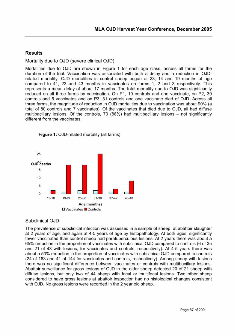
MLA OJD Harvest Year Conference, December 2005
Page 87 of 200
ResultsMortality due to OJD (severe clinical OJD)Mortalities due to OJD are shown in Figure 1 for each age class, across all farms for theduration of the trial. Vaccination was associated with both a delay and a reduction in OJD-related mortality. OJD mortalities in control sheep began at 23, 14 and 19 months of agecompared to 41, 23 and 43 months in vaccinates on farms 1, 2 and 3 respectively. Thisrepresents a mean delay of about 17 months. The total mortality due to OJD was significantlyreduced on all three farms by vaccination. On P1, 10 controls and one vaccinate, on P2, 39controls and 5 vaccinates and on P3, 31 controls and one vaccinate died of OJD. Across allthree farms, the magnitude of reduction in OJD mortalities due to vaccination was about 90% (atotal of 80 controls and 7 vaccinates). Of the vaccinates that died due to OJD, all had diffusemultibacillary lesions. Of the controls, 70 (88%) had multibacillary lesions – not significantlydifferent from the vaccinates.
Subclinical OJDThe prevalence of subclinical infection was assessed in a sample of sheep at abattoir slaughterat 2 years of age, and again at 4-5 years of age by histopathology. At both ages, significantlyfewer vaccinated than control sheep had paratuberculous lesions. At 2 years there was about a65% reduction in the proportion of vaccinates with subclinical OJD compared to controls (6 of 35and 21 of 43 with lesions, for vaccinates and controls, respectively). At 4-5 years there wasabout a 50% reduction in the proportion of vaccinates with subclinical OJD compared to controls(24 of 163 and 41 of 144 for vaccinates and controls, respectively). Among sheep with lesionsthere was no significant difference between vaccinates or controls with multibacillary lesions.Abattoir surveillance for gross lesions of OJD in the older sheep detected 20 of 21 sheep withdiffuse lesions, but only two of 44 sheep with focal or multifocal lesions. Two other sheepconsidered to have gross lesions at abattoir inspection had no histological changes consistentwith OJD. No gross lesions were recorded in the 2 year old sheep.
Figure 1: OJD-related mortality (all farms)
0
5
10
15
20
25
13-18 19-24 25-30 31-36 37-42 43-48
Age (months)
OJD deaths
Vaccinates Controls
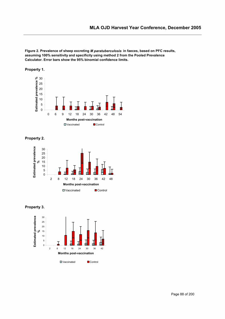
MLA OJD Harvest Year Conference, December 2005
Page 88 of 200
Property 1.
Property 2.
Property 3.
FIgure 2. Prevalence of sheep excreting M paratuberculosis in faeces, based on PFC results, assuming 100% sensitivity and specificity using method 2 from the Pooled Prevalence Calculator. Error bars show the 95% binomial confidence limits.
0
5
10
15
20
25
30
0 6 9 12 18 24 30 36 42 48 54
Months post-vaccination
Estim
ated
pre
vale
nce
%
Vaccinated Control
05
1015202530
2 8 12 18 24 30 36 42 48
Months post-vaccination
Estim
ated
pre
vale
nce
%
Vaccinated Control
0
5
10
15
20
25
30
2 8 12 18 24 30 36 42
Months post-vaccination
Estim
ated
pre
vale
nce
%
Vaccinated Control
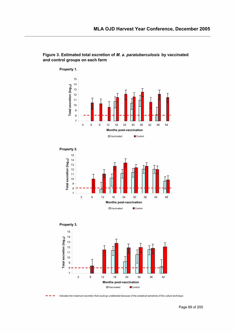
MLA OJD Harvest Year Conference, December 2005
Page 89 of 200
Property 1.
Property 2.
Property 3.
Indicates the maximum excretion that could go undetected because of the analytical sensitivity of the culture technique
Figure 3. Estimated total excretion of M. a. paratuberculosis by vaccinated and control groups on each farm
7
8
9
10
11
12
13
14
15
0 6 9 12 18 24 30 36 42 48 54
Months post-vaccination
Tota
l exc
retio
n (lo
g 10)
Vaccinated Control
7
8
9
10
11
12
13
14
15
2 8 12 18 24 30 36 42 48
Months post-vaccination
Tota
l exc
retio
n (lo
g 10)
Vaccinated Control
7
8
9
10
11
12
13
14
15
2 8 12 18 24 30 36 42
Months post-vaccination
Tota
l exc
retio
n (lo
g 10)
Vaccinated Control
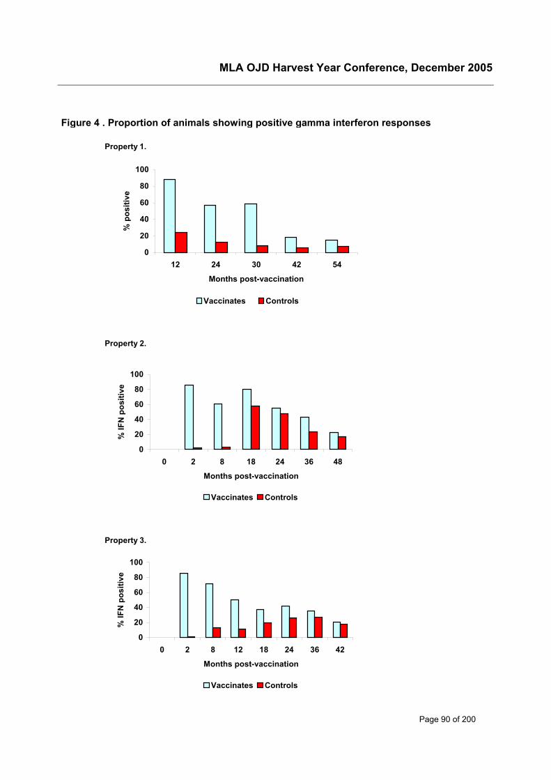
MLA OJD Harvest Year Conference, December 2005
Page 90 of 200
Figure 4 . Proportion of animals showing positive gamma interferon responses
Property 1.
Property 2.
Property 3.
0
20
40
60
80
100
0 2 8 18 24 36 48
Months post-vaccination
% IF
N p
ositi
ve
Vaccinates Controls
0
20
40
60
80
100
0 2 8 12 18 24 36 42
Months post-vaccination
% IF
N p
ositi
ve
Vaccinates Controls
0
20
40
60
80
100
12 24 30 42 54
Months post-vaccination
% p
ositi
ve
Vaccinates Controls
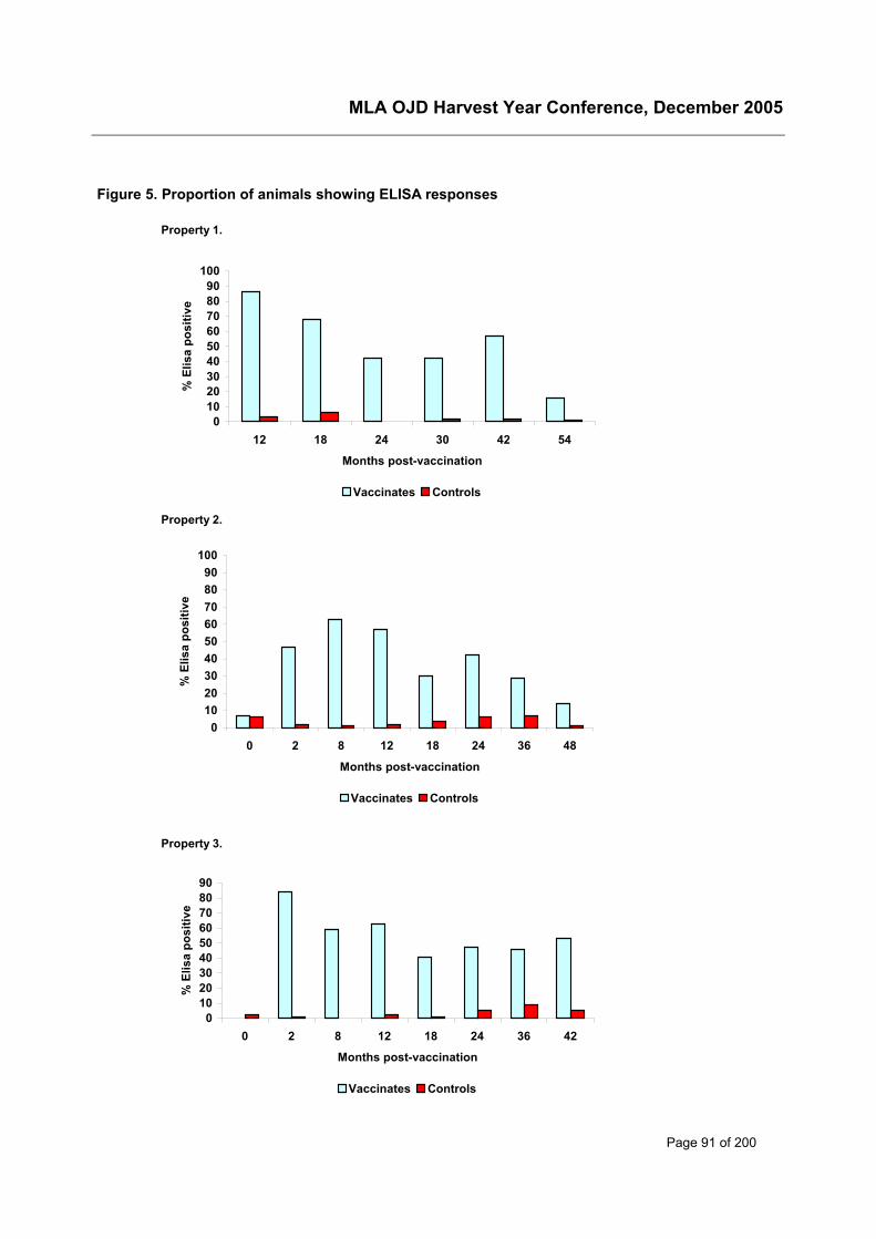
MLA OJD Harvest Year Conference, December 2005
Page 91 of 200
Figure 5. Proportion of animals showing ELISA responses
Property 1.
Property 2.
Property 3.
0102030405060708090
100
12 18 24 30 42 54
Months post-vaccination
% E
lisa
posi
tive
Vaccinates Controls
0102030405060708090
100
0 2 8 12 18 24 36 48
Months post-vaccination
% E
lisa
posi
tive
Vaccinates Controls
0102030405060708090
0 2 8 12 18 24 36 42
Months post-vaccination
% E
lisa
posi
tive
Vaccinates Controls
MLA OJD Harvest Year Conference, December 2005
Page 92 of 200
Excretion of M. a. paratuberculosisFigure 2 shows the estimated prevalence of shedders on each farm at each sampling timeobtained from the pooled prevalence calculator (http://www.ausvet.com.au/pprev/). There wereobvious differences between the three properties. P1 had the lowest prevalence of sheepexcreting M. ptb. P2, had the highest prevalence and showed a distinct peak , reaching 26% incontrol sheep at 24 mths pv. By 48 mths, the prevalence in these control sheep had dropped toless than 2%, presumably because many of the infected sheep had died. Despite thesedifferences, the proportion of positive PFC results in the vaccinated sheep was less than that ofthe controls at every sampling time after 2 months pv on each farm.
The absence or reduction of faecal shedding in the vaccinated groups compared to the controlsat each sampling time was very clear on farms P1 and P3. On these farms, there was nodetectable excretion of M. ptb by the vaccinated groups until 18 months pv (about 21 months ofage), whereas excretion, was detected in the controls as early as 6 or 8 months pv (9 -11months of age). The reduction in prevalence of excreters averaged about 90%, and wasmaintained throughout the trial. The results from these two farms thus clearly demonstrate adelay and reduction in faecal excretion of M. ptb, with maintenance of the protection for theeconomic life of the sheep. The results for P2 differed in that there was a smaller delay in thestart of shedding, and at the final 2 samplings at 42 and 48 mths pv, there was little difference inprevalence between the vaccinated and control groups. The peak prevalence for excreters overthe life of the sheep was, however, very much lower, and delayed, in the vaccinated group.
Figure 3 shows the total daily amount of M. ptb excreted by each group at each sampling time.This is a measure of the potential for environmental contamination with the organism, and thusthe risk to subsequent generations of sheep. At each sampling time throughout the trial on P1,and up to 30 months pv on the other farms, the numbers of M. ptb excreted by the vaccinatedgroup were at least one log lower than the controls. Note, however, that the actual amount of M.ptb excreted can be very much higher than indicated by prevalence data alone. This may be areflection of the large contribution of a single (or a few) multibacillary infected sheep.
Immunological responsesFigures 4 and 5 show the proportions of sheep with positive IFN-γ responses and ELISAantibody at each sampling time on each farm. The stimulation of both the cell mediated andhumoral immune systems by vaccination is evident.
Cell-mediated (IFN-γ) responses (Figure 4) - In controls, positive IFN-γ responses were detectedby 8-12 months pv and were likely to be due to environmental exposure to M. ptb. Theproportion of vaccinates with positive IFN-γ responses was maximum at the first pv test on eachfarm, and then declined. These proportions remained higher than in the controls at everysubsequent sampling time. Positive pv IFN-γ response was negatively associated with sheddingand with infection. No significant association with OJD-mortality was found, but there were veryfew vaccinated sheep that died of OJD.
Humoral responses - The pattern of ELISA results (Figure 5) was similar on P1 & P3, with highlevels of seroconversion in vaccinates, which declined slowly for about 18 months, and thenlevelled out. On P2, the most severely affected property, antibodies to M. ptb were present in 6-7% of lambs at the time of vaccination. This indicates the presence of passive maternalantibody, which may have affected the ELISA responses to vaccination. On this property fewersheep responded initially, and by 8 months pv the percentage of reactors peaked at only 63%,in comparison with levels above 80% on the other two farms. At the 24 months pv samplingthere was a large increase in the percentage of vaccinates with positive ELISA results, which
MLA OJD Harvest Year Conference, December 2005
Page 93 of 200
may indicate an immune memory effect as vaccinates are exposed to increasing levels ofinfection. Among control sheep, seroconversion occurred from about 12 months and theproportion of ELISA positives was also greatest at 24 months pv. These seroconversions likelyreflect the development of advancing disease. Positive pv ELISA response was negativelyassociated with shedding, with infection and with OJD-mortality.
The effect of detectable maternal antibodies on the subsequent development of ELISA and IFN-γ responses in vaccinated sheep was assessed by comparing the post-vaccinal immunologicalresults for lambs with and without maternal antibody on P2. The presence of detectableantibodies was negatively correlated with CMI responsiveness following treatment. A similartrend was observed with regard to ELISA results. To ascertain whether the association ofmaternal antibodies with reduced post-vaccinal CMI responses was of practical significance, weexamined the association of detectable maternal antibodies on the subsequent OJD status inboth vaccinated and unvaccinated sheep, and no significant effect was found.
Vaccination site lesionsInjection site lesions were not identified in any control sheep, but 42% of vaccinated lambs hadlesions at 2 months post vaccination. The prevalence of lesions decreased to a minimum 12months pv, and then remained steady at approximately 20% of vaccinates having palpablelesions until 3-4 years of age. The low percentage of palpable lesions at 12 months pv was mostlikely associated with the sheep carrying lots of wool, so that lesions were harder to detect. Noassociation was found between the presence of a palpable post-vaccinal lesion with excretion ofM. ptb, infection status, nor with OJD-mortality. Thus the presence or absence of a pv lesionwas of little use to predict efficacy of vaccination in individual sheep. At both the hogget cull andfinal slaughter, post-vaccinal lesions did not result in significant losses on the slaughter floor.
Comparison of IFN-γ, ELISA and post-vaccinal lesions as indicators of immune response -Among vaccinated sheep, the presence of a post-vaccinal lesion (2mths pv), IFN-γ responsesand ELISA responses (2 mths pv, P2,3 and 12 mths pv, P1) were compared using the kappastatistic. Although all three may be considered as indicators of vaccine response, there was nosignificant correlation between them, except for IFN-γ and ELISA on P3.
Production measurementsData used were from surviving sheep at each sampling time, so that loss of production due toOJD mortalities is not included in this analysis.
Liveweight and condition score – GLM analysis for liveweight, with weight at the time ofvaccination included as a covariate, revealed significantly higher liveweight gain for controlsheep compared to vaccinates at 2, 6 and 12 months pv. The magnitude of the differences wassmall, however (0.22, 0.34 and 0.75 kg at each time period respectively). In older sheep therewere no significant differences, except at 30 months pv, when the controls were again heavierthan the vaccinates. There were highly significant differences in weight gains between farms,but farm and vaccination interactions were not significant, except at 30 months pv. GLManalyses were also used to compare liveweight of infected sheep with those of sheep notdetectably infected. This examines the effect of subclinical OJD at the time of measurement.Infected sheep were significantly lighter, 1-2 kg at 24, 30 and 42 months pv, but there were nodifferences at other times. GLM analyses for condition score, with respect to vaccination orinfection status, yielded similar findings to liveweight, except at 30 months pv, when thevaccinates scored higher than the controls.
Fleece characteristics - There were no differences between controls and vaccinates in averagegreasy fleece weight or fibre diameter in 2001, nor in 2002. At the 2003 shearing, when sheep
MLA OJD Harvest Year Conference, December 2005
Page 94 of 200
were 3-4 years old, greasy fleece weight for vaccinates was slightly higher (0.123 kg) thancontrols. There were no differences in fibre diameter at any sampling time. With respect toinfection status, there were no significant differences at any sampling time.
DiscussionThis trial has been the first detailed, objective and controlled examination of the efficacy ofvaccination of sheep against paratuberculosis on commercial farms. In most overseas work todate, properly controlled studies have not been done, and it has not been possible to isolate theeffects of vaccination from other aspects of control programs. Preliminary results from this trialfacilitated the registration of GudairTM in Australia in 2002.
By project end there had been only seven confirmed OJD mortalities in vaccinates compared to80 from the controls (a reduction of about 90%), and OJD mortalities in the vaccinated sheepwere delayed by a mean of 17 months. This alone would justify the use of vaccination and willprovide immediate benefit to producers who are experiencing significant OJD-related mortalityin their flocks.
The PFC results demonstrated a delay and reduction in faecal excretion of M. a.paratuberculosis in vaccinated sheep, with maintenance of the protection for the economic lifeof the sheep. The reduction in prevalence of shedders was about 90%, and the total amount ofM. a. paratuberculosis organisms shed onto pasture was also reduced by more than 90% atmost sampling times. Reduced environmental contamination may lead to reduction in OJDprevalence in subsequent generations of sheep 2. Vaccination also increased the age at whichshedding commenced by about 12 months, thus providing a much larger window of safety formanagement of pasture contamination 6. These findings indicate that the estimates used inmodelling studies 7 that show vaccination should over time reduce within flock OJD prevalencewere reasonable, and underscore the incorporation of vaccination into assurance programs.
Development of immune responses was demonstrated following vaccination, and both pv IFN-γand ELISA responses of individual sheep were negatively correlated with subsequent faecalshedding, OJD mortality and infection status. Post-vaccinal ELISA response was at least asgood as pv IFN-γ response in predicting the subsequent OJD status of vaccinated sheep.Humoral responses are not considered in themselves to be protective 5, but the results from thistrial indicate that ELISA response may be a suitable marker for ‘vaccination take’.
There was no indication of any short term adverse clinical effect of vaccination. Vaccination sitelesions were persistent, but were not a major concern to processors. The presence of a pvlesion was of little use to predict efficacy of vaccination in individual sheep, but the persistenceof lesions in a proportion of vaccinates might act as a marker for flock vaccination.
A small reduction in the weight gains of vaccinates over the first 12 months pv was seen. Themechanism for this is unclear. Post-vaccinal lesions may be painful and/or reduce the animals’enthusiasm for grazing, but when vaccinated sheep with lesions were compared to thosewithout lesions no significant differences were found. Presumably a systemic reaction tovaccination, and/or some grossly unobservable local reaction was responsible for the lowerweight gains. The lower weight gain in young sheep is unlikely to be an economic disincentiveto vaccination in a fine-wool merino enterprise, but could be of concern in fat lamb operations.Vaccinates cut slightly more wool than controls at 3-4 years of age, but as no differences wereseen at other times, increased fleece yields could not be used to support vaccination. However,these measurements of production parameters only include surviving sheep at each samplingtime. Thus they ignore the considerable effects of vaccination in reducing OJD mortality.
MLA OJD Harvest Year Conference, December 2005
Page 95 of 200
There were differences between the three farms in the pattern of OJD expression during thetrial. Farm 2 appeared to have had a very high level of M. ptb challenge, with the highestproportion of OJD mortalities, and the highest proportion of subclinical OJD in culled hoggets(87% among controls). The proportion of IFN-γ reactors in controls peaked at 58% at 18months pv, and prevalence of shedders reached 26% at 24 months. Farm 3 was less severelyinfected, and the peaks in mortality, subclinical infection and IFN-γ reactors occurred later. Farm1 had the lowest numbers of mortalities, of shedders, and of IFN-γ reactors, and on this farm,the vaccinated group had no detectable shedders at several of the later sampling times.
Several aspects of the design of this trial should be considered when translating the findingsinto management and control recommendations for the wider industry.
Firstly, we investigated the effect of vaccination only against a very heavy challenge, because ofthe difficulty in detecting and quantifying M. ptb infection in low prevalence flocks, where few orno OJD mortalities are recognised. Moreover, in the current trial, unvaccinated control sheepwere run with the vaccinates, providing an on-going high level of challenge. Under normal fieldconditions, the entire cohort of an age group or mob would be vaccinated, so that this additionalchallenge from cohorts would be reduced. Thus, the current trial was a “worst-case scenario”from the perspective of disease challenge. Intuitively, one may expect that vaccination shouldbe more effective when other management procedures are used concurrently to reduce M. ptbchallenge, but this cannot be inferred from the current trial.
Secondly, in these experiments, we examined a single cohort of sheep over 4 to 5 years. In thereal world situation, a new crop of lambs would be raised and vaccinated each year. Theamount of M. ptb exposure would be expected to be successively reduced, with a reduction inthe opportunity for transmission of the disease to future generations. Any effects of vaccinationon successive generations of lambs over time is not reflected in the results from the currentstudy. Some useful insights on the likely long term effects of vaccination can be gained from theexperience in Iceland, where vaccination of sheep was introduced only after more traditionalcontrol measures, such as quarantine, and test and cull programs, had failed 3. There,vaccination has been an effective control measure and was shown to reduce mortality by 94%,but despite long term mandatory vaccination, OJD infection remains widespread.
There are also a number of potential concerns for the use of vaccination in control programs forOJD highlighted by the current trial.
All of the vaccinates that died had severe multibacillary infection, and would have beenexcreting enormous numbers of M. ptb leading up to their death. While there were very few ofthese sheep, the contribution of just a single sheep with multibacillary infection to environmentalcontamination is large. Just one, or a few, sheep with multibacillary lesions may have also beenresponsible for the relatively large amounts of M. ptb excreted by vaccinated groups at somesampling times. If this occurred at a critical time (eg in a ewe in the lambing paddock whenpasture was short and many young lambs were beginning to graze) a whole cohort of youngsusceptible animals may be exposed to large numbers of M. ptb. Another important observationwas that the effect of vaccination on subclinical disease, although significant, was less than theeffect on clinical disease, and vaccination did not decrease the proportion of subclinicallyaffected sheep that had multibacillary lesions. These findings have particular relevance whenvaccinated sheep from infected farms are moved to areas of low prevalence for OJD, and implythat sustained vaccine use will be necessary to avoid recrudescence in infected flocks
In this trial, vaccination was seen to provide life-long protection. However, because of the on-going high challenge with M. ptb, a natural boosting effect may have been operating. Theincrease in the proportion of vaccinated sheep with positive IFN-γ responses at 18 mths pv on
MLA OJD Harvest Year Conference, December 2005
Page 96 of 200
P2, and 24 mths pv on P3, is indicative of such an effect. Whether vaccinated sheep in a low orzero challenge environment would still be protected if suddenly exposed after several years tohigh levels of M. ptb is unknown.
ConclusionsIn flocks with a high prevalence of OJD, vaccination with Gudair™ stimulated specific immuneresponses and significantly reduced and delayed both OJD-related mortality and the excretionof M. ptb. Protection was maintained for the economic life of the sheep. Thus vaccination willprovide immediate benefit to producers who are experiencing significant OJD-related mortality,and should provide benefit to subsequent generations of sheep. However, ignoring theconsiderable OJD-related mortality, there were only small effects of either vaccination orsubclinical OJD on production parameters.
High levels of M. ptb excretion by vaccinates on some occasions, multibacillary lesions in thefew vaccinates that died from OJD, and persistence of subclinical OJD in a significant proportionof vaccinates, indicate risk that some vaccinated sheep could transmit the disease. This hasimplications for translocation of vaccinated sheep to very low prevalence areas, and implies thatsustained vaccine use will be necessary to avoid recrudescence in heavily infected flocks.
AcknowledgementsWe thank Ian Lugton for initial establishment of the trial, Kevin Thornberry for assistance duringsample collection, Idris Barchia for biometrical advice, and staff from the Orange AgriculturalInstitute, CSL Animal Health and from EMAI for the detailed laboratory investigations. Specialthanks are due to Shayne Fell for his diligence with the OJD cultures. Research funding wasprovided through Meat and Livestock Australia as part of the NOJDP.
References1. Cranwell MP. Control of Johne's disease in a flock of sheep by vaccination. Vet Rec 1993;
133(9):219-20.
2. Eppleston J, Reddacliff L, Windsor P, Links I, Whittington R. Preliminary observations on theprevalence of sheep shedding Mycobacterium avium subsp. paratuberculosis after 3 yearsof a vaccination program for ovine Johne’s disease . Aust Vet J 2005; 83(10):35-6.
3. Fridriksdottir V, Gunnarsson E, Sigurdarson S, Gudmundsdottir KB. Paratuberculosis inIceland: epidemiology and control measures, past and present. Vet Microbiol 2000; 77(3-4):263-7.
4. Garcia Marin JF, Tellechea J, Gutierrez M, Corpa JM, Perez V. Evaluation of two vaccines(killed and attenuated) against small ruminant paratuberculosis. 6th International Colloquiumon Paratuberculosis, Melbourne, Australia 1999; 234-41.
5. Perez V, Tellechea J, Badiola JJ, Gutierrez M, Marin JFG. Relation between serologicresponse and pathologic findings in sheep with naturally acquired paratuberculosis. Am JVet Res 1997; 58:799-803 .
6. Reddacliff LA. Grazing management and Ovine Johne's disease: insights from recentresearch into pathogenesis and epidemiology . Proceedings of the Australian SheepVeterinary Society, Gold Coast Conference 2005; 15:134-9.
7. Sergeant ESG. Modelling the spread of ovine Johne’s disease in infected flocks. In:Proceedings of the Australian Sheep Veterinary Society 2002; 12.
8. West DM. Johne's disease in New Zealand: history from first cases to current situation.

MLA OJD Harvest Year Conference, December 2005
Page 97 of 200
Allworth B. Proceedings of the Fourth International Congress for Sheep Veterinarians.Australian Sheep Veterinary Society, 1997: 151-4.
MLA OJD Harvest Year Conference, December 2005
Page 98 of 200
On-farm impacts of vaccination with GudairTMJeff EpplestonCentral Tablelands Rural Lands Protection BoardPO Box 20, Bathurst NSW 2795Phone: 02 6331 1377; Fax: 02 6331 6915Email: [email protected]
AbstractIn addition to the major vaccination trial conducted in three high prevalence flocks (TrialOJD.009), two field studies and several surveys and strategic samplings have been undertakenin other vaccinating flocks. These studies were conducted to better assess the full on-farmimpact of vaccination in a range of commercial situations. Both a survey of 50 early vaccineusers and a whole farm vaccination study confirm the major reduction in clinical diseasemeasured in Trial OJD.009. A preliminary study in seven high prevalence flocks in which avaccination program has commenced, suggests a 50% reduction in shedding after three yearsof vaccination. Results from a longer term monitoring program in 12 flocks with variable initialdisease prevalence will be available in 2-3 years time. Vaccination site lesions have producedinfrequent adverse effects on-farm, but a survey of a small number of abattoir consignments ofvaccinates suggests they will be of little consequence at slaughter. A survey of vaccinatorsindicated an exposure rate to Gudair of around 1 in 7,000 doses applied and a number of casesof self-injection injury have been reported.
IntroductionGudairTM was first released in Australia under permit in late 1999 for use in a major trial, (knownas the ‘Intensive vaccination trial’ - MLA project OJD.009 1) funded under the National OvineJohne’s Disease Program (NOJDP), aimed at determining the efficacy of vaccinating Merinolambs against OJD. Soon afterwards the vaccine was also supplied, again under permit, toowners of heavily infected flocks that had been experiencing large OJD-related losses (>5%).Following encouraging results from Trial OJD.009, GudairTM was registered in April 2002 forwider use by the sheep industry and since the completion of the NOJDP, vaccination againstOJD has been actively encouraged as a means of significantly reducing the on-farm impact ofthe disease and the risk associated with the purchase of re-stocker sheep.
The detailed outcomes from Trial 009 are being presented at this conference by LeslieReddacliff. In brief the trial showed that in flocks with a high prevalence of OJD, vaccination withGudair™ stimulated specific immune responses and significantly reduced and delayed bothOJD-related mortality and the excretion of M. a. paratuberculosis (M. ptb). Protection wasmaintained for the economic life of the sheep.
Trial 009 was conducted in commercial flocks. However the application of the vaccinationprogram was not reflecting commonly used management practices and in fact could beconsidered ‘worst-case’ scenario. The trial was conducted in one generation of sheep only (thefirst to be vaccinated on each property) and under extreme challenge from M. ptb. The trialflocks had a very high initial prevalence of OJD, the vaccinates were run with unvaccinatedcontrols and OJD clinical sheep from other mobs were grazed with the trial sheep at a timewhen they were young and susceptible to infection. Under normal farming practices all lambs inthe cohort would be vaccinated and other management procedures would be used concurrentlyto reduce M. ptb challenge, i.e. they would graze pastures with the lowest levels ofcontamination and not co-graze with older unvaccinated sheep. In addition many flocks would
MLA OJD Harvest Year Conference, December 2005
Page 99 of 200
also commence vaccination at lower disease prevalence and as the program proceeds anincreasing number of previous generations will have been vaccinated, resulting in decreasinglevels of pasture contamination. Therefore it is important that the outcomes from Trial 009 beconfirmed in a larger number of commercial flocks.
Further, while Trial 009 has demonstrated clear benefits from vaccination, it has also identifiedtwo potentially negative outcomes: i) that shedding continues at a level that is likely to beinfective, and ii) that persistent vaccination site lesions occur in around 25% of vaccinatedsheep. These issues resulted in the establishment of two additional field trials funded by theNOJDP outlined previously2.
In addition to these two field studies a number of other surveys and samplings aimed at a betterunderstanding of vaccination have been conducted in commercial flocks without the limitationsimposed by the experimental design used in Trial 009. This paper will outline the results ofthese studies and discuss them in relation to the on-farm impact of vaccination on the incidenceof clinical and subclinical disease, the level of shedding, vaccination site lesions and vaccinesafety issues.
Reduced clinical and subclinical diseaseThe most prominent and practically beneficial outcome from Trial 009 was the observation thatvaccination reduced mortalities due to OJD by 90% in these high prevalence flocks. Anotherhigh prevalence flock Trial OJD.015 (Whole flock vaccination3) estimated a similar reduction inmortalities in the two years following the implementation of whole flock vaccination. Anecdotallymost producers and animal health staff in high prevalence areas confirm that the incidence ofOJD-mortalities decreases rapidly following the commencement of a vaccination program.
Confirmation of reduced mortalities is also provided by a survey conducted in early 2003, of thefirst 50 high prevalence flocks allowed to vaccinate under permit, concurrent with Trial 009.Thirty four of the 37 respondents (74% response rate) ran a self-replacing Merino enterpriseand because all had commenced vaccination in 2000 most (21 - 62%) had all their sheepvaccinated, including four properties where the whole flock had been vaccinated before 16weeks of age. Of the 13 flocks still containing non-vaccinates, nine had only the oldest drop leftunvaccinated, while the remainder still contained two or three of the oldest drops asunvaccinated sheep.
Flock owners were asked to estimate the proportion of sheep dying with OJD-consistent(wasting) symptoms for each age group. While these data are subjective, all the flock ownershad previously observed significant mortalities in their flocks and were experienced in identifyingclinical sheep. In general these estimates agreed with the significant reduction in clinicaldisease observed in Trial 009. The incidence of mortality increased with age at vaccination. Theproportion of flocks reporting mortalities in cohorts vaccinated before 4 months of age, was lessthan in cohorts vaccinated after 4 months of age (13 of 33 – 39% vs. 43 of 49 – 88%; P<0.01).Accurate data on the magnitude of reductions in losses was not obtained, however mostreported reductions of more than 75% in sheep vaccinated as lambs compared to previouslosses in unvaccinated sheep of the same age.
In terms of subclinical effects, Trial 009 estimated a 70% reduction in the level of subclinicalinfection amongst vaccinates compared with controls, which is less than the 90% reductionobserved in clinical disease. In Trial 009 there was no significant production loss associatedwith subclinical infection which contrasts with the well documented reduction in milk productionin subclinically infected dairy cattle, and a report from New Zealand which estimatedsignificantly lower lamb and wool production in subclinically infected sheep. Again owners of
MLA OJD Harvest Year Conference, December 2005
Page 100 of 200
high prevalence flocks often report anecdotal improvements in production in vaccinatescompared to previously unvaccinated generations.
Changes in sheddingTrial 009 showed a delay and a 90% reduction in both the prevalence and total excretion of M.ptb in vaccinates. However in these high prevalence flocks the level of excretion detectedwould be likely to result in infection if sheep were moved to an uninfected flock. Modelling hassuggested that the rate of shedding would fall rapidly with successive generations of vaccinates4
but this needed to be confirmed in the field. As a result, a long-term prospective trial has begunto monitor the rate of reduction in shedding from successive generations of vaccinated Merinosheep in flocks with varying initial levels of infection (Monitoring shedding in high and lowprevalence flocks - MLA 0332), but final results will not be available for another 3 years.
Monitoring shedding in high and low prevalence flocks - Trial OJD.033In Trial 009 the impact of vaccination was only assessed in the vaccinated generation andwhere vaccinates had been exposed to extremely high levels of M. ptb. The potential value invaccination is likely to become evident over an extended period following vaccination of anumber of successive lamb crops. In addition the period required to reduce the risk of diseasetransfer in traded vaccinated sheep is likely to be dependent on the disease prevalence at thetime of commencing a vaccination control program. Many producers are likely to commence avaccination program when within flock disease prevalence is lower and the impact ofvaccination in these flocks is still unclear. The objective of this trial is to document the rate ofdecline in OJD risk following the introduction of a vaccination strategy in flocks of varyingdisease prevalence.
Twelve self-replacing Merino flocks were identified and classified as high, medium or lowprevalence (4 flocks per category) based on previous test results and owner estimates ofmortality. In each flock the age groups were identified into 3 cohorts (1 and 2, 3 and 4, and 5and 6 year olds), with the 2 older groups being sampled every 2 years to estimate disease(shedding) prevalence. At each consecutive sampling another age group will have beenvaccinated as lambs so that the following comparisons can be made.
Sampling 1 3/4 yo non-vaccinates vs. 5/6 yo non-vaccinates
Sampling 2 3/4 yo vaccinates vs. 5/6 yo non-vaccinates
Sampling 3 3/4 yo vaccinates vs. 5/6 yo vaccinates
Comparisons between the same age groups at different samplings will provide an estimate ofdisease prevalence in each flock at the commencement of vaccination, and will estimate anychange in prevalence that occurs in subsequent generations.
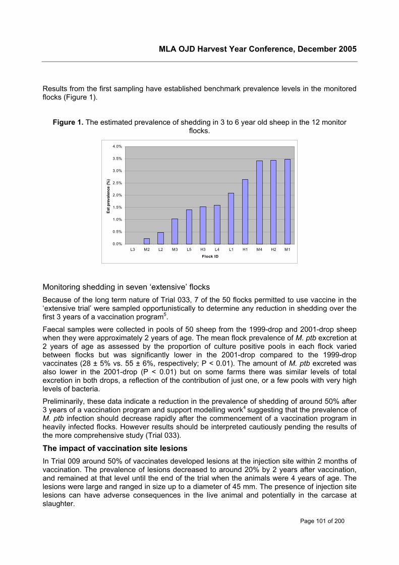
MLA OJD Harvest Year Conference, December 2005
Page 101 of 200
Results from the first sampling have established benchmark prevalence levels in the monitoredflocks (Figure 1).
Figure 1. The estimated prevalence of shedding in 3 to 6 year old sheep in the 12 monitorflocks.
0.0%
0.5%
1.0%
1.5%
2.0%
2.5%
3.0%
3.5%
4.0%
L3 M2 L2 M3 L5 H3 L4 L1 H1 M4 H2 M1
Flock ID
Est p
reva
lenc
e (%
)
Monitoring shedding in seven ‘extensive’ flocksBecause of the long term nature of Trial 033, 7 of the 50 flocks permitted to use vaccine in the‘extensive trial’ were sampled opportunistically to determine any reduction in shedding over thefirst 3 years of a vaccination program5.
Faecal samples were collected in pools of 50 sheep from the 1999-drop and 2001-drop sheepwhen they were approximately 2 years of age. The mean flock prevalence of M. ptb excretion at2 years of age as assessed by the proportion of culture positive pools in each flock variedbetween flocks but was significantly lower in the 2001-drop compared to the 1999-dropvaccinates (28 ± 5% vs. 55 ± 6%, respectively; P < 0.01). The amount of M. ptb excreted wasalso lower in the 2001-drop (P < 0.01) but on some farms there was similar levels of totalexcretion in both drops, a reflection of the contribution of just one, or a few pools with very highlevels of bacteria.
Preliminarily, these data indicate a reduction in the prevalence of shedding of around 50% after3 years of a vaccination program and support modelling work4 suggesting that the prevalence ofM. ptb infection should decrease rapidly after the commencement of a vaccination program inheavily infected flocks. However results should be interpreted cautiously pending the results ofthe more comprehensive study (Trial 033).
The impact of vaccination site lesionsIn Trial 009 around 50% of vaccinates developed lesions at the injection site within 2 months ofvaccination. The prevalence of lesions decreased to around 20% by 2 years after vaccination,and remained at that level until the end of the trial when the animals were 4 years of age. Thelesions were large and ranged in size up to a diameter of 45 mm. The presence of injection sitelesions can have adverse consequences in the live animal and potentially in the carcase atslaughter.
MLA OJD Harvest Year Conference, December 2005
Page 102 of 200
Adverse consequences in live vaccinatesSeveral reports from producers of adverse reactions in live sheep resulting from the presence oflesions have been collated:
• One producer had to apply a preventative flystrike treatment to his Merino weaners inDecember,10 weeks post vaccination, after 30 were struck near fistulating injection sites.
• Several producers reported problems associated with injection site lesions when the vaccinewas applied at a site other than the recommended neck area. A ram developed a largelesion near the brisket, likely to result in mating difficulties. Another producer reported thatvaccinating lambs on the cheek resulted in the development of lesions on the face that couldimpair grazing efficacy.
• In 4 flocks 5-6% of lambs or weaners died after developing paralysis following apparentdeep injection near the vertebrae resulting in focal chronic inflammation presumed to havecaused compression of the spinal cord.
Producers therefore should be encouraged to follow the recommended site of vaccination andto ensure that sheep are adequately restrained prior to treatment.
The impact of lesions at slaughter – Trial OJD.032Because of the prevalence of lesions observed in Trial 009 and reports particularly from NewZealand of discounts being applied to vaccinated carcases, a survey of 20 consignments ofvaccinates was conducted at slaughter to determine the variation in the prevalence of lesionsand their impact on carcase value 2.
The prevalence of lesions observed in the slaughter survey was 18% for mutton and 65% forlamb carcases. However there was considerable variation in the prevalence of lesions observedamongst consignments of mutton sheep (Figure 2; range 1 – 41%), but this was not related toage at vaccination or to the interval between vaccination and slaughter.
Figure 2. The prevalence of injection site lesions by consignment.
0%
10%
20%
30%
40%
50%
60%
70%
Mutton Lamb
% w
ith le
sion
s
MLA OJD Harvest Year Conference, December 2005
Page 103 of 200
The value of the trim removed on the slaughter chain was insignificant and the labour cost of itsremoval was nil. No carcase was downgraded to a lower value grade. This study was conductedat a time of low sheep supply, in carcases vaccinated at the recommended site high on theneck. In these circumstances OJD vaccination site lesions are not a significant cost toproducers or to the processing industry and will represent only a very small proportion of thetotal cost of OJD control by vaccination.
Self-injectionWhile a more detailed examination of vaccine safety will be presented separately at thisconference, a discussion of the on-farm impacts of vaccination cannot ignore the risks ofvaccine exposure and self-injection. As part of the survey of the first 50 flocks approved to usevaccine described previously, data on the incidence of vaccine exposure was also collected.Respondents reported they had vaccinated a total of 155,523 sheep, which included onecontractor who had vaccinated 63,000 sheep. There were five reports of incidents where theoperator had splashed themselves with vaccine, and 16 instances of needle stick injury withoutinjection. There were no reports of vaccine self-injection. This represents an exposure rate of 1in 7,406 and a needle stick rate of 1 in 9,720. If the contractor is deleted from the data set, theexposure rate and needle stick rate amongst farmer vaccinators increased to 1 in 4,406 and 1 in5,783 respectively. There were no reports of detrimental outcomes to exposed users in thisdata.
Reports of six cases of accidental self-injection resulting in medical intervention includingsurgical removal of injected material have been collated elsewhere.
These data suggest that accidental exposure is not rare in Australia and steps such asadequate restraint of lambs, minimising the number of activities undertaken when vaccinating,improved vaccination technique and seeking immediate medical attention following exposureshould be stressed to vaccine users.
Implications for industryThe benefits at a flock level of vaccinating against OJD in high prevalence flocks are obvious;rates of infections and shedding are rapidly reduced to levels that are insignificant to the ownerof the infected flock. However, in these flocks the continued existence of low numbers ofvaccinates with multibacillary disease means that the risk of disease spread remains in the flockshould vaccination be discontinued or in other flocks should the vaccinates be traded.
The value of vaccination in low prevalence flocks, whether low prevalence at the beginning of avaccination program or because of an ongoing vaccination program, is less evident. Intuitivelythe cumulative effects of vaccination on shedding over generations should mean the continuedreduction in risk associated with vaccinated sheep. Indeed the reduction in shedding measuredin the seven high prevalence ‘extensive trial flocks’ is encouraging but the final results from the12 flocks with varying levels of infection in Trial 033 will be more informative. In this regard it isimportant that the prevalence of gross lesions be monitored by abattoir surveillance as this islikely to provide useful information on changes in regional within-flock prevalence as districtvaccination proceeds.
While the prevalence of vaccination site lesions is significant, the cost of these to the sheepindustry appears to be of little concern provided the recommended site of injection is used. Twoimportant extension messages for producers should be i) the importance of vaccinating high onthe neck, and ii) strategies available for reducing the risks and impacts of vaccine exposure tothe vaccinator.
MLA OJD Harvest Year Conference, December 2005
Page 104 of 200
AcknowledgmentsThese studies were the result of much collaboration between Sydney University, NSW DPI andRLPB’s. We acknowledge the assistance of the many OJD-affected producers in these on-farmstudies.
References1 Eppleston J, Reddacliff LA, Windsor PA, Whittington RJ and Jones S. (2005) Efficacy of akilled Mycobacterium paratuberculosis vaccine for the control of OJD in Australian sheep flocks.Proc 8th Int. Coll. Paratuberculosis : In press.
2 Eppleston J, Reddacliff LA, Windsor PA, Whittington RJ and Jones S. (2004) Field studies onvaccination for the control of OJD in Australia – An overview. Proc Aust Sheep Vet Soc 14:56-59.
3 McGregor H, Windsor PA and Abbott KA (2003) A longitudinal study of ovine Johne’sdisease: estimation of the mortality rate two years after whole-flock vaccination. Proc AustSheep Vet Soc 13:102-106.
4 Sergeant E (2003) Cost effectiveness of vaccination against ovine Johne’s disease ininfected flocks. Proc Aust Sheep Vet Soc 13:123-127.
5 Eppleston J, Reddacliff LA, Windsor PA, Links I and Whittington RJ (2005) Preliminaryobservations on the prevalence of sheep shedding Mycobacterium avium subsp.paratuberculosis after 3 years of a vaccination program for ovine Johne’s disease (OJD) AustVet J 83:35-36.

MLA OJD Harvest Year Conference, December 2005
Page 105 of 200
Public Health and National Programs
MLA OJD Harvest Year Conference, December 2005
Page 106 of 200
Review of National Level Prevention and Controlfrom 8th International Colloquium on Paratuberculosis
Luzia RastRMB 417GUNDAGAI NSW 2722Phone: 02 69 441 588Fax: 02 69 441 876E-mail: [email protected]
BackgroundPapers and posters were presented from Holland(3), Switzerland(1), Australia(2), USA(2),Italy(1), Sweden(2), France/Holland(1), Israel(1) and the Czech Republic(1). Most work wasfocused on the dairy cattle industry. Australia and Sweden also reported work done on the beefcattle and sheep industries.
It was recognized that the potential link between Crohn’s disease in humans andparatuberculosis remains unresolved and that once this question is answered, futureapproaches to JD control in livestock may change.
Generally in countries with large livestock numbers and endemic JD it was accepted thateradication of paratuberculosis on a national level was technically not possible, necessary oreconomic. Therefore the main focus of paratuberculosis control needs to be to reduce theeconomic losses due to the disease. Any national program needs to actively involve industry inthe decision making and needs a communication and education strategy for the program to bewidely accepted. Until the potential public health issue is resolved the main effort needs to be toreduce M. ptb levels in foods that could transmit M. ptb to humans if contained in highconcentrations, mainly dairy products.
An exception to this was work from Sweden and Switzerland. Sweden has a very lowprevalence of JD in cattle and sheep and hence their control programs are aimed at protectingthis status by strict import controls and a ‘stamping out’ policy should JD be detected.
Switzerland recognized that there is a need to better define the prevalence of JD and itsgeographical distribution and to conduct a cost benefit analysis, prior to making any decision fora national JD control program.
Dutch StudiesTwo modeling studies were presented:
1. Development of a milk quality assurance program for paratuberculosis: stochasticsimulation of within-herd infection dynamics and economics by Weber M. F. et al.; and
2. Development of a milk quality assurance program for paratuberculosis: from within-andbetween herd dynamics to economic decision analysis by van Roermund H.J.W., et al.
These studies recognized that due to the as yet unresolved issue of M. ptb role in Crohn’sdisease in humans, M. ptb infection in cattle is of concern to the dairy industry and a milk qualityassurance program for paratuberculosis may reduce the potential risk of transmission tohumans through consumption of milk products. Importantly the goal of the milk qualityassurance program is to reduce the concentration of M. ptb in milk rather than to eradicate M.ptb. A milk quality assurance program is more cost effective than certification, surveillance and

MLA OJD Harvest Year Conference, December 2005
Page 107 of 200
control programs aimed at low risk trade of cattle and elimination of M. ptb at the herd level.Herds were categorised into green (M. ptb levels below a certain level) and red herds (infectionconfirmed resulting in M. ptb levels in milk above a certain level) depending on test results.(Entry and maintenance/surveillance)
These modeling studies concluded that:
1. Herd examination by ELISA for entry into the quality assurance program and continuedsurveillance by ELISA effectively ensures the quality of bulk milk by ensuring theconcentration of M. ptb did not exceed a maximum acceptable concentration(<103 M.ptb /L milk) in closed herds.
2. Faecal culture test was more efficient in identifying cattle for culling (red herds)
3. Additional preventative measures reduced within herd spread and increased theproportion of herds that could be certified as low M. ptb in bulk milk.
4. Low M. ptb bulk milk certified herds need to exclude animal trade to retain that status.
5. Management measures are less important when trade is restricted but are alwaysimportant in herds that are above minimum level (red herds)
6. Price difference between low level M. ptb milk (green herds) and higher level M. ptbmilk (red herds) is important to encourage farmers to have an incentive to join theprogram.
The results of each modeling study depended on some assumptions. They included amount ofbacteria in milk and effect of management on the within herd transmission of infection.However these assumptions were studied in a sensitivity analysis.
A third paper presented an overview of paratuberculosis control in the Netherlands(Paratuberculosis control in the Netherlands; the target and an overview of the activities.Franken P.)
Initially a control program was focused on preventative management and certification ofinfection free herds to prevent within and between herd transmission of paratuberculosis. Anew approach is based on limiting M. ptb infection in dairy products, mainly milk, since it iseconomically the most important product. The program needs to be cost effective and farmerfriendly, simple to manage and easy to communicate.
It is based on a three step program:
• A first test for classification of the herd
• A second test for surveillance of the previously negative tested herds
• A third test for control of herds that have tested positive.
Test used is ELISA of all animals older than 3 years (blood or milk)
Combined with control measures (such as careful movement of stock, culling of infected cowsand their lastborn calves) the numbers of negative testing herds are maintained.
MLA OJD Harvest Year Conference, December 2005
Page 108 of 200
Evaluation of intervention strategies for control of Johne’s disease inSwitzerland (Antognoli M.C. et al)
The prevalence of JD in Switzerland is not well known despite several small scale studies andmandatory reporting of clinical cases. It is suspected that disease is under reported and thatmany farmers and veterinarians have limited knowledge on JD.
The hypothetical link between BJD and human Crohn’s disease, the economic losses due toJD in the dairy industry and potential non-tariff trade barriers stimulated research into the cost,benefit and disadvantages of initiating a program for control of JD in Swiss cattle.
The animal health population institute at Colorado State University and the Swiss FederalVeterinary Office collaborated in a study to provide scientifically sound recommendations forthe selection of intervention strategies for the control of JD in Switzerland.
The study evaluated various testing strategies (herd sample test with ELISA and follow up ofreactors with faecal culture, whole herd ELISA testing, whole herd faecal culture testing,pooled faecal culture testing) and found that classification of herd status was not feasible withany of these strategies if within herd prevalence was <5% due to poor sensitivity of the testsand small herd sizes.
Management aimed at reducing exposure of susceptible calves to contaminated manure wouldbe more effective in controlling disease.
The study recommended the need for more complete and updated information on theprevalence and geographic distribution of JD in Switzerland before making a decision on anynational control strategies. An abattoir survey, culturing abdominal lymph nodes was proposedto achieve this.
US studiesA national Johne’s disease veterinary certification program: Key to success by McDonald J., etal.
In 2002 the US Department of Agriculture created a national JD control program andmandated that veterinarians in each state must be educated and certified to implement thecontrol program. The US needed a program that would provide a uniform base of knowledgeand deliver a constant message on JD diagnosis and control. An on-line certificate program forveterinarians was developed with JD experts from across the country. The program developedis in modular format with four basic information modules and two modules with nationalprogram information. While the certificate program was developed nationally itsimplementation was the responsibility of states and allowed for adaptation to state specificpractices, policies and procedures. The program was launched in February 2004 and sincehas been adopted by 35 states and has trained and certified 752 veterinarians.
A poster titled ‘Laboratory proficiency in testing for antibodies to Mycobacterium aviumsubspecies paratuberculosis: a new approach for the United States’, by Lombard J.E. et al.was presented
A new laboratory proficiency testing for M. ptb antibodies has been adopted and is based on ascoring system that uses qualitative (positive/negative) and quantitative (Z-scores) results. Itmakes allowance for the expected variability near the cutoff of test results. Z-scoring uses themedian and interquartile range values from participating labs’ results in the analysis. Thisscore can then be used to evaluate inter and intra lab variation in ELISA readings and allowsmore scrutiny of sample values and can be used to provide labs with feedback on consistency.

MLA OJD Harvest Year Conference, December 2005
Page 109 of 200
Australian studies‘Increasing involvement of herd owners in controlling paratuberculosis through assurancebased trading’ by Kennedy D. et al and ‘The Victorian OJD management and control program’,by Tobin F. et al were the two papers presented from Australia.
The first paper outlined the significant change in approach to JD control on a national levelfrom voluntary herd/flock certification to provide low risk replacements, regulatory control ofidentified infected or high risk herds/flocks and movement restrictions, to an assurance basedcontrol program that is driven by all (affected and non- affected) producers and therefore ismore equitable and encourages disease prevention and management.
An assurance based credit system has been developed for the sheep industry and is based ona quantitative risk assessment at the flock level. Scores based on background prevalence inthe region, test results and vaccination history are allocated to each flock or consignment ofsheep and declared in writing at the point of sale.
In the dairy cattle industry a herd scoring system (dairy assurance score) based on results ofherd testing and prevalence in the geographic region of origin is being developed.
JD is endemic in both these industries in Australia. The systems also provide pathways forimproving the scores by controlling the disease.
Surveillance in the beef cattle industry has detected very little JD in beef herds that had nocontact with dairy herds. The low risk nature in the beef sector has been recognised by a newassurance category for beef only herds that is based on biosecurity alone.
The alpaca industry appears to have successfully controlled disease in the 1990’s andsubsequently no or very little infection has been confirmed. Therefore this industry is in a goodposition to concentrate on protecting itself. A voluntary quality assurance system is achievedby individual animal identification, movement records and herd biosecurity.
Success of these schemes will require a continued communication and auditing system todetect any problems, to educate users and to confirm the confidence in the schemes.
The second paper by Tobin et al. outlined the progress in OJD management and control inVictoria since 2002. It outlined how OJD management had moved from a largely governmentsponsored effort at eradication, heavily reliant on regulatory bureaucratic processes with littleindustry support, to an industry sponsored management program supported by government.
Key factors in the success of the OJD management program in Victoria were:
• Appointment of an industry representative OJD advisory committee
• Development of a financial assistance package to provide incentive to owners ofinfected flocks to actively manage the disease
• Extensive and ongoing communication process
• Collaboration with all stakeholders
• Social support mechanism for affected producers
• Flexibility of program and ability to promptly adjust program based on feedback ofstakeholders
MLA OJD Harvest Year Conference, December 2005
Page 110 of 200
French/Dutch modelling studyA modelling study was conducted with the aim to evaluate if truly JD free herds acquired JDcertification and kept their status over time by Ezanno P et al. The Dutch program was used toconstruct a deterministic model of the evolution of the number of herds per health status over25 years.
Results showed that certification programs that use tests with perfect specificity, or confirmpositive results with a test with perfect specificity, to reduce false positive results maintainednumbers of truly free certified herds over the 25 year modeling time, while at the same timedetecting or clearing infected herds. Test and cull strategies alone will not lead to a rapidincrease in certified and truly M. ptb free herds but restrictions on purchase of animals fromherds at the highest level of certification and a decrease in within herd prevalence throughherd management is also needed to increase the number of truly JD free certified herds overthe 25 year modeling period.
Swedish studiesTwo posters were presented:
1. Control of paratuberculosis in live cattle and in semen imported into Sweden 1995 to 2004by, Holmstrom A. and Stenlund S.; and
2. Screening the Swedish sheep population for paratuberculosis Sternberg S., et al.
Because Sweden has a very low prevalence of JD in its cattle population their main effort is toprotect this status by minimising the risk of introducing infection from other countries. WhenSweden joined the EU in 1995 they applied additional assurances and control measures. Avoluntary import control program advises farmers to import semen or embryos instead of liveanimals. It further recommends that semen or embryos originate from tested herds. In the dairyindustry it is prohibited to purchase and introduce live animals from other countries.
These animal programs have successfully limited the number of live animals being imported.Since 1995 only 278 animals have been imported to Sweden and they all tested negative byfaecal culture and serology. Most herds of origin were also tested negative by serology. Since1998 there have been no dairy cattle introduced into Sweden.
JD is a notifiable disease in Sweden and has never been detected in Swedish sheep. Oneimported ram was detected as JD infected while in quarantine in 1999.
Since 1993 annual screening of the sheep population for JD has occured. Serology (AGID)was used until 2004 and since then faecal culture is being used. In addition all suspect clinicalcases are investigated. No infection has been detected to date.
Johne’s disease control program in the Israeli dairy herds (Koren O., et al)Israel has initiated a voluntary control program for JD in 2003 that aims at detecting infectedherds and provides management solutions to reduce and prevent herd infection.
More than half of the dairy herds in Israel are infected with JD but the within herd sero-prevalence is generally low.
The control program consists of: obligation of herd to follow a management program, wholeherd testing by ELISA (serum or milk) with faecal culture follow up of positive sero-reactorsand risk assessment. The herd is then classified on a risk scale (1-8) and a managementprogram aimed at reducing this risk is initiated.
MLA OJD Harvest Year Conference, December 2005
Page 111 of 200
Application of a pilot control program against paratuberculosis in an alpineregion in northern Italy (Liandris E. et al)Eighty-one dairy herds in this region were tested by ELISA and only 8 herds had no reactors.Prevalence amongst adult cattle was estimated at 19%.
On the basis of these results, a voluntary control program was implemented aimed atdecreasing the disease prevalence within the herds. The program was aimed at beingeconomic (cost and labour).
A risk assessment was completed first and then farmers implemented management strategiesto reduce disease transmission. Cattle are tested prior to parturition with ELISA. Faeces of anycow with a doubtful pre-parturition serology result is tested after parturition with real time PCR.All positive animals are recommended to be culled.
Paratuberculosis in cattle in the Czech Republic: current situation results ofnational control program and the main risk factors (Pavlik I. et al.)The Czech Republic has 1.7 million cattle on 5,405 farms (1998 figures). Around 3,500 herdsare considered as economically significant for milk production. Paratuberculosis wasintroduced through importation of cattle and by 2004 paratuberculosis was diagnosed in justunder 20% of these imported cattle.
Since 1998 a nationally subsidised control program is being implemented. It is based on faecalculture testing of all animals older than 18 months twice a year. Paratuberculosis is controlledin about 25-30 herds.
The main risk factors for JD spread are:
• Widely used practice of feeding calves with mixed colostrums and milk
• Use of progeny of infected cows for breeding
• Purchase of cattle with unknown status
The control program is hoping to encourage farmers to address these risk factors.
MLA OJD Harvest Year Conference, December 2005
Page 112 of 200
Review of Prevention and Control at a Herd Levelfrom 8th International Colloquium on Paratuberculosis
Peter Windsora and Jeff Epplestona Farm Animal Health University of Sydney PMB 3, Camden, NSW 2570
IntroductionTheme 3a was one of the largest themes of the colloquium with 37 papers submitted for eitheroral or poster presentation. These were accompanied by 2 review papers presented at the endof the Theme 3a session. In addition one of the workshops held after the conference was onvaccination and additional information was presented and discussed in some detail whichassisted integration of some of the new information into current thinking. For example, thesuccess of the Australian vaccine research has increased the likelihood of re-examination ofvaccination in the control of bovine paratuberculosis in the USA. Twenty four of the papers dealtwith control of paratuberculosis in dairy cattle herds, mostly in Europe or the US. Six paperswere on control in goat herds and there were 7 papers on control in sheep flocks, including 3papers presented on Australian vaccine work conducted under the National OJD Controlprogram (NOJDCP).
The major questions addressed in this theme were;
i) Are JD control programs cost-effective?
ii) What are the main risk factors in the spread of the disease?
iii) Which tests should be used in ‘test and cull’ programs?
iv) Can animal treatments reduce the risk of disease transmission?
v) Can treatment of faeces reduce the risk of disease transmission?
vi) Does vaccination effectively control paratuberculosis?
Summary of findings from papers and posters(i) Are JD control programs cost-effective?The likelihood of producers adopting voluntary control &/or eradication programs forparatuberculosis will depend on the cost effectiveness of the control program adopted. Fivepapers presented data on the cost, either in production or financial terms of running infectedherds. All presented data documenting significant costs associated with infection. For examplein the US dairy industry one paper documented a 4 fold reduction in lifetime milk production inELISA positive cows and an increased risk of other diseases during their lifetime; anothermeasured significantly reduced milk production in heavy shedders. In his review paper ScottWells of the USA concluded that in heavily infected herds the production costs do warrant thecost of effective control programs.
However there are varying levels of intervention possible to control paratuberculosis in cattleand another 3 papers modelled control options to determine the ‘best’ program for producers inan environment where control is voluntary. In general these investigations concluded that oftenprograms involving improved calf hygiene and minimal testing were the most cost effective forproducers, rather than the strict application of an intensive test and cull program. Russell Bush
MLA OJD Harvest Year Conference, December 2005
Page 113 of 200
of Australia presented the only paper on sheep. He developed an economic model aimed atdetermining the on-farm financial effects of different control options.
It was noted however that the outcomes from modelling were likely to change under mandatorycontrol programs should, in the future, human health concerns dictate this type of approach.
(ii) What are the main risk factors in the spread of the disease?Four papers examined dairy industry survey data for associations between herd managementfactors and measures of within herd JD prevalence. Most confirmed previously identified riskfactors such as herd size and purchasing replacement cows. Herd hygiene, particularly thoseactivities aimed at reducing the exposure of calves to contamination from the dairy environmentand from pooled colostrum or milk, was identified as important for minimising herd prevalence.In this regard a survey of Irish dairy practices identified poor calf biosecurity as a major limitationto controlling the on-farm impact of JD.
The importance of segregating offspring from their dams at birth was demonstrated in a Spanishdairy goat herd where the infection rate of kids not separated from their mothers was 73%compared to 26% in separated kids Two papers reported a significant reduction in JDprevalence in dairy goat herds following the implementation of a program of post partum kidseparation and feeding with artificial colostrum and milk.
In a survey of Italian dairy farms the level of disease was positively associated with the ironcontent of the drinking water suggesting that further investigation into this relationship could beworthwhile.
(iii) Which tests should be used in ‘test and cull’ programs?Ten papers or posters addressed the issue of tests involved in herd control programs. Broadlythey could be divided into studies examining the difficulties of using current testing regimes forcontrolling paratuberculosis at the herd level, or in a number of cases, early herd prevalencedata in countries where control programs have only recently or are yet to commence.
One study examined the interferon gamma test. It was evaluated in goats in Norway with testsperformed a day after collection, comparing results from unvaccinated herds withoutparatuberculosis, vaccinated herds without paratuberculosis and vaccinated herds with a historyof paratuberculosis. Despite re-sampling being required when 5% of animals in control sampleswere found to be positive, the study concluded that the low response rate of 1% of positiveanimals in 1,200 animals from herds free of paratuberculosis, suggests a role for this test inmonitoring unvaccinated herds.
Several studies examined the ELISA combined with faecal culture or PCR to investigate the riskof milk from herds of differing paratuberculosis status, or to more rapidly predict the status of atested animal. Classification of 668 dairy cows as low, medium or high risk based on four annualherd screens using the ELISA and one annual herd screen by faecal culture, found that 4 of 386cows classified as low risk (never positive) had PCR positive milk, posing a serious risk ofdisease transmission. Another study used a combined screening test used on milk using bulkmilk serology (SvanovirTM) followed by capture-PCR to detect M. ptb. DNA. This approach wasconsidered a viable option to protect the consumer by excluding M. ptb. containing milk from themanufacturing process. In a study using the mean ELISA adjusted optical density measures asa cut-off for groups of cattle with negative, low and high faecal shedding culture results, the M.ptb. shedding status in 2,578 adult cattle from six herds was predicted. The mean ELISA cut-offfor a positive test for negative, medium or high shedders was 3.2, 9.7 and 1,600 respectively.This suggests the ELISA adjusted OD measures can provide a more rapid prediction of theprobability of a cow shedding M. ptb. than the 8-16 weeks required for culture.
MLA OJD Harvest Year Conference, December 2005
Page 114 of 200
One study examined the sensitivity of pooling of faceal samples for culture from cattle atslaughter, using pools of 3, 5, 8, 10 and 15 samples per pool. The study found that pools of 10were of adequate sensitivity (59%) but pools of 5 were optimum (67%).
A survey of ruminants in several districts of northern India for incidence of M. ptb. using ELISA(Bison type antigen) found prevalence of antibody as high as 40.3%, 42.6% and 50.0% inbuffalo, cattle and goats respectively across the region, with higher rates of infection found inbovids when tested by faecal (62.5%) or milk culture (70.3%). Further, examination of goat kidsbetween 0 and 6 months using culture and PCR on faeces and tissues at slaughter, plus ELISAon serum, identified high rates of infection with Bison type M. ptb. up to 56%. In Portugal, apreliminary study of paratuberculosis in cheese producing sheep identified that 5.6% of 1,220animals were positive by ELISA, with 14 of 145 animals positive by AGID, all of which had grosslesions of bowel thickening but only 7 having histological lesions of paratuberculosis. Sixisolates of M. ptb. were obtained from these 14 sheep, with molecular identification to follow.
(iv) Can animal treatments reduce the risk of disease transmission?A paper by Whitlock et al presented results of a randomised double blind trial in 12 calvesdosed with M. ptb. between days 7 and 9 then treated twice daily with 35mg Monensin in theirmilk replacer until euthanasia between days 65 and 67. The study suggested that calves fedMonensin had a 55-72% reduction in both passive shedding and tissue culture of M. ptb. At70mg/day, this higher rate of Monensin content than is normally added to calf starter may havea role in reducing the bioburden of M. ptb. in neonatal calves, probably through direct action onthe mycobacterial cell.
(v) Can treatment of faeces reduce the risk of disease transmission?As M. ptb. is known to survive in faeces for extended periods in the environment, separateexperiments on survival of the organism in compost and following calcium hydroxide treatmentwas examined. In a short paper by Gobec et al. from Slovenia, survival of M. ptb. in sheepmanure following composting in a bioreactor vessel was considerably shortened, with positivePCR results for M. ptb. up to 7 days only in the composting system versus the full 21 days of theexperiment when faeces was left in open storage. Treatment with calcium hydroxide (to raisethe pH to 12.7) of sterile faecal slurry spiked with M. ptb. (109 cfu in 300ml) from Irish dairyherds resulted in a reduction but not elimination of the organism from the slurry up to 72hrs aftertreatment. These observations confirm the survival of M. ptb. in environmental samples,suggesting such treatments are impractical methods of inactivating the organism.
(vi) Does vaccination effectively control paratuberculosis?The Australian paper by Eppleston et al which evaluated use of GudairTM to control OJDpresents convincing data that supports vaccination as a key tool for the control of OJD at theflock level. Over the productive life of Australian merino sheep in 3 high prevalence flocks,vaccination reduced mortalities by 90% and delayed and reduced faecal shedding of M. ptb. by90%, although the few vaccinates that developed OJD had multibacillary disease and excretedinfectious doses of the organism. Vaccination site lesions occurred in about 50% of vaccinatesbut declined to between 20-25% during the course of the 5 year study. The potential costs of thevaccination site lesions were found to be negligible following investigations of 20 lines of sheepat slaughter, where 65% of lamb carcases and 18% of mutton had vaccine lesions.
Three posters presented data on the new vaccine SILIRUMTM in cattle. The vaccine is alsoproduced by CZ Veterinaria of Spain, and is a killed vaccine but uses a different adjuvant to thatin GudairTM in an attempt to reduce vaccination site lesions. A field study was conducted inSpain in a Friesian herd of 468 animals with 12% annual clinical cases of BJD, with 75% of all
MLA OJD Harvest Year Conference, December 2005
Page 115 of 200
cattle vaccinated and 26% retained as controls for 30 months post-vaccination. More than 90%of vaccinates produced persistent cellular and humoral immune responses to the vaccine andno new cases of BJD occurred from 6 months post-vaccination. The percentage of the 192animals culled during the trial with lesions of BJD was significantly higher in the controls thanvaccinates, with 92.3% of controls having lesions compared to 56.6% of vaccinates, indicating asignificant reduction in BJD in the herd following vaccination. A pilot field study with SILIRUMwas also conducted in a herd of 113 cattle with a clinical incidence of BJD of 8% and faecalculture prevalence of 5.5%. After one year of control, no new clinical cases of BJD haveoccurred, the faecal culture prevalence is 3.6% and no interference with intradermal testing fortuberculosis has occurred, suggesting expansion of the trial to a maximum of 50 herds is likely.SILIRUMTM was also evaluated in 10 of 18 calves that were challenged orally with 6.9 x 10¹⁰ cfuof M. ptb. following vaccination without adverse reactions (although my questioning of theauthor of the poster revealed that all vaccinates had injection site lesions up to 15cm). Cellularand humoral responses were evaluated with interferon-gamma results detected at 30 days andstill high at 330 days post-vaccination. The calves were killed at 180 and 330 days post-vaccination and only one vaccinate had a diffuse lesion of BJD, although several had lesionsmainly in lymphoid tissues. Isolation of M. ptb. was reduced in vaccinates, suggesting thisvaccine will reduce bacterial load and severity and numbers of lesions in challenged calves.
What can we conclude from current research into herd level control?The review and perspectives session on herd control provided papers by Scott Wells (scientistsview) and George Caldow (consultants view), focused on the reasons for controllingparatuberculosis and prospects for herd level control, particularly in dairy herds. The US dairyherd prevalence of 20-40% from 1996 NAHM survey is considered to be only half the trueprevalence and although the within herd prevalence is mostly below 5%, some have up to 50%.Expensive test and cull programs appear to have been largely unsuccessful due to failure torecognise and test for the disease and inadequate compliance with control procedures, plus theperceived costs of joining the control program. At current milk and beef prices, there is noincentive to join a control program unless there is a high prevalence in the herd.
The cost of paratuberculosis control in positive herds with prevalence below 10% is estimatedas $97/cow, compared to costs of $245/cow where the prevalence exceeds 10%, althoughthese figures may underestimate the true costs due to difficulty of measuring subclinical losses.Key findings on transmission of the disease include risks from faeces and pooled colostrum. Ininfected herds, survival of M. ptb. has been recorded in bovine slurry at 50C for over 8 months.Further, in infected herds, the farm environment is culture-positive in 95% of herds (with at leastone positive pool of cow faecal samples) with cow alleyways 77% positive, manure storageareas 68% positive, calving areas 21% positive and cow sick pens 18% culture positive, areevidence of the risk of environmental transmission. However the risk of colostrum and rawwaste milk is significant, with 27% of subclinically infected cows found to have culture positivesupramammary lymph nodes and 12% having culture-positive raw milk.
So what are the best control strategies for infected dairies? Firstly, define the status. Currentlythere is debate as to the best method to do this, with ELISA tests compared to PFC and theprospect that environmental sampling may provide the cheapest screening test. Secondly,encourage infected herds to enter a Voluntary Johne’s Disease Herd Status Program (VJDHSP)or equivalent, to:
1. Test/cull: problem being that can currently only detect animals when they are infectious
2. Vaccinate: possible problem being there is limited proof that vaccination reducesshedding in cattle
MLA OJD Harvest Year Conference, December 2005
Page 116 of 200
3. Reduce exposure of herd to M. ptb. Options include the possibility of neonatal calftreatment with monensin to decrease risk of infection and to test and remove positives(especially the ‘supershedders’) although is this cost effective?
However the most important strategies are implementing calf management, including avoidinguse of pooled colostrums, segregated heifer rearing and closing the herd to introductions ofpossible undetected infected carriers. This suggests that test and management is a moreappropriate approach to control, except in high prevalence herds where test and cull may resultin more rapid control. However further work on vaccination in cattle is required to determine ifthe results showing rapid reduction of shedding in sheep after vaccination can be applied tocattle control.
In conclusion, the tools are available to reduce the within-herd prevalence of infection in sheepflocks and in dairy and beef cattle operations to minimise economic loss to cattle producers.However we need further information from clinical trials and research to fine-tune effectivemanagement programs in cattle as it is currently unclear whether we are able to provide cost-effective eradication of Johne’s disease from cattle herds. Effective control of paratuberculosisand elimination of clinical cases will be necessary if M. paratuberculosis is conclusively shownto be a public health risk to protect human health and satisfy public demands for food safety.
MLA OJD Harvest Year Conference, December 2005
Page 117 of 200
Review of Implications for Public Health8th International Colloquium on Paratuberculosis
Sally SpenceLocked Bag 21ORANGE NSW 2800Ph: 02 6391 3630; Fax: 02 6361 9976Email: [email protected]
BackgroundThe possibility that Mycobacterium avium subsp. paratuberculosis (MAP) causes humandisease is the principle driver for implementing control and eradication programs in animalpopulations in many countries.
Papers and posters presented under the theme ‘Implications for public health’ at the 8th
International Colloquium on Paratuberculosis addressed a number of issues. These were:
• the presence of MAP organisms in patients with Crohn’s disease and other diseases
• the presence of MAP organisms in the environment and food products
• the impact of pasteurisation on the survival of MAP organisms in milk.
Summary of papers and postersa) Human disease and MAPBoth Crohn’s disease and paratuberculosis are increasing in prevalence throughout the world.Crohn’s disease is more prevalent in developed countries (6 cases per 100,000 in the USA andNorthern Europe) than in the third world (1 case per 100,000).
Many Colloquium attendees appeared to accept that MAP is, at the very least, an antigenprovoking an autoimmune process in genetically susceptible people. Little discussion was heardsuggesting that MAP is a primary infectious agent causing human disease.
CT Dow of the USA postulated that MAP acts as a ‘superantigen’ in individuals with theCARD15 mutation which confers susceptibility to Crohn’s disease. Mutations on the samechromosome are associated with Blau syndrome, a rare familial, juvenile, systemicgranulomatosis with primary clinical findings of uveitis, arthritis and dermatitis. Dr Dow foundthat all 6 granulomatous tissue samples examined from 5 individuals with Blau syndrome wereIS900 positive.
Dr Dow also postulated that MAP acts as an immune antigen to trigger Type 1 diabetes. Helinked this to the finding that early exposure to cows milk increases the risk of acquiring Type 1diabetes in the genetically at risk.
Some papers demonstrated an association between the detection of MAP and Crohn’s diseasewhile others found that there was no association between MAP and Crohn’s disease.
Analysing blood samples using a nested PCR Elguezabal et al in Spain did not find anyassociation between Inflammatory Bowel Disease and MAP. They found a high frequency ofMAP carriers in the healthy population: 41.5% of Inflammatory Bowel Disease patients and42.5% of normal controls were IS900 positive.
MLA OJD Harvest Year Conference, December 2005
Page 118 of 200
An Italian study by Taddei et al failed to culture MAP in biopsy samples from 30 patients withinflammatory bowel disease (including 8 Crohn’s disease patients) and 80 healthy patients.
A Mexican study by Favila-Humara et al found that a greater proportion of Crohn’s diseasepatients with granulomas were MAP IS900 positive than those without granulomas. Nine of 14patients were positive.
Singh et al in India cultured MAP from the stools of 2 people suffering clinical bowel disordersand was unable to culture MAP from the stools of 6 healthy individuals.
The review by Dr Ole Thomsen argued against a link between MAP and Crohn’s disease. Hementioned that a wide range of chemotherapies have been trialled on Crohn’s disease patientswithout success. He also pointed out that Crohn’s disease patients respond toimmunosuppressive therapy while TB is reactivated by such therapy. He mentioned thatinoculating animals with material from Crohn’s patients does not result in disease in the animals.
b) MAP in the environment and food productsA study by Pickup et al found that 32.3% of water samples from the Taff river in Wales wereIS900 positive over a 12 month period. There was a significant relationship between positivesamples and rainfall on the preceding 6 days. They were able to culture MAP from 38.7% of thepositive samples. Following DNA examination, they concluded that a different strain of MAP waspresent in the positive water samples from which they had been unable to culture MAP. MAPwas also isolated from sediment taken from reservoirs on the Taff river and from 1 of 70 housewater tanks sampled. They linked the distribution of Crohn’s patients in Cardiff to wind directionand postulated that aerosols carrying MAP from the river Taff was responsible for thatdistribution.
MAP was detected by IS900 based PCR or by culture from 8% of raw water samples enteringwater treatment plants in Northern Ireland by Rowe et al. No significant correlation was foundbetween numbers of coliforms or faecal coliforms and the presence of MAP. They found thatMAP was readily engulfed and concentrated by several types of protozoa. They also found thatchlorine caused a larger drop in MAP numbers when the organism is free than when it isengulfed by protozoa. They suggested that the protozoa may help MAP survive in water andprotect it from chlorination.
Hruska et al from the Czech Republic cultured MAP from 2% of powdered milk samples anddetected IS900 in 49% of powdered milk samples. The powdered milk sampled had originatedin 7 different EU countries. They had also sampled the milk from a small number of cowsshedding MAP in the faeces. Most of these cows were shedding MAP in their milk but in all thecases they tested only one quarter was shedding MAP.
Irene Grant discussed the presence of MAP organisms in food in her review presentation. Shesaid that MAP had not been detected in sheep milk but it had been detected in goats milk. Shepointed out that cattle infected with MAP can have disseminated MAP organisms in lymphnodes, muscle and blood consequently localised infection can be spread through a whole batchof beef by mincing.
c) MAP and pasteurisationA number of papers addressed the issue of the impact of pasteurisation on survival of MAP.Many studies demonstrated that some MAP organisms survive commercial pasteurisation but 5to 7 log reductions in the numbers of organisms does occur.
One study found that homogenisation of milk prior to pasteurisation did not contribute to theinactivation of MAP organisms.

MLA OJD Harvest Year Conference, December 2005
Page 119 of 200
High hydrostatic pressure milk treatment was found to be at least as effective as pasteurisationfor MAP inactivation.
Microfiltration can only be used with skim milk but does remove 98% of organisms.
Herman et al demonstrated that clumping of MAP organisms in milk or faeces was not thereason some MAP organisms survive pasteurisation. They were able to demonstrate that heatactivation of MAP growth does occur. They found that heating milk results in a shorter time toMAP growth being detected but does not increase the total number of positive samples ifculturing continues for 12 months.
ConclusionsThe question of whether MAP causes disease in humans was not answered at the Colloquium.
It was postulated that MAP acts as an antigen which can trigger Crohn’s disease and otherautoimmune diseases such as Blau syndrome and Type 1 diabetes in genetically susceptibleindividuals. MAP was demonstrated to be present in granulomatous lesions sampled in Blaudisease patients.
Of the papers examining an association between Crohn’s disease and the presence of MAP inhumans some demonstrated an association while others did not.
Several papers demonstrated that MAP was readily found in water ways in England and Irelandand that some organisms survive chlorine treatment.
MAP IS900 was detected in 49% of Czeck powdered milk and MAP was cultured from 2% of thesamples.
Several studies demonstrated that some MAP organisms survive commercial pasteurisation butpasteurisation results in a 5 to 7 log reduction in the number of MAP organisms present in milk.
MLA OJD Harvest Year Conference, December 2005
Page 120 of 200
Crohn’s Treatment Trial
Warwick Selby
Clinical Associate ProfessorAW Morrow Gastroenterology & Liver Centre,Royal Prince Alfred HospitalPhone: 02 9519 9550Email: [email protected]
An infectious cause for Crohn's disease has been sought since the disease was first describedin 1932. Recent attention has focussed on Mycobacterial avium subspecies paratuberculosis(M. ptb) since its isolation from 3 of 11 patients with Crohn's disease in 1984. Numerous reportshave appeared since then describing detection of the organism in tissue by PCR and culture. Ithas also been detected in blood and even breast milk. However, while some workers haveconfirmed these findings other have not found differences between Crohn's disease andcontrols. Epidemiological and other factors also provide contradictions if M. ptb is the cause ofCrohn's disease and hence the role of the organism remains controversial.
Conventional anti-tuberculous antibiotic regimens are generally ineffective in patients withCrohn's disease. However, M. ptb appears to exist in a cell wall deficient form and this,combined with its intracellular location and slow growth, make it resistant to these antibiotics.Anecdotal open-label studies using antibiotics that are effective against MAP have reportedfavourable results. However, there have been no large controlled trials until now. The Australiantrial of anti-paratuberculosis therapy (APT) randomised 213 patients to either clarithromycin,rifabutin and clofazimine or matching placebos in addition to 16 weeks of a reducing dose oforal prednisolone. Patients in remission after that time continued antibiotics or placebos for upto two years. They were then followed for a further one year off trial medications. Although ashort term benefit was found for the antibiotic combination at 16 weeks this was not maintainedat one or two years of treatment. One year after the trial agents were ceased, only 13.7% ofthose who were on active treatment and 9.0% of those on placebo were in remission, a non-significant difference. These findings argue against a role of active infection with MAP as asignificant cause in the majority of patients with Crohn's disease.

MLA OJD Harvest Year Conference, December 2005
Page 121 of 200
Epidemiology, Economics&
Grazing Management
MLA OJD Harvest Year Conference, December 2005
Page 122 of 200
Epidemiology of ovine Johne’s diseasecross species transmission and survival of the organism in
the environmentRichard Whittingtona, Barbara Moloney and Catherine TaragelaFaculty of Veterinary Science, The University of Sydney425 Werombi Rd, Camden NSW 2570Phone: 02 9351 1619; Fax: 02 9351 1618E-mail: [email protected]
BackgroundIn 1995 gaps in knowledge about the behaviour of ovine Johne’s disease (OJD) in populationsof sheep precluded tight recommendations being made to limit spread of the disease. Thereforewhen the national control program commenced, research was commissioned by Meat &Livestock Australia (MLA) to reduce the number of unknown factors. This paper covers aspectsof the epidemiological research program that are not presented elsewhere in this volume.
Some important questions in 1995 related to the:
• Differences between Johne’s disease in cattle and sheep
- Are the diseases in sheep and cattle caused by different organisms?
- Can OJD and bovine Johne’s disease (BJD) be managed separately in Australia?
• Possible reservoirs of OJD in cattle and goats
- Are cattle/goats susceptible to OJD?
- Are cattle/goats suitable reservoir hosts for OJD?
- Can OJD be controlled in the presence of cattle/goats?
• Possible reservoirs of OJD in the environment
- How long does the organism survive in the environment?
- Do climatic factors explain the lack of OJD in western NSW and QLD?
- Does soil type influence the occurrence of OJD?
Sheep and cattleThe success of control based on depopulation and decontamination of farms with multiplegrazing species depended on the segregation of infection within individual species. In Australia,where sheep and cattle are the predominant grazing species and where M. avium subspeciesparatuberculosis ((M. ptb)) was believed not to be spread commonly between these species, amanagement option was to graze cattle on pastures during the decontamination period afterdepopulation of OJD infected sheep.
The first projects commissioned by MLA were laboratory based, molecular, epidemiologicalstudies to confirm that Johne’s disease (JD) in sheep and cattle in Australia were due todifferent organisms and had quite separate epidemiological pathways. A significant aspectabout this work was that it was conducted before the sheep strain could be cultured routinely. Ina comprehensive study, molecular fingerprinting using RFLP (Restriction Fragment LengthPolymorphism) analysis, which required large amounts of DNA, was mostly undertaken on
MLA OJD Harvest Year Conference, December 2005
Page 123 of 200
organisms extracted laboriously from the lamina propria of sheep intestines that had beenscreened for infection using histopathology (1). This research confirmed that sheep wereinfected with a dominant strain known as the S strain while cattle were infected with a variety ofcattle (or C) strains (3, 10). In this regard the pattern of OJD and BJD in Australia appeared toresemble that in New Zealand (2), where S and C strains were first described, and wasdissimilar to the situation in Europe where sheep, cattle and other ruminants were most ofteninfected with various C strains (8). An explanation for this difference may be the relatively recentintroduction of OJD (S strain) into Australia and the concentration of BJD in Australia in the dairyindustry which has had very little contact with the sheep industry. These findings enabledgreater confidence in the management of both OJD and BJD in Australia.
Rapid molecular tests based on DNA sequence polymorphisms in the IS1311 gene were soondeveloped to facilitate routine differentiation of S and C strains, including retrospective testing offormalin-fixed paraffin-embedded tissues in laboratory archives (6, 9, 14). Further studies havebeen undertaken to identify the extent of genomic differences between S and C strains in aneffort to better define the microbial factors underlying the observed epidemiological differences(see Ian Marsh, this volume).
During the 1980’s and early 1990’s there were several anomalous cases of JD in cattle in NSWin which tissue or faecal cultures were negative. These were investigated using the IS1311method on archival tissues and S strain infection was confirmed from three NSW properties.Consequently, MLA commissioned a further project to determine the prevalence of disease incattle exposed to sheep infected with OJD and the likely risk factors (7). In this study, 1,808cattle from 12 properties were tested by ELISA (for the presence of M. ptb antibodies) andfaecal culture. All properties had a known history of JD in sheep and were mostly selected fromparticipants in a trial being conducted within the Australian National Ovine Johne’s DiseaseControl program. All properties had young cattle likely to be susceptible to JD. All animals gavenegative results on serology; only one animal from a herd of 349 gave a single positive faecalculture result, with histopathology being negative, which suggested passive excretion of M. ptborganisms. However, two properties undergoing testing for the cattle Market AssuranceProgram each had two serological reactor animals and all four animals were faecal culturepositive (S strain). Three were subsequently diagnosed as histologically positive for JD. One ofthese properties did not have sheep present on it, but OJD was present on a neighbouring farmadjacent to the calving paddock. The other property was located at Berry on the NSW SouthCoast, which is outside the known OJD infected areas, but had raised heifers from weaning tospringing on a known OJD infected property in the Central Tablelands. These observationsconfirmed that the risk of transmission was low except where young cattle were reared incontact with infected sheep. Overall the risk of transmission of OJD from sheep to cattle wasassessed as low, but could not be precisely determined. The prevalence of OJD in cattleexposed as calves to infected sheep could not be accurately determined but one estimate wasthat up to 0.4% to 0.6% of cattle running on OJD infected properties may be infected with M. ptb(S strain), but the upper confidence limit was as high as 30%.
Sheep and goatsJD was first reported in goats in Australia in 1977 and the infection was confined mainly to dairyherds (4). These infections were due to C strains. In 1995, OJD was suspected as the cause ofJD in a cashmere buck but it was not until 1996 that infection of a fibre goat with the S-strain ofM. ptb was confirmed. The results of existing field and laboratory investigations weresummarised and cross-sectional or prospective surveys were conducted on three extensivelygrazed farms where sufficient time (> 2 years) was likely to have elapsed for spread to haveoccurred. Samples were collected for ELISA and/or gel test and faecal culture from animals
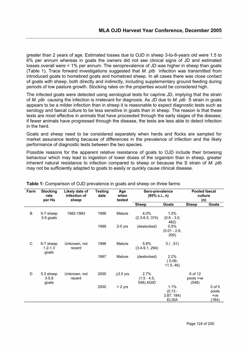
MLA OJD Harvest Year Conference, December 2005
Page 124 of 200
greater than 2 years of age. Estimated losses due to OJD in sheep 3-to-8-years old were 1.5 to6% per annum whereas in goats the owners did not see clinical signs of JD and estimatedlosses overall were < 1% per annum. The seroprevalence of JD was higher in sheep than goats(Table 1). Trace forward investigations suggested that M. ptb infection was transmitted fromintroduced goats to homebred goats and homebred sheep. In all cases there was close contactof goats with sheep, both directly and indirectly, including supplementary ground feeding duringperiods of low pasture growth. Stocking rates on the properties would be considered high.
The infected goats were detected using serological tests for caprine JD, implying that the strainof M. ptb causing the infection is irrelevant for diagnosis. As JD due to M. ptb S strain in goatsappears to be a milder infection than in sheep it is reasonable to expect diagnostic tests such asserology and faecal culture to be less sensitive in goats than in sheep. The reason is that thesetests are most effective in animals that have proceeded through the early stages of the disease;if fewer animals have progressed through the disease, the tests are less able to detect infectionin the herd.
Goats and sheep need to be considered separately when herds and flocks are sampled formarket assurance testing because of differences in the prevalence of infection and the likelyperformance of diagnostic tests between the two species.
Possible reasons for the apparent relative resistance of goats to OJD include their browsingbehaviour which may lead to ingestion of lower doses of the organism than in sheep, greaterinherent natural resistance to infection compared to sheep or because the S strain of M. ptbmay not be sufficiently adapted to goats to easily or quickly cause clinical disease.
Table 1: Comparison of OJD prevalence in goats and sheep on three farmsFarm Stocking
rateper Ha
Likely date ofinfection of
sheep
Testingdate
Agewhentested
Sero-prevalence(95% c.i., n)
Pooled faecalculture
(n)Sheep Goats Sheep Goats
B 5-7 sheep3-5 goats
1982-1983 1996 Mature 4.0%(2.3-6.5, 374)
1.5%(0.6 - 3.0,
482)1999 2-5 yrs (destocked) 0.5%
(0.01 - 2.8,200)
C 6-7 sheep1.2-1.3goats
Unknown, notrecent
1996 Mature 5.8%(3.4-9.1, 294)
0 ( ,51)
1997 Mature (destocked) 2.2%( 0.06-
11.5, 46)
D 5.3 sheep3-5.6goats
Unknown, notrecent
2000 >3.5 yrs 2.7%(1.5 - 4.5,548) AGID
6 of 12pools +ve
(548)2000 > 2 yrs 1.1%
(0.13 -3.87, 184)
ELISA
0 of 5pools+ve
(184)
MLA OJD Harvest Year Conference, December 2005
Page 125 of 200
Survival of the organism in the environmentWhile research was being conducted on the feasibility of eradication of OJD by whole flockdepopulation, resting of pasture and restocking with healthy sheep (see Pat Taylor, thisvolume), research was also conducted on the survival of the organism. Data on the resistanceof the organism were reported in 1944 (5) for bovine faeces exposed in a bowl in London. Theorganism survived for about nine months. Other reports suggest a trend for prolonged survivalexcept where urine is also present, or in silage with low pH or high ammonia levels (reviewed in(13)). These data come from the northern hemisphere where livestock are commonly housedindoors during winter on straw bedding and from experimental models without field validation.
In this MLA project, the survival of M. ptb was studied by culture of faecal material sampled atintervals for up to 117 weeks from soil and grass in pasture plots and boxes (13). Survival for upto 55 weeks was observed in a dry shaded environment, with much shorter survival in unshadedlocations. Moisture and application of lime to soil did not affect survival. The organism survivedfor up to 24 weeks on grass that germinated through infected faecal material applied to the soilsurface in shaded boxes and for up to nine weeks on grass in 70% shade. Survival of M. ptb inthe environment is finite, consistent with its taxonomic description as an obligate parasite ofanimals. As a rough guide, about 90% of the organisms deposited on a paddock in sheepfaeces die off each month.
In a related experiment, the survival of the organism was studied in dam water and sediment inlarge water troughs which were placed in either a semi-exposed location or in a shadedlocation, and compared to survival in faecal material and soil in the shaded location (11).Survival, in water and/or sediment in the shade, was observed for 48 weeks compared to 36weeks in the semi-exposed location. Survival in sediment was 12 to 26 weeks longer thansurvival in the water column. Survival in soil and faecal material in the shaded terrestrialenvironment was only 12 weeks. The results suggest that water may be a significant reservoir ofM. ptb infection. Further research on the biology of the organism in aquatic environments iswarranted. Animal health authorities will need to provide appropriate advice to farmers tominimise exposure of livestock to potentially infected water sources. Survival of the organism inwater destined for human consumption will need to be addressed if the organism is found to beinvolved in the aetiology of Crohn’s disease.
The aim of a further experiment was to determine whether M. ptb could be isolated from soil-pasture, faecal, water and sediment samples on farms before and after removal of OJD infectedsheep (12). The organism was recovered from approximately 20% of such samples from sixproperties. The positive samples were from a range of locations; however, low lying areastended to have larger numbers of organisms. When the same sites were sampled again aboutfive months later, only one was culture positive, and none were culture positive > 12 monthslater. The organism was rarely recovered from water samples on infected farms. Of 96 watersamples, 90 sediment samples and 93 soil samples from farms that had been destocked ofinfected sheep/goats for 9-24 months, one sediment sample from a farm in Victoria (destockedfor 12 months) and two sediment samples from a farm in New South Wales (destocked for 10and 19 months) were culture positive. Recontamination from cattle or water could not beexcluded as a cause of the positive cultures from the second farm. Improved culture methodsare needed for critical surveys and to study the movement and fate of the organism in theenvironment.
MLA OJD Harvest Year Conference, December 2005
Page 126 of 200
Conclusions These studies enabled closure of previous gaps in knowledge regarding OJD and animal healthauthorities were able to develop recommendations for control of OJD with greater confidence. Inparticular, the important role that cattle can play in rotational grazing with sheep was of low riskof interference with OJD control whereas goats were established as a threat to OJD control. Thesurvival of the organism in the environment was lengthy, but finite. Useful degrees of pasturedecontamination could be achieved in only a few months, but complete sterilisation wouldrequire more than a year. This knowledge, when combined with data from actual OJDtransmission studies (see McGregor, this volume), has led to the development of rationalrecommendations for grazing management to reduce the impact of OJD.
References1. Choy, E., R. J. Whittington, I. Marsh, J. Marshall, and M. T. Campbell. 1998. A method for
purification and characterisation of Mycobacterium avium subsp. paratuberculosis from theintestinal mucosa of sheep with Johne's disease. Veterinary Microbiology 64:51-60.
2. Collins, D. M., D. M. Gabric, and G. W. de Lisle. 1990. Identification of two groups ofMycobacterium paratuberculosis strains by restriction endonuclease analysis and DNAhybridization. Journal of Clinical Microbiology 28:1591-1596.
3. Cousins, D. V., S. N. Williams, A. Hope, and G. J. Eamens. 2000. DNA fingerprinting ofAustralian isolates of Mycobacterium avium subsp paratuberculosis using IS900 RFLP.Australian Veterinary Journal 78:184-190.
4. Eamens, G. J. 1989. Johne's disease in goats, p. 105-112. In A. R. Milner and P. R. Wood(ed.), Johne's disease. Current Trends in Research, Diagnosis and Management. CSIROPublications, Melbourne.
5. Lovell, R., M. Levi, and J. Francis. 1944. Studies on the survival of Johne's bacilli. Journalof Comparative Pathology 54:120-129.
6. Marsh, I., R. Whittington, and D. Cousins. 1999. PCR-restriction endonuclease analysis foridentification and strain typing of Mycobacterium avium subsp. paratuberculosis and M.avium subsp. avium based on polymorphisms in IS1311. Molecular and Cellular Probes13:115-126.
7. Moloney, B. J., and R. J. Whittington. 2002. Cross Species Transmission of Ovine Johne'sDisease - Phase 2 Cattle. Meat and Livestock Australia, Sydney.
8. Pavlik, I., L. Bejckova, M. Pavlas, Z. Rozsypalova, and S. Koskova. 1995. Characterizationby restriction endonuclease analysis and DNA hybridization using IS900 of bovine, ovine,caprine and human dependent strains of Mycobacterium paratuberculosis isolated invarious localities. Veterinary Microbiology 45:311-318.
9. Whittington, R., I. Marsh, E. Choy, and D. Cousins. 1998. Polymorphisms in IS1311, aninsertion sequence common to Mycobacterium avium and M. avium subsp.paratuberculosis, can be used to distinguish between and within these species. Molecularand Cellular Probes 12:349-358.
10. Whittington, R. J., A. F. Hope, D. J. Marshall, C. A. Taragel, and I. Marsh. 2000. Molecularepidemiology of Mycobacterium avium subsp. paratuberculosis: IS900 restriction fragmentlength polymorphism and IS1311 polymorphism analyses of isolates from animals and ahuman in Australia. Journal of Clinical Microbiology 38:3240-3248.
MLA OJD Harvest Year Conference, December 2005
Page 127 of 200
11. Whittington, R. J., I. B. Marsh, and L. A. Reddacliff. 2005. Survival of Mycobacterium aviumsubsp. paratuberculosis in dam water and sediment. Applied & EnvironmentalMicrobiology. 71:5304-5308.
12. Whittington, R. J., I. B. Marsh, P. J. Taylor, D. J. Marshall, C. Taragel, and L. A. Reddacliff.2003. Isolation of Mycobacterium avium subsp. paratuberculosis from environmentalsamples collected from farms before and after destocking sheep with paratuberculosis.Australian Veterinary Journal 81:559-563.
13. Whittington, R. J., D. J. Marshall, P. J. Nicholls, I. B. Marsh, and L. A. Reddacliff. 2004.Survival and dormancy of Mycobacterium avium subsp. paratuberculosis in theenvironment. Applied & Environmental Microbiology 70:2989-3004.
14. Whittington, R. J., L. Reddacliff, I. Marsh, and V. Saunders. 1999. Detection ofMycobacterium avium subsp. paratuberculosis in formalin-fixed paraffin-embeddedintestinal tissue by IS900 polymerase chain reaction. Australian Veterinary Journal 77:392-397.
MLA OJD Harvest Year Conference, December 2005
Page 128 of 200
Development of grazing management strategies for thecontrol of ovine Johne’s disease
H McGregora, K Abbott and O Dhungyel, R WhittingtonaSydney University,Faculty of Veterinary Science,425 Werombi Rd Camden, NSWPhone: 02 9351 1792; Fax: 02 9351 1618Email: [email protected]
IntroductionOn high prevalence, OJD infected properties in NSW annual mortality rates up to 21% havebeen estimated, with over 70% of these mortalities due to OJD. [1, 2] The annual costs from OJDrelated mortalities have been estimated across 12 infected properties in NSW and areconsiderable, with a reduction in gross margin on average of 6.4%[1] In heavily infected flocks itis common for animals to start to die from OJD as early as 18 months of age, well before thepeak of their productivity. Practical options for control of OJD are necessary to reduce thiseconomic loss. Based on recent studies on farms where OJD is well established, the use ofvaccine has proven to be a financially viable control option as it reduces mortalities, shedding ofinfectious organisms and lost income due to OJD. [3] However, there is evidence that the use ofvaccine alone may not be the optimal approach for control. Where OJD prevalence is low, thecost of vaccination makes its widespread use less economically attractive. Control of OJDthrough grazing management will provide additional and complementary options to vaccine useto reduce the levels of exposure experienced by young sheep, and thus the severity of infection.
Two extensive field-based, epidemiological studies were conducted between 1999 and 2004 tofacilitate the development and validation of grazing management protocols for the control ofOJD. In the first study (MLA OJD.002a) the role of the ewe as a source of infection for lambsand the relative benefits of the provision of low contamination pastures in the pre-weaning andpost-weaning periods were evaluated. The second study, (MLA OJD.028) which commenced in2002, was conducted to determine whether age of sheep at exposure and level of pasturecontamination affects the development of OJD.
MethodsExperimental siteThe experimental site was at the University of Sydney farm, Arthursleigh, in the southerntablelands of NSW. The site comprised secure paddocks and a laneway system which enabledtwo replicates for each treatment. Erosion gullies were fenced to exclude livestock and theseand the laneways were incorporated in ways which assisted in the separation of paddocks withdifferent levels of OJD contamination. The pasture on the experimental site was unimproved,consisting of native grasses and volunteer-introduced species and weeds. There were fewshade trees on the site and, generally, all or most parts of all paddocks did not have shade. Areticulated water system was installed to service experimental paddocks. Water was originallysupplied from a tank serviced by a fenced dam within the site and then later due to increasingdrought conditions from the Wollondilly river.
Pasture contamination history
MLA OJD Harvest Year Conference, December 2005
Page 129 of 200
Experimental pastures had been grazed by cattle only, for 18 months prior to the start ofexperiment 002a. The cattle were tested on 2 occasions 2 years apart, and were negative in theJohne’s disease absorbed ELISA. The OJD seroprevalence of the sheep grazing the pastureprior to this 18-month period was 0.25%. Prior to trial 002a which commenced in September1999, pastures were considered to have negligible levels of M. ptb infection. At the start of eachtrial paddocks to be infected were grazed by OJD infected sheep. These sheep were identifiedfrom high prevalence infected properties and tested for active shedding, initially by ZN stainingof faecal smears or AGID serology and retrospectively by individual faecal culture. For trial002a, infected ewes grazed designated paddocks prior to and during the lambing and pre-weaning period. For trial 028 infected donor sheep were co-grazed with trial sheep for the initial14.5 weeks of the trial. The number and the faecal culture status of OJD infected animalsgrazing a paddock for a designated time determined the pasture contamination rate. Thecontamination history of each paddock was calculated from the numbers of ‘shedders’ grazingthe area for a designated period of time. Paddocks required to be largely free from infectionwere not grazed at all or were grazed only by the clean trial sheep.
Experimental DesignMLA OJD.002a (Table 1)Pregnant Merino ewes were sourced for this experiment from two flocks with similar bloodlinesin NSW, one testing negative over serial tests for OJD (flock U) and the other a known highprevalence flock (flock I). The principle experimental sheep were the lambs born onto theexperimental site to infected (flock I) and uninfected (flock U) ewes. Lambs were raised toweaning age on pastures of 3 levels of contamination, low, medium and high, determined bydam infection status and grazing history. At weaning lambs were stratified on the basis ofweight, sex and pre-weaning exposure history and allocated to post weaning paddocks withhigh or low levels of contamination. Treatment groups were based on history of dam infectionstatus, pre-weaning and post-weaning exposure. The 7 treatment groups were replicated, eachreplicate containing 36 animals. Once allocated to a treatment group, animals remained withinthat group and replicate for the duration of the trial. Blood and faecal samples were collected 3monthly and then 6 weekly from lamb marking for the duration of the trial to monitor the onset ofshedding. Lambs were euthanased at 3 years of age and samples collected for histopathologyand tissue culture to determine prevalence and severity of infection.
MLA OJD.028A total of 840 female Merino sheep of three age groups, lambs, hoggets and adults wereobtained from an uninfected property. Sheep were stratified on bodyweight and age andrandomly allocated to a treatment group so that there were 35 of each age. Each mixed agegroup mob of 105 sheep was grazed in a 20 hectare paddock within the experimental site(treatment group). The infected donor sheep were co-grazed with each mob at predeterminedstocking rates to provide four levels of contamination, control (very low), low, medium and high(0, 0.1, 0.35 and 0.7 infected sheep per hectare respectively). Each of the contaminationtreatments was replicated once. Pooled faecal culture and agar gel immunodiffusion assayswere conducted on all sheep at regular intervals during the trial, while individual faecal culture,ELISA and gamma-interferon assays were applied to subgroups at some time points. Theanimals remained in the experiment for 2.5 years and were then euthanased and sampled forhistopathology and tissue culture to establish the prevalence of infection with M. ptb.
ResultsMLA OJD.002a (Table 1)
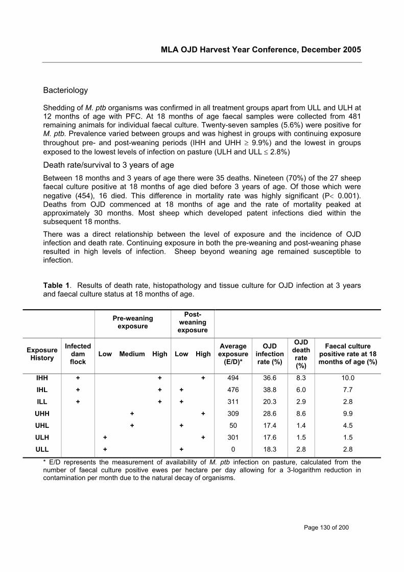
MLA OJD Harvest Year Conference, December 2005
Page 130 of 200
Bacteriology
Shedding of M. ptb organisms was confirmed in all treatment groups apart from ULL and ULH at12 months of age with PFC. At 18 months of age faecal samples were collected from 481remaining animals for individual faecal culture. Twenty-seven samples (5.6%) were positive forM. ptb. Prevalence varied between groups and was highest in groups with continuing exposurethroughout pre- and post-weaning periods (IHH and UHH ≥ 9.9%) and the lowest in groupsexposed to the lowest levels of infection on pasture (ULH and ULL ≤ 2.8%)
Death rate/survival to 3 years of ageBetween 18 months and 3 years of age there were 35 deaths. Nineteen (70%) of the 27 sheepfaecal culture positive at 18 months of age died before 3 years of age. Of those which werenegative (454), 16 died. This difference in mortality rate was highly significant (P< 0.001).Deaths from OJD commenced at 18 months of age and the rate of mortality peaked atapproximately 30 months. Most sheep which developed patent infections died within thesubsequent 18 months.
There was a direct relationship between the level of exposure and the incidence of OJDinfection and death rate. Continuing exposure in both the pre-weaning and post-weaning phaseresulted in high levels of infection. Sheep beyond weaning age remained susceptible toinfection.
Table 1. Results of death rate, histopathology and tissue culture for OJD infection at 3 yearsand faecal culture status at 18 months of age.
* E/D represents the measurement of availability of M. ptb infection on pasture, calculated from thenumber of faecal culture positive ewes per hectare per day allowing for a 3-logarithm reduction incontamination per month due to the natural decay of organisms.
Pre-weaningexposure
Post-weaningexposure
ExposureHistory
Infecteddamflock
Low Medium High Low HighAverageexposure
(E/D)*
OJDinfectionrate (%)
OJDdeathrate(%)
Faecal culturepositive rate at 18months of age (%)
IHH + + + 494 36.6 8.3 10.0
IHL + + + 476 38.8 6.0 7.7
ILL + + + 311 20.3 2.9 2.8
UHH + + 309 28.6 8.6 9.9
UHL + + 50 17.4 1.4 4.5
ULH + + 301 17.6 1.5 1.5
ULL + + 0 18.3 2.8 2.8
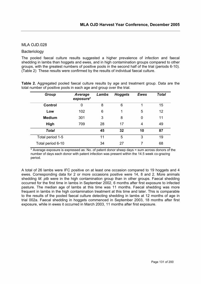
MLA OJD Harvest Year Conference, December 2005
Page 131 of 200
MLA OJD.028BacteriologyThe pooled faecal culture results suggested a higher prevalence of infection and faecalshedding in lambs than hoggets and ewes, and in high contamination groups compared to othergroups, with the greatest numbers of positive pools in the second half of the trial (periods 6-10).(Table 2) These results were confirmed by the results of individual faecal culture.
Table 2. Aggregated pooled faecal culture results by age and treatment group. Data are thetotal number of positive pools in each age and group over the trial.
Group Averageexposure*
Lambs Hoggets Ewes Total
Control 0 8 6 1 15
Low 102 6 1 5 12
Medium 301 3 8 0 11
High 709 28 17 4 49
Total 45 32 10 87
Total period 1-5 11 5 3 19
Total period 6-10 34 27 7 68
* Average exposure is expressed as: No. of patent donor sheep days = sum across donors of thenumber of days each donor with patent infection was present within the 14.5 week co-grazingperiod.
A total of 26 lambs were IFC positive on at least one occasion compared to 19 hoggets and 4ewes. Corresponding data for 2 or more occasions positive were 14, 8 and 2. More animalsshedding M. ptb were in the high contamination group than in other groups. Faecal sheddingoccurred for the first time in lambs in September 2002, 6 months after first exposure to infectedpasture. The median age of lambs at this time was 11 months. Faecal shedding was morefrequent in lambs in the high contamination treatment at this time and later. This is comparableto the results of the pooled faecal culture detecting shedding in lambs at 12 months of age intrial 002a. Faecal shedding in hoggets commenced in September 2003, 18 months after firstexposure, while in ewes it occurred in March 2003, 11 months after first exposure.
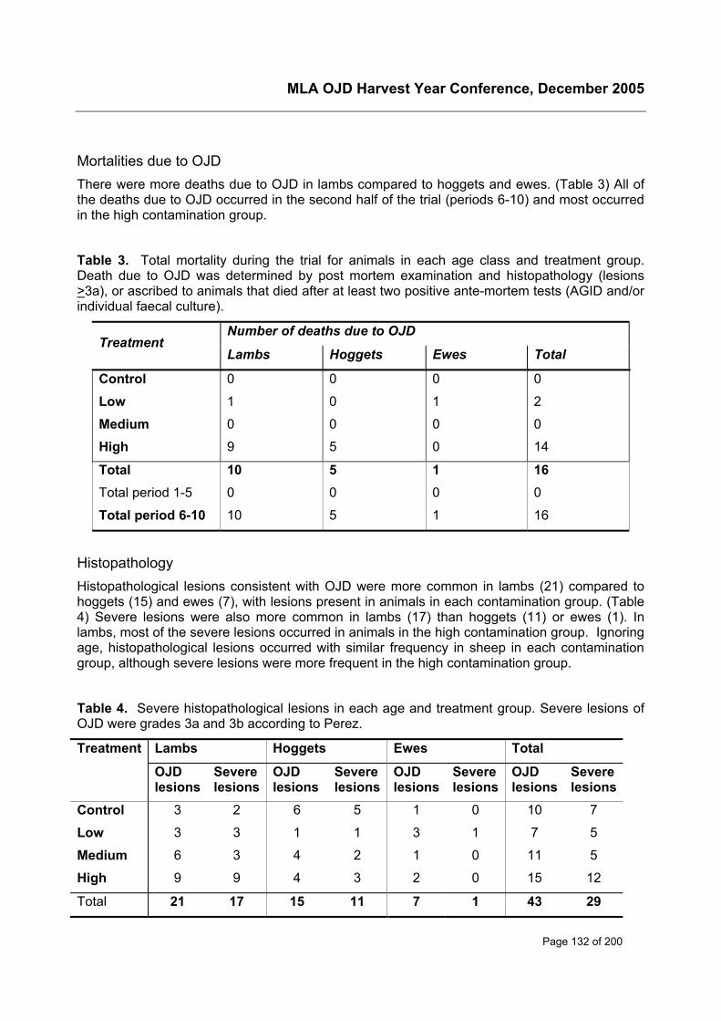
MLA OJD Harvest Year Conference, December 2005
Page 132 of 200
Mortalities due to OJDThere were more deaths due to OJD in lambs compared to hoggets and ewes. (Table 3) All ofthe deaths due to OJD occurred in the second half of the trial (periods 6-10) and most occurredin the high contamination group.
Table 3. Total mortality during the trial for animals in each age class and treatment group.Death due to OJD was determined by post mortem examination and histopathology (lesions>3a), or ascribed to animals that died after at least two positive ante-mortem tests (AGID and/orindividual faecal culture).
Number of deaths due to OJDTreatment
Lambs Hoggets Ewes Total
Control 0 0 0 0
Low 1 0 1 2
Medium 0 0 0 0
High 9 5 0 14
Total 10 5 1 16Total period 1-5 0 0 0 0
Total period 6-10 10 5 1 16
HistopathologyHistopathological lesions consistent with OJD were more common in lambs (21) compared tohoggets (15) and ewes (7), with lesions present in animals in each contamination group. (Table4) Severe lesions were also more common in lambs (17) than hoggets (11) or ewes (1). Inlambs, most of the severe lesions occurred in animals in the high contamination group. Ignoringage, histopathological lesions occurred with similar frequency in sheep in each contaminationgroup, although severe lesions were more frequent in the high contamination group.
Table 4. Severe histopathological lesions in each age and treatment group. Severe lesions ofOJD were grades 3a and 3b according to Perez.
Lambs Hoggets Ewes TotalTreatment
OJDlesions
Severelesions
OJDlesions
Severelesions
OJDlesions
Severelesions
OJDlesions
Severelesions
Control 3 2 6 5 1 0 10 7
Low 3 3 1 1 3 1 7 5
Medium 6 3 4 2 1 0 11 5
High 9 9 4 3 2 0 15 12
Total 21 17 15 11 7 1 43 29
MLA OJD Harvest Year Conference, December 2005
Page 133 of 200
SerologyThere was a similar number of AGID reactors among lambs and hoggets, and a lower numberamong ewes. Ignoring age, the highest number of AGID reactors occurred among sheep in thehigh contamination group, but the trend did not apply to lambs or ewes. Most of the positiveresults occurred in the second half of the trial in periods 6-10.
ConclusionsThe results from both experiments confirm that post weaning lambs remain highly susceptible toinfection. Lambs are highly susceptible to infection compared to adult sheep, evidenced bygreater prevalence of severe histological lesions and mortality in this age group, particularlywhen exposed to high levels of infection. Similarly faecal shedding of the organism was morecommon when animals were first exposed as lambs. The results from MLA OJD.028 suggestthat transmission of M. ptb occurred very quickly after first exposure, with patent infectionestablished in the lambs within 6 months of first exposure. This work confirms that there is astrong relationship between the level of natural exposure to M. ptb, age and the subsequentincidence of OJD infection. It should therefore be possible to reduce the incidence or severity ofOJD by reducing the levels of exposure in young sheep in the pre-weaning and immediate post-weaning periods. Recommendations for flock testing to detect infection are dependant on theage of the animals. However, the use of faecal culture to detect infection may also be a validtool to predict future disease and mortalities, particularly in young animals. These results onlyallow an estimation of how long lambs may remain susceptible but a reasonable hypothesiswould be that susceptibility will start to decline from around 12 months of age. The use ofinfected, actively shedding, co-grazer sheep to successfully create an infected environment inboth experiments demonstrates that even relatively low numbers of clinically affected sheep perhectare (0.1-0.7 in OJD.028) will lead to transmission of infection to susceptible sheep. Thisfinding illustrates the importance of monitoring for clinical sheep frequently and culling themfrom all mobs to reduce the levels of M. ptb on pasture. The occurrence of OJD in controlgroups in both experiments demonstrated that lateral spread of the disease is a serious issueand it is not necessary for infected sheep to graze the same immediate area for transmission tooccur if infected sheep are in neighboring paddocks. It is also important to note that all agegroups of sheep may become infected even following exposure to relatively low levels ofinfection on pasture. This finding suggests that highly susceptible individuals probably existwithin all age groups, therefore transmission of disease through shedding is a possibility even insheep exposed to relatively low levels of infection as adults.
RecommendationsIt is possible to recommend several approaches to control OJD based on the data from thisstudy:
• Minimise exposure of lambs and hoggets to heavily contaminated pasture
• As adult sheep are more resistant to infection and disease impacts of OJD, if cleanpastures are scarce, they, rather than younger animals, should be grazed oncontaminated pasture
• Cull adult sheep with clinical signs of OJD, or conduct PFC to obtain evidence of patentOJD infection and manage or cull these mobs to reduce the level of pasturecontamination and future mortalities.

MLA OJD Harvest Year Conference, December 2005
Page 134 of 200
• Manage pastures by spelling, grazing with adult cattle or grazing with approvedvaccinates in order to reduce the contamination level for lambs, weaners and youngsheep
• In all infected flocks including those vaccinated, regularly observe all classes of sheepand identify and remove individuals that develop signs of weight loss to reduce theenormous contribution of ‘multibacillary shedders’ to the level of contamination
• Maintain boundary fencing to reduce the likelihood of lateral spread from an infectedneighbour. As a single fence line does not prevent lateral spread, consider doublefencing with wide laneways or segregation of critical pastures using a tree belt, gully orother natural feature. Maintain high value lambs in paddocks well away from paddocksknown to contain infected sheep.
Endoparasite control on prepared, low infectivity pastures needs to be considered to avoid aconflicting effect from OJD control and management practices. Extra vigilance with endoparasitecontrol regimes may be necessary where weaner animals are at risk of exposure to potentiallyhigh levels of endoparasite infection on prepared pastures. The use of young sheep for thepreparation of low infectivity pastures for M. ptb may lead to increased risk from high levels ofcontamination with endoparasites. The use of adult sheep (vaccinated as lambs) to reduce M.ptb levels on pasture will produce an environment with a lower risk for endoparasite infection butmay be a higher risk option for M. ptb control. Spelling, crops or grazing adult cattle may be thesafest approach for preparation of low contamination pastures for OJD control.
AcknowledgementsTrials OJD.002a and OJD.028 were both funded through Meat and Livestock Australia.
The cooperation, support and expertise of staff at Sydney University is gratefully acknowledged,especially Anna Waldron and Craig Kristo. The support of “Arthursleigh” farm staff through thefield trial phases of these projects was critical for their successful completion.
References1. Bush R. A study of the Biological and Economic impact of OJD in affected sheep flocks in
NSW. MLA OJD research and Development Update 2004; 46-50.
2. H McGregor, PA Windsor, KA Abbott. A longitudinal study of Ovine Johne’s disease:estimation of the mortality rate two years after whole-flock vaccination. Annual conference ofthe Australian Sheep Veterinary Society, Proceedings 2003;13:102-106.
3. Sergeant E. Cost effectiveness of vaccination against ovine Johne’s disease in infectedflocks. Annual conference of the Australian Sheep Veterinary Society, Proceedings 2003;13:123-127.
4. McGregor H, Abbott KA, and Whittington RJ. Development of grazing managementstrategies for the control of ovine Johne’s disease. Annual conference of the AustralianSheep Veterinary Society, Proceedings 2004; 60-65
5. Whittington RJ, Marsh I, Turner MJ et al. Rapid detection of Mycobacteriumparatuberculosis in clinical samples from ruminants and in spiked environmental samples bymodified BACTEC 12B radiometric culture and direct confirmation by IS900 PCR. J ClinMicrobiol 1999; 37:1077-1083
MLA OJD Harvest Year Conference, December 2005
Page 135 of 200
Within flock spread of OJD - a case study from a sheep flocklocated in southeastern New South Wales
L Rasta, RJ WhittingtonaGundagai Rural Lands Protection BoardPO Box 21,Gundagai NSW 2722Phone: +61 2 6944 1588; Fax: +61 2 6944 1867Email: [email protected]
AbstractOn farm investigation and monitoring for ovine Johne’s disease (OJD) was performed in arecently infected flock of around 3,000 sheep, between 1997 and 2002 to better understandOJD prevalence, distribution and spread on this farm and to plan practical disease control andintervention strategies.
Tests used were serology (AGID), pooled faecal culture (PFC) and histopathology, includingpartial and then whole flock testing using PFC annually three times. Faecal shedding ofMycobacterium paratuberculosis (M. ptb) commenced in home bred sheep around six to sevenyears after a single introduction of a mob of 410 infected sheep in 1993. For at least sevenyears there was clustering of infection and shedding within one or two age groups only. Sheepin these age groups appeared to have been exposed to mycobacterial contamination at an earlyage (<12 months) and commenced shedding at five years of age or older. Groups that wereexposed to contamination as adults did not shed detectable levels of M. ptb during the studyperiod.
These results provided indirect evidence of the finite duration of survival of M. ptb on pastureand the influence of age on susceptibility of sheep to develop patent M. ptb infection.
A further feature of the epidemiology in this flock was the slow transmission and spread of M.ptb, related to the long incubation period (exposure to shedding interval) of five years and theabsence of clinical signs throughout the study period.
The findings suggest that partial flock culling, selective grazing management and vaccinationcould lead to a reduction in mycobacteria contamination on-farm, possibly to a level at whichpatent M. ptb infection no longer occurs. Better understanding of disease spread within flocksover time through flock profiling using PFC will help in devising surveillance strategies (includingtesting protocols for market assurance testing) that account for clustering of infection as well asvery slow transmission and spread of infection through a flock.
IntroductionOJD is a chronic wasting disease caused by Mycobacterium paratuberculosis (M. ptb). Themajor mode of transmission of bacteria is by the faecal-oral route. Once ingested, it usuallytakes at least 12 months before a patent infection establishes and detectable levels of bacteriaare shed via faeces (Chaitaweesub et al, 1999). Appearance of clinical signs of chronic wastingusually takes many years (Whittington et al, 2001). The organism remains viable in theenvironment for up to about 12 months but infectious doses may be available for a lesser period(Whittington, 2001). The principle means of spread of OJD between flocks is though movementof infected sheep. Less is known about transmission and spread of M. ptb in sheep than cattle,but calves are reported to be more susceptible to infection than mature cattle and the samemight apply to sheep (Whittington et al, 2001).
MLA OJD Harvest Year Conference, December 2005
Page 136 of 200
There is little objective information regarding the spread of Mptb within an infected flock. It isassumed that most sheep are exposed via pasture, and that all animals in a flock have an equalchance of becoming exposed. Surveillance programs to detect infection entail sampling basedon this assumption and ignore potential clustering of infection within mobs or incubation periodsof more than three years. Negative test results following random sampling across a flock willhave lower than stated confidence if the infection is confined to some mobs only, rather thanevenly distributed throughout a flock. This will reduce the accuracy of testing to detect OJDinfection in flocks but also testing to show absence of disease for market assurance purposes.In some areas, recommendations for control of OJD are based on whole flock destocking orquarantine and other trading restrictions to reduce further spread. These measures make noallowance for the possibility that infection is confined to subgroups or mobs within a flock. Theavailability of pooled faecal culture (PFC) (Whittington et al, 2000) has enabled relativelyinexpensive testing of whole flocks, to better determine the source and distribution of infection.
The aim of this study was to apply whole flock testing (flock profiling) over time to determine theprevalence, distribution and spread of infection in a recently infected flock, with a view toplanning practical on-farm intervention strategies for disease control.
Material and methodsManagement and flock structureThe farm consisted of 1,739 hectares of improved pasture and was located in southeasternNSW, a 600 mm average annual rainfall area. The main enterprise was sheep breeding, butaround 700 beef cattle were also grazed and 250-300 hectares were cropped annually. Thesheep flock consisted of a commercial Bond breed flock of approximately 3,000 breeding ewesand a Bond stud of about 300 ewes. Lambing was in April/May. Wether lambs were sold toslaughter as fat lambs from December onwards each year. Culls were sold to slaughter annuallyin September/October and usually included the entire oldest age group (six year olds). Prior totheir first joining, the ewes were classed and around 50 were selected each year to enter thestud. Sheep were segregated based on age and run in separate mobs, up to six years old. Thecommercial and stud ewes were run separately and mobs were grazed in a rotational pattern,fitting in with pasture availability and the cropping program. Other than the introduction of someMerino sheep (described below) and a once off purchase of five Dorset rams in 1997 no sheepwere routinely introduced.
History of OJD acquisitionIn 1993 the owner purchased 410 mixed age fine wool Merino ewes from farm X, located inwhat is now recognised as the high prevalence OJD area in NSW. On the study farm the Merinosheep had no direct contact with the Bond flocks. OJD was confirmed on farm X in late 1996.
In early 1996 the owner of the study farm sold 287 of the Merino ewes to another farm (farm F)and the remainder to slaughter. Any remaining progeny from these Merino ewes were sold toslaughter in December 1996.In October 1996, 237 merino ewes on farm F were tested with AGID and there were ninereactors. Three reactors were examined at post mortem and histology on intestinal tissueshowed lesions consistent with multibacillary Johne’s disease in all three sheep. The acid fastorganisms in the formalin-fixed paraffin-embedded tissues of each sheep were identified as Sstrain M. ptb.
MLA OJD Harvest Year Conference, December 2005
Page 137 of 200
Eventually, in March 2000, OJD was confirmed on the study farm in homebred 5-year old Bondewes (see results). Whole flock testing of adult sheep by PFC commenced later that year andwas repeated annually three times in total. Methods for testing for OJD and further details aredescribed in Rast and Whittington, 2005.
ResultsThe patterns of testing and infection on the study farm are provided in Figure 1 and Table 1.
Testing prior to 2000Testing commenced on the study farm in 1997, as potentially infected sheep had beenintroduced from farm X in 1993.
• In 1997, 50 homebred 1992-born Bond sheep, which may have had contact withcontaminated land, were tested using the AGID test, with negative results.
• In June 1998, 460 homebred Bond sheep, born in 1993 and 1994, were tested using theAGID and PFC, with negative results.
• In March 2000, OJD was confirmed in 1995-born homebred Bond ewes byhistopathology following serology (AGID) tests of 450 sheep (Table 1, Figure 1).
Testing was required on three occasions over a four-year period to confirm infection inhomebred sheep. Formalin-fixed paraffin-embedded tissues from one sheep with multibaciliarylesions were examined by PCR and were confirmed to contain S strain of M. ptb.
Testing in 2000Sheep born from 1994 to 2000 were present and all sheep older than two years were testedwith PFC in June 2000. Shedding of M. ptb was confirmed in the 1995-born commercial ewes(prevalence 2.5%). This was the age group known to be infected based on AGID tests andsubsequent post mortem examination and histopathology testing, performed earlier in the sameyear. All results from other age groups were negative (Figure 1 and Table 1).
Testing in 2001Sheep born from 1996 to 2001 were present and all sheep older than 12 months were sampledfor PFC in September 2001. Shedding of M. ptb was detected in the 1996-born commercialBond ewes (prevalence 0.8%) and 1999-born stud ewes (prevalence 1.4%) (Figure 1 and Table1).
Testing in 2002Sheep born from 1996 to 2001 were present and all sheep older than 12 months were testedusing PFC in September-October 2002. Shedding was confirmed again in the 1996-borncommercial ewes and prevalence had increased from 0.8% to 2.5%. The 1997- borncommercial ewes were also positive (prevalence 0.7%). All other results were negative,including the 1999-born stud ewes that had tested positive the previous year (Table 1 andFigure 1).
Disease management on the study farmAfter notification of infection on Farm X (source of putative infected Merino sheep) but prior toOJD confirmation on the study farm
The owner identified the land used to graze the putative infected introduced Merinosheep and their progeny and ceased sheep grazing on this land in 1996. He applied lime

MLA OJD Harvest Year Conference, December 2005
Page 138 of 200
at a rate of 2.5 tonnes per hectare and used the land for cattle grazing or croppinginstead.
After OJD confirmation on the study farm in 2000
The first whole flock test by PFC in 2000 established that only the 1995-born sheep wereshedding detectable levels of M. ptb at the time of sampling. Faecal samples werecollected in June, however the serology and histopathology results of multibacillary OJDfrom a limited number of sheep in that age group in March 2000 made it likely that theywere shedding by then. Therefore lambs born to these ewes in April/May 2000 were athigh risk of exposure and infection. A recommendation was made in 2000 to cull theentire 1995-born age group, including the lambs born to them. The owner sold allremaining 1995 born ewes (207 ewes) to slaughter in October 2000, and the lambs asthey reached market weight between October and December 2000.
At that stage it was considered that the 1999-born sheep were potentially at high risk ofinfection through contamination from M. ptb shed by the infected 1995-born sheep in2000 or earlier. The 1996 and 1997 born sheep were also considered at risk byexposure to contamination from the introduced Merino sheep (Figure 1). However cullingof other age groups considered at risk of infection was not an economically viable option.
It was recommended to use the land on which the confirmed infected and shedding1995-born age group had run, for purposes other than sheep grazing. The owner wasable to identify and use the paddock these sheep had run in from the beginning of 1999to late 2000 for cropping and cattle grazing in the following years.
During 2001
In 2001,use of Gudair® vaccine was approved on the study farm and a vaccinationprogram began. All ewe lambs born in 2001 and the 1999-born commercial and studewes were vaccinated at the time of faecal sampling. Other age groups were notvaccinated due to the doubtful efficacy of vaccine administered post exposure to adultsand the cost.
During 2002
As the test results from the 2001 sampling showed an apparent increase in shedding, allsheep (including adults) considered at risk but not already shedding were vaccinated.These were the 1998-, 2000- and 2002-born ewes and all rams.
DiscussionObservations and test results support the following scenario:
1) Infection was introduced onto the study farm with Merino sheep purchased in 1993 and thissource of contamination was removed in 1996
2) Shedding in infected merino sheep occurred prior to their detection, and infectiveconcentrations of M. ptb originating from the Merino sheep were present in the environment onthe study farm from 1995 to 1997. This is based on a 12-month survival period on pasture afterremoval of the infected sheep (Whittington et al, 2004)
3) Only sheep less than 12 months old when exposed to M. ptb developed patent infections,that is progressed to shed M. ptb, during the study period
4) Shedding commenced when sheep were five years or older, a significantly longer time periodthan the shortest reported 12 months incubation period in sheep (Chaitaweesub et al, 1999).
MLA OJD Harvest Year Conference, December 2005
Page 139 of 200
The source of infection on the study farm was most likely environmental contamination with M.ptb from some of the 410 Merino sheep and their progeny introduced from farm X in 1993 andsold in 1996. No other sources of infection are known or likely. Although the Merino sheep werenever tested while present on the study farm, these introduced sheep were likely to be sheddingM. ptb because 8 months after their sale to farm F the prevalence of sero-positive sheep was3.8%. Estimation of the true prevalence of infection from these results is problematical becauseof the reported extreme variation in sensitivity of the AGID (Sergeant et al, 2003; Sacks et al1989). Shedding as early as 1995/1996 by these sheep can be assumed because theseropositive sheep examined on farm F had multibacillary intestinal lesions, which are highlycorrelated with shedding of M. ptb in faeces (Whittington et al, 1999). Negative test results in thehome-bred Bond sheep born in 1994 or earlier (therefore four to six year old when tested)strengthens the assumption that the Merino sheep introduced from farm X did not shedsufficient numbers of M. ptb prior to 1994 to contaminate the environment and transmit infectionto lambs present on the study farm at that time.
Whole flock testing using PFC of all adult sheep in 2000 confirmed that shedding was limited tothe 1995-born age group. It is possible that contamination originating from the Merino sheepwas very localised and only the 1995-born commercial Bond ewes were exposed (i.e. bychance). Alternatively - and more likely considering subsequent test results - age susceptibilityto infection meant that only lambs exposed to contamination developed patent infection. If this isthe case and if the Merino sheep produced sufficient contamination with M. ptb from 1995 andlasting until 12 months after their departure from the study farm in 1996, then sheep born in1995, 1996 and 1997 were at risk of infection, but only the1995-born sheep were old enough toshed at the time of sampling in 2000.
Results from testing in 2001 and 2002 support this hypothesis, and showed that in 2001shedding was occurring in the 1996-born sheep and in 2002 in 1997-born sheep.Contamination left by the Merino sheep was probably quite low, leading to lower ingested dosesof M. ptb, which would explain the late onset of shedding (5 year old) in the 1995-, 1996- and1997-born age groups (Whittington et al, 2001). Also these sheep were in good body conditionand perhaps other environmental or genetic effects contributed to late onset of shedding. It isalso possible that the 1996- and 1997-born sheep became infected as adults fromcontamination by homebred Bond ewes that were confirmed to shed at the 2000 and 2001testing (Figure1). However we consider this unlikely due to insufficient time for development oflesions and shedding and the low prevalence of shedding (Table 1).
At the 2001 testing, shedding was confirmed in the 1999-born stud ewes but not in thecommercial ewes of the same age (Table 1). The source of infection of this age group isuncertain. Infection could have been transmitted by chance exposure to high contamination(produced by homebred sheep shedding in 2000, 2001 or earlier). This is unlikely because theprevalence in homebred sheep was low (see Table 1). Intrauterine infection may have been thesource as their dams could have been the 1995- or 1996-born infected ewes. Howeverintrauterine transmission is improbable unless sheep are clinically infected, and there were noclinically infected sheep observed on the study farm (Lambeth et al, 2004). The same mob,when retested in 2002 was apparently no longer shedding bacteria. Intermittent shedding mayreflect either stage of disease (Whittington et al, 2001), passive shedding of ingested organismsacquired from contaminated pasture or perhaps the effect of vaccination (Eppleston et al, 2003).Alternatively, the sheep being responsible for shedding could have died during the intervalbetween the two tests.
MLA OJD Harvest Year Conference, December 2005
Page 140 of 200
ConclusionThe history and detailed and extensive testing available on this farm, allowed identification ofthe time period over which mycobacterial contamination occurred by the introduced infectedMerino sheep and highlights the slow spread of infection within the home bred flock. OJDinfection was clustered, remaining limited to one or two age groups for at least seven years afterthe infection was introduced.
Opportunities for disease control and risks to effective surveillance are apparent retrospectively.On this farm, it may have been possible to stop transmission of OJD through timelyimplementation of disease management such as selective culling, strategic grazing andvaccination. If these disease control strategies had been implemented earlier than was possibleon this farm, disease transmission may have been stopped.
In retrospect, action that could have been taken in 1996/97 that may have led to elimination ofthe infection from the study farm would include:
Implementation of an infected flock profile immediately upon identification of the infectedintroduced sheep
Culling of these sheep and their progeny
Culling of home bred sheep born during the preceding and following 12 months,corresponding to the period of contamination, and prior to reaching 12 months of age
Implementation of a vaccination program of all remaining sheep and annually of lambs atmarking time
Whole flock testing using PFC (infected flock profile) about 5 years after commencementof the program to monitor flock infection status
Vaccination was used on the study farm and the owner plans to vaccinate ewe lambs annuallyat marking or weaning. Based on recent research results with Gudair ® vaccine underAustralian conditions, vaccination on this farm is likely reduce the levels of M. ptb contaminationas well as delaying the onset of shedding (Eppleston et al, 2003). It may be that in very lowchallenge situations such as this, vaccination eventually prevents shedding of detectable levelsof M. ptb. Further testing would be needed to confirm this.
This study also demonstrates how difficult effective OJD surveillance may be unless exacthistory is known and the correct age groups or mobs are tested or unless testing occursrepeatedly over a long period of time. Clustering of infection reduces the confidence in anegative test outcome if sampling is random. This has significant implications for marketassurance program testing (Whittington et al, 2001) and surveillance testing with a view toassess the OJD risk of a flock or mob of sheep intended for sale.
AcknowledgmentsThis study was supported by Meat & Livestock Australia. Gordon and Jill Forsyth, who lost theirstud business through OJD quarantine and despite their emotional and financial hardship,cooperated with investigations over many years and always made us welcome. Peter Windsorand Rob Walker are thanked for useful discussions, while Evan Sergeant provided aspreadsheet for calculation of prevalence from pooled samples. Georgina Marshall, BenMcDonald, Col Elphick, and Tim Jones assisted in the field, while the efforts of Deb Walker,Shayne Fell and Anna Waldron with pooled faecal culture and Leslie Reddacliff and VanessaSaunders with strain typing from paraffin blocks was greatly appreciated.
MLA OJD Harvest Year Conference, December 2005
Page 141 of 200
ReferencesChaitaweesub P., Abbott K.A., Whittington R.J., 1999. Shedding of organisms and sub-clinicaleffects on production in pre-clinical Merino sheep affected with ovine paratuberculosis. In: EJBManning, MT Collins Proceedings of the Sixth International Colloquium on Paratuberculosis, ed.Madison: International Association for Paratuberculosis. 126-31.
Eppleston J., Reddacliff L., Windsor P., 2003. An update on the efficacy of Gudair ™ vaccine inAustralia. Proceedings of the Australian Sheep Veterinary Society - Cairns Conference:Australian Sheep Veterinary Society. 107-13
Hope A.F., Kluver P.F., Jones S.L., 2000. Sensitivity and specificity of two serological tests forthe detection of ovine paratuberculosis. Australian Veterinary Journal ;78:850-856.
Lambeth C, Reddacliff L, Windsor P, 2004. Intrauterine transmission of Mycobacterium aviumsubsp. paratuberculosis in sheep. Australian Veterinary Journal ;82: 504-508.
Perez V., Marin J.F.G., Badiola J.J.,1996. Description and classification of different types oflesion associated with natural paratuberculosis infection in sheep. Journal of ComparativePathology; 114:107-122.
Rast L., Whittington R.J., 2005. Longitudinal study of the spread of ovine Johne's disease in asheep flock in southeastern New south Wales. Australian Veterinary Journal;83 (4): 227-232.
Sacks J.M., Bolin S.R., Crowder S.V., 1989. Prevalence estimation from pooled samples.American Journal of Veterinary Research ;50:205-206
Sergeant E.S., Marshall D.J., Eamens G.J.,2003. Evaluation of an absorbed ELISA and anagar-gel immuno-diffusion test for ovine paratuberculosis in sheep in Australia. Prev Vet Med;61:235-248.
Stephens L.R.,1987. Johne's disease (paratuberculosis). In: LA Corner, TJ Bagust AustralianStandard Diagnostic Techniques for Animal Diseases, ed. East Melbourne: Standing Committeeon Agriculture and Resource Management, Commonwealth Scientific and Industrial ResearchOrganisation Publications .
Whittington R.J., Sergeant E.S.G., 2001. Progress towards understanding the spread, detectionand control of Mycobacterium avium subsp. paratuberculosis in animal populations. AustralianVeterinary Journal;79:267-278
Whittington R.J., 2001. Survival of Johne's Disease in the Environment. North Sydney: Meat &Livestock Australia. 140 pp.
Whittington R.J., Fell S., Walker D., 2000. Use of pooled fecal culture for sensitive andeconomic detection of Mycobacterium avium subsp paratuberculosis infection in flocks ofsheep. Journal of Clinical Microbiology ;38:2550-2556.
Whittington R.J., Marsh I., McAllister S., 1999. Evaluation of modified BACTEC 12B radiometricmedium and solid media for the culture of Mycobacterium avium subsp. paratuberculosis fromsheep. Journal of Clinical Microbiology ;37:1077-1083.
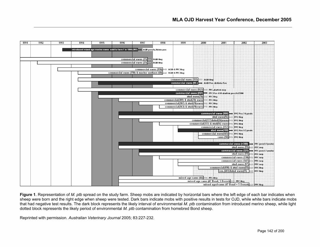
MLA OJD Harvest Year Conference, December 2005
Page 142 of 200
Figure 1. Representation of M. ptb spread on the study farm. Sheep mobs are indicated by horizontal bars where the left edge of each bar indicates whensheep were born and the right edge when sheep were tested. Dark bars indicate mobs with positive results in tests for OJD, while white bars indicate mobsthat had negative test results. The dark block represents the likely interval of environmental M. ptb contamination from introduced merino sheep, while lightdotted block represents the likely period of environmental M. ptb contamination from homebred Bond sheep.
Reprinted with permission. Australian Veterinary Journal 2005; 83:227-232.
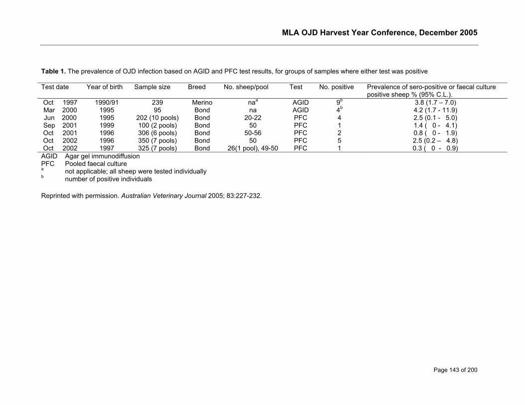
MLA OJD Harvest Year Conference, December 2005
Page 143 of 200
Table 1. The prevalence of OJD infection based on AGID and PFC test results, for groups of samples where either test was positive
Test date Year of birth Sample size Breed No. sheep/pool Test No. positive Prevalence of sero-positive or faecal culturepositive sheep % (95% C.L.).
Oct 1997 1990/91 239 Merino naa AGID 9b 3.8 (1.7 – 7.0)Mar 2000 1995 95 Bond na AGID 4b 4.2 (1.7 - 11.9)Jun 2000 1995 202 (10 pools) Bond 20-22 PFC 4 2.5 (0.1 - 5.0)Sep 2001 1999 100 (2 pools) Bond 50 PFC 1 1.4 ( 0 - 4.1)Oct 2001 1996 306 (6 pools) Bond 50-56 PFC 2 0.8 ( 0 - 1.9)Oct 2002 1996 350 (7 pools) Bond 50 PFC 5 2.5 (0.2 – 4.8)Oct 2002 1997 325 (7 pools) Bond 26(1 pool), 49-50 PFC 1 0.3 ( 0 - 0.9)
AGID Agar gel immunodiffusionPFC Pooled faecal culturea not applicable; all sheep were tested individuallyb number of positive individuals
Reprinted with permission. Australian Veterinary Journal 2005; 83:227-232.
MLA OJD Harvest Year Conference, December 2005
Page 144 of 200
Risk factors for OJD prevalence in infected flocks
Dhand NKa, Eppleston J, Whittington RJ and Toribio J-ALMLaFaculty of Veterinary Science, The University of Sydney,425 Werombi Road, Camden NSW 2570 AustraliaPhone: 02 9351 1610; Fax: 02 9351 1693Email: [email protected]
IntroductionThe level of clinical disease experienced due to ovine Johne’s disease (OJD) appears to varyconsiderably between infected sheep flocks in Australia, even in those having apparently similarcharacteristics. Some inter-flock variation may be due to differences in time since OJD infection andpast sheep purchasing history. However, there appear to be other factors capable of affecting theclinical expression of disease in the flock. Only a small number of studies have been conducted toinvestigate these factors (Lugton, 2004; Mainar-Jaime and Vazquez-Boland, 1998; Reviriego et al.,2000). The one risk factor study conducted in Australia (Lugton, 2004) could not make conclusiverecommendations due to limitations of the study design as outcome variables in this study werebased on the subjective information provided by the producers. Investigation of risk factors in a 12-farm mortality study (MLA OJD.023) identified some potential risk factors for increased prevalence,but interpretation was constrained by the small sample size (Toribio et al., 2004). In MLA projectOJD.028, high levels of pasture contamination and exposure of young sheep were the main riskfactors identified but this study was not designed to investigate a range of factors (Whittington andMcGregor, 2005). In the absence of concrete scientific information, there is speculation amongfarmers about the potential effect of some factors like sheep management practices, soil pH,micronutrient levels and soil type. On the other hand, though vaccination appears to be an effectivecontrol strategy, it is relatively expensive and may not be economically viable for all flocks. Thusthere is a strong need to investigate risk factors for clinical expression of disease that may helpdevelopment of recommendations for on-farm control of OJD in the absence of vaccination, or as anadjunct to vaccination.
MethodsThe project consisted of a cross-sectional study on 92 infected properties in Australia that metspecific selection criteria:
1. Self-replacing Merino flocks
2. Located in all identified OJD infected areas/regions in Australia including NSW, Victoria,Kangaroo Island, Tasmania (including Flinders Island) and Western Australia
3. Flock infected for 3 or more years
4. Non-vaccinated 3-year old, 3 & 4-year old (6-8 tooth) or 4 & 5-year old sheep present in flock
5. ≥ 210 sheep in the 3-year old, 3 & 4-year old or 4 & 5-year old cohort (as 7 pools of 30 sheepwere used to classify flocks as high or low prevalence)
A cohort represented the sheep of a specific age and sex group in the flock. Usually only one cohortwas selected from every flock, however if on the day of faecal sampling, 210 sheep of same age andsex were not present in the flock, then samples from another age or sex group, representingadditional cohorts, were also selected.
MLA OJD Harvest Year Conference, December 2005
Page 145 of 200
Mycobacterium avium subsp. paratuberculosis (M. ptb) in faeces was detected by pooled faecalculture (PFC) (Whittington et al., 2000a) and confirmed by polymerase chain reaction and restrictionendonuclease analysis. Animal level prevalence for each sheep cohort, based on PFC results, wascalculated by a method for variable pool size (Williams and Moffitt, 2001) using the PooledPrevalence Calculator available online at http://www.ausvet.com.au/pprev (Sergeant, 2004). Basedon OJD prevalence, the cohorts were categorised into low (<2%), medium (2-10%) and high (>10%)prevalence categories to create outcome variables.
Information about potential risk factors and confounders was collected by face-to-face interview witheach producer using a pretested questionnaire and from 3 soil samples collected from paddocksgrazed by the cohort sheep. Univariable analyses using the logistic regression SAS LOGISTICprocedure (Stokes et al., 2000) were performed to investigate the unconditional association betweenoutcome variable and each explanatory variable. The variables significant in the univariableanalyses (P<0.25) were then tested in the multivariable model first by forward selection and then bybackward removal (retaining only variables with P<0.10) to investigate their relationship with OJDprevalence level after allowing for other variables and confounders. Separate final models were builtfor husbandry and soil variables.
ResultsIn total, 233 known OJD-infected flocks were investigated to identify eligible flocks that met thespecific selection criteria and 92 flocks were enrolled in the study by 31 July, 2004. Visits to eachenrolled flock for faecal and soil sample collection, and producer interviews were completed by 21stDecember, 2004.
For better data consistency and reliability, a sub dataset was prepared after excluding cohortscomprised of 3 or lesser faecal pools as well as those of 5 year old age group. This datasetconsisted of records of 98 cohorts representing 87 flocks and was used in the present analyses.Average OJD prevalence based on PFC among these 98 sheep cohorts was 14.8% (median 4.5%,range 0 to 58.9%) and the number of sheep cohorts categorised as low (<2%) , medium (2-10%)and high (>10%) prevalence were 24, 52 and 22, respectively.
In the final model, there was a strong relationship between the OJD prevalence and the level of OJDmortality as well as with sex (Table 1). Stocking rate of the lambing paddock and number of Gudair®vaccinated sheep drops present in the flock were the main husbandry risk factors significantlyassociated with OJD prevalence level. In addition, presence of wildlife aside from kangaroos andrabbits, and application of fertilizer other than single super, molybdenum super and lime were alsoassociated with cohort OJD prevalence. Among the soil risk factors investigated (Table 4), OJDprevalence was higher in flocks raised on soils with higher phosphorus buffering index (≥70) andhigher clay percentage (>15%).
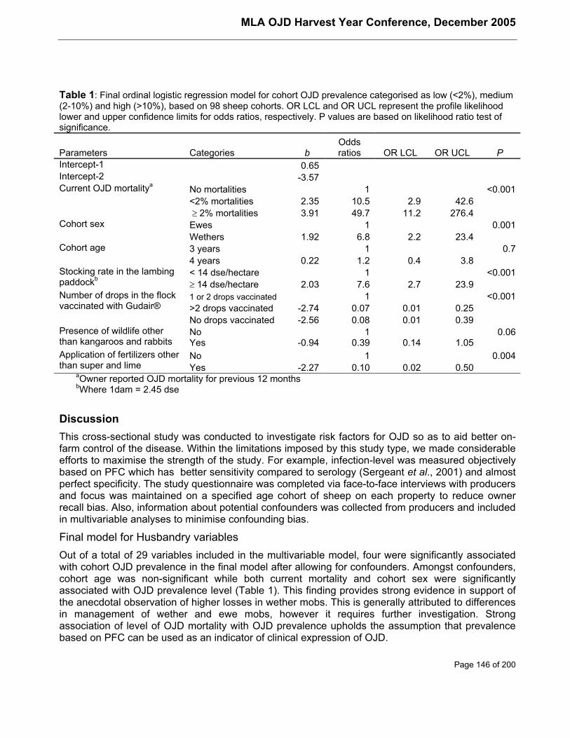
MLA OJD Harvest Year Conference, December 2005
Page 146 of 200
Table 1: Final ordinal logistic regression model for cohort OJD prevalence categorised as low (<2%), medium(2-10%) and high (>10%), based on 98 sheep cohorts. OR LCL and OR UCL represent the profile likelihoodlower and upper confidence limits for odds ratios, respectively. P values are based on likelihood ratio test ofsignificance.
Parameters Categories bOddsratios OR LCL OR UCL P
Intercept-1 0.65Intercept-2 -3.57Current OJD mortalitya No mortalities 1 <0.001
<2% mortalities 2.35 10.5 2.9 42.6 ≥ 2% mortalities 3.91 49.7 11.2 276.4
Cohort sex Ewes 1 0.001Wethers 1.92 6.8 2.2 23.4
Cohort age 3 years 1 0.74 years 0.22 1.2 0.4 3.8< 14 dse/hectare 1 <0.001Stocking rate in the lambing
paddockb≥ 14 dse/hectare 2.03 7.6 2.7 23.91 or 2 drops vaccinated 1 <0.001>2 drops vaccinated -2.74 0.07 0.01 0.25
Number of drops in the flockvaccinated with Gudair®
No drops vaccinated -2.56 0.08 0.01 0.39No 1 0.06Presence of wildlife other
than kangaroos and rabbits Yes -0.94 0.39 0.14 1.05No 1 0.004Application of fertilizers other
than super and lime Yes -2.27 0.10 0.02 0.50aOwner reported OJD mortality for previous 12 monthsbWhere 1dam = 2.45 dse
DiscussionThis cross-sectional study was conducted to investigate risk factors for OJD so as to aid better on-farm control of the disease. Within the limitations imposed by this study type, we made considerableefforts to maximise the strength of the study. For example, infection-level was measured objectivelybased on PFC which has better sensitivity compared to serology (Sergeant et al., 2001) and almostperfect specificity. The study questionnaire was completed via face-to-face interviews with producersand focus was maintained on a specified age cohort of sheep on each property to reduce ownerrecall bias. Also, information about potential confounders was collected from producers and includedin multivariable analyses to minimise confounding bias.
Final model for Husbandry variablesOut of a total of 29 variables included in the multivariable model, four were significantly associatedwith cohort OJD prevalence in the final model after allowing for confounders. Amongst confounders,cohort age was non-significant while both current mortality and cohort sex were significantlyassociated with OJD prevalence level (Table 1). This finding provides strong evidence in support ofthe anecdotal observation of higher losses in wether mobs. This is generally attributed to differencesin management of wether and ewe mobs, however it requires further investigation. Strongassociation of level of OJD mortality with OJD prevalence upholds the assumption that prevalencebased on PFC can be used as an indicator of clinical expression of OJD.
MLA OJD Harvest Year Conference, December 2005
Page 147 of 200
Vaccination with Gudair®The numbers of sheep drops vaccinated with Gudair® in the flock were significantly associated withOJD prevalence4. The flocks with >2 vaccinated drops had significantly lower cohort OJDprevalence as compared to those with only 1-2 vaccinated drops (Table 1). It may be an indication ofpossible reduction in infection level on the property with several drops of vaccinated sheep sheddinglower M. ptb. Similar observations were also made in a vaccination trial as a significant reduction inmortality risk was reported in the 3rd and 4th years after commencement of vaccination (McGregor,2004). In contrast, the OJD prevalence in flocks not vaccinating at all was lower than in thereference group (1-2 drops vaccinated). This could be due to the possibility that producers in non-vaccinating flocks may not be going for vaccination as their flocks were not experiencing highlosses. Only 21.4% (3 of 14) of cohorts of non-vaccinating flocks reported high current mortalities incomparison to 38.1% cohorts (32 of the 84) of vaccinating flocks. Overall, vaccination of sheep withGudair® appears to reduce the prevalence of OJD over time.
Stocking Rate of the Lambing paddockHigh stocking rate in the lambing paddock/s (≥14 dse /hectare) was associated with higher cohortOJD prevalence. This is a very important finding and highlights the importance of lambs being themost susceptible age group in a sheep flock. Dams may be at increased stress due to high stockingrate coupled with parturition and lactation. Heightened stress may lead to higher level of M. ptbfaecal shedding, which may in turn lead to higher M. ptb pasture and environmental contamination inthe lambing paddock. Consequently, higher doses at an age when sheep are more susceptible toinfection increase both the probability of infection and the probability of establishment of infectionleading to a greater likelihood of the clinical expression of the disease. Our findings complement theresults from the MLA OJD.028 project that concluded that lambs were the most susceptible agegroup and if exposed to high levels of contamination, may develop severe infection leading to death(Whittington and McGregor, 2005). Although stocking rate has been investigated in previous cross-sectional studies (Daniels et al., 2002; Lugton, 2004; Reviriego et al., 2000), this is the first study toidentify an association between Johne’s disease and higher stocking rate. Toribio et al. (2004)reported a tentative link between OJD mortality and stocking rate but conclusions were guarded dueto small sample size.
The stocking rate in the weaning, hogget and adult paddock, and overall stocking rate of the flockwere also significant risk factors by themselves in the univariable analysis. However, these variableswere not significant in the multivariable model due to multicollinearity with lambing paddock stockingrate. Interestingly, if lambing paddock stocking rate was temporarily removed from the model theneach of the stocking rate variables added by turn to the final model, was significant after allowing forother confounders and variables. It indicates that stocking rate is a major risk factor for OJD andhence should be considered in formulating any disease control strategy.
4 All sheep cohorts selected for the study were required to be non-vaccinated and the results reported here areabout the number of drops vaccinated in the flock in question.
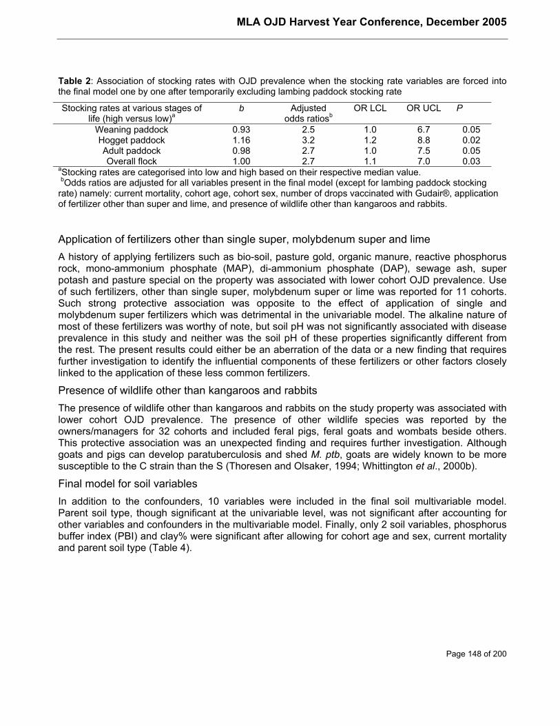
MLA OJD Harvest Year Conference, December 2005
Page 148 of 200
Table 2: Association of stocking rates with OJD prevalence when the stocking rate variables are forced intothe final model one by one after temporarily excluding lambing paddock stocking rate
Stocking rates at various stages oflife (high versus low)a
b Adjustedodds ratiosb
OR LCL OR UCL P
Weaning paddock 0.93 2.5 1.0 6.7 0.05Hogget paddock 1.16 3.2 1.2 8.8 0.02Adult paddock 0.98 2.7 1.0 7.5 0.05Overall flock 1.00 2.7 1.1 7.0 0.03
aStocking rates are categorised into low and high based on their respective median value. bOdds ratios are adjusted for all variables present in the final model (except for lambing paddock stockingrate) namely: current mortality, cohort age, cohort sex, number of drops vaccinated with Gudair®, applicationof fertilizer other than super and lime, and presence of wildlife other than kangaroos and rabbits.
Application of fertilizers other than single super, molybdenum super and limeA history of applying fertilizers such as bio-soil, pasture gold, organic manure, reactive phosphorusrock, mono-ammonium phosphate (MAP), di-ammonium phosphate (DAP), sewage ash, superpotash and pasture special on the property was associated with lower cohort OJD prevalence. Useof such fertilizers, other than single super, molybdenum super or lime was reported for 11 cohorts.Such strong protective association was opposite to the effect of application of single andmolybdenum super fertilizers which was detrimental in the univariable model. The alkaline nature ofmost of these fertilizers was worthy of note, but soil pH was not significantly associated with diseaseprevalence in this study and neither was the soil pH of these properties significantly different fromthe rest. The present results could either be an aberration of the data or a new finding that requiresfurther investigation to identify the influential components of these fertilizers or other factors closelylinked to the application of these less common fertilizers.
Presence of wildlife other than kangaroos and rabbitsThe presence of wildlife other than kangaroos and rabbits on the study property was associated withlower cohort OJD prevalence. The presence of other wildlife species was reported by theowners/managers for 32 cohorts and included feral pigs, feral goats and wombats beside others.This protective association was an unexpected finding and requires further investigation. Althoughgoats and pigs can develop paratuberculosis and shed M. ptb, goats are widely known to be moresusceptible to the C strain than the S (Thoresen and Olsaker, 1994; Whittington et al., 2000b).
Final model for soil variablesIn addition to the confounders, 10 variables were included in the final soil multivariable model.Parent soil type, though significant at the univariable level, was not significant after accounting forother variables and confounders in the multivariable model. Finally, only 2 soil variables, phosphorusbuffer index (PBI) and clay% were significant after allowing for cohort age and sex, current mortalityand parent soil type (Table 4).
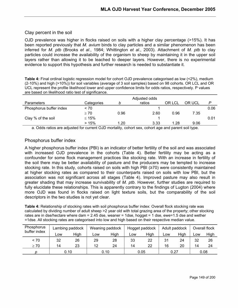
MLA OJD Harvest Year Conference, December 2005
Page 149 of 200
Clay percent in the soilOJD prevalence was higher in flocks raised on soils with a higher clay percentage (>15%). It hasbeen reported previously that M. avium binds to clay particles and a similar phenomenon has beeninferred for M. ptb (Brooks et al., 1984; Whittington et al., 2003). Attachment of M. ptb to clayparticles could increase the availability of the organism to sheep by maintaining it in the upper soillayers rather than allowing it to be leached to deeper layers. However, there is no experimentalevidence to support this hypothesis and further research is needed to substantiate it.
Table 4: Final ordinal logistic regression model for cohort OJD prevalence categorised as low (<2%), medium(2-10%) and high (>10%)) for soil variables (average of 3 soil samples) based on 98 cohorts. OR LCL and ORUCL represent the profile likelihood lower and upper confidence limits for odds ratios, respectively. P valuesare based on likelihood ratio test of significance.
Parameters Categories bAdjusted odds
ratios OR LCL OR UCL PPhosphorus buffer index < 70 1 0.06
≥ 70 0.96 2.60 0.96 7.35Clay % of the soil ≤ 15% 1 0.01
> 15% 1.20 3.33 1.28 9.06a. Odds ratios are adjusted for current OJD mortality, cohort sex, cohort age and parent soil type.
Phosphorus buffer indexA higher phosphorus buffer index (PBI) is an indicator of better fertility of the soil and was associatedwith increased OJD prevalence in the cohorts (Table 4). Better fertility may be acting as aconfounder for some flock management practices like stocking rate. With an increase in fertility ofthe soil there may be better availability of pasture and the producers may be tempted to increasestocking rate. In this study, cohorts raised on soils with high PBI (≥70) were consistently maintainedat higher stocking rates as compared to their counterparts raised on soils with low PBI, but theassociation was not significant across all stages (Table 4). Improved pasture may also result ingreater shading that may increase survivability of M. ptb. However, further studies are required tofully elucidate these relationships. This is apparently contrary to the findings of Lugton (2004) wheremore OJD was found in flocks raised on light texture soils, but the comparability of the soildescriptors in the two studies is not yet clear.
Table 4: Relationship of stocking rates with soil phosphorus buffer index: Overall flock stocking rate wascalculated by dividing number of adult sheep >2 year old with total grazing area of the property; other stockingrates are in dse/hectare where dam = 2.45 dse, weaner = 1dse, hogget = 1 dse, ewe=1.5 dse and wether=1dse. All stocking rates are categorised into low and high based on their respective median value.
Lambing paddock Weaning paddock Hogget paddock Adult paddock Overall flockPhosphorusbuffer index
Low High Low High Low High Low High Low High< 70 32 26 29 28 33 22 31 24 32 26≥ 70 14 23 12 24 14 22 16 20 14 24
p 0.10 0.10 0.05 0.27 0.08
MLA OJD Harvest Year Conference, December 2005
Page 150 of 200
Conclusions and recommendationsThe strong association of current mortality with OJD prevalence based on PFC indicates that thelatter can be used to objectively measure the clinical expression of OJD. This is the first study toconfirm the anecdotal observation of higher OJD levels in wethers than ewes. The high stockingrates, particularly in the lambing paddock, are associated with higher OJD prevalence and producersshould be advised to take necessary measures to reduce the stocking rates to lessen the OJDprevalence. Also, vaccination appears to reduce the prevalence over time and continued vaccinationprogramme should be recommended. The association of disease prevalence with fertile soils is anew finding and needs further investigation.
ReferencesBrooks, R.W., George, K.L., Parker, B.C., Falkinham, J.O., 3rd, Gruff, H., 1984, Recovery and
survival of nontuberculous mycobacteria under various growth and decontaminationconditions. Can J Microbiol 30, 1112-1117.
Daniels, M.J., Hutchings, M.R., Allcroft, D.J., McKendrick, I.J., Greig, A., 2002, Risk factors forJohne's disease in Scotland - the results of a survey of farmers. Veterinary Record 150, 135-139.
Lugton, I.W., 2004, Cross-sectional study of risk factors for the clinical expression of ovine Johne'sdisease on New South Wales farms. Aust Vet J 82, 355-365.
Mainar-Jaime, R.C., Vazquez-Boland, J.A., 1998, Factors associated with seroprevalence toMycobacterium paratuberculosis in small-ruminant farms in the Madrid region (Spain).Preventive Veterinary Medicine 34, 317-327.
McGregor, H., 2004. A longitudinal study of OJD and the effects of whole flock vaccination withGudair (TM). In: Ovine Johne's Disease: An update of Australian and International Research,Sydney, Australia, March 2004, pp. 2-4.
Reviriego, F.J., Moreno, M.A., Dominguez, L., 2000, Soil type as a putative risk factor of ovine andcaprine paratuberculosis seropositivity in Spain. Prev Vet Med 43, 43-51.
Sergeant, E. 2004. Pooled Prevalence Calculator (Aus Vet Animal Health Services).
Sergeant, E.S.G., Whittington, R.J., More, S.J., 2001, Sensitivity and specificity of pooled faecalculture and serology as flock-screening tests for detection of ovine paratuberculosis inAustralia. Preventive Veterinary Medicine 52, 199-211.
Stokes, M.E., Davis, C.S., Koch, G.G., 2000, Categorical Data Analysis using The SAS system,Second Edition. SAS Institute Inc., Cary, N.C.
Thoresen, O.F., Olsaker, I., 1994, Distribution and hybridization patterns of the insertion elementIS900 in clinical isolates of Mycobacterium paratuberculosis. Veterinary Microbiology 40,293-303.
Toribio, J.A., Bush, R., Windsor, P. 2004. A study of the biological and economic impact of OJD inaffected sheep flocks in NSW. A report submitted to Meat and Livestock Australia.
Whittington, R., McGregor, H. 2005. Epidemiology of ovine Johne’s disease 2 – pasturecontamination level and age susceptibility A report submitted to Meat and LivestockAustralia.
MLA OJD Harvest Year Conference, December 2005
Page 151 of 200
Whittington, R.J., Fell, S., Walker, D., McAllister, S., Marsh, I., Sergeant, E., Taragel, C.A., Marshall,D.J., Links, I.J., 2000a, Use of pooled fecal culture for sensitive and economic detection ofmycobacterium avium subsp. paratuberculosis infection in flocks of sheep. Journal of ClinicalMicrobiology 38, 2550-2556.
Whittington, R.J., Hope, A.F., Marshall, D.J., Taragel, C.A., Marsh, I., 2000b, Molecularepidemiology of Mycobacterium avium subsp. paratuberculosis: IS900 restriction fragmentlength polymorphism and IS1311 polymorphism analyses of isolates from animals and ahuman in Australia. Journal of Clinical Microbiology 38, 3240-3248.
Whittington, R.J., Marsh, I.B., Taylor, P.J., Marshall, D.J., Taragel, C., Reddacliff, L.A., 2003,Isolation of Mycobacterium avium subsp paratuberculosis from environmental samplescollected from farms before and after destocking sheep with paratuberculosis. AustralianVeterinary Journal 81, 559-563.
Williams, C., Moffitt, C., 2001, A critique of methods of sampling and reporting pathogens inpopulations of fish. Journal of Aquatic Animal Health 13, 300-309.
MLA OJD Harvest Year Conference, December 2005
Page 152 of 200
Economic modelling of the impact of OJD
Russell Busha, Jenny-Ann Toribio, Peter Windsor and Stewart WebsteraFaculty of Veterinary Science, University of SydneyPMB 3 Camden, NSW 2570Phone: 02 9351 1785; Fax: 02 9351 1693E-mail: [email protected]
IntroductionTo date the work conducted to estimate the financial impact of OJD in Australia has been based onboth farm-level (NSW Farmers, 1997; Webster & Hall, 2000; Webster, 2002) and industry-levelstudies (Hassall and Associates, 2000; Hassall and Associates, 2003; Sergeant, 2002; Sergeant,2003). To assist sheep producers to better manage OJD it is more important that they understandthe on-farm financial impact of the disease and associated implications, rather than the industry-wide financial impact.
As an infected flock cannot return to an uninfected status without prohibitive cost (Taylor & Webster,2005), the difference between infected and uninfected enterprise profit has little meaning. Of greatervalue is the proportion of the total cost of OJD that is avoidable through disease control strategiessuch as vaccination and management (McInerney et al., 1992; Webster & Mullen, 2000).
Gross margin (GM) calculations are commonly used by Australian sheep producers when budgetingand planning. They are a simple and quick method of providing a direct comparison of the relativeprofitability of similar enterprises (Dijkhuizen & Morris, 1997) and can be used to analyse actualenterprise performance under different disease scenarios.
A GM model was developed to predict the on-farm financial impact of OJD for Merino and first orsecond cross prime lamb enterprises, comparing non-infected, infected (status quo) and infected(vaccination) disease scenarios within Australia. The model estimates changes in profit due toincreased mortalities as a result of OJD, and the benefits and costs of control through the use ofGudairTM vaccination, including a breakeven point.
MethodsSpreadsheets for the GM model were constructed in MS Excel 2000 with the more complexworksheet functions programmed using MS Excel Visual Basic 6.0. The model allows potential usersto enter information that is either specific to their own farm enterprise or of interest whencontemplating a change in enterprise mix.
The model user enters information describing the farm carrying capacity, enterprise mix, income andcosts of production as well as OJD status. Choices of individual GM budgets include Merino fine(19µm), medium (21µm) and strong (23µm) wool enterprises as well as 1st and 2nd cross lambproduction. Merino sheep are further separated into ewe and wether enterprises. The userdetermines the size of each enterprise by entering the number of breeding ewes (Merino, 1st or 2nd
cross) or wethers (Merino). Flock numbers and mortalities are simulated throughout the year.
Relevant production and husbandry information is entered to gain the income and expenditure foreach enterprise. The user is able to enter specific information or can opt to use preset values basedon information current in 2005. Drenching regimes for internal parasite control are preset and arespecific for the likely location of each enterprise (NSW Agriculture, 2002).
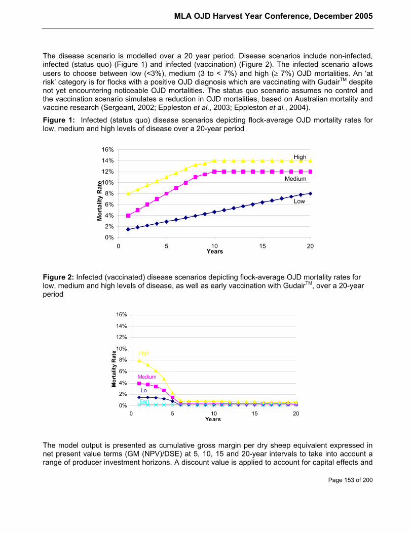
MLA OJD Harvest Year Conference, December 2005
Page 153 of 200
The disease scenario is modelled over a 20 year period. Disease scenarios include non-infected,infected (status quo) (Figure 1) and infected (vaccination) (Figure 2). The infected scenario allowsusers to choose between low (<3%), medium (3 to < 7%) and high (≥ 7%) OJD mortalities. An ‘atrisk’ category is for flocks with a positive OJD diagnosis which are vaccinating with GudairTM despitenot yet encountering noticeable OJD mortalities. The status quo scenario assumes no control andthe vaccination scenario simulates a reduction in OJD mortalities, based on Australian mortality andvaccine research (Sergeant, 2002; Eppleston et al., 2003; Eppleston et al., 2004).
Figure 1: Infected (status quo) disease scenarios depicting flock-average OJD mortality rates forlow, medium and high levels of disease over a 20-year period
0%
2%
4%
6%
8%
10%
12%
14%
16%
0 5 10 15 20
Low
Medium
High
Years
Mor
talit
y R
ate
Figure 2: Infected (vaccinated) disease scenarios depicting flock-average OJD mortality rates forlow, medium and high levels of disease, as well as early vaccination with GudairTM, over a 20-yearperiod
0%
2%
4%
6%
8%
10%
12%
14%
16%
0 5 10 15 20
Lo
Medium
High
Years
Mor
talit
y R
ate
Earl
The model output is presented as cumulative gross margin per dry sheep equivalent expressed innet present value terms (GM (NPV)/DSE) at 5, 10, 15 and 20-year intervals to take into account arange of producer investment horizons. A discount value is applied to account for capital effects and
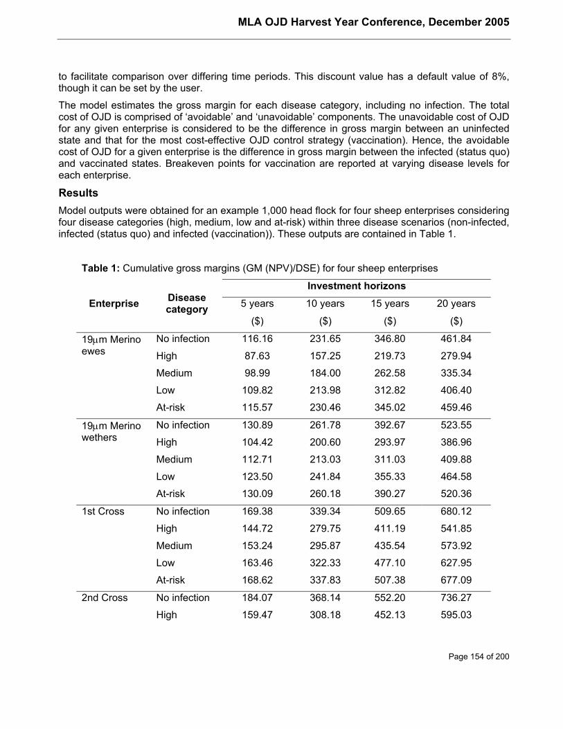
MLA OJD Harvest Year Conference, December 2005
Page 154 of 200
to facilitate comparison over differing time periods. This discount value has a default value of 8%,though it can be set by the user.
The model estimates the gross margin for each disease category, including no infection. The totalcost of OJD is comprised of ‘avoidable’ and ‘unavoidable’ components. The unavoidable cost of OJDfor any given enterprise is considered to be the difference in gross margin between an uninfectedstate and that for the most cost-effective OJD control strategy (vaccination). Hence, the avoidablecost of OJD for a given enterprise is the difference in gross margin between the infected (status quo)and vaccinated states. Breakeven points for vaccination are reported at varying disease levels foreach enterprise.
ResultsModel outputs were obtained for an example 1,000 head flock for four sheep enterprises consideringfour disease categories (high, medium, low and at-risk) within three disease scenarios (non-infected,infected (status quo) and infected (vaccination)). These outputs are contained in Table 1.
Table 1: Cumulative gross margins (GM (NPV)/DSE) for four sheep enterprises
Investment horizons
5 years 10 years 15 years 20 yearsEnterprise Diseasecategory
($) ($) ($) ($)
No infection 116.16 231.65 346.80 461.8419µm Merinoewes High 87.63 157.25 219.73 279.94
Medium 98.99 184.00 262.58 335.34
Low 109.82 213.98 312.82 406.40
At-risk 115.57 230.46 345.02 459.46
No infection 130.89 261.78 392.67 523.5519µm Merinowethers High 104.42 200.60 293.97 386.96
Medium 112.71 213.03 311.03 409.88
Low 123.50 241.84 355.33 464.58
At-risk 130.09 260.18 390.27 520.36
1st Cross No infection 169.38 339.34 509.65 680.12
High 144.72 279.75 411.19 541.85
Medium 153.24 295.87 435.54 573.92
Low 163.46 322.33 477.10 627.95
At-risk 168.62 337.83 507.38 677.09
2nd Cross No infection 184.07 368.14 552.20 736.27
High 159.47 308.18 452.13 595.03
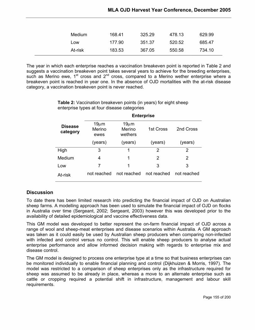
MLA OJD Harvest Year Conference, December 2005
Page 155 of 200
Medium 168.41 325.29 478.13 629.99
Low 177.90 351.37 520.52 685.47
At-risk 183.53 367.05 550.58 734.10
The year in which each enterprise reaches a vaccination breakeven point is reported in Table 2 andsuggests a vaccination breakeven point takes several years to achieve for the breeding enterprises,such as Merino ewe, 1st cross and 2nd cross, compared to a Merino wether enterprise where abreakeven point is reached in year one. In the absence of OJD mortalities with the at-risk diseasecategory, a vaccination breakeven point is never reached.
Table 2: Vaccination breakeven points (in years) for eight sheepenterprise types at four disease categories
Enterprise
Diseasecategory
19µmMerinoewes
19µmMerinowethers
1st Cross 2nd Cross
(years) (years) (years) (years)
High 3 1 2 2
Medium 4 1 2 2
Low 7 1 3 3
At-risk not reached not reached not reached not reached
DiscussionTo date there has been limited research into predicting the financial impact of OJD on Australiansheep farms. A modelling approach has been used to simulate the financial impact of OJD on flocksin Australia over time (Sergeant, 2002; Sergeant, 2003) however this was developed prior to theavailability of detailed epidemiological and vaccine effectiveness data.
This GM model was developed to better represent the on-farm financial impact of OJD across arange of wool and sheep-meat enterprises and disease scenarios within Australia. A GM approachwas taken as it could easily be used by Australian sheep producers when comparing non-infectedwith infected and control versus no control. This will enable sheep producers to analyse actualenterprise performance and allow informed decision making with regards to enterprise mix anddisease control.
The GM model is designed to process one enterprise type at a time so that business enterprises canbe monitored individually to enable financial planning and control (Dijkhuizen & Morris, 1997). Themodel was restricted to a comparison of sheep enterprises only as the infrastructure required forsheep was assumed to be already in place, whereas a move to an alternate enterprise such ascattle or cropping required a potential shift in infrastructure, management and labour skillrequirements.
MLA OJD Harvest Year Conference, December 2005
Page 156 of 200
The model suggests a vaccination breakeven point is achieved in two to three years for breedingenterprises if the level of OJD is high. If the level of OJD is low a vaccination breakeven point isachieved in three years for either a 1st cross or 2nd cross enterprise and in seven years for a Merinoewe enterprise. Although all breeding enterprises vaccinate lambs, the Merino ewe enterprise takesthe longest time to reach a vaccination breakeven point as a greater number of young sheep areretained annually. With Merino wethers a vaccination breakeven point is reached in year one for alldisease categories due to introduced vaccinated replacement hoggets providing an immediateresponse in reducing OJD mortalities. In the absence of OJD mortalities in the at-risk diseasecategory, a vaccination breakeven point is not reached within the model’s 20-year time frame for anyof the enterprises.
The total cost of OJD for the high disease level scenario across all enterprises is due to a highnumber of OJD mortalities. The avoidable cost of OJD, resulting from the use of vaccination,reduced OJD mortalities and improved the cumulative GM for an infected (vaccination) flockcompared to an infected (status quo) flock. At a medium and low disease level the total cost of OJDand the avoidable costs of OJD associated with vaccination are reduced for all enterprises due tolower OJD mortality rates. In the at-risk disease category there is a minimal reduction in cumulativeGM associated with the total cost of OJD due to the assumption that an annual OJD mortality rate of0.2% is possibly being experienced. There is also a negative return to vaccination due to the cost ofvaccination not being compensated by a reduced OJD mortality rate. Benefits from vaccination arelikely to be associated with the market advantage these at-risk vaccinated animals could command,however this benefit was not calculated.
In addition to the costs associated with OJD mortality rates and control through vaccination andmanagement it is suggested that sub-clinical losses should also be considered when establishing allthe productivity losses associated with a disease (Bennett, 2003). However, these may be difficult toestablish as there continues to be some debate regarding the existence and financial impact of sub-clinical losses. Reductions in bodyweight have been recorded around 12 months prior to death inone study (MLA.OJD.002A), leading to infected sheep being 32% (12kg) lighter at death andproducing 6% less wool annually than similar animals free of the disease. However, this may not bea consistent finding. In a separate study investigating the efficacy of vaccination withGudairTM(MLA.OJD.009), small reductions in live weight gain were found in vaccinated lambs in thefirst year following vaccination. Even so, over the course of the 5-year trial there was little differencein weight or condition score, nor in fleece parameters, between vaccinates and non-vaccinates.These conflicting observations suggest that the extent of sub-clinical losses is variable betweenflocks. Regardless of these findings, as subclinical losses of 6% less wool has been identified in atleast one study, it is probable that economic losses reported in this model may be an underestimateof the actual losses.
The extent of trading losses for individual farms primarily depends on the disease status, enterprisemix, production system and market forces/demands. The financial impact of trading losses for a farmprimarily selling sheep for slaughter will be minimal compared to a farm on-selling animals to otherproducers. With assurance based trading (Australian Animal Health Council, 2004) there is anopportunity for Australian sheep producers with infected flocks to improve their trading positionthrough the use of vaccination as part of their on-farm management of OJD.
MLA OJD Harvest Year Conference, December 2005
Page 157 of 200
Application for industryThis GM model provides Australian sheep producers and their advisors with an accurate on-farmestimate of the total and avoidable cost of OJD for Merino as well as first or second cross primelamb enterprises. Breakeven points for vaccination inform affected producers of the length of timerequired for a return on their investment in vaccination, providing a useful decision making tool whendeveloping on-farm strategies for the control of OJD at different disease levels.
ReferencesAustralian Animal Health Council (2004), Johne's disease information centre: The Assurance BasedCredit (ABC) scheme - OJD, Australian Animal Health Council Ltd., Canberra, Australiahttp://www.aahc.com.au/jd/abc_scheme.htm, (retrieved July 2004).
Bennett, R. (2003), The 'direct costs' of livestock disease: the development of a system of modelsfor the analysis of 30 endemic livestock diseases in Great Britain. Journal of Agricultural Economics54: 55-71.
Dijkhuizen, A. A. & Morris, R. S. (1997), Animal health economics - principles and applications. PostGraduate Foundation, University of Sydney, Sydney, pp. 30-87.
Eppleston, J., Reddacliff, L., PA, W., Whittington, R. & Jones, S. (2004), Field studies on vaccinationfor the control of OJD in Australia - an overview, In: Proceedings of the Australian Sheep VeterinarySociety Annual Meeting, Vol. 14, Canberra, pp. 56-59.
Eppleston, J., Reddacliff, L., Windsor, P., Whittington, R. & Jones, S. (2003), An update on theefficacy of GudairTM OJD vaccine in Australia, In: Proceedings of the Australian Sheep VeterinarySociety Annual Meeting, Vol. 13, Cairns, pp. 107-113.
Hassall and Associates (2000), Financial impacts and forms of assistance for OJD affectedsheepbreeders, In: Report prepared for NSW Ovine Johne's Disease Industry Advisory Committee.
Hassall and Associates (2003), Costs of OJD for Australian sheep producers and options forassistance, In: Final report prepared for Australian Wool Innovation Limited.
McInerney, J., Howe, K. S. & Schepers, J. A. (1992), A framework for the economic analysis ofdisease in farm livestock. Preventive Veterinary Medicine 13: 137-154.
NSW Agriculture (2002), Sheep Budgets, New South Wales Department of Primary Industries,Orange, Australia http://www.agric.nsw.gov.au/reader/sheepbud, (retrieved June 2003).
NSW Farmers (1997), The financial and emotional affects of ovine Johne's disease (OJD) onaffected sheep breeders: Results from case studies with OJD affected sheep breeders. NSWFarmers' Association, Sydney.
Sergeant, E. S. G. (2002), Modelling the spread of ovine Johne's disease in infected flocks, In:Proceedings of the Australian Sheep Veterinary Society Annual Meeting, Vol. 12, Adelaide, pp. 10 -13.
Sergeant, E. S. G. (2003), Cost-effectiveness of vaccination against ovine Johne's disease ininfected flocks, In: Proceedings of the Australian Sheep Veterinary Society Annual Meeting, Vol. 13,Cairns, pp. 123-127.
Taylor, P. & Webster, S. R. (2005), Evaluation of Eradication Strategies for Ovine Johne's Disease,Project OJD.001. Meat and Livestock Australia, North Sydney.

MLA OJD Harvest Year Conference, December 2005
Page 158 of 200
Webster, S. R. (2002), Private incentives for voluntary on-farm management of ovine Johne'sdisease, In: Australian Agricultural and Resource Economics Society 46th Annual Conference,Canberra.
Webster, S. R. & Hall, D. G. (2000), The financial consequences of ovine Johne's disease for NewSouth Wales sheep producers. Asian-Australian Journal of Animal Science 13: Suppl C: 465-468.
Webster, S. R. & Mullen, J. (2000), Animal disease economics: The case of ovine Johne's disease inNew South Wales, In: Australian Agricultural and Resource Economics Society 44th AnnualConference, Sydney.
MLA OJD Harvest Year Conference, December 2005
Page 159 of 200
Genomic and proteomic comparative study of the sheep andcattle strains of Mycobacterium avium subsp. paratuberculosis
Ian Marsha, John Bannantine, Mark Tizard and Richard WhittingtonaElizabeth Macarthur Agricultural InstitutePMB 8 Camden, NSW 2570, AustraliaPhone: 02 4640 6502; Fax: 02 4640 6384Email: [email protected]
AbstractIn Australia and other countries the distinction between ovine Johne’s disease (OJD) and bovineJohne’s disease (BJD) at the microbiological level plays an important role in the development andimplementation of control and evaluation programmes for both forms of this insidious disease.However, the differences between the sheep and cattle strains of Mycobacterium avium subsp.paratuberculosis (M. ptb) at the DNA level that result in the different host specificities are stillextremely poorly understood. To date the DNA techniques that exist to differentiate these strainsprovide excellent tools for epidemiological investigations and diagnosis of Johne’s disease but haveyielded little if any insight into the relationship between genotype and phenotype. In this study anintensive comparison was made of the sheep and cattle strains of M. ptb using a variety of modernDNA and protein based techniques including: representational difference analysis (RDA),polymerase chain reaction (PCR) and sequencing, microarray, two dimensional electrophoresisproteomics and surface enhanced laser desorption-ionisation (SELDI) proteomics. The majority ofthese techniques were used in conjunction with the recently completed M. ptb K10 genomesequence in order to identify those genes or proteins with unique characteristics in either strain.Using these techniques 12 single nucleotide polymorphisms, 3 large polymorphisms and a numberof proteomic differences were observed between the sheep and cattle strains of M. ptb. Theseresults confirm earlier studies that differentiate the sheep and cattle strains of M. ptb and continue tosupport management strategies that rely on this distinction.
Introduction and backgroundAfter more than 100 years of research on M. ptb and Johne’s disease, much still remains a mysteryabout this elusive organism and the disease it causes. However, what is clear is that Johne’sdisease presents itself differently in cattle and sheep with respect to clinical, pathological andepidemiological features [34]. Early epidemiological studies identified that cases of Johne’s diseasein sheep and cattle were caused by different strains of M. ptb [27] [28]. The strains isolated fromsheep were difficult to grow on primary culture [27] [18] and were readily identifiable by their orangepigmentation [28]. Problems associated with primary culture of sheep strains persisted [9] but havebeen overcome more recently [33]. DNA based studies of M. ptb isolates from a range of hosts,using restriction fragment length polymorphism of genomic DNA, confirmed the existence of twodistinct groups of M. ptb [9]. These are now commonly referred to as either sheep strains (S) orcattle strains (C). However, the differences observed between the S and C strains to date do notprovide genotypic explanation of their altered phenotypes and divergent host specificity [9] [7] [31][8] [14].
To protect disease free flocks and herds within infected districts and the non-infected regions,control programs have been developed in Australia including the National Ovine Johne’s diseaseControl and Evaluation Program (NOJDP). The success of control programs is dependent on theability to make sound decisions regarding on-farm management practices and the movement of
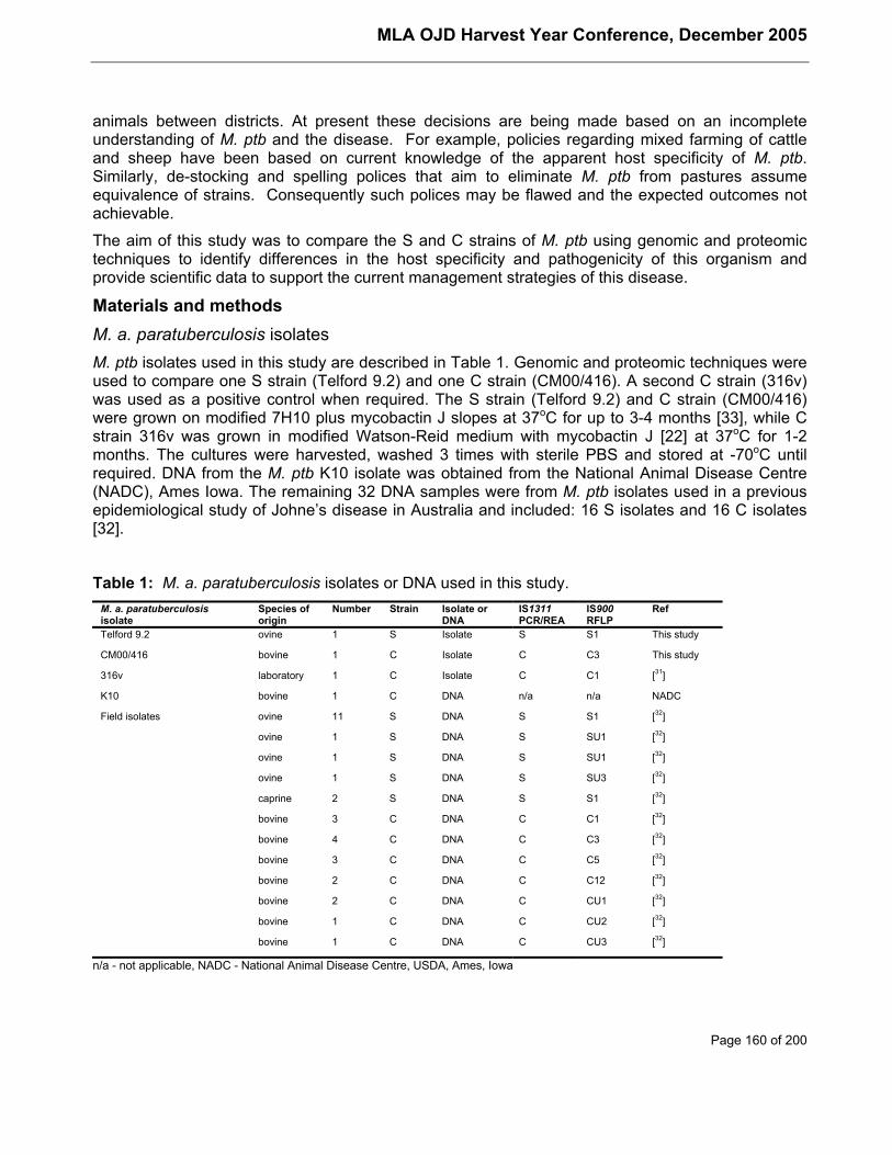
MLA OJD Harvest Year Conference, December 2005
Page 160 of 200
animals between districts. At present these decisions are being made based on an incompleteunderstanding of M. ptb and the disease. For example, policies regarding mixed farming of cattleand sheep have been based on current knowledge of the apparent host specificity of M. ptb.Similarly, de-stocking and spelling polices that aim to eliminate M. ptb from pastures assumeequivalence of strains. Consequently such polices may be flawed and the expected outcomes notachievable.
The aim of this study was to compare the S and C strains of M. ptb using genomic and proteomictechniques to identify differences in the host specificity and pathogenicity of this organism andprovide scientific data to support the current management strategies of this disease.
Materials and methodsM. a. paratuberculosis isolatesM. ptb isolates used in this study are described in Table 1. Genomic and proteomic techniques wereused to compare one S strain (Telford 9.2) and one C strain (CM00/416). A second C strain (316v)was used as a positive control when required. The S strain (Telford 9.2) and C strain (CM00/416)were grown on modified 7H10 plus mycobactin J slopes at 37oC for up to 3-4 months [33], while Cstrain 316v was grown in modified Watson-Reid medium with mycobactin J [22] at 37oC for 1-2months. The cultures were harvested, washed 3 times with sterile PBS and stored at -70oC untilrequired. DNA from the M. ptb K10 isolate was obtained from the National Animal Disease Centre(NADC), Ames Iowa. The remaining 32 DNA samples were from M. ptb isolates used in a previousepidemiological study of Johne’s disease in Australia and included: 16 S isolates and 16 C isolates[32].
Table 1: M. a. paratuberculosis isolates or DNA used in this study.M. a. paratuberculosisisolate
Species oforigin
Number Strain Isolate orDNA
IS1311PCR/REA
IS900RFLP
Ref
Telford 9.2 ovine 1 S Isolate S S1 This study
CM00/416 bovine 1 C Isolate C C3 This study
316v laboratory 1 C Isolate C C1 [31]
K10 bovine 1 C DNA n/a n/a NADC
Field isolates ovine 11 S DNA S S1 [32]
ovine 1 S DNA S SU1 [32]
ovine 1 S DNA S SU1 [32]
ovine 1 S DNA S SU3 [32]
caprine 2 S DNA S S1 [32]
bovine 3 C DNA C C1 [32]
bovine 4 C DNA C C3 [32]
bovine 3 C DNA C C5 [32]
bovine 2 C DNA C C12 [32]
bovine 2 C DNA C CU1 [32]
bovine 1 C DNA C CU2 [32]
bovine 1 C DNA C CU3 [32]
n/a - not applicable, NADC - National Animal Disease Centre, USDA, Ames, Iowa
MLA OJD Harvest Year Conference, December 2005
Page 161 of 200
DNA extraction and strain confirmation of M. a. paratuberculosis strain typesDNA extraction was performed as described [21] The concentration of the DNA was calculated byspectrophotometry using the formula (µg/mL=A260x D.F.x 50) where D.F. is the dilution factor. DNAsamples were stored at 4oC. Strain identity of the Telford 9.2 and CM00/416 isolates was confirmedby IS1311 PCR/REA [20] and IS900 RFLP [5].
Representational difference analysis (RDA)An RDA protocol was developed to compare the S (Telford 9.2) and C (CM00/416) strains of M. ptb[21]. Four rounds of RDA were undertaken using the S strain as the (tester) and the C strain as the(driver) at tester to driver ratios of 1:40, 1:400, 1:4000 and 1:40,000. RDA products were cloned,sequenced and used to query the M. ptb K10 database (formerly located at http://www.tigr.com)and M. a. avium 104 database (http://www.tigr.com) using the BLASTn algorithm [1] (NationalCentre for Biotechnology Information, NCBI) for identification. An extensive PCR and sequencingbased strategy was used to confirm or refute the RDA sequences as divergent loci in the S and Cstrains [21]. Confirmation of divergent regions was achieved by BstE II RFLP analysis with a dig-labelled probe produced from the C strain and corresponding to the deleted region in the S strain[21]. Just prior to the completion of this study the entire M. ptb K10 genome was made publiclyavailable (GenBank accession, AE016958) and BLASTn and BLASTx searches of the K10 genomeand GenBank were performed to identify the genes included in the S strain deletion.
Identification of single nucleotide polymorphisms (SNPs) by polymerase chain reaction(PCR) and sequencing of selected genesThirty loci across 29 genes (Table 2) were used in a PCR and sequencing based comparison of theS (Telford 9.2) and C (CM00/416) strains of M. ptb, based on an experimental design used for theM. tuberculosis complex [26]. Genes selected were either the full or partial gene sequencesavailable for M. ptb in GenBank at the time this study commenced or were derived from othermycobacterial species for which homologous loci were identified in the incomplete M. ptb K10genome. The desA1 gene in the C strain has been reported to be disrupted by a copy of IS900 [3]and therefore was examined by PCR on its own (desA1) and in conjunction with IS900(desA1+IS900) to determine if desA1 in the S strain has also been disrupted by IS900.
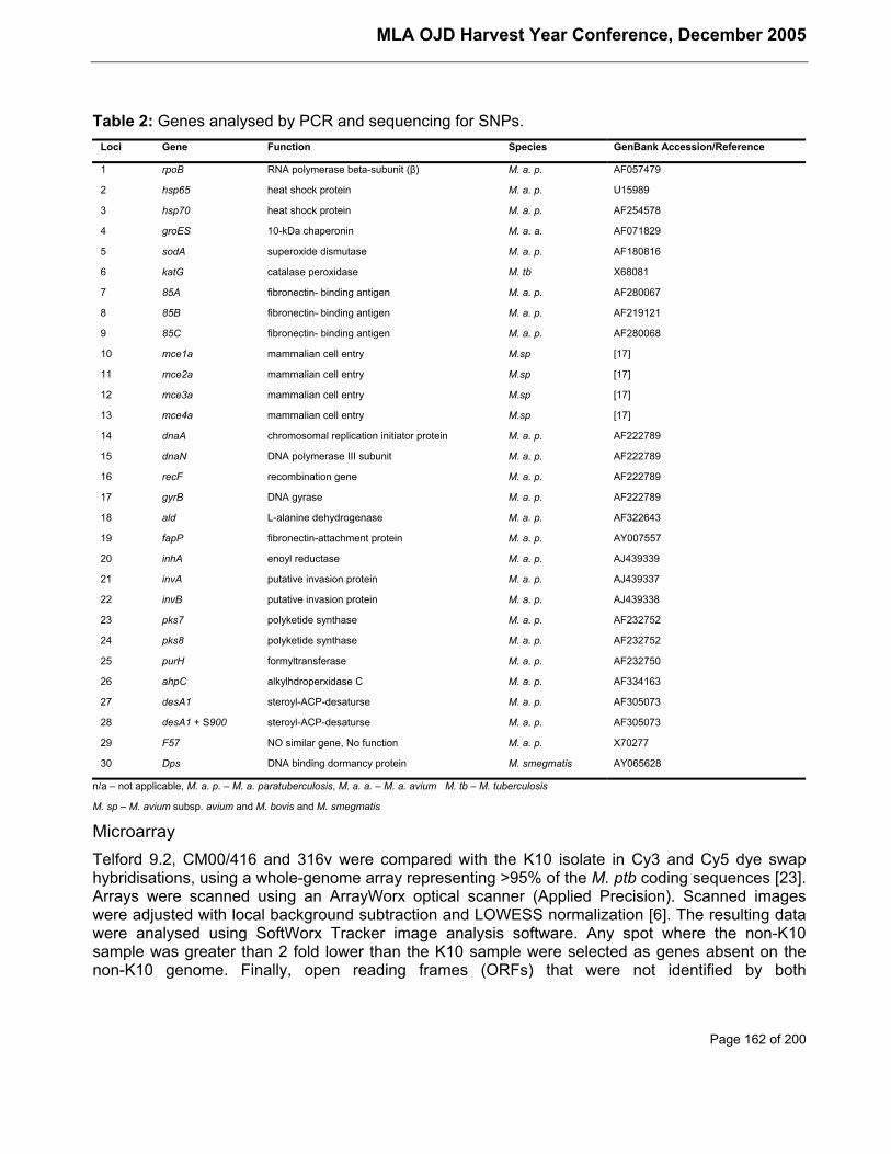
MLA OJD Harvest Year Conference, December 2005
Page 162 of 200
Table 2: Genes analysed by PCR and sequencing for SNPs.Loci Gene Function Species GenBank Accession/Reference
1 rpoB RNA polymerase beta-subunit (β) M. a. p. AF057479
2 hsp65 heat shock protein M. a. p. U15989
3 hsp70 heat shock protein M. a. p. AF254578
4 groES 10-kDa chaperonin M. a. a. AF071829
5 sodA superoxide dismutase M. a. p. AF180816
6 katG catalase peroxidase M. tb X68081
7 85A fibronectin- binding antigen M. a. p. AF280067
8 85B fibronectin- binding antigen M. a. p. AF219121
9 85C fibronectin- binding antigen M. a. p. AF280068
10 mce1a mammalian cell entry M.sp [17]
11 mce2a mammalian cell entry M.sp [17]
12 mce3a mammalian cell entry M.sp [17]
13 mce4a mammalian cell entry M.sp [17]
14 dnaA chromosomal replication initiator protein M. a. p. AF222789
15 dnaN DNA polymerase III subunit M. a. p. AF222789
16 recF recombination gene M. a. p. AF222789
17 gyrB DNA gyrase M. a. p. AF222789
18 ald L-alanine dehydrogenase M. a. p. AF322643
19 fapP fibronectin-attachment protein M. a. p. AY007557
20 inhA enoyl reductase M. a. p. AJ439339
21 invA putative invasion protein M. a. p. AJ439337
22 invB putative invasion protein M. a. p. AJ439338
23 pks7 polyketide synthase M. a. p. AF232752
24 pks8 polyketide synthase M. a. p. AF232752
25 purH formyltransferase M. a. p. AF232750
26 ahpC alkylhdroperxidase C M. a. p. AF334163
27 desA1 steroyl-ACP-desaturse M. a. p. AF305073
28 desA1 + S900 steroyl-ACP-desaturse M. a. p. AF305073
29 F57 NO similar gene, No function M. a. p. X70277
30 Dps DNA binding dormancy protein M. smegmatis AY065628
n/a – not applicable, M. a. p. – M. a. paratuberculosis, M. a. a. – M. a. avium M. tb – M. tuberculosis
M. sp – M. avium subsp. avium and M. bovis and M. smegmatis
MicroarrayTelford 9.2, CM00/416 and 316v were compared with the K10 isolate in Cy3 and Cy5 dye swaphybridisations, using a whole-genome array representing >95% of the M. ptb coding sequences [23].Arrays were scanned using an ArrayWorx optical scanner (Applied Precision). Scanned imageswere adjusted with local background subtraction and LOWESS normalization [6]. The resulting datawere analysed using SoftWorx Tracker image analysis software. Any spot where the non-K10sample was greater than 2 fold lower than the K10 sample were selected as genes absent on thenon-K10 genome. Finally, open reading frames (ORFs) that were not identified by both
MLA OJD Harvest Year Conference, December 2005
Page 163 of 200
hybridisations or that were not represented by at least 2 spot replicates were censored and notincluded in further analysis.
Each of the ORFs identified by microarray analysis as deleted from the S strain were evaluated byPCR to confirm their presence or absence in the S and C strains. PCR was also performed on anumber of other ORFs not identified by the microarray but residing within or immediately flanking theregions that were identified. The latter was undertaken to determine the limits of each of the S straindeletions. The amplified products from the S strain were sequenced and used in BLASTn searchesof the M. ptb K10 genome to identify the terminal ends of each deletion. Thirty two M. ptb fieldisolates (16 S strain and 16 C strain field isolates, Table 1) were evaluated using the S straindeletion PCR assays. Finally, the 3 DNA sequences derived from the M. ptb K10 genome sequencecorresponding to the regions deleted in the S strains were used in BLASTn searches to identify thepresence or absence of these sequences in the M. a. avium 104 genome (http://www.tigr.com).
Two dimensional electrophoresis proteomicsFor 1D and 2D comparisons proteins were extracted using a method based on [30]. Briefly, proteinswere extracted using 0.1 µm zirconium beads in conjunction with 3 x 45 sec pulses at maximumspeed (6.5) in a Bio 101 Fastprep. For 1D experiments protein samples were extracted directly intosterile purified water and diluted 1:1 in standard SDS-PAGE buffer. For 2D experiments, proteinswere extracted directly into standard 2D buffers. All samples were centrifuged at 103,320 g for 1 hrat 15oC prior to 2D electrophoresis. Protein concentrations were determined using a Bradford assay.Electrophoresis was performed in 15 cm x 16 cm (W x H) 12% polyacrylamide gels in a Hoefer SE600 vertical slab gel electrophoresis unit for both 1D and 2D electrophoresis. Isoelectric focusing(IEF) was achieved with 11 cm IPG strips pH 4-7 passively re-hydrated. IEF was undertaken in aBioRad Protean IEF unit. Two-dimensional experiments were run in triplicate to determine thereproducibility of this protocol. Eight protein spots (2 sheep and 6 cattle) chosen based on beingeither unique or demonstrating differential expression together with 3 reference proteins from eachstrain were analysed by mass spectrometry (QSTAR2, Bioanalytical Mass Spectrometry facility atthe University of New South Wales).
Surface enhanced laser desorption-ionisation (SELDI) proteomicsProteins were extracted directly into extraction/reducing buffer (8M urea, 1% CHAPS in PBS) usingthe same protocol as for the 2D experiments. S and C strain protein samples were diluted inextraction/reducing buffer and run in duplicate at two concentrations, 1.5 µg per spot and 10 µg perspot on 4 ProteinChips including: CM10 (weak cation exchange, at pH 4 and pH 7), H50(hydrophobic), IMAC30-Cu (copper affinity) and Q10 (strong anion exchange, at pH 7 and pH 9). AllProteinChips were prepared using the manufacturers’ recommended protocols and scanned with aProteinChip reader (Ciphergen Model PBS IIC) that had been pre-calibrated according to themanufacturer’s recommendations. Laser intensity and deflector sensitivity were determinedempirically, after which spot and chip protocols were prepared to scan each ProteinChip. Theresulting data was then analysed using Ciphergen ProteinChip Software version 3.2. Spectra wereexamined and only those protein peaks with peak intensity equal or greater than 1 were considered.
ResultsRepresentational difference analysisThree bands identified as theoretically divergent DNA regions were cloned and sequenced (RDA1,RDA3 and RDA4)[21]. RDA1 (229 bp) contained a single G∅T base difference in the S strainrelative to the C strain, producing a serine∅alanine amino acid substitution in the MAP1381
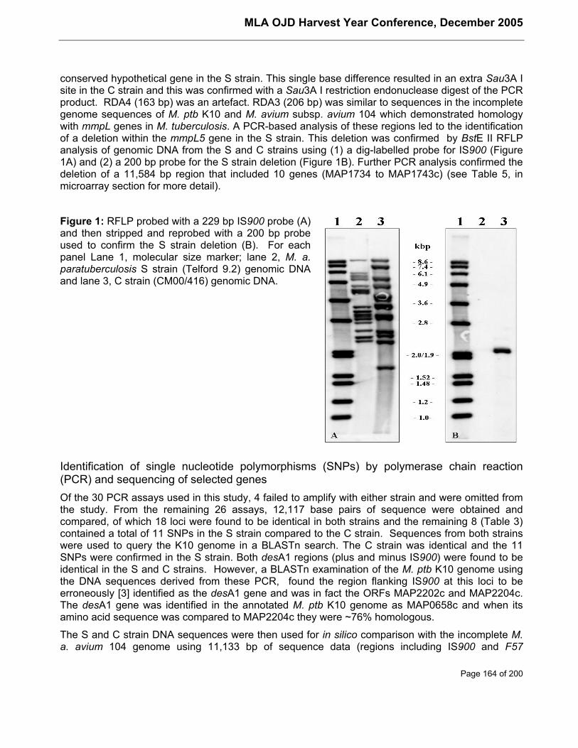
MLA OJD Harvest Year Conference, December 2005
Page 164 of 200
conserved hypothetical gene in the S strain. This single base difference resulted in an extra Sau3A Isite in the C strain and this was confirmed with a Sau3A I restriction endonuclease digest of the PCRproduct. RDA4 (163 bp) was an artefact. RDA3 (206 bp) was similar to sequences in the incompletegenome sequences of M. ptb K10 and M. avium subsp. avium 104 which demonstrated homologywith mmpL genes in M. tuberculosis. A PCR-based analysis of these regions led to the identificationof a deletion within the mmpL5 gene in the S strain. This deletion was confirmed by BstE II RFLPanalysis of genomic DNA from the S and C strains using (1) a dig-labelled probe for IS900 (Figure1A) and (2) a 200 bp probe for the S strain deletion (Figure 1B). Further PCR analysis confirmed thedeletion of a 11,584 bp region that included 10 genes (MAP1734 to MAP1743c) (see Table 5, inmicroarray section for more detail).
Figure 1: RFLP probed with a 229 bp IS900 probe (A)and then stripped and reprobed with a 200 bp probeused to confirm the S strain deletion (B). For eachpanel Lane 1, molecular size marker; lane 2, M. a.paratuberculosis S strain (Telford 9.2) genomic DNAand lane 3, C strain (CM00/416) genomic DNA.
Identification of single nucleotide polymorphisms (SNPs) by polymerase chain reaction(PCR) and sequencing of selected genesOf the 30 PCR assays used in this study, 4 failed to amplify with either strain and were omitted fromthe study. From the remaining 26 assays, 12,117 base pairs of sequence were obtained andcompared, of which 18 loci were found to be identical in both strains and the remaining 8 (Table 3)contained a total of 11 SNPs in the S strain compared to the C strain. Sequences from both strainswere used to query the K10 genome in a BLASTn search. The C strain was identical and the 11SNPs were confirmed in the S strain. Both desA1 regions (plus and minus IS900) were found to beidentical in the S and C strains. However, a BLASTn examination of the M. ptb K10 genome usingthe DNA sequences derived from these PCR, found the region flanking IS900 at this loci to beerroneously [3] identified as the desA1 gene and was in fact the ORFs MAP2202c and MAP2204c.The desA1 gene was identified in the annotated M. ptb K10 genome as MAP0658c and when itsamino acid sequence was compared to MAP2204c they were ~76% homologous.
The S and C strain DNA sequences were then used for in silico comparison with the incomplete M.a. avium 104 genome using 11,133 bp of sequence data (regions including IS900 and F57
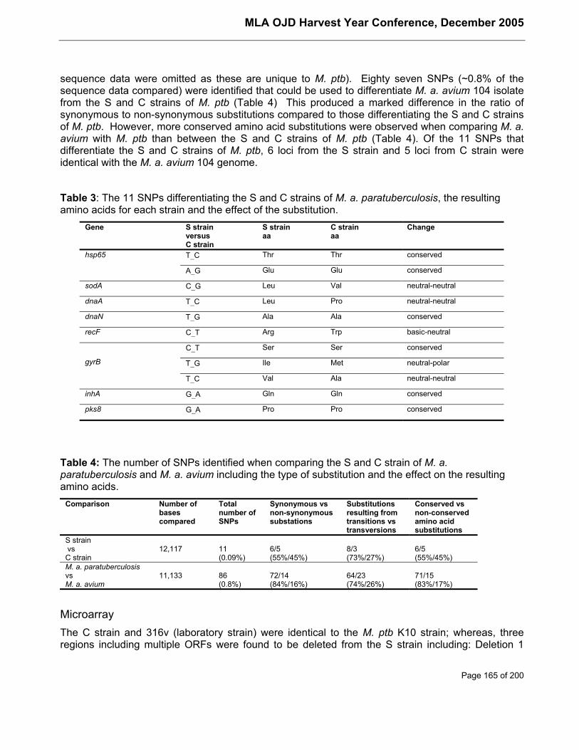
MLA OJD Harvest Year Conference, December 2005
Page 165 of 200
sequence data were omitted as these are unique to M. ptb). Eighty seven SNPs (~0.8% of thesequence data compared) were identified that could be used to differentiate M. a. avium 104 isolatefrom the S and C strains of M. ptb (Table 4) This produced a marked difference in the ratio ofsynonymous to non-synonymous substitutions compared to those differentiating the S and C strainsof M. ptb. However, more conserved amino acid substitutions were observed when comparing M. a.avium with M. ptb than between the S and C strains of M. ptb (Table 4). Of the 11 SNPs thatdifferentiate the S and C strains of M. ptb, 6 loci from the S strain and 5 loci from C strain wereidentical with the M. a. avium 104 genome.
Table 3: The 11 SNPs differentiating the S and C strains of M. a. paratuberculosis, the resultingamino acids for each strain and the effect of the substitution.
Gene S strainversusC strain
S strainaa
C strainaa
Change
T_C Thr Thr conservedhsp65
A_G Glu Glu conserved
sodA C_G Leu Val neutral-neutral
dnaA T_C Leu Pro neutral-neutral
dnaN T_G Ala Ala conserved
recF C_T Arg Trp basic-neutral
C_T Ser Ser conserved
T_G Ile Met neutral-polargyrB
T_C Val Ala neutral-neutral
inhA G_A Gln Gln conserved
pks8 G_A Pro Pro conserved
Table 4: The number of SNPs identified when comparing the S and C strain of M. a.paratuberculosis and M. a. avium including the type of substitution and the effect on the resultingamino acids.
Comparison Number ofbasescompared
Totalnumber ofSNPs
Synonymous vsnon-synonymoussubstations
Substitutionsresulting fromtransitions vstransversions
Conserved vsnon-conservedamino acidsubstitutions
S strain vsC strain
12,117 11(0.09%)
6/5(55%/45%)
8/3(73%/27%)
6/5(55%/45%)
M. a. paratuberculosisvsM. a. avium
11,133 86(0.8%)
72/14(84%/16%)
64/23(74%/26%)
71/15(83%/17%)
MicroarrayThe C strain and 316v (laboratory strain) were identical to the M. ptb K10 strain; whereas, threeregions including multiple ORFs were found to be deleted from the S strain including: Deletion 1
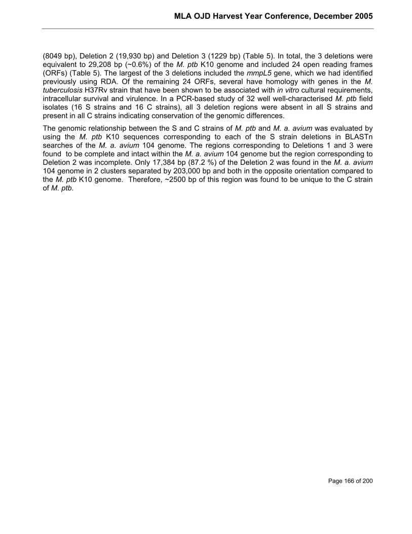
MLA OJD Harvest Year Conference, December 2005
Page 166 of 200
(8049 bp), Deletion 2 (19,930 bp) and Deletion 3 (1229 bp) (Table 5). In total, the 3 deletions wereequivalent to 29,208 bp (~0.6%) of the M. ptb K10 genome and included 24 open reading frames(ORFs) (Table 5). The largest of the 3 deletions included the mmpL5 gene, which we had identifiedpreviously using RDA. Of the remaining 24 ORFs, several have homology with genes in the M.tuberculosis H37Rv strain that have been shown to be associated with in vitro cultural requirements,intracellular survival and virulence. In a PCR-based study of 32 well well-characterised M. ptb fieldisolates (16 S strains and 16 C strains), all 3 deletion regions were absent in all S strains andpresent in all C strains indicating conservation of the genomic differences.
The genomic relationship between the S and C strains of M. ptb and M. a. avium was evaluated byusing the M. ptb K10 sequences corresponding to each of the S strain deletions in BLASTnsearches of the M. a. avium 104 genome. The regions corresponding to Deletions 1 and 3 werefound to be complete and intact within the M. a. avium 104 genome but the region corresponding toDeletion 2 was incomplete. Only 17,384 bp (87.2 %) of the Deletion 2 was found in the M. a. avium104 genome in 2 clusters separated by 203,000 bp and both in the opposite orientation compared tothe M. ptb K10 genome. Therefore, ~2500 bp of this region was found to be unique to the C strainof M. ptb.
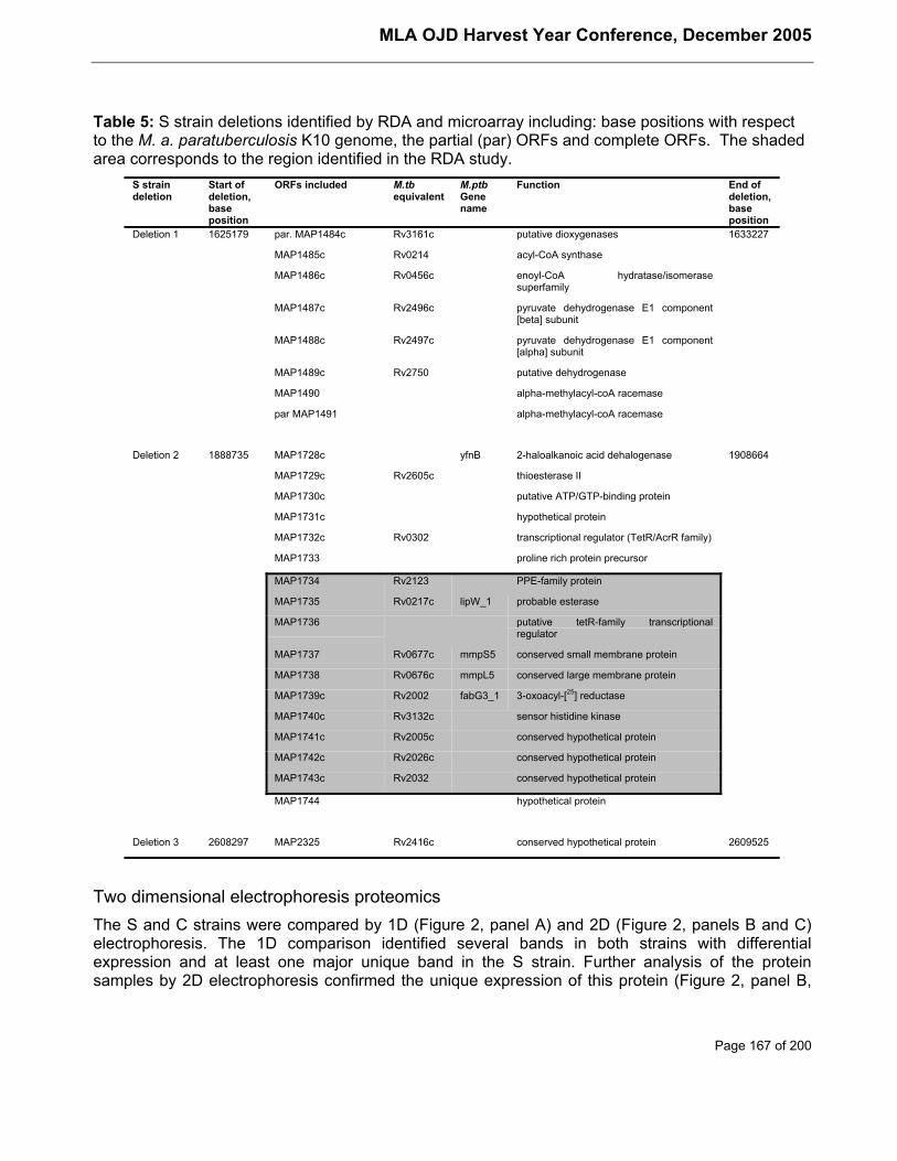
MLA OJD Harvest Year Conference, December 2005
Page 167 of 200
Table 5: S strain deletions identified by RDA and microarray including: base positions with respectto the M. a. paratuberculosis K10 genome, the partial (par) ORFs and complete ORFs. The shadedarea corresponds to the region identified in the RDA study.
S straindeletion
Start ofdeletion,baseposition
ORFs included M.tbequivalent
M.ptbGenename
Function End ofdeletion,baseposition
Deletion 1 1625179 par. MAP1484c Rv3161c putative dioxygenases 1633227
MAP1485c Rv0214 acyl-CoA synthase
MAP1486c Rv0456c enoyl-CoA hydratase/isomerasesuperfamily
MAP1487c Rv2496c pyruvate dehydrogenase E1 component[beta] subunit
MAP1488c Rv2497c pyruvate dehydrogenase E1 component[alpha] subunit
MAP1489c Rv2750 putative dehydrogenase
MAP1490 alpha-methylacyl-coA racemase
par MAP1491 alpha-methylacyl-coA racemase
Deletion 2 1888735 MAP1728c yfnB 2-haloalkanoic acid dehalogenase 1908664
MAP1729c Rv2605c thioesterase II
MAP1730c putative ATP/GTP-binding protein
MAP1731c hypothetical protein
MAP1732c Rv0302 transcriptional regulator (TetR/AcrR family)
MAP1733 proline rich protein precursor
MAP1734 Rv2123 PPE-family protein
MAP1735 Rv0217c lipW_1 probable esterase
MAP1736 putative tetR-family transcriptionalregulator
MAP1737 Rv0677c mmpS5 conserved small membrane protein
MAP1738 Rv0676c mmpL5 conserved large membrane protein
MAP1739c Rv2002 fabG3_1 3-oxoacyl-[25] reductase
MAP1740c Rv3132c sensor histidine kinase
MAP1741c Rv2005c conserved hypothetical protein
MAP1742c Rv2026c conserved hypothetical protein
MAP1743c Rv2032 conserved hypothetical protein
MAP1744 hypothetical protein
Deletion 3 2608297 MAP2325 Rv2416c conserved hypothetical protein 2609525
Two dimensional electrophoresis proteomicsThe S and C strains were compared by 1D (Figure 2, panel A) and 2D (Figure 2, panels B and C)electrophoresis. The 1D comparison identified several bands in both strains with differentialexpression and at least one major unique band in the S strain. Further analysis of the proteinsamples by 2D electrophoresis confirmed the unique expression of this protein (Figure 2, panel B,
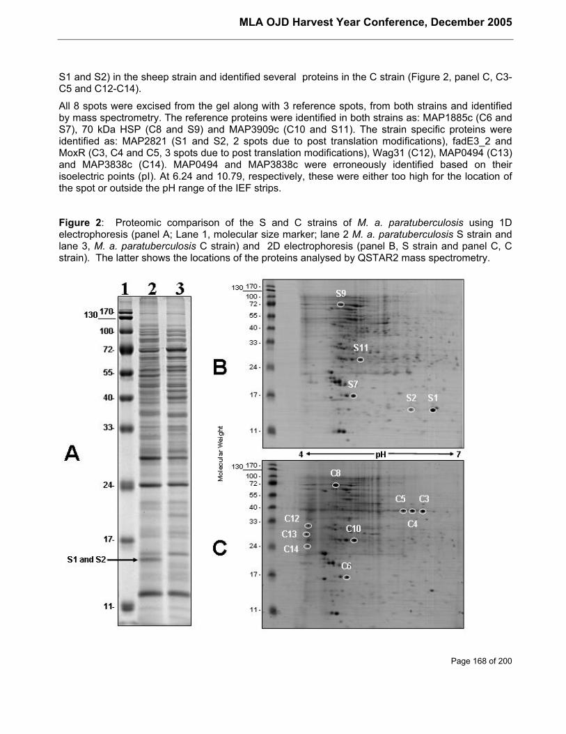
MLA OJD Harvest Year Conference, December 2005
Page 168 of 200
S1 and S2) in the sheep strain and identified several proteins in the C strain (Figure 2, panel C, C3-C5 and C12-C14).
All 8 spots were excised from the gel along with 3 reference spots, from both strains and identifiedby mass spectrometry. The reference proteins were identified in both strains as: MAP1885c (C6 andS7), 70 kDa HSP (C8 and S9) and MAP3909c (C10 and S11). The strain specific proteins wereidentified as: MAP2821 (S1 and S2, 2 spots due to post translation modifications), fadE3_2 andMoxR (C3, C4 and C5, 3 spots due to post translation modifications), Wag31 (C12), MAP0494 (C13)and MAP3838c (C14). MAP0494 and MAP3838c were erroneously identified based on theirisoelectric points (pI). At 6.24 and 10.79, respectively, these were either too high for the location ofthe spot or outside the pH range of the IEF strips.
Figure 2: Proteomic comparison of the S and C strains of M. a. paratuberculosis using 1Delectrophoresis (panel A; Lane 1, molecular size marker; lane 2 M. a. paratuberculosis S strain andlane 3, M. a. paratuberculosis C strain) and 2D electrophoresis (panel B, S strain and panel C, Cstrain). The latter shows the locations of the proteins analysed by QSTAR2 mass spectrometry.
MLA OJD Harvest Year Conference, December 2005
Page 169 of 200
Surface enhanced laser desorption-ionisation (SELDI) proteomicsSELDI spectra were obtained with all 4 ProteinChips at both 1.5 µg per spot and 10.0 µg per spotloading concentrations. However, protein peaks were only observed in the 5,000 to 20,000 kDarange. Numerous peaks were observed that were common to both samples but closer examinationof the spectra identified many unique peaks in the S strain and a few in the C strain (Table 6, U).Evidence was also observed for differential expression of a number of proteins (Table 6, E). Severalunique protein peaks, of similar molecular weight, had been identified in the S strain with 2 or moreProteinChips including a 5.2 kDa protein peak using the CM10 and IMAC30 ProteinChips and a 13.9kDa protein peak using the H50, IMAC30 and Q10 ProteinChips. A 9.6kDa protein peak wasidentified as unique in the S strain by the CM10 and Q10 ProteinChips, but as differentiallyexpressed by the IMAC30 ProteinChip. Finally, a differentially expressed 17.7 kDa protein wasidentified by the IMAC30 and Q10 ProteinChips. No unique or differentially expressed protein peakswere identified in the C strain with more than 1 ProteinChip.
Table 6: Summary of the SELDI results. The expanded spectra for each ProteinChip were used toidentify protein peaks in the S and C strain of M. a. paratuberculosis as either unique (U) orrepresenting a marked difference in expression (E). The size of each peak identified is given in kDa.
1.5 µg of protein 10.0 µg of proteinProteinChip
Telford 9.2
kDa
CM00/416
kDa
Telford 9.2
kDa
CM00/416
kDa
CM10 (pH 4) 13.9 U 5.2, 6.0, 14.4 U
8.7, 9.0, 9.3, 10.2, 14.7 E
CM10 (pH 7) 5.2, 6.0, 9.6 U 8.37 E 5.2, 6.0 U
14.7 E
H50 13.9 U 16.3 U 10.9, 12.9(a) U
13.9(a) E
IMAC30 (Cu) 5.2, 5.3, 5.5(a), 13.9(a) U 5.2, 5.3, 5.9, 13.9, 14.3 U
9.6, 17.7, 17.9 E 9.6, 17.7(a) E
Q10 (pH 7) 6.9, 13.9 U 7.36 E 9.4, 13.9 U 14.3 U
17.7 E
Q10 (pH 9) 8.9, 9.0, 9.6, 13.9 E 13.9 U
9.5(a) U
(a) Protein peak was < 1 in peak intensity but readily identifiable
DiscussionPreviously unidentified differences between the S and C strains of M. ptb were identified using acombination of modern DNA and protein based techniques. RDA has already been used to identifyunique regions present in the S strain but absent in the C strain [14]. In this study two furtherdifferences were discovered in the S strain using RDA including a single base difference inMAP1381 and a large-scale deletion that included the mmpL 5 gene. The latter was fullycharacterised as Deletion 2 by microarray analysis. Deletion 2 along with two others identified bymicroarray, are the first large-scale genomic differences to be reported that differentiate the S and Cstrains of M. ptb. In total, the 3 deletions were equivalent to 29,208 bp (~0.6%) of the M. ptb K10
MLA OJD Harvest Year Conference, December 2005
Page 170 of 200
genome and included 24 open reading frames (ORFs) (Table 5). PCR analysis of 32 field isolatesconfirmed these deletions to be highly conserved in the S strain. Homologues of many of the ORFsin Deletions 1-3 have been identified in M. tuberculosis and shown to play important roles inpathogenicity, host cell invasion and intracellular survival as well as in vitro culture. The latter maybe important given the marked differences in the cultural characteristics of these strains.
The pdhABC genes in M. tuberculosis, for which the ORFs MAP1487c and MAP1488c (Deletion 1)bear homology, encode subunits of pyruvate dehydrogenase, which converts pyruvate to acetyl-coenzyme A (CoA) and carbon dioxide, and have been shown to be up regulated during nutrientstarvation [2]. Similarly the ORFs MAP1740c and MAP1742c (Deletion 2) appear to be homologuesof M. tuberculosis genes Rv3132c and Rv2026c, respectively, which were considered to beessential for optimal in vitro growth [24]. Interestingly, homologues in M. tuberculosis of many of theORFs in Deletion 2 have been studied in association with the DevR-DevS two-component regulatorysystem, thought to be involved in intracellular survival. This observation of M. tuberculosis and theclustering of MAP1740c to MAP1743c would indicate that these genes may form part of an operoninvolved in the intracellular response to the host environment and that the loss of this operon mayexplain the shift in host specificity and possibly the in vitro growth requirements of the S strain of M.ptb. Of particular interest was the absence of the mmpS5 (MAP1737) and mmpL5 genes (MAP1738)as part of Deletion 2 in the S strain. MmpL proteins in M. tuberculosis have been shown to beessential for intracellular survival and growth, are involved in pathogenicity and contribute to host-pathogen interactions [4] [12] [11] [16] [19] [15]. Furthermore, attenuated strains of M. bovis that lackthe mmpL 4 gene were found to exhibit equivalent protection to M. bovis BCG in guinea pigschallenged with M. tuberculosis.[10]
The smallest deletion identified in this study, Deletion 3, included only one ORF, MAP2325, whichshares homology with Rv2416c in M. tuberculosis. Rv2416 has been described as an enhancedintracellular survival (eis) protein thought to be an important M. tuberculosis immunogen [13] presentonly in pathogenic mycobacteria [29] but as yet not confirmed in M. ptb.
In an in silico analysis, DNA sequences from the C strain corresponding to the S strain deletionswere used to compare M. ptb with M. a. avium. Sequence for deletions 1 and 3 was complete andintact within the M. a. avium 104 genome; whereas, Deletion 2 was found to be only 87.2 % (17,384bp) complete. The missing 2546 bp of this region that includes the ORFs corresponding to MAP1730to MAP1732 are therefore unique to the C strain. This makes this deletion a very interesting targetfor further research on the divergence of M. ptb and M. a. avium and on the strains of M. ptb.
PCR and sequencing revealed eleven SNPs that can be used to differentiate the S and C strains.Five of these produced non-conserved amino acid substitutions in the S strain; however further workis required to identify what effect each of these may have on the S strain phenotype. Interestingly, aten fold increase in the number of SNPs differentiating M. ptb from M. a. avium suggests that thedivergence between the S and C strains of M. ptb is evolutionarily recent compared with thedivergence between M. a. avium and M. ptb. More work is required in this area to confirm thishypothesis.
Proteomic techniques demonstrated their usefulness in identifying the protein expressioncharacteristics of the S and C strains under routine culture conditions. The results indicated theusefulness of combining techniques given that two dimensional electrophoresis is more suited toproteins >20 kDa and SELDI is more useful for proteins <20 kDa when whole cell protein samplesare examined. However, both techniques would benefit from fractionation techniques to simplify theprotein samples. Interestingly, the protein identified by 2D electrophoresis in the S strain, MAP2821
MLA OJD Harvest Year Conference, December 2005
Page 171 of 200
was also evident in both the 1D electrophoresis (Figure 2, panel A) and SELDI (protein peak at~14.0 kDa) results, which demonstrates the reproducibility of these techniques.
The results from this study have clearly demonstrated that many genomic differences includingdeletions equivalent to ~0.6% of the genome exist between the S and C strains of M. ptb. Theseresults confirm and support earlier studies that differentiate the S and C strains of M. ptb and can beused to reassure animal industries that rely on this strain difference for the recommendedmanagement practices used to combat Johne’s disease. Furthermore, a number of importantgenomic loci have been identified that can be used to differentiate between the S and C strains.Differences in a number of important genes require further examination to understand what role theymay play in pathogenicity, intracellular and environmental survival and specific host-pathogeninteractions.
AcknowledgementsThis project was funded by Meat & Livestock Australia. Development of the microarray was fundedby the USDA-CSREES JDIP grant subcontracted to John P. Bannantine and Michael L. Paustian.The authors would like to thank: Leslie Reddacliff, Alison Collins, Shayne Fell, Sue Austin and themicrobiology staff at the NSW Department of Primary Industries, Elizabeth Macarthur AgriculturalInstitute), Anna Waldron, Deb Taylor and Lyrissa Diforei (University of Sydney), Michael Paustian(National Animal Disease Centre, USDA-ARS, Ames, IA, USA) and Kathy Grainger (CSIROLivestock Industries, Australian Animal Health Laboratory, Geelong, Victoria Australia).
References[1] S.F. Altschul, T.L. Madden, A.A. Schaffer, J. Zhang, Z. Zhang, W. Miller, D.J. Lipman.
Gapped BLAST and PSI-BLAST: a new generation of protein database search programs,Nucleic Acids Res, 25, (1997) 3389-402.
[2] J.C. Betts, P.T. Lukey, L.C. Robb, R.A. McAdam, K. Duncan. Evaluation of a nutrientstarvation model of Mycobacterium tuberculosis persistence by gene and protein expressionprofiling, Mol Microbiol, 43, (2002) 717-31.
[3] T.J. Bull, J. Hermon-Taylor, I. Pavlik, F. El-Zaatari, M. Tizard. Characterization of IS900 lociin Mycobacterium avium subsp.paratuberculosis and development of multiplex PCR typing,Microbiology, 146 ( Pt 9), (2000) 2185-97.
[4] L.R. Camacho , D. Ensergueix, E. Perez, B. Gicquel, C. Guilhot. Identification of a virulencegene cluster of Mycobacterium tuberculosis by signature-tagged transposon mutagenesis,Mol Microbiol, 34, (1999) 257-67.
[5] E. Choy, R.J. Whittington, I. Marsh, J. Marshall, M.T. Campbell. A method for purification andcharacterisation of Mycobacterium avium subsp.paratuberculosis from the intestinal mucosaof sheep with Johne's disease, Vet Microbiol, 64, (1998) 51-60.
[6] W.S. Cleveland, S.J. Devlin. Locally weighted regression: an approach to regression analysisby local fitting, Journal of the American Statistical Association, 83, (1988) 596-610.
[7] D.M. Collins , S. Cavaignac, G.W. de Lisle. Use of four DNA insertion sequences tocharacterize strains of the Mycobacterium avium complex isolated from animals, Mol CellProbes, 11, (1997) 373-80.
[8] D.M. Collins , M. De Zoete, S.M. Cavaignac. Mycobacterium avium subsp.paratuberculosisstrains from cattle and sheep can be distinguished by a PCR test based on a novel DNAsequence difference, J Clin Microbiol, 40, (2002) 4760-2.
MLA OJD Harvest Year Conference, December 2005
Page 172 of 200
[9] D.M. Collins , D.M. Gabric, G.W. de Lisle. Identification of two groups of Mycobacteriumparatuberculosis strains by restriction endonuclease analysis and DNA hybridization, J ClinMicrobiol, 28, (1990) 1591-6.
[10] D.M. Collins, B. Skou, S. White, S. Bassett, L. Collins, R. For, K. Hurr, G. Hotter, G.W. deLisle . Generation of attenuated Mycobacterium bovis strains by signature-taggedmutagenesis for discovery of novel vaccine candidates, Infect Immun, 73, (2005) 2379-86.
[11] S.E. Converse, J.D. Mougous, M.D. Leavell, J.A. Leary, C.R. Bertozzi, J.S. Cox. MmpL8 isrequired for sulfolipid-1 biosynthesis and Mycobacterium tuberculosis virulence, Proc NatlAcad Sci U S A, 100, (2003) 6121-6.
[12] J.S. Cox, B. Chen, M. McNeil, W.R. Jacobs Jr. Complex lipid determines tissue-specificreplication of Mycobacterium tuberculosis in mice, Nature, 402, (1999) 79-83.
[13] J.L. Dahl, J. Wei, J.W. Moulder, S. Laal, R.L. Friedman. Subcellular localization of theIitracellular survival-enhancing EIS protein of Mycobacterium tuberculosis, Infect Immun, 69,(2001) 4295-302.
[14] K. Dohmann, B. Strommenger, K. Stevenson, L. de Juan, J. Stratmann, V. Kapur, T.J. Bull,G.F. Gerlach. Characterization of genetic differences between Mycobacterium aviumsubsp.paratuberculosis type I and type II isolates, J Clin Microbiol, 41, (2003) 5215-23.
[15] P. Domenech , M.B. Reed, C.E. Barry 3rd. Contribution of the Mycobacterium tuberculosisMmpL protein family to virulence and drug resistance, Infect Immun, 73, (2005) 3492-501.
[16] P. Domenech , M.B. Reed, C.S. Dowd, C. Manca, G. Kaplan, C.E. Barry 3rd. The role ofMmpL8 in sulfatide biogenesis and virulence of Mycobacterium tuberculosis, J Biol Chem,279, (2004) 21257-65.
[17] Y. Haile, D.A. Caugant, G. Bjune, H.G. Wiker. Mycobacterium tuberculosis mammalian cellentry operon (mce) homologs in Mycobacterium other than tuberculosis (MOTT), FEMSImmunol Med Microbiol, 33, (2002) 125-32.
[18] N.H. Hole. Johne's disease, Advances in Veterinary Science, 4, (1958) 341-387.
[19] G. Lamichhane, S. Tyagi, W.R. Bishai. Designer Arrays for Defined Mutant Analysis ToDetect Genes Essential for Survival of Mycobacterium tuberculosis in Mouse Lungs, InfectImmun, 73, (2005) 2533-40.
[20] I. Marsh, R. Whittington, D. Cousins. PCR-restriction endonuclease analysis for identificationand strain typing of Mycobacterium avium subsp.paratuberculosis and Mycobacterium aviumsubsp. avium based on polymorphisms in IS1311, Mol Cell Probes, 13, (1999) 115-26.
[21] I.B. Marsh, R.J. Whittington. Deletion of an mmpL gene and multiple associated genes fromthe genome of the S strain of Mycobacterium avium subsp. paratuberculosis identified byrepresentational difference analysis and in silico analysis, Mol Cell Probes, Accepted, (2005).
[22] N.E. Morrison. Circumvention of the mycobactin requirement of Mycobacteriumparatuberculosis, J Bacteriol, 89, (1965) 762-7.
[23] M.L. Paustian, V. Kapur, J.P. Bannantine. Comparative genomic hybridizations revealgenetic regions within the Mycobacterium avium Complex that are divergent fromMycobacterium avium subsp.paratuberculosis Isolates, J Bacteriol, 187, (2005) 2406-15.
[24] C.M. Sassetti, D.H. Boyd, E.J. Rubin. Genes required for mycobacterial growth defined by
MLA OJD Harvest Year Conference, December 2005
Page 173 of 200
high density mutagenesis, Mol Microbiol, 48, (2003) 77-84.
[25] M.L. Schaeffer, G. Agnihotri, H. Kallender, P.J. Brennan, J.T. Lonsdale. Expression,purification, and characterization of the Mycobacterium tuberculosis acyl carrier protein,AcpM, Biochim Biophys Acta, 1532, (2001) 67-78.
[26] S. Sreevatsan, X. Pan, K.E. Stockbauer, N.D. Connell, B.N. Kreiswirth, T.S. Whittam, J.M.Musser. Restricted structural gene polymorphism in the Mycobacterium tuberculosis complexindicates evolutionarily recent global dissemination, Proc Natl Acad Sci U S A, 94, (1997)9869-74.
[27] A.W. Taylor. Ovine paratuberculosis (Johne's disease of sheep), Journal of ComparativePathology, 55, (1945) 41-44.
[28] A.W. Taylor . Varieties of Mycobacterium johnei isolated from sheep, J Pathol Bacteriol, 63,(1951) 333-6.
[29] J. Wei, J.L. Dahl, J.W. Moulder, E.A. Roberts, P. O'Gaora, D.B. Young, R.L. Friedman.Identification of a Mycobacterium tuberculosis gene that enhances mycobacterial survival inmacrophages, J Bacteriol, 182, (2000) 377-84.
[30] W.B. White, D.L. Whipple, J.R. Stabel, C.A. Bolin. Comparison of cellular and extracellularproteins expressed by various isolates of Mycobacterium paratuberculosis and othermycobacterial species, Am J Vet Res, 55, (1994) 1399-405.
[31] R. Whittington, I. Marsh, E. Choy, D. Cousins. Polymorphisms in IS1311, an insertionsequence common to Mycobacterium avium and M. avium subsp. paratuberculosis, can beused to distinguish between and within these species, Mol Cell Probes, 12, (1998) 349-58.
[32] R.J. Whittington, A.F. Hope, D.J. Marshall, C.A. Taragel, I. Marsh. Molecular epidemiology ofMycobacterium avium subsp. paratuberculosis: IS900 restriction fragment lengthpolymorphism and IS1311 polymorphism analyses of isolates from animals and a human inAustralia, J Clin Microbiol, 38, (2000) 3240-8.
[33] R.J. Whittington, I. Marsh, S. McAllister, M.J. Turner, D.J. Marshall, C.A. Fraser. Evaluationof modified BACTEC 12B radiometric medium and solid media for culture of Mycobacteriumavium subsp. paratuberculosis from sheep, J Clin Microbiol, 37, (1999) 1077-83.
[34] R.J. Whittington, E.S. Sergeant. Progress towards understanding the spread, detection andcontrol of Mycobacterium avium subsp. paratuberculosis in animal populations, Aust Vet J,79, (2001) 267-78.
MLA OJD Harvest Year Conference, December 2005
Page 174 of 200
A survey of potential wildlife reservoirs forMycobacterium paratuberculosis
Patrick KluverUniversity of Melbourne, Mackinnon Project250 Princes FreewayWerribeePhone: 03 97312343; Fax: 03 9731 2388E-mail: [email protected]
1.1 Introduction
In any disease control program eradication of disease depends upon the protection of disease freeanimals from any potential source of infection. Sylvatic hosts have the potential to thwart eradicationcampaigns. Wild bovids and ungulates are a source of bovine tuberculosis for North Americanherds. Eradication of bovine tuberculosis has been hampered by two non ruminant wildlifereservoirs, possums in New Zealand and badgers in England. A survey of potential wildlifereservoirs in Australia was conducted between 1997 and 1999, when eradication of ovine Johne’sdisease (OJD) was seriously contemplated.
The presence of a common wildlife reservoir of Johne's disease (JD) could render the eradication ofdisease impossible. In Scotland, rabbits have been demonstrated to be infected with Mycobacteriumparatuberculosis (Grieg et al., 1997). In order to assess the risk posed to JD control due to wildlife,the survey described in this paper examined rabbits and another herbivore commonly found onAustralian pastoral lands, the eastern grey kangaroo.
To survey European rabbits (Oryctolagus cuniculus) and eastern grey kangaroos(Macropus giganteus) in areas with endemic JD for the presence of M. paratuberculosis (M. ptb).
1.3 Method
RabbitsIn 3 areas where JD was present in livestock, rabbits were collected and examined for the presenceof M. ptb. Property A was a series of three contiguous farms in the Ensay valley of Victoria. Theseproperties had a purported long history of OJD and significant mortalities due to clinical OJD. Astudy examining sheep from these properties revealed JD prevalence of 21-37 % (Hope et al.,1999). These properties were destocked in January 1997 and rabbits were sampled in March of thatyear. Property B was a commercial beef operation of 200 breeders between Geelong and Ballarat,with a history of JD of at least three years. In 1996 this property had 9 clinical cases of JD. The farmalso ran a commercial merino flock and had a small flock of 9-10 feral goats. The final property,Property C, was a commercial dairy farm of 131 milkers in the Peterborough region of Victoria. Thefirst case of JD on the farm was diagnosed in 1996. A whole herd investigation in 1999 conducted atthe same time as the rabbit survey resulted in a faecal culture positive rate of 10%.
Farms were chosen on the basis of a high prevalence of JD in sheep or cattle, a high reported rabbitdensity in close contact with stock and the willingness of the manager to assist in the survey.
1.2 Aim

MLA OJD Harvest Year Conference, December 2005
Page 175 of 200
MLA OJD Harvest Year Conference, December 2005
Page 176 of 200
Table 1: Summary of rabbit sampling.
Property Enterprise Location Number ofspecimens
No of collections
A Merino Ensay Valley 100 rabbits, 1 hare. 3 nights, 18/3/97-20/3/97.
B Beef Cattle South Ballarat 104 rabbits, 1 hare3 goats.
3 nights, 5/3/99,15/06/99, 4/06/99.
C Dairy Cattle Peterborough 106 rabbits, 2hares.
3 nights 3/06/99,21/07/99, 21/06/99.
Rabbits were shot at night and necropsied within 12 hours of death. A minimum of 100 animalswere examined from each area. Weight, sex, and any gross pathological changes were recorded.Twenty cm sections of ileum, ileo-caecal junction, ileum and mesenteric lymph nodes were takenand halved for histology and culture; faeces, when present, were stored for culture. Fresh tissuesamples were pooled, stomached and processed as one sample. Samples were processedaccording to the relevant Australian Standard Diagnostic Technique (Tennant et al., 1996).andinoculated in BACTEC 12B (Becton Dickinson) media supplemented with Mycobactin J (AlliedMonitor), 17% eggyolk and PANTA plus (Becton Dickinson). Histological sections and faeces werestored for future reference.
KangaroosEastern grey kangaroos, which were being culled for pest management purposes in a central regionof Victoria, were necropsied and sampled for the presence of JD from April 1997 to November 1998.This central Victorian region had a history of endemic JD.
The farm where the kangaroos were being culled was approximately 1000 hectares, in 2 portions,consisting of volcanic plains and sedimentary hills. Improved pastures and dams, for water, providedfor the self-replacing Merino flock and some breeding cattle. OJD had been confirmed by necropsyafter the owner had reported losses due to wasting in adult sheep. Following detection of infectionon the property, six neighbouring properties were confirmed as also infected, along with numbers ofclose-by farms. One neighbouring farm had a bovine JD infection confirmed in 1992, and was thendestocked of cattle until 1997. On another neighbouring property clinical cases in sheep werecommon, with gross intestinal lesions characteristic of JD detected in more than 18% of ewes and10% of wethers at slaughter.
The sample farm, adjoining farms and roadsides had heavily treed areas providing an ideal habitatfor large mobs of kangaroos that readily moved between adjoining properties. The shot sampleswere from adult and sub-adult kangaroos. Juvenile animals under 1 year of age were excluded forthe purpose of this survey.
Sex, location, estimation of age, and any gross pathological changes were recorded. Sections ofileum, ileo-caecal valve, proximal colon, caecum and mesenteric lymph nodes were taken forhistology, and fresh sections and faeces for culture. Initially all sections were cultured individually butfor the last 35 animals sampled, lymph nodes and gut sections were pooled separately and
MLA OJD Harvest Year Conference, December 2005
Page 177 of 200
processed as two cultures. The culture technique used was in accordance with the AustralianStandard Diagnostic Test recommendations (Tennant et al., 1996).
A minimum of 100 animals (kangaroos or rabbits) were examined from each site. The sample sizewas selected to detect a 3% infection level with 95% confidence limits.
1.4 Results
RabbitsIn total 310 rabbits, 4 hares and 3 feral goats were examined by culture.
M. ptb was not detected in any of the samples, giving a point estimate of prevalence of 0 (95% CI 0-1.2%). Eight of 310 samples demonstrated growth of organisms other than M. ptb
Table 2: Prevalence of Johne’s disease in rabbits.
Property Number ofspecimens
Prevalence(CI 95% Wilson)
A 100 rabbits, 1 hare. 0(0-3.7%).
B 104 rabbits, 1 hare3 goats.
0(0-3.6%).
C 106 rabbits, 2hares.
0(0-3.5%)
Total 310 rabbits 0(0-1.2%)
Eastern grey kangaroosThirty-seven female and sixty-three males were sampled. Five faecal culture samples had someevidence of bacterial growth in BACTEC, which was shown to be faecal contaminants; all otherfaecal samples and tissue samples were found to be negative.
There was no evidence of JD in the kangaroos examined (95% CI 0-3.7%).
1.5 Discussion
Several reviews have described the occurrence of Johne's disease in wildlife and zoologicalcollections (Chiodini et al., 1984, Morgan 1999). Wild populations of Bighorn Sheep; Rockymountain goat; and axis, fallow, white tailed, red, sika, roe deer have been reported as beinginfected with M. ptb (Williams et al., 1979, Riemann et al., 1979, Chiodini et al., 1983, Hillermark1966, Reid et al., 1996).
Rabbits have been experimentally infected with JD. In two similar studies when 1 day old rabbitswere orally dosed with ~108 organisms 3 of 16 (19%) and 9 of 21 (43%) had histological evidence ofparatuberculosis at necropsy 9-12 months after inoculation (Mokresh et al., 1989, Mokresh et al.,1990). These studies demonstrate that rabbits are susceptible to M. ptb but provide little informationof the natural ecology of the disease due to the large infective dose and age of the animals infected.

MLA OJD Harvest Year Conference, December 2005
Page 178 of 200
There are several reports of JD in wild populations of rabbits in Scotland. A rabbit with intestinallesions resembling JD was described on a Scottish deer farm with endemic JD in 1990 (Angus1990). No cultural confirmation was performed but the lesions closely resembled those seen withinfection in cattle. A total of 32 rabbits were examined on the farm with only one animal having anyevidence of JD. The JD exposure of these rabbits was due to a ’major outbreak’ of JD in red deer(Cervus elaphus) on an experimental farm where rabbits were in ’plague proportions’ requiringregular culling.
The first confirmed report of wild rabbits being naturally infected with M. ptb was in the summer of1995 (Anon 1995). Researchers at the SAC Perth investigated rabbits on four farms in the Taysideregion of Scotland. The rabbits were collected on 3 properties where JD was known and a fourthproperty where no clinical signs of the disease had been observed (Grieg et al., 1997). A total of 33were sampled with 23 of these being culture positive for M. ptb (one of which was faecal culturepositive only); 19 had histological evidence of JD and 13 were both culturally and histologicallypositive.
A wider survey was carried out 1996 in Scotland (Grieg et al., 1999). This survey involved shotsamples collected from farms with and without a history of JD. It involved surveys in 7 counties andagain included the Tayside region. The survey revealed little infection in areas outside the originalsurvey area of Tayside. Of the 169 rabbits collected from counties other than Tayside, there wasonly one confirmed as M. ptb positive, and this rabbit was from a farm with no known history of JD.Eighty-seven of the rabbits from outside Tayside were from 10 farms with known JD; M. ptb was notisolated from any of these rabbits. Two of the 87 rabbits from 2 of these10 farms had histologicalchanges with acid fast bacilli (AFB) present. Eighty rabbits from 8 farms with no known history of JDwere also tested, with one culturally positive animal and 2 rabbits showing histological changes withthe presence of AFB (it is not stated whether the animal is the same). RFLP strain typing revealedthat all isolates tested except one were type B_C17, the predominant RFLP type in cattle in the UK.HPLC typing revealed some overlap of strains between species on one farm and none on the othertested.
It does appear from the wider Scottish survey that JD in rabbits outside the Tayside region isuncommon. The Tayside county appears to be a hot spot of lagomorph JD. Evidence of JD has alsobeen detected in this area in stoats (Mustela erminea) (3 of 5 culture positive) and red fox (Vulpesvulpes) (8 of 9 culture positive) (Beard et al., 1990). This is in the area of high prevalence of JD inrabbits, which is presumably the source of infection for the carnivores.
The prevalence and occurrence of JD in rabbits in the Tayside region does appear to be unusual.The significance is yet to be determined and the reason this area has such a high prevalence of JDis unknown. The farms were originally surveyed because of the concordance of high levels of JDand the heavy density of rabbits. The association of lagomorph JD in this area may be due to anumber of reasons; a close interaction between a large rabbit and an infected ruminant populationswith heavy exposure of rabbits to M. ptb, the age structure of the rabbit populations, a highlypathogenic strain of M. ptb or rabbit adapted strain, a highly susceptible strain of rabbit in this area,or an unspecified environmental factor in this region which renders ruminants and rabbits highlysusceptible to JD.
Major differences will exist between the ecology of wild populations of rabbits between differentareas. In Australia, the proportion of animals that survive over 2 years of age is highly variable andcan range from 7-17% with an annual survival of adults ranging from 40-60% (Williams et al., 1995).The dynamics of a population will greatly affect the expression of a chronic disease such as JD withpopulations with high turnover and very few adults over 2 years of age being very unlikely to show
MLA OJD Harvest Year Conference, December 2005
Page 179 of 200
expression of disease. No information about the ecology and population dynamics of the rabbits inScotland was available, although Abbott in his review suggests that rabbit densities are higher in theTayside region than normally found in Australia (Abbott 2000).
There is no evidence that strains of M. ptb differ in their pathogenicity. It is suggested that somestrains, which are more commonly isolated from a particular species, demonstrate a host preferencefor different species, but this may be the result of an opportunity to disseminate within a species, asopposed to a strain being more pathogenic to a particular host.
A similar argument exists for host susceptibility. Breed susceptibility has been suggested in the pastbut it is difficult to separate breed susceptibility from greater dissemination within a breed (Chiodiniet al., 1983).
A similar study in rabbits on OJD infected properties was conducted by the University of Sydney(Abbott 2000). The survey included 300 rabbits collected from 8 properties. None of the 300 rabbitshad any evidence of infection with M. ptb.
Table 3: Combined results of Victorian and NSW surveys for Johne’s disease in rabbits.
Total Sampled Prevalence %
(95% CI)
Ovine Johne’s disease 400 0
(0-0.95)
Bovine Johne’s disease 210 0
(0-1.8)
Johne’s disease 610 0
(0-0.63)
When the results of our study are combined with those from the University of Sydney study, theestimated prevalence of M. ptb in rabbits in Australia is 0 with an upper 95% confidence limit of0.63% samples (Table 3). It is very unlikely that M. ptb exists in rabbits in Australia to any significantdegree.
Paratuberculosis has been recorded in a wide range of species; no published cases have beenrecorded in marsupials.
There have been recorded cases of marsupials, including Tree Kangaroos (Dendrolagus matschei)and Tammar wallabies (Macropus eugenii) infected with bacteria from the M avium complex and Mgenavense, (M Lynch pers com, Montali et al., 1998, Peet et al., 1982) in zoological collections andM bovis in brush tailed possums (Trichosurus vulpecula) in New Zealand (Davidson 1991). Ingeneral marsupials are thought to be more susceptible to mycobacteria than eutherian mammals.So it is logical to look at marsupials as a possible reservoir or carrier species for M. ptb.
The eastern grey kangaroo is predominantly a grazing animal with specific food preferencesrestricted to grasses and forbs. Their dietary habits do overlap those of sheep and they are often inclose association with sheep, sharing grazing land.
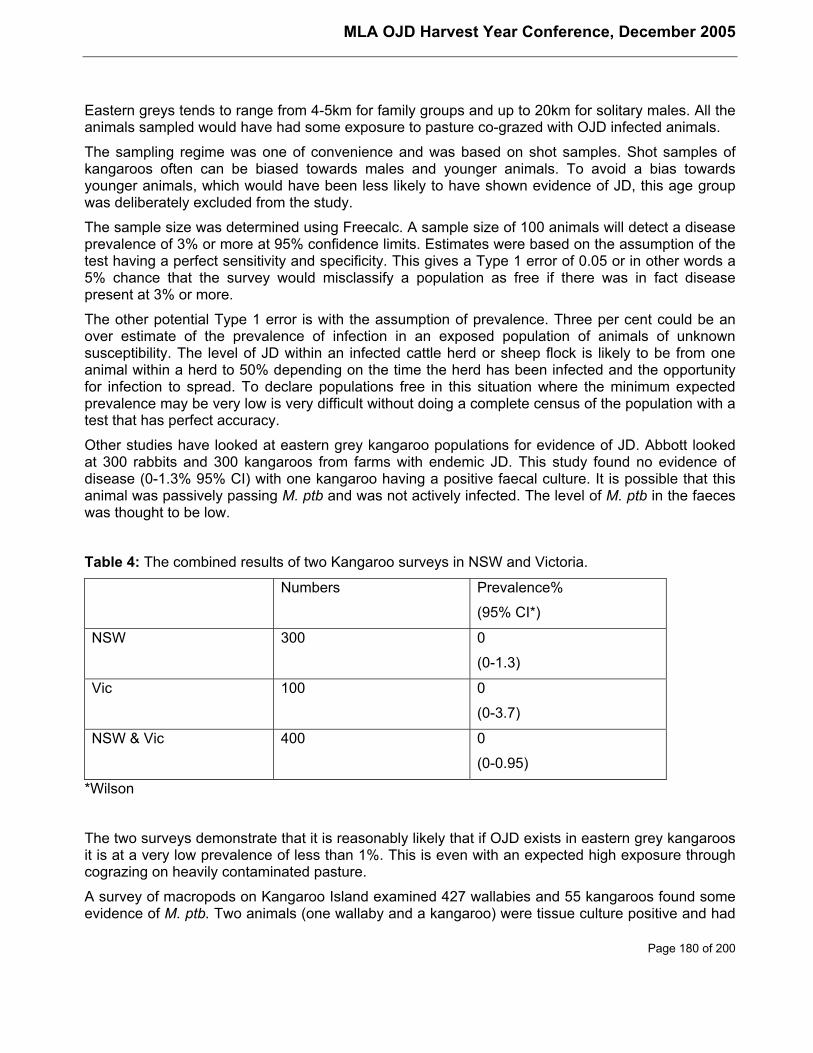
MLA OJD Harvest Year Conference, December 2005
Page 180 of 200
Eastern greys tends to range from 4-5km for family groups and up to 20km for solitary males. All theanimals sampled would have had some exposure to pasture co-grazed with OJD infected animals.
The sampling regime was one of convenience and was based on shot samples. Shot samples ofkangaroos often can be biased towards males and younger animals. To avoid a bias towardsyounger animals, which would have been less likely to have shown evidence of JD, this age groupwas deliberately excluded from the study.
The sample size was determined using Freecalc. A sample size of 100 animals will detect a diseaseprevalence of 3% or more at 95% confidence limits. Estimates were based on the assumption of thetest having a perfect sensitivity and specificity. This gives a Type 1 error of 0.05 or in other words a5% chance that the survey would misclassify a population as free if there was in fact diseasepresent at 3% or more.
The other potential Type 1 error is with the assumption of prevalence. Three per cent could be anover estimate of the prevalence of infection in an exposed population of animals of unknownsusceptibility. The level of JD within an infected cattle herd or sheep flock is likely to be from oneanimal within a herd to 50% depending on the time the herd has been infected and the opportunityfor infection to spread. To declare populations free in this situation where the minimum expectedprevalence may be very low is very difficult without doing a complete census of the population with atest that has perfect accuracy.
Other studies have looked at eastern grey kangaroo populations for evidence of JD. Abbott lookedat 300 rabbits and 300 kangaroos from farms with endemic JD. This study found no evidence ofdisease (0-1.3% 95% CI) with one kangaroo having a positive faecal culture. It is possible that thisanimal was passively passing M. ptb and was not actively infected. The level of M. ptb in the faeceswas thought to be low.
Table 4: The combined results of two Kangaroo surveys in NSW and Victoria.
Numbers Prevalence%
(95% CI*)
NSW 300 0
(0-1.3)
Vic 100 0
(0-3.7)
NSW & Vic 400 0
(0-0.95)
*Wilson
The two surveys demonstrate that it is reasonably likely that if OJD exists in eastern grey kangaroosit is at a very low prevalence of less than 1%. This is even with an expected high exposure throughcograzing on heavily contaminated pasture.
A survey of macropods on Kangaroo Island examined 427 wallabies and 55 kangaroos found someevidence of M. ptb. Two animals (one wallaby and a kangaroo) were tissue culture positive and had
MLA OJD Harvest Year Conference, December 2005
Page 181 of 200
histological evidence of acid fast bacilli in lymph nodes. Six animals were tissue culture positive buthad no histological evidence of acid fast bacilli and 6 ’histo positive’ animals were tissue culturenegative, although 4 had lesions which were described as either severe granulomatous enteritis orhad moderate to numerous acid fast bacilli. There was no evidence of M. ptb in the faeces.
These results are difficult to interpret. Certainly there appears to be no evidence that these animalsare shedding the bacteria, so pose little if any risk as far as spreading JD. In sheep and cattle it isnot unusual to find animals which have no histological evidence of disease but from which M. ptbcan be isolated. These animals are either in the early stages of the disease with focalhistopathological lesions not detected due to sampling, are infected but controlling the disease, or itis contamination from the gut lumen although in this survey the gut contents were washed out.
The culturally negative animals with advanced or proliferating lesions are unusual in sheep andcattle. One animal had evidence of Mycobacterium genavense on PCR which is a difficult organismto culture. It may be that all these animals which had proliferative lesions were in fact infected withother fastidious non paratuberculosis Mycobacteria.
1.6 Implications
With 400 eastern grey kangaroos and 610 rabbits now sampled on the mainland of Australia, withoutany evidence of the presence of disease, it seems highly unlikely that these animals play anysignificant part in the epidemiology of the disease in Australia.
Although no animals were found infected with M. ptb in this survey it does not rule out that easterngrey kangaroos or rabbits could become infected with paratuberculosis in Australia. It does appearfrom the Scottish surveys that the disease in rabbits at least can be highly clustered. This surveywas not designed to look for any clustering of disease in areas; instead it was biased towardsproperties with reasonably high exposure and looked at the likely prevalence under normalconditions.
1.7 References
Abbott, K.A. (2000). Prevalence of Johne’s disease in rabbits and kangaroos. MLA. Report ProjectTR.050.
Anon. (1995). High profile for animal welfare at the 1995 conference of the AVTRW. VeterinaryRecord. 136, 23: 575-578.
Angus, K.W. (1990). Intestinal lesions resembling paratuberculosis in a wild rabbit (Oryctolaguscuniculus). Journal of Comparative Pathology. 103:101-105.
Beard, P.M., Henderson, D., Danials M.J., et al. (1999). Evidence of paratuberculosis in fox (Vulpesvulpes) and stoat (Mustela erminea). Veterinary Record. 145, 21:612-613.
Chiodini, R.J., van Kruiningen, H.J. and Merkal, R.S. (1984). Ruminant paratuberculosis (Johne’sdisease): The current status and future prospects. Cornell Veterinarian. 74:218-262.
Greig, A., Stevenson, K., Henderson, D., Perez, V., Hughes, V., Pavlik, I., Hines, M.E. II.,McKendrick, I. and Sharp, J.M. (1999). Epidemiological study of paratuberculosis in wild rabbits inScotland. Journal of Clinical Microbiology. 37:1746-1751.
Greig, A., Stevenson, K., Perez, V., Pirie, A.A., Grant, J.M. and Sharp, J.M. (1997).Paratuberculosis in wild rabbits (Oryctolagus cuniculus). Veterinary Record. 140:141-143.

MLA OJD Harvest Year Conference, December 2005
Page 182 of 200
Hillermark, K. (1966). A disease resembling paratuberculosis (Johne’s disease) in roe deer(Capreolus capreolus L.). Acta Veterinaria Scandanavica. 7:330-363.
Hope, A.F., Kluver, P.F. Jones, S.J. Condron, R.J.. (2000) Accuracy of two serological tests for thediagnosis of ovine paratuberculosis. (submitted for publication)
Mokresh, A.H. and Butler, D.G. (1990). Granulomatous enteritis following oral inoculation ofnewborn rabbits with Mycobacterium. paratuberculosis of bovine origin. Canadian Journal ofVeterinary Research. 54:313-319.
Mokresh, A.H., Czuprynski, C.J. and Butler, D.G. (1989). A rabbit model for study of Mycobacterium.paratuberculosis infection. Infection and Immunity. 57:3798-3807.
Montali, R.J., Bush, M., Cromie, R., et al (1998). Primary Mycobacterium avium Complex InfectionsCorrelate with Lowered Cellular Immune Reactivity in Matschie’s Tree Kangaroos (Dendrolagusmatschiei). The Journal of Infectious diseases. 178:1719-25.
Morgan I. (1999) A Literature Review of Cross-Species Infections with Mycobacterium.paratuberculosis. Report Meat and Livestock Australia.
Peet, R.L., Dickson J.L. (1982). Mycobacterium intracellurae infection in wallabies. Australianveterinary Journal. 58:215.
Reid, H.W., Stevenson, K., Challans, J.A., Ramage C., Hitchcock, D., Sharp, J.M., (1996).Mycobacterial infections of free-living deer in Scotland. Proceedings of the Sixth InternationalColloquium on Paratuberculosis.
Riemann, H., Zaman, M.R., Ruppanner, R., Aalund, O., Jorgensen, J.B., Worsaae, H. and Behymer,D. (1979). Paratuberculosis in cattle and free-living exotic deer. Journal of the American VeterinaryMedical Association. 174:841-843.
Williams, E.S., Spraker, T.R. and Schoonveld, G.G. (1979). Paratuberculosis (Johne’s disease) inbighorn sheep and a Rocky Mountain goat in Colorado. Journal of Wildlife Diseases. 15:221-227.
MLA OJD Harvest Year Conference, December 2005
Page 183 of 200
Review of Epidemiologyfrom 8th International Colloquium on Paratuberculosis
Paul FreemanPO Box 158CASINO NSW 2470Phone: 02 6662 1544; Fax: 02 6662 6012Email: [email protected]
BackgroundThis report only examines the 39 papers and posters which were included in the epidemiologystream by the conference organisers. A large number of studies reported on in other streams hadsome epidemiological content but they will not be included in this report.
There was broad range of topics covered in the epidemiology theme, and this report collates similarstudy topics into arbitrary groupings. The nature of epidemiology as an integrative disciplineprecludes summarising an overall theme for the studies included in this stream as all the themescovered in the colloquium contained epidemiological elements and any summary would be toolengthy to be useful.
Transmission studiesResults from three studies presented at the colloquium suggest that shedding by calves may bemore epidemiologically significant than previously thought.
In an experimental study in Holland, (Van Roemund et al) two groups of five week old calves housedwith faecal culture positive cows for 3 months, all became infected and 50% of the infected calveswere shedding during this period. In one group, 60% of shedding calves continued to shed M. ptbduring a subsequent second 3 month period after the adult cows had been removed and thencontinued to shed until they were slaughtered at 2 years of age.
This study also demonstrated successful horizontal transmission in calves. Four shedding calvesinfected 2 out of 4 naïve week old calves housed with them during the second three month periodwhen the adult shedders had been removed.
Similarly an American field study (Bolton et al) looking at faecal shedding in calves in seven infecteddairy herds found M. ptb in faeces in 2% of samples from all four age groups (0-3months, 4-6months, 7-14months, 15-24months ) of calves in the study herds. This study is currently 40%complete and to date 83% of the positive faecal samples were from calves less than 14months ofage and 73% were from faecal culture positive dams.
In another Dutch study (Weber et al) based on individual faecal culture results from 37,151 head ofcattle in 373 dairy herds and using survival analysis techniques an estimated 5-14% of cattle in highprevalence herds (Apparent prevalence >10%) shed before 2 years of age. The shedding level waslower in lower prevalence herds.
MLA OJD Harvest Year Conference, December 2005
Page 184 of 200
Herd prevalence (%) Estimated % shedding at < 2yrs
<5% .4-2%
5-10% 4%
>10% 5-14%
These experimental findings are reinforced by an US modelling study (Mitchell et al) whichexamined the determinants for the maintenance of infection within infected dairy cattle herds,especially low prevalence herds. This model showed that aggressive test and cull strategies will noteradicate infection in the short term and that inclusion of calf faecal shedding and variable adultshedding rates (including super-shedders) in the model made the outputs more realistic.
These findings are consistent with field experience where infection is still present in some lowprevalence herds that have been on test and cull programs and stringent calf management for manyyears as discussed by Whitlock et al (below).
Whitlock et al presented a poster study supporting the modelling work of Mitchell et al describedabove. Using an extensive dataset collected over 20 years from an infected low prevalence dairyherd that had been using an aggressive test and cull program coupled with stringent managementpractices, these authors suggested that calf to calf transmission may be a significant factor in themaintenance of infection in this herd and made quantitative estimates of the transmission efficiencyof cow-calf and calf-calf transmission.
Age susceptibility in sheep has been less studied than in cattle but an Australian study (Rast et al)provides evidence that age susceptibility does occur in sheep and that management practices (egvaccination, strategic grazing or strategic culling) targeting subgroups within the flock, may be amore appropriate approach to management of OJD infected flocks where records are sufficientlyaccurate to define which groups have been exposed.
Role of non ruminant hosts in the epidemiology of M. ptb infections1. Studies on the host range of Mycobacterium avium subsp paratuberculosisFive studies looked at the host range of M. ptb and showed that the organism is common among awide variety of vertebrates and invertebrates but does not appear to cause clinical disease in most.The host range may just reflect the organism’s ubiquity.
One comment made by Elizabeth Manning in her study was that “Contamination of the farmenvironment on infected farms by infected wildlife is negligible when compared to the volume ofcontaminated manure produced by infected domestic livestock. They may have someepidemiological significance in M. ptb free farms located in the geographic vicinity of infected farms.”
M. ptb has been demonstrated in a wide range of hosts in localities where infected livestock residebut evidence for maintenance of infection in wildlife populations in the absence of livestock isabsent, except possibly in rabbits in Scotland. All these studies reported at the colloquium havebeen carried out in areas where high levels of infection occur or are suspected. Much lower isolationrates would be expected in areas where infection is less common or environmental conditions notfavourable for survival of the organism in the environment.
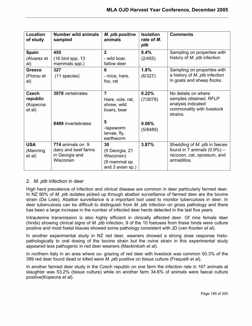
MLA OJD Harvest Year Conference, December 2005
Page 185 of 200
Locationof study
Number wild animalssampled
M. ptb positiveanimals
Isolationrate of M.ptb
Comments
Spain(Alvarez etal)
455(16 bird spp, 13mammals spp.)
2- wild boar,fallow deer
0.4%(2/455)
Sampling on properties withhistory of M. ptb infection
Greece(Florou etal)
327 (11 species)
6- mice, hare,fox, rat
1.8%(6/327)
Sampling on properties witha history of M. ptb infectionin goats and sheep flocks.
Czechrepublic(Kopecnaet al)
3078 vertebrates
8489 invertebrates
7Hare, vole, rat,shrew, wildboars, bear
5-tapewormlarvae, fly,earthworm
0.22%(7/3078)
0.06%(5/8489)
No details on wheresamples obtained. RFLPanalysis indicatedcommonality with livestockstrains.
USA(Manninget al)
774 animals on 9dairy and beef farmsin Georgia andWisconsin
30(9 Georgia, 21Wisconsin)(9 mammal sp.and 3 avian sp.)
3.87% Shedding of M. ptb in faecesfound in 7 animals (0.9%) –raccoon, cat, opossum, andarmadillos.
2. M. ptb infection in deerHigh herd prevalence of infection and clinical disease are common in deer particularly farmed deer.In NZ 90% of M. ptb isolates picked up through abattoir surveillance of farmed deer are the bovinestrain (De Lisle). Abattoir surveillance is a important tool used to monitor tuberculosis in deer. Indeer tuberculosis can be difficult to distinguish from M. ptb infection on gross pathology and therehas been a large increase in the number of infected deer herds detected in the last five years.
Intrauterine transmission is also highly efficient in clinically affected deer. Of nine female deer(hinds) showing clinical signs of M. ptb infection, 9 of the 10 foetuses from these hinds were culturepositive and most foetal tissues showed some pathology consistent with JD (van Kooten et al).
In another experimental study in NZ red deer, weaners showed a strong dose response histo-pathologically to oral dosing of the bovine strain but the ovine strain in this experimental studyappeared less pathogenic in red deer weaners (Mackintosh et al).
In northern Italy in an area where co- grazing of red deer with livestock was common 50.3% of the399 red deer found dead or killed were M. ptb positive on tissue culture (Fraquelli et al).
In another farmed deer study in the Czech republic on one farm the infection rate in 167 animals atslaughter was 53.2% (tissue culture) while on another farm 34.6% of animals were faecal culturepositive(Kopecna et al).
MLA OJD Harvest Year Conference, December 2005
Page 186 of 200
Case reports of the pathology of M. ptb infection were also reported in Mouflon sheep and hog deer.
3. M. ptb infection in rabbits.Judge et al reported on a high prevalence area in Scotland where the site infection prevalence levelsin wild rabbit populations was 39.7% and 23% respectively. These sites had previously had infectiondemonstrated in both wild rabbits and in cattle and sheep.
The same authors also demonstrated vertical, horizontal and pseudo-vertical transmission in thewild rabbit populations and isolated M. ptb from a wide variety of rabbit tissues including thereproductive tract, foetus, placenta as well as milk and faeces. However clinical disease was notrecorded in any of the rabbits studied.
The authors calculated the probability of vertical transmission at 0.458 and believe that infection canbe maintained in the wild rabbit population in the absence of livestock but did not provide anyexperimental evidence to support this view.
In a separate study by the same authors, calves and lambs were grazed on pastures contaminatedby varied concentrations of rabbit faeces. While sheep appeared to avoid ingesting rabbit faecesduring grazing, calves did not and a single rabbit faecal pellet could constitute an infective dose for acalf.
Risk factor and descriptive studiesProgress in the diagnosis and eradication of BJD in a subtropical area in Australia over a 30 yearstudy period was one of the oral presentations featured in the epidemiology stream. (Freeman et al)
Of the 78 herds in the study with a history of an infected herd status at some time more than halfhad successfully eradicated the disease from the herd. Most were beef herds and either partial ortotal de-stocking were essential elements of the eradication programs in the study herds. Lateraltransmission of infection between herds in this study only occurred through introduction of infectedanimals or by sharing contaminated facilities. Water or feed contamination, straying and roadsidewalking were not found to be risk factors for infection in the 78 herds.
Evidence was also presented showing that infection was being detected earlier and eradicated morequickly than had been the case in the past due to greater awareness, more flexible programs andfinancial assistance packages.
Another Australian study by Dhand et al looked at the risk factors for OJD prevalence level in 92infected flocks and using producer interviews evaluated the importance of various flock andmanagement practices for the risk of low medium or high within-flock prevalence.
In a report by Aly et al from the USA, ELISA results for 625 dairy dam-daughter pairs werecompared and it was found that daughters of positive dams had a 6.6 times greater risk of beingseropositive reinforcing the importance of culling the progeny of seropositive animals.
Sampling of alpine water supplies in Italy from areas grazed during summer by cattle originatingfrom a high prevalence area did not indicate that water was a significant risk factor for the spread ofM. ptb infection (Pozzato et al).
Prevalence studiesA structured survey of Austrian cattle in 2002-2003 ((Khol et al) showed a 19% seroprevalence inindividual cattle using an ELISA which was an increase on the seroprevalence detected in a 1999survey. Microbiological testing detected 4 culture positive animals.
MLA OJD Harvest Year Conference, December 2005
Page 187 of 200
In a regional survey in Italy 35% of 100 dairy/beef cattle herds were seropositive to the ELISA whilea survey of 400 cattle at an abattoir in Iran found 2.25% (9/400) were tissue culture positive.
In India, of 50 buffaloes sampled at slaughter 48% (24/50) were tissue culture positive while 46.7%of 167 serum samples from buffalo were seropositive to the ELISA.
These studies do illustrate that M. ptb infection is present in most countries who take the time to lookfor it and that it appears to be on the increase. The occurrence of infection in buffaloes in the tropicsdoes suggest that the organism is adapted to a wide range of environmental conditions.
Molecular epidemiologyStudies in the USA, Mexico, Germany, the Czech Republic, Greece and Italy all reported on the useof molecular techniques such as Restriction Fragment Length Polymorphism (RFLP) in order tocharacterise the heterogeneity of M. ptb strains and where multiple M. ptb hosts are involved thelikely source of infection for those hosts.
A number of new RFLP types were reported on, and in one cattle herd in Germany with multipleintroductions, 15 M. ptb strains were found representing 8 RFLP types. The German study alsofrequently encountered multiple strains in infected individuals.
RFLP is also useful to monitor the spread of M. ptb between regions and provide evidence for thesource of infection. It would assist tracing investigations particularly where new infections aredetected in low prevalence areas.
Economic and production studiesAspects of the financial costs associated with OJD infection in sheep flocks were examined in twoAustralian studies.
One study (Bush et al) looked at the costs associated with sheep mortalities due to OJD in 12 sheepflocks in NSW and found that on average OJD accounted for about two thirds of the total annualfinancial costs due to sheep deaths, and the gross margin for infected flocks was $10.90/ha lessthan that for uninfected flocks.
Also significant was the variation in mortality rates due to OJD.
Year Average Mortality rate Range
2002 6.2% 2.1%-17.5%
2003 7.8% 1.8%-14.6%
The second a case control study (McGregor et al) examined the impact of OJD infection onproduction parameters (body weight, reproduction, wool growth and quality) and showed that OJDhas a significant production impact that increases as infection progresses from sub-clinical toclinical.
Diagnostic test evaluationEvaluation of JD diagnostic tests such as faecal culture and ELISAs is limited by a lack ofknowledge of the true infection status of individuals in a population (gold standard- i.e. tissue cultureat slaughter).

MLA OJD Harvest Year Conference, December 2005
Page 188 of 200
Test evaluation using non-gold standard methods (comparing test results in 2 or more populationsusing conditionally independent tests) was discussed in several studies (Gardener et al). Whilethese methodologies are reasonably precise when the prevalence in the populations beingcompared differs significantly, much larger sample sizes are needed when the prevalences are moresimilar.
A recently developed computer program to assist in estimates of the within flock prevalence basedon pooled faecal culture results was also featured. This program uses seven different methodologiesto create prevalence estimates and confidence intervals and the limitations and appropriateness ofthe different methodologies in field research were discussed (Toribio et al).
MLA OJD Harvest Year Conference, December 2005
Page 189 of 200
Epidemiology of Johne’s disease: Recent developments andfuture trends
Evan SG SergeantAusVet Animal Health Services69 Turner Crescent Orange NSW 2800 AustraliaPhone: 02 6362 1598; Fax: 02 6369 1473Email: [email protected]
AbstractDue to the prolonged incubation period and associated difficulties in studying Johne’s disease (JD),the epidemiology of this condition is still not well understood, despite the efforts of many researchersover the years. However, with the advent of new technologies and methods, progress inunderstanding the epidemiology of this complex disease is still being made.
Recent studies have focused on factors affecting transmission of infection, particularly levels ofexcretion, intrauterine transmission and transmission via semen and milk in sheep, and the role ofrecently infected young cattle in transmission. Risk factors for herd and animal level infection arealso of considerable interest, although no clear pattern of identifiable risk factors have emerged,other than herd size and numbers of introductions. Further studies on transmission and risk factorswill hopefully progressively clarify some of these issues over the next few years.
The currently available screening tests for JD are limited by the chronic nature of the disease, andtheir relatively poor sensitivity, particularly in the early stages of infection. Recent developmentshave led to refinement of methodologies and sampling strategies to improve the efficiency and cost-effectiveness of the current tests, particularly for herd and flock screening. However, newtechnologies and techniques are required before any substantial improvement in test performance atthe individual animal level can be expected. These advances will hopefully arise over the next fewyears from ongoing studies using genomic and proteomic techniques to identify suitable testingtargets indicative of early infection.
Simulation modelling has also been used extensively in recent years to help understand theepidemiology of JD and for the evaluation of management and certification options. As better dataon epidemiological parameters becomes available the models are becoming more sophisticated andshould become increasingly more reliable.
IntroductionJohne’s disease, or paratuberculosis, is a syndrome characterised by chronic, granulomatousenteritis affecting mainly ruminants, and is caused by Mycobacterium avium subsp. paratuberculosis(MAP). The disease is characterised by infection of young animals early in life and a long incubationperiod, so that clinical disease usually only occurs in adults. JD is now recognised as an increasinglyimportant source of economic loss to producers and has become the focus of national and on-farmcontrol programs in the last 10-20 years (Kennedy and Benedictus, 2001).
Because of the prolonged incubation period and associated difficulties in studying JD, theepidemiology of this condition is still not well understood, despite the efforts of many researchersover the years. However, with the advent of new technologies and methods, progress inunderstanding of the epidemiology of this complex disease is still being made, and this paper
MLA OJD Harvest Year Conference, December 2005
Page 190 of 200
summarises some of the advances made over the last few years, including some of the findingspresented at this colloquium.
This paper focuses primarily on transmission of infection, risk factors for infection, developments intesting technologies and strategies and modelling of JD.
TransmissionSpread of paratuberculosis is primarily via the faeco-oral route, with clinically affected animalsexcreting large numbers of organisms and causing significant environmental contamination. Younganimals are exposed to faecal contamination of the udder, fodder and the environment, providingample opportunity for exposure. Although the faeco-oral route appears to be the main method oftransmission, other methods can also be important in some situations.
Important factors affecting the successful transmission of MAP include the level of excretion byinfected animals, the route of excretion, the source of exposure and the susceptibility of exposedanimals.
1. Excretion levelsRecent research has attempted to quantify patterns of excretion of MAP from infected animals. Thelevel and frequency of MAP excretion depend on the stage of disease and the type of lesions. Forexample, sheep with multibacillary lesions were found to be excreting MAP organisms continuously,whereas companion animals from the same flock, but which did not have detectable lesions,excreted MAP organisms only intermittently, possibly due to undetectable infection or due to passivepassaging of organisms through the gut (Whittington et al., 2000b). In the same research, the meanexcretion rate for five sheep (4 with multibacillary lesions and 1 with no detectable lesions) was inexcess of 108 organisms per gram of faeces (>8 x 10¹⁰ per sheep per day), considerably higher thanprevious estimates of 105 – 106 per gram of faeces in cattle (Jorgensen, 1982).
2. Route of excretionAs discussed above, the primary route of excretion is via faeces. However, MAP excretion via milkand semen and intra-uterine infection of the foetus have been demonstrated previously in cattle(Stehman, 1996; Sweeney, 1996) and also more recently in sheep (Eppleston and Whittington,2001; Lambeth et al., 2004 ). MAP excretion via these routes is usually more frequent in animalswith more advanced disease or that are showing clinical signs (Sweeney, 1996; Lambeth et al.,2004 ).
3. Source of infectionUntil recently, the main source of infection has generally been assumed to be older animals in theclinical or pre-clinical phase of disease. Clinical cases of JD often excrete very large numbers oforganisms (see above), and therefore represent the main source of exposure. Similarly, adultanimals are more likely to be in more advanced stages of disease, and are therefore also a higherrisk for intrauterine and trans-mammary transmission.
However, recent research has shown that calves and younger age cattle may excrete MAPorganisms soon after infection (Bolton et al., 2005; van Roermund and de Jong, 2005; Weber et al.,2005a). Modelling of MAP transmission has also supported the hypothesis that infected calvesprovide an important contribution to the spread of infection in infected cattle herds (van Roermund etal., 2002; Mitchell et al., 2005). Although this source of exposure may be of lesser significance inheavily infected herds, it suggests that young animals from infected herds or flocks are an important
MLA OJD Harvest Year Conference, December 2005
Page 191 of 200
factor in disease transmission and may pose a greater risk of spreading infection than waspreviously considered likely.
Studies of environmental samples from infected dairies have also identified high-risk areas forenvironmental contamination with MAP, and that levels of MAP contamination were correlated withpercentage of culture-positive faecal-pools in the dairy herd (Raizman et al., 2004). Areas with highproportions of positive samples included cow alleyways and manure storage areas, while lower (butstill important) proportions of positive samples were observed for the calving area and sick cowpens. Proportions of positive samples were lowest in water run-off and post-weaned calf areas.
4. Susceptibility of exposed animalsIn cattle, young calves have generally been regarded as highly susceptible to MAP infection, withsusceptibility declining with age (Sweeney, 1996). There has been a general assumption that asimilar situation exists for sheep, although this has been based on extrapolation from cattle ratherthan on any sheep-based research. However, recent research is starting to shed more light on thesituation in sheep. In one study, there was no difference in infection rates of lambs, weaners oradults following similar levels of exposure to MAP under natural conditions of heavy contamination(Reddacliff et al., 2004). This finding suggests that any age-resistance effect in sheep might not beas clearly defined as in cattle, and that under conditions of heavy challenge adult sheep can be justas susceptible to infection as young sheep. In contrast, observations on one recently infectedproperty in Australia found that infection was strongly clustered in sheep exposed as lambs and thatinfection was not detectable in sheep exposed as adults, suggesting that some age-relatedresistance does occur but may be dependent on level of exposure. Additional research is stillrequired to further clarify this situation, but age-related resistance to infection in sheep appears to beless-pronounced than is assumed to be the case in cattle.
Risk factors for Johne’s disease infectionThe identification of risk factors for JD presents significant challenges to researchers, particularlybecause of the chronic nature of the disease, and the lack of reliable diagnostic tests for detection ofinfection. In addition, introduction of infected animals is generally recognised as the main risk factorfor introduction of infection, so that most studies have concentrated on factors associated withprevalence or severity of disease in infected herds or flocks, rather than the presence or absence ofinfection.
1. CattleA number of recent studies have been undertaken to investigate possible risk factors for JD in cattle.Because of the limitations of test performance, these studies generally focused on risk factors forseropositivity for JD (at either cow or herd level), rather than for confirmed infection, and were mainlyin dairy cattle. Risk factors that were identified included:
• herd size (Hirst et al., 2004; Muskens et al., 203);
• purchasing of replacements and rate of importation of replacements (Chi et al., 202; Hirst etal., 2004); and
• previous occurrence of clinical signs of JD (Hirst et al., 2004).
Increased risk of infection with increasing herd size could be related to the larger numbers ofintroductions into the herd (Hirst et al., 2004) or to increased cattle density resulting in greaterenvironmental challenges (Muskens et al., 203). Purchasing of replacement animals is a logical riskfactor for herd-infection with JD and the greater the number of animals introduced the greater the

MLA OJD Harvest Year Conference, December 2005
Page 192 of 200
likelihood that an infected animal will be introduced. In contrast, the occurrence of clinical signs ofJD is probably a reflection of the presence of disease and perhaps of a higher prevalence ofdisease, rather than a risk factor for infection.
In addition, in one study in beef cattle herds in Texas, USA (Roussel et al., 2005), risk factors forseropositive animals included:
• species of cattle (seroprevalence was higher in Bos indicus than Bos taurus)
• geographic location in the State of Texas; and
• water source (seroprevalence was higher for cattle watered on a running stream or river).
However, only 7% of the seropositive cattle were positive on follow-up faecal culture, so that manyof the serological reactions are likely to be false-positive results. Therefore, the risk factors identifiedare not necessarily associated with the occurrence of JD, and could be associated with some otherfactor leading to false positive serological reactions, highlighting the problems of basing JD studieson the results of serology.
One study, undertaken in Scotland, investigated risk factors for the suspected occurrence of JD onindividual farms based on official records and owner knowledge of farm history to identify case andnon-case farms, rather than using serology (Daniels et al., 2002). Risk factors identified for casefarms included:
• herd size (total number of sheep and cattle);
• frequency of application of manure to pasture;
• number of rabbits present;
• wildlife access to stored feed; and
• type of water supply for cattle (piped or piped and open vs open only).
The effect of some of these factors was unexpected, and may have been due to confounding byfarmers on infected farms taking active control measures. For example, application of manure topasture ‘often’ was associated with a lower risk of JD than application sometimes or never. Similarly,use of an open water source was associated with a reduced risk compared to use of piped water,despite the use of open water sources being potentially subject to contamination and hence posing ahigher theoretical risk.
Finally, one Australian study investigated the occurrence of clinical cases in infected dairy herds(Ridge et al., 2005). This study found no association between the continued occurrence of clinicalcases or serological reactors and three recommended control measures for JD in dairy cattle. Thecontrol measures considered were: early removal of calf from dam; separating unweaned calvesfrom adult cattle and effluent from adult cattle; and not grazing weaned calves on pastures grazedby adult cattle or that have been treated with effluent from adult cattle. The lack of significant effectof these measures might have been due to the relatively small sample size in the study. The studydid identify that feeding of antibiotic contaminated waste milk and providing water to calves wereassociated with an increased occurrence of cases, while allowing cows to calve in paddocks wasassociated with decreased occurrence of cases. The reasons for these associations are unclear, butit is interesting to note that all of these factors are associated with the early neonatal period,supporting previous hypotheses that this is the most critical period for infection of calves.
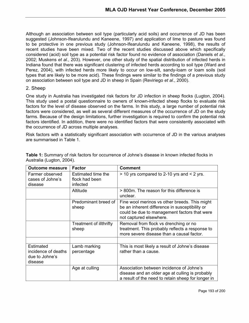
MLA OJD Harvest Year Conference, December 2005
Page 193 of 200
Although an association between soil type (particularly acid soils) and occurrence of JD has beensuggested (Johnson-Ifearulundu and Kaneene, 1997) and application of lime to pasture was foundto be protective in one previous study (Johnson-Ifearulundu and Kaneene, 1998), the results ofrecent studies have been mixed. Two of the recent studies discussed above which specificallyconsidered (acid) soil type as a potential risk factor found no evidence of association (Daniels et al.,2002; Muskens et al., 203). However, one other study of the spatial distribution of infected herds inIndiana found that there was significant clustering of infected herds according to soil type (Ward andPerez, 2004), with infected herds more likely to occur on low-silt, sandy-loam or loam soils (soiltypes that are likely to be more acid). These findings were similar to the findings of a previous studyon association between soil type and JD in sheep in Spain (Reviriego et al., 2000).
2. SheepOne study in Australia has investigated risk factors for JD infection in sheep flocks (Lugton, 2004).This study used a postal questionnaire to owners of known-infected sheep flocks to evaluate riskfactors for the level of disease observed on the farms. In this study, a large number of potential riskfactors were considered, as well as several different measures of the occurrence of JD on the studyfarms. Because of the design limitations, further investigation is required to confirm the potential riskfactors identified. In addition, there were no identified factors that were consistently associated withthe occurrence of JD across multiple analyses.
Risk factors with a statistically significant association with occurrence of JD in the various analysesare summarised in Table 1.
Table 1: Summary of risk factors for occurrence of Johne’s disease in known infected flocks inAustralia (Lugton, 2004).
Outcome measure Factor CommentFarmer observedcases of Johne’sdisease
Estimated time theflock had beeninfected
> 10 yrs compared to 2-10 yrs and < 2 yrs.
Altitude > 800m. The reason for this difference isunclear.
Predominant breed ofsheep
Fine wool merinos vs other breeds. This mightbe an inherent difference in susceptibility orcould be due to management factors that werenot captured elsewhere.
Treatment of illthriftysheep
Removal from flock vs drenching or notreatment. This probably reflects a response tomore severe disease than a causal factor.
Estimatedincidence of deathsdue to Johne’sdisease
Lamb markingpercentage
This is most likely a result of Johne’s diseaserather than a cause.
Age at culling Association between incidence of Johne’sdisease and an older age at culling is probablya result of the need to retain sheep for longer in
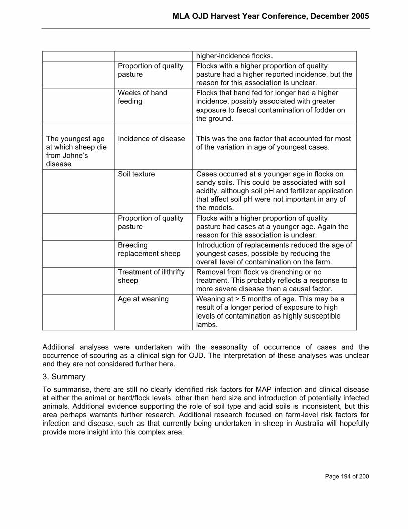
MLA OJD Harvest Year Conference, December 2005
Page 194 of 200
higher-incidence flocks.Proportion of qualitypasture
Flocks with a higher proportion of qualitypasture had a higher reported incidence, but thereason for this association is unclear.
Weeks of handfeeding
Flocks that hand fed for longer had a higherincidence, possibly associated with greaterexposure to faecal contamination of fodder onthe ground.
The youngest ageat which sheep diefrom Johne’sdisease
Incidence of disease This was the one factor that accounted for mostof the variation in age of youngest cases.
Soil texture Cases occurred at a younger age in flocks onsandy soils. This could be associated with soilacidity, although soil pH and fertilizer applicationthat affect soil pH were not important in any ofthe models.
Proportion of qualitypasture
Flocks with a higher proportion of qualitypasture had cases at a younger age. Again thereason for this association is unclear.
Breedingreplacement sheep
Introduction of replacements reduced the age ofyoungest cases, possible by reducing theoverall level of contamination on the farm.
Treatment of illthriftysheep
Removal from flock vs drenching or notreatment. This probably reflects a response tomore severe disease than a causal factor.
Age at weaning Weaning at > 5 months of age. This may be aresult of a longer period of exposure to highlevels of contamination as highly susceptiblelambs.
Additional analyses were undertaken with the seasonality of occurrence of cases and theoccurrence of scouring as a clinical sign for OJD. The interpretation of these analyses was unclearand they are not considered further here.
3. SummaryTo summarise, there are still no clearly identified risk factors for MAP infection and clinical diseaseat either the animal or herd/flock levels, other than herd size and introduction of potentially infectedanimals. Additional evidence supporting the role of soil type and acid soils is inconsistent, but thisarea perhaps warrants further research. Additional research focused on farm-level risk factors forinfection and disease, such as that currently being undertaken in sheep in Australia will hopefullyprovide more insight into this complex area.

MLA OJD Harvest Year Conference, December 2005
Page 195 of 200
Limitations of current testing for Johne’s diseaseOne of the greatest difficulties in understanding the epidemiology of JD is the limitations of theavailable screening tests. These limitations are mainly associated with the chronic nature of thedisease, and the fact that current tests have very poor sensitivity in the early stages of infection.Currently available tests are mainly based on serology (eg ELISA or AGID), faecal culture in solid orliquid media and PCR testing on faeces, milk or tissue samples for the detection of the unique DNAsequence IS900.
Recent research on screening tests for Johne’s disease has focused mainly on:
• optimising sampling strategies to achieve acceptable sensitivity at reduced cost;
• adapting existing tests to new samples or approaches; or
• refining and improving existing tests and technologies.
Pooling of faecal samples for culture is being investigated or applied in a number of countries andfor both sheep and cattle (Whittington et al., 2000a; Tavornpanich et al., 2004; Fyock et al., 2005a;Weber et al., 2004; van Schaik et al., 2003b; Antognoli et al., 2005). Pooled culture is primarily usedas a tool for identification of infected herds or flocks or conversely for certification of low-risk herdsand flocks. It has also been evaluated for identification of individual infected animals for culling, buthas limited value for this purpose depending on prevalence of infection (van Schaik et al., 2003b).Environmental sampling has also been used as an alternative to faecal sampling for theidentification of high-risk areas on dairy farms and for evaluating the effectiveness of controlprogrammes (Fyock et al., 2005a; Raizman et al., 2004; Lombard et al., 2005).
Johne’s disease ELISA’s have also been adapted, particularly for use with milk instead of bloodsamples. These have been used or proposed for both individual cow testing and for bulk milktesting. Although the milk ELISA shows promise as an alternative individual animal test to serology(Nielsen et al., 2002; Hendrick et al., 2005; Collins et al., 2005), it is likely to be of limited value as abulk milk test for surveillance purposes (Nielsen et al., 2000; Weber et al., 2005b). Despite this,simulation studies suggest that bulk milk screening might be a useful tool to support milk qualityassurance programmes (Weber et al., 2005b). Similarly, culture of bulk milk samples has also beenevaluated, but was found to be less sensitive than bulk-milk PCR (Stabel et al., 2002).
Some existing tests have also been adapted or improved by uptake of newer technologies. Forexample, liquid culture media have the advantage of supporting more rapid growth of MAP andhence shorter culture periods for confirmation of infection (Fyock et al., 2005b). Early liquid culturesystems relied on radioactivity in the media for detection of growth. Newer technologies have nowallowed the use non-radioactive media, providing a more useful and efficient culture process with thepotential to replace existing solid and liquid media systems (van Schaik et al., 2003a; Fyock et al.,2005b).
Despite these advances that have been made in broadening the application and efficiency of testingsystems, there have been no fundamental changes to the approaches used, so that the availabletests are still limited in their ability to detect early infection in individual animals.
MLA OJD Harvest Year Conference, December 2005
Page 196 of 200
Role of ModelingConsidering the difficulties and cost involved in conducting large studies on JD, modeling of thedisease has become increasingly popular in recent years. Most of these models are based on coreaspects of our understanding of the epidemiology of JD (for example age-related resistance toinfection, the effect of calf-management practices and the long pre-clinical period) to predict thecourse and dynamics of infection in infected herds or flocks. Most models also incorporatestochastic elements to reflect uncertainty about some of the epidemiological parameters. Themodels have been used for a number of different purposes, including:
• to further investigate the epidemiology and dynamics of the disease in infected herds(Pouillot et al., 2004; Mitchell et al., 2005);
• to evaluate alternative strategies for management and control of JD in infected herds(Groenendaal et al., 2002; van Schaik et al., 2003b; Dorshorst and Collins, 2005); and
• to evaluate national or industry-wide strategies for herd-certification and diseasemanagement (Weber et al., 2004; Weber et al., 2005b; Tavornpanich and Gardner, 2005).
Although such models have proved very useful in supporting recommendations for on-farm controlor for development of national control and certification programmes, they are still limited by theunderlying assumptions and the lack of detailed knowledge of some aspects of JD epidemiology. Asour understanding of the epidemiology of this complex disease improves so will the reliability andutility of the various models being used.
Future perspectivesWhat can we expect (or hope for) from the future for our understanding of JD epidemiology?
1. Further research and understanding of transmission mechanisms and risk factors forinfection, including age-susceptibility and infectious dose, the role of environmental factorssuch as soil type and the identification of other risk factors that can be manipulated to helpcontrol the disease.
2. More basic research into the epidemiology and pathogenesis of JD utilising new technologiessuch as genomics and proteomics.
3. Development of new tests and testing technologies based on the outcomes of thefundamental research and other new technologies being developed in the bio-medical field.New tests are likely to focus particularly on the early detection of infection.
4. Sophisticated models based on sound epidemiological data to predict disease transmissionand progression and the impact of a complex mix of control measures on occurrence of bothinfection and clinical disease.
Perhaps the day is coming when the farmer can use a pen-side test to identify at-risk animals beforetheir first joining and log onto the internet and answer some simple questions about his/her farm toreceive customised advice (based on a sophisticated model running remotely) on how best tomanage the disease in the current farm environment, for both individual infected animals and theherd as a whole.
MLA OJD Harvest Year Conference, December 2005
Page 197 of 200
AcknowledgmentI wish to acknowledge the financial support of Meat and Livestock Australia and AusVet AnimalHealth Services who supported my attendance at the Colloquium and to the Colloquium OrganisingCommittee for their invitation to present this paper.
ReferencesAntognoli, M.C., Jemmi, T., Hill, A.E., Ochs, H., Salman, M.D., 2005. Evaluation of interventionstrategies for control of Johne's disease in Switzerland. Proceedings of the 8th InternationalColloquium on Paratuberculosis. Madison, Wisconsin.
Bolton, M.W., Grooms, D.L., Kaneene, J.B., Mitchell, R.M., Stehman, S.M., Whitlock, R.H.,Benedictus, A., Schukken, Y.H., 2005. Fecal shedding of Mycobacterium avium subsp.paratuberculosis (MAP) in calves: Implications for disease control and management. Proceedings ofthe 8th International Colloquium on Paratuberculosis. Madison, Wisconsin.
Chi, J., VanLeeuwen, J.A., Weersink, A., Keefe, G.P., 202. Management factors related toseroprevalences to bovine viral-diarrhoea virus, bovine-leukosis virus, Mycobacterium aviumsubspecies paratuberculosis, and Neospora caninum in dairy herds in the Canadian Maritimes.Prev. Vet. Med. 55, 57-68.
Collins, M.T., Wells, S.J., Petrini, K.R., Collins, J.E., Schultz, R.D., Whitlock, R.H., 2005. Evaluationof five antibody detection tests for diagnosis of bovine paratuberculosis. Clin. Diagn. Lab. Immunol.12, 685-692.
Daniels, M.J., Hutchings, M.R., Allcroft, D.J., McKendrick, J., Greig, A., 2002. Risk factors forJohne's disease in Scotland--the results of a survey of farmers. Vet. Rec. 150, 135-9.
Dorshorst, N.C., Collins, M.T., 2005. Decision analysis model for paratuberculosis control incommercial dairy herds. Proceedings of the 8th International Colloquium on Paratuberculosis.Madison, Wisconsin.
Eppleston, J., Whittington, R.J., 2001. Isolation of Mycobacterium avium subsp paratuberculosisfrom the semen of rams with clinical Johne's disease. Aust. Vet. J. 79, 776-777.
Fyock, B.A., Smith, J., Hoving, E., Schukken, Y., Whitlock, R., 2005a. Whole herd fecal pools toassess herd MAP bio-burden compared to composite environmental samples. Proceedings of the8th International Colloquium on Paratuberculosis. Madison, Wisconsin.
Fyock, T.L., Sweeney, R.W., Whitlock, R.H., 2005b. MGIT, Liquid culture system for detection ofMAP in bovine fecal samples. Proceedings of the 8th International Colloquium on Paratuberculosis.Madison, Wisconsin.
Groenendaal, H., Nielen, M., Jalvingh, A.W., Horst, S.H., Galligan, D.T., Hesselink, J.W., 2002. Asimulation of Johne's disease control. Prev. Vet. Med. 54, 225-245.
Hendrick, S.H., Duffield, T.E., Kelton, D.E., Leslie, K.E., Lissemore, K.D., Archambault, M., 2005.Evaluation of enzyme-linked immunosorbent assays performed on milk and serum samples fordetection of paratuberculosis in lactating dairy cows. J. Am. Vet. Med. Assoc. 226, 424-8.
Hirst, H.L., Garry, F.B., Morley, P.S., Salman, M.D., Dinsmore, R.P., Wagner, B.A., McSweeney,K.D., Goodell, G.M., 2004. Seroprevalence of Mycobacterium avium subsp paratuberculosisinfection among dairy cows in Colorado and herd-level risk factors for seropositivity. J. Am. Vet.Med. Assoc. 225, 97-101.
MLA OJD Harvest Year Conference, December 2005
Page 198 of 200
Johnson-Ifearulundu, Y.J., Kaneene, J.B., 1997. Relationship between soil type and Mycobacteriumparatuberculosis. J. Am. Vet. Med. Assoc. 210, 1735-1740.
Johnson-Ifearulundu, Y.J., Kaneene, J.B., 1998. Management-related factors for M.paratuberculosis infection in Michigan, USA, dairy herds. Prev. Vet. Med. 37, 41-54.
Jorgensen, J.B., 1982. An improved medium for culture of Mycobacterium paratuberculosis frombovine faeces. Acta Vet Scand 23,
Kennedy, D.J., Benedictus, G., 2001. Control of Mycobacterium avium subsp paratuberculosisinfection in agricultural species. Rev. Sci. Tech. 20, 151-179.
Lambeth, C., Reddacliff, L.A., Windsor, P., Abbott, K.A., McGregor, H., Whittington, R.J., 2004.Intrauterine and transmammary transmission of Mycobacterium avium subsp paratuberculosis insheep. Aust. Vet. J. 82, 504-8.
Lombard, J.E., Smith, R.L., Wagner, B.A., McCluskey, B.J., 2005. Evaluation of environmentalsampling to determine distribution and herd infection status for Mycobacterium avium subspeciesparatuberculosis. Proceedings of the 8th International Colloquium on Paratuberculosis. Madison,Wisconsin.
Lugton, I.W., 2004. A cross-sectional study of risk factors for the clinical expression of ovine Johne'sdisease on New South Wales farms. Aust. Vet. J. 82, 355-365.
Mitchell, R.M., Stehman, S.M., Whitlock, R.H., Benedictus, A., Schukken, Y.H., 2005. A deterministicmathematical model of Mycobacterium avium subsp. paratuberculosis (MAP) transmission oncommercial US dairy farms. Proceedings of the 8th International Colloquium on Paratuberculosis.Madison, Wisconsin.
Muskens, J., Elbers, A.R., van Weering, H.J., Noordhuizen, J.P., 203. Herd management practicesassociated with paratuberculosis seroprevalence in Dutch dairy herds. Journal of VeterinaryMedicine Series B. 50, 372-7.
Nielsen, S.S., Enevoldsen, C., Grohn, Y.T., 2002. The Mycobacterium avium subsp.paratuberculosis ELISA response by parity and stage of lactation. Prev. Vet. Med. 54, 1-10.
Nielsen, S.S., Thamsborg, S.M., Houe, H., Bitsch, V., 2000. Bulk-tank milk ELISA antibodies forestimating the prevalence of paratuberculosis in Danish dairy herds. Prev. Vet. Med. 44, 1-7.
Pouillot, R., Dufour, B., Durand, B., 2004. A deterministic and stochastic simulation model for intra-herd paratuberculosis transmission. Vet Res 35, 53-68.
Raizman, E.A., Wells, S.J., Godden, S.M., Bey, R.F., Oakes, M.J., Bentley, D.C., Olsen, K.E., 2004.The distribution of Mycobacterium avium ssp. paratuberculosis in the environment surroundingMinnesota dairy farms. J Dairy Sci 87, 2959-66.
Reddacliff, L.A., McGregor, H., Abbott, K., Whittington, R.J., 2004. Field evaluation of tracer sheepfor the detection of early natural infection with Mycobacterium avium subsp paratuberculosis. Aust.Vet. J. 82, 426-33.
Reviriego, F.J., Moreno, M.A., Domínguez, L., 2000. Soil type as a putative risk factor of ovine andcaprine paratuberculosis seropositivity in Spain. Prev. Vet. Med. 43, 43-51.
Ridge, S.E., Baker, I.M., Hannah, M., 2005. Effect of compliance with recommended calf-rearingpractices on control of bovine Johne's disease. Aust. Vet. J. 83, 85-90.
Roussel, A.J., Libal, M.C., Whitlock, R.L., Hairgrove, T.B., Barling, K.S., Thompson, J.A., 2005.
MLA OJD Harvest Year Conference, December 2005
Page 199 of 200
Prevalence of and risk factors for paratuberculosis in purebred beef cattle. J. Am. Vet. Med. Assoc.226, 773-8.
Stabel, J.R., Wells, S.J., Wagner, B.A., 2002. Relationships between fecal culture, ELISA, and bulktank milk test results for Johne's disease in US dairy herds. J Dairy Sci 85, 525-31.
Stehman, S.M., 1996. Paratuberculosis in small ruminants, deer, and South American camelids. Vet.Clin. North Am. Food Anim. Pract. 12, 441-455.
Sweeney, R.W., 1996. Transmission of paratuberculosis. Vet. Clin. North Am. Food Anim. Pract. 12,305-312.
Tavornpanich, S., Gardner, I.A., 2005. Evaluation of various testing programs for certification ofparatuberculosis-free herds by simulation modeling. Proceedings of the 8th InternationalColloquium on Paratuberculosis. Madison, Wisconsin.
Tavornpanich, S., Gardner, I.A., Anderson, R.J., Shin, S., Whitlock, R.H., Fyock, T., Adaska, J.M.,Walker, R.L., Hietala, S.K., 2004. Evaluation of microbial culture of pooled fecal samples fordetection of Mycobacterium avium subsp paratuberculosis in large dairy herds. Am. J. Vet. Res. 65,1061-1070.
van Roermund, H.J.W., de Jong, M.C.M., 2005. Horizontal-transmission experiment ofparatuberculosis. Proceedings of the 8th International Colloquium on Paratuberculosis. Madison,Wisconsin.
van Roermund, H.J.W., Vos, A.M., de Jong, M.C.M. , 2002. Within-herd transmission ofparatuberculosis and the possible role of infectious calves. Proceedings of the 7th InternationalColloquium on Paratuberculosis.
van Schaik, G., Rossiter, C.R., Stehman, S.M., Shin, S.J., Schukken, Y.H., 2003a. Longitudinalstudy to investigate variation in results of repeated ELISA and culture of fecal samples forMycobacterium avium subsp paratuberculosis in commercial dairy herds. Am. J. Vet. Res. 64, 479-484.
van Schaik, G., Stehman, S.M., Schukken, Y.H., Rossiter, C.R., Shin, S.J., 2003b. Pooled fecalculture sampling for Mycobacterium avium subsp. paratuberculosis at different herd sizes andprevalence. J. Vet. Diagn. Invest. 15, 233-41.
Ward, M.P., Perez, A.M., 2004. Soil types associated with bovine paratuberculosis in Indiana cattleherds. Am. J. Vet. Res. 65, 10-14.
Weber, M.F., Groenendaal, H., van Roermund, H.J. , Nielen, M., 2004. Simulation of alternatives forthe Dutch Johne's disease certification-and-monitoring program. Prev. Vet. Med. 62, 1-17.
Weber, M.F., Kogut, J., de Bree, J., van Schaik, G., 2005a. Evidence for Mycobacterium aviumsubsp. paratuberculosis shedding in young stock. Proceedings of the 8th International Colloquiumon Paratuberculosis. Madison, Wisconsin.
Weber, M.F., van Roermund, H.J.W., Velthuis, A.G.J., de Koeijer, A.A., de Jong, M.C.M., Nielen, M.,2005b. Development of a milk quality assurance programme for paratuberculosis: Stochasticsimulation of within-herd infection dynamics and economics. Proceedings of the 8th InternationalColloquium on Paratuberculosis. Madison, Wisconsin.

MLA OJD Harvest Year Conference, December 2005
Page 200 of 200
Whittington, R.J., Fell, S., Walker, D., McAllister, S., Marsh, I., Sergeant, E., Taragel, C., Marshall,D.J., Links, I.J., 2000a. Use of pooled fecal culture for sensitive and economic detection ofMycobacterium avium subsp. paratuberculosis infection in flocks of sheep . J. Clin. Microbiol. 38,2550-2556.
Whittington, R.J., Reddacliff, L.A., Marsh, I., McAllister, S., Saunders, V., 2000b. Temporal patternsand quantification of excretion of Mycobacterium avium subsp paratuberculosis in sheep withJohne's disease. Aust. Vet. J. 78, 34-37.