Embed Size (px)
Citation preview
1AN ANALYSIS OFOBSTRUCTIVE JAUNDICE
Dissertation submitted to
THE TAMIL NADUDR. M.G.R. MEDICAL UNIVERSITY
CHENNAI 600 032
With fulfillment of the RegulationsFor the Award of the Degree of
M.S. GENERAL SURGERY(BRANCH I)
DEPARTMENT OF SURGERYGOVERNMENT KILPAUK MEDICAL COLLEGE
CHENNAI 600 010
MARCH 2009
CERTIFICATE
This is to certify that this dissertation in “AN ANALYSIS OF OBSTRUCTIVE
JAUNDICE” is a work done by DR.J.EMMANUEL THAS, under my guidance during the
period 2006-2008. This has been submitted in partial fulfillment of the award of M.S.
Degree in General Surgery (Branch-I) by the Tamilnadu Dr. M.G.R. Medical University,
Chennai 600 032.
PROF. DR. G. GUNASEELAN, M.S. PROF. DR. S. UDAYAKUMAR, M.S.,PROFESSOR AND HEAD OF THE DEPARTMENT PROFESSOR AND UNIT CHIEFDEPARTMENT OF SURGERY DEPARTMENT OF SURGERYGOVERNMENT KILPAUK MEDICAL GOVERNMENT KILPAUK MEDICAL COLLEGE, CHENNAI COLLEGE, CHENNAI
THE DEANProf. Dr. M. DHANAPAL, M.D., D.M.,
Government Kilpauk Medical College and HospitalChennai 600 010
ACKNOWLEDGEMENT
I thank the DEAN of Kilpauk Medical College and Hospital, Prof. Dr. M.
DHANAPAL, M.D., D.M., (Cardiology) for permitting me to conduct this study in the Department of
General Surgery of the Government Kilpauk Medical College and Hospital, Chennai.
I thank Prof. G. GUNASEELAN, M.S., Head of Department of General Surgery for helping
and guiding me during the study.
My heartful gratitude to Prof. S. UDAYAKUMAR, M.S., for his esteemed guidance and
valuable suggestions. It is my privileged duty to profusely thank my teacher, guide and mentor
underwhom I have the great honour to work as a postgraduate student.
I acknowledge the invaluable advice and guidance received from
Prof. P. RAVI M.S,Professor of Surgery.
I am greatly indebted to my Unit Assistant Professors
Dr.T.S.JAYASHREE,D.G.O., M.S., Dr. S. THIRUNAVUKKARASU, M.S., who have put in
countless hours in guiding me in many aspects of this study and also in honing my surgical skills.
I thank the Surgical Registrar, Department of General Surgery , Dr
.A.K.RAJENDRAN .,M.S for his help throughout the study period.
I thank the Department of Surgical Gastroenterology and Surgical Oncology in Government
Royapettah Hospital, Chennai.

Last but not the least, my heartfelt gratitude to my patients without whom this study could not
have been possible and also to Medical Records Department of Government Royapettah Hospital for
their timely help.
CONTENTS
INTRODUCTION 2
DEFINITION 4
AIM OF THE STUDY 6
MATERIALS AND METHODS 8
REVIEW OF LITERATURE
ANATOMY OF HEPATOBILIARY SYSTEM 11
SURGICALLY RELEVANT VARIATIONS IN ANATOMY 15
AETIOLOGY OF BILIARY OBSTRUCTION 18
PATHOPHYSIOLOGY OF BILIARY TRACT OBSTRUCTION 24
CLINICAL PRESENTATION 33
INVESTIGATION 38
PRE OPERATIVE PREPARATIONS 50
OPERATIVE PROCEDURES 53
RESULTS AND OBSERVATIONS 58
DISCUSSION 64
CONCLUSION 68
ANNEXURE
PROFORMA
BIBILIOGRAPHY
INTRODUCTION
Surgical or obstructive jaundice is basically a biochemical derangement resulting in physiological
changes due to non deliverance of bile into the intestinal lumen as a consequence of anatomical
alterations caused by a variety of pathologies involving the biliary tract, which is mostly incurable with
medicines, sometimes palliated by endoscopic procedures and majority of the time cured or palliated
by surgery.
Surgical jaundice is a common entity seen in surgical wards or in general practice. The
challenge it poses in the diagnosis is totally elusive occasionally, more so in the therapeutic aspect & its
outcome.
It is one of the few areas where measures are started, along with diagnostic evaluations, towards
preventing and managing the complications.
Even though the advancements in this area have occurred in leaps and bounds, resulting in
improved long term results, it still poses to be deceptive in the optimal management with wide
variations.
DEFINITION
DEFINITIO
Jaundice is a generic term for the yellow pigmentation of the skin, mucous membranes, or
sclera that is caused by a heterogeneous group of disorders due to hyperbilirubinemia. Although
surgical jaundice is characterised by hyperbilirubinemia and dilated bile duct it is clearly inadequate to
equate the two. Similarly obstruction and the dilation of the common bile duct are not synonymous,
thought they complement each other.
Complete biliary obstruction produces jaundice.
Incomplete obstruction produces symptoms and biochemical changes but may or may not be
associated with features of clinical jaundice.
Chronic incomplete obstruction with or without clinical symptoms or biochemical features, produces
pathological changes in bile ducts or liver.
Segmental obstruction involves one or more isolated segments of the biliary tree which
may take the form of complete, intermittent or chronic incomplete obstruction.
AIM OF THE STUDYAIM OF THE STUDY
To study the incidence, etiopathogenesis and progression of disease in obstructive
jaundice.
To investigate and analyse all patients with obstructive jaundice and to prepare them for
surgical intervention.
To plan for surgical intervention, either curative or palliative.
To follow up the patients to understand the progression of the disease, after surgical
intervention and to analyse the outcome.
MATERIALS AND METHODS=TERIALS AND
Obstructive jaundice patients admitted in all the four surgical units of Govt.
Royapettah Hospital,Kilpauk Medical College, Chennai. between May 2006 to October
2008 were studied and evaluated.
On admission a detailed history and clinical assessment of the problems were
made. Preliminary biochemical investigations were carried in all the patients followed
by real-time ultrasonography and ERCP if necessary. In all patients CT scan of
abdomen was carried out to assess the operability.
If operable lesion were detected patients underwent a careful preoperative
preparation. Histopathological examination was conducted in relevant patients. They
were followed in the post operative period and subsequent to their discharge.
The various causes for the obstructive jaundice in our hospital was evaluated. A
comparison was made with other studies regarding the incidence, mode of presentation,
prognosis and survival. The results were compared and graphically represented and a
conclusion was arrived from it.
REVIEW OF LITERATURE
ANATOMY OF HEPATOBILIARY TRACT
ANATOMY OF HEPATOBILANATOMY OF THE HEPATOBILIARY SYSTEM
Liver is the largest gland in the body weighing about 1500 gms and receiving
1500 ml. of blood per min. It arises from the foregut endoderm. It has three surfaces,
two lobes and eight segments, four for each side, each side being separated by Cantlie’s
line or main portal scissura, thus leaving a smaller right liver than the right lobe.
It has the following ligamentous attachments;
Falciform ligament
Right and left triangular ligaments
Coronary ligaments
Gastrohepatic omentum
The biliary tract begins as blind channels or canaliculi, that ramify between the
hepatocytes, formed by the specialised structures in the cell membranes of adjacent
hepatocytes, and drain into the interlobular bile duct in the portal triad which forms the
periphery of the functional unit called lobule, centered around central vein.
The right hepatic duct is formed by the intrahepatic union of dorsocaudal and
ventrocranial branches (segments V-VIII) and the left duct by the medial and lateral
branches.
EXTRAHEPATIC BILIARY APPARATUS :
Consists of
Hepatic ducts - right, left and common
Gall bladder
Cystic duct
Common bile duct
Gall Bladder
It is a pear shaped reservoir of 10 cm length with capacity of 50 ml. Situated in
the gall bladder fossa in the inferior surface of liver. It is covered with peritoneum
variably.
It has three parts :
Fundus - has the poorest blood supply;
Body
Neck or infundibulum - frequently has the abnormal dilatation called
Hartmann’s pouch in the posteromedial wall, which may adhere to the
surrounding structures obscuring the anatomy of the region.
Cystic duct :
Variable course of 3-4 cm. Usually, downward and backward, joining the
common hepatic duct, guarded by a false valve formed by the mucous fold called valve
of Heister.
Hepatic ducts :
Right and left unite to form common duct (usually extrahepatically 90% within 1
cm.) which is about 3 cm. At the porta, they are arranged behind forwards as portal
vein, hepatic artery and hepatic ducts.
Common bile duct:
Anatomically starts distal to cystic duct junction but surgically from the union of
right and left hepatic ducts. It measures about 10-12 cm. In length with average diameter
of 7 mm. (Range 4-10mm). It is divisible into supraduodenal, retroduodenal,
intrapancreatic and intraduodenal. It unites with the main pancreatic duct to form the
ampulla and opens into the major papilla in the posteromedial wall of II part of
duodenum.
SURGICALLY RELEVANT VARIATIONS IN ANATOMY
SURGICALLY RELEThe knowledge of segmental anatomy is deployed in the hepatic resections. The hepatic arterial, portal venous and hepatic bile duct branches conform to
the segment organisation excepting the hepatic vein.The rex recessus in the umbilical fissure in the anterosuperior surface, is followed by the ligamentum teres to access the segment III and the left duct in the round ligament approach for bypass in inoperable cholangio-carcinoma.
A thorough knowledge of not just the anatomy but also the variations is needed to
avoid complications. In Gall Bladder, commonest anomaly is the “Phrygian cap”.
Others include agenesis, floating GB, sausage shaped GB, positional anomalies,
trabeculated GB.
Bile duct anomalies include
- aberrant, supernumerary or accessory ducts.
- variations in the confluence of major intrahepatic ducts.
- variations in the course and termination of cystic duct.
- variations in the union with pancreatic duct at the termination
of CBD.
Vascular anomalies :
Of importance is the cystic artery, which is normally a branch of right hepatic
artery, emerging from behind the common hepatic duct.
- Accessory cystic artery
- Low origin of the cystic artery.
- Abnormal origin from other vessels
- Short cystic artery with a looped right hepatic artery called
The Moynihan’s hump.
- Close lie of the right hepatic artery to the cystic duct with a
Short cystic artery - most dangerous anomaly.
The relevance of peri choledochal arterial plexus, formed by the duodenal
branch of gastroduodenal and right hepatic arteries, in the prevention of strictures should
be understood.
17AETIOLOGY OF BILIARY
OBSTRUCTION
AETIOLOGY OF BILIARY OBSTRUCTION
AETIOLOGY OF BILIARY OBSTRUCTION
It is important to recognise the various causes of biliary obstruction, since they
produce many subtle clinical syndrome whose true nature may go unrecognised, the
clinician should recognise at least 4 categories of biliary tract obstruction discussed
before :-
- Complete biliary tract obstruction
- Intermittent obstruction
- Chronic incomplete obstruction
- Segmental obstruction
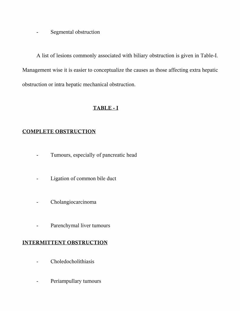
A list of lesions commonly associated with biliary obstruction is given in Table-I.
Management wise it is easier to conceptualize the causes as those affecting extra hepatic
obstruction or intra hepatic mechanical obstruction.
TABLE - I
COMPLETE OBSTRUCTION
- Tumours, especially of pancreatic head
- Ligation of common bile duct
- Cholangiocarcinoma
- Parenchymal liver tumours
INTERMITTENT OBSTRUCTION
- Choledocholithiasis
- Periampullary tumours
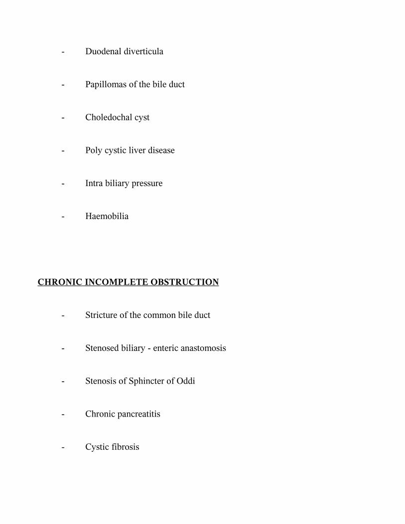
- Duodenal diverticula
- Papillomas of the bile duct
- Choledochal cyst
- Poly cystic liver disease
- Intra biliary pressure
- Haemobilia
CHRONIC INCOMPLETE OBSTRUCTION
- Stricture of the common bile duct
- Stenosed biliary - enteric anastomosis
- Stenosis of Sphincter of Oddi
- Chronic pancreatitis
- Cystic fibrosis
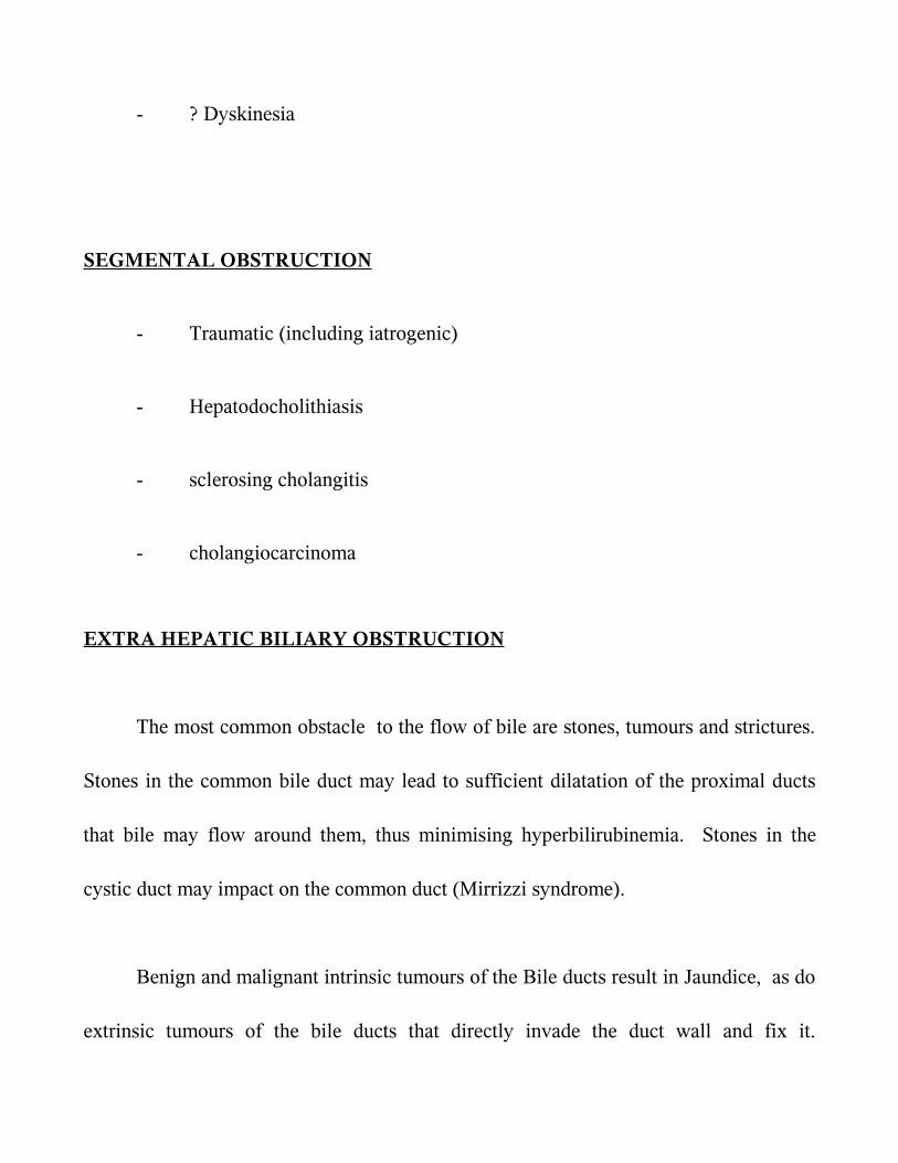
- ? Dyskinesia
SEGMENTAL OBSTRUCTION
- Traumatic (including iatrogenic)
- Hepatodocholithiasis
- sclerosing cholangitis
- cholangiocarcinoma
EXTRA HEPATIC BILIARY OBSTRUCTION
The most common obstacle to the flow of bile are stones, tumours and strictures.
Stones in the common bile duct may lead to sufficient dilatation of the proximal ducts
that bile may flow around them, thus minimising hyperbilirubinemia. Stones in the
cystic duct may impact on the common duct (Mirrizzi syndrome).
Benign and malignant intrinsic tumours of the Bile ducts result in Jaundice, as do
extrinsic tumours of the bile ducts that directly invade the duct wall and fix it.
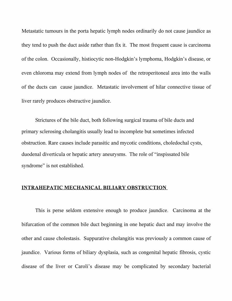
Metastatic tumours in the porta hepatic lymph nodes ordinarily do not cause jaundice as
they tend to push the duct aside rather than fix it. The most frequent cause is carcinoma
of the colon. Occasionally, histiocytic non-Hodgkin’s lymphoma, Hodgkin’s disease, or
even chloroma may extend from lymph nodes of the retroperitoneal area into the walls
of the ducts can cause jaundice. Metastatic involvement of hilar connective tissue of
liver rarely produces obstructive jaundice.
Strictures of the bile duct, both following surgical trauma of bile ducts and
primary sclerosing cholangitis usually lead to incomplete but sometimes infected
obstruction. Rare causes include parasitic and mycotic conditions, choledochal cysts,
duodenal diverticula or hepatic artery aneurysms. The role of “inspissated bile
syndrome” is not established.
INTRAHEPATIC MECHANICAL BILIARY OBSTRUCTION
This is perse seldom extensive enough to produce jaundice. Carcinoma at the
bifurcation of the common bile duct beginning in one hepatic duct and may involve the
other and cause cholestasis. Suppurative cholangitis was previously a common cause of
jaundice. Various forms of biliary dysplasia, such as congenital hepatic fibrosis, cystic
disease of the liver or Caroli’s disease may be complicated by secondary bacterial
infection of the bile ducts. Even solitary non parasitic cysts may obstruct the biliary
tree.
Chronic pancreatitis is recognised as a cause of cholestasis, mainly as a result of
cholangiographic or ultrasonographic demonstration of formidable narrowing of
adjacent bile duct. The protracted jaundice usually results from large amounts of
alcohol damaging the liver, since despite the chronic pancreatitis, the bile duct is almost
normal. Hydatid cysts can obstruct ducts.
In cystic fibrosis of the pancreas, inspissated mucus in ductules and ducts may
cause partial obstruction without jaundice. Metastatic carcinoma is frequently associated
with cholestasis, so are lymphomas and Hodgkin’s disease.
Cholestatic jaundice is a serious problem after transplantation of the liver.
In the immediate postoperative period, it may be caused by complications mechanical or
infectious, at the biliary anastamosis. A few weeks postoperatively, rejection
phenomenon may be the reason. Eventually bile ducts may disappear and the lesion may
resemble primary biliary cirrhosis.
PATHOPHYSIOLOGY OF BILIARY TRACT OBSTRUCTION
PATHOPHYSIOLOGY OBiliary obstruction results in many physiological, pathological, biochemical and functional changes which are considered in detail below.
PHYSICAL EFFECTS
The normal secretary pressure of bile is 120-250 mm. H2O (11-8-24.5 kPa).
When the intra biliary pressure is raised to more than 300 mm. H2O (29.4 kPa), there is
total inhibition of bile secretion. However the effects on secretion of bile salts,
cholesterol and phopholipids are unequal and the return of the secretary functions
following relief of the bile duct obstruction may be asynchronous. The degree of
proximal biliary dilatation depends on nature and duration of obstruction and also on
pliability of the extra hepatic bile ducts and of supporting skeleton of intra hepatic bile
ducts. The former may be restricted in inflammation and fibrosis; the latter may be
enhanced by impaired hepato cellular function raising suspicion of portal hypertension.
These two pathologies can cause a lack of proximal biliary dilatation inspite of organic
distal lesion.
EFFECT ON BLOOD FLOW
Though presumably obstruction of bile ducts should increase intrahepatic
hydrostatic pressure and produce an elevation of hepatic sinusoidal pressure, thus
adversely affecting hepatic tissue perfusion; this however has been disproved in animal
experimental studies. Chronic obstruction, however has been demonstrated to lead to
fibrosis with secondary portal hypertension.
PATHOLOGICAL EFFECTS
DUCTULAR CHANGES
The initial changes occur at the canalicular level and are mediated by high local
concentration of bile salts. There is dilatation of centrilobular bile canaliculus, bile
thrombi are present in the lumen. In more prolonged cholestasis the bile ducts and
ductules appear to be increased probably both by lengthening and tortuosity and by
sprouting of small bile ductules secondary to inflammation; these changes are probably
due to lithocholate or altered portal micro circulation.
FIBROTIC CHANGES
Intra hepatic fibrosis occurs secondary to cholangitis which causes laying down of
reticulin fibres which is followed by formation of hard collagen. The changes in
extrahepatic ducts are a sequence of mucosal atrophy and squamous metaplasia followed
by inflammation and ultimately fibrosis in the subepithelial layers of the ducts.
HEPATOCYTE CHANGES
Cholestasis causes local high concentration of bile salts which inhibit the enzyme
cytochrome p-450 causing damage along the biliary pole of the Hepatocyte. This
initially results in “feathery necrosis” and in its most severe form is associated with
leukocyte infiltration resulting in “Biliary piecemeal necrosis”.
BIOCHEMICAL EFFECTS
BILIRUBIN
The rise in serum bilirubin varies in intensity with the type of obstruction -
whether complete / intermittent / chronic complete / segmental. Persistent absence of
urobilinogen from urine is strong evidence of obstructive jaundice and bilirubin is
usually present in the urine until the secondary hepatocellular disease becomes
advanced. It has been proposed that it is bile concentration gradient that opposes the
transport of bilirubin into the canaliculus, and this gradient may be produced by stasis in
the biliary tree. The mechanism by which conjugated bilirubin reflexes into sinusoidal
plasma in cholestasis is unclear.
BILE ACIDS
Complete biliary tract obstruction will interrupt the enterohepatic circulation of
bile salts. Serum bile acids may be very high ranging from 4-60 times the normal .
Nevertheless the synthesis itself is impaired. There is an increased urinary excretion of
bile acids but this still amounts to a very small proportion of faceal excretion. In
addition abnormal bile acids are produced by the liver, including ursodeoxycholate,
which are more easily Excreted in urine than the normal bile acids. The pruritus of
cholestasis is probably caused by deposition of bile acids in the skin. External biliary
drainage of the obstructed tract results in depletion of the bile salt pool and its
replacement may have a beneficial effect on the clearance of bilirubin and other moities
excreted by mechanisms dependent on bile salt related canalicular flow.
ALKALINE PHOSPHATASE
This is a sensitive marker of biliary obstruction but its lack of specificity is a clear
pitfall. In th liver, the isoenzymes of alkaline phosphatase exist in the plasma membrane
of the microvilli of the canaliculus. Sources of other isoenzymes are bone, intestine,
kidney and placenta. The return to normal of alkaline phosphatase after relief of biliary
obstruction may be a good index of successful therapy in majority of cases. A simple
practical application of the measurement of alkaline phosphatase in the complex post
operative patient, particularly following biliary reconstruction or major hepatic trauma
elevation of serum billirubin in the absence of raising alkaline phosphatase may indicate
an intraperitoneal biliary collection while parallel increases of bilirubin and alkaline
phosphatase suggest a degree of obstruction.
OTHER BIOCHEMICAL PARAMETERS
SERUM AMINO TRANSFERASES
Gross elevation of aminotransferase is an index of hepatocellular damage. Minor
elevation is common, however, in extrahepatic biliary obstruction, and may even rise
earlier than alkaline phosphatase in case of acute obstruction. The levels fall to normal
nearly so, within several days, despite depending jaundice. However in later stage they
may rise again, and this is an adverse prognostic feature, indicating prolonged
obstruction with superadded intrabiliary infection or invasive sepsis from suppurative
cholangitis. Gamma Glutamyl transpeptidase tends to parallel alkaline phosphatase and
is sometimes used as an alternative to the isoenzyme fractionation of alkaline
phosphatase.
SERUM PROTEINS
Depression of the serum albumin is a common feature in patients with malignant
biliary obstruction, particularly in prolonged nutritional impairment. Elevation of
globulins particularly the IgG fraction, may be seen in severe parenchymal liver disease
due to long standing obstruction. Gross elevation of gamma globulins may be related to
porta-systemic shunting and should raise a strong suspicion of secondary portal
hypertension.
LIPID METABOLISM
This is grossly altered but most of the changes are unhelpful because of lack of
specificity. Cholesterol may be elevated inlong standing biliary obstruction.
29
FUNCTIONAL EFFECTSThe fundamental dearrangements due to cholestasis are reflected in the
hepatocellular function and reticulo endothelial function.
HEPATOCELLULAR FUNCTION
The alterations in hepatocellular function occur due to
1. Retained bile salts – which have a toxic effect on the cellular
Component cytochrome P-450 and transform it into inactive
Cytochrome P-420.
2. Retained bilirubin – which inhibits mono oxygenease activity
3. Other moities - disruption of mitochondrial functions.
Though various enzymes which are markers of hepatocellular dysfunction
are routinely used, a dynamic indicator of liver function would be a valuable adjunct to
assessment of patients who are candidates for major surgery. The clearance of antipyrine
( a minor analgesic almost entirely oxidized in the liver) and paracetamol (which is
conjugated to glucoronide and sulphate esters in the liver) are effective parameters of
liver function. Abnormal glucose tolerance is also seen and may be due to defects in the
hepatic alkaline cycle.
RETICULO ENDOTHELIAL FUNCTION
Kupffer cells lining the sinusoids have a major role in the inactivation of
endotoxins, the abnormal lipopolysaccharides derived from cell wall of gram negative
bacteria in the gut. In biliary obstruction this function is impaired and systemic
endotoxemia occurs more frequently in jaundiced patients in comparision to non
jaundiced patients. This fact has been demonstrated in animal experiments.
Endotoxemia is associated with postoperative renal failure in the jaundice patient; hence
these results are of therapeutic significance.
SECONDARY EFFECTS
INFECTION :
The normal biliary tract is sterile. Following surgical or radiological intervention
of the obstructed biliary tract the bile culture rate increases. Therefore fluid suppurative
cholangitis is commoner in malignant obstruction rather than benign biliary obstruction.
The most common infecting organisms are Escherichia coli and Streptococcus faecalis,
this spectrum may change following radiological intubation or surgery.
ATROPHY
The liver atrophy may be uniform, lobar or segmental. Uniform atrophy occurs in
presence of malnutrition or partial occlusion of the main stem of portal vein.
31
Segmental or lobal atrophy occurs due to portal venous occlusion / bile duct
occlusion / proximity to space occupying lesion / (rarely) unilobular veno-occlusive
disease. Atrophy can be determined radiologically by isotope or CT scans or
transhepatic cholangiograms.
32 CLINICAL PRESENTATIONCLINICAL PRESENTATION
CLINICAL PRESENTATION
The presenting feature may be in many ways in any combination of the
following :
JAUNDICE
A generic word from French meaning yellow. It is due to the deposition of bile
pigments in sclera, skin, and mucous membranes. The predilection of sclera is due to
the high affinity of the elastin, to bilirubin, in the sclera.
WEIGHT LOSS & ANOREXIA
Presence of these two factors are more commonly indicative of malignancy where
the history is usually progressive.
PAIN
The site and type of pain vary with the underlying pathology e.g., Pain in
epigastrium radiating to back indicates a pancreatic pathology, where pain is usually due
to involvement of retropancreatic nerves and pancreatic duct obstruction.
PRURITIS
It strongly suggests extrahepatic obstructive jaundice, even in infective pathology
it indicates presence of cholestatic component. It may begin abruptly and is worse in
the nights but not usually relieved by ointments, lotions etc.
34
FEVER
Fever and rigors indicate cholangitis and high degree of suspicion is needed as it
needs urgent attention. Care should be taken to rule out other causes of fever.
DYSPEPSIA
Quite commonly seen in gallstones esp. to fatty foods, but cannot attribute to any
other disorders, specifically. Bloating may be seen in obstruction of duodenum.
HIGH COLOURED URINE
Due to bilirubinuria. It may be noted several days before the onset of jaundice.
The darkest specimen is the first passed urine in the morning.
PALE STOOLS
Absence of the bilirubin metabolites lead to clay coloured to pale yellow stools,
the colour varying with the degree of cholestasis.
MALAISE AND FATIGABILITY
May be due to poor nutrition or due to underlying psychiatric disorder eg.
Depression in pancreatic carcinoma which has to be differentiated.
35
PHYSICAL SIGNS
JAUNDICE
The depth of jaundice depends on the type and duration of obstruction e.g.
Progressive and deep jaundice in carcinoma head of pancreas and intermittent in
periampullary carcinoma and choledocholithiasis.
DISTENDED GALL BLADDER
Palpable gall bladder in the presence of jaundice goes more in favour of
obstruction caused by a malignancy. Courvoisier’s law reinforces this.
PALPABLE LIVER
Liver is usually enlarged in cases of pure extrahepatic obstruction, absence of
which should prompt the clinician to look for other pathologies. Nodularities over the
liver suggest malignant involvement, esp if obstructive type of jaundice, and point to a
extrahepatic cause.
MASS
Both benign and malignant causes can present with mass abdomen, viz
choledochal cyst and carcinoma head of pancreas respectively. The character of the
mass indicates the type of lesion.
36
ASCITES
This usually signifies extensive spread of a malignant disease, however this can
happen in the absence of peritoneal metastasis too, like due to portal vein obstruction at
the porta hepatis.
37
INVESTIGATIONSINVESTIGATIONS
INVESTIGATIONS
It is to reiterate that history and examination alone can give a diagnosis in 80% of
the patients.
BIOCHEMICAL
BILIRUBIN
Their intensity differs with the type of obstruction. Persistence or absence of
urobilinogen in urine is a strong evidence of obstructive jaundice.
Associated secondary hepatocellular disease can produce bilirubin in urine.
Clinically jaundice is apparent when the level of bilirubin is above 2mg% or 3mg
% when the jaundice is rising rapidly. Also in convalescence it is seen until the level
falls to 1.5 mg% as it still remains bound to sclera.
Bilirubin in urine is a sensitive screening test. Yellow foam on shaking the urine
and its reaction with diazotized 2,4 dichloroaniline or a stable diazonium salt in the
presence of acid buffer produce a colour. This is the basis of van den bergh reaction.
Urine urobilinogen is characteristically absent in obstructive jaundice as entry into
bowel is needed for its production, and subsequent absorption to appear in urine.
39
I
The van den Bergh reaction differentiates the type of jaundice. A direct reaction
indicates immediate colour change (in cholestasis) while pre-treatment with alcohol or
urea is needed to produce a colour change in haemolytic jaundice.
BILE ACIDS
Obstruction of bile flow can cause an increase in the bile acid level by 4-60 fold
but at the same time impairs the synthesis. But the abnormal bile acid secretion
increases which is more avidly excreted in urine along with normal acids too.
ALKALINE PHOSPHATASE
It is sensitive but lacks specificity in obstructive jaundice. This is elaborated also
by bone, leucocytes, kidney, reticulo-endothelial cells, intestine, placenta and tumours,
which can be differentiated by electrophoresis. It is a plasma membrane enzyme that
rises with jaundice to reach a plateau 2-4 times normal. But it decreases slowly when
jaundice fades. This is a good indicator of the adequacy of treatment.
Post operatively, in patients following hepatobiliary surgery, a rising bilirubin
with this enzyme indicates obstruction or if the bilirubin alone increases it indicates
collection intra-abdominally.
40
SERUM AMINOTRANSFERASES
Serum aspartate aminotransferase (AST : Syn : SGOT- sr glutamic oxaloacetic
tramsaminase) and serum alanine amino transferase (ALT; syn SGPT - sr glutamic
pyruvic transaminase) are indicators of hepatic damage. They may rise earlier than
alkaline phosphatase in acute obstruction but return to normalcy even if obstruction
persists. ALT is more specific than AST.
SERUM PROTEINS
Decrease in albumin levels is seen in malignancy, with poor nutrition also
contributing to it. While elevations of globulins esp gamma IgG fraction is seen in
parenchymal liver disease and in porta-systemic shunting and should raise the suspicion
of secondary portal hypertension.
41
IMAGING STUDIES
It ranges from simple X-ray to sophisticated studies like magnetic resonance
imaging and radionuleotide studies.
PLAIN X-RAY ABDOMEN
Can diagnose
- Calculous in the biliary tree
- Gallstone ileus and gas in biliary tree
- Calcification elsewhere like in pancreas and in gall bladder wall
(Best detected by plain X-ray) or milk of calcium bile.
- Acute emphysematous cholecystitis.
BARIUM MEAL SERIES
- Widening of C-loop, PAD sign in carcinoma head of pancreas
- reversed 3 sign-in periampullary carcinoma
- smooth filling defects in the ampulla due to stone
- irregular filling defect in duodenal growth
INTRAVENOUS CHOLANGIOGRAPHY
It uses sodium iodopamide (biligraphin) which has more concentration of iodine.
About 20 ml. in infusion form is given. Iotroxate meglumine is also used.
It is superior in visualising the biliary system but inferior for GB. It is an accurate
technique for diagnosis of cystic duct obstruction.
42REALTIME ULTRASONOGRAPHY
It is the first line investigation for biliary tract and pancreatic diseases and in
cholestatic jaundice.
ADVANTAGES
- It detects gallstones and gallbladder diseases.
- It detects lesions of biliary tract and hepatic parenchymal
diseases.
- It detects lesions in pancreas
- accuracy of diagnosis of extrahepatic obstruction is >90%
DISADVANTAGES
- Inferior to oral cholecystography for adenomyomatosis of
Gall bladder
- Cannot give the exact site and extent of the lesion causing
Cholestasis.
- It is unsatisfactory in the following
- obese
- ascites
- following previous surgery
- gaseous distension of upper abdominal viscera
By convention the diameters of the CBD is measured just above the junction of
the cystic duct.A CBD whose diameter exceeding 10mm. is considered dilated. In the
absence of contrast e.g. USG or MRCP, diameter above 7mm indicates dilatation.
43
COMPUTED TOMOGRAPHY
Reserved for patients for whom USG has failed.
ADVANTAGES
- Contrast enhancement increases diagnostic yield.
- High dose contrast helical CT has better detection of solid
lesions of extrahepatic bile ducts, liver and pancreas.
DISADVANTAGES
- Cost and radiation exposure
PERCUTANEOUS TRANSHEPATIC CHOLANGIOGRAPHY
Used for visualization of the biliary tract in the jaundiced patient and can be
modified to allow percutaneous transhepatic drainage and insertion of prostheses, where
the drainage is only attempted palliatively and not as preoperative procedure as
decompression offers no advantage. Success rates in dilated duct system is approx
100%
It is done with 22 G Chiba needle under sedation with fluoroscopic control.
Systemic antibiotics are needed. It is used when ERCP fails or does not provide
information on the proximal duct system due to obstruction.
COMPLICATIONS
- Septicaemia - Bile leak
- Haemorrhage and hemobilia - Pneumothorax
- Intrahepatic arterio-portal fistula - Bile embolisation
44
ENDOSCOPIC RETROGRADE CHOLANGIOPANCREATICOGRAPHY
It provides useful information in cholestatic jaundice irrespective of the dilatation
of biliary tree. It is done through a side viewing endoscope. It is both diagnostic and
therapeutic (stone removal, stenting, sphincterotomy)
It is very accurate in diagnosing
- tumours of the bile ducts and pancreas
- sclerosing cholangitis
- ductal calculi
DISADVANTAGES
- needs technical expertise,
- complications (haemorrhage, pancreatitis, cholangitis, duodenal
Perforation, cholecystitis, gallstone ileus).
45
MAGNETIC RESONANCE CHOLANGIOPANCREATOGRAPHY
PRINCIPLE : The static bile ensures very high signal in T2 weighted images, in
contrast to the surrounding liver with blood supply and hence produces no signal.
It is very useful in
- Biliary calculi - detects as small as 2 mm. stone. It is of choice in the
Diagnosis of intrahepatic stones.
- Choledochal cyst - it displays all types of cysts
- Bileduct injury - outlines both duct and collection. So ERCP needed.
- Bileduct tumours - esp. High bile duct tumours, detects extension
Of tumours along intrahepatic ducts, i.e. Complete staging, and
Assesses patency of hepaticojejunostomy after surgery.
- Pancreatic disease - highly accurate for pancreatic divisum, and in
Ampullary and cystic tumours.
MRCP > ERCP ERCP > MRCP
- for duct stones in gall stone - minimal disease chronic
Pancreatitis pancreatitis
- high bile duct tumours - when intervention is planned
- to differentiate the cause of common - to check whether bile duct
Hepatic duct obstruction viz GB ca, injury is active or not
46
BILIARY SCINTISCANNING
Compounds used are 99m Tc-labelled iminodiacetic acid (IDA) and
diethylacetanilido - iminodiacetic acid (EHIDA). They are selectively taken up by
hepatocytes and secreted in bile, which is imaged by a gamma camera. This is not
influenced by the presence of cholestasis.
- It is the most accurate test for acute cholecystitis irrespective of
the nature.
- Normal gall bladder scan virtually rules out (100%) cholecystitis
while false positive study is not uncommon.
- Only indication for its routine use in jaundiced patient is in
Neonates for biliary atresia.
- It can however differentiate the various types of jaundice. Viz.
Complete or partial obstruction and hepatocellular varieties.
- Functional evaluation of bilio-enteric anastomoses.
- To check bile leaks.
INTRAOPERATIVE FLOUROCHOLANGIOGRAPHY
It is commonly performed through cystic duct and is now considered a part
Of cholecystectomy.
It provides the best mapping of the biliary tree. The commonest artefact is air
bubble.
47
Criteria for normal cholangiogram.
- unequivocal flow into duodenum
- both intra and extra hepatic duct should be visualized
- no filling defects
- main portion of the duct is of normal calibre
- narrow terminal portion
- no excess retrograde filling of extrahepatic duct.
T-TUBE CHOLANGIOGRAPHY
It is usually performed after CBD exploration 7-10 days with a water soluble
contrast. It has been replaced by completion choledochoscopy.
Before injecting contrast, T-tube and the extrahepatic bile duct is filled with 60
ml. Of saline to remove air bubbles.
CHOLEDOCHOSCOPY
It is now an integral part of common bile duct exploration. Both rigid and flexible
scopes are used.
ADVANTAGES :
- Better evaluation of intradochal pathology
- Allows biopsy and removal of impacted stones
- Can be passed via T-tube in post op cases replacing
Cholangiography
48Gallstones
DISADVANTAGES
- needs expertise and can be traumatic.
LAPAROSCOPY
Demands to have its routine use in all patients with jaundice of malignant
etiology. It allows visualization of all the organs and biopsy and cytology can be
obtained. It can preclude unnecessary laparotomies in advanced cases.
49
PREOPERATIVE PREPARATION
All the patients are placed under the category of urgent cases. Adequate timing of
intervention and preparation of the patient are essential as delays more than 4 weeks
increase morbidity and mortality.
PREVENTION OF INFECTIVE COMPLICATIONS
The normal biliary tract and the human bile are sterile and obstruction predisposes
to infection more with stones than with malignant cause. Aerobic organisms are more
common and most commonly due to gram negative bacilli. Post op infection in the form
of cholangitis and septicemia occurs with infected bile and needs prophylaxis, which in
general is given for high risk cases. They are :
- all jaundiced patients - patients with rigors and pyrexia
- elderly - undergoing emergency biliary procedures
- CBD stones if not jaundiced - secondary biliary intervention
CORRECTING COAGULATION DISORDERS
The most common disorder of coagulation is prolonged prothrombin time due to
deficiency of Vit-K dependent factos. The intramuscular injection of phytomenadione
10-20 mg. Will reverse it in 1-3 days. FFP can be used perioperatively where the
reason can also be due to DIC due to endotoxemia.
51
PREVENTION OF RENAL FAILURE
The underlying mechanism is probably reduced glomerular filtration or
endotoxemia. Good hydration and pre op natriuresis reduces the incidence. Both
osmotic and loop diuretics are effective to maintain an output of above 40 ml./hr. Pre op
oral chenodeoxycholate and lactulose, act by decreasing the absorption of endotoxin,
give variable results.
FLUID AND ELECTROLYTE CORRECTION
Hypokalemia is the frequent abnormality and in general isotonic saline use should
be restricted as the exchangeable body sodium is increased. Oral diet is the safest and
used whenever possible. Parenteral nutrition should be restricted to grossly
malnourished patients coz of infective risk. High intake of carbohydrates and low
aromatic aminoacids are advised.
PREVENTION OF HEPATIC ENCEPHALOPATHY
It is common in patients with prolonged complete large bile duct obstruction or
with chronic hepatocellular disease. If jaundice is above 15gm% or if the patient shows
signs of impending failure a period of decompression is needed, in the form of
sphincterotomy for periampullary growth or stenting for duct obstruction. Correction of
hypokaelemia restriction of sedatives, hypnotics, analgesics and infections are crucial.
52
OPERATIVE PROCEDURES
OPERATIVE PROCEDURES
OPERATIVE PROCEDURES
CHOLEDOCHOLITHIASIS WITH CHOLELITHIASIS
There are basically two options.
(1) ERCP - Sphincterotomy or papillotomy with stone retrieval
With CBD stenting followed by open / laparoscopic cholecystectomy.
(2) Open / laparoscopic cholecystectomy with CBD exploration with
T-tube drainage.
CARCINOMA HEAD OF PANCREAS
Curative surgery
There are four options :
1. Whipple operation (pancreaticoduodenectomy)
Removal of head and neck of pancreas with duodenum, distal half of
stomach, lower common bile duct, gall bladder and upper jejunum with as much
regional nodes as possible.
2. Pylorus preserving Pancreaticoduodenectomy
Introduced to modify the post gastrectomy symptoms associated with
antrectomy. It differs technically from Whipple’s by preservation of blood supply to
proximal duodenum.
3. Total Pancreatectomy (Total Pancreaticoduodenectomy)
Along with Whipple’s excision of spleen, body and tail of pancreas,
thorough regional lymphadenectomy.
4. Regional Pancreatectomy
Entails extirpation of transpancreatic portion of portal vein, celiac axis,
superior mesenteric artery and middle colic vessels with total pancreatectomy.
PERIAMPULLARY CARCINOMA
1. Pancreaticodenectomy
This is the procedure of choice and done when ever the patient is fit and the
lesion operable.
2. Local excision ;
Used for malignant lesions if
A) the patient is unfit for major surgery
B) tumour limited to ampulla of vater with favourable grading
And no infiltration.
3. Transduodenal excision.
Only if the lesion is benign as diagnosed by preoperative biopsy or
papillectomy endoscopically.
CHOLANGIOCARCINOMA
Treatment depends on the site of the growth
1. Middle third :
Excised from just below the confluence down to duodenum with
pancreaticoduodenectomy in continuity with associated hepatoduodenal and
retroduodenal nodes.
55 2. Distal third
They follow the treatment of periampullary carcinoma viz.
Pancreaticoduodenectomy
3. Hilar lesion
If along the right or left ducts, it needs resection including lobes. If it is
beyond the junction of two ducts reconstruction with hepaticojejunostomy is planned.
CARCINOMA GALL BLADDER
Surgical options depend on the staging
1. Extended cholecystectomy - For stage I-III i.e., Upto serosal involvement,
cholecystectomy with 3 cm. Of hepatic parenchyma with lymph node clearance.
2. Even more aggressive approach of right lobectomy with extensive
lymphadenectomy is done for advanced disease.
PALLIATIVE OPERATIONS
They are done for incurable disease not with the intent to improve survival but to
better the quality of life.
The options are
1. Cholecystojejunostomy
2. Choledochoduodenostomy
3. Choledochogastrostomy
4. Choledochojejunostomy
5. Cholecystoduodenostomy
The choice of procedure depends on the type of the disease.
Of common use in cholecystojejunostomy with Roux-en-Y loop or jejunal
loop. Gastrojejunostomy is performed where there is a probability of gastric outlet
obstruction.
CHOLEDOCHAL CYST
The procedure of choice is surgical excision with Roux-en-Y anastomosis.
Hepaticojejunostomy, choledochoduodenostomy, choledochojejunostomy, are some of
the procedures used.
57RESULTS AND OBSERVATIONSRESULTS AND OBSERVATIONS59
RESULTS AND OBSERVATIONS
From May 2006 to October 2008 we had 57 patients of biliary obstruction in our
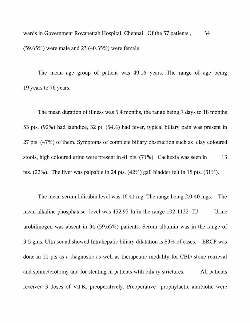
wards in Government Royapettah Hospital, Chennai. Of the 57 patients , 34
(59.65%) were male and 23 (40.35%) were female.
The mean age group of patient was 49.16 years. The range of age being
19 years to 76 years.
The mean duration of illness was 5.4 months, the range being 7 days to 18 months
53 pts. (92%) had jaundice, 32 pt. (54%) had fever, typical biliary pain was present in
27 pts. (47%) of them. Symptoms of complete biliary obstruction such as clay coloured
stools, high coloured urine were present in 41 pts. (71%). Cachexia was seen in 13
pts. (22%). The liver was palpable in 24 pts. (42%) gall bladder felt in 18 pts. (31%).
The mean serum bilirubin level was 16.41 mg. The range being 2.0-40 mgs. The
mean alkaline phosphatase level was 452.95 Iu in the range 102-1132 IU. Urine
urobilinogen was absent in 34 (59.65%) patients. Serum albumin was in the range of
3-5 gms. Ultrasound showed Intrahepatic biliary dilatation is 83% of cases. ERCP was
done in 21 pts as a diagnostic as well as therapeutic modality for CBD stone retrieval
and sphincterotomy and for stenting in patients wtih biliary strictures. All patients
received 3 doses of Vit.K. preoperatively. Preoperative prophylactic antibiotic were
used in all .
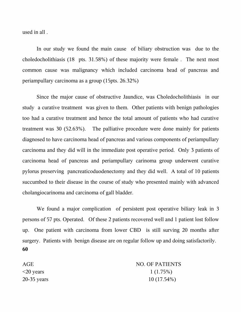
In our study we found the main cause of biliary obstruction was due to the
choledocholithiasis (18 pts. 31.58%) of these majority were female . The next most
common cause was malignancy which included carcinoma head of pancreas and
periampullary carcinoma as a group (15pts. 26.32%)
Since the major cause of obstructive Jaundice, was Choledocholithiasis in our
study a curative treatment was given to them. Other patients with benign pathologies
too had a curative treatment and hence the total amount of patients who had curative
treatment was 30 (52.63%). The palliative procedure were done mainly for patients
diagnosed to have carcinoma head of pancreas and various components of periampullary
carcinoma and they did will in the immediate post operative period. Only 3 patients of
carcinoma head of pancreas and periampullary carinoma group underwent curative
pylorus preserving pancreaticoduodenectomy and they did well. A total of 10 patients
succumbed to their disease in the course of study who presented mainly with advanced
cholangiocarinoma and carcinoma of gall bladder.
We found a major complication of persistent post operative biliary leak in 3
persons of 57 pts. Operated. Of these 2 patients recovered well and 1 patient lost follow
up. One patient with carcinoma from lower CBD is still surving 20 months after
surgery. Patients with benign disease are on regular follow up and doing satisfactorily.
60 AGE NO. OF PATIENTS<20 years 1 (1.75%)20-35 years 10 (17.54%)
36-50 years 18 (31.58%)51-65 years 23 (40.35%)>65 years 5 (8.77%) Total 57AGE DISTRIBUTIONBILIRUBIN LEVELS NO. OF PATIENTS<5 mg% 2 (3.51%)5-10 mg% 19 (33.33%) 11-20 mg% 19 (33.33%)21-30 mg% 7 (12.28%)> 31 mg% 10 (17.54%)BILIRUBIN DISTRIBUTION6162
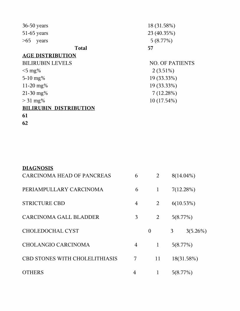
DIAGNOSISCARCINOMA HEAD OF PANCREAS 6 2 8(14.04%)
PERIAMPULLARY CARCINOMA 6 1 7(12.28%)
STRICTURE CBD 4 2 6(10.53%)
CARCINOMA GALL BLADDER 3 2 5(8.77%)
CHOLEDOCHAL CYST 0 3 3(5.26%)
CHOLANGIO CARCINOMA 4 1 5(8.77%)
CBD STONES WITH CHOLELITHIASIS 7 11 18(31.58%)
OTHERS 4 1 5(8.77%)
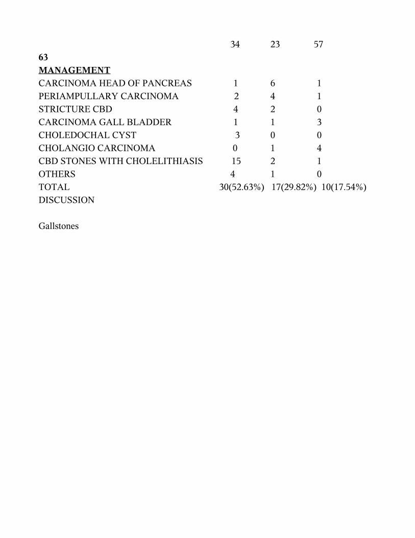
34 23 5763MANAGEMENTCARCINOMA HEAD OF PANCREAS 1 6 1PERIAMPULLARY CARCINOMA 2 4 1STRICTURE CBD 4 2 0CARCINOMA GALL BLADDER 1 1 3CHOLEDOCHAL CYST 3 0 0CHOLANGIO CARCINOMA 0 1 4CBD STONES WITH CHOLELITHIASIS 15 2 1OTHERS 4 1 0TOTAL 30(52.63%) 17(29.82%) 10(17.54%)DISCUSSIONDISCUSSIONGallstones
DISCUSSION
Jaundice is a distressing problem for any human, more so when they are ignorant
of the severe underlying pathology. The presentation is usually very late with many
patients trying self medications. It is attributable also to the paucity of symptoms. Onset
of specific symptoms also implies that the disease is advanced and involvement of vital
structures.
Comparing the studies elsewhere too , the observation in our study also shows an
increase in incidence in males in general and benign conditions being more common in
female. The age of onset of disease is also low including the malignancies. The overall
average age of incidence of surgical jaundice is 49 years and the average age for
malignant diseases is 52 years. The lowest age recorded for a male patient is 19 years
and for a female is 25 years both diagnosed to be with choledocholithiasis .
Comparing with S.Agal et al of Mumbai who studied 62 cases of malignant
etiology and M.Kannan et al of Chennai who studied 455 cases of both benign and
malignant etiology there is more or less equal age incidence .
65
In our study liver was palpable in 42% of patients only while in Benjamin series it was
palpable in 50% of the patients and in the Popper series is 54%.The gallbladder was
palpable in 31% of our patients while in Benjamin series it was palpable in 50% of the
icteric patients and 62.20% of those with pancreatic malignancies.
Ultrasound was done in all our patients . It showed dilatation of intrahepatic
biliary radicles in 83% of patients. All the choledocholithiais patients underwent
cholecystectomy and choledocholithotomy either by ERCP sphincterotomy and stone
retrieval or CBD exploration with T - Tube are doing well.
Of the 15 patients wtih pancreatic carcinoma ,10 patients underwent bypass
procedures and 3 patients underwent Whipples pancreatico duodenectomy followed by
chemotherapy.
Comparing to other studies of Benjamin and Popper , our study showed equal
curative rates in the management of other benign extrahepatic biliary tract obstructive
lesions such as stricture of the Common Bile Duct , Choledochal cyst and patients
presenting with chronic pancreatic bile duct stricture.
We had 2 patients with Hepatocellular Carcinoma who were referred to higher
centres for evaluation and management. Only 1-12 % of HCC patients manifest
obstructive jaundice as the initial complain. Identification of this group of patients is
clinically important, because surgical treatment may be beneficial.
In our study , we did not attempt any modality of preoperative biliary drainage for
any amount of jaundice mainly in patients with malignant cause of obstructive jaundice
since various studies have shown no change in the survival benefits with this
procedure.Hence ERCP and stenting was done only for benign cases with features of
cholangitis and sepsis who recovered very well.
We had 10 deaths in the follow up and those under investigations. These patients
were mainly in their terminal stage of their illness and the underlying pathology was
mainly advanced cholangiocarcinoma and advanced carcinoma of the gallbladder.
Though the death percentage was a little higher compared to other studies done
elsewhere it cannot be considered significant as the study group was small .
67CONCLUSION
CONCLUSION The following conclusions were made :
* Obstructive jaundice was more common in men. Benign
conditions such as choledocholithiasis was common in female
and malignant conditions were common in male.
* Most of them in the late fifth and sixth decades of life
* Choledocholithiaisis was found to be the most common cause of
obstrutive jaundice; tumours (pancreatic carcinoma ,
periampullary carcinoma and cholangiocarcinoma ) are next frequent
causes.
* Biliary obstruction due to metastasis is not uncommon.
* Ultrasound followed by endoscopic retrograde
cholangiopancreatography and Computed Tomography are the
investigation of choice.
* Patients with benign pathology have a favourable outcome and most
of them had improved remarkably and had returned to normal activity.
• Palliative bypass procedures are the final outcome in most malignancies causing
biliary obstruction;their use being not in prolonging life span but in giving
a better quality of life.
•
PROFORMA
NAME : AGE: SEX :
ADDRESS: UNIT: O.P/I.P No.:
Socioeconomic status:
D.O.A: D.O.D.:
PRESENTING COMPLAINTS :
SYMPTOMS :
SIGNS :
GENERAL
Built : Pallor : Icterus :
Pedal oedema : Scratch marks: Hydration:

Signs of liver failure Clubbing BP :
PR:
ABDOMEN
- Jaundice- Pain- Malaise- Loss of wt & appetite- Itching - High coloured urine- Pale stools- Fever- Mass- Others– Palpable liver- Palpable gall bladder- Mass- TendernessGallstones
- Ascites
- Others
OTHER SYSTEMS :
INVESTIGATIONS
Blood :
Complete haemogram - Hb., TC, DC, ESR
Sugar, Urea
Serum electolytes - Na, K, Cl, HCO3
Serum creatinine
Liver function tests - Sr. Bilirubin (total & direct)
Sr. Proteins (A:G. Ratio)
Enzymes (SGOT, SGPT, SAP)
Bleeding & Clotting time, Prothrombin time.
Urine :
Routine - alb, sugar, deposits,
BS, BP, Urobilinogen
Imaging :
X-ray chest P.A. & abdomen A.P.
USG abdomen
CT scan
ERCP
Others
Gallstones
PREOPERATIVE PREPARATION
IV fluids, Vitamin K, Nutrition, Antibiotics
OPERATIVE PROCEDURE
Dt: Surgeon : Anaesthetist
Anaesthesia
Procedure
Findings :
Recovery
Post Operative complications / morbidity
Relief of symptoms :
FOLLOW UP :
Gallstones BIBLIOGRAPHYBIBLIOGRAPHYGallstones
BIBLIOGRAPHY
1. Greim H, Trulzsch D Czygan P et al, Mechanism of cholestasis :6 bile
salts in human livers with or without biliary obstruction.
Gastroenterology, 1972; 63 : 846-850.
2. Hand B.H. Nanatomy and function of extrahepatic biliary system, Clin.
Gastroenterol 1973; 2:3-8.
3. Northover J.M.A. Terblanche J. Applied surgical anatomy of the
Biliary tree IN Blumgart L.H.(ed): The biliary tract (Clinical Surgery
International; V-5). New york, Churchil Livingstone 1982 1-16.
4. Wood Mc D. Eponyms in biliary surgery. Am J Surg. 1979; 138:
746 - 754.
5. Boyden EA, Phyrgian cap in cholecystography - congenital anomaly of
The gall bladder. Am J Roentogenol, 1935; 33:589, 602
6. Braasch JW, Congenital anomalies of the gall bladder and bile ducts.
Surg. Clin. North Am. 1958; 38 : 627-630.
7. Brooks F P. The secretion of bile. Am. J. Dig. Dis. 1969; 14;343-350.

8. Elliot W H, Hydge P , Ametabolic pathways of bile acid synthesis,
Am. J. Surg. 1971, 51:568-571.
9. Almond H R, Viahcevic ZR, Bell CC Jr et al. Bile acid pools, kinetics
And biliary lipid composition before and after cholecystectomy.N.
Engl. J. Med. 1973; 1213-1215.
Gallstones
Ono K, Watanabe N, Suzuki K et a; Bile Flow mechanisms in Man. Arch. Surg. 1968;
96 : 869-871
Toouli J. Watts J.M. Actions of chlecystokinin, pancreozymin, secretin and gastrin on
extrahepatic biliary motility in vitro. Ann Surg. 1973; 175: 439-443.
Price CP, Sammons H G. The nature of serum alkaline phosphatases in liver disease. J
Clin Pathol 1974; 27: 392-398.
Knill - Jones, RP, Cochrane K M, Sokhi G S, Russel R L, Blumbgart LH, Early
Dagnosis of Jaundice. A computer and clinical study Br. J.Surg. 1975; 62 : 654-655.
Benjamin I S, Ryan C J, McLay A L C, Horne C H W, Blumgart L H. The effects of
portacaval shunting and portacaval.
Price C.P. Sammons H G. An interpretatation of serum alkaline phosphatase isoenzyme
patterns in patients with obstructive liver disease. J. Clin. Pathol. 1976; 29: 976-980.
Boey J H, Way LW, Acute cholangitis, Ann. Surg. 1980; 191 : 264-268.
Bismuth H, mart R, Current concepts in cancer : carcinoma of the biliary tract N. Eng. J.
Med. 1979; 301 : 704-709.
Benjamin I R. The obstructed biliary tract in Blumgart L H(ed): The
Biliary tract (clinical surgery internation; (v-5) New York. Churchhill Livingstone 1982;
157-182
Yamaguchi M. Congenital Choledochal cyst. Analysis of 1,433 patients in Japanese
literature. Am. J. Surg. 1980; 140:653-656.
Thaler M M, Cryptogenic liver disease in young infants in popper H, Schafner F (eds).
Progress in liver disease. Vol 5, new York; Grure &
Stratton. 1976; 476-493.
Wisloff F, Jacobson J, Osnes M, Stenosis of the common bile duct in thechronic
pancreatitis. Br. J. Surg. 1982; 68:52-54.
Mason G R, Bacteriolog and antibiotic selection t/in biliar surgery, Arch. Surg. 1968;
97: 533-537.
Popper H, Schaffer F, Cholestasis In Bookus gastroenterology 4th Ed. (Ed in chief) Berk
J.E. Philadelphia, W.B. Saunders Co. 1985; 2697-2731.
Allison M.E.M., Et al Renal function and other factors in obstructive jaundice. Br. J.
Surg. Vol. 66, 392-397, 1979.
Varma H.N. Management of Surgical Jaundice - Association of Surgeons of India,
CME, Calcutta 1980.
Blumgart, L.H. Biliary tract obstruction - New approaches to old problem, Am.J. Surg.
135, 19, 1990.
Evaluation of palliative procedures of pancreatic cancer. Am. J. Surg. 141:430-433,
1996.
Post operative renal failure in obstructive jaundice; effect of manitol diuresis. Brit. Med.
J. 1:82-89, 1995.Gallstones