Embed Size (px)
Citation preview
Journal of Cancer Treatment and Research 2016; 4(2): 16-20
http://www.sciencepublishinggroup.com/j/jctr
doi: 10.11648/j.jctr.20160402.11
ISSN: 2376-7782 (Print); ISSN: 2376-7790 (Online)
Obstructive Jaundice, Study of 33 Cases in Department of Visceral Surgery, Donka National Hospital
Abdoulaye Korse Balde1, Oumar Taibata Balde
1, Ahmed Boubacar Barry
1, Soriba Naby Camara
2,
Hamidou Sylla1, Aissatou Taran Diallo
3, Amadou Dioulde Diallo
1, Fode Lansana Camara
1,
Alpha Madiou Barry1, Sneha Ballah
4
1Department of Visceral Surgery, University Gamal Abdel Nasser of Conakry, Conakry, Guinea 2Department of Visceral Surgery, Friendship Hospital of Kipe, University Gamal Abdel Nasser of Conakry, Conakry Guinea 3Department of General Surgery, University Gamal Abdel Nasser of Conakry Conakry, Guinea 4Department of General Surgery, Sir Seewoosagur Ramgoolam North Hospital, University of Mauritius, Mauritius
Email address: [email protected] (S. Ballah), [email protected] (A. K. Balde), [email protected] (O. T. Balde)
To cite this article: Abdoulaye Korse Balde, Oumar Taibata Balde, Ahmed Boubacar Barry, Soriba Naby Camara, Hamidou Sylla, Aissatou Taran Diallo,
Amadou Dioulde Diallo, Fode Lansana Camara, Alpha Madiou Barry, Sneha Ballah. Obstructive Jaundice, Study of 33 Cases in Department
of Visceral Surgery, Donka National Hospital. Journal of Cancer Treatment and Research. Vol. 4, No. 2, 2016, pp. 16-20.
doi: 10.11648/j.jctr.20160402.11
Received: June 12, 2016; Accepted: June 20, 2016; Published: July 23, 2016
Abstract: Icterus or jaundice is a yellow staining of the skin and mucous membranes which occurs secondary to elevated
bilirubin levels in the blood. Obstructive jaundice corresponds to a mechanical obstacle in the biliary pathways. The definitive
diagnosis of obstructive jaundice in preoperative patients remains a challenge. The aim of this study was to present a number
of epidemiological aspects to highlight the problems faced with diagnosis and treatment in order to ameliorate the management
and prognosis of patients with obstructive jaundice. It was a retrospective study conducted on 17 male patients and 16 female
patients with an average age of 51-54 years, who were surgically treated for obstructive jaundice. The main clinical
presentation was characterized by icterus, pruritus and abdominal pain. Pancreatic head tumors are most commonly responsible
for the presentation of obstructive jaundice (63.64%), followed by jaundice secondary to choledocholithiasis (9.06%), and
stones in vesicular system (6.06%). The surgical intervention of choice was a palliative choledochoduodenostomy to divert
flow of biliary juices.
Keywords: Obstructive Jaundice, Epidemiology, Diagnostic, Treatment, Conakry, Donka
1. Introduction
Obstructive jaundice is the consequence of bile stasis
resulting from obstruction of bile flow by stones, tumors or
inflammation [1]. The most common etiologies of
obstructive jaundice are Cholelithiasis and pancreatic cancer,
identified in 40% of total cases. Jaundice is visible on
examination, and recognized by the yellow coloration of the
skin, cornea and mucous membranes when serum bilirubin
levels rise above than 2.5 to 3 mg per dL (42.8 to 51.3 µmol
per L. Icterus is the most common manifestation of a
mechanical obstruction in the biliary tract. Other
accompanying signs include white stools, dark colored urine,
abdominal pain and skin pruritus. It is a serious medical
complication as bile reflux can be toxic, cause coagulation
defects, renal insufficiency and decrease sensitivity of the
humoral or cellular response of the immune system. [2] This
most often presents acutely with obstruction secondary to
tumor growths). Biochemical liver function tests show
elevations in the direct bilirubin, alkaline phosphatase (ALP),
and γ-glutamyltransferase (GGT) values and aspartate
transferase (AST) and to a lesser degree, alanine transferase
(ALT) values. Common noninvasive imaging studies include
trans abdominal ultrasound scan (US), computed
tomographic (CT) scan, and magnetic resonance
cholangiopancreatography (MRCP). Endoscopic ultrasound
(EUS) and endoscopic retrograde cholangiopancreatography
(ERCP) are important for both diagnostic and therapeutic
17 Abdoulaye Korse Balde et al.: Obstructive Jaundice, Study of 33 Cases in Department of
Visceral Surgery, Donka National Hospital
reasons. [1] Despite the difference in recognition of the
nature of obstructive jaundice in different medical centers,
finding the causative factor responsible the obstruction is an
obligatory preoperative process, as wrongly chosen surgical
procedures may have deleterious outcomes [4]
The etiological diagnosis remainsto be an
enduringproblem in the departmental context, as reflected by
the lack of morphological and biological tests for conducting
clinical investigations. [4] This situation is coupled with the
scarcity of most advanced and recent treatment resources
which are otherwise already in use in developed countries.
This particular context has been the most compelling reason
for us to write this article with the goal was of reporting our
experience while comparing with similar medical literature,
in the surgical service in the department of visceral surgery at
the National hospital of Conakry in Guinea.
2. Methodology
2.1. Patient and Demographic Data
Data were collected retrospectively from 1st January 2008
to 31December 2012. The study included 23 patients total
comprising of 17 male and 16 female subjects with a sex
ratio of 1.06:1. The data collected from the patients included
age, sex, clinical signs and symptoms, length of hospital stay,
echography results; computer tomographic imaging,
preoperative diagnosis, treatment modalities and their results.
Those with no echography results, and with inoperable
disease were excluded from the study.
2.2. Diagnosis Standard
On admission, all patients underwent complete standard
blood test examinations, including serum bilirubin levels.
Patients were considered eligible for the study when they
presented obstructive jaundice confirmed by instrumental
tests and increased bilirubin serum levels (total bilirubin >2.0
mg/dL). Biochemical tests such as Serum levels of CA19-9
and CEA were also measured on admission. Computer
tomography imaging and endoscopic ultrasound were
performed for all patients to further investigate the etiology
of the obstructive jaundice [5]
2.3. Etiology
The main etiologic findings in our study related to the
obstructive jaundice were periampullary (including pancreatic)
malignancy, cholangiocarcinoma, cholelithiasis and others.
2.4. Management
2.4.1. Indication for Surgery
In our study the main indication for surgery was to relieve
the itching, the pruritus and to improve the quality of life of
patient.
2.4.2. Contraindication for Surgery
The main contraindication for the surgery was that of
tumor infiltration into the portal vein, the hepatic vein and
the celiac trunk.
3. Results
Out of a total of 4127 operations, 33 were performed for
treating obstructive jaundice (0.8%). The subjects studied
included 17 male and 16 female patients at a sex ratio of
1.06, with age ranging from 5 to 56 years.
The major clinical signs were icterus in all patients,
change in general physical appearance was noticeable with
anorexia in 30 cases (90%), weight loss in 29 cases (87%),
weakness and fatigue in 24 cases (72%). The gallbladder was
palpable in 10 cases (30%) and anal swelling noted in 1
patient (3.03%).
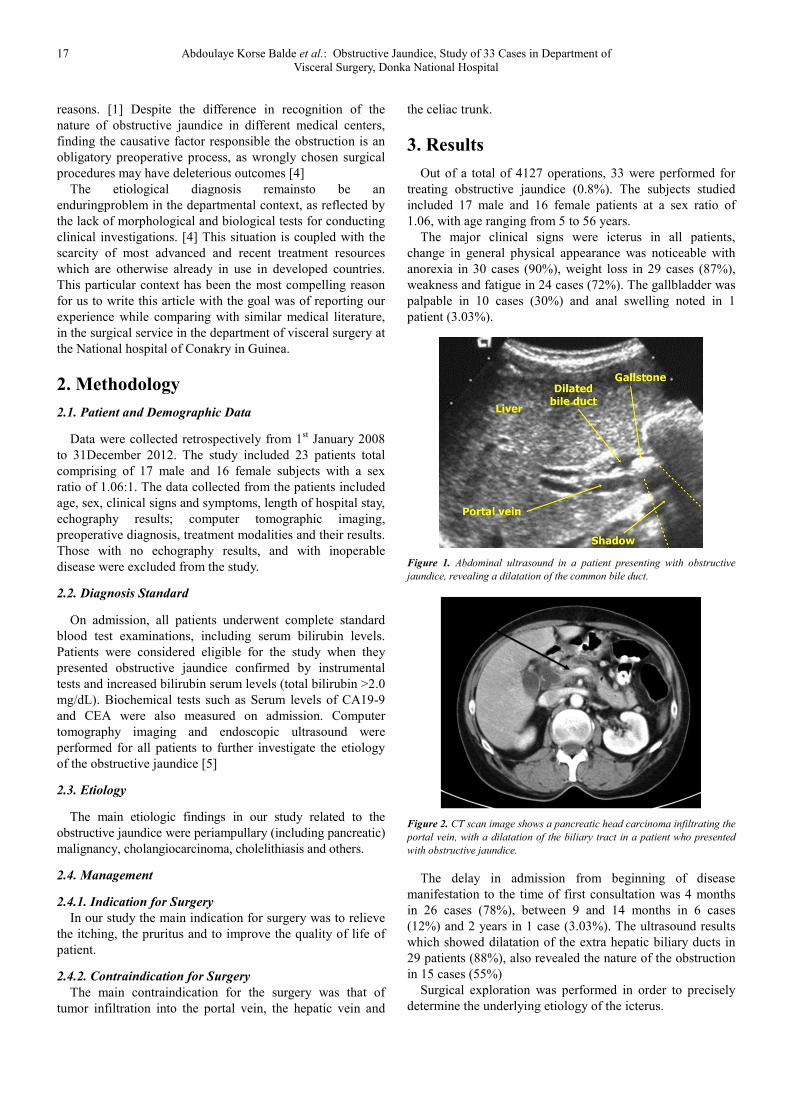
Figure 1. Abdominal ultrasound in a patient presenting with obstructive
jaundice, revealing a dilatation of the common bile duct.
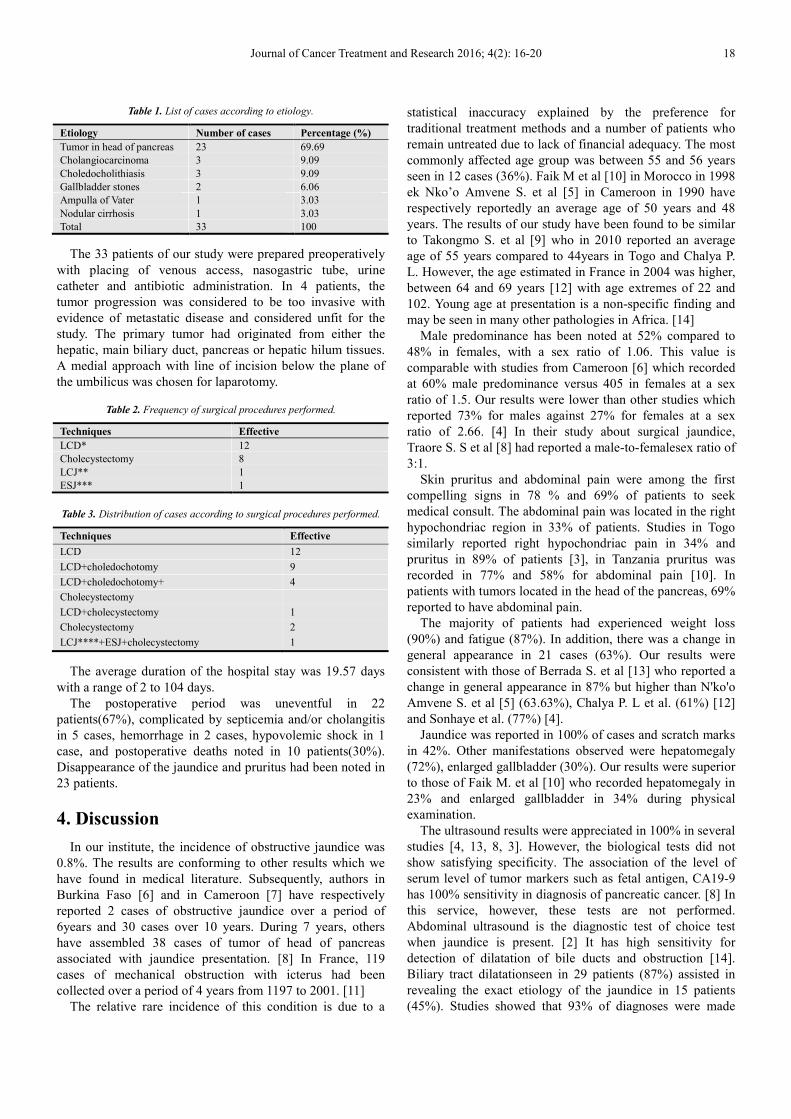
Figure 2. CT scan image shows a pancreatic head carcinoma infiltrating the
portal vein, with a dilatation of the biliary tract in a patient who presented
with obstructive jaundice.
The delay in admission from beginning of disease
manifestation to the time of first consultation was 4 months
in 26 cases (78%), between 9 and 14 months in 6 cases
(12%) and 2 years in 1 case (3.03%). The ultrasound results
which showed dilatation of the extra hepatic biliary ducts in
29 patients (88%), also revealed the nature of the obstruction
in 15 cases (55%)
Surgical exploration was performed in order to precisely
determine the underlying etiology of the icterus.
Journal of Cancer Treatment and Research 2016; 4(2): 16-20 18
Table 1. List of cases according to etiology.
Etiology Number of cases Percentage (%)
Tumor in head of pancreas 23 69.69
Cholangiocarcinoma 3 9.09
Choledocholithiasis 3 9.09
Gallbladder stones 2 6.06
Ampulla of Vater 1 3.03
Nodular cirrhosis 1 3.03
Total 33 100
The 33 patients of our study were prepared preoperatively
with placing of venous access, nasogastric tube, urine
catheter and antibiotic administration. In 4 patients, the
tumor progression was considered to be too invasive with
evidence of metastatic disease and considered unfit for the
study. The primary tumor had originated from either the
hepatic, main biliary duct, pancreas or hepatic hilum tissues.
A medial approach with line of incision below the plane of
the umbilicus was chosen for laparotomy.
Table 2. Frequency of surgical procedures performed.
Techniques Effective
LCD* 12
Cholecystectomy 8
LCJ** 1
ESJ*** 1
Table 3. Distribution of cases according to surgical procedures performed.
Techniques Effective
LCD 12
LCD+choledochotomy 9
LCD+choledochotomy+ 4
Cholecystectomy
LCD+cholecystectomy 1
Cholecystectomy 2
LCJ****+ESJ+cholecystectomy 1
The average duration of the hospital stay was 19.57 days
with a range of 2 to 104 days.
The postoperative period was uneventful in 22
patients(67%), complicated by septicemia and/or cholangitis
in 5 cases, hemorrhage in 2 cases, hypovolemic shock in 1
case, and postoperative deaths noted in 10 patients(30%).
Disappearance of the jaundice and pruritus had been noted in
23 patients.
4. Discussion
In our institute, the incidence of obstructive jaundice was
0.8%. The results are conforming to other results which we
have found in medical literature. Subsequently, authors in
Burkina Faso [6] and in Cameroon [7] have respectively
reported 2 cases of obstructive jaundice over a period of
6years and 30 cases over 10 years. During 7 years, others
have assembled 38 cases of tumor of head of pancreas
associated with jaundice presentation. [8] In France, 119
cases of mechanical obstruction with icterus had been
collected over a period of 4 years from 1197 to 2001. [11]
The relative rare incidence of this condition is due to a
statistical inaccuracy explained by the preference for
traditional treatment methods and a number of patients who
remain untreated due to lack of financial adequacy. The most
commonly affected age group was between 55 and 56 years
seen in 12 cases (36%). Faik M et al [10] in Morocco in 1998
ek Nko’o Amvene S. et al [5] in Cameroon in 1990 have
respectively reportedly an average age of 50 years and 48
years. The results of our study have been found to be similar
to Takongmo S. et al [9] who in 2010 reported an average
age of 55 years compared to 44years in Togo and Chalya P.
L. However, the age estimated in France in 2004 was higher,
between 64 and 69 years [12] with age extremes of 22 and
102. Young age at presentation is a non-specific finding and
may be seen in many other pathologies in Africa. [14]
Male predominance has been noted at 52% compared to
48% in females, with a sex ratio of 1.06. This value is
comparable with studies from Cameroon [6] which recorded
at 60% male predominance versus 405 in females at a sex
ratio of 1.5. Our results were lower than other studies which
reported 73% for males against 27% for females at a sex
ratio of 2.66. [4] In their study about surgical jaundice,
Traore S. S et al [8] had reported a male-to-femalesex ratio of
3:1.
Skin pruritus and abdominal pain were among the first
compelling signs in 78 % and 69% of patients to seek
medical consult. The abdominal pain was located in the right
hypochondriac region in 33% of patients. Studies in Togo
similarly reported right hypochondriac pain in 34% and
pruritus in 89% of patients [3], in Tanzania pruritus was
recorded in 77% and 58% for abdominal pain [10]. In
patients with tumors located in the head of the pancreas, 69%
reported to have abdominal pain.
The majority of patients had experienced weight loss
(90%) and fatigue (87%). In addition, there was a change in
general appearance in 21 cases (63%). Our results were
consistent with those of Berrada S. et al [13] who reported a
change in general appearance in 87% but higher than N'ko'o
Amvene S. et al [5] (63.63%), Chalya P. L et al. (61%) [12]
and Sonhaye et al. (77%) [4].
Jaundice was reported in 100% of cases and scratch marks
in 42%. Other manifestations observed were hepatomegaly
(72%), enlarged gallbladder (30%). Our results were superior
to those of Faik M. et al [10] who recorded hepatomegaly in
23% and enlarged gallbladder in 34% during physical
examination.
The ultrasound results were appreciated in 100% in several
studies [4, 13, 8, 3]. However, the biological tests did not
show satisfying specificity. The association of the level of
serum level of tumor markers such as fetal antigen, CA19-9
has 100% sensitivity in diagnosis of pancreatic cancer. [8] In
this service, however, these tests are not performed.
Abdominal ultrasound is the diagnostic test of choice test
when jaundice is present. [2] It has high sensitivity for
detection of dilatation of bile ducts and obstruction [14].
Biliary tract dilatationseen in 29 patients (87%) assisted in
revealing the exact etiology of the jaundice in 15 patients
(45%). Studies showed that 93% of diagnoses were made
19 Abdoulaye Korse Balde et al.: Obstructive Jaundice, Study of 33 Cases in Department of
Visceral Surgery, Donka National Hospital
with ultrasound at a sensitivity of 100% and specificity of
80%. [3, 7] However, ultrasound is less efficient in
determining the nature of the stenosis if the obstruction is not
caused by a tumor [14]. Not all cases of obstructive jaundice
can be diagnosed based solely on ultrasound results thus
driving surgeons to opt for exploratory laparotomy. [7].
Despite the advent of advanced and more complex imaging
techniques, the hospitals in this developing section of the
world are unable to afford the cost of supplying expensive
equipment [2]. Ultrasound imaging results are variable
depending on the experience and skill of the physician
performing the technique. [15] The definite etiology of the
condition is rarely determined by clinical and imaging tests
alone and therefore, laparotomy is the only alternative to
finding the cause. [7]
The principal etiologies of jaundice discovered during
surgery were tumor in head of pancreas in 23 cases (63%),
cholethiasis 9%. Our results were superior to Takongmo S. et
al [9] who reported 53.3% and 6.6 % for tumors of the
pancreas and cholelithiasis respectively, and Sonhaye et al.
[4] with 46.03% and 19.05% respectively, but inferior to
Chalya P. L. et al. [12] who had 64.7% for tumors in
pancreatic head and 62.5% for cholelithiasis.
Principal palliative management comprised of a diversion
of the biliary duct in 30 cases (90.9%), with laterolateral
choledoduodenostomy in majority of patients (87%). A
choledotomy was performed in 14 cases (42%) for stone
extraction and was followed by a choledochoduodenostomy
in 11 cases (33%).
Takongmo S. et al [9] similarly performed diversion of
biliary tract (90%), but with 56.6% of
choledochoduodenostomies, 26.6% of cholecystectomy et
6.6% of choledochotomy. Alincourt A. et al [13] placed
biliary prosthesis in all of their patients while Vidal V. et al
[11] did a percutaneous drainage.
The choice for performing choledochoduodenostomy is
related to its relative simplicity and rapidity of completion of
the procedure, thus, being appropriate for patients in poor
health. [15]
Curative surgery for tumors of the head of the pancreas
(5%) is rarely considered as first choice. In cases of
malignant obstruction, if an early diagnosis is made then
curative resection may be possible, otherwise only palliative
procedures are performed; in inoperable malignant cases
endoprosthesis may be used to relieve the obstruction. [8,
16]. Choledochoduodenostomy or hepatojejunostomy are
both related to a significantly higher mortality rate [16]. The
use of biliary prosthesis is preferred over surgery to prevent
unnecessary laparotomies [11, 13, 8]. However, the result is
short-lived as regular stent replacement is required [15].
While endoscopic intervention is not yet available in the
local hospitals, laparotomy gives way to ample visualization
of the tumor and assists to decide on its resectability [13, 8].
The procedures of choice in 3 cases (9.09%) of biliary
stones were cholecystectomy, choledocoduodenostomy and
choledocotomy. The management of the biliary stones is
based on the competence of the surgeon and clinical
presentation. The choice for the procedure can be either
ERCP with sphincterotomy and extraction of stones or a
complete surgical approach with cholecystectomy and
cholangiography with laparoscopic exploration of biliary
tracts. [19]. The technique of biliary diversion prevents the
recurrence of obstruction by stones [20].
The signs have regressed in 23 patients (69%) but
persisted in 7 patients (21%), with 10 cases of death reported
(30%). However, 1 post-surgical patient had deteriorating
health condition despite disappearance of jaundice. Berrada
S. et al [15] had reported complete disappearance of jaundice
and pruritus in 98% and mortality rate of 8,7% compared to
Tankongmo S. et al [9] with a lower mortality rate of 6,6%.
The high mortality rates are accounted for by the
presentation of the disease in its late course. [21]. Post-
operative mortality rates are lower in patients had received
prosthesis, which is possible to perform in 70-95% of cases,
either via endoscopic or laparoscopic route or percutaneous
route with radiological assistance, and associated with a
combined mortality rate of 3% only [12].
5. Conclusion
Mechanical obstructive jaundice is a rare pathology in the
service. It has a male predominance and arises mostly around
the age of 50. Ultrasound sonography represents an
important paraclinical diagnostic test to visualize the
obstruction along the biliary tract.
In cases where malignancies are the causes, presence of
icterus signifies an advanced tumor progression stage. Due to
the lack of endoscopic diagnostic and treatment techniques,
definite diagnosis can only be made in perioperative phase.
The management chiefly consists of surgical intervention
through a palliative deviation of biliary tract. Therefore,
improvement of the prognosis and chance of recovery
depend on the necessity of making an early diagnosis and
rapid intervention for jaundice.
Abbreviations
LCD= laterolateral choledochoduodenostomy (side-to-side
choledochoduodenostomy)
LCJ= laterolateral choledochojejunostomy (side-to-side
choldochojejunostomy)
ESJ= end to side jejunojejunostomy (end-to-side
jejunostomy)
LC= laterolateral choledochojejunostomy (side-to-side
choledochojejunostomy)
References
[1] Reem Zeyad Sharaiha MD, MScand Zhiping Li MDObstructive Jaundice Current Surgical Therapy, 427-429 Copyright © 2014
[2] Fattorusso V.; Ritter O.: VADEMECUM CLINIQUE: du diagnostic au traitement Masson; 1988; 12ème édition: 976-978
Journal of Cancer Treatment and Research 2016; 4(2): 16-20 20
[3] Valls C.: L'ictère nu: rôle du radiologue dans la prise en charge diagnostic et thérapeutique Journal de la radiologie; Avril 2006; vol 87; N°4-C2: 460-478
[4] Sonhaye L. et al: L'échographie dans la cholestase extra-hépatique chez l'adulte à Lomé JOURNAL AFRICAIN D'IMAGERIE MEDICAL; 2012; VOL3; N°9 http://jaim-online.net/index.php/jaim/article/view/120/pdf_61 consulté le 1er février 2015
[5] N'ko'oAmvene S.; Juimo A. G.; Malonga E. E: Ictères obstructifs à Yaoundé: exploration radiologique Médecine d'Afrique Noire; 1990; 37(12): 783-786
[6] Daniele Marrelli M. D., Stefano Caruso M. D., Corrado Pedrazzani M. D., Alessandro Neri M. D., Eduardo Fernandes M. D., M. R. C. S., Mario Marini M. D., Enrico Pinto M.D.and Franco Roviello M. D. CA19-9 serum levels in obstructive jaundice: clinical value in benign and malignant conditionsAmerican Journal of Surgery, The, 2009-09-01, Volume 198, Issue 3, Pages 333-339, Copyright © 2009 Elsevier Inc.
[7] Vienne A.; Obertin O.; Chaussade S.; Dousset B.; Prat F.: Ictères néoplasiques: Pour ou contre le drainage biliaire préopératoire ? Cancero dig.; 2010; Vol2 N°3: 214-223
[8] Traoré S. S.; Ouédraogo D.; Ilboudo P. D.; Kafando R.; Dakouré R.; Sanou A. Les ictères chirurgicaux au Centre Hospitalier National Yalgado Ouédraogo (CHNYO) Ouagadougou (BF). Burkina Médical; 1997; vol 1: 19-22
[9] Takongmo S.; Guifo M. L.; PisohTangnyin C.; Talla P.; Monabang C.; Essame-Oyono J. L.; Sosso M. A. Prise en charge des ictères obstructifs à Yaoundé. Analyse d'une série de trente cas. Health sci. Dis; 2010; vol 11; (20) http://www.hsd-fmsb.org/index.php/hsd/article/view/50/pdf_90. consulté le 11 février 2014
[10] Faik M. et coll.: Cancer de la tête du pancréas au stade d'ictère (à propos de 38 cas) Médecine du Maghreb 1998; N°72: 6-8
[11] Vidal V.; Ho C. S.; Petit P.: Complications infectieuses précoces au cours des drainages biliaires percutanés transhépatiques, J. radiol 2004; Edition Française de radiologie; 85: 1707-9
[12] Chalya P. L.; Kanumba E. S.; MabulaMchembe Etiological spectrum and treatment outcome of Obstructive jaundice at a University teaching Hospital in northwestern Tanzania: A diagnostic and therapeutic challenges BMC Research Notes 2011, 4: 147
[13] Alincourt A.; Hamy A.; Thibaud C.; Redon H.; Paineau J.; Lerat F.: Ictères obstructifs néoplasiques: apport des prothèses métalliques percutanées GastroenterolClinBiol 2000; 24: 770-775
[14] Koffi E.; Yenon K.; Ehua S.; Coulibaly A.; Kouassi J. C.; Kanga M.: La lithiase de la voie biliaire principale en milieu ivoirien. Médecine d'Afrique Noire; 1999; 46 (2): 114-118
[15] Berrada S.; D'Klissy M.; Ridai M.; Zerouali N. O Place de la déviation bilio-digestive dans le traitement du cancer de la tête du pancréas. Médecine du Maghreb 1992; N°36: 21-23
[16] Napoléon B.: Apport des nouvelles techniques endoscopiques dans l'exploration des sténoses de la VBP Post'U; 2012: 95-100
[17] Takongmo S.; Nko'Amveme S.; Biwole M.; Essame J. L.; Masso-Misse P.; Malonga E. Une démarche diagnostic des cancers du pancréas exocrine en milieu tropical Médecine d'Afrique Noire; 1994; 41 (1): 56-59
[18] Sauvanet A.: Sténoses biliaires ou duodénales: chirurgie ou endoscopie. Les journées EPU Paris VII- journée de gastroentérologie; janvier 2006: 52-57
[19] Yenon K.; Benchelial Z.; Huten N.: Résultats du traitement laparoscopique de la lithiase du cholédoque: notre expérience à propos d'une série de 62 cas Rev. Int. Sc. Med.; 2006; vol 8 N°1: 18-22
[20] Breda Y.; Heng T. K.; Faucompret S.; Louis C.; Deligny M.: Intérêt de la déviation bilio-digestive dans la pathologie biliaire extrême-orientale: étude rétrospective sur 5ans à l'hôpital CALMETTE DE PHONOM PENH (Royaume du Cambodge) Médecine Tropicale; 2000; 60.4: 114-118
[21] Trigui B. et coll.: Facteurs pronostiques des cancers avancés du pancreas. Analyse multifactorielle et score prédictif de survie Ann Chir 2000; 125: 625-30, Copyright © 2014