Embed Size (px)
Citation preview
PUBLISHED VERSION
Fairhurst, Katherine; Strickland, Andrew David; Bridgewater, Franklin Herbert G.; Maddern, Guy John Painless obstructive jaundice secondary to a common bile duct abscess: A delayed sequela of cholecystectomy, HPB Surgery, 2009; 2009:Article ID 628197. Copyright © 2009 Katherine Fairhurst et al.
This is an open access article distributed under the Creative Commons Attribution License, which permits unrestricted use, distribution, and reproduction in any medium, provided the original work is properly cited.
http://hdl.handle.net/2440/75545
PERMISSIONS
http://www.hindawi.com/journals/hpb/guidelines/
Open Access authors retain the copyrights of their papers, and all open access articles are distributed under the terms of the Creative Commons Attribution license, which permits unrestricted use, distribution and reproduction in any medium, provided that the original work is properly cited.
13th August 2013
Hindawi Publishing CorporationHPB SurgeryVolume 2009, Article ID 628197, 3 pagesdoi:10.1155/2009/628197
Case Report
Painless Obstructive Jaundice Secondary to a Common Bile DuctAbscess: A Delayed Sequela of Cholecystectomy
Katherine Fairhurst, Andrew Strickland, Franklin H. G. Bridgewater, and Guy J. Maddern
The Queen Elizabeth Hospital, University of Adelaide Discipline of Surgery, Woodville, South Australia, SA 5011, Australia
Correspondence should be addressed to Guy J. Maddern, [email protected]
Received 14 September 2009; Revised 20 October 2009; Accepted 16 November 2009
Recommended by Guntars Pupelis
Complications related to cholecystectomy are well described. Most occur in the early postoperative period and are recognisedeither at the time of, or shortly after surgery. Clinical sequelae occurring years following cholecystectomy are rare and infrequentlyreported. In addition, most delayed complications are related to the continuing presence or new formation of gallstones. In thispaper we present a unique case of an abscess of the common bile duct wall, presenting with painless obstructive jaundice morethan 30 years following an open cholecystectomy, without the presence of gallstones. The clinical presentation, investigations, andtreatment are discussed with a review of other relevant reported cases in the literature.
Copyright © 2009 Katherine Fairhurst et al. This is an open access article distributed under the Creative Commons AttributionLicense, which permits unrestricted use, distribution, and reproduction in any medium, provided the original work is properlycited.
1. Introduction
Commonly reported complications of cholecystectomyinclude damage to the common bile duct [1], bile leaksfrom the gallbladder bed or cystic duct stump [2], droppedgallstones [3], and damage to other structures such as thesmall bowel, liver, or diaphragm [4]. The complications oflaparoscopic surgery have lessened with growing technicalexpertise, and laparoscopic cholecystectomy is now consid-ered the gold standard [5]. However, the essential steps ofa cholecystectomy remain the same. These steps includeadequate visualisation of the structures to obtain “the criticalview,” secure occlusion of the cystic duct and artery, goodhaemostasis, and the treatment of any bile leaks from thegallbladder bed, or bile or stone spillages from an iatrogeni-cally perforated gallbladder. Inadequate performance of anyof these steps can lead to complications many years followingthe initial operation, as presented in this paper, and as suchattention to operative detail is of paramount importance.
2. Case Report
A 70-year-old female presented to a rural hospital withpainless jaundice. She had undergone a routine open chole-cystectomy 30 years earlier at the same hospital. Biochemical
markers showed an obstructive pattern: Bilirubin 155 μmol/L(6–24), ALP 262 U/L (30–110), ALT 272 U/L (0–55), AST161 U/L (0–45). The white cell count was within the normalrange at 6.65 × 10⁹/L (4–11), and the CRP was mildlyelevated at 11 mg/L (<8.0). In the 30-year interval, she hadexperienced no other episodes of jaundice, abdominal pain,or unexplained fever and had undergone no other abdominalsurgery. Unfortunately the hospital records (on microfilm)were incomplete so no operation note from the cholecystec-tomy 30 years previously was available. The anaesthetic chartand drug chart were retrieved and revealed that no antibi-otics were given and no specific intraoperative difficulty orpostoperative complications were recorded in the clinicalrecords. The patient also confirmed an uneventful recovery.
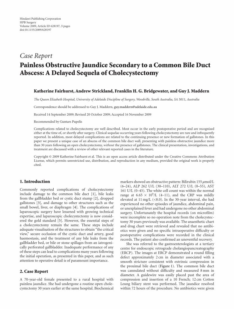
She was referred to the gastroenterologists at a tertiarycentre for endoscopic retrograde cholangiopancreatography(ERCP). The images at ERCP demonstrated a round fillingdefect approximately 2 cm in diameter associated with asmooth stricture consistent with extrinsic compression inthe proximal bile duct (Figure 1). The common bile ductwas cannulated without difficulty and measured 8 mm indiameter. A guidewire was easily placed past the area ofcompression and insertion of a 10 French; 12 cm CottonLeung biliary stent was performed. The jaundice resolvedwithin 72 hours of the procedure. No antibiotics were given
2 HPB Surgery
12′′LAO 0◦CRAN 0◦MAG 1.67SID 106 cmZoom 100%87 kV 10.1 mAs 40.5 ms
Figure 1: Stricture of the proximal bile duct at ERCP.
at the time of the ERCP. No malignant cells were identifiedfrom the aspirated bile sent for histology. The patient wasdischarged the day following the ERCP and referral to thehepatopancreaticobiliary surgical team was made for furtherinvestigation into the cause of the jaundice.
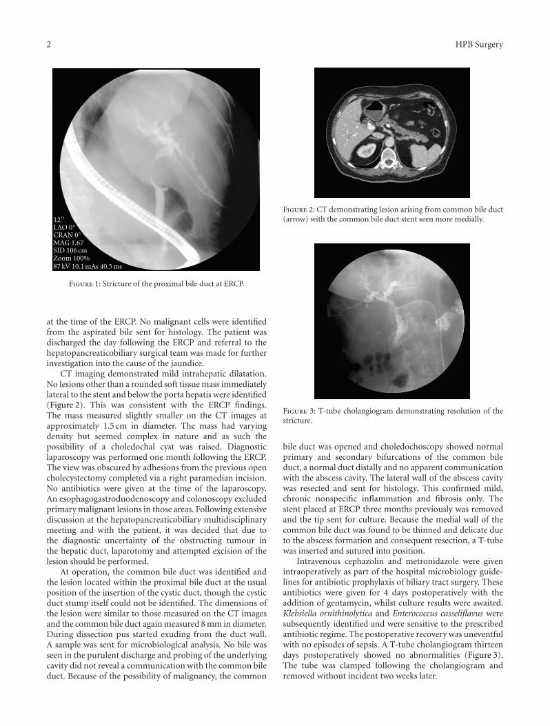
CT imaging demonstrated mild intrahepatic dilatation.No lesions other than a rounded soft tissue mass immediatelylateral to the stent and below the porta hepatis were identified(Figure 2). This was consistent with the ERCP findings.The mass measured slightly smaller on the CT images atapproximately 1.5 cm in diameter. The mass had varyingdensity but seemed complex in nature and as such thepossibility of a choledochal cyst was raised. Diagnosticlaparoscopy was performed one month following the ERCP.The view was obscured by adhesions from the previous opencholecystectomy completed via a right paramedian incision.No antibiotics were given at the time of the laparoscopy.An esophagogastroduodenoscopy and colonoscopy excludedprimary malignant lesions in those areas. Following extensivediscussion at the hepatopancreaticobiliary multidisciplinarymeeting and with the patient, it was decided that due tothe diagnostic uncertainty of the obstructing tumour inthe hepatic duct, laparotomy and attempted excision of thelesion should be performed.
At operation, the common bile duct was identified andthe lesion located within the proximal bile duct at the usualposition of the insertion of the cystic duct, though the cysticduct stump itself could not be identified. The dimensions ofthe lesion were similar to those measured on the CT imagesand the common bile duct again measured 8 mm in diameter.During dissection pus started exuding from the duct wall.A sample was sent for microbiological analysis. No bile wasseen in the purulent discharge and probing of the underlyingcavity did not reveal a communication with the common bileduct. Because of the possibility of malignancy, the common
Figure 2: CT demonstrating lesion arising from common bile duct(arrow) with the common bile duct stent seen more medially.
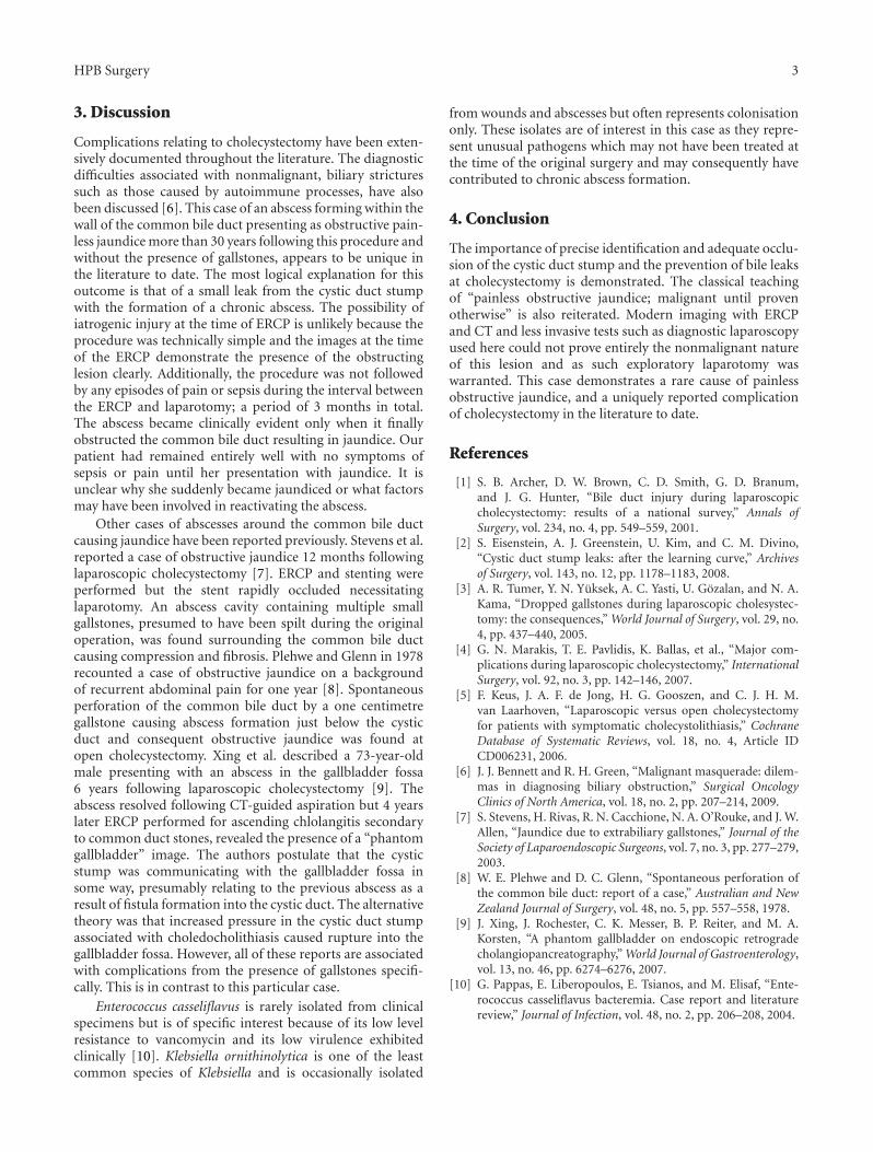
Figure 3: T-tube cholangiogram demonstrating resolution of thestricture.
bile duct was opened and choledochoscopy showed normalprimary and secondary bifurcations of the common bileduct, a normal duct distally and no apparent communicationwith the abscess cavity. The lateral wall of the abscess cavitywas resected and sent for histology. This confirmed mild,chronic nonspecific inflammation and fibrosis only. Thestent placed at ERCP three months previously was removedand the tip sent for culture. Because the medial wall of thecommon bile duct was found to be thinned and delicate dueto the abscess formation and consequent resection, a T-tubewas inserted and sutured into position.
Intravenous cephazolin and metronidazole were givenintraoperatively as part of the hospital microbiology guide-lines for antibiotic prophylaxis of biliary tract surgery. Theseantibiotics were given for 4 days postoperatively with theaddition of gentamycin, whilst culture results were awaited.Klebsiella ornithinolytica and Enterococcus casseliflavus weresubsequently identified and were sensitive to the prescribedantibiotic regime. The postoperative recovery was uneventfulwith no episodes of sepsis. A T-tube cholangiogram thirteendays postoperatively showed no abnormalities (Figure 3).The tube was clamped following the cholangiogram andremoved without incident two weeks later.
HPB Surgery 3
3. Discussion
Complications relating to cholecystectomy have been exten-sively documented throughout the literature. The diagnosticdifficulties associated with nonmalignant, biliary stricturessuch as those caused by autoimmune processes, have alsobeen discussed [6]. This case of an abscess forming within thewall of the common bile duct presenting as obstructive pain-less jaundice more than 30 years following this procedure andwithout the presence of gallstones, appears to be unique inthe literature to date. The most logical explanation for thisoutcome is that of a small leak from the cystic duct stumpwith the formation of a chronic abscess. The possibility ofiatrogenic injury at the time of ERCP is unlikely because theprocedure was technically simple and the images at the timeof the ERCP demonstrate the presence of the obstructinglesion clearly. Additionally, the procedure was not followedby any episodes of pain or sepsis during the interval betweenthe ERCP and laparotomy; a period of 3 months in total.The abscess became clinically evident only when it finallyobstructed the common bile duct resulting in jaundice. Ourpatient had remained entirely well with no symptoms ofsepsis or pain until her presentation with jaundice. It isunclear why she suddenly became jaundiced or what factorsmay have been involved in reactivating the abscess.
Other cases of abscesses around the common bile ductcausing jaundice have been reported previously. Stevens et al.reported a case of obstructive jaundice 12 months followinglaparoscopic cholecystectomy [7]. ERCP and stenting wereperformed but the stent rapidly occluded necessitatinglaparotomy. An abscess cavity containing multiple smallgallstones, presumed to have been spilt during the originaloperation, was found surrounding the common bile ductcausing compression and fibrosis. Plehwe and Glenn in 1978recounted a case of obstructive jaundice on a backgroundof recurrent abdominal pain for one year [8]. Spontaneousperforation of the common bile duct by a one centimetregallstone causing abscess formation just below the cysticduct and consequent obstructive jaundice was found atopen cholecystectomy. Xing et al. described a 73-year-oldmale presenting with an abscess in the gallbladder fossa6 years following laparoscopic cholecystectomy [9]. Theabscess resolved following CT-guided aspiration but 4 yearslater ERCP performed for ascending chlolangitis secondaryto common duct stones, revealed the presence of a “phantomgallbladder” image. The authors postulate that the cysticstump was communicating with the gallbladder fossa insome way, presumably relating to the previous abscess as aresult of fistula formation into the cystic duct. The alternativetheory was that increased pressure in the cystic duct stumpassociated with choledocholithiasis caused rupture into thegallbladder fossa. However, all of these reports are associatedwith complications from the presence of gallstones specifi-cally. This is in contrast to this particular case.
Enterococcus casseliflavus is rarely isolated from clinicalspecimens but is of specific interest because of its low levelresistance to vancomycin and its low virulence exhibitedclinically [10]. Klebsiella ornithinolytica is one of the leastcommon species of Klebsiella and is occasionally isolated
from wounds and abscesses but often represents colonisationonly. These isolates are of interest in this case as they repre-sent unusual pathogens which may not have been treated atthe time of the original surgery and may consequently havecontributed to chronic abscess formation.
4. Conclusion
The importance of precise identification and adequate occlu-sion of the cystic duct stump and the prevention of bile leaksat cholecystectomy is demonstrated. The classical teachingof “painless obstructive jaundice; malignant until provenotherwise” is also reiterated. Modern imaging with ERCPand CT and less invasive tests such as diagnostic laparoscopyused here could not prove entirely the nonmalignant natureof this lesion and as such exploratory laparotomy waswarranted. This case demonstrates a rare cause of painlessobstructive jaundice, and a uniquely reported complicationof cholecystectomy in the literature to date.
References
[1] S. B. Archer, D. W. Brown, C. D. Smith, G. D. Branum,and J. G. Hunter, “Bile duct injury during laparoscopiccholecystectomy: results of a national survey,” Annals ofSurgery, vol. 234, no. 4, pp. 549–559, 2001.
[2] S. Eisenstein, A. J. Greenstein, U. Kim, and C. M. Divino,“Cystic duct stump leaks: after the learning curve,” Archivesof Surgery, vol. 143, no. 12, pp. 1178–1183, 2008.
[3] A. R. Tumer, Y. N. Yuksek, A. C. Yasti, U. Gozalan, and N. A.Kama, “Dropped gallstones during laparoscopic cholesystec-tomy: the consequences,” World Journal of Surgery, vol. 29, no.4, pp. 437–440, 2005.
[4] G. N. Marakis, T. E. Pavlidis, K. Ballas, et al., “Major com-plications during laparoscopic cholecystectomy,” InternationalSurgery, vol. 92, no. 3, pp. 142–146, 2007.
[5] F. Keus, J. A. F. de Jong, H. G. Gooszen, and C. J. H. M.van Laarhoven, “Laparoscopic versus open cholecystectomyfor patients with symptomatic cholecystolithiasis,” CochraneDatabase of Systematic Reviews, vol. 18, no. 4, Article IDCD006231, 2006.
[6] J. J. Bennett and R. H. Green, “Malignant masquerade: dilem-mas in diagnosing biliary obstruction,” Surgical OncologyClinics of North America, vol. 18, no. 2, pp. 207–214, 2009.
[7] S. Stevens, H. Rivas, R. N. Cacchione, N. A. O’Rouke, and J. W.Allen, “Jaundice due to extrabiliary gallstones,” Journal of theSociety of Laparoendoscopic Surgeons, vol. 7, no. 3, pp. 277–279,2003.
[8] W. E. Plehwe and D. C. Glenn, “Spontaneous perforation ofthe common bile duct: report of a case,” Australian and NewZealand Journal of Surgery, vol. 48, no. 5, pp. 557–558, 1978.
[9] J. Xing, J. Rochester, C. K. Messer, B. P. Reiter, and M. A.Korsten, “A phantom gallbladder on endoscopic retrogradecholangiopancreatography,” World Journal of Gastroenterology,vol. 13, no. 46, pp. 6274–6276, 2007.
[10] G. Pappas, E. Liberopoulos, E. Tsianos, and M. Elisaf, “Ente-rococcus casseliflavus bacteremia. Case report and literaturereview,” Journal of Infection, vol. 48, no. 2, pp. 206–208, 2004.

Submit your manuscripts athttp://www.hindawi.com
Hindawi Publishing Corporationhttp://www.hindawi.com Volume 2013
ObesityJournal of
Hindawi Publishing Corporation http://www.hindawi.com Volume 2013Hindawi Publishing Corporation http://www.hindawi.com Volume 2013
The Scientific World Journal
Hindawi Publishing Corporationhttp://www.hindawi.com Volume 2013
MediatorsinflaMMation
of
ISRN Anesthesiology
Hindawi Publishing Corporationhttp://www.hindawi.com Volume 2013
Evidence-Based Complementary and Alternative Medicine
Volume 2013Hindawi Publishing Corporationhttp://www.hindawi.com
OphthalmologyJournal of
Hindawi Publishing Corporationhttp://www.hindawi.com Volume 2013
Hindawi Publishing Corporationhttp://www.hindawi.com Volume 2013
Computational and Mathematical Methods in Medicine
ISRN Allergy
Hindawi Publishing Corporationhttp://www.hindawi.com Volume 2013
BioMed Research International
Hindawi Publishing Corporationhttp://www.hindawi.com Volume 2013
International Journal of
EndocrinologyHindawi Publishing Corporationhttp://www.hindawi.com
Volume 2013
ISRN Addiction
Hindawi Publishing Corporationhttp://www.hindawi.com Volume 2013
Hindawi Publishing Corporationhttp://www.hindawi.com
OncologyJournal of
Volume 2013
ISRN AIDS
Hindawi Publishing Corporationhttp://www.hindawi.com Volume 2013
Hindawi Publishing Corporationhttp://www.hindawi.com Volume 2013
Oxidative Medicine and Cellular Longevity
Diabetes ResearchJournal of
Hindawi Publishing Corporationhttp://www.hindawi.com Volume 2013
Clinical &DevelopmentalImmunology
Hindawi Publishing Corporationhttp://www.hindawi.com
Volume 2013
Hindawi Publishing Corporationhttp://www.hindawi.com Volume 2013
Gastroenterology Research and Practice
Hindawi Publishing Corporationhttp://www.hindawi.com Volume 2013
ISRN Biomarkers
PPARRe sea rch
Hindawi Publishing Corporationhttp://www.hindawi.com Volume 2013