Embed Size (px)
Citation preview
S-1
Supporting Information
Phase-Controlled Electrochemical Activity of Epitaxial Mg-Spinel Thin Films Zhenxing Feng
1,2, *, Xiao Chen
3, Liang Qiao
6, Albert L. Lipson
1,2, Timothy T. Fister1,2, Li Zeng
3,
Chunjoong Kim2,7
, Tanghong Yi2,7
, Niya Sa1,2, Danielle L. Proffit
1,2, Anthony K. Burrell1,2, Jordi
Cabana2,5
, Brian J. Ingram1,2, Michael D. Biegalski
4, Michael J. Bedzyk
3,4 5, Paul Fenter1,2,*
1Chemical Science and Engineering Division, 2Joint Center for Energy Storage Research
(JCESR), Argonne National Laboratory, Lemont, Illinois, 60439, United States
3Applied Physics Program, 4Department of Materials Science and Engineering, 5Department of
Physics and Astronomy, Northwestern University, Evanston, IL 60208, USA
6Center for Nanophase Materials Science, Oak Ridge National Laboratory, Oak Ridge,
Tennessee, 37831, United States
7Department of Chemistry, University of Illinois at Chicago, Chicago, IL, 60607, United States
*Corresponding author email addresses: [email protected] (Z. Feng), [email protected] (P. Fenter)
Index Page
Supplementary Methods S-2 – S-3
Table S1 S-4
Figures S1 – S8 S-5 – S-10
S-2
Supplementary Methods
Pulsed Laser Deposition (PLD) Target Synthesis and Growth.
The MgMn2O4 (MMO) and La0.7Sr0.3FeO3 (LSFO) targets with 2 inch diameter were
synthesized using solid-state methods from stoichiometric mixtures of MgO and Mn2O3 (Alfa
Aesar, USA) powders for MMO, as well as La2O3, SrCO3, and Fe2O3 (Alfa Aesar, USA)
powders for LSFO, respectively. Both targets were calcinated at 1100 °C in air for 72 hours. The
TiC was a commercial target (PVD products, Inc). The MMO target was mixed phase, with the
majority as tetragonal phase, as confirmed by X-ray diffraction (XRD) in Figure S1.
Epitaxial TiC thin films on MgO(001) were prepared at 700 °C under vacuum condition
for 10000 pulses (~50 nm). The LSFO thin films on MgO(001) were prepared at 750 °C under
200 mTorr O2 for 7500 pulses (~50 nm). The MMO thin films on both TiC and LSFO films were
prepared at 500 °C under 10 mTorr O2 for 10000 pulses (~70 nm, estimated from the MMO
growth rate). PLD was done under the following conditions: KrF excimer laser (λ = 248 nm), 10
Hz pulse rate, ~1.5J/cm2 energy density. Reflection high-energy electron diffraction (RHEED)
was utilized for diagnostic in situ monitoring of the film growth.
The successful growth of different pure phases of MMO on either LSFO or TiC can be
seen from the specular θ-2θ scan in Figure S3.
X-ray Characterization.
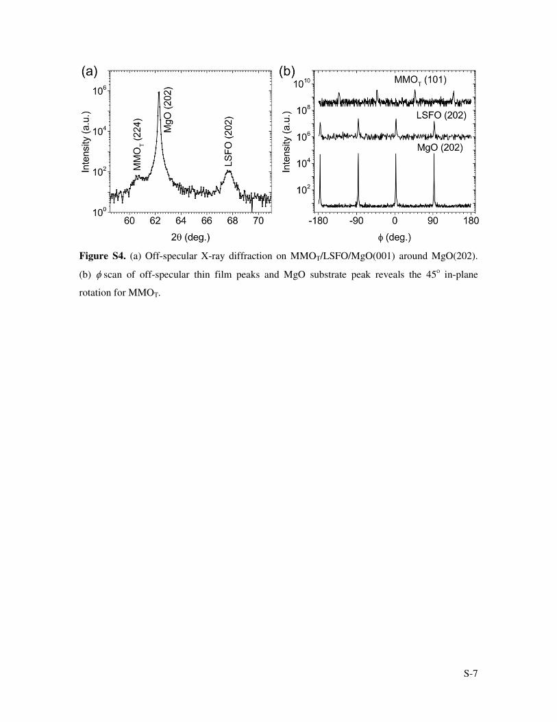
Based on the specular XRD (Figure 1) and off-specular XRD (Figure S4) measurements
on tetragonal phase MMO (MMOT), the following epitaxial relationship of plane directions has
been obtained: MMOT[001]//LSFO[001]//MgO[001] and MMOT[100]//LSFO[110]//MgO[110].
The MMOT film has a 45° in-plane rotation with respect to LSFO film and MgO substrate. In
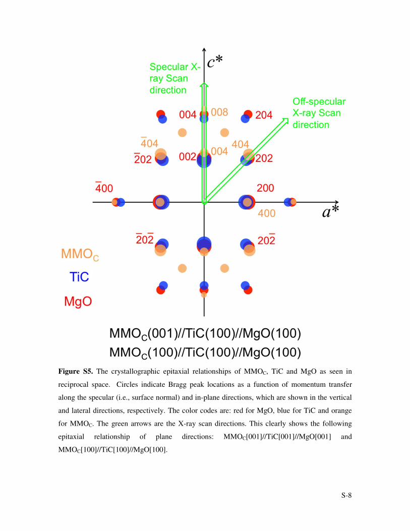
contrast, the cubic phase MMO (MMOC) has different epitaxial alignment with respect to the
substrates. As shown in Figure S5, the green arrows are the X-ray scan directions. The observed
peaks for corresponding thin films and MgO substrate in Figure 1c and 1d clearly indicate the
reciprocal diffraction patterns in Figure S5, and thus the following epitaxial relationship:
MMOC[001]//TiC[001]//MgO[001] and MMOC[100]//TiC[100]//MgO[100].
X-ray Photoelectron Spectroscopy Analysis.
The XPS spectra for Mn 2p3/2 can be decomposed and fitted quantitatively for MMOC at
charged (Figure 4) and uncharged (Figure S9) conditions according to multiplet theory.[2] The

S-3
data are fitted to three chemical states, namely Mn(II), Mn(III) and Mn(IV). The spectrum from
the standard MnO2 powder sample is best fit with Mn(IV) only.
S-4
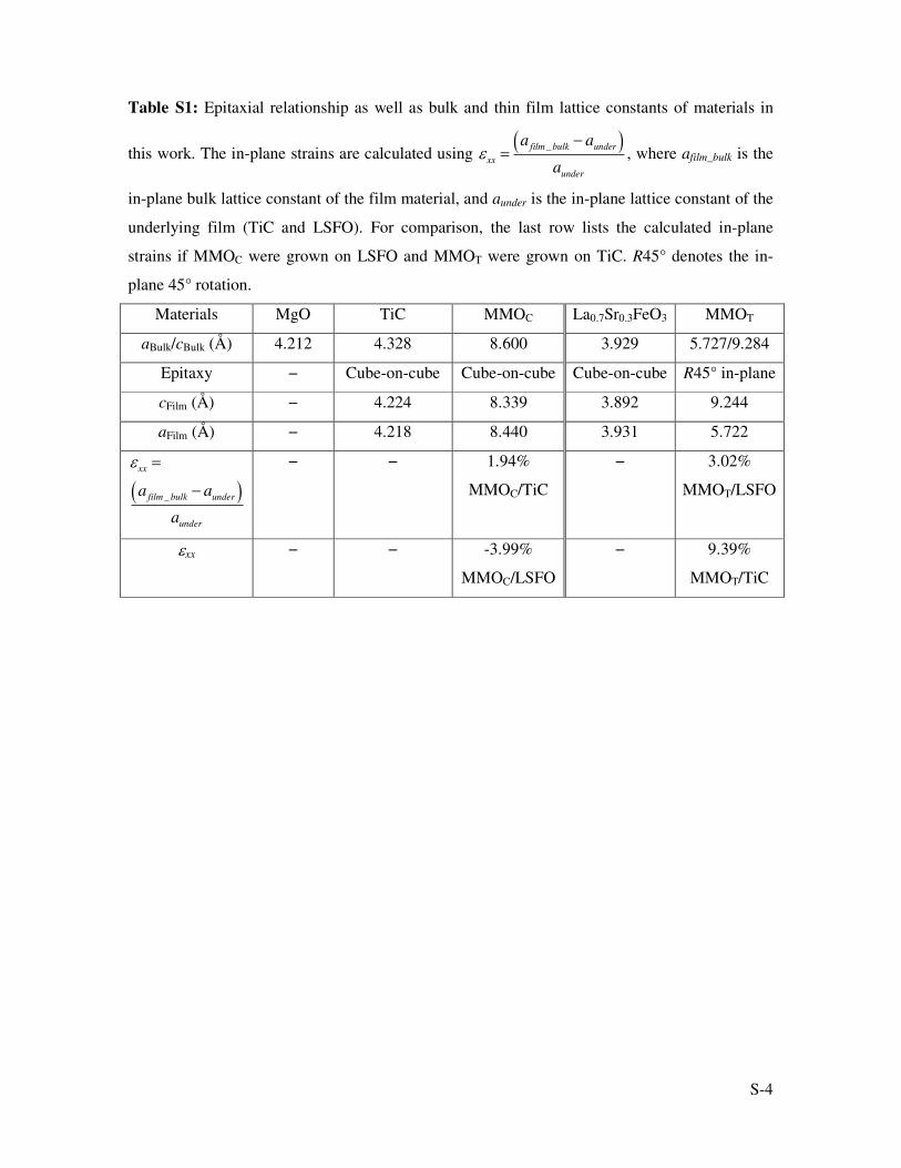
Table S1: Epitaxial relationship as well as bulk and thin film lattice constants of materials in
this work. The in-plane strains are calculated using , where afilm_bulk is the
in-plane bulk lattice constant of the film material, and aunder is the in-plane lattice constant of the
underlying film (TiC and LSFO). For comparison, the last row lists the calculated in-plane
strains if MMOC were grown on LSFO and MMOT were grown on TiC. R45° denotes the in-
plane 45° rotation.
Materials MgO TiC MMOC La0.7Sr0.3FeO3 MMOT
aBulk/cBulk (Å) 4.212 4.328 8.600 3.929 5.727/9.284
Epitaxy − Cube-on-cube Cube-on-cube Cube-on-cube R45° in-plane
cFilm (Å) − 4.224 8.339 3.892 9.244
aFilm (Å) − 4.218 8.440 3.931 5.722
− −
1.94%
MMOC/TiC
− 3.02%
MMOT/LSFO
εxx − − -3.99%
MMOC/LSFO
− 9.39%
MMOT/TiC
εxx =a film _ bulk − aunder( )
aunder
εxx =
a film _ bulk − aunder( )aunder
S-5
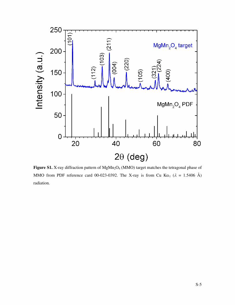
Figure S1. X-ray diffraction pattern of MgMn2O4 (MMO) target matches the tetragonal phase of
MMO from PDF reference card 00-023-0392. The X-ray is from Cu Kα1 (λ = 1.5406 Å)
radiation.
S-6
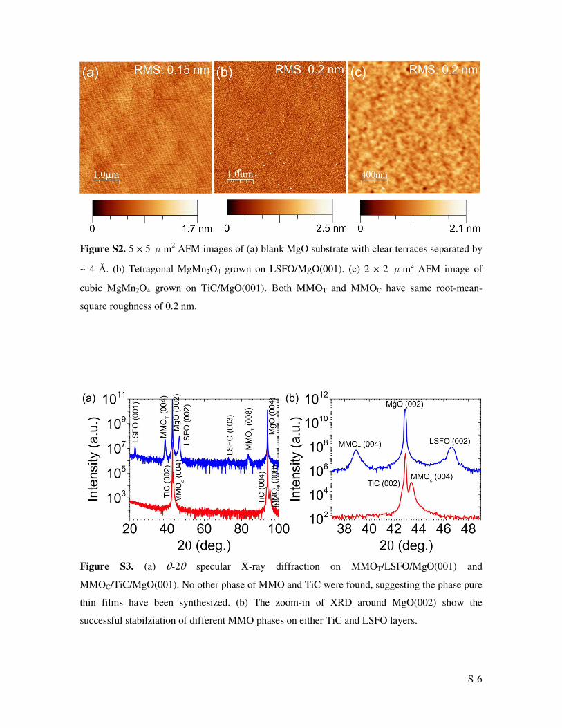
Figure S2. 5 × 5 μm2 AFM images of (a) blank MgO substrate with clear terraces separated by
~ 4 Å. (b) Tetragonal MgMn2O4 grown on LSFO/MgO(001). (c) 2 × 2 μm2 AFM image of
cubic MgMn2O4 grown on TiC/MgO(001). Both MMOT and MMOC have same root-mean-
square roughness of 0.2 nm.
Figure S3. (a) θ-2θ specular X-ray diffraction on MMOT/LSFO/MgO(001) and
MMOC/TiC/MgO(001). No other phase of MMO and TiC were found, suggesting the phase pure
thin films have been synthesized. (b) The zoom-in of XRD around MgO(002) show the
successful stabilziation of different MMO phases on either TiC and LSFO layers.
S-7
Figure S4. (a) Off-specular X-ray diffraction on MMOT/LSFO/MgO(001) around MgO(202).
(b) φ scan of off-specular thin film peaks and MgO substrate peak reveals the 45o in-plane
rotation for MMOT.
S-8
Figure S5. The crystallographic epitaxial relationships of MMOC, TiC and MgO as seen in
reciprocal space. Circles indicate Bragg peak locations as a function of momentum transfer
along the specular (i.e., surface normal) and in-plane directions, which are shown in the vertical
and lateral directions, respectively. The color codes are: red for MgO, blue for TiC and orange
for MMOC. The green arrows are the X-ray scan directions. This clearly shows the following
epitaxial relationship of plane directions: MMOC[001]//TiC[001]//MgO[001] and
MMOC[100]//TiC[100]//MgO[100].
S-9
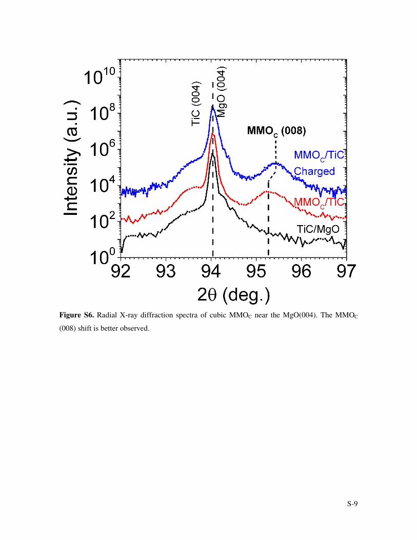
Figure S6. Radial X-ray diffraction spectra of cubic MMOC near the MgO(004). The MMOC
(008) shift is better observed.
S-10
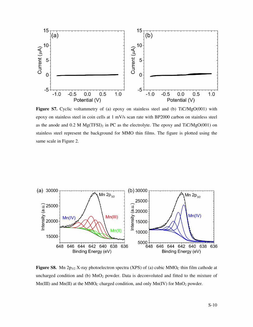
Figure S7. Cyclic voltammetry of (a) epoxy on stainless steel and (b) TiC/MgO(001) with
epoxy on stainless steel in coin cells at 1 mV/s scan rate with BP2000 carbon on stainless steel
as the anode and 0.2 M Mg(TFSI)2 in PC as the electrolyte. The epoxy and TiC/MgO(001) on
stainless steel represent the background for MMO thin films. The figure is plotted using the
same scale in Figure 2.
Figure S8. Mn 2p3/2 X-ray photoelectron spectra (XPS) of (a) cubic MMOC thin film cathode at
uncharged condition and (b) MnO2 powder. Data is deconvoluted and fitted to the mixture of
Mn(III) and Mn(II) at the MMOC charged condition, and only Mn(IV) for MnO2 powder.


![EPOXY RESINS - Krishna districtkrishna.nic.in/PDFfiles/MSME/Chemical/EPOXY RESINS[1].pdf · EPOXY RESINS CONTENTS SECTION I ... PROJECT COST AND PROFITABILITY PROJECTIONS ... Epoxy](https://img.pdfslide.us/doc/110x75/5aa5b17b7f8b9ab4788d7c0f/epoxy-resins-krishna-resins1pdfepoxy-resins-contents-section-i-project.jpg)


![Dana Cable Trays/Troughs [Hot Dip Galvanized/Stainless Steel/Aluminum/Epoxy Painted/Offshore/Marine] - UAE/INDIA/QATAR/LIBYA/AFRICA/SAUDI ARABIA](https://img.pdfslide.us/doc/110x75/558e7a3d1a28ab5d2c8b45fe/dana-cable-traystroughs-hot-dip-galvanizedstainless-steelaluminumepoxy-paintedoffshoremarine-uaeindiaqatarlibyaafricasaudi-arabia.jpg)