Embed Size (px)
Citation preview
12/15/12 Ev ernote Web
1/9https://www.ev ernote.com/edit/83b401d4-f 884-42f 9-97f 0-a8f 6bccc25db#st=p&n=83b401d4-f 884-42f …
Cajal, Ramon Santiago y
Saturday, December 15 2012, 10:57 AM
Santiago Ramón y Cajal (1852-1934)
Citation:DeFelipe, J (2002) History of Neuroscience: Santiago Ramón y Cajal (1852-1934)), IBRO Historyof Neuroscience [http://www.ibro.info/Pub/Pub_Main_Display.asp?LC_Docs_ID=3456]Accessed: date
Javier DeFelipe
Santiago Ramón y Cajal (Figure 1) was born on 1 May 1852 in 'Petilla de Aragón' (Figure 2), a smallvillage in the North of Spain, and he died on 17 October 1934 in Madrid, having become one of themost outstanding neuroscientists of all time.
Figure 1: Cajal and his histological preparations. From photographs in the Cajal Museum.
After studying medicine at the Faculty of Medicine in Zaragoza, Cajal was appointed to the chairof Descriptive and General Anatomy at the University of Valencia in 1883. In 1887 he moved to theUniversity of Barcelona where he was elected to the chair of Histology and Pathological Anatomy.Finally, Cajal moved to the University of Madrid where he occupied the chair of Histology andPathological Anatomy until his retirement. Cajal received numerous prizes, honorary degrees anddistinctions, but undoubtedly the most important was the Nobel Prize for Physiology or Medicine hereceived in 1906. To describe the work of Cajal is a rather difficult task, because, unlike othergreat scientists, he is not known for one single discovery, but rather for his many and importantcontributions to our knowledge of the organization of the nervous system. Those readersinterested in his life would be wise to consult his autobiography (Cajal, 1917), where there is alsoa brief description of his main discoveries and theoretical ideas.
12/15/12 Ev ernote Web
2/9https://www.ev ernote.com/edit/83b401d4-f 884-42f 9-97f 0-a8f 6bccc25db#st=p&n=83b401d4-f 884-42f …
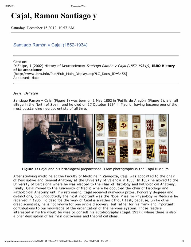
Figure 2: The village of Petilla de Aragón (Navarra, Spain) and the house where Cajal was born.Photographs taken in 2001, kindly supplied by Dr Pedro Uhalte.
The detailed study of the nervous system began in the middle of the nineteenth century. BeforeCajal's discoveries, very little was known about the individual elements of the nervous system, andthe connections between its different parts were purely speculative. The origin of nerve fibers wasa mystery, and it was speculated that they arose from the gray matter independently of the nervecells (neurons). This lack of knowledge was mainly due to the fact that appropriate methods forvisualizing neurons were not available. The early methods of staining only permitted thevisualization of neuronal cell bodies, a small portion of their proximal processes, and some isolatedand rather poorly stained fibers. However, in 1873 the method of Camillo Golgi (1843-1926) wasdeveloped, and for the first time it was possible to observe neurons in their entirety in histologicalpreparations: soma, dendrites and axon. Indeed, Golgi-stained neurons displayed the mostexquisite morphological details, which ultimately led to their characterization and classification, aswell as to the study of their possible connections. In 1906 Golgi was awarded the Nobel Prize forPhysiology or Medicine for discovering this technique, and Cajal shared this Nobel Prize for hismasterful interpretations of the Golgi preparations he had prepared.
Unlike other scientists of his era, Cajal's scientific career did not commence under the direction ofa more senior distinguished scientist, but rather he became a prominent neurohistologist of his ownmaking. For practical purposes, we can divide the career of Cajal into three major phases(DeFelipe and Jones, 1991).
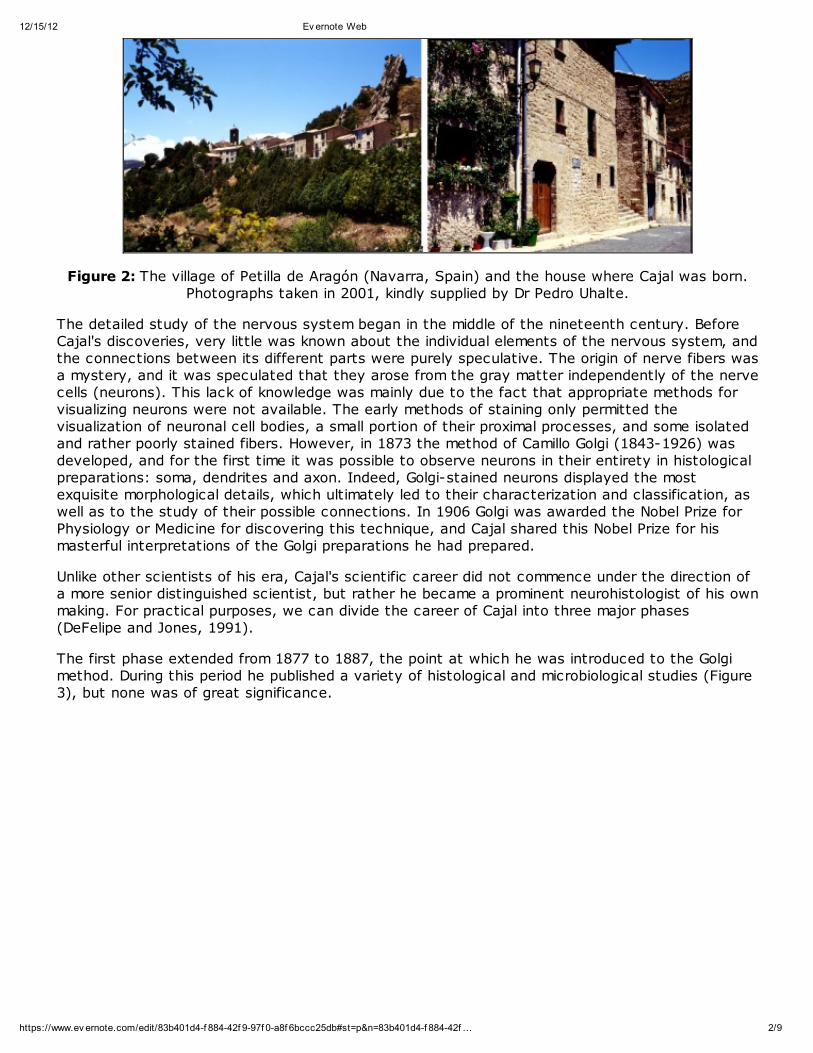
The first phase extended from 1877 to 1887, the point at which he was introduced to the Golgimethod. During this period he published a variety of histological and microbiological studies (Figure3), but none was of great significance.
12/15/12 Ev ernote Web
3/9https://www.ev ernote.com/edit/83b401d4-f 884-42f 9-97f 0-a8f 6bccc25db#st=p&n=83b401d4-f 884-42f …
Figure 3: Drawing by Cajal showing nerve terminations in frog muscles, published in 1881 (Estudiosanatómicos. Observaciones microscópicas sobre las terminaciones nerviosas en los músculos
voluntarios. Zaragoza: El diario católico).
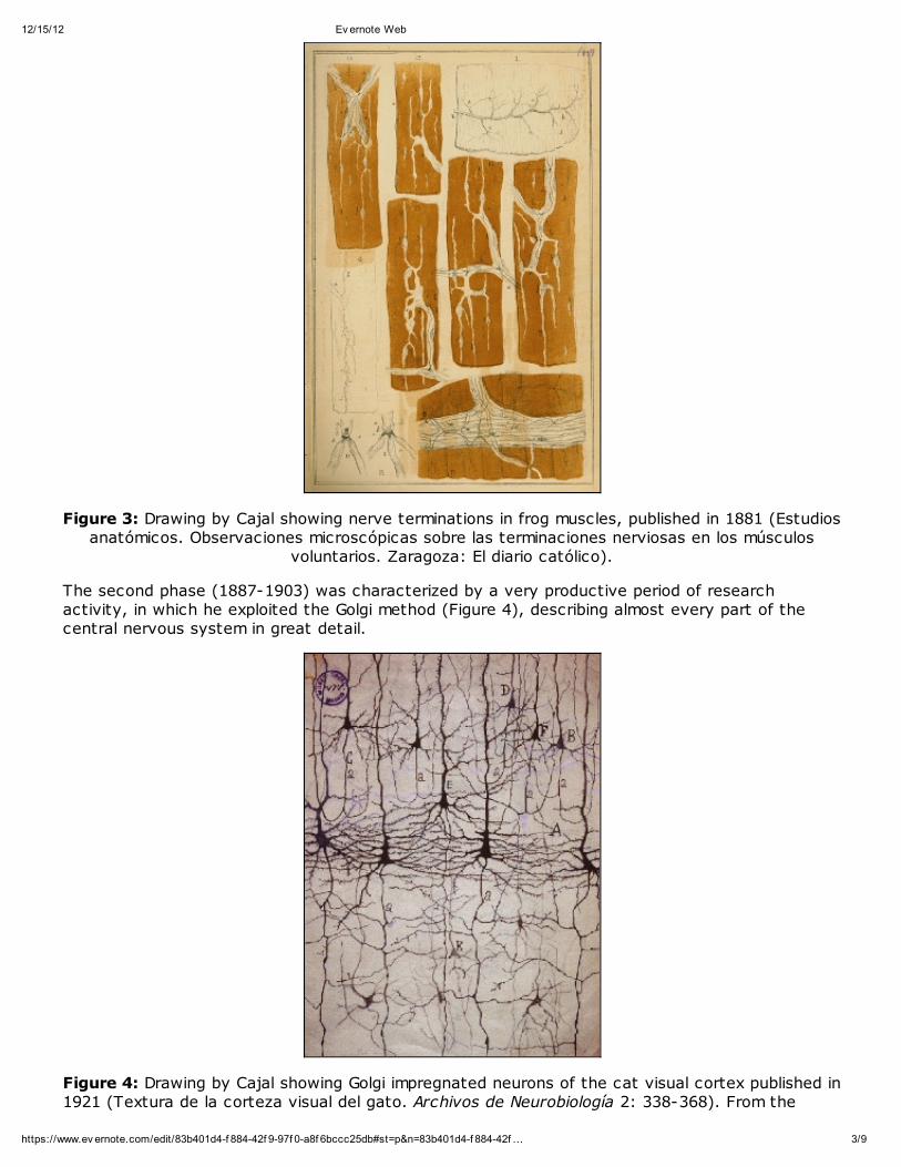
The second phase (1887-1903) was characterized by a very productive period of researchactivity, in which he exploited the Golgi method (Figure 4), describing almost every part of thecentral nervous system in great detail.
Figure 4: Drawing by Cajal showing Golgi impregnated neurons of the cat visual cortex published in1921 (Textura de la corteza visual del gato. Archivos de Neurobiología 2: 338-368). From the

12/15/12 Ev ernote Web
4/9https://www.ev ernote.com/edit/83b401d4-f 884-42f 9-97f 0-a8f 6bccc25db#st=p&n=83b401d4-f 884-42f …
collection of original drawings housed in the Museo Cajal (Cajal Museum).
Cajal was introduced in 1887 to the Golgi method during a visit to the private laboratory, inValencia, of the well-known psychiatrist and neurologist Luis Simarro. Cajal was so impressed bythe beautiful and complete staining of neurons that he made a radical change in his scientificcareer (see Favorite Sentences, nos. 1 and 3). His descriptions of the nervous system based onhis observations using the Golgi method were so accurate that his classic book Histologie (Cajal,1909, 1911), in which these studies are summarized, is still a reference book in all neurosciencelaboratories. Also, during the first few years of this phase of his life, Cajal found much evidence infavor of the 'Neuron Doctrine', which contrasted with the other more commonly accepted principleof the 'Reticular Theory'. The 'Neuron Doctrine', which now forms the fundamental organizationaland functional principle of the nervous system, states that the neuron is the anatomical,physiological, genetic and metabolic unit of the nervous system. In contrast, the 'Reticular Theory'suggested that the nervous system consisted of a diffuse nerve network formed by theanastomosing branches of nerve cell processes (either both dendritic and axonal, or only axonal),with the cell somata principally playing a role in nourishment (for reviews, see Shepherd, 1991;Jones, 1994).
The third phase of Cajal's career began in 1903 with his discovery of the reduced silver nitratemethod, and ended with his death in 1934. This period was also very productive and was devotedmainly to the theme of traumatic degeneration and regeneration of the nervous system (Figure 5).
Figure 5: Schematic drawing of Cajal showing "the flow of currents in a mutilated pyramidal cell(A) furnished with hypertrophic recurrent collaterals", published in 1914 (Estudios sobre la
degeneración y regeneración del sistema nervioso, Vol. 2. Madrid: Moya). From the collection oforiginal drawings housed in the Museo Cajal (Cajal Museum).
He published numerous scientific papers about this subject that were of great relevance, and thatwere summarized in another classic book, Degeneration and Regeneration (Cajal, 1913-1914).During this phase of his life, Cajal also published some important papers on the structure of the
12/15/12 Ev ernote Web
5/9https://www.ev ernote.com/edit/83b401d4-f 884-42f 9-97f 0-a8f 6bccc25db#st=p&n=83b401d4-f 884-42f …
retina and optic centers of invertebrates.
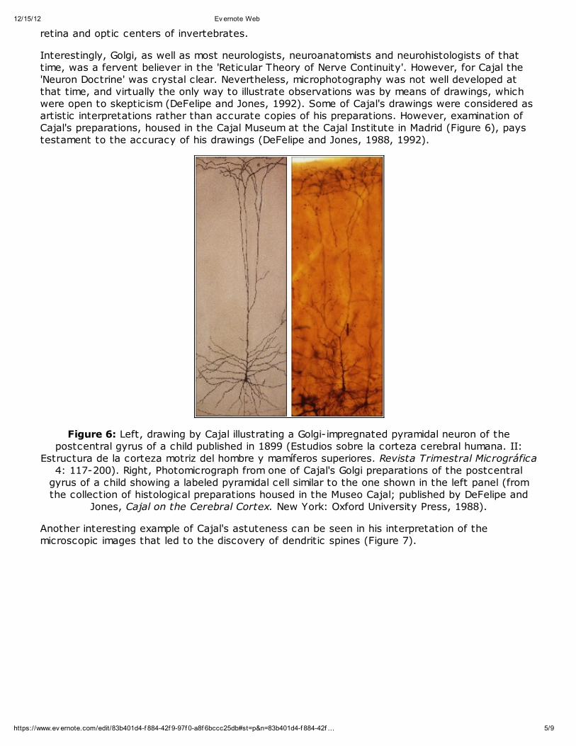
Interestingly, Golgi, as well as most neurologists, neuroanatomists and neurohistologists of thattime, was a fervent believer in the 'Reticular Theory of Nerve Continuity'. However, for Cajal the'Neuron Doctrine' was crystal clear. Nevertheless, microphotography was not well developed atthat time, and virtually the only way to illustrate observations was by means of drawings, whichwere open to skepticism (DeFelipe and Jones, 1992). Some of Cajal's drawings were considered asartistic interpretations rather than accurate copies of his preparations. However, examination ofCajal's preparations, housed in the Cajal Museum at the Cajal Institute in Madrid (Figure 6), paystestament to the accuracy of his drawings (DeFelipe and Jones, 1988, 1992).
Figure 6: Left, drawing by Cajal illustrating a Golgi-impregnated pyramidal neuron of thepostcentral gyrus of a child published in 1899 (Estudios sobre la corteza cerebral humana. II:
Estructura de la corteza motriz del hombre y mamíferos superiores. Revista Trimestral Micrográfica4: 117-200). Right, Photomicrograph from one of Cajal's Golgi preparations of the postcentral
gyrus of a child showing a labeled pyramidal cell similar to the one shown in the left panel (fromthe collection of histological preparations housed in the Museo Cajal; published by DeFelipe and
Jones, Cajal on the Cerebral Cortex. New York: Oxford University Press, 1988).
Another interesting example of Cajal's astuteness can be seen in his interpretation of themicroscopic images that led to the discovery of dendritic spines (Figure 7).

12/15/12 Ev ernote Web
6/9https://www.ev ernote.com/edit/83b401d4-f 884-42f 9-97f 0-a8f 6bccc25db#st=p&n=83b401d4-f 884-42f …
Figure 7: Drawing from Cajal illustrating types of dendritic spines published in 1933 (¿Neuronismo oreticularismo? Las pruebas objetivas de la unidad anatómica de las células nerviosas. Archivos deNeurobiología 13: 1-144). "Types of collateral spines of cerebral pyramids. A, rabbit; B, child of
two months; C, spines of a one-month-old cat (visual region); D, portion of a dendrite of a spinalmotor neuron of a cat in a phase before end feet are formed."
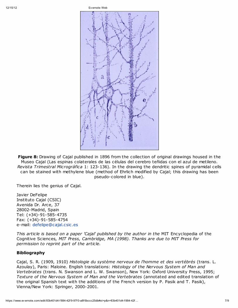
There were two different views at the time but for Cajal these structures represented fundamentalcomponents of spiny neurons. However, some authors (including Golgi himself) interpreted thespines as artifacts produced by the Golgi method: "a superficial precipitate, like a crystallization ofneedles, fortuitously deposited on the dendritic surface". As a result, the latter authors did notinclude spines in their drawings. Cajal explored different methods of staining to demonstrate thatthe dendritic spines were real morphological dispositions. Using the methylene blue method, heshowed that the dendritic surfaces of pyramidal cells were covered with spines (Figure 8), therebydemonstrating that they were not artifacts of the Golgi method. Although Cajal had the samemicroscopes and produced similar histological preparations with comparable quality of staining asthe majority of the neurohistologists of his time, he saw differently than they did.
12/15/12 Ev ernote Web
7/9https://www.ev ernote.com/edit/83b401d4-f 884-42f 9-97f 0-a8f 6bccc25db#st=p&n=83b401d4-f 884-42f …
Figure 8: Drawing of Cajal published in 1896 from the collection of original drawings housed in theMuseo Cajal (Las espinas colaterales de las células del cerebro teñidas con el azul de metileno.
Revista Trimestral Micrográfica 1: 123-136). In the drawing the dendritic spines of pyramidal cellscan be stained with methylene blue (method of Ehrlich modified by Cajal; this drawing has been
pseudo-colored in blue).
Therein lies the genius of Cajal.
Javier DeFelipe Instituto Cajal (CSIC)Avenida Dr. Arce, 37 28002-Madrid, SpainTel: (+34)-91-585-4735Fax: (+34)-91-585-4754e-mail: [email protected]
This article is based on a paper 'Cajal' published by the author in the MIT Encyclopedia of theCognitive Sciences, MIT Press, Cambridge, MA (1998). Thanks are due to MIT Press forpermission to reprint part of the article.
Bibliography
Cajal, S. R. (1909, 1910) Histologie du système nerveux de l'homme et des vertébrés (trans. L.Azoulay), Paris: Maloine. English translations: Histology of the Nervous System of Man andVertebrates (trans. N. Swanson and L. W. Swanson), New York: Oxford University Press, 1995;Texture of the Nervous System of Man and the Vertebrates (annotated and edited translation ofthe original Spanish text with the additions of the French version by P. Pasik and T. Pasik),Vienna/New York: Springer, 2000-2001.
12/15/12 Ev ernote Web
8/9https://www.ev ernote.com/edit/83b401d4-f 884-42f 9-97f 0-a8f 6bccc25db#st=p&n=83b401d4-f 884-42f …
Cajal, S. R. (1913-1914) Estudios sobre la degeneración y regeneración del sistema nervioso,Madrid: Moya. English translation: Degeneration and Regeneration of the Nervous System (trans.and edited Raoul M. May), London: Oxford University Press, 1928. Reprinted and edited withadditional translations by DeFelipe, J. and Jones, E. G., Cajal's Degeneration and Regeneration ofthe Nervous System, New York: Oxford University Press, 1991.
Cajal, S. R. (1917) Recuerdos de mi vida, Vol. 2, Historia de mi labor científica. Madrid: Moya.There is an English translation: Recollections of my life (trans. E. H. Craigie with the assistance ofJ. Cano), Philadelphia: American Philosophical Society, 1937. Reprinted Cambridge, MA: MIT Press,1989.
DeFelipe, J. and Jones, E. G. (1988) Cajal on the Cerebral Cortex. New York: Oxford UniversityPress.
DeFelipe, J. and Jones, E. G. (1991), Cajal's Degeneration and Regeneration of the NervousSystem. New York: Oxford University Press.
DeFelipe, J. and Jones, E. G. (1992) 'Santiago Ramón y Cajal and methods in neurohistology',Trends Neurosci 15: 237-246.
Jones, E. G. (1994) 'The neuron doctrine', Journal of History of Neuroscience 3: 3-20.
Shepherd, G. M. (1991) Foundations of the Neuron Doctrine. New York: Oxford University Press.
Favorite sentences
1. Cajal said of the Golgi method:
"I expressed in former paragraphs the surprise which I experienced upon seeing with my own eyesthe wonderful revelatory powers of the chrome-silver reaction and the absence of any excitementin the scientific world aroused by its discovery. How can one explain such strange indifference?Today, when I am better acquainted with the psychology of scientific men, I find it very natural …Out of respect for the master, no pupil is wont to use methods of investigation which he has notlearned from him. As for the great investigators, they would consider themselves dishonoured ifthey worked with the methods of others."
Recuerdos de mi vida, Vol. 2, Historia de mi labor científica. Madrid: Moya, 1917, p. 76.
2. Differences between the brain of humans and nonhuman mammals:
"The opinion generally accepted at that time that the differences between the brain of[nonhuman] mammals (cat, dog, monkey, etc) and that of man are only quantitative, seemed tome unlikely and even a little offensive to the human dignity … But do not articulate language, thecapability of abstraction, the ability to create concepts, and, finally, the art of inventing ingeniousinstrument … seem to indicate (even admitting fundamental structural correspondences with theanimals) the existence of original resources, of something qualitatively new which justify thepsychological nobility of homo sapiens? … My investigations showed that the functional superiorityof the human brain is intimately bound up with the prodigious abundance and unusual wealth offorms of the so-called neurons with short axon."
Recuerdos de mi vida, Vol. 2, Historia de mi labor científica. Madrid: Moya, 1917, pp. 345-346,350.
3. The cerebral cortex and pyramidal cells:
"the cerebral cortex is similar to a garden filled with trees, the pyramidal cells, which, thanks tointelligent culture, can multiply their branches, sending their roots deeper and producing more andmore varied and exquisite flowers and fruits."
The Cronian Lecture: La fine structure des centres nerveux. Proceedings of the Royal Society of
12/15/12 Ev ernote Web
9/9https://www.ev ernote.com/edit/83b401d4-f 884-42f 9-97f 0-a8f 6bccc25db#st=p&n=83b401d4-f 884-42f …
London 55: 444-468, 1984. Translated by DeFelipe and Jones, Cajal on the Cerebral Cortex. NewYork: Oxford University Press, 1988, p. 87.
4. Plasticity:
"Cerebral gymnastics are not capable of improving the organization of the brain by increasing thenumber of cells, because it is known that the nerve cells after the embryonic period have lost theproperty of proliferation; but it can be admitted as very probable that mental exercise leads to agreater development of the dendritic apparatus and of the system of axonal collaterals in the mostutilized cerebral regions. In this way, associations already established among certain groups ofcells would be notably reinforced by means of the multiplication of the small terminal branches ofthe dendritic appendages and axonal collaterals; but, in addition, completely new intercellularconnections could be established thanks to the new formation of [axonal] collaterals anddendrites."
The Cronian Lecture: La fine structure des centres nerveux. Proceedings of the Royal Society ofLondon 55: 444-468, 1984. Translated by DeFelipe and Jones, Cajal on the Cerebral Cortex. NewYork: Oxford University Press, 1988, p. 87.